Cloning and transcriptional analysis of the reveals stable nifD, nifDK ...
Upload
sergio-torresCategory
view
213download
1
BioMed CentralBMC Microbiology
ss
Open AcceResearch articleAlkane-induced expression, substrate binding profile, and immunolocalization of a cytochrome P450 encoded on the nifD excision element of Anabaena 7120Sergio Torres†1, Conrad R Fjetland†2 and Peter J Lammers*†1Address: 1Department of Chemistry and Biochemistry, New Mexico State University, Las Cruces, NM, USA and 2University of Texas at Austin, Department of Chemistry and Biochemistry, Austin, TX, USA
Email: Sergio Torres - [email protected]; Conrad R Fjetland - [email protected]; Peter J Lammers* - [email protected]
* Corresponding author †Equal contributors
AbstractBackground: Alkanes have been hypothesized to act as universal inducers of bacterialcytochrome P450 gene expression. We tested this hypothesis on an unusual P450 gene (cyp110)found on a conserved 11 kilobase episomal DNA element of unknown function found infilamentous cyanobacteria. We also monitored the binding of potential substrates to the P450protein and explored the distribution of P450 protein in vegetative cells and nitrogen-fixingheterocysts using immuno-electron microscopy.
Results: Hexadecane treatments resulted in a two-fold increase in mRNA, and a four-fold increasein P450 protein levels relative to control cultures. Hexane, octane and dodecane were toxic andinduced substantial changes in membrane morphology. Long-chain saturated and unsaturated fattyacids were shown to bind the CYP110 protein using a spectroscopic spin-shift assay, but alkanesdid not bind. CYP110 protein was detected in vegetative cells but not in differentiated heterocystswhere nitrogen fixation occurs.
Conclusion: Hexadecane treatment was an effective inducer of CYP110 expression incyanobacteria. Based on substrate binding profiles and amino acid sequence similarities it ishypothesized that CYP110 is a fatty acid ω-hydroxylase in photosynthetic cells. CYP110 was foundassociated with membrane fractions unlike other soluble microbial P450 proteins, and in this regardCYP110 more closely resembles eukarytotic P450s. Substrate stablization is an unlikely mechanismfor alkane induction because alkanes did not bind to purified CYP110 protein.
BackgroundAnabaena sp. strain PCC 7120 (Anabaena 7120) is an obli-gate photoautotrophic cyanobacterium that reducesatmospheric nitrogen in terminally differentiated cellsknown as heterocysts [1]. Approximately every tenth cellalong a filament differentiates into a heterocyst undernitrogen-fixing conditions. Three developmentally regu-
lated DNA rearrangements occur within the heterocystchromosome which excise DNA elements integratedwithin the nifD, fdxN and hupL genes [2,3]. The elementinterrupting the nifD gene is 11,268 base pairs in lengthand carries its own site-specific recombinase, the xisA gene[4]. The nifD element is found in most heterocyst formingcyanobacteria integrated within the nifD gene [5,6]. It is
Published: 24 March 2005
BMC Microbiology 2005, 5:16 doi:10.1186/1471-2180-5-16
Received: 06 January 2005Accepted: 24 March 2005
This article is available from: http://www.biomedcentral.com/1471-2180/5/16
© 2005 Torres et al; licensee BioMed Central Ltd. This is an Open Access article distributed under the terms of the Creative Commons Attribution License (http://creativecommons.org/licenses/by/2.0), which permits unrestricted use, distribution, and reproduction in any medium, provided the original work is properly cited.
Page 1 of 12(page number not for citation purposes)

BMC Microbiology 2005, 5:16 http://www.biomedcentral.com/1471-2180/5/16
absent from all non-heterocystous strains examined todate [7,8]. While factors driving the evolution of the nifDelement and its associated genes remain obscure, anessential role in diazotrophic growth can be ruled outsince an Anabaena strain cured of the nifD element wasshown to fix nitrogen under aerobic conditions in hetero-cysts of normal morphology [9].
The complete DNA sequence of the nifD element revealslittle about the biological functions it encodes. Nine openreading frames are known (see Fig. 1) only one of whichcan be identified by sequence homology: the first reportedcyanobacterial cytochrome P450 gene, cyp110 [10]. Thecyp110 gene is widely conserved in the heterocyst formingcyanobacteria including Anabaena, Nostoc and Calothrix/Fremyella. The amino acid sequence of CYP110 is mostsimilar to the mammalian P450 family 4 fatty acid omega-hydroxylases and P450BM3 from Bacillus megatarium,another fatty acid omega hydroxylase. However, the met-abolic significance of fatty acid omega hydroxylation inbacteria remains obscure. Even for the well-characterizedP450BM3 enzyme, the identity of the in vivo substrate(s) ofthis protein and the pathway involved are not known[11]. Studies suggest n-alkanes act as multi-purposemicrobial P450 inducers and our preliminary data sug-gested this was also true for cyp110 [12,13]. Moreover, abiosensor for hexadecane detection based on changes in
cyp110 mRNA from Anabeana variabilias has beendescribed [14]. Here we report on the pattern of expres-sion of the cyp110 mRNA and protein in Anabaena 7120,substrate binding characteristics of CYP110, the effects ofalkanes on cellular morphology, and the distribution ofCYP110 in vegetative cells versus heterocysts using elec-tron microscopy.
ResultsHexadecane-dependent induction of cyp110 transcripts in nitrogen-fixing and ammonia-supplemented culturesWe first tested the effects of hexane, octane, dodecane andhexadecane on the growth of Anabaena 7120 cultures. Allalkane additions in this study were carried out at 0.2% (v/v), the recommended concentration for microbial P450induction [12]. In preliminary experiments, we deter-mined that Anabaena 7120 exhibited rapid chlorosis with0.2% hexane and octane. These treatments yielded onlyhighly degraded RNA and were not further analyzed (datanot shown). RNA samples from control, dodecane andhexadecane treated cultures were size fractionated andhybridized with cyp110. A psbA1 specific probe was usedto assess RNA quality and for normalization.
The cyp110 probe identified a range of transcript sizes witha maximum of 9 kb seen in mRNA from both nitrogen-fix-ing and ammonia supplemented Anabaena 7120 cultures
Genetic Map of the nif D ElementFigure 1Genetic Map of the nif D Element. The positions of known nif D element genes and other open reading frames are indi-cated with arrows showing the orientation. LB = left border, RB = right border, H = HindIII sites. The numbers inside the dou-ble lines designate the HindIII fragment subclones of the nifD element derived from a 17 kbp EcoRI fragment, An 207. The cyp110 probe was made from the An207.4 HindIII fragment. The complete sequence of the nif D element is deposited in Gen-bank (U38537).
Page 2 of 12(page number not for citation purposes)
BMC Microbiology 2005, 5:16 http://www.biomedcentral.com/1471-2180/5/16
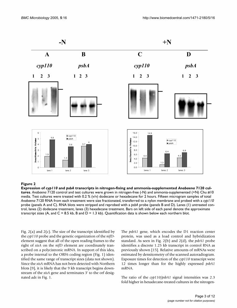
Fig. 2(a) and 2(c). The size of the transcript identified bythe cyp110 probe and the genetic organization of the nifD-element suggest that all of the open reading frames to theright of xisA on the nifD element are coordinately tran-scribed on a polycistronic mRNA. In support of this idea,a probe internal to the ORF6 coding region (Fig. 1) iden-tified the same range of transcript sizes (data not shown).Since the xisA mRNA has not been detected with Northernblots [9], it is likely that the 9 kb transcript begins down-stream of the xisA gene and terminates 3' to the orf desig-nated adx in Fig. 1.
The psbA1 gene, which encodes the D1 reaction centerprotein, was used as a load control and hybridizationstandard. As seen in Fig. 2(b) and 2(d), the psbA1 probeidentifies a discrete 1.25 kb transcript in control RNA aspreviously shown [15]. Relative amounts of mRNAs wereestimated by densitometry of the scanned autoradiogram.Exposure times for detection of the cyp110 transcript were12 times longer than for the highly expressed psbA1mRNA.
The ratio of the cyp110/psbA1 signal intensities was 2.3fold higher in hexadecane-treated cultures in the nitrogen-
Expression of cyp110 and psbA transcripts in nitrogen-fixing and ammonia-supplemented Anabaena 7120 culturesFigure 2Expression of cyp110 and psbA transcripts in nitrogen-fixing and ammonia-supplemented Anabaena 7120 cul-tures. Anabaena 7120 control and test cultures were grown in nitrogen-free (-N) and ammonia-supplemented (+N) Chu #10 media. Test cultures were treated with 0.2 % (v/v) dodecane or hexadecane for 2 hours. Fifteen microgram samples of total Anabaena 7120 RNA from each treatment were size fractionated, transferred to a nylon membrane and probed with a cyp110 probe (panels A and C). RNA blots were stripped and reprobed with a psbA probe (panels B and D). Lanes (1) untreated con-trol, lanes (2) dodecane treatment, lanes (3) hexadecane treatment. Bars on left side of each panel denote the approximate transcript sizes (A, and C = 8.5 kb, B and D = 1.3 kb). Quantification data is shown below each northern blot.
-N +N_________
A B C D
cyp110 psbA cyp110 psbA
1 2 3 1 2 3 1 2 3 1 2 3
4.4
1.3
4.5
14.9
6.46.8
0.0
2.0
4.0
6.0
8.0
10.0
12.0
14.0
16.0
lanes 1 lanes 2 lanes 3
cyp110
psbA
1.9
1.3
3.7
4.8
3.64
0
1
2
3
4
5
6
lane 1 lane 2 lane 3
cyp110
psbA
Page 3 of 12(page number not for citation purposes)

BMC Microbiology 2005, 5:16 http://www.biomedcentral.com/1471-2180/5/16
fixing culture (panels A&B) while a 2.2-fold increase wasobserved in the ammonium-supplemented culture (pan-els C&D). The dodecane treatment had little effect oncyp110 mRNA levels in either nitrogen-fixing or ammo-nium supplemented cultures. Similar results wereobtained in 2 separate experiments with RNA isolatedfrom independent cultures. The doubling of steady-statecyp110 mRNA levels in response to hexadecane treatmentwas independent of nitrogen status of the culture, consist-ent with previous data showing the entire nifD element isnot required for nitrogen fixation in heterocysts [9].
CYP110 protein is only detected in hexadecane-treated cultures and localized in the membrane fractionThe hexadecane effect on cyp110 gene expression was fur-ther examined using CYP110-specific antibodies to probefor the presence of P450 protein in nitrogen-fixing Ana-baena 7120 cultures. As shown in Fig. 3A, the 0.2% hexa-
decane treatment resulted in the appearance of animmuno-reactive protein band of 50 kDa that co-migrated with a purified CYP110 protein sample. TheP450 protein band intensity increased at least 2 foldwithin 2 hours of treatment, and at least 4 fold 24 hoursafter treatment with hexadecane when compared to thebackground, zero-time expression shown in lane 3 (Fig.3C). A western blot with protein samples from anuntreated control culture failed to detect any immuno-reactive P450 protein (Fig. 3B). These results were repli-cated with 2 independent cultures.
Heterologous expression of the CYP110 protein in E. coliproduced a substantial amount of membrane associatedP450 protein [16]. This is a clear distinction betweenCYP110 and its closest sequence homolog, P450BM3,which has been extensively studied in part because it is asoluble P450 model protein [11]. To determine if CYP110
Expression of CYP110 protein in Anabaena 7120Figure 3Expression of CYP110 protein in Anabaena 7120. Proteins were fractionated on 12% polyacrylamide gels, blotted to nitrocellulose and probed with rabbit anti-CYP110 antibody. Panel A displays samples treated with 0.2 % n-hexadecane. Panel B, control untreated samples taken at the same time points. Lanes 1 contain 600 ng of purified recombinant CYP110 protein. Lanes 2 are blank. Lanes 3 through 7 contain 50 µg of crude protein taken at 0, 2, 12, 24, and 36 hours. Panel C shows the den-sitometry trace volumes for the CYP110 protein in Panel A. Panel D is a Western blot showing cyp110-reactive proteins from the 36 hour hexadecane time point after separation into the soluble and membrane fractions. Lane 1: 50 µg of the cytosolic fraction and lane 4: 50 µg of the membrane fraction.
0.3
0.70.6
1.31.4
0.0
0.5
1.0
1.5
2.0
lane 3 lane 4 lane 5 lane 6 lane 7Den
sito
met
ryT
race
1 2
50 kDa
50 kDa
1 2 3 4 5 6 7 1 2 3 4 5 6 7
A B
CD
Page 4 of 12(page number not for citation purposes)

BMC Microbiology 2005, 5:16 http://www.biomedcentral.com/1471-2180/5/16
is membrane associated in Anabaena 7120, protein frommembrane and soluble fractions separated by centrifuga-tion were subjected to Western blot analysis. As shown inFig. 3D, the membrane fraction appeared to contain all ofthe immuno-reactive P450 protein in the Anabaena 7120.This stands in contrast to P450BM3 and most other micro-bial P450 proteins, and in this regard, CYP110 moreclosely resembles the eukaryotic P450 proteins.
Spectral characterization of CYP110 protein and substrate binding assayAbsorption spectra studies were recorded for CYP110 pro-tein expressed and purified from E. coli carrying a pUC-based expression vector [16]. As shown in Fig. 4A, theCYP110 protein has a typical cytochrome P450 absorb-ance profile in the visible region for the oxidized, reducedand carbon monoxide bound states. CYP110 also showsthe characteristic peak at 450 nm in the CO differencespectrum (Fig. 4B). We monitored the visible spectrum
CYP110 Absorbance Spectra, Titrated Spectral Shift, and Double Reciprocal Plot in the presence and absence of various hydrocarbonsFigure 4CYP110 Absorbance Spectra, Titrated Spectral Shift, and Double Reciprocal Plot in the presence and absence of various hydrocarbons. The absorbance characteristics were monitored for the purified soluble CYP110 from E. coli. The sample was diluted 1:5 in buffer A with 0.2 % deoxycholate. To reduce the P450, 5 mg of sodium dithionate was added to the sample. Carbon monoxide was then bubbled into the sample for 30 seconds. Panel A. Oxidized, reduced and carbon monoxide bound spectra. Panel B. Carbon monoxide difference spectrum with a peak at 450 nm. Panel C. The oxidized spectra for 3 µM CYP110 in buffer A + 0.2 % deoxycholate and titrated with tetradecanoic acid. The concentrations of tetradecanoic acid shown are 0, 0.1, 0.2, 0.5, 0.7, and 1.0 mM. Panel D. (Top) The difference spectra generated for CYP110 when titrated with tetradecanoic acid. The bottom plot for Panel D shows the spectrum without fatty acid subtracted from each spectra with fatty acid. The double reciprical plot of 1/∆Abs vs 1/S is shown and the X-intercept is calculated by linear regression. Ks is defined as -1/X-intercept. ∆Abs for each fatty acid concentration is defined as the resulting value at 388 nm.
CO Bound
Reduced
Oxidized
400 420 440 460 480 500
0.1
0.2
0.3
0.4
0.5
0.6
-0.2
-0.1
0.0
0.1
0.2
0.3
0.4
0.5
400 420 440 460 480 500
Wavelength (nm)Wavelength (nm)
Ab
sorb
an
c
e
∆A
bso
rban
ce
A B
350 370 390 410 430 450
0.200
0.300
0.400
0.500
Wavelength (nm)
Wavelength (nm)
1/S
Ab
sorb
an
ce
∆A
bso
rban
ce
1/∆
Ab
s
C D
Page 5 of 12(page number not for citation purposes)

BMC Microbiology 2005, 5:16 http://www.biomedcentral.com/1471-2180/5/16
while titrating a CYP110 solution with tetradecanoic acid.As shown in Fig. 4C, this resulted in a blue shift of thepeak absorbance in the oxidized spectrum to 388 nm,similar to previously described P450 substrate-dependentType I spectral shifts. This spectral shift was the same ineither buffer A or buffer A + 0.2% deoxycholate. Whenbuffer A was supplemented with 0.2% Triton X-100, nospectral shift was observed (data not shown). The titrationyields the binding constant (Ks) for each substrate pro-ducing a spectral shift. A typical difference spectrum gen-erated from the titration and the resulting doublereciprocal plot are shown in Fig. 4D.
The calculated binding constants for the interaction ofCYP110 with potential substrate molecules of differenthydrocarbon chain length are listed in Table 1. For the sat-urated fatty acids tested, CYP110 displays the highestaffinity for tetradecanoic acid with a Ks of 0.091 mM. Theunsaturated fatty acids also bound well to CYP110. Over-all, CYP110 had the highest affinity for alpha-linolenicacid (Ks 0.062 mM) and arachidonic acid (Ks = 0.051
mM), long chain fatty acids with 3 and 4 double bonds,respectively. For comparison, P450BM3 has a Ks of 0.002mM for arachidonic acid [17]. The Ks values towards ara-chidonic acid for the mammalian cytochrome P450s fromfamily 4 are in the same range [18].
Fatty amines and fatty amides are known to interact withP450 ω-hydroxylases via Type II spectral shifts [19,20],and this was also true for CYP110. The minimal chainlength for this interaction was 5 since n-butylamine didnot cause a spectral shift while n-pentylamine did bind.There was a clear trend for tighter binding with longerhydrocarbon chain length, as the CYP110-dodecylamineinteraction had the lowest tested Ks value (0.012 mM).CYP110 bound fatty amines 10 fold better than P450non,which has a binding constant of 1.3 mM towardsoctylamine [21]. Twelve-aminododecanoic acid causes aType I spectral shift instead of a Type II suggesting analtered mode of binding. Lu et al. [20] showed that termi-nal amino fatty acid derivatives interact weakly with theheme iron of cytochromes P450.
Table 1: Spectral Shift and Binding Constants of CYP110 with SeveralCompounds.
Compound Type Ks (mM) Compound Type Ks (mM)
Octanoic Acid None - Butyl Amine None -Nonanoic Acid None - Pentyl Amine II 0.54Decanoic Acid I 6.3 Hexyl Amine II 0.32
Undecanoic Acid I 5.2 Heptyl Amine II 0.102Dodecanoic Acid I 1.9 Ocytl Amine II 0.081Tridecanoic Acid I 0.85 Nonyl Amine II 0.062
Tetradecanoic Acid I 0.091 Decyl Amine II 0.041Pentadecanoic Acid I 0.21 Dodecyl Amine II 0.012Hexadecanoic Acid I 0.50Heptadecanoic Acid I 1.1Octadecanoic Acid I 3.1 12-Aminododecanoic
AcidI 0.416
Arachidonic Acid I 0.051 I9-Octadecenoic Acid I 2.8 Putrescine None -9,12-Octadecadienoic
AcidI 0.72 Cetyltrimethyl
Ammonium BromideNone -
9,12,15-Octadecatrienoic Acid
I 0.062 n-Alkanes C6-C18 None -
11-Dodecenoic Acid None -Sodium Dodecyl
SulfateI - Saturated Fatty
Alcohols C6-C18None -
Lauryl Amide I 2.21N-methyl Lauryl
AmideI 2.56 11-
Hydroxydodecanoic Acid
None -
N,N-Dimethyl Lauryl Amide
I 2.79 12-Hydroxydocecanoic
Acid
None -
Methyl Dodecanoate None - N-LaurylSacosine I 2.0
Page 6 of 12(page number not for citation purposes)

BMC Microbiology 2005, 5:16 http://www.biomedcentral.com/1471-2180/5/16
The binding of CYP110 towards other fatty acid deriva-tives was also examined. Table 1 displays a list of com-pounds that displayed no spectral shift, including fattyalcohols or saturated n-alkanes. No spectral shift wasobserved with 11- or 12-hydroxydodecanoic acid or withmethyl laurate. The unsaturated fatty acid 11-dodecenoicacid was also tested with no shift observed. Two otheramines that did not display spectral shifts were thepolyamine, putrescine, and cetyltrimethylammoniumbromide (CTAB). Sodium dodecyl sulfate (SDS) doesbind CYP110 and causes a Type I spectral shift. Althoughno binding constant was determined, it was observed thatthe maximal shift to 388 nm occurred below 0.5 mM andreverted back to 416 nm above this concentration, a phe-nomenon likely to be linked to the critical micelle concen-tration for SDS (data not shown).
Alkane effects on morphology and localization of CYP110Given the toxicity of hexane to Anabaena 7120, transmis-sion electron microscopy was used to document changesin cell structure associated with the alkane treatmentsshown to induce the CYP110 expression. Vegetative cellsfrom control, dodecane and hexadecane treated culturesare shown in Fig. 5a,5b and 5c respectively. Many darkspherical inclusions are seen in the dodecane-treated cells(Fig. 5b). The dodecane treatment also distorts and thick-ens the cellular and photosynthetic membranes, creatinga ruffled effect in both outer and inner membranes. Hex-adecane treatment did not have the same dramatic effecton membrane morphology, however the close stacking ofphotosynthetic membranes seen in the control micro-graph (Fig. 5a) is lost with hexadecane treatment (Fig. 4c).
Gold-conjugated anti-CYP110 antibodies were used todetermine the relative protein expression levels in vegeta-tive cells with and without alkane treatment and to assessvegetative cell versus heterocysts expression patterns Ana-baena 7120. Immunoelectron micrographs showed agreater number of anti-CYP110 polyclonal gold conju-gates in dodecane and hexadecane vegetative cells (Fig. 5eand 5f) than in control samples (Fig. 5d). Expression lev-els were quantified by counting gold particles in multiplemicrographs as shown in Table 2. Immunogold particlesmost often clustered around dark-staining material inAnabaena 7120 vegetative cells grown in the presence of0.2% (v/v) dodecane or hexadecane (Fig. 5e,f). We foundfewer immunogold particles/cluster in dodecane-treatedsamples (avg. = 22) than hexadecane treated samples(avg. = 50). Heterocysts showed significantly fewer goldparticles than vegetative cells, and the number of gold par-ticles in heterocysts did not increase with hexadecanetreatment (Fig. 5g, Table 2).
Nonspecific immuno-staining was estimated by incubat-ing sections with preimmune rabbit IgG or with polyclo-
nal anti-CYP110 antibodies that had been pre-adsorbedwith purified CYP110 protein produced in E. coli. Verylow background was seen in both the preimmune rabbitIgG primary incubation (Fig. 5h), and sections treatedwith preabsorbed anti-CYP110 antibodies (Fig. 5i). Noclusters of immunogold conjugates were seen in any ofthe control sections. These results clearly show the major-ity of CYP110 protein is localized in vegetative cells andcorroborate Northern and Western data showing elevatedCYP110 expression in hexadecane-treated cultures.
DiscussionMost well characterized microbial P450s enzymes wereisolated from heterotrophic organisms and are involvedin the oxidative metabolism of xenobiotic compounds forenergy and growth. Examples include camphor-degradingP450CAM [22] and two herbicide inducible P450 genes inStreptomyces griseolus [23]. There is no evidence to date forcytochrome P450-dependent oxidative metabolism in thecyanobacteria. A P450-dependent N-demethylationactivity is known in unicellular green algae that activatesthe pro-herbicide metflurazon into norflurazon [24]. Twogenera of marine cyanobacteria Microcoleus and Phormid-ium were isolated from oil spills along the Arabian sea andreported to contribute to oil degradation [25]. While thefirst step in alkane degradation in many microbial systemsis a P450-dependent hydroxylation [11,26,27], there areno data on the mode of alkane metabolism norconfirmation of hydrocarbon oxidation by axenic isolatesof these cyanobacterial strains. Recent evidence does showthat mixtures of these cyanobacteria and organotrophicbacteria of the genera Rhodococcus, Arthrobacter, Pseu-domonas, and Bacillus are more effective than either groupalone towards alkane oxidation in inorganic growthmedia [28].
The available evidence suggests that CYP110 does not par-ticipate in the degradation of alkanes. CYP110 has lowoverall sequence similarity to other known alkane hydrox-ylase enzymes like the CYP52 family [29]. Alkanes arequite toxic to Anabaena 7120, and purified CYP110 pro-tein expressed in E. coli binds fatty acids but not alkanesusing a spin-shift assay. The membrane localization andspin-shift binding data suggest that long-chain polyunsat-urated fatty acids are the most likely endogenous sub-strates for CYP110. The endogenous function of CYP110in Anabaena 7120 remains a mystery. Sequencecomparisons indicate the protein is related to the fattyacid omega hydroxylase proteins. One possibility is thatω-hydroxylated fatty acids are further oxidized to dicarbo-xylic acids which then undergo β-oxidation from bothends yielding acetate, as demonstrated in eukaryotic per-oxisomes [30].
Page 7 of 12(page number not for citation purposes)

BMC Microbiology 2005, 5:16 http://www.biomedcentral.com/1471-2180/5/16
Ultrastructure and immunocytochemical detection of CYP110 in alkane treated Anabaena 7120Figure 5Ultrastructure and immunocytochemical detection of CYP110 in alkane treated Anabaena 7120. Nitrogen-fixing cultures were grown with shaking in continuous light at 30 C with ambient CO2. Test cultures were treated with 0.2% dodecane or 0.2% hexadecane for 30 days. (a) TEM of untreated vegetative cell, (b) TEM of dodecane-treated vegetative cell, (c) TEM of hexadecane-treated vegetative cell, (d) untreated control vegetative cell incubated with anti-CYP110 antiserum, (e) dodecane-treated vegetative cell incubated with anti-Cyp111 antiserum, (f) hexadecane-treated vegetative cell incubated with anti-CYP110 antiserum, (g) hexadecane treated heterocyst incubated with anti-CYP110 antiserum, (h) dodecane-treated vege-tative cell incubated with non-immune rabbit IgG, (i) dodecane-treated vegetative cell incubated with anti-CYP110 antiserum previously adsorbed with purified CYP110.
d
g
e
a
Page 8 of 12(page number not for citation purposes)

BMC Microbiology 2005, 5:16 http://www.biomedcentral.com/1471-2180/5/16
Cytochrome P450 proteins in diazotrophic bacteria havebeen suggested to play an ancillary role in nitrogen fixa-tion. Many years ago, Appleby proposed that a cyto-chrome P450 enzyme functions as an alternative high-affinity oxidase in an efficient, leghemoglobin-facilitatedpathway of oxidative phosphorylation in bacteroids ofBradyrhizobium japonicum [31]. The idea was based onspectroscopic evidence for the presence of a P450 proteinin B. japonicum bacteroids but not in aerobically growncells and the blocking of the high-affinity oxidase systemby the P450 inhibitor, N-phenylimidazole. More recentlyboth bacteroids and anaerobically grown B. japonicumcells were shown to contain two immunologically distinctcytochromes P450, which were cloned and designatedcyp112 and cyp114 [32]. Neither protein is expressed inaerobically grown B. japonicum cells, consistent with theAppleby hypothesis. Transposon mutagenesis of thecyp112 locus was found to block expression of bothCyp112 and Cyp114. Nevertheless, the mutant strain pro-duced effective nodules on soybeans. These results showneither protein is essential for symbiotic function underthe conditions of plant growth used in these experiments.Subsequent analysis of the B. japonicum P450 clusteridentified a third P450 gene and other open readingframes suggesting an operon involved in terpenoid bio-synthesis [33]. Like the Anabaena 7120 strain missing thenifD element, a quantitative phenotype related to nitro-gen fixation under specific culture conditions cannot beexcluded from the available evidence. B. japonicum andAnabaena 7120 are not the only diazotrophic bacteria har-boring cytochrome P450 genes near clusters of nitrogenfixation-related genes. The large Sym plasmid from Rhizo-bium sp. NGR234 confers the ability to associate symbiot-ically with plants. It contains two cytochrome P450 geneswhose function(s) are not known [34].
ConclusionAlkanes have been hypothesized to act as universal induc-ers of microbial P450 genes [12]. Consistent with thishypothesis, we observed a small but reproducible 2-foldenhancement of cyp110 transcription induced by 0.2%hexadecane. We did not test lower concentrations of hex-ane or dodecane to determine if non-toxic concentrations
would have P450-inducing activity in Anabaena 7120. Wecannot rule out the possibility that the lower solubility oflonger chain alkanes in aqueous solutions may have beenmore important for the activity of hexadecane as aninducer than chain-length per se. The CYP110 protein dis-played the highest affinity towards long-chain unsatu-rated fatty acids and was localized primarily inphotosynthetic vegetative cells of Anabaena 7120. Unlikemost prokaryotic P450 proteins, CYP110 was found to beassociated with membrane fractions consistent with thesolubility characteristics of its preferred substrates.
MethodsCyanobacterial growthFor RNA and protein isolation, cultures of Anabaena 7120(4 L) were grown in Chu #10 media with continuouslight, stirred at 30°C and bubbled with a 1% CO2/air mix-ture illuminated at approximately 100 µE s-1 m-1. Nitrogensupplemented cultures were grown with 2.5 mMNH4(SO4)2 to suppress heterocyst differentiation. Treat-ments were carried out after cultures reached A700 = 0.7.Control and test cultures (0.2 % dodecane or hexadecane[v/v] for 2 hours) were maintained in the same growthconditions during the treatment interval.
RNA methodsAn amended RNA isolation procedure [37] was used fortotal RNA isolation. Briefly, cells were cooled by addingan equal volume of ice followed by transferring the cellpaste into a mortar and freezing with liquid nitrogen.Cells were ground to a fine powder and placed in 10 mLRNA lysis buffer-pH 5.2 (0.1 M Tris, 0.1 M LiCl, 5 mMEDTA, 0.1 M NaCl, 0.1 M sodium acetate, 1 % SDS, 0.2%β-mercaptoethanol). After vortexing (2 min), 5 mL hot(60 C) NTE saturated-phenol, pH 6.0, was added, fol-lowed by 3 min of vortexing. Samples were placed at 60°Cfor 10 min, vortexed and 7 mL chloroform:isoamyl alco-hol (24:1) added and the aqueous phase was subjected torepeated phenol/chloroform extractions until no visiblematerial remained at the interface. Total RNA wasprecipitated overnight in 2 M LiCl at 4°C. RNA pelletswere washed with 2 M LiCl and ethanol precipitated. After
Table 2: Detection of anti-CYP110 polyclonal gold conjugates.
Culture conditions CYP110-gold conjugates(Avg. no. of gold particles)
Vegetative cell Heterocyst cell
Anabaena 7120 untreated 8 5Anabaena 7120 dodecane treatment 22 5Anabaena 7120 hexadecane treatment 50 5
Page 9 of 12(page number not for citation purposes)

BMC Microbiology 2005, 5:16 http://www.biomedcentral.com/1471-2180/5/16
a 70% ethanol wash, the RNA was brought up in diethylpyrocarbonate treated water.
Samples of total RNA (15 µg) were separated by agaroseformaldehyde denaturing gel electrophoresis. The gel wasequilibrated twice in 10× SSC for 15 min and RNA wastransferred onto a nylon membrane (Boehringer Man-nheim) by capillary transfer. Probes were labeled usingthe DIG-High Prime Mix (Boehringer Mannheim) follow-ing the manufactures' recommendations. The cyp110probe was derived from the 2 kbp HindIII pAn 207.4 DNAfragment (Fig. 1) containing most of the cyp110 genesequence [10]. The psbA probe used is a 2 kbp pAn 625EcoR1 fragment [15] containing the coding region for theD1 reaction center protein in photosystem II. RNA blotswere first probed with cyp110, stripped, and reprobed withthe psbA probe. The hybridized probes were detectedusing the CDP-Star (Boehringer Mannheim) chemilumi-nescent substrate, followed by autoradiography. Prehy-bridizations, hybridizations, and washes were carried outas described by the manufacturer. Signal intensities fromnorthern blots were quantified using Intelligent Quantifi-cation software (Genomic Solutions).
P450 protein analysisProteins for Western blotting were isolated from N2-fixingAnabaena 7120 cultures treated with 0.2% hexadecane.Cultures (2 L) were grown to an A 700 = 1.0 – 1.2 prior tohexadecane addition. Fractions of 250 mL were collectedat 0, 2, 12, 24, 36, 48 and 60 h after treatment. Cells wereharvested by centrifugation and resuspended in 5 mL ofprotein buffer: 20 mM MOPS (pH 7.4), 10% glycerol, 1mM DTT, 20 mM KCl, 1 mM PMSF, 1 mM apoprotin and1 mM pepstatin. Samples were lysed by three passagesthrough a French pressure cell. Soluble and membranefractions were separated by centrifugation at 100,000 × gfor 1.5 h. Pelleted membranes were resuspended in pro-tein buffer supplemented with 0.2% deoxycholate by gen-tle shaking overnight at 4°C. The CYP110 protein wasexpressed from a pUC8 expression vector and purified asdescribed elsewhere [16]. Antiserum to this protein wasraised in a New Zealand white rabbit using subcutaneousinjections of 100 µg purified CYP110 protein in Freund'scomplete adjuvant. Two identical booster injections weremade at 3 and 6 weeks after the first. Immune serum wascollect at 6 and 12 weeks after the initial injection. Proteinsamples (50 µg) were separated on 12% SDS-PAGE [38]and blotted using standard methods [39]. Antiserumraised against P450 protein was used at a 1:100 dilutionand detected with a peroxidase-linked anti-rabbit second-ary antibody (Amersham) and the ECL Western Blot sys-tem (Amersham) using X-ray film.
Oxidized, reduced and carbon monoxide reduced spectraThe purified soluble fraction of CYP110 was diluted 1:10in buffer A (20 mM MOPS pH 7.4, 20% Glycerol, 2 mMDTT, 20 mM KCl, and 2 mM PMSF). The oxidized spectrawas measured with a Hewlett Packard Diode Array Spec-trophotometer. Five milligrams of sodium dithionite wasadded, and the reduced spectra measured. CO was thenbubbled into the reduced sample at a rate of 1 bubble persecond for 30 seconds. The oxidized spectra was sub-tracted from the CO reduced spectra to yield the peak at450 nm. The concentration of cytochrome P450 wasdetermined by using the extinction coefficient, 91 mM-
1cm-1, as determined for rat liver P450 [40].
Substrate binding studiesSpectrophotometric titration of CYP110 with fatty acids,fatty amines, fatty amides, fatty alcohols, and n-alkaneswith varying chain lengths (C5-C20) was preformed inbuffer A with and without 0.2 % deoxycholate. Purifiedsoluble CYP110 was diluted to a concentration of 3 µM,and the baseline spectrum was measured. Substrates werethen titrated into the P450 from a 100 mM stock solutionin ethanol. The n-alkanes were solubilized via the methodof Scheller et al. [29] by sonication of the stock solutionfor 10 minutes on ice to enhance solubility. The substrateswere titrated to concentrations varying from 5 µM to 2mM depending upon the compound being tested. Fattyacids, amides, alcohols, esters and n-alkanes were titratedfrom 50 µM to 2 mM while fatty amines were titrated from1 µM to 700 µM.
Immunocytochemical electron microscopyNitrogen-fixing Anabaena 7120 batch cultures were grownat ambient CO2 levels the presence or absence of 0.2%dodecane or hexadecane (v/v) under continuous lightwith stirring at 30 C for 30 days. Immediately upon har-vesting, cells were fixed in 4% paraformaldehyde, 0.6%glutaraldehyde, 0.033 M phosphate, 0.1 M sucrose for 2hours at 4°C. Samples were then washed three times in7% sucrose, 0.033 M phosphate buffer. Following anascending ethanol dehydration series (50%, 70%, 95%),the samples were infiltrated in LR white acrylic plastic(Electron Microscopy Sciences). Cells were then embed-ded in gelatin capsules for 24 hours at 50°C. SolidifiedLR-white samples were sectioned with a diamond knifeand placed on carbon coated, nitrocellulose-covered,nickel grids. Sections were blocked with 1% BSA/10%goat serum in TBST (0.1% Tween, 16.5 mM Tris (pH 7.5),137 mM NaCl, 2.7 mM KCl). The sections were incubated(2 h at room temperature) with 35 µL of the followingantibodies: A. Anti-CYP110 polyclonal sera used in west-ern blot analyses (1:400 dilution in BSA-TBST), B. preim-mune rabbit IgG (1:400 dilution in BSA-TBST), or C.Immune serum preabsorbed with 2.5 mg purifiedCYP110 protein [16]. Following the primary antibody
Page 10 of 12(page number not for citation purposes)

BMC Microbiology 2005, 5:16 http://www.biomedcentral.com/1471-2180/5/16
treatment sections were washed at least four times in BSA-TBST and incubated for one hour (room temperature) ina drop of secondary antibody goat anti-rabbit IgG(Sigma). The sections were then viewed on a Hitachi H7000 transmission electron microscope at 80 kV.
Conventional electron microscopyFollowing growth conditions described for immunocyto-chemical studies, approximately 107 Anabaena 7120 cellswere pelleted and fixed by placing in 1 mL 2.5% glutaral-dehyde in 0.1 M cacodylate buffer (pH 7.4) at room tem-peratures for 1 hour. Cells were washed twice withcacodylate buffer (15 min/wash) and embedded with 1%agar. Agar embedded samples were cut into small cubes(~1 mm) and postfixed in 1% osmium tetroxide 0.1 Mcacodylate (pH 7.4) for two hours at room temperature.Samples were rinsed with deionized water and dehydratedin an ascending ethanol series. Cells were infiltrated with50% Spurr's media (Electron Microscopy Sciences): 50%absolute ethanol, followed by at least four more infiltra-tions with fresh 100% Spurr's media. To allow polymeri-zation of the Spurr's media, samples were incubatedovernight at 70 C. The samples were sectioned using a dia-mond knife and picked up on copper grids for viewing asabove.
Authors' contributionsST conducted the Northern and immuno-localizationexperiments, analyzed and organized the data and com-posed initial drafts for portions of the manuscript. CRFconducted the protein purification, western and substrate-binding experiments, analyzed and organized the dataand composed initial drafts for portions of the manu-script. PJL conceived the project, provided overall guid-ance, and wrote the final version of the manuscript. Allthree authors read and approved the manuscript.
AcknowledgementsThe authors wish to acknowledge the skillful assistance of Hank Adams and the NMSU Electron Microscope Facility. This work was supported by NIH Grant GM08136–23.
References1. Wolk CP, Ernst A, Elhai J: Heterocyst metabolism and develop-
ment. In The Molecular Biology of Cyanobacteria Edited by: Bryant D.Dordrecht. The Netherlands: Kluwer Academic Publishers;1994:769-823.
2. Golden JW, Robinson SJ, Haselkorn R: Rearrangement of nitro-gen fixation genes during heterocyst differentiation in thecyanobacterium. Anabaena 1985, 314:419-423.
3. Carrasco CD, Buettner JA, Golden JW: Programed DNA rear-rangement of a cyanobacterial hupL gene in heterocysts. ProcNatl Acad Sci USA 1995, 92:791-795.
4. Lammers PJ, Golden JW, Haselkorn R: Identification andsequence of a gene required for a developmentally regulatedDNA excision in Anabaena. Cell 1986, 44:905-911.
5. Haselkorn R: Developmentally regulated gene rearrange-ments in prokaryotes. Annu Rev Genet 1992, 26:113-130.
6. Carrasco CD, Golden JW: Heterocyst-specific DNA rearrange-ments of nif operons in Anabaena cylindrica and Nostoc spstrain MAC. Microbiology 1995, 141:2479-2487.
7. Barnum SR, Gendel SM: Organization of nitrogen fixation genesin a nonheterocystous filamentous cyanobacterium. FEMSMicrobiol Lett 1985, 29:339-342.
8. Kallas T, Coursin T, Rippka R: Different organization of nif genesin nonheterocystous and heterocystous cyanobacteria. PlantMol Biol 1985, 5:321-329.
9. Brusca JS, Chastain CJ, Golden JW: Expression of the Anabaenasp. Strain PCC 7120 xisA gene from a heterologous pro-moter Results in excision of the nifD element. J Bacteriol 1990,172:3925-3931.
10. Lammers PJ, McLaughlin S, Papin S, Trujillo-Provencio C, Ryncarz AJ:Developmental rearrangement of cyanobacterial nif genes:nucleotide sequence, open reading frames, and cytochromeP-450 homology of the Anabaena sp. strain PCC 7120 nifDelement. J Bacteriol 1990, 172:6981-6990.
11. Fulco A: P450BM3 and other inducible bacterial P450 cyto-chromes: Biochemistry and regulation. Ann Rev Pharm Toxicol1991, 31:177-203.
12. Asperger O, Wirkner K, Schmidt M, Flechsig E: Detection ofdiverse cytochrome P450-dependent bio-oxygenation cata-lysts in microorganisms using a multipurpose inducer. Bioca-talysis 1994, 10:233-246.
13. Torres S, Fjetland CR, Lammers PJ: Expression of cyp110 andImmunolocalization of a cyanobacterial cytochrome P450 inAnabaena sp. strain PCC 7120. FASEB J 1997, 11:P247.
14. Asai R, Nakamura C, Ikebukuro K, Karube I, Miyake J: A bioassay todetect contaminant-induced messenger RNA using a tran-scriptomics approach: Detection of RT-PCR-amplified sin-gle-stranded DNA based on the SPR sensor incyanobacteria. Anal Lett 2003, 36:1475-1491.
15. Vrba JM, Curtis SE: Characterization of a four-member psbAgene family from the cyanobacterium Anabaena PCC 7120.Plant Mol Biol 1989, 14:81-92.
16. Fjetland CR: Characterization of CYP110: A cytochrome P450from Anabaena sp. Strain PCC 7120. In PhD Dissertation NewMexico State University; 1999.
17. Capdevila JH, Falck JR, Estabrook RW: Cytochrome P450 and thearachidonate cascade. FASEB J 1992, 6:731-736.
18. Sawamurav A, Kusunose E, Satouchi K, Kusunose M: Catalyticproperties of rabbit kidney fatty acid omega-hydroxylasecytochrome P-450ka2 (CYP4A7). Biochim Biophys Act 1993,1168:30-36.
19. Alterman MA, Chaurasia CS, Lu P, Hardwick JP: Fatty acid discrim-ination and omega-hydroxylation by cytochrome P450 4A1and a cytochrome P4504A1/NADPH-P450 reductase fusionprotein. Arch Biochem Biophys 1995:289-296.
20. Lu P, Alterman MA, Chaurasia CS, Bambal RB, Hanzlik RP: Heme-coordinating analogs of lauric acid as inhibitors of fatty acidomega-hydroxylation. Arch Biochem Biophys 1997, 37:1-7.
21. Mueller EJ, Loida PJ, Sligar SG: Cytochrome P450, Structure,Mechanism and Biochemistry:. Plenum Press, New York;1995:83-124.
22. Koga H, Rauchfuss B, Gunsalus IC: P450camgene cloning andexpression in Pseudomonas putida and Escherichia coli. BiochemBiophys Res Commun 1985, 130:412-417.
23. Omer CA, Lenstra R, Litle PJ, Dean C, Tepperman JM, Leto KJ,Romesser JA, O'Keefe DP: Gene for two herbicide-induciblecytochromes P450 from Streptomyces griseolus. J Bacteriol 1990,172:3335-3345.
24. Theis T, Backhaus T, Bossmann B, Grimme LH: Xenobioticbiotransformation in unicellular green algae. Plant Physiol 1996,112:361-370.
25. Al Hassan ARH, Sorkhoh NA, Al Bader DA, Radwan SS: Utilizationof hydrocarbons by cyanobacteria from microbial mats onoily coasts of the Arabian gulf. Appl Microbiol Biotechnol1994:615-619.
26. Cardini G, Jurtshuk P: Cytochrome P450 involvement in theoxidation of n-octane by cell free extracts of Corynebacte-rium sp. Strain 7E1C. J Biol Chem 1968, 243:6070-6072.
27. Asperger O, Muller R, Kleber HP: Reconstitution of n-alkane oxi-dizing activity of a purified cytochrome P450 from Acineto-bacter calcoaceticus strain EB104. In Developments in Biochemistry,Cytochrome P450: Biochemistry, Biophysics, and Induction Edited by:Vereczkey L, Magyar K. New York, NY, Elsevier Science Publishers;1985.
Page 11 of 12(page number not for citation purposes)

BMC Microbiology 2005, 5:16 http://www.biomedcentral.com/1471-2180/5/16
Publish with BioMed Central and every scientist can read your work free of charge
"BioMed Central will be the most significant development for disseminating the results of biomedical research in our lifetime."
Sir Paul Nurse, Cancer Research UK
Your research papers will be:
available free of charge to the entire biomedical community
peer reviewed and published immediately upon acceptance
cited in PubMed and archived on PubMed Central
yours — you keep the copyright
Submit your manuscript here:http://www.biomedcentral.com/info/publishing_adv.asp
BioMedcentral
28. Al Hassan RH, Al Bader DA, Sorkhoh NA, Radwan SS: Evidence forn-alkane consumption and oxidation by filamentouts cyaon-bacteria from oil contaminated coasts of the Arabian gulf.Marine Biol 1988, 130:521-527.
29. Scheller U, Zimmer T, Kargel E, Schunck WH: Characterization ofthe n-alkane and fatty-acid hydroxylating cytochrome-p450forms 52A3 and 52A4. Arch Biochem Biophys 1996, 328:245-254.
30. Imai Y: Characterization of rabbit liver cytochrome P-450(laurate omega-1 hydroxylase) synthesized in transformedyeast cells. J Biochem 1988, 103:143-148.
31. Appleby CA, Truner GL, Macnicol PK: Involvement of oxyleghae-moglobin and cytochrome P450 in an efficient oxidativephosphorylation pathway which supports nitrogen fixation inRhizobium. Biochim Biophys Acta 1975, 387:461-474.
32. Tully RE, Keister DL: Cloning and mutagenesis of a cyto-chrome-P-450 locus from Bradyrhizobium japonicum that isexpressed anaerobically and symbiotically. Appl EnvironMicrobiol 1993:4136-4142.
33. Tully RE, Vanberkum P, Lovins KW, Keister DL: Identification andsequencing of a cytochrome-P450 gene-cluster fromBradyrhizobium-japonicum. Biochim et Biophys Acta 1998,1398:243-255.
34. Freiberg C, Fellay R, Bairoch A, Broughton WJ, Rosenthal A, PerretX: Molecular basis of symbiosis between Rhizobium andlegumes. Nature 1997, 387:394-401.
35. Asperger O, Steinbrenner H, Lehmann A, Petsch M, Griengl H:Induction and functional role of cytochromes P450 in the fil-amentous fungi Mortierella alpina ATCC 8979 and Cunning-hamella blakesleeana DSM 1906 during hydroxylation ofcycloalkylbenzoxazoles. Applied Microbiology and Biotechnology1999, 51:516-522.
36. Barrick JE, Corbino KA, Winkler WC, Nahvi A, Mandal M, Collins J,Lee M, Roth A, Sudarsan N, Jona I, Wickiser JK, Breaker RR: NewRNA motifs suggest an expanded scope for riboswitches inbacterial genetic control. Proc Natl Acad Sci USA 2004,101:6421-6426.
37. Wilkins TA, Smart LB: Isolation of RNA from plant tissue. In ALaboratory Guide to RNA Edited by: Krieg PA. New York, NY: J Wiley& Sons; 1996:21-24.
38. Laemmli UK: Cleavage of structural proteins during theassembly of the head of the bacteriophage T4. Nature 1970,227:680-685.
39. Harlowe E, Lane D: Antibodies: A laboratory manual. ColdSpring Harbor, NY: Cold Spring Harbor Laboratory; 1988.
40. Omura T, Sato R: The Carbon Monoxide-binding Pigment ofLiver Microsomes. ii. Solubilization, Purification, andProperties. J Biol Chem 1964, 239:2370-2378.
Page 12 of 12(page number not for citation purposes)