Algoritma ACLS 2010 OK
32
7/26/2019 Algoritma ACLS 2010 OK http://slidepdf.com/reader/full/algoritma-acls-2010-ok 1/32 Highlights of the 2010 A m e r i c a n H e a r t A s s o c i a t i o n Guidelines for CPR and ECC Major Issues Affecting All Rescuers 1 Lay Rescuer Adult CPR 3 Healthcare Provider BLS 5 Electrical Therapies 9 CPR Techniques and Devices 12 Advanced Cardiovascular Life Support 13 Acute Coronary Syndromes 17 Stroke 18 Pediatric Basic Life Support 18 Pediatric Advanced Life Support 20 Neonatal Resuscitation 22 Ethical Issues 24 Education, Implementation, and Teams 25 First Aid 26 Summary 28 Contents
-
Upload
irfan-ners-maulana -
Category
Documents
-
view
219 -
download
0
Transcript of Algoritma ACLS 2010 OK
7/26/2019 Algoritma ACLS 2010 OK
http://slidepdf.com/reader/full/algoritma-acls-2010-ok 1/32
Highlights of the 2010
Am e r i ca n H e a r t As s o c i a t i o n
Guidelines for CPR and ECC
Major Issues Affecting
All Rescuers 1
Lay Rescuer Adult CPR 3
Healthcare Provider BLS 5
Electrical Therapies 9
CPR Techniques and Devices 12
Advanced Cardiovascular
Life Support 13
Acute Coronary Syndromes 17
Stroke 18
Pediatric Basic Life Support 18
Pediatric Advanced LifeSupport 20
Neonatal Resuscitation 22
Ethical Issues 24
Education, Implementation,
and Teams 25
First Aid 26
Summary 28
Contents

7/26/2019 Algoritma ACLS 2010 OK
http://slidepdf.com/reader/full/algoritma-acls-2010-ok 2/32
© 2010 American Heart Association
Editor
Mary Fran Hazinski, RN, MSN
Associate Editors
Leon Chameides, MD
Robin Hemphill, MD, MPH
Ricardo A. Samson, MDStephen M. Schexnayder, MD
Elizabeth Sinz, MD
Contributor
Brenda Schoolfield
Guidelines Writing Group Chairs and Cochairs
Michael R. Sayre, MD
Marc D. Berg, MD
Robert A. Berg, MD
Farhan Bhanji, MD
John E. Billi, MD
Clifton W. Callaway, MD, PhD
Diana M. Cave, RN, MSN, CEN
Brett Cucchiara, MD
Jeffrey D. Ferguson, MD, NREMT-P
Robert W. Hickey, MD
Edward C. Jauch, MD, MS
John Kattwinkel, MD
Monica E. Kleinman, MD
Peter J. Kudenchuk, MD
Mark S. Link, MD
Laurie J. Morrison, MD, MSc
Robert W. Neumar, MD, PhD
Robert E. O’Connor, MD, MPH
Mary Ann Peberdy, MD
Jeffrey M. Perlman, MB, ChB
Thomas D. Rea, MD, MPH
Michael Shuster, MD
Andrew H. Travers, MD, MSc
Terry L. Vanden Hoek, MD
7/26/2019 Algoritma ACLS 2010 OK
http://slidepdf.com/reader/full/algoritma-acls-2010-ok 3/32
Highlights of the 2010 AHA Guidelines for CPR and ECC
This “Guidelines Highlights” publication summarizes
the key issues and changes in the 2010
American Heart Association (AHA) Guidelines for
Cardiopulmonary Resuscitation (CPR) and Emergency
Cardiovascular Care (ECC). It has been developed for
resuscitation providers and for AHA instructors to focus on
resuscitation science and guidelines recommendations that
are most important or controversial or will result in changes in
resuscitation practice or resuscitation training. In addition, it
provides the rationale for the recommendations.
Because this publication is designed as a summary, it does
not reference the supporting published studies and does
not list Classes of Recommendations or Levels of Evidence.
For more detailed information and references, the reader is
encouraged to read the 2010 AHA Guidelines for CPR and
ECC, including the Executive Summary,1 published online
in Circulation in October 2010 and to consult the detailed
summary of resuscitation science in the 2010 International
Consensus on CPR and ECC Science With Treatment
Recommendations, published simultaneously in Circulation2
and Resuscitation.3
This year marks the 50th anniversary of the first peer-reviewed
medical publication documenting survival after closed
chest compression for cardiac arrest,4 and resuscitation
experts and providers remain dedicated to reducing death
and disability from cardiovascular diseases and stroke.
Bystanders, first responders, and healthcare providers all
play key roles in providing CPR for victims of cardiac arrest.
In addition, advanced providers can provide excellent
periarrest and postarrest care.
The 2010 AHA Guidelines for CPR and ECC are based onan international evidence evaluation process that involved
hundreds of international resuscitation scientists and experts
who evaluated, discussed, and debated thousands of peer-
reviewed publications. Information about the 2010 evidence
evaluation process is contained in Box 1.
MAJOR ISSUES AFFECTING
ALL RESCUERS
This section summarizes major issues in the 2010 AHA
Guidelines for CPR and ECC, primarily those in basic life
support (BLS) that affect all rescuers, whether healthcare
providers or lay rescuers. The 2005 AHA Guidelines for CPR
and ECC emphasized the importance of high-quality chest
compressions (compressing at an adequate rate and depth,
allowing complete chest recoil after each compression, and
minimizing interruptions in chest compressions). Studies
published before and since 2005 have demonstrated that (1) the
quality of chest compressions continues to require improvement
although implementation of the 2005 AHA Guidelines for CPR
and ECC has been associated with better CPR quality and
greater survival; (2) there is considerable variation in survival
from out-of-hospital cardiac arrest across emergency medical
services (EMS) systems; and (3) most victims of out-of-hospital
sudden cardiac arrest do not receive any bystander CPR. The
changes recommended in the 2010 AHA Guidelines for CPR
and ECC attempt to address these issues and also make
recommendations to improve outcome from cardiac arrest
through a new emphasis on post–cardiac arrest care.
Continued Emphasis on High-Quality CPR
The 2010 AHA Guidelines for CPR and ECC once again
emphasize the need for high-quality CPR, including
• A compression rate of at least 100/min (a change from
“approximately” 100/min)
• A compression depth of at least 2 inches (5 cm) in adults
and a compression depth of at least one third of the anterior-posterior diameter of the chest in infants and children
(approximately 1.5 inches [4 cm] in infants and 2 inches
[5 cm] in children). Note that the range of 1½ to 2 inches is
no longer used for adults, and the absolute depth specified
for children and infants is deeper than in previous versions of
the AHA Guidelines for CPR and ECC.
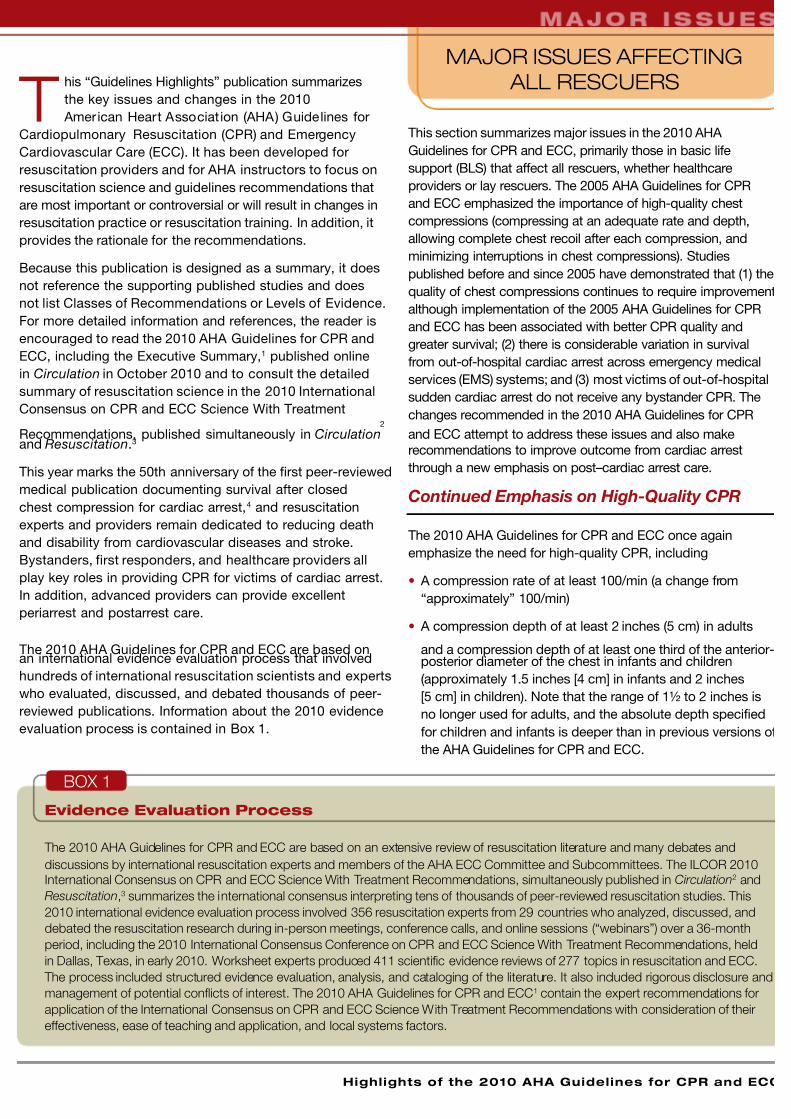
Evidence Evaluation Process
The 2010 AHA Guidelines for CPR and ECC are based on an extensive review of resuscitation literature and many debates and
discussions by international resuscitation experts and members of the AHA ECC Committee and Subcommittees. The ILCOR 2010
International Consensus on CPR and ECC Science With Treatment Recommendations, simultaneously published in Circulation2 and
Resuscitation,3 summarizes the international consensus interpreting tens of thousands of peer-reviewed resuscitation studies. This
2010 international evidence evaluation process involved 356 resuscitation experts from 29 countries who analyzed, discussed, and
debated the resuscitation research during in-person meetings, conference calls, and online sessions (“webinars”) over a 36-month
period, including the 2010 International Consensus Conference on CPR and ECC Science With Treatment Recommendations, held
in Dallas, Texas, in early 2010. Worksheet experts produced 411 scientific evidence reviews of 277 topics in resuscitation and ECC.
The process included structured evidence evaluation, analysis, and cataloging of the literature. It also included rigorous disclosure and
management of potential conflicts of interest. The 2010 AHA Guidelines for CPR and ECC1 contain the expert recommendations for
application of the International Consensus on CPR and ECC Science With Treatment Recommendations with consideration of their
effectiveness, ease of teaching and application, and local systems factors.
BOX 1
7/26/2019 Algoritma ACLS 2010 OK
http://slidepdf.com/reader/full/algoritma-acls-2010-ok 4/32
A m e r i c a n H e a r t A s s o c i a t i o n A m e r i c a n H e a r t A s s o c i a t i o n
• Allowing for complete chest recoil after each compression
• Minimizing interruptions in chest compressions
• Avoiding excessive ventilation
There has been no change in the recommendation for a
compression-to-ventilation ratio of 30:2 for single rescuers of
adults, children, and infants (excluding newly born infants). The
2010 AHA Guidelines for CPR and ECC continue to recommend
that rescue breaths be given in approximately 1 second. Oncean advanced airway is in place, chest compressions can be
continuous (at a rate of at least 100/min) and no longer cycled
with ventilations. Rescue breaths can then be provided at
about 1 breath every 6 to 8 seconds (about 8 to 10 breaths per
minute). Excessive ventilation should be avoided.
A Change From A-B-C to C-A-B
The 2010 AHA Guidelines for CPR and ECC recommend a
change in the BLS sequence of steps from A-B-C (Airway,
Breathing, Chest compressions) to C-A-B (Chest compressions,
Airway, Breathing) for adults, children, and infants (excluding the
newly born; see Neonatal Resuscitation section). This fundamental
change in CPR sequence will require reeducation of everyone
who has ever learned CPR, but the consensus of the authors and
experts involved in the creation of the 2010 AHA Guidelines for
CPR and ECC is that the benefit will justify the effort.
Why: The vast majority of cardiac arrests occur in adults,
and the highest survival rates from cardiac arrest are reported
among patients of all ages who have a witnessed arrest and
an initial rhythm of ventricular fibrillation (VF) or pulseless
ventricular tachycardia (VT). In these patients, the critical
initial elements of BLS are chest compressions and early
defibrillation. In the A-B-C sequence, chest compressionsare often delayed while the responder opens the airway to
give mouth-to-mouth breaths, retrieves a barrier device, or
gathers and assembles ventilation equipment. By changing the
sequence to C-A-B, chest compressions will be initiated sooner
and the delay in ventilation should be minimal (ie, only the time
required to deliver the first cycle of 30 chest compressions, or
approximately 18 seconds; when 2 rescuers are present for
resuscitation of the infant or child, the delay will be even shorter).
Most victims of out-of-hospital cardiac arrest do not receive
any bystander CPR. There are probably many reasons for this,
but one impediment may be the A-B-C sequence, which starts
with the procedures that rescuers find most difficult, namely,
opening the airway and delivering breaths. Starting with chest
compressions might encourage more rescuers to begin CPR.
Basic life support is usually described as a sequence of
actions, and this continues to be true for the lone rescuer.
Most healthcare providers, however, work in teams, and
team members typically perform BLS actions simultaneously.
For example, one rescuer immediately initiates chest
compressions while another rescuer gets an automated
external defibrillator (AED) and calls for help, and a third
rescuer opens the airway and provides ventilations.
Healthcare providers are again encouraged to tailor rescue
actions to the most likely cause of arrest. For example,
if a lone healthcare provider witnesses a victim suddenly
collapse, the provider may assume that the victim has had a
primary cardiac arrest with a shockable rhythm and should
immediately activate the emergency response system,
retrieve an AED, and return to the victim to provide CPR
and use the AED. But for a presumed victim of asphyxial
arrest such as drowning, the priority would be to provide
chest compressions with rescue breathing for about 5 cycles
(approximately 2 minutes) before activating the emergency
response system.
Two new parts in the 2010 AHA Guidelines for CPR and ECC
are Post–Cardiac Arrest Care and Education, Implementation,
and Teams. The importance of post–cardiac arrest care isemphasized by the addition of a new fifth link in the AHA
ECC Adult Chain of Survival (Figure 1). See the sections
Post–Cardiac Arrest Care and Education, Implementation,
and Teams in this publication for a summary of key
recommendations contained in these new parts.
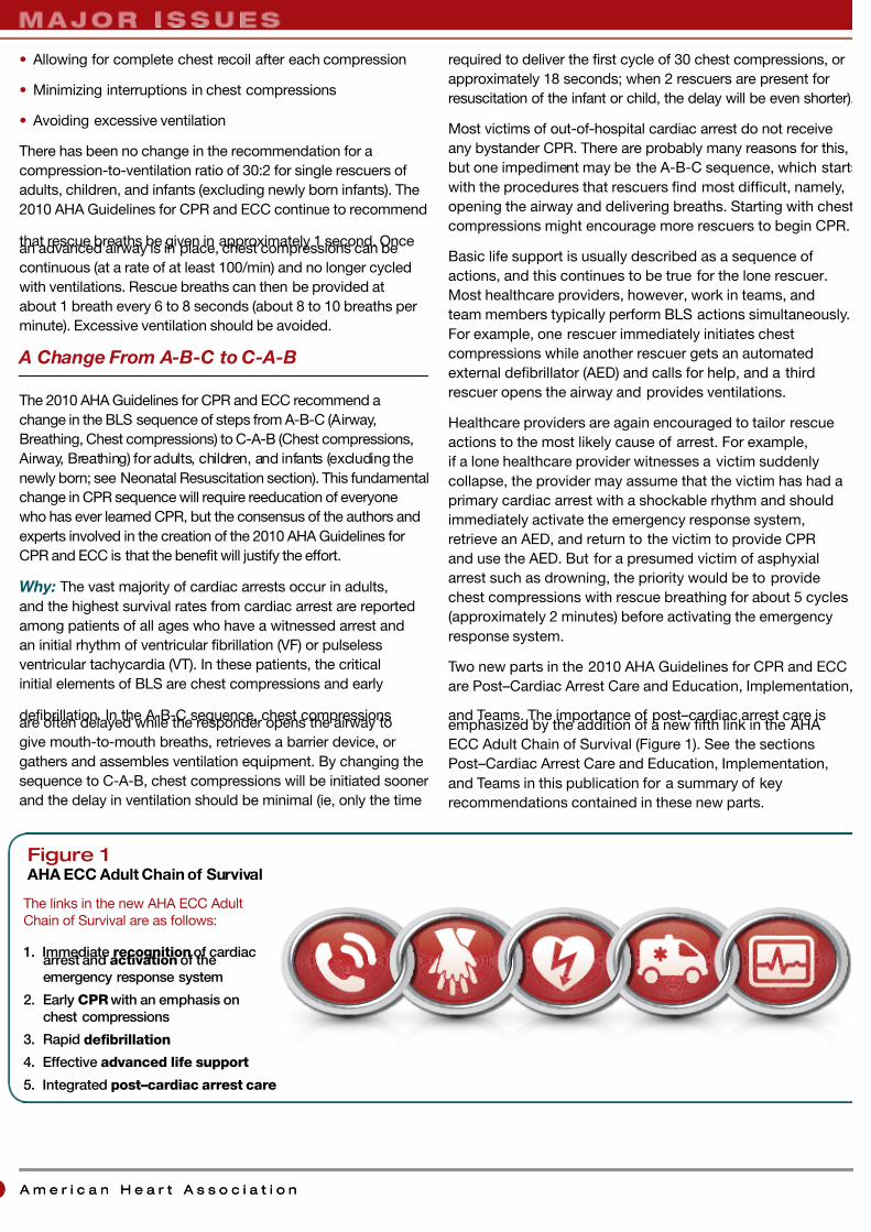
Figure 1 AHA ECC Adult Chain of Survival
The links in the new AHA ECC Adult
Chain of Survival are as follows:
1. Immediate recognition of cardiacarrest and activation of the
emergency response system
2. Early CPR with an emphasis on
chest compressions
3. Rapid defibrillation
4. Effective advanced life support
5. Integrated post–cardiac arrest care

7/26/2019 Algoritma ACLS 2010 OK
http://slidepdf.com/reader/full/algoritma-acls-2010-ok 5/32
Highlights of the 2010 AHA Guidelines for CPR and ECC
Figure 2Simplified Adult BLS Algorithm
LAY RESCUER
ADULT CPR
Summary of Key Issues and Major Changes
Key issues and major changes for the 2010 AHA Guidelines for
CPR and ECC recommendations for lay rescuer adult CPR are
the following:
• The simplified universal adult BLS algorithm has been
created (Figure 2).
• Refinements have been made to recommendations for
immediate recognition and activation of the emergency
response system based on signs of unresponsiveness, as
well as initiation of CPR if the victim is unresponsive with no
breathing or no normal breathing (ie, victim is only gasping).
• “Look, listen, and feel for breathing” has been removed from
the algorithm.
• Continued emphasis has been placed on high-quality CPR(with chest compressions of adequate rate and depth,
allowing complete chest recoil after each compression,
minimizing interruptions in compressions, and avoiding
excessive ventilation).
• There has been a change in the recommended sequence
for the lone rescuer to initiate chest compressions before
giving rescue breaths (C-A-B rather than A-B-C). The lone
rescuer should begin CPR with 30 compressions rather than
2 ventilations to reduce delay to first compression.
• Compression rate should be at least 100/min (rather than
“approximately” 100/min).
• Compression depth for adults has been changed from the
range of 1½ to 2 inches to at least 2 inches (5 cm).
These changes are designed to simplify lay rescuer training
and to continue to emphasize the need to provide early chest
compressions for the victim of a sudden cardiac arrest. More
information about these changes appears below. Note: In the
following topics, changes or points of emphasis for lay rescuers
that are similar to those for healthcare providers are noted with
an asterisk (*).
Emphasis on Chest Compressions*
2010 (New): If a bystander is not trained in CPR, the bystander
should provide Hands-Only™ (compression-only) CPR for
the adult victim who suddenly collapses, with an emphasis to
“push hard and fast” on the center of the chest, or follow the
directions of the EMS dispatcher. The rescuer should continue
Hands-Only CPR until an AED arrives and is ready for use or
EMS providers or other responders take over care of the victim.
All trained lay rescuers should, at a minimum, provide chestcompressions for victims of cardiac arrest. In addition, if
the trained lay rescuer is able to perform rescue breaths,
compressions and breaths should be provided in a ratio of
30 compressions to 2 breaths. The rescuer should continue
CPR until an AED arrives and is ready for use or EMS providers
take over care of the victim.
2005 (Old): The 2005 AHA Guidelines for CPR and ECC
did not provide different recommendations for trained versus
untrained rescuers but did recommend that dispatchers provide
compression-only CPR instructions to untrained bystanders.
The 2005 AHA Guidelines for CPR and ECC did note that if
the rescuer was unwilling or unable to provide ventilations, the
rescuer should provide chest compressions only.
Why: Hands-Only (compression-only) CPR is easier for an
untrained rescuer to perform and can be more readily guided
by dispatchers over the telephone. In addition, survival rates
from cardiac arrests of cardiac etiology are similar with either
Hands-Only CPR or CPR with both compressions and rescue
breaths. However, for the trained lay rescuer who is able, the
recommendation remains for the rescuer to perform both
compressions and ventilations.
© 2010 American Heart Association
Unresponsive
No breathing or
no normal breathing
(only gasping)
Activate
emergency
response
Start CPR
Get
defibrillator
Check rhythm/
shock if
indicated
Repeat every 2 minutes
P u s h
H a r d
•
P u s h
F a s t

7/26/2019 Algoritma ACLS 2010 OK
http://slidepdf.com/reader/full/algoritma-acls-2010-ok 6/32
A m e r i c a n H e a r t A s s o c i a t i o n
Change in CPR Sequence: C-A-B Rather
Than A-B-C*
2010 (New): Initiate chest compressions before ventilations.
2005 (Old): The sequence of adult CPR began with opening of
the airway, checking for normal breathing, and then delivery of
2 rescue breaths followed by cycles of 30 chest compressions
and 2 breaths.
Why: Although no published human or animal evidencedemonstrates that starting CPR with 30 compressions
rather than 2 ventilations leads to improved outcome, chest
compressions provide vital blood flow to the heart and
brain, and studies of out-of-hospital adult cardiac arrest
showed that survival was higher when bystanders made
some attempt rather than no attempt to provide CPR. Animal
data demonstrated that delays or interruptions in chest
compressions reduced survival, so such delays or interruptions
should be minimized throughout the entire resuscitation. Chest
compressions can be started almost immediately, whereas
positioning the head and achieving a seal for mouth-to-mouth
or bag-mask rescue breathing all take time. The delay in
initiation of compressions can be reduced if 2 rescuers arepresent: the first rescuer begins chest compressions, and the
second rescuer opens the airway and is prepared to deliver
breaths as soon as the first rescuer has completed the first
set of 30 chest compressions. Whether 1 or more rescuers are
present, initiation of CPR with chest compressions ensures that
the victim receives this critical intervention early, and any delay
in rescue breaths should be brief.
Elimination of “Look, Listen, and Feel
for Breathing”*
2010 (New): “Look, listen, and feel” was removed from the
CPR sequence. After delivery of 30 compressions, the lone
rescuer opens the victim’s airway and delivers 2 breaths.
2005 (Old): “Look, listen, and feel” was used to assess
breathing after the airway was opened.
Why: With the new “chest compressions first” sequence, CPR
is performed if the adult is unresponsive and not breathing
or not breathing normally (as noted above, lay rescuers will
be taught to provide CPR if the unresponsive victim is “not
breathing or only gasping”). The CPR sequence begins with
compressions (C-A-B sequence). Therefore, breathing is briefly
checked as part of a check for cardiac arrest; after the first set
of chest compressions, the airway is opened, and the rescuer
delivers 2 breaths.
Chest Compression Rate: At Least
100 per Minute*
2010 (New): It is reasonable for lay rescuers and healthcare
providers to perform chest compressions at a rate of at least
100/min.
2005 (Old): Compress at a rate of about 100/min.
Why: The number of chest compressions delivered per
minute during CPR is an important determinant of return
of spontaneous circulation (ROSC) and survival with good
neurologic function. The actual number of chest compressions
delivered per minute is determined by the rate of chest
compressions and the number and duration of interruptions in
compressions (eg, to open the airway, deliver rescue breaths,
or allow AED analysis). In most studies, more compressions are
associated with higher survival rates, and fewer compressions
are associated with lower survival rates. Provision of adequate
chest compressions requires an emphasis not only on an
adequate compression rate but also on minimizing interruptions
to this critical component of CPR. An inadequate compression
rate or frequent interruptions (or both) will reduce the total
number of compressions delivered per minute. For further
information, see Box 2.
Chest Compression Depth*
2010 (New): The adult sternum should be depressed at least 2
inches (5 cm).
2005 (Old): The adult sternum should be depressed
approximately 1½ to 2 inches (approximately 4 to 5 cm).
Why: Compressions create blood flow primarily by increasing
intrathoracic pressure and directly compressing the heart.
Compressions generate critical blood flow and oxygen and
energy delivery to the heart and brain. Confusion may result
when a range of depth is recommended, so 1 compression
Number of Compressions Delivered
Affected by Compression Rate andby Interruptions
The total number of compressions delivered during resuscitation
is an important determinant of survival from cardiac arrest.
The number of compressions delivered is affected by the
compression rate and by the compression fraction (the portion
of total CPR time during which compressions are performed);
increases in compression rate and fraction increase the total
compressions delivered, whereas decreases in compression
rate or compression fraction decrease the total compressions
delivered. Compression fraction is improved if you reduce
the number and length of any interruptions in compressions,
and it is reduced by frequent or long interruptions in chestcompressions. An analogy can be found in automobile travel.
When you travel in an automobile, the number of miles you
travel in a day is affected not only by the speed that you drive
(your rate of travel) but also by the number and duration of any
stops you make (interruptions in travel). During CPR, you want
to deliver effective compressions at an appropriate rate (at least
100/min) and depth, while minimizing the number and duration
of interruptions in chest compressions. Additional components
of high-quality CPR include allowing complete chest recoil after
each compression and avoiding excessive ventilation.
BOX 2
7/26/2019 Algoritma ACLS 2010 OK
http://slidepdf.com/reader/full/algoritma-acls-2010-ok 7/32
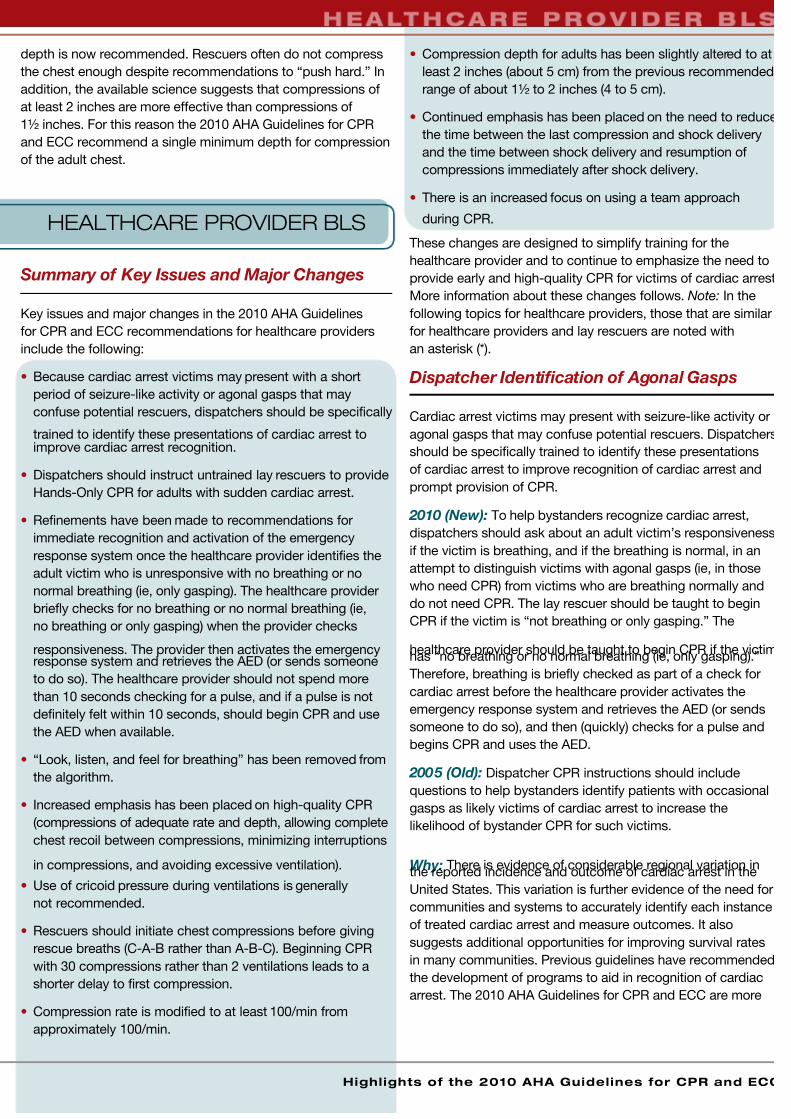
depth is now recommended. Rescuers often do not compress
the chest enough despite recommendations to “push hard.” In
addition, the available science suggests that compressions of
at least 2 inches are more effective than compressions of
1½ inches. For this reason the 2010 AHA Guidelines for CPR
and ECC recommend a single minimum depth for compression
of the adult chest.
HEALTHCARE PROVIDER BLS
Summary of Key Issues and Major Changes
Key issues and major changes in the 2010 AHA Guidelines
for CPR and ECC recommendations for healthcare providers
include the following:
• Because cardiac arrest victims may present with a short
period of seizure-like activity or agonal gasps that may
confuse potential rescuers, dispatchers should be specifically
trained to identify these presentations of cardiac arrest toimprove cardiac arrest recognition.
• Dispatchers should instruct untrained lay rescuers to provide
Hands-Only CPR for adults with sudden cardiac arrest.
• Refinements have been made to recommendations for
immediate recognition and activation of the emergency
response system once the healthcare provider identifies the
adult victim who is unresponsive with no breathing or no
normal breathing (ie, only gasping). The healthcare provider
briefly checks for no breathing or no normal breathing (ie,
no breathing or only gasping) when the provider checks
responsiveness. The provider then activates the emergencyresponse system and retrieves the AED (or sends someone
to do so). The healthcare provider should not spend more
than 10 seconds checking for a pulse, and if a pulse is not
definitely felt within 10 seconds, should begin CPR and use
the AED when available.
• “Look, listen, and feel for breathing” has been removed from
the algorithm.
• Increased emphasis has been placed on high-quality CPR
(compressions of adequate rate and depth, allowing complete
chest recoil between compressions, minimizing interruptions
in compressions, and avoiding excessive ventilation).
• Use of cricoid pressure during ventilations is generally
not recommended.
• Rescuers should initiate chest compressions before giving
rescue breaths (C-A-B rather than A-B-C). Beginning CPR
with 30 compressions rather than 2 ventilations leads to a
shorter delay to first compression.
• Compression rate is modified to at least 100/min from
approximately 100/min.
• Compression depth for adults has been slightly altered to at
least 2 inches (about 5 cm) from the previous recommended
range of about 1½ to 2 inches (4 to 5 cm).
• Continued emphasis has been placed on the need to reduce
the time between the last compression and shock delivery
and the time between shock delivery and resumption of
compressions immediately after shock delivery.
• There is an increased focus on using a team approach
during CPR.
These changes are designed to simplify training for the
healthcare provider and to continue to emphasize the need to
provide early and high-quality CPR for victims of cardiac arrest
More information about these changes follows. Note: In the
following topics for healthcare providers, those that are similar
for healthcare providers and lay rescuers are noted with
an asterisk (*).
Dispatcher Identification of Agonal Gasps
Cardiac arrest victims may present with seizure-like activity or
agonal gasps that may confuse potential rescuers. Dispatchers
should be specifically trained to identify these presentations
of cardiac arrest to improve recognition of cardiac arrest and
prompt provision of CPR.
2010 (New): To help bystanders recognize cardiac arrest,
dispatchers should ask about an adult victim’s responsiveness
if the victim is breathing, and if the breathing is normal, in an
attempt to distinguish victims with agonal gasps (ie, in those
who need CPR) from victims who are breathing normally and
do not need CPR. The lay rescuer should be taught to begin
CPR if the victim is “not breathing or only gasping.” The
healthcare provider should be taught to begin CPR if the victimhas “no breathing or no normal breathing (ie, only gasping).”
Therefore, breathing is briefly checked as part of a check for
cardiac arrest before the healthcare provider activates the
emergency response system and retrieves the AED (or sends
someone to do so), and then (quickly) checks for a pulse and
begins CPR and uses the AED.
2005 (Old): Dispatcher CPR instructions should include
questions to help bystanders identify patients with occasional
gasps as likely victims of cardiac arrest to increase the
likelihood of bystander CPR for such victims.
Why: There is evidence of considerable regional variation inthe reported incidence and outcome of cardiac arrest in the
United States. This variation is further evidence of the need for
communities and systems to accurately identify each instance
of treated cardiac arrest and measure outcomes. It also
suggests additional opportunities for improving survival rates
in many communities. Previous guidelines have recommended
the development of programs to aid in recognition of cardiac
arrest. The 2010 AHA Guidelines for CPR and ECC are more
Highlights of the 2010 AHA Guidelines for CPR and ECC

7/26/2019 Algoritma ACLS 2010 OK
http://slidepdf.com/reader/full/algoritma-acls-2010-ok 8/32
A m e r i c a n H e a r t A s s o c i a t i o n
specific about the necessary components of resuscitation
systems. Studies published since 2005 have demonstrated
improved outcome from out-of-hospital cardiac arrest,
particularly from shockable rhythms, and have reaffirmed the
importance of a stronger emphasis on immediate provision
of high-quality CPR (compressions of adequate rate and
depth, allowing complete chest recoil after each compression,
minimizing interruptions in chest compressions, and avoiding
excessive ventilation).
To help bystanders immediately recognize cardiac arrest,
dispatchers should specifically inquire about an adult
victim’s absence of response, if the victim is breathing, and
if any breathing observed is normal. Dispatchers should be
specifically educated in helping bystanders detect agonal
gasps to improve cardiac arrest recognition.
Dispatchers should also be aware that brief generalized
seizures may be the first manifestation of cardiac arrest. In
summary, in addition to activating professional emergency
responders, the dispatcher should ask straightforward
questions about whether the patient is responsive and
breathing normally to identify patients with possible cardiacarrest. Dispatchers should provide Hands-Only (compression-
only) CPR instructions to help untrained bystanders initiate
CPR when cardiac arrest is suspected (see below).
Dispatcher Should Provide CPR Instructions
2010 (New): The 2010 AHA Guidelines for CPR and ECC more
strongly recommend that dispatchers should instruct untrained
lay rescuers to provide Hands-Only CPR for adults who
are unresponsive with no breathing or no normal breathing.
Dispatchers should provide instructions in conventional CPR
for victims of likely asphyxial arrest.
2005 (Old): The 2005 AHA Guidelines for CPR and ECC
noted that telephone instruction in chest compressions alone
may be preferable.
Why: Unfortunately, most adults with out-of-hospital cardiac
arrest do not receive any bystander CPR. Hands-Only
(compression-only) bystander CPR substantially improves
survival after adult out-of-hospital cardiac arrests compared
with no bystander CPR. Other studies of adults with cardiac
arrest treated by lay rescuers showed similar survival rates
among victims receiving Hands-Only CPR versus those
receiving conventional CPR (ie, with rescue breaths).
Importantly, it is easier for dispatchers to instruct untrained
rescuers to perform Hands-Only CPR than conventional CPR
for adult victims, so the recommendation is now stronger
for them to do so, unless the victim is likely to have had an
asphyxial arrest (eg, drowning).
Cricoid Pressure
2010 (New): The routine use of cricoid pressure in cardiac
arrest is not recommended.
2005 (Old): Cricoid pressure should be used only if the victim
is deeply unconscious, and it usually requires a third rescuer
not involved in rescue breaths or compressions.
Why: Cricoid pressure is a technique of applying pressure to
the victim’s cricoid cartilage to push the trachea posteriorly
and compress the esophagus against the cervical vertebrae.
Cricoid pressure can prevent gastric inflation and reduce the
risk of regurgitation and aspiration during bag-mask ventilation,
but it may also impede ventilation. Seven randomized studies
showed that cricoid pressure can delay or prevent the
placement of an advanced airway and that some aspiration
can still occur despite application of cricoid pressure. In
addition, it is difficult to appropriately train rescuers in use of
the maneuver. Therefore, the routine use of cricoid pressure in
cardiac arrest is not recommended.
Emphasis on Chest Compressions*
2010 (New): Chest compressions are emphasized for
both trained and untrained rescuers. If a bystander is not
trained in CPR, the bystander should provide Hands-Only
(compression-only) CPR for the adult who suddenly collapses,
with an emphasis to “push hard and fast” on the center of
the chest, or follow the directions of the emergency medical
dispatcher. The rescuer should continue Hands-Only CPR
until an AED arrives and is ready for use or EMS providers
take over care of the victim.
Optimally all healthcare providers should be trained in BLS. In
this trained population, it is reasonable for both EMS and in-
hospital professional rescuers to provide chest compressions
and rescue breaths for cardiac arrest victims.
2005 (Old): The 2005 AHA Guidelines for CPR and ECC
did not provide different recommendations for trained and
untrained rescuers and did not emphasize differences in
instructions provided to lay rescuers versus healthcare
providers, but did recommend that dispatchers provide
compression-only CPR instructions to untrained bystanders. In
addition, the 2005 AHA Guidelines for CPR and ECC noted that
if the rescuer was unwilling or unable to provide ventilations,
the rescuer should provide chest compressions. Note that the
AHA Hands-Only CPR statement was published in 2008.
Why: Hands-Only (compression-only) CPR is easier for
untrained rescuers to perform and can be more readily guided
by dispatchers over the telephone. However, because thehealthcare provider should be trained, the recommendation
remains for the healthcare provider to perform both
compressions and ventilations. If the healthcare provider is
unable to perform ventilations, the provider should activate the
emergency response system and provide chest compressions.
Activation of Emergency Response System
2010 (New): The healthcare provider should check for
response while looking at the patient to determine if breathing

7/26/2019 Algoritma ACLS 2010 OK
http://slidepdf.com/reader/full/algoritma-acls-2010-ok 9/32
Highlights of the 2010 AHA Guidelines for CPR and ECC
is absent or not normal. The provider should suspect cardiac
arrest if the victim is not breathing or only gasping.
2005 (Old): The healthcare provider activated the emergency
response system after finding an unresponsive victim. The
provider then returned to the victim and opened the airway and
checked for breathing or abnormal breathing.
Why: The healthcare provider should not delay activation of
the emergency response system but should obtain 2 pieces of
information simultaneously: the provider should check the victim
for response and check for no breathing or no normal breathing.
If the victim is unresponsive and is not breathing at all or has no
normal breathing (ie, only agonal gasps), the provider should
activate the emergency response system and retrieve the AED if
available (or send someone to do so). If the healthcare provider
does not feel a pulse within 10 seconds, the provider should
begin CPR and use the AED when it is available.
Change in CPR Sequence: C-A-B Rather
Than A-B-C*
2010 (New): A change in the 2010 AHA Guidelines for CPRand ECC is to recommend the initiation of chest compressions
before ventilations.
2005 (Old): The sequence of adult CPR began with opening
of the airway, checking for normal breathing, and then delivering
2 rescue breaths followed by cycles of 30 chest compressions
and 2 breaths.
Why: Although no published human or animal evidence
demonstrates that starting CPR with 30 compressions
rather than 2 ventilations leads to improved outcome, chest
compressions provide the blood flow, and studies of out-of-
hospital adult cardiac arrest showed that survival was higherwhen bystanders provided chest compressions rather than no
chest compressions. Animal data demonstrate that delays or
interruptions in chest compressions reduce survival, so such
delays and interruptions should be minimized throughout the
entire resuscitation. Chest compressions can be started almost
immediately, whereas positioning the head and achieving a
seal for mouth-to-mouth or bag-mask rescue breathing all take
time. The delay in initiation of compressions can be reduced if 2
rescuers are present: the first rescuer begins chest compressions,
and the second rescuer opens the airway and is prepared to
deliver breaths as soon as the first rescuer has completed the
first set of 30 chest compressions. Whether 1 or more rescuersare present, initiation of CPR with chest compressions ensures
that the victim receives this critical intervention early.
Elimination of “Look, Listen, and Feel
for Breathing”*
2010 (New): “Look, listen, and feel for breathing” was
removed from the sequence for assessment of breathing after
opening the airway. The healthcare provider briefly checks
for breathing when checking responsiveness to detect signs
of cardiac arrest. After delivery of 30 compressions, the lone
rescuer opens the victim’s airway and delivers 2 breaths.
2005 (Old): “Look, listen, and feel for breathing” was used to
assess breathing after the airway was opened.
Why: With the new chest compression–first sequence, CPR is
performed if the adult victim is unresponsive and not breathing
or not breathing normally (ie, not breathing or only gasping)
and begins with compressions (C-A-B sequence). Therefore,
breathing is briefly checked as part of a check for cardiac
arrest. After the first set of chest compressions, the airway is
opened and the rescuer delivers 2 breaths.
Chest Compression Rate: At Least 100 per Minute*
2010 (New): It is reasonable for lay rescuers and healthcare
providers to perform chest compressions at a rate of at least
100/min.
2005 (Old): Compress at a rate of about 100/min.
Why: The number of chest compressions delivered per
minute during CPR is an important determinant of ROSC andsurvival with good neurologic function. The actual number of
chest compressions delivered per minute is determined by the
rate of chest compressions and the number and duration of
interruptions in compressions (eg, to open the airway, deliver
rescue breaths, or allow AED analysis). In most studies, delivery
of more compressions during resuscitation is associated with
better survival, and delivery of fewer compressions is associated
with lower survival. Provision of adequate chest compressions
requires an emphasis not only on an adequate compression rate
but also on minimizing interruptions to this critical component of
CPR. An inadequate compression rate or frequent interruptions
(or both) will reduce the total number of compressions deliveredper minute. For further information, see Box 2 on page 4.
Chest Compression Depth*
2010 (New): The adult sternum should be depressed at least 2
inches (5 cm).
2005 (Old): The adult sternum should be depressed 1½ to 2
inches (approximately 4 to 5 cm).
Why: Compressions create blood flow primarily by increasing
intrathoracic pressure and directly compressing the heart.
Compressions generate critical blood flow and oxygen andenergy delivery to the heart and brain. Confusion may result
when a range of depth is recommended, so 1 compression
depth is now recommended. Rescuers often do not adequately
compress the chest despite recommendations to “push hard.”
In addition, the available science suggests that compressions
of at least 2 inches are more effective than compressions
of 1½ inches. For this reason the 2010 AHA Guidelines for CPR
and ECC recommend a single minimum depth for compression
of the adult chest, and that compression depth is deeper than
in the old recommendation.
7/26/2019 Algoritma ACLS 2010 OK
http://slidepdf.com/reader/full/algoritma-acls-2010-ok 10/32
A m e r i c a n H e a r t A s s o c i a t i o n
Team Resuscitation
2010 (New): The steps in the BLS algorithm have traditionally
been presented as a sequence to help a single rescuer
prioritize actions. There is increased focus on providing
CPR as a team because resuscitations in most EMS and
healthcare systems involve teams of rescuers, with rescuersperforming several actions simultaneously. For example, one
rescuer activates the emergency response system while a
second begins chest compressions, a third is either providing
ventilations or retrieving the bag-mask for rescue breathing,
and a fourth is retrieving and setting up a defibrillator.
2005 (Old): The steps of BLS consist of a series of sequential
assessments and actions. The intent of the algorithm is to
present the steps in a logical and concise manner that will be
easy for each rescuer to learn, remember, and perform.
Why: Some resuscitations start with a lone rescuer who
calls for help, whereas other resuscitations begin with several
willing rescuers. Training should focus on building a team
as each rescuer arrives, or on designating a team leader if
multiple rescuers are present. As additional personnel arrive,
responsibilities for tasks that would ordinarily be performed
sequentially by fewer rescuers may now be delegated to a team
of providers who perform them simultaneously. For this reason,
BLS healthcare provider training should not only teach individua
skills but should also teach rescuers to work in effective teams.
Comparison of Key Elements of Adult, Child,
and Infant BLS
As in the 2005 AHA Guidelines for CPR and ECC, the 2010 AHA
Guidelines for CPR and ECC contain a comparison table that lists
the key elements of adult, child, and infant BLS (excluding CPR fo
newly born infants). These key elements are included in Table 1.
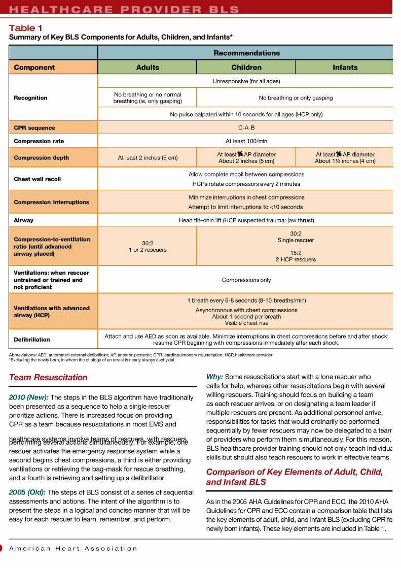
Recommendations
Component Adults Children Infants
Recognition
Unresponsive (for all ages)
No breathing or no normalbreathing (ie, only gasping)
No breathing or only gasping
No pulse palpated within 10 seconds for all ages (HCP only)
CPR sequence C-A-B
Compression rate At least 100/min
Compression depth At least 2 inches (5 cm) At least ¹⁄ ³ AP diameter About 2 inches (5 cm)
At least ¹⁄ ³ AP diameter About 1½ inches (4 cm)
Chest wall recoil Allow complete recoil between compressions
HCPs rotate compressors every 2 minutes
Compression interruptionsMinimize interruptions in chest compressions
Attempt to limit interruptions to <10 seconds
Airway Head tilt–chin lift (HCP suspected trauma: jaw thrust)
Compression-to-ventilation
ratio (until advanced
airway placed)
30:21 or 2 rescuers
30:2Single rescuer
15:22 HCP rescuers
Ventilations: when rescuer
untrained or trained and
not proficient
Compressions only
Ventilations with advanced
airway (HCP)
1 breath every 6-8 seconds (8-10 breaths/min)
Asynchronous with chest compressions About 1 second per breath
Visible chest rise
Defibrillation Attach and use AED as soon as available. Minimize interruptions in chest compressions before and after shock;
resume CPR beginning with compressions immediately after each shock.
Abbreviations: AED, automated external defibrillator; AP, anterior-posterior; CPR, cardiopulmonary resuscitation; HCP, healthcare provider.*Excluding the newly born, in whom the etiology of an arrest is nearly always asphyxial.
Table 1Summary of Key BLS Components for Adults, Children, and Infants*

7/26/2019 Algoritma ACLS 2010 OK
http://slidepdf.com/reader/full/algoritma-acls-2010-ok 11/32
Highlights of the 2010 AHA Guidelines for CPR and ECC
ELECTRICAL
THERAPIES
The 2010 AHA Guidelines for CPR and ECC have been
updated to reflect new data regarding defibrillation and
cardioversion for cardiac rhythm disturbances and the use of
pacing in bradycardia. These data largely continue to support
the recommendations in the 2005 AHA Guidelines for CPR
and ECC. Therefore, no major changes were recommended
regarding defibrillation, cardioversion, and pacing. Emphasis on
early defibrillation integrated with high-quality CPR is the key to
improving survival from sudden cardiac arrest.
Summary of Key Issues and Major Changes
Main topics include
• Integration of AEDs into the Chain of Survival system for
public places
• Consideration of AED use in hospitals
• AEDs can now be used in infants if a manual defibrillator is
not available
• Shock first versus CPR first in cardiac arrest
• 1-shock protocol versus 3-shock sequence for VF
• Biphasic and monophasic waveforms
• Escalating versus fixed doses for second and
subsequent shocks
• Electrode placement
• External defibrillation with implantablecardioverter-defibrillator
• Synchronized cardioversion
Automated External Defibrillators
Community Lay Rescuer AED Programs
2010 (Slightly Modified): Cardiopulmonary resuscitation
and the use of AEDs by public safety first responders are
recommended to increase survival rates for out-of-hospital
sudden cardiac arrest. The 2010 AHA Guidelines for CPR and
ECC again recommend the establishment of AED programs
in public locations where there is a relatively high likelihood of
witnessed cardiac arrest (eg, airports, casinos, sports facilities).
To maximize the effectiveness of these programs, the AHA
continues to emphasize the importance of organizing, planning,
training, linking with the EMS system, and establishing a
process of continuous quality improvement.
2005 (Old): The 2005 AHA Guidelines for CPR and ECC
identified 4 components for successful community lay rescuer
AED programs:
• A planned and practiced response, typically requiring
oversight by a healthcare provider
• Training of anticipated rescuers in CPR and use of the AED
• A link with the local EMS system
• A program of ongoing quality improvement
There is insufficient evidence to recommend for or against the
deployment of AEDs in homes.
In-Hospital Use of AEDs
2010 (Reaffirmed 2005 Recommendation): Despite
limited evidence, AEDs may be considered for the hospital
setting as a way to facilitate early defibrillation (a goal of shock
delivery ≤3 minutes from collapse), especially in areas where
staff have no rhythm recognition skills or defibrillators are used
infrequently. Hospitals should monitor collapse-to–first shock
intervals and resuscitation outcomes.
AED Use in Children Now Includes Infants
2010 (New): For attempted defibrillation of children 1 to 8
years of age with an AED, the rescuer should use a pediatricdose-attenuator system if one is available. If the rescuer
provides CPR to a child in cardiac arrest and does not have an
AED with a pediatric dose-attenuator system, the rescuer should
use a standard AED. For infants (<1 year of age), a manual
defibrillator is preferred. If a manual defibrillator is not available,
an AED with pediatric dose attenuation is desirable. If neither is
available, an AED without a dose attenuator may be used.
2005 (Old): For children 1 to 8 years of age, the rescuer
should use a pediatric dose-attenuator system if one is
available. If the rescuer provides CPR to a child in cardiac
arrest and does not have an AED with a pediatric attenuator
system, the rescuer should use a standard AED. There are
insufficient data to make a recommendation for or against the
use of AEDs for infants <1 year of age.
Why: The lowest energy dose for effective defibrillation in
infants and children is not known. The upper limit for safe
defibrillation is also not known, but doses >4 J/kg (as high
as 9 J/kg) have effectively defibrillated children and animal
models of pediatric arrest with no significant adverse effects.
Automated external defibrillators with relatively high-energy
doses have been used successfully in infants in cardiac arrest
with no clear adverse effects.
Shock First vs CPR First
2010 (Reaffirmed 2005 Recommendation): When any
rescuer witnesses an out-of-hospital arrest and an AED is
immediately available on-site, the rescuer should start CPR
with chest compressions and use the AED as soon as possible.
Healthcare providers who treat cardiac arrest in hospitals and
other facilities with on-site AEDs or defibrillators should provide
immediate CPR and should use the AED/defibrillator as soon
as it is available. These recommendations are designed to
7/26/2019 Algoritma ACLS 2010 OK
http://slidepdf.com/reader/full/algoritma-acls-2010-ok 12/32
A m e r i c a n H e a r t A s s o c i a t i o n0
support early CPR and early defibrillation, particularly when an
AED or defibrillator is available within moments of the onset of
sudden cardiac arrest. When an out-of-hospital cardiac arrest is
not witnessed by EMS personnel, EMS may initiate CPR while
checking the rhythm with the AED or on the electrocardiogram
(ECG) and preparing for defibrillation. In such instances, 1½
to 3 minutes of CPR may be considered before attempted
defibrillation. Whenever 2 or more rescuers are present, CPR
should be provided while the defibrillator is retrieved.
With in-hospital sudden cardiac arrest, there is insufficient
evidence to support or refute CPR before defibrillation.
However, in monitored patients, the time from VF to shock
delivery should be under 3 minutes, and CPR should be
performed while the defibrillator is readied.
Why: When VF is present for more than a few minutes, the
myocardium is depleted of oxygen and energy. A brief period
of chest compressions can deliver oxygen and energy to the
heart, increasing the likelihood that a shock will both eliminate
VF (defibrillation) and be followed by ROSC. Before the
publication of the 2005 AHA Guidelines for CPR and ECC,
2 studies suggested the potential benefit of CPR first ratherthan shock first. In both studies, although 1½ to 3 minutes of
CPR before shock delivery did not improve overall survival from
VF, the CPR-first strategy did improve survival among victims
with VF if the EMS call-to-arrival interval was 4 to 5 minutes
or longer. However, 2 subsequent randomized controlled
trials found that CPR before attempted defibrillation by EMS
personnel was not associated with a significant difference
in survival to discharge. One retrospective study did find an
improved neurologic status at 30 days and at 1 year when
immediate CPR was compared with immediate defibrillation in
patients with out-of-hospital VF.
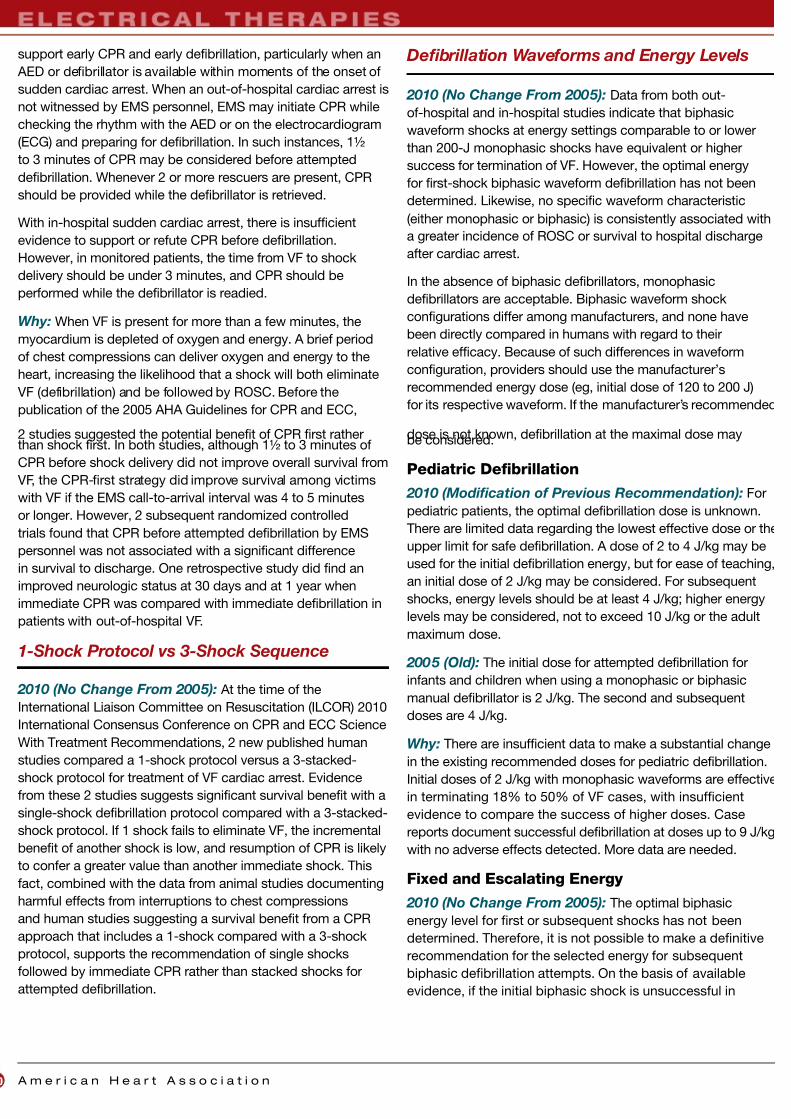
1-Shock Protocol vs 3-Shock Sequence
2010 (No Change From 2005): At the time of the
International Liaison Committee on Resuscitation (ILCOR) 2010
International Consensus Conference on CPR and ECC Science
With Treatment Recommendations, 2 new published human
studies compared a 1-shock protocol versus a 3-stacked-
shock protocol for treatment of VF cardiac arrest. Evidence
from these 2 studies suggests significant survival benefit with a
single-shock defibrillation protocol compared with a 3-stacked-
shock protocol. If 1 shock fails to eliminate VF, the incremental
benefit of another shock is low, and resumption of CPR is likely
to confer a greater value than another immediate shock. This
fact, combined with the data from animal studies documenting
harmful effects from interruptions to chest compressions
and human studies suggesting a survival benefit from a CPR
approach that includes a 1-shock compared with a 3-shock
protocol, supports the recommendation of single shocks
followed by immediate CPR rather than stacked shocks for
attempted defibrillation.
Defibrillation Waveforms and Energy Levels
2010 (No Change From 2005): Data from both out-
of-hospital and in-hospital studies indicate that biphasic
waveform shocks at energy settings comparable to or lower
than 200-J monophasic shocks have equivalent or higher
success for termination of VF. However, the optimal energy
for first-shock biphasic waveform defibrillation has not been
determined. Likewise, no specific waveform characteristic
(either monophasic or biphasic) is consistently associated with
a greater incidence of ROSC or survival to hospital discharge
after cardiac arrest.
In the absence of biphasic defibrillators, monophasic
defibrillators are acceptable. Biphasic waveform shock
configurations differ among manufacturers, and none have
been directly compared in humans with regard to their
relative efficacy. Because of such differences in waveform
configuration, providers should use the manufacturer’s
recommended energy dose (eg, initial dose of 120 to 200 J)
for its respective waveform. If the manufacturer’s recommended
dose is not known, defibrillation at the maximal dose maybe considered.
Pediatric Defibrillation
2010 (Modification of Previous Recommendation): For
pediatric patients, the optimal defibrillation dose is unknown.
There are limited data regarding the lowest effective dose or the
upper limit for safe defibrillation. A dose of 2 to 4 J/kg may be
used for the initial defibrillation energy, but for ease of teaching,
an initial dose of 2 J/kg may be considered. For subsequent
shocks, energy levels should be at least 4 J/kg; higher energy
levels may be considered, not to exceed 10 J/kg or the adult
maximum dose.
2005 (Old): The initial dose for attempted defibrillation for
infants and children when using a monophasic or biphasic
manual defibrillator is 2 J/kg. The second and subsequent
doses are 4 J/kg.
Why: There are insufficient data to make a substantial change
in the existing recommended doses for pediatric defibrillation.
Initial doses of 2 J/kg with monophasic waveforms are effective
in terminating 18% to 50% of VF cases, with insufficient
evidence to compare the success of higher doses. Case
reports document successful defibrillation at doses up to 9 J/kg
with no adverse effects detected. More data are needed.
Fixed and Escalating Energy
2010 (No Change From 2005): The optimal biphasic
energy level for first or subsequent shocks has not been
determined. Therefore, it is not possible to make a definitive
recommendation for the selected energy for subsequent
biphasic defibrillation attempts. On the basis of available
evidence, if the initial biphasic shock is unsuccessful in

7/26/2019 Algoritma ACLS 2010 OK
http://slidepdf.com/reader/full/algoritma-acls-2010-ok 13/32
Highlights of the 2010 AHA Guidelines for CPR and ECC
terminating VF, subsequent energy levels should be at least
equivalent, and higher energy levels may be considered,
if available.
Electrode Placement
2010 (Modification of Previous Recommendation):
For ease of placement and education, the anterior-lateral pad
position is a reasonable default electrode placement. Any
of 3 alternative pad positions (anterior-posterior, anterior–left infrascapular, and anterior–right infrascapular) may be
considered on the basis of individual patient characteristics.
Placement of AED electrode pads on the victim’s bare chest in
any of the 4 pad positions is reasonable for defibrillation.
2005 (Old): Rescuers should place AED electrode pads on the
victim’s bare chest in the conventional sternal-apical (anterior-
lateral) position. The right (sternal) chest pad is placed on the
victim’s right superior-anterior (infraclavicular) chest, and the
apical (left) pad is placed on the victim’s inferior-lateral left
chest, lateral to the left breast. Other acceptable pad positions
are placement on the lateral chest wall on the right and left
sides (biaxillary) or the left pad in the standard apical position
and the other pad on the right or left upper back.
Why: New data demonstrate that the 4 pad positions
(anterior-lateral, anterior-posterior, anterior–left infrascapular,
and anterior–right infrascapular) appear to be equally effective
to treat atrial or ventricular arrhythmias. Again, for ease of
teaching, the default position taught in AHA courses will not
change from the 2005 recommended position. No studies
were identified that directly evaluated the effect of placement
of pads or paddles on defibrillation success with the endpoint
of ROSC.
Defibrillation With Implantable
Cardioverter-Defibrillator
2010 (New): The anterior-posterior and anterior-lateral
locations are generally acceptable in patients with implanted
pacemakers and defibrillators. In patients with implantable
cardioverter-defibrillators or pacemakers, pad or paddle
placement should not delay defibrillation. It might be
reasonable to avoid placing the pads or paddles directly over
the implanted device.
2005 (Old): When an implantable medical device is located
in an area where a pad would normally be placed, position the
pad at least 1 inch (2.5 cm) away from the device.
Why: The language of this recommendation is a bit softer
than the language used in 2005. There is the potential for
pacemaker or implantable cardioverter-defibrillator malfunction
after defibrillation when the pads are in close proximity to
the device. One study with cardioversion demonstrated that
positioning the pads at least 8 cm away from the device did
not damage device pacing, sensing, or capturing. Pacemaker
spikes with unipolar pacing may confuse AED software and
may prevent VF detection (and therefore shock delivery). The
key message to rescuers is that concern about precise pad or
paddle placement in relation to an implanted medical device
should not delay attempted defibrillation.
Synchronized Cardioversion
Supraventricular Tachyarrhythmia
2010 (New): The recommended initial biphasic energy dose
for cardioversion of atrial fibrillation is 120 to 200 J. The initial
monophasic dose for cardioversion of atrial fibrillation is 200 J.
Cardioversion of adult atrial flutter and other supraventricular
rhythms generally requires less energy; an initial energy of
50 to 100 J with either a monophasic or a biphasic device is
often sufficient. If the initial cardioversion shock fails, providers
should increase the dose in a stepwise fashion.
2005 (Old): The recommended initial monophasic energy
dose for cardioversion of atrial fibrillation is 100 to 200 J.
Cardioversion with biphasic waveforms is now available, but
the optimal doses for cardioversion with biphasic waveforms
have not been established with certainty. Extrapolation frompublished experience with elective cardioversion of atrial
fibrillation with the use of rectilinear and truncated exponential
waveforms supports an initial dose of 100 to 120 J with
escalation as needed. This initial dose has been shown to
be 80% to 85% effective in terminating atrial fibrillation. Until
further evidence becomes available, this information can be
used to extrapolate biphasic cardioversion doses to other
tachyarrhythmias.
Why: The writing group reviewed interim data on all biphasic
studies conducted since the 2005 AHA Guidelines for CPR
and ECC were published and made minor changes to update
cardioversion dose recommendations. A number of studiesattest to the efficacy of biphasic waveform cardioversion
of atrial fibrillation with energy settings from 120 to 200 J,
depending on the specific waveform.
Ventricular Tachycardia
2010 (New): Adult stable monomorphic VT responds well to
monophasic or biphasic waveform cardioversion (synchronized
shocks at initial energies of 100 J. If there is no response to the
first shock, it may be reasonable to increase the dose in a step-
wise fashion. No interim studies were found that addressed this
rhythm, so the recommendations were made by writing group
expert consensus.
Synchronized cardioversion must not be used for treatment
of VF because the device is unlikely to sense a QRS wave,
and thus, a shock may not be delivered. Synchronized
cardioversion should also not be used for pulseless VT or
polymorphic VT (irregular VT).These rhythms require delivery of
high-energy unsynchronized shocks (ie, defibrillation doses).

7/26/2019 Algoritma ACLS 2010 OK
http://slidepdf.com/reader/full/algoritma-acls-2010-ok 14/32
A m e r i c a n H e a r t A s s o c i a t i o n2
2005 (Old): There was insufficient evidence to recommend a
biphasic dose for cardioversion of monomorphic VT. The 2005
AHA Guidelines for CPR and ECC recommended use of an
unsynchronized shock for treatment of the unstable patient
with polymorphic VT.
Why: The writing group agreed that it would be helpful to add
a biphasic dose recommendation to the 2010 AHA Guidelines
for CPR and ECC for cardioversion of monomorphic VT but
wanted to emphasize the need to treat polymorphic VT as
unstable and as an arrest rhythm.
Fibrillation Waveform Analysis to
Predict Outcome
2010 (No Change From 2005): The value of VF waveform
analysis to guide defibrillation management during resuscitation
is uncertain.
Pacing
2010 (No Change From 2005): Pacing is not routinely
recommended for patients in asystolic cardiac arrest. In
patients with symptomatic bradycardia with a pulse, it is
reasonable for healthcare providers to be prepared to initiate
transcutaneous pacing in patients who do not respond to
drugs. If transcutaneous pacing fails, transvenous pacing
initiated by a trained provider with experience in central
venous access and intracardiac pacing is probably indicated.
CPR TECHNIQUES AND DEVICES
Summary of Key Issues and Major Changes
To date, no CPR device has consistently been shown to be
superior to standard conventional (manual) CPR for out-of-
hospital BLS, and no device other than a defibrillator has
consistently improved long-term survival from out-of-hospital
cardiac arrest. This part of the 2010 AHA Guidelines for CPR
and ECC does contain summaries of recent clinical trials.
CPR Techniques
Alternatives to conventional manual CPR have been
developed in an effort to enhance perfusion during
resuscitation from cardiac arrest and to improve survival.
Compared with conventional CPR, these techniques typically
require more personnel, training, and equipment, or they
apply to a specific setting. Some alternative CPR techniques
may improve hemodynamics or short-term survival when
used by well-trained providers in selected patients.
2010 (New): The precordial thump should not be used for
unwitnessed out-of-hospital cardiac arrest. The precordial
thump may be considered for patients with witnessed,
monitored, unstable VT (including pulseless VT) if a defibrillator
is not immediately ready for use, but it should not delay CPR
and shock delivery.
2005 (Old): No recommendation was provided previously.
Why: A precordial thump has been reported to convert
ventricular tachyarrhythmias in some studies. However,
2 larger case series found that the precordial thump did
not result in ROSC for cases of VF. Reported complications
associated with precordial thump include sternal fracture,
osteomyelitis, stroke, and triggering of malignant arrhythmias
in adults and children. The precordial thump should not delay
initiation of CPR or defibrillation.
CPR Devices
Several mechanical CPR devices have been the focus of
recent clinical trials. Initiation of therapy with these devices (ie,
application and positioning of the device) has the potential
to delay or interrupt CPR for the victim of cardiac arrest, so
rescuers should be trained to minimize any interruption of
chest compressions or defibrillation and should be retrained
as needed.
Use of the impedance threshold device improved ROSC
and short-term survival in adults with out-of-hospital cardiac
arrest, but it has not improved long-term survival in patients
with cardiac arrest.
One multicenter, prospective, randomized controlled trial
comparing load-distributing band CPR (AutoPulse® ) with
manual CPR for out-of-hospital cardiac arrest demonstrated
no improvement in 4-hour survival and worse neurologic
outcome when the device was used. Further studies are
required to determine if site-specific factors and experience
with deployment of the device could influence its efficacy.
There is insufficient evidence to support the routine use of
this device.
Case series employing mechanical piston devices have
reported variable degrees of success. Such devices may be
considered for use when conventional CPR would be difficultto maintain (eg, during diagnostic studies).
To prevent delays and maximize efficiency, initial training,
ongoing monitoring, and retraining programs should be
offered on a frequent basis to providers using CPR devices.

7/26/2019 Algoritma ACLS 2010 OK
http://slidepdf.com/reader/full/algoritma-acls-2010-ok 15/32
Highlights of the 2010 AHA Guidelines for CPR and ECC
ADVANCED CARDIOVASCULAR
LIFE SUPPORT
Summary of Key Issues and Major Changes
The major changes in advanced cardiovascular life support
(ACLS) for 2010 include the following:
• Quantitative waveform capnography is recommended for
confirmation and monitoring of endotracheal tube placement
and CPR quality.
• The traditional cardiac arrest algorithm was simplified and an
alternative conceptual design was created to emphasize the
importance of high-quality CPR.
• There is an increased emphasis on physiologic monitoring to
optimize CPR quality and detect ROSC.
• Atropine is no longer recommended for routine use in the
management of pulseless electrical activity (PEA)/asystole.
• Chronotropic drug infusions are recommended as an
alternative to pacing in symptomatic and unstable bradycardia.
• Adenosine is recommended as safe and potentially
effective for both treatment and diagnosis in the initial
management of undifferentiated regular monomorphic wide-
complex tachycardia.
• Systematic post–cardiac arrest care after ROSC should
continue in a critical care unit with expert multidisciplinary
management and assessment of the neurologic andphysiologic status of the patient. This often includes the use
of therapeutic hypothermia.
Capnography Recommendation
2010 (New): Continuous quantitative waveform capnography
is now recommended for intubated patients throughout the
periarrest period. When quantitative waveform capnography
is used for adults, applications now include recommendations
for confirming tracheal tube placement and for monitoring CPR
quality and detecting ROSC based on end-tidal carbon dioxide
(PETCO2 ) values (Figures 3A and 3B).
50
37.5
25
12.5
0
m m H g
Before intubation Intubated
1-minute interval
A.Capnography to confirm endotracheal tube placement. This capnography tracing displays the partial pressure of exhaled carbon dioxide
(PETCO2 ) in mm Hg on the vertical axis over time when intubation is performed. Once the patient is intubated, exhaled carbon dioxide is detected,
confirming tracheal tube placement. The PETCO2 varies during the respiratory cycle, with highest values at end-expiration.
50
37.5
25
12.5
0
m m H g
CPR ROSC
1-minute interval
B.Capnography to monitor effectiveness of resuscitation efforts. This second capnography tracing displays the PETCO
2 in mm Hg on the
vertical axis over time. This patient is intubated and receiving CPR. Note that the ventilation rate is approximately 8 to 10 breaths per minute.
Chest compressions are given continuously at a rate of slightly faster than 100/min but are not visible with this tracing. The initial PETCO2
is less than 12.5 mm Hg during the first minute, indicating very low blood flow. The P ETCO2 increases to between 12.5 and 25 mm Hg during
the second and third minutes, consistent with the increase in blood flow with ongoing resuscitation. Return of spontaneous circulation (ROSC)
occurs during the fourth minute. ROSC is recognized by the abrupt increase in the PETCO2 (visible just after the fourth vertical line) to over
40 mm Hg, which is consistent with a substantial improvement in blood flow.
Figure 3Capnography Waveforms
7/26/2019 Algoritma ACLS 2010 OK
http://slidepdf.com/reader/full/algoritma-acls-2010-ok 16/32
A m e r i c a n H e a r t A s s o c i a t i o n4
2005 (Old):
An exhaled carbon dioxide (CO2 ) detector or an
esophageal detector device was recommended to confirm
endotracheal tube placement. The 2005 AHA Guidelines for
CPR and ECC noted that PETCO2 monitoring can be useful as a
noninvasive indicator of cardiac output generated during CPR.
Why: Continuous waveform capnography is the most reliable
method of confirming and monitoring correct placement of
an endotracheal tube. Although other means of confirming
endotracheal tube placement are available, they are not more
reliable than continuous waveform capnography. Patients are
at increased risk of endotracheal tube displacement duringtransport or transfer; providers should observe a persistent
capnographic waveform with ventilation to confirm and monitor
endotracheal tube placement.
Because blood must circulate through the lungs for CO2 to
be exhaled and measured, capnography can also serve as a
physiologic monitor of the effectiveness of chest compressions
and to detect ROSC. Ineffective chest compressions (due
to either patient characteristics or rescuer performance) are
associated with a low PETCO2. Falling cardiac output or rearrest
in the patient with ROSC also causes a decrease in PETCO2. In
contrast, ROSC may cause an abrupt increase in PETCO2.
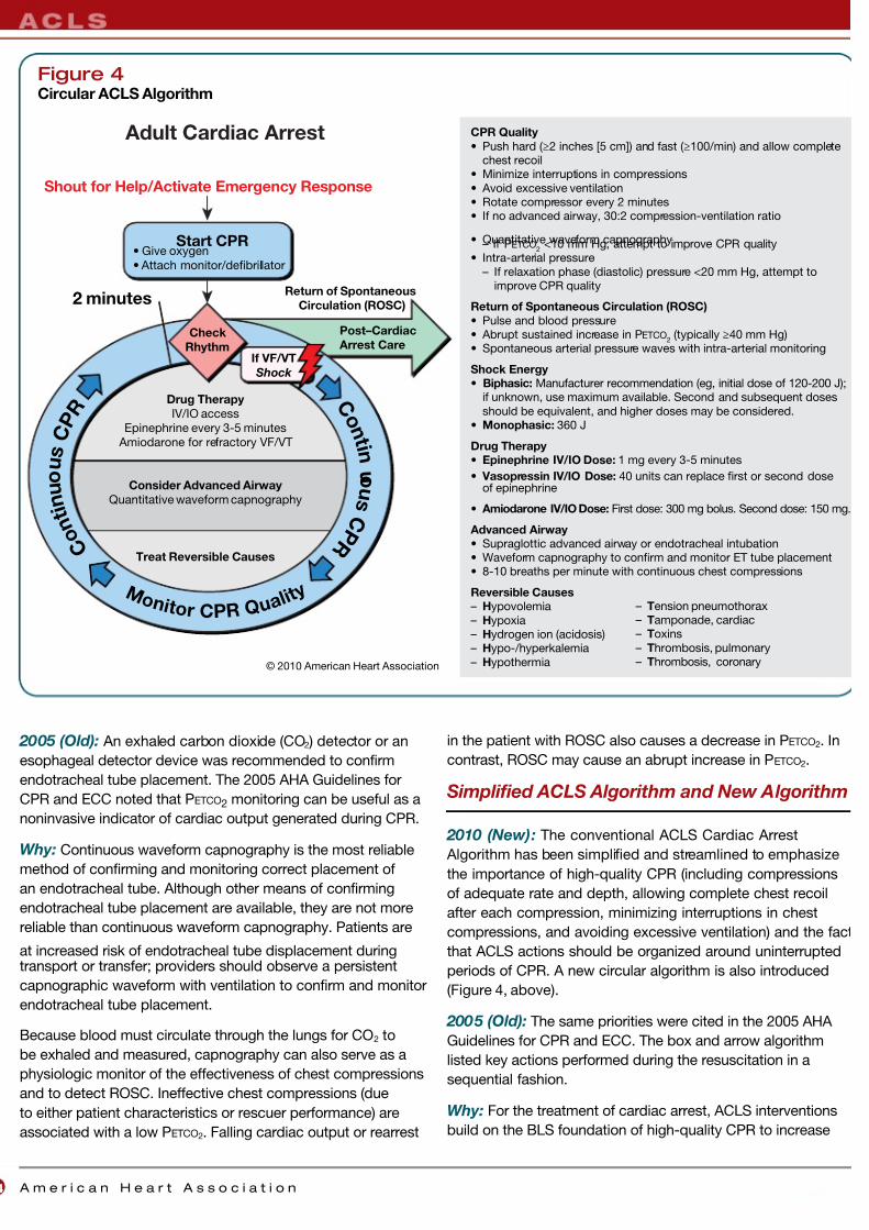
Simplified ACLS Algorithm and New Algorithm
2010 (New): The conventional ACLS Cardiac Arrest
Algorithm has been simplified and streamlined to emphasize
the importance of high-quality CPR (including compressions
of adequate rate and depth, allowing complete chest recoil
after each compression, minimizing interruptions in chest
compressions, and avoiding excessive ventilation) and the fact
that ACLS actions should be organized around uninterrupted
periods of CPR. A new circular algorithm is also introduced
(Figure 4, above).
2005 (Old):
The same priorities were cited in the 2005 AHA
Guidelines for CPR and ECC. The box and arrow algorithm
listed key actions performed during the resuscitation in a
sequential fashion.
Why: For the treatment of cardiac arrest, ACLS interventions
build on the BLS foundation of high-quality CPR to increase
© 2010 American Heart Association
CPR Quality
• Push hard (≥2 inches [5 cm]) and fast (≥100/min) and allow completechest recoil
• Minimize interruptions in compressions
• Avoid excessive ventilation• Rotate compressor every 2 minutes• If no advanced airway, 30:2 compression-ventilation ratio
• Quantitative waveform capnography– If PETCO2 <10 mm Hg, attempt to improve CPR quality
• Intra-arterial pressure
– If relaxation phase (diastolic) pressure <20 mm Hg, attempt toimprove CPR quality
Return of Spontaneous Circulation (ROSC)
• Pulse and blood pressure• Abrupt sustained increase in PETCO
2 (typically ≥40 mm Hg)
• Spontaneous arterial pressure waves with intra-arterial monitoring
Shock Energy
• Biphasic: Manufacturer recommendation (eg, initial dose of 120-200 J);if unknown, use maximum available. Second and subsequent doses
should be equivalent, and higher doses may be considered.• Monophasic: 360 J
Drug Therapy
• Epinephrine IV/IO Dose: 1 mg every 3-5 minutes
• Vasopressin IV/IO Dose: 40 units can replace first or second doseof epinephrine
• Amiodarone IV/IO Dose: First dose: 300 mg bolus. Second dose: 150 mg.
Advanced Airway
• Supraglottic advanced airway or endotracheal intubation• Waveform capnography to confirm and monitor ET tube placement• 8-10 breaths per minute with continuous chest compressions
Reversible Causes
– Hypovolemia
– Hypoxia– Hydrogen ion (acidosis)– Hypo-/hyperkalemia
– Hypothermia
Check
Rhythm
Drug Therapy
IV/IO access
Epinephrine every 3-5 minutes
Amiodarone for refractory VF/VT
Consider Advanced Airway
Quantitative waveform capnography
Treat Reversible Causes
Adult Cardiac Arrest
Post–Cardiac
Arrest Care
Start CPR• Give oxygen
• Attach monitor/defibrillator
2 minutes
If VF/VT
Shock
Return of Spontaneous
Circulation (ROSC)
Shout for Help/Activate Emergency Response
C o n
t i n u o u s
C P R
C o n
t i n
u o u s
C P
R
M o n i t o r
CP R
Q u a l i t y – Tension pneumothorax
– Tamponade, cardiac– Toxins
– Thrombosis, pulmonary– Thrombosis, coronary
Figure 4Circular ACLS Algorithm
7/26/2019 Algoritma ACLS 2010 OK
http://slidepdf.com/reader/full/algoritma-acls-2010-ok 17/32
Highlights of the 2010 AHA Guidelines for CPR and ECC
the likelihood of ROSC. Before 2005, ACLS courses assumed
that excellent CPR was provided, and they focused mainly
on added interventions of manual defibrillation, drug therapy,
and advanced airway management, as well as alternative
and additional management options for special resuscitation
situations. Although adjunctive drug therapy and advanced
airway management are still part of ACLS, in 2005 the
emphasis in advanced life support (ALS) returned to the basics,
with an increased emphasis on what is known to work: high-
quality CPR (providing compressions of adequate rate anddepth, allowing complete chest recoil after each compression,
minimizing interruptions in chest compressions, and avoiding
excessive ventilation). The 2010 AHA Guidelines for CPR and
ECC continue this emphasis. The 2010 AHA Guidelines for
CPR and ECC note that CPR is ideally guided by physiologic
monitoring and includes adequate oxygenation and early
defibrillation while the ACLS provider assesses and treats
possible underlying causes of the arrest. There is no definitive
clinical evidence that early intubation or drug therapy improves
neurologically intact survival to hospital discharge.
De-emphasis of Devices, Drugs, and
Other Distracters
Both ACLS algorithms use simple formats that focus on
interventions that have the greatest impact on outcome. To
that end, emphasis has been placed on delivery of high-quality
CPR and early defibrillation for VF/pulseless VT. Vascular
access, drug delivery, and advanced airway placement, while
still recommended, should not cause significant interruptions in
chest compressions and should not delay shocks.
New Medication Protocols
2010 (New): Atropine is not recommended for routine use in
the management of PEA/asystole and has been removed from
the ACLS Cardiac Arrest Algorithm. The treatment of PEA/
asystole is now consistent in the ACLS and pediatric advanced
life support (PALS) recommendations and algorithms.
The algorithm for treatment of tachycardia with pulses has been
simplified. Adenosine is recommended in the initial diagnosis
and treatment of stable, undifferentiated regular, monomorphic
wide-complex tachycardia (this is also consistent in ACLS and
PALS recommendations). It is important to note that adenosine
should not be used for irregular wide-complex tachycardias
because it may cause degeneration of the rhythm to VF.
For the treatment of the adult with symptomatic and unstable
bradycardia, chronotropic drug infusions are recommended as
an alternative to pacing.
2005 (Old): Atropine was included in the ACLS Pulseless
Arrest Algorithm: for a patient in asystole or slow PEA, atropine
could be considered. In the Tachycardia Algorithm, adenosine
was recommended only for suspected regular narrow-complex
reentry supraventricular tachycardia. In the Bradycardia
Algorithm, chronotropic drug infusions were listed in the
algorithm after atropine and while awaiting a pacer or if pacing
was ineffective.
Why: There are several important changes regarding
management of symptomatic arrhythmias in adults. Available
evidence suggests that the routine use of atropine during PEA or
asystole is unlikely to have a therapeutic benefit. For this reason,
atropine has been removed from the Cardiac Arrest Algorithm.
On the basis of new evidence of safety and potential efficacy,
adenosine can now be considered in the initial assessmentand treatment of stable, undifferentiated regular, monomorphic
wide-complex tachycardia when the rhythm is regular. For
symptomatic or unstable bradycardia, intravenous (IV) infusion
of chronotropic agents is now recommended as an equally
effective alternative to external transcutaneous pacing when
atropine is ineffective.
Organized Post–Cardiac Arrest Care
2010 (New): Post–Cardiac Arrest Care is a new section
in the 2010 AHA Guidelines for CPR and ECC. To improve
survival for victims of cardiac arrest who are admitted to ahospital after ROSC, a comprehensive, structured, integrated,
multidisciplinary system of post–cardiac arrest care should be
implemented in a consistent manner (Box 3). Treatment should
include cardiopulmonary and neurologic support. Therapeutic
hypothermia and percutaneous coronary interventions (PCIs)
should be provided when indicated (see also Acute Coronary
Syndromes section). Because seizures are common after
cardiac arrest, an electroencephalogram for the diagnosis
of seizures should be performed with prompt interpretation
as soon as possible and should be monitored frequently or
continuously in comatose patients after ROSC.
2005 (Old): Post–cardiac arrest care was included withinthe ACLS section of the 2005 AHA Guidelines for CPR
and ECC. Therapeutic hypothermia was recommended to
improve outcome for comatose adult victims of witnessed
out-of-hospital cardiac arrest when the presenting rhythm
was VF. In addition, recommendations were made to optimize
hemodynamic, respiratory, and neurologic support, identify
and treat reversible causes of arrest, monitor temperature,
and consider treatment for disturbances in temperature
regulation. However, there was limited evidence to support
these recommendations.
Why: Since 2005, two nonrandomized studies with concurrent
controls and other studies using historic controls have
indicated the possible benefit of therapeutic hypothermia
after in-hospital cardiac arrest and out-of-hospital cardiac
arrest with PEA/asystole as the presenting rhythm. Organized
post–cardiac arrest care with an emphasis on multidisciplinary
programs that focus on optimizing hemodynamic, neurologic,
and metabolic function (including therapeutic hypothermia)
may improve survival to hospital discharge among victims who
achieve ROSC after cardiac arrest either in or out of hospital.
Although it is not yet possible to determine the individual effect
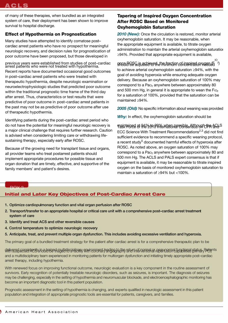
7/26/2019 Algoritma ACLS 2010 OK
http://slidepdf.com/reader/full/algoritma-acls-2010-ok 18/32
A m e r i c a n H e a r t A s s o c i a t i o n6
of many of these therapies, when bundled as an integrated
system of care, their deployment has been shown to improve
survival to hospital discharge.
Effect of Hypothermia on Prognostication
Many studies have attempted to identify comatose post–
cardiac arrest patients who have no prospect for meaningful
neurologic recovery, and decision rules for prognostication of
poor outcome have been proposed, but those developed in
previous years were established from studies of post–cardiacarrest patients who were not treated with hypothermia.
Recent reports have documented occasional good outcomes
in post–cardiac arrest patients who were treated with
therapeutic hypothermia, despite neurologic examination or
neuroelectrophysiologic studies that predicted poor outcome
within the traditional prognostic time frame of the third day
after arrest. Thus, characteristics or test results that were
predictive of poor outcome in post–cardiac arrest patients in
the past may not be as predictive of poor outcome after use
of therapeutic hypothermia.
Identifying patients during the post–cardiac arrest period who
do not have the potential for meaningful neurologic recovery is
a major clinical challenge that requires further research. Caution
is advised when considering limiting care or withdrawing life-
sustaining therapy, especially early after ROSC.
Because of the growing need for transplant tissue and organs,
all provider teams who treat postarrest patients should
implement appropriate procedures for possible tissue and
organ donation that are timely, effective, and supportive of the
family members’ and patient’s desires.
Tapering of Inspired Oxygen Concentration
After ROSC Based on Monitored
Oxyhemoglobin Saturation
2010 (New): Once the circulation is restored, monitor arterial
oxyhemoglobin saturation. It may be reasonable, when
the appropriate equipment is available, to titrate oxygen
administration to maintain the arterial oxyhemoglobin saturation
≥94%. Provided that appropriate equipment is available,
once ROSC is achieved, the fraction of inspired oxygen (FIO
2 )should be adjusted to the minimum concentration needed
to achieve arterial oxyhemoglobin saturation ≥94%, with the
goal of avoiding hyperoxia while ensuring adequate oxygen
delivery. Because an oxyhemoglobin saturation of 100% may
correspond to a PaO2 anywhere between approximately 80
and 500 mm Hg, in general it is appropriate to wean the FIO2
for a saturation of 100%, provided that the saturation can be
maintained ≥94%.
2005 (Old): No specific information about weaning was provided
Why: In effect, the oxyhemoglobin saturation should be
maintained at 94% to 99% when possible. Although the ACLSTask Force of the 2010 International Consensus on CPR and
ECC Science With Treatment Recommendations2,3 did not find
sufficient evidence to recommend a specific weaning protocol,
a recent study5 documented harmful effects of hyperoxia after
ROSC. As noted above, an oxygen saturation of 100% may
correspond to a PaO2 anywhere between approximately 80 and
500 mm Hg. The ACLS and PALS expert consensus is that if
equipment is available, it may be reasonable to titrate inspired
oxygen on the basis of monitored oxyhemoglobin saturation to
maintain a saturation of ≥94% but <100%.
Initial and Later Key Objectives of Post–Cardiac Arrest Care
1. Optimize cardiopulmonary function and vital organ perfusion after ROSC
2. Transport/transfer to an appropriate hospital or critical care unit with a comprehensive post–cardiac arrest treatment
system of care
3. Identify and treat ACS and other reversible causes
4. Control temperature to optimize neurologic recovery
5. Anticipate, treat, and prevent multiple organ dysfunction. This includes avoiding excessive ventilation and hyperoxia.
The primary goal of a bundled treatment strategy for the patient after cardiac arrest is for a comprehensive therapeutic plan to be
delivered consistently in a trained multidisciplinary environment leading to the return of normal or near-normal functional status. Patientswith suspected ACS should be triaged to a facility with coronary angiography and interventional reperfusion capabilities (primary PCI)
and a multidisciplinary team experienced in monitoring patients for multiorgan dysfunction and initiating timely appropriate post–cardiac
arrest therapy, including hypothermia.
With renewed focus on improving functional outcome, neurologic evaluation is a key component in the routine assessment of
survivors. Early recognition of potentially treatable neurologic disorders, such as seizures, is important. The diagnosis of seizures
may be challenging, especially in the setting of hypothermia and neuromuscular blockade, and electroencephalographic monitoring has
become an important diagnostic tool in this patient population.
Prognostic assessment in the setting of hypothermia is changing, and experts qualified in neurologic assessment in this patient
population and integration of appropriate prognostic tools are essential for patients, caregivers, and families.
BOX 3

7/26/2019 Algoritma ACLS 2010 OK
http://slidepdf.com/reader/full/algoritma-acls-2010-ok 19/32
Special Resuscitation Situations
2010 (New): Fifteen specific cardiac arrest situations now
have specific treatment recommendations. The topics reviewed
include asthma, anaphylaxis, pregnancy, morbid obesity (new),
pulmonary embolism (new), electrolyte imbalance, ingestion of
toxic substances, trauma, accidental hypothermia, avalanche
(new), drowning, electric shock/lightning strikes, PCI (new),
cardiac tamponade (new), and cardiac surgery (new).
2005 (Old): Ten specific situations related to patient
compromise (ie, periarrest conditions) were included.
Why: Cardiac arrest in special situations may require special
treatments or procedures beyond those provided during
normal BLS or ACLS. These conditions occur infrequently,
so it is difficult to conduct randomized clinical trials to
compare therapies. As a result, these unique situations call
for experienced providers to go beyond basics, using clinical
consensus and extrapolation from limited evidence. The topics
covered in the 2005 AHA Guidelines for CPR and ECC have
been reviewed, updated, and expanded to 15 specific cardiac
arrest situations. Topics include significant periarrest treatment
that may be important to prevent cardiac arrest or that require
treatment beyond the routine or typical care defined in the BLS
and ACLS guidelines.
ACUTE CORONARY SYNDROMES
Summary of Key Issues and Major Changes
The 2010 AHA Guidelines for CPR and ECC recommendationsfor the evaluation and management of acute coronary syndromes
(ACS) have been updated to define the scope of treatment for
healthcare providers who care for patients with suspected or
definite ACS within the first hours after onset of symptoms.
The primary goals of therapy for patients with ACS are
consistent with those in previous AHA Guidelines for CPR and
ECC and AHA/American College of Cardiology Guidelines,
which include
• Reducing the amount of myocardial necrosis that occurs in
patients with acute myocardial infarction, thus preserving
left ventricular function, preventing heart failure, and limitingother cardiovascular complications
• Preventing major adverse cardiac events: death, nonfatal
myocardial infarction, and the need for urgent revascularization
• Treating acute, life-threatening complications of ACS,
such as VF, pulseless VT, unstable tachycardias, and
symptomatic bradycardias
Within this context, several important strategies and
components of care are defined.
Systems of Care for Patients With ST-Segment
Elevation Myocardial Infarction
A well-organized approach to ST-segment elevation myocardia
infarction (STEMI) care requires integration of community, EMS
physician, and hospital resources in a bundled STEMI system
of care. This includes educational programs for recognition
of ACS symptoms, development of EMS protocols for initial
call center instruction and out-of-hospital intervention, and
emergency department (ED) and hospital-based programs for
intrafacility and interfacility transport once ACS is diagnosed
and definitive care is determined.
Out-of-Hospital 12-Lead ECGs
An important and key component of STEMI systems of care
is the performance of out-of-hospital 12-lead ECGs with
transmission or interpretation by EMS providers and with
advance notification of the receiving facility. Use of out-
of-hospital 12-lead ECGs has been recommended by the
AHA Guidelines for CPR and ECC since 2000 and has been
documented to reduce time to reperfusion with fibrinolytictherapy. More recently, out-of-hospital 12-lead ECGs have
also been shown to reduce the time to primary PCI and can
facilitate triage to specific hospitals when PCI is the chosen
strategy. When EMS or ED physicians activate the cardiac
care team, including the cardiac catheterization laboratory,
significant reductions in reperfusion times are observed.
Triage to Hospitals Capable of
Performing PCI
These recommendations provide criteria for triage of patients to
PCI centers after cardiac arrest.
Comprehensive Care for Patients After Cardiac
Arrest With Confirmed STEMI
or Suspected ACS
The performance of PCI has been associated with favorable
outcomes in adult patients resuscitated from cardiac arrest. It
is reasonable to include cardiac catheterization in standardized
post–cardiac arrest protocols as part of an overall strategy
to improve neurologically intact survival in this patient group.
In patients with out-of-hospital cardiac arrest due to VF,
emergent angiography with prompt revascularization of the
infarct-related artery is recommended. The ECG may be
insensitive or misleading after cardiac arrest, and coronary
angiography after ROSC in subjects with arrest of presumed
ischemic cardiac etiology may be reasonable, even in the
absence of a clearly defined STEMI. Clinical findings of coma
in patients before PCI are common after out-of-hospital
cardiac arrest and should not be a contraindication to
consideration of immediate angiography and PCI (see also
Post–Cardiac Arrest Care section).
Highlights of the 2010 AHA Guidelines for CPR and ECC
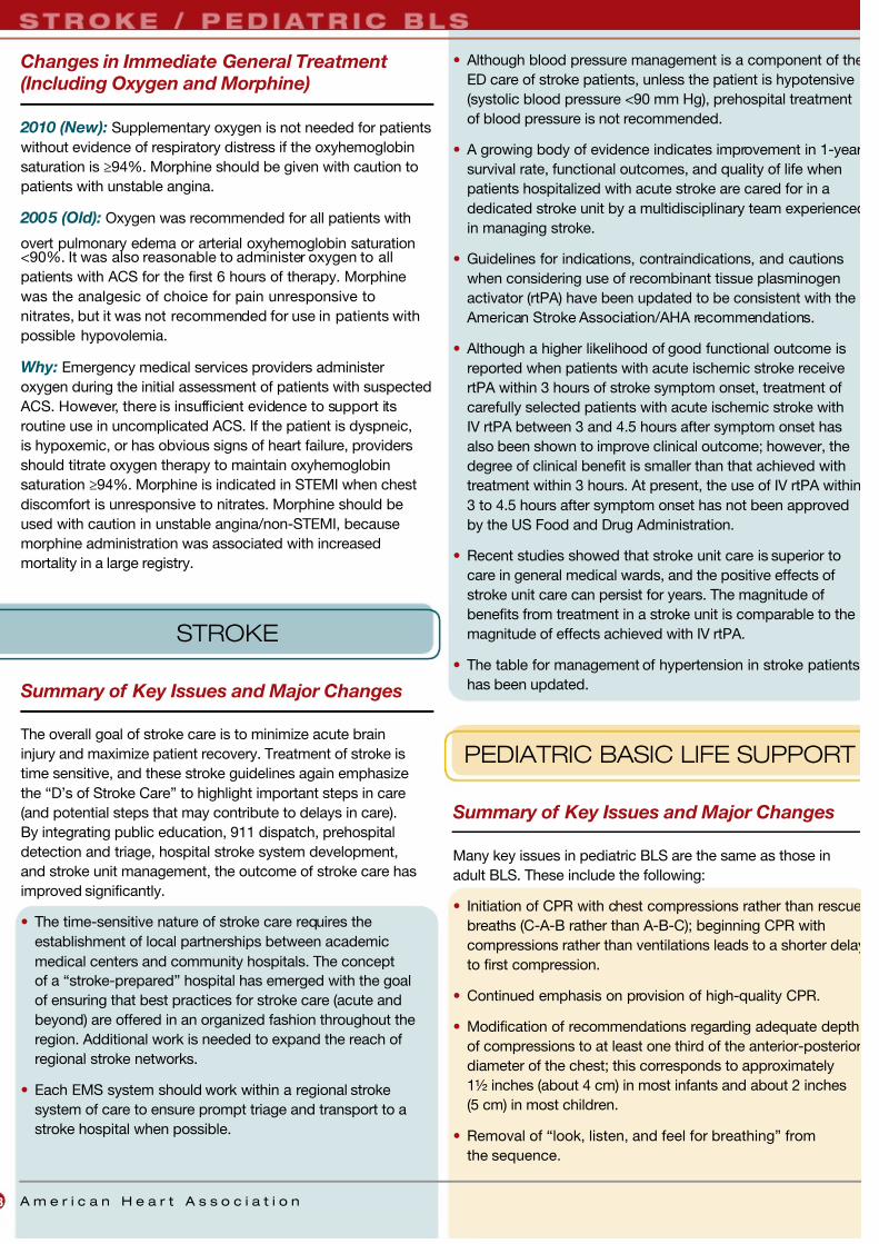
7/26/2019 Algoritma ACLS 2010 OK
http://slidepdf.com/reader/full/algoritma-acls-2010-ok 20/32
A m e r i c a n H e a r t A s s o c i a t i o n8
Changes in Immediate General Treatment
(Including Oxygen and Morphine)
2010 (New): Supplementary oxygen is not needed for patients
without evidence of respiratory distress if the oxyhemoglobin
saturation is ≥94%. Morphine should be given with caution to
patients with unstable angina.
2005 (Old): Oxygen was recommended for all patients with
overt pulmonary edema or arterial oxyhemoglobin saturation<90%. It was also reasonable to administer oxygen to all
patients with ACS for the first 6 hours of therapy. Morphine
was the analgesic of choice for pain unresponsive to
nitrates, but it was not recommended for use in patients with
possible hypovolemia.
Why: Emergency medical services providers administer
oxygen during the initial assessment of patients with suspected
ACS. However, there is insufficient evidence to support its
routine use in uncomplicated ACS. If the patient is dyspneic,
is hypoxemic, or has obvious signs of heart failure, providers
should titrate oxygen therapy to maintain oxyhemoglobin
saturation ≥94%. Morphine is indicated in STEMI when chest
discomfort is unresponsive to nitrates. Morphine should be
used with caution in unstable angina/non-STEMI, because
morphine administration was associated with increased
mortality in a large registry.
STROKE
Summary of Key Issues and Major Changes
The overall goal of stroke care is to minimize acute brain
injury and maximize patient recovery. Treatment of stroke is
time sensitive, and these stroke guidelines again emphasize
the “D’s of Stroke Care” to highlight important steps in care
(and potential steps that may contribute to delays in care).
By integrating public education, 911 dispatch, prehospital
detection and triage, hospital stroke system development,
and stroke unit management, the outcome of stroke care has
improved significantly.
• The time-sensitive nature of stroke care requires the
establishment of local partnerships between academic
medical centers and community hospitals. The concept
of a “stroke-prepared” hospital has emerged with the goal
of ensuring that best practices for stroke care (acute and
beyond) are offered in an organized fashion throughout the
region. Additional work is needed to expand the reach of
regional stroke networks.
• Each EMS system should work within a regional stroke
system of care to ensure prompt triage and transport to a
stroke hospital when possible.
• Although blood pressure management is a component of the
ED care of stroke patients, unless the patient is hypotensive
(systolic blood pressure <90 mm Hg), prehospital treatment
of blood pressure is not recommended.
• A growing body of evidence indicates improvement in 1-year
survival rate, functional outcomes, and quality of life when
patients hospitalized with acute stroke are cared for in a
dedicated stroke unit by a multidisciplinary team experienced
in managing stroke.
• Guidelines for indications, contraindications, and cautions
when considering use of recombinant tissue plasminogen
activator (rtPA) have been updated to be consistent with the
American Stroke Association/AHA recommendations.
• Although a higher likelihood of good functional outcome is
reported when patients with acute ischemic stroke receive
rtPA within 3 hours of stroke symptom onset, treatment of
carefully selected patients with acute ischemic stroke with
IV rtPA between 3 and 4.5 hours after symptom onset has
also been shown to improve clinical outcome; however, the
degree of clinical benefit is smaller than that achieved with
treatment within 3 hours. At present, the use of IV rtPA within
3 to 4.5 hours after symptom onset has not been approved
by the US Food and Drug Administration.
• Recent studies showed that stroke unit care is superior to
care in general medical wards, and the positive effects of
stroke unit care can persist for years. The magnitude of
benefits from treatment in a stroke unit is comparable to the
magnitude of effects achieved with IV rtPA.
• The table for management of hypertension in stroke patients
has been updated.
PEDIATRIC BASIC LIFE SUPPORT
Summary of Key Issues and Major Changes
Many key issues in pediatric BLS are the same as those in
adult BLS. These include the following:
• Initiation of CPR with chest compressions rather than rescue
breaths (C-A-B rather than A-B-C); beginning CPR with
compressions rather than ventilations leads to a shorter delay
to first compression.
• Continued emphasis on provision of high-quality CPR.
• Modification of recommendations regarding adequate depth
of compressions to at least one third of the anterior-posterior
diameter of the chest; this corresponds to approximately
1½ inches (about 4 cm) in most infants and about 2 inches
(5 cm) in most children.
• Removal of “look, listen, and feel for breathing” from
the sequence.
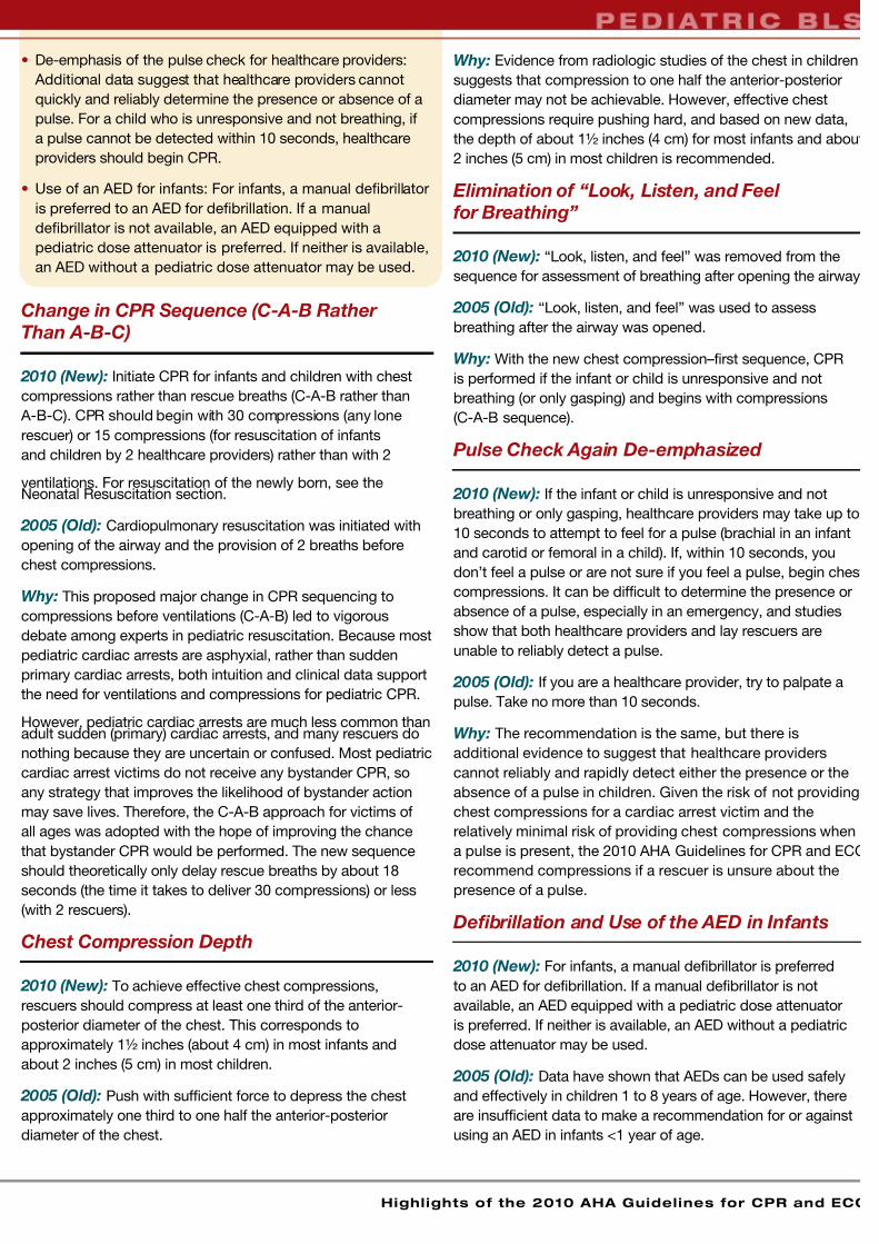
7/26/2019 Algoritma ACLS 2010 OK
http://slidepdf.com/reader/full/algoritma-acls-2010-ok 21/32
Highlights of the 2010 AHA Guidelines for CPR and ECC
• De-emphasis of the pulse check for healthcare providers:
Additional data suggest that healthcare providers cannot
quickly and reliably determine the presence or absence of a
pulse. For a child who is unresponsive and not breathing, if
a pulse cannot be detected within 10 seconds, healthcare
providers should begin CPR.
• Use of an AED for infants: For infants, a manual defibrillator
is preferred to an AED for defibrillation. If a manual
defibrillator is not available, an AED equipped with a
pediatric dose attenuator is preferred. If neither is available,
an AED without a pediatric dose attenuator may be used.
Change in CPR Sequence (C-A-B Rather
Than A-B-C)
2010 (New): Initiate CPR for infants and children with chest
compressions rather than rescue breaths (C-A-B rather than
A-B-C). CPR should begin with 30 compressions (any lone
rescuer) or 15 compressions (for resuscitation of infants
and children by 2 healthcare providers) rather than with 2
ventilations. For resuscitation of the newly born, see theNeonatal Resuscitation section.
2005 (Old): Cardiopulmonary resuscitation was initiated with
opening of the airway and the provision of 2 breaths before
chest compressions.
Why: This proposed major change in CPR sequencing to
compressions before ventilations (C-A-B) led to vigorous
debate among experts in pediatric resuscitation. Because most
pediatric cardiac arrests are asphyxial, rather than sudden
primary cardiac arrests, both intuition and clinical data support
the need for ventilations and compressions for pediatric CPR.
However, pediatric cardiac arrests are much less common thanadult sudden (primary) cardiac arrests, and many rescuers do
nothing because they are uncertain or confused. Most pediatric
cardiac arrest victims do not receive any bystander CPR, so
any strategy that improves the likelihood of bystander action
may save lives. Therefore, the C-A-B approach for victims of
all ages was adopted with the hope of improving the chance
that bystander CPR would be performed. The new sequence
should theoretically only delay rescue breaths by about 18
seconds (the time it takes to deliver 30 compressions) or less
(with 2 rescuers).
Chest Compression Depth
2010 (New): To achieve effective chest compressions,
rescuers should compress at least one third of the anterior-
posterior diameter of the chest. This corresponds to
approximately 1½ inches (about 4 cm) in most infants and
about 2 inches (5 cm) in most children.
2005 (Old): Push with sufficient force to depress the chest
approximately one third to one half the anterior-posterior
diameter of the chest.
Why: Evidence from radiologic studies of the chest in children
suggests that compression to one half the anterior-posterior
diameter may not be achievable. However, effective chest
compressions require pushing hard, and based on new data,
the depth of about 1½ inches (4 cm) for most infants and about
2 inches (5 cm) in most children is recommended.
Elimination of “Look, Listen, and Feel
for Breathing”
2010 (New): “Look, listen, and feel” was removed from the
sequence for assessment of breathing after opening the airway
2005 (Old): “Look, listen, and feel” was used to assess
breathing after the airway was opened.
Why: With the new chest compression–first sequence, CPR
is performed if the infant or child is unresponsive and not
breathing (or only gasping) and begins with compressions
(C-A-B sequence).
Pulse Check Again De-emphasized
2010 (New): If the infant or child is unresponsive and not
breathing or only gasping, healthcare providers may take up to
10 seconds to attempt to feel for a pulse (brachial in an infant
and carotid or femoral in a child). If, within 10 seconds, you
don’t feel a pulse or are not sure if you feel a pulse, begin chest
compressions. It can be difficult to determine the presence or
absence of a pulse, especially in an emergency, and studies
show that both healthcare providers and lay rescuers are
unable to reliably detect a pulse.
2005 (Old): If you are a healthcare provider, try to palpate a
pulse. Take no more than 10 seconds.
Why: The recommendation is the same, but there is
additional evidence to suggest that healthcare providers
cannot reliably and rapidly detect either the presence or the
absence of a pulse in children. Given the risk of not providing
chest compressions for a cardiac arrest victim and the
relatively minimal risk of providing chest compressions when
a pulse is present, the 2010 AHA Guidelines for CPR and ECC
recommend compressions if a rescuer is unsure about the
presence of a pulse.
Defibrillation and Use of the AED in Infants
2010 (New): For infants, a manual defibrillator is preferred
to an AED for defibrillation. If a manual defibrillator is not
available, an AED equipped with a pediatric dose attenuator
is preferred. If neither is available, an AED without a pediatric
dose attenuator may be used.
2005 (Old): Data have shown that AEDs can be used safely
and effectively in children 1 to 8 years of age. However, there
are insufficient data to make a recommendation for or against
using an AED in infants <1 year of age.

7/26/2019 Algoritma ACLS 2010 OK
http://slidepdf.com/reader/full/algoritma-acls-2010-ok 22/32
A m e r i c a n H e a r t A s s o c i a t i o n0
Why: Newer case reports suggest that an AED may be safe
and effective in infants. Because survival requires defibrillation
when a shockable rhythm is present during cardiac arrest,
delivery of a high-dose shock is preferable to no shock. Limited
evidence supports the safety of AED use in infants.
PEDIATRIC ADVANCED
LIFE SUPPORT
Summary of Key Issues and Major Changes
• Many key issues in the review of the PALS literature resulted
in refinement of existing recommendations rather than
new recommendations; new information is provided for
resuscitation of infants and children with selected congenital
heart defects and pulmonary hypertension.
• Monitoring capnography/capnometry is again recommended
to confirm proper endotracheal tube position and may
be useful during CPR to assess and optimize the quality of
chest compressions.
• The PALS cardiac arrest algorithm was simplified to
emphasize organization of care around 2-minute periods of
uninterrupted CPR.
• The initial defibrillation energy dose of 2 to 4 J/kg of either
monophasic or biphasic waveform is reasonable; for ease of
teaching, a dose of 2 J/kg may be used (this dose is the same
as in the 2005 recommendation). For second and subsequent
doses, give at least 4 J/kg. Doses higher than 4 J/kg (not
to exceed 10 J/kg or the adult dose) may also be safe and
effective, especially if delivered with a biphasic defibrillator.
• On the basis of increasing evidence of potential harm
from high oxygen exposure, a new recommendation has
been added to titrate inspired oxygen (when appropriate
equipment is available), once spontaneous circulation
has been restored, to maintain an arterial oxyhemoglobin
saturation ≥94% but <100% to limit the risk of hyperoxemia.
• New sections have been added on resuscitation of
infants and children with congenital heart defects,
including single ventricle, palliated single ventricle, and
pulmonary hypertension.
• Several recommendations for medications have been
revised. These include not administering calcium except in
very specific circumstances and limiting the use of etomidate
in septic shock.
• Indications for postresuscitation therapeutic hypothermia
have been clarified somewhat.
• New diagnostic considerations have been developed for
sudden cardiac death of unknown etiology.
• Providers are advised to seek expert consultation, if
possible, when administering amiodarone or procainamide
to hemodynamically stable patients with arrhythmias.
• The definition of wide-complex tachycardia has been
changed from >0.08 second to >0.09 second.
Recommendations for Monitoring Exhaled CO 2
2010 (New): Exhaled CO2 detection (capnography orcolorimetry) is recommended in addition to clinical assessment
to confirm tracheal tube position for neonates, infants, and
children with a perfusing cardiac rhythm in all settings (eg,
prehospital, ED, intensive care unit, ward, operating room) and
during intrahospital or interhospital transport (Figure 3A on
page 13). Continuous capnography or capnometry monitoring,
if available, may be beneficial during CPR to help guide therapy
especially the effectiveness of chest compressions (Figure 3B
on page 13).
2005 (Old): In infants and children with a perfusing rhythm,
use a colorimetric detector or capnography to detect exhaled
CO2 to confirm endotracheal tube position in the prehospital
and in-hospital settings and during intrahospital and
interhospital transport.
Why: Exhaled CO2 monitoring (capnography or colorimetry)
generally confirms placement of the endotracheal tube in
the airway and may more rapidly indicate endotracheal tube
misplacement/displacement than monitoring of oxyhemoglobin
saturation. Because patient transport increases the risk for
tube displacement, continuous CO2 monitoring is especially
important at these times.
Animal and adult studies show a strong correlation between
PETCO2 concentration and interventions that increase cardiac
output during CPR. PETCO2 values consistently <10 to 15 mm Hg
suggest that efforts should be focused on improving chest
compressions and making sure that ventilation is not
excessive. An abrupt and sustained rise in PETCO2 may be
observed just before clinical identification of ROSC, so use
of PETCO2 monitoring may reduce the need to interrupt chest
compressions for a pulse check.
Defibrillation Energy Doses
2010 (New): It is acceptable to use an initial dose of 2 to 4 J/kg
for defibrillation, but for ease of teaching, an initial dose of 2J/kg may be used. For refractory VF, it is reasonable to increase
the dose. Subsequent energy levels should be at least 4 J/kg,
and higher energy levels, not to exceed 10 J/kg or the adult
maximum dose, may be considered.
2005 (Old): With a manual defibrillator (monophasic or
biphasic), use a dose of 2 J/kg for the first attempt and 4 J/kg
for subsequent attempts.

7/26/2019 Algoritma ACLS 2010 OK
http://slidepdf.com/reader/full/algoritma-acls-2010-ok 23/32
Highlights of the 2010 AHA Guidelines for CPR and ECC
Why: More data are needed to identify the optimal energy
dose for pediatric defibrillation. Limited evidence is available
about effective or maximum energy doses for pediatric
defibrillation, but some data suggest that higher doses may be
safe and potentially more effective. Given the limited evidence
to support a change, the new recommendation is a minor
modification that allows higher doses up to the maximum dose
most experts believe is safe.
Limiting Oxygen to Normal Levels After Resuscitation
2010 (New): Once the circulation is restored, monitor
arterial oxyhemoglobin saturation. It may be reasonable,
when the appropriate equipment is available, to titrate
oxygen administration to maintain the arterial oxyhemoglobin
saturation ≥94%. Provided appropriate equipment is available,
once ROSC is achieved, adjust the FIO2 to the minimum
concentration needed to achieve arterial oxyhemoglobin
saturation ≥94%, with the goal of avoiding hyperoxia while
ensuring adequate oxygen delivery. Because an arterial
oxyhemoglobin saturation of 100% may correspond to aPaO2 anywhere between approximately 80 and 500 mm Hg, in
general it is appropriate to wean the FIO2 when the saturation is
100%, provided the saturation can be maintained ≥94%.
2005 (Old): Hyperoxia and the risk for reperfusion injury were
addressed in the 2005 AHA Guidelines for CPR and ECC in
general, but recommendations for titration of inspired oxygen
were not as specific.
Why: In effect, if equipment to titrate oxygen is available,
titrate oxygen to keep the oxyhemoglobin saturation 94%
to 99%. Data suggest that hyperoxemia (ie, a high PaO2 )
enhances the oxidative injury observed after ischemia-reperfusion such as occurs after resuscitation from cardiac
arrest. The risk of oxidative injury may be reduced by
titrating the FIO2 to reduce the PaO2 (this is accomplished by
monitoring arterial oxyhemoglobin saturation) while ensuring
adequate arterial oxygen content. Recent data from an adult
study5 demonstrated worse outcomes with hyperoxia after
resuscitation from cardiac arrest.
Resuscitation of Infants and Children With
Congenital Heart Disease
2010 (New): Specific resuscitation guidance has been
added for management of cardiac arrest in infants and
children with single-ventricle anatomy, Fontan or hemi-Fontan/
bidirectional Glenn physiology, and pulmonary hypertension.
2005 (Old): These topics were not addressed in the 2005 AHA
Guidelines for CPR and ECC.
Why: Specific anatomical variants with congenital heart
disease present unique challenges for resuscitation. The 2010
AHA Guidelines for CPR and ECC outline recommendations in
each of these clinical scenarios. Common to all scenarios is the
potential early use of extracorporeal membrane oxygenation as
rescue therapy in centers with this advanced capability.
Management of Tachycardia
2010 (New): Wide-complex tachycardia is present if the QRS
width is >0.09 second.
2005 (Old): Wide-complex tachycardia is present if the QRS
width is >0.08 second.
Why: In a recent scientific statement,6 QRS duration was
considered prolonged if it was >0.09 second for a child under the
age of 4 years, and ≥0.1 second was considered prolonged for
a child between the ages of 4 and 16 years. For this reason, the
PALS guidelines writing group concluded that it would be most
appropriate to consider a QRS width >0.09 second as prolonged
for the pediatric patient. Although the human eye is not likely to
appreciate a difference of 0.01 second, a computer interpretation
of the ECG can document the QRS width in milliseconds.
Medications During Cardiac Arrest and Shock
2010 (New): The recommendation regarding calcium
administration is stronger than in past AHA Guidelines: routine
calcium administration is not recommended for pediatric
cardiopulmonary arrest in the absence of documented
hypocalcemia, calcium channel blocker overdose,
hypermagnesemia, or hyperkalemia. Routine calcium
administration in cardiac arrest provides no benefit and
may be harmful.
Etomidate has been shown to facilitate endotracheal intubation
in infants and children with minimal hemodynamic effect but
is not recommended for routine use in pediatric patients withevidence of septic shock.
2005 (Old): Although the 2005 AHA Guidelines for CPR
and ECC noted that routine administration of calcium does
not improve the outcome of cardiac arrest, the words “is not
recommended” in the 2010 AHA Guidelines for CPR and ECC
provide a stronger statement and indicate potential harm.
Etomidate was not addressed in the 2005 AHA Guidelines for
CPR and ECC.
Why: Stronger evidence against the use of calcium during
cardiopulmonary arrest resulted in increased emphasis on
avoiding the routine use of this drug except for patients withdocumented hypocalcemia, calcium channel blocker overdose
hypermagnesemia, or hyperkalemia.
Evidence of potential harm from the use of etomidate
in both adults and children with septic shock led to the
recommendation to avoid its routine use in this setting.
Etomidate causes adrenal suppression, and the endogenous
steroid response may be critically important in patients with
septic shock.
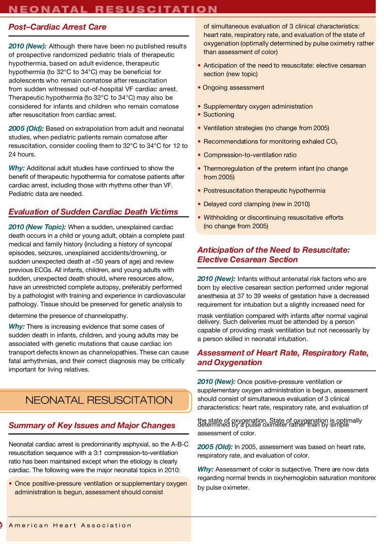
7/26/2019 Algoritma ACLS 2010 OK
http://slidepdf.com/reader/full/algoritma-acls-2010-ok 24/32
A m e r i c a n H e a r t A s s o c i a t i o n
Post–Cardiac Arrest Care
2010 (New): Although there have been no published results
of prospective randomized pediatric trials of therapeutic
hypothermia, based on adult evidence, therapeutic
hypothermia (to 32°C to 34°C) may be beneficial for
adolescents who remain comatose after resuscitation
from sudden witnessed out-of-hospital VF cardiac arrest.
Therapeutic hypothermia (to 32°C to 34°C) may also be
considered for infants and children who remain comatose
after resuscitation from cardiac arrest.
2005 (Old): Based on extrapolation from adult and neonatal
studies, when pediatric patients remain comatose after
resuscitation, consider cooling them to 32°C to 34°C for 12 to
24 hours.
Why: Additional adult studies have continued to show the
benefit of therapeutic hypothermia for comatose patients after
cardiac arrest, including those with rhythms other than VF.
Pediatric data are needed.
Evaluation of Sudden Cardiac Death Victims
2010 (New Topic): When a sudden, unexplained cardiac
death occurs in a child or young adult, obtain a complete past
medical and family history (including a history of syncopal
episodes, seizures, unexplained accidents/drowning, or
sudden unexpected death at <50 years of age) and review
previous ECGs. All infants, children, and young adults with
sudden, unexpected death should, where resources allow,
have an unrestricted complete autopsy, preferably performed
by a pathologist with training and experience in cardiovascular
pathology. Tissue should be preserved for genetic analysis to
determine the presence of channelopathy.
Why: There is increasing evidence that some cases of
sudden death in infants, children, and young adults may be
associated with genetic mutations that cause cardiac ion
transport defects known as channelopathies. These can cause
fatal arrhythmias, and their correct diagnosis may be critically
important for living relatives.
NEONATAL RESUSCITATION
Summary of Key Issues and Major Changes
Neonatal cardiac arrest is predominantly asphyxial, so the A-B-C
resuscitation sequence with a 3:1 compression-to-ventilation
ratio has been maintained except when the etiology is clearly
cardiac. The following were the major neonatal topics in 2010:
• Once positive-pressure ventilation or supplementary oxygen
administration is begun, assessment should consist
of simultaneous evaluation of 3 clinical characteristics:
heart rate, respiratory rate, and evaluation of the state of
oxygenation (optimally determined by pulse oximetry rather
than assessment of color)
• Anticipation of the need to resuscitate: elective cesarean
section (new topic)
• Ongoing assessment
• Supplementary oxygen administration
• Suctioning
• Ventilation strategies (no change from 2005)
• Recommendations for monitoring exhaled CO2
• Compression-to-ventilation ratio
• Thermoregulation of the preterm infant (no change
from 2005)
• Postresuscitation therapeutic hypothermia
• Delayed cord clamping (new in 2010)
• Withholding or discontinuing resuscitative efforts
(no change from 2005)
Anticipation of the Need to Resuscitate:
Elective Cesarean Section
2010 (New): Infants without antenatal risk factors who are
born by elective cesarean section performed under regional
anesthesia at 37 to 39 weeks of gestation have a decreased
requirement for intubation but a slightly increased need for
mask ventilation compared with infants after normal vaginaldelivery. Such deliveries must be attended by a person
capable of providing mask ventilation but not necessarily by
a person skilled in neonatal intubation.
Assessment of Heart Rate, Respiratory Rate,
and Oxygenation
2010 (New): Once positive-pressure ventilation or
supplementary oxygen administration is begun, assessment
should consist of simultaneous evaluation of 3 clinical
characteristics: heart rate, respiratory rate, and evaluation of
the state of oxygenation. State of oxygenation is optimallydetermined by a pulse oximeter rather than by simple
assessment of color.
2005 (Old): In 2005, assessment was based on heart rate,
respiratory rate, and evaluation of color.
Why: Assessment of color is subjective. There are now data
regarding normal trends in oxyhemoglobin saturation monitored
by pulse oximeter.
2 A m e r i c a n H e a r t A s s o c i a t i o n
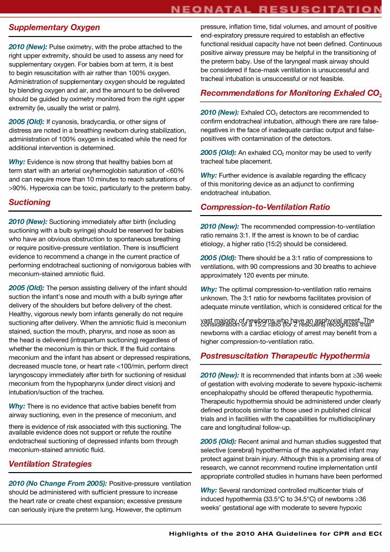
7/26/2019 Algoritma ACLS 2010 OK
http://slidepdf.com/reader/full/algoritma-acls-2010-ok 25/32
Highlights of the 2010 AHA Guidelines for CPR and ECC
Supplementary Oxygen
2010 (New): Pulse oximetry, with the probe attached to the
right upper extremity, should be used to assess any need for
supplementary oxygen. For babies born at term, it is best
to begin resuscitation with air rather than 100% oxygen.
Administration of supplementary oxygen should be regulated
by blending oxygen and air, and the amount to be delivered
should be guided by oximetry monitored from the right upper
extremity (ie, usually the wrist or palm).
2005 (Old): If cyanosis, bradycardia, or other signs of
distress are noted in a breathing newborn during stabilization,
administration of 100% oxygen is indicated while the need for
additional intervention is determined.
Why: Evidence is now strong that healthy babies born at
term start with an arterial oxyhemoglobin saturation of <60%
and can require more than 10 minutes to reach saturations of
>90%. Hyperoxia can be toxic, particularly to the preterm baby.
Suctioning
2010 (New): Suctioning immediately after birth (including
suctioning with a bulb syringe) should be reserved for babies
who have an obvious obstruction to spontaneous breathing
or require positive-pressure ventilation. There is insufficient
evidence to recommend a change in the current practice of
performing endotracheal suctioning of nonvigorous babies with
meconium-stained amniotic fluid.
2005 (Old): The person assisting delivery of the infant should
suction the infant’s nose and mouth with a bulb syringe after
delivery of the shoulders but before delivery of the chest.
Healthy, vigorous newly born infants generally do not require
suctioning after delivery. When the amniotic fluid is meconium
stained, suction the mouth, pharynx, and nose as soon as
the head is delivered (intrapartum suctioning) regardless of
whether the meconium is thin or thick. If the fluid contains
meconium and the infant has absent or depressed respirations,
decreased muscle tone, or heart rate <100/min, perform direct
laryngoscopy immediately after birth for suctioning of residual
meconium from the hypopharynx (under direct vision) and
intubation/suction of the trachea.
Why: There is no evidence that active babies benefit from
airway suctioning, even in the presence of meconium, and
there is evidence of risk associated with this suctioning. Theavailable evidence does not support or refute the routine
endotracheal suctioning of depressed infants born through
meconium-stained amniotic fluid.
Ventilation Strategies
2010 (No Change From 2005): Positive-pressure ventilation
should be administered with sufficient pressure to increase
the heart rate or create chest expansion; excessive pressure
can seriously injure the preterm lung. However, the optimum
pressure, inflation time, tidal volumes, and amount of positive
end-expiratory pressure required to establish an effective
functional residual capacity have not been defined. Continuous
positive airway pressure may be helpful in the transitioning of
the preterm baby. Use of the laryngeal mask airway should
be considered if face-mask ventilation is unsuccessful and
tracheal intubation is unsuccessful or not feasible.
Recommendations for Monitoring Exhaled CO 2
2010 (New): Exhaled CO2 detectors are recommended to
confirm endotracheal intubation, although there are rare false-
negatives in the face of inadequate cardiac output and false-
positives with contamination of the detectors.
2005 (Old): An exhaled CO2 monitor may be used to verify
tracheal tube placement.
Why: Further evidence is available regarding the efficacy
of this monitoring device as an adjunct to confirming
endotracheal intubation.
Compression-to-Ventilation Ratio
2010 (New): The recommended compression-to-ventilation
ratio remains 3:1. If the arrest is known to be of cardiac
etiology, a higher ratio (15:2) should be considered.
2005 (Old): There should be a 3:1 ratio of compressions to
ventilations, with 90 compressions and 30 breaths to achieve
approximately 120 events per minute.
Why: The optimal compression-to-ventilation ratio remains
unknown. The 3:1 ratio for newborns facilitates provision of
adequate minute ventilation, which is considered critical for the
vast majority of newborns who have an asphyxial arrest. Theconsideration of a 15:2 ratio (for 2 rescuers) recognizes that
newborns with a cardiac etiology of arrest may benefit from a
higher compression-to-ventilation ratio.
Postresuscitation Therapeutic Hypothermia
2010 (New): It is recommended that infants born at ≥36 weeks
of gestation with evolving moderate to severe hypoxic-ischemic
encephalopathy should be offered therapeutic hypothermia.
Therapeutic hypothermia should be administered under clearly
defined protocols similar to those used in published clinical
trials and in facilities with the capabilities for multidisciplinary
care and longitudinal follow-up.
2005 (Old): Recent animal and human studies suggested that
selective (cerebral) hypothermia of the asphyxiated infant may
protect against brain injury. Although this is a promising area of
research, we cannot recommend routine implementation until
appropriate controlled studies in humans have been performed
Why: Several randomized controlled multicenter trials of
induced hypothermia (33.5°C to 34.5°C) of newborns ≥36
weeks’ gestational age with moderate to severe hypoxic
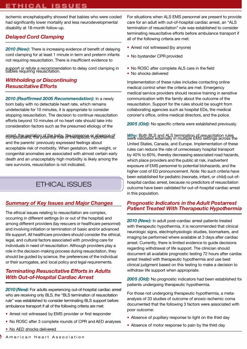
7/26/2019 Algoritma ACLS 2010 OK
http://slidepdf.com/reader/full/algoritma-acls-2010-ok 26/32
A m e r i c a n H e a r t A s s o c i a t i o n4
ischemic encephalopathy showed that babies who were cooled
had significantly lower mortality and less neurodevelopmental
disability at 18-month follow-up.
Delayed Cord Clamping
2010 (New): There is increasing evidence of benefit of delaying
cord clamping for at least 1 minute in term and preterm infants
not requiring resuscitation. There is insufficient evidence to
support or refute a recommendation to delay cord clamping inbabies requiring resuscitation.
Withholding or Discontinuing
Resuscitative Efforts
2010 (Reaffirmed 2005 Recommendation): In a newly
born baby with no detectable heart rate, which remains
undetectable for 10 minutes, it is appropriate to consider
stopping resuscitation. The decision to continue resuscitation
efforts beyond 10 minutes of no heart rate should take into
consideration factors such as the presumed etiology of the
arrest, the gestation of the baby, the presence or absence ofcomplications, the potential role of therapeutic hypothermia,
and the parents’ previously expressed feelings about
acceptable risk of morbidity. When gestation, birth weight, or
congenital anomalies are associated with almost certain early
death and an unacceptably high morbidity is likely among the
rare survivors, resuscitation is not indicated.
ETHICAL ISSUES
Summary of Key Issues and Major Changes
The ethical issues relating to resuscitation are complex,
occurring in different settings (in or out of the hospital) and
among different providers (lay rescuers or healthcare personnel)
and involving initiation or termination of basic and/or advanced
life support. All healthcare providers should consider the ethical,
legal, and cultural factors associated with providing care for
individuals in need of resuscitation. Although providers play a
role in the decision-making process during resuscitation, they
should be guided by science, the preferences of the individual
or their surrogates, and local policy and legal requirements.
Terminating Resuscitative Efforts in Adults
With Out-of-Hospital Cardiac Arrest
2010 (New): For adults experiencing out-of-hospital cardiac arrest
who are receiving only BLS, the “BLS termination of resuscitation
rule” was established to consider terminating BLS support before
ambulance transport if all of the following criteria are met:
• Arrest not witnessed by EMS provider or first responder
• No ROSC after 3 complete rounds of CPR and AED analyses
• No AED shocks delivered
For situations when ALS EMS personnel are present to provide
care for an adult with out-of-hospital cardiac arrest, an “ALS
termination of resuscitation” rule was established to consider
terminating resuscitative efforts before ambulance transport if
all of the following criteria are met:
• Arrest not witnessed (by anyone)
• No bystander CPR provided
• No ROSC after complete ALS care in the field
• No shocks delivered
Implementation of these rules includes contacting online
medical control when the criteria are met. Emergency
medical service providers should receive training in sensitive
communication with the family about the outcome of the
resuscitation. Support for the rules should be sought from
collaborating agencies such as hospital EDs, the medical
coroner’s office, online medical directors, and the police.
2005 (Old): No specific criteria were established previously.
Why: Both BLS and ALS termination of resuscitation ruleswere validated externally in multiple EMS settings across the
United States, Canada, and Europe. Implementation of these
rules can reduce the rate of unnecessary hospital transport
by 40% to 60%, thereby decreasing associated road hazards,
which place providers and the public at risk, inadvertent
exposure of EMS personnel to potential biohazards, and the
higher cost of ED pronouncement. Note: No such criteria have
been established for pediatric (neonate, infant, or child) out-of-
hospital cardiac arrest, because no predictors of resuscitation
outcome have been validated for out-of-hospital cardiac arrest
in this population.
Prognostic Indicators in the Adult Postarrest Patient Treated With Therapeutic Hypothermia
2010 (New): In adult post–cardiac arrest patients treated
with therapeutic hypothermia, it is recommended that clinical
neurologic signs, electrophysiologic studies, biomarkers, and
imaging be performed where available at 3 days after cardiac
arrest. Currently, there is limited evidence to guide decisions
regarding withdrawal of life support. The clinician should
document all available prognostic testing 72 hours after cardiac
arrest treated with therapeutic hypothermia and use best
clinical judgment based on this testing to make a decision to
withdraw life support when appropriate.
2005 (Old): No prognostic indicators had been established for
patients undergoing therapeutic hypothermia.
For those not undergoing therapeutic hypothermia, a meta-
analysis of 33 studies of outcome of anoxic-ischemic coma
documented that the following 3 factors were associated with
poor outcome:
• Absence of pupillary response to light on the third day
• Absence of motor response to pain by the third day
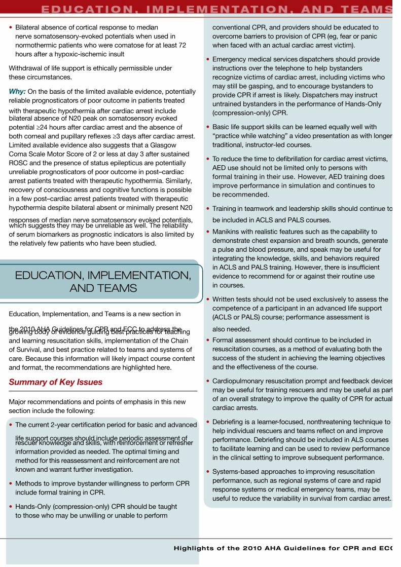
7/26/2019 Algoritma ACLS 2010 OK
http://slidepdf.com/reader/full/algoritma-acls-2010-ok 27/32
• Bilateral absence of cortical response to median
nerve somatosensory-evoked potentials when used in
normothermic patients who were comatose for at least 72
hours after a hypoxic-ischemic insult
Withdrawal of life support is ethically permissible under
these circumstances.
Why: On the basis of the limited available evidence, potentially
reliable prognosticators of poor outcome in patients treated
with therapeutic hypothermia after cardiac arrest includebilateral absence of N20 peak on somatosensory evoked
potential ≥24 hours after cardiac arrest and the absence of
both corneal and pupillary reflexes ≥3 days after cardiac arrest.
Limited available evidence also suggests that a Glasgow
Coma Scale Motor Score of 2 or less at day 3 after sustained
ROSC and the presence of status epilepticus are potentially
unreliable prognosticators of poor outcome in post–cardiac
arrest patients treated with therapeutic hypothermia. Similarly,
recovery of consciousness and cognitive functions is possible
in a few post–cardiac arrest patients treated with therapeutic
hypothermia despite bilateral absent or minimally present N20
responses of median nerve somatosensory evoked potentials,which suggests they may be unreliable as well. The reliability
of serum biomarkers as prognostic indicators is also limited by
the relatively few patients who have been studied.
EDUCATION, IMPLEMENTATION,
AND TEAMS
Education, Implementation, and Teams is a new section in
the 2010 AHA Guidelines for CPR and ECC to address thegrowing body of evidence guiding best practices for teaching
and learning resuscitation skills, implementation of the Chain
of Survival, and best practice related to teams and systems of
care. Because this information will likely impact course content
and format, the recommendations are highlighted here.
Summary of Key Issues
Major recommendations and points of emphasis in this new
section include the following:
• The current 2-year certification period for basic and advanced
life support courses should include periodic assessment ofrescuer knowledge and skills, with reinforcement or refresher
information provided as needed. The optimal timing and
method for this reassessment and reinforcement are not
known and warrant further investigation.
• Methods to improve bystander willingness to perform CPR
include formal training in CPR.
• Hands-Only (compression-only) CPR should be taught
to those who may be unwilling or unable to perform
conventional CPR, and providers should be educated to
overcome barriers to provision of CPR (eg, fear or panic
when faced with an actual cardiac arrest victim).
• Emergency medical services dispatchers should provide
instructions over the telephone to help bystanders
recognize victims of cardiac arrest, including victims who
may still be gasping, and to encourage bystanders to
provide CPR if arrest is likely. Dispatchers may instruct
untrained bystanders in the performance of Hands-Only
(compression-only) CPR.
• Basic life support skills can be learned equally well with
“practice while watching” a video presentation as with longer
traditional, instructor-led courses.
• To reduce the time to defibrillation for cardiac arrest victims,
AED use should not be limited only to persons with
formal training in their use. However, AED training does
improve performance in simulation and continues to
be recommended.
• Training in teamwork and leadership skills should continue to
be included in ACLS and PALS courses.
• Manikins with realistic features such as the capability to
demonstrate chest expansion and breath sounds, generate
a pulse and blood pressure, and speak may be useful for
integrating the knowledge, skills, and behaviors required
in ACLS and PALS training. However, there is insufficient
evidence to recommend for or against their routine use
in courses.
• Written tests should not be used exclusively to assess the
competence of a participant in an advanced life support
(ACLS or PALS) course; performance assessment is
also needed.
• Formal assessment should continue to be included in
resuscitation courses, as a method of evaluating both the
success of the student in achieving the learning objectives
and the effectiveness of the course.
• Cardiopulmonary resuscitation prompt and feedback devices
may be useful for training rescuers and may be useful as part
of an overall strategy to improve the quality of CPR for actual
cardiac arrests.
• Debriefing is a learner-focused, nonthreatening technique to
help individual rescuers and teams reflect on and improve
performance. Debriefing should be included in ALS courses
to facilitate learning and can be used to review performance
in the clinical setting to improve subsequent performance.
• Systems-based approaches to improving resuscitation
performance, such as regional systems of care and rapid
response systems or medical emergency teams, may be
useful to reduce the variability in survival from cardiac arrest.
Highlights of the 2010 AHA Guidelines for CPR and ECC

7/26/2019 Algoritma ACLS 2010 OK
http://slidepdf.com/reader/full/algoritma-acls-2010-ok 28/32
A m e r i c a n H e a r t A s s o c i a t i o n6
Two Years Is Too Long an Interval for Skills
Practice and Reassessment
2010 (New): Skill performance should be assessed during
the 2-year certification with reinforcement provided as needed.
The optimal timing and method for this reassessment and
reinforcement are not known.
Why: The quality of rescuer education and frequency of
retraining are critical factors in improving the effectiveness ofresuscitation. Ideally, retraining should not be limited to 2-year
intervals. More frequent renewal of skills is needed, with a
commitment to maintenance of certification similar to that
embraced by many healthcare-credentialing organizations.
Instructors and participants should be aware that successful
completion of any AHA ECC course is only the first step
toward attaining and maintaining competence. American
Heart Association ECC courses should be part of a larger
continuing education and continuous quality improvement
process that reflects the needs and practices of individuals and
systems. The best method to help rescuers maintain required
resuscitation skills is currently unknown.
Learning to Mastery
2010 (New): New CPR prompt and feedback devices may be
useful for training rescuers and as part of an overall strategy
to improve the quality of CPR in actual cardiac arrests and
resuscitations. Training for the complex combination of skills
required to perform adequate chest compressions should focus
on demonstrating mastery.
Why: Maintaining focus during CPR on the 3 characteristics of
rate, depth, and chest recoil while minimizing interruptions is
a complex challenge even for highly trained professionals andaccordingly must receive appropriate attention in training. The
2010 AHA Guidelines for CPR and ECC have placed renewed
emphasis on ensuring that chest compressions are performed
correctly. Training simply to “push hard and push fast” may not
be adequate to ensure excellent chest compressions. Use of
CPR prompt and feedback devices during training can improve
learning and retention.
Overcoming Barriers to Performance
2010 (New): Training should address barriers that interfere
with bystander willingness to attempt CPR.
Why: Many fears of potential rescuers can be alleviated by
education about actual risks to the resuscitation provider and to
the arrest victim. Education may help people previously trained
in BLS to be more likely to attempt resuscitation. Frequent
responses identified in studies of actual bystanders are fear and
panic, and training programs must identify methods to reduce
these responses. Emergency medical services dispatcher
instructions should identify and use methods that have proven
effective in educating and motivating potential providers to act.
Learning Teamwork Skills in ACLS and PALS
2010 (New): Advanced life support training should include
training in teamwork.
Why: Resuscitation skills are often performed simultaneously,
and healthcare providers must be able to work collaboratively
to minimize interruptions in chest compressions. Teamwork
and leadership skills continue to be important, particularly for
advanced courses that include ACLS and PALS providers.
AED Training Not Required for Use
2010 (New): Use of an AED does not require training, although
training does improve performance.
Why: Manikin-based studies have demonstrated that AEDs
can be operated correctly without prior training. Allowing the
use of AEDs by untrained bystanders can be beneficial and
may be lifesaving. Because even minimal training has been
shown to improve performance in simulated cardiac arrests,
training opportunities should be made available and promoted
for the lay rescuer.
Continuous Quality Improvement for
Resuscitation Programs
2010 (New): Resuscitation systems should establish ongoing
systems of care assessment and improvement.
Why: There is evidence of considerable regional variation in
the reported incidence and outcome of cardiac arrest in the
United States. This variation is further evidence of the need for
communities and systems to accurately identify each instance
of treated cardiac arrest and measure outcomes. It also
suggests additional opportunities for improving survival rates in
many communities.
Community and hospital-based resuscitation programs should
systematically monitor cardiac arrests, the level of resuscitation
care provided, and outcome. Continuous quality improvement
includes systematic evaluation and feedback, measurement
or benchmarking and interpretation, and efforts to optimize
resuscitation care and help to narrow the gaps between ideal
and actual resuscitation performance.
FIRST AID
The 2010 First Aid Guidelines were once again codeveloped by
the AHA and the American Red Cross (ARC). The 2010 AHA/ARC
Guidelines for First Aid are based on worksheets (topical
literature reviews) on selected topics, under the auspices of
an International First Aid Science Advisory Board made up of
representatives from 30 first aid organizations; this process is
different from that used for the ILCOR International Consensus

7/26/2019 Algoritma ACLS 2010 OK
http://slidepdf.com/reader/full/algoritma-acls-2010-ok 29/32
Highlights of the 2010 AHA Guidelines for CPR and ECC
on CPR and ECC Science With Treatment Recommendations
and was not part of the ILCOR process.
For the purposes of the 2010 AHA/ARC Guidelines for First
Aid, the International First Aid Science Advisory Board defined
first aid as the assessments and interventions that can be
performed by a bystander (or by the victim) with minimal or no
medical equipment. A first aid provider is defined as someone
with formal training in first aid, emergency care, or medicine
who provides first aid.
Summary of Key Issues and Major Changes
Key topics in the 2010 AHA/ARC Guidelines for First
Aid include
• Supplementary oxygen administration
• Epinephrine and anaphylaxis
• Aspirin administration for chest discomfort (new)
• Tourniquets and bleeding control
• Hemostatic agents (new)
• Snakebites
• Jellyfish stings (new)
• Heat emergencies
Topics covered in the 2010 Guidelines but with no new
recommendations since 2005 are the use of inhalers for
breathing difficulties, seizures, wounds and abrasions, burns
and burn blisters, spine stabilization, musculoskeletal injuries,
dental injuries, cold emergencies, and poison emergencies.
Supplementary Oxygen
2010 (No Change From 2005): Routine administration
of supplementary oxygen is not recommended as a first aid
measure for shortness of breath or chest discomfort.
2010 (New): Supplementary oxygen administration
should be considered as part of first aid for divers with
a decompression injury.
Why: As in 2005, no evidence was found that showed a benefit
of supplementary oxygen administration as a first aid measure
to victims with shortness of breath or chest discomfort.
Evidence was found (new for 2010) of a possible benefit ofsupplementary oxygen for divers with a decompression injury.
Epinephrine and Anaphylaxis
2010 (New): New in 2010 is the recommendation that
if symptoms of anaphylaxis persist despite epinephrine
administration, first aid providers should seek medical
assistance before administering a second dose of epinephrine.
2005 (Old): As in 2005, the 2010 AHA/ARC Guidelines for
First Aid recommend that first aid providers learn the signs and
symptoms of anaphylaxis and the proper use of an epinephrine
autoinjector so they can aid the victim.
Why: Epinephrine can be lifesaving for a victim of anaphylaxis,
but approximately 18% to 35% of victims who have the
signs and symptoms of anaphylaxis may require a second
dose of epinephrine. The diagnosis of anaphylaxis can be a
challenge, even for professionals, and excessive epinephrine
administration may produce complications (eg, worsening
of myocardial ischemia or arrhythmias) if given to patients
who do not have anaphylaxis (eg, if administered to a patient
with ACS). Therefore, the first aid provider is encouraged to
activate the EMS system before administering a second dose
of epinephrine.
Aspirin Administration for Chest Discomfort
2010 (New): First aid providers are encouraged to activate
the EMS system for anyone with chest discomfort. While
waiting for EMS to arrive, first aid providers should advise
the patient to chew 1 adult (non–enteric-coated) or 2 low-
dose “baby” aspirins if the patient has no history of allergy to
aspirin and no recent gastrointestinal bleeding.
Why: Aspirin is beneficial if the chest discomfort is due to an
ACS. It can be very difficult even for professionals to determine
whether chest discomfort is of cardiac origin. The administration
of aspirin must therefore never delay EMS activation.
Tourniquets and Bleeding Control
2010 (No Change From 2005): Because of the potential
adverse effects of tourniquets and difficulty in their proper
application, use of a tourniquet to control bleeding of the
extremities is indicated only if direct pressure is not effective
or possible and if the first aid provider has proper training in
tourniquet use.
Why: There has been a great deal of experience with using
tourniquets to control bleeding on the battlefield, and there
is no question that they work under proper circumstances
and with proper training. However, there are no data on
tourniquet use by first aid providers. The adverse effects of
tourniquets, which can include ischemia and gangrene of
the extremity, as well as shock and even death, appear to be
related to the amount of time tourniquets remain in place, and
their effectiveness is partially dependent on tourniquet type.
In general, specially designed tourniquets are better thanimprovised ones.
Hemostatic Agents
2010 (New): The routine use of hemostatic agents to control
bleeding as a first aid measure is not recommended at this time.
Why: Despite the fact that a number of hemostatic agents
have been effective in controlling bleeding, their use is not
recommended as a first aid method of bleeding control

7/26/2019 Algoritma ACLS 2010 OK
http://slidepdf.com/reader/full/algoritma-acls-2010-ok 30/32
A m e r i c a n H e a r t A s s o c i a t i o n8
because of significant variability in effectiveness and the
potential for adverse effects, including tissue destruction with
induction of a proembolic state and potential thermal injury.
Snakebites
2010 (New): Applying a pressure immobilization bandage
with a pressure between 40 and 70 mm Hg in the upper
extremity and between 55 and 70 mm Hg in the lower
extremity around the entire length of the bitten extremity is aneffective and safe way to slow lymph flow and therefore the
dissemination of venom.
2005 (Old): In 2005, use of pressure immobilization bandages
to slow the spread of the toxin was recommended only for
victims of bites by snakes with neurotoxic venom.
Why: Effectiveness of pressure immobilization has now also been
demonstrated for bites by other venomous American snakes.
Jellyfish Stings
2010 (New): To inactivate venom load and prevent furtherenvenomation, jellyfish stings should be liberally washed with
vinegar (4% to 6% acetic acid solution) as soon as possible
and for at least 30 seconds. After the nematocysts are removed
or deactivated, the pain from jellyfish stings should be treated
with hot-water immersion when possible.
Why: There are 2 actions necessary for treatment of jellyfish
stings: preventing further nematocyst discharge and pain relief.
A number of topical treatments have been used, but a critical
evaluation of the literature shows that vinegar is most effective
for inactivation of the nematocysts. Immersion with water, as
hot as tolerated for about 20 minutes, is most effective for
treating the pain.
Heat Emergencies
2010 (No Change From 2005): First aid for heat cramps
includes rest, cooling off, and drinking an electrolyte-
carbohydrate mixture that can include juice, milk, or a
commercial electrolyte-carbohydrate drink. Stretching, icing,
and massaging the painful muscles may be helpful. Heat
exhaustion must be vigorously treated by having the victim lie
down in a cool place, removing as many of the victim’s clothes
as possible, cooling, preferably by immersing the victim in cold
water, and activating EMS. Heat stroke requires emergencytreatment by EMS providers and will require treatment with IV
fluids. The first aid provider should not try to force the victim of
heat stroke to drink fluids.
Why: The 2010 AHA/ARC Guidelines for First Aid have divided
heat emergencies into 3 categories of increasing severity: heat
cramps, heat exhaustion, and, the most severe, heat stroke.
Signs of heat stroke include those of heat exhaustion plus
signs of central nervous system involvement. As a result, heat
stroke requires emergency care including IV fluid therapy.
SUMMARY
In the years since the publication of the 2005 AHA Guidelines
for CPR and ECC, many resuscitation systems and
communities have documented improved survival for victims
of cardiac arrest. However, too few victims of cardiac arrest
receive bystander CPR. We know that CPR quality must be
high and that victims require excellent post–cardiac arrest
care by organized teams with members who function welltogether. Education and frequent refresher training are likely
the keys to improving resuscitation performance. In this 50th
year since the publication of the landmark Kouwenhoven, Jude
and Knickerbocker description of successful closed chest
compression,4 we must all rededicate ourselves to improving
the frequency of bystander CPR and the quality of all CPR and
post–cardiac arrest care.
REFERENCES
1. Field JM, Hazinski MF, Sayre M, et al. Part 1: Executive Summary of
2010 AHA Guidelines for CPR and ECC. Circulation. In press.
2. Hazinski MF, Nolan JP, Billi JE, et al. Part 1: Executive Summary:
2010 International Consensus on Cardiopulmonary Resuscitation
and Emergency Cardiovascular Care Science With Treatment
Recommendations.Circulation. In press.
3. Nolan JP, Hazinski MF, Billi JE, et al. Part 1: Executive Summary:
2010 International Consensus on Cardiopulmonary Resuscitation
and Emergency Cardiovascular Care Science With Treatment
Recommendations.Resuscitation. In press.
4. Kouwenhoven WB, Jude JR, Knickerbocker GG. Closed-chest cardiac
massage. JAMA. 1960;173:1064-1067.
5. Kilgannon JH, Jones AE, Shapiro NI, et al. Association between arteria
hyperoxia following resuscitation from cardiac arrest and in-hospital
mortality. JAMA. 2010;303:2165-2171.
6. Surawicz B, Childers R, Deal BJ, et al. AHA/ACCF/HRS
Recommendations for the Standardization and Interpretation of the
Electrocardiogram, Part III: Intraventricular Conduction Disturbances.
Circulation. 2009;119:e235-e240.

7/26/2019 Algoritma ACLS 2010 OK
http://slidepdf.com/reader/full/algoritma-acls-2010-ok 31/32

7/26/2019 Algoritma ACLS 2010 OK
http://slidepdf.com/reader/full/algoritma-acls-2010-ok 32/32
For more information on
other American Heart Association
programs contact us:
877-AHA-4CPR
www.heart.org/cpr