Alginate microbeads are coagulation compatible, while … · Full length article Alginate...
10
Full length article Alginate microbeads are coagulation compatible, while alginate microcapsules activate coagulation secondary to complement or directly through FXII Caroline Gravastrand a , Shamal Hamad b , Hilde Fure b , Bjørg Steinkjer a , Liv Ryan a , Josè Oberholzer c , John D. Lambris d , Igor Lacík e , Tom Eirik Mollnes a,b,f,g,h , Terje Espevik a , Ole-Lars Brekke b,f , Anne Mari Rokstad a,i,j,⇑ a Centre of Molecular Inflammation Research, and Department of Cancer Research and Molecular Medicine, Norwegian University of Science and Technology, Trondheim, Norway b Research Laboratory, Nordland Hospital, 8092 Bodø, Norway c Department of Surgery/Division of Transplantation, University of Illinois at Chicago, IL, USA d Department of Pathology and Laboratory Medicine, University of Pennsylvania, Philadelphia, PA 19104, USA e Department for Biomaterials Research, Polymer Institute of the Slovak Academy of Sciences, Bratislava, Slovakia f Faculty of Health Sciences, K.G. Jebsen Thrombosis Research and Expertise Center, The Arctic University of Norway, Tromsø, 9037 Tromsø, Norway g Department of Immunology, Oslo University Hospital, Rikshospitalet, 0424 Oslo, Norway h K.G. Jebsen Inflammatory Research Center, University of Oslo, 0424 Oslo, Norway i Clinic of Surgery, Centre for Obesity, St. Olavs University Hospital, Trondheim, Norway j Central Norway Regional Health Authority, Norway article info Article history: Received 23 March 2017 Received in revised form 5 May 2017 Accepted 30 May 2017 Available online 30 May 2017 Keywords: Alginate microcapsules Coagulation Complement Cross-talk Factor XII Tissue factor abstract Alginate microspheres are presently under evaluation for future cell-based therapy. Their ability to induce harmful host reactions needs to be identified for developing the most suitable devices and effi- cient prevention strategies. We used a lepirudin based human whole blood model to investigate the coag- ulation potentials of alginate-based microspheres: alginate microbeads (Ca/Ba Beads), alginate poly-L- lysine microcapsules (APA and AP microcapsules) and sodium alginate-sodium cellulose sulfate-poly( methylene-co-cyanoguanidine) microcapsules (PMCG microcapsules). Coagulation activation measured by prothrombin fragments 1 + 2 (PTF1.2) was rapidly and markedly induced by the PMCG microcapsules, delayed and lower induced by the APA and AP microcapsules, and not induced by the Ca/Ba Beads. Monocytes tissue factor (TF) expression was similarly activated by the microcapsules, whereas not by the Ca/Ba Beads. PMCG microcapsules-induced PTF1.2 was abolished by FXII inhibition (corn trypsin inhi- bitor), thus pointing to activation through the contact pathway. PTF1.2 induced by the AP and APA micro- capsules was inhibited by anti-TF antibody, pointing to a TF driven coagulation. The TF induced coagulation was inhibited by the complement inhibitors compstatin (C3 inhibition) and eculizumab (C5 inhibition), revealing a complement-coagulation cross-talk. This is the first study on the coagulation potentials of alginate microspheres, and identifies differences in activation potential, pathways and pos- sible intervention points. Statement of significance Alginate microcapsules are prospective candidate materials for cell encapsulation therapy. The material surface must be free of host cell adhesion to ensure free diffusion of nutrition and oxygen to the encap- sulated cells. Coagulation activation is one gateway to cellular overgrowth through deposition of fibrin. Herein we used a physiologically relevant whole blood model to investigate the coagulation potential of alginate microcapsules and microbeads. The coagulation potentials and the pathways of activation were depending on the surface properties of the materials. Activation of the complement system could also be http://dx.doi.org/10.1016/j.actbio.2017.05.052 1742-7061/Ó 2017 Acta Materialia Inc. Published by Elsevier Ltd. All rights reserved. ⇑ Corresponding author at: Centre of Molecular Inflammation Research, and Department of Cancer Research and Molecular Medicine, Norwegian University of Science and Technology, Trondheim, Norway. E-mail address: [email protected] (A.M. Rokstad). Acta Biomaterialia 58 (2017) 158–167 Contents lists available at ScienceDirect Acta Biomaterialia journal homepage: www.elsevier.com/locate/actabiomat
Transcript of Alginate microbeads are coagulation compatible, while … · Full length article Alginate...
Acta Biomaterialia 58 (2017) 158–167
Contents lists available at ScienceDirect
Acta Biomaterialia
journal homepage: www.elsevier .com/locate /ac tabiomat
Full length article
Alginate microbeads are coagulation compatible, while alginatemicrocapsules activate coagulation secondary to complement or directlythrough FXII
http://dx.doi.org/10.1016/j.actbio.2017.05.0521742-7061/� 2017 Acta Materialia Inc. Published by Elsevier Ltd. All rights reserved.
⇑ Corresponding author at: Centre of Molecular Inflammation Research, and Department of Cancer Research and Molecular Medicine, Norwegian University of SciTechnology, Trondheim, Norway.
E-mail address: [email protected] (A.M. Rokstad).
Caroline Gravastrand a, Shamal Hamad b, Hilde Fure b, Bjørg Steinkjer a, Liv Ryan a, Josè Oberholzer c,John D. Lambris d, Igor Lacík e, Tom Eirik Mollnes a,b,f,g,h, Terje Espevik a, Ole-Lars Brekke b,f,Anne Mari Rokstad a,i,j,⇑aCentre of Molecular Inflammation Research, and Department of Cancer Research and Molecular Medicine, Norwegian University of Science and Technology, Trondheim, NorwaybResearch Laboratory, Nordland Hospital, 8092 Bodø, NorwaycDepartment of Surgery/Division of Transplantation, University of Illinois at Chicago, IL, USAdDepartment of Pathology and Laboratory Medicine, University of Pennsylvania, Philadelphia, PA 19104, USAeDepartment for Biomaterials Research, Polymer Institute of the Slovak Academy of Sciences, Bratislava, Slovakiaf Faculty of Health Sciences, K.G. Jebsen Thrombosis Research and Expertise Center, The Arctic University of Norway, Tromsø, 9037 Tromsø, NorwaygDepartment of Immunology, Oslo University Hospital, Rikshospitalet, 0424 Oslo, NorwayhK.G. Jebsen Inflammatory Research Center, University of Oslo, 0424 Oslo, NorwayiClinic of Surgery, Centre for Obesity, St. Olavs University Hospital, Trondheim, NorwayjCentral Norway Regional Health Authority, Norway
a r t i c l e i n f o
Article history:Received 23 March 2017Received in revised form 5 May 2017Accepted 30 May 2017Available online 30 May 2017
Keywords:Alginate microcapsulesCoagulationComplementCross-talkFactor XIITissue factor
a b s t r a c t
Alginate microspheres are presently under evaluation for future cell-based therapy. Their ability toinduce harmful host reactions needs to be identified for developing the most suitable devices and effi-cient prevention strategies. We used a lepirudin based human whole blood model to investigate the coag-ulation potentials of alginate-based microspheres: alginate microbeads (Ca/Ba Beads), alginate poly-L-lysine microcapsules (APA and AP microcapsules) and sodium alginate-sodium cellulose sulfate-poly(methylene-co-cyanoguanidine) microcapsules (PMCG microcapsules). Coagulation activation measuredby prothrombin fragments 1 + 2 (PTF1.2) was rapidly and markedly induced by the PMCG microcapsules,delayed and lower induced by the APA and AP microcapsules, and not induced by the Ca/Ba Beads.Monocytes tissue factor (TF) expression was similarly activated by the microcapsules, whereas not bythe Ca/Ba Beads. PMCGmicrocapsules-induced PTF1.2 was abolished by FXII inhibition (corn trypsin inhi-bitor), thus pointing to activation through the contact pathway. PTF1.2 induced by the AP and APA micro-capsules was inhibited by anti-TF antibody, pointing to a TF driven coagulation. The TF inducedcoagulation was inhibited by the complement inhibitors compstatin (C3 inhibition) and eculizumab(C5 inhibition), revealing a complement-coagulation cross-talk. This is the first study on the coagulationpotentials of alginate microspheres, and identifies differences in activation potential, pathways and pos-sible intervention points.
Statement of significance
Alginate microcapsules are prospective candidate materials for cell encapsulation therapy. The materialsurface must be free of host cell adhesion to ensure free diffusion of nutrition and oxygen to the encap-sulated cells. Coagulation activation is one gateway to cellular overgrowth through deposition of fibrin.Herein we used a physiologically relevant whole blood model to investigate the coagulation potential ofalginate microcapsules and microbeads. The coagulation potentials and the pathways of activation weredepending on the surface properties of the materials. Activation of the complement system could also be
ence and
C. Gravastrand et al. / Acta Biomaterialia 58 (2017) 158–167 159
involved, thus emphasizing a complement-coagulation cross-talk. Our findings points to complementand coagulation inhibition as intervention point for preventing host reactions, and enhance functionalcell-encapsulation devices.
� 2017 Acta Materialia Inc. Published by Elsevier Ltd. All rights reserved.
1. Introduction
Alginate microspheres are currently under development as pro-tecting devices for islet transplantation in diabetes type 1 treat-ment. As for biomaterials in general [1], the devices are underconstant attack by the host defense system [2]. Defining low-activating materials and inhibitory points to reduce the pressurefrom the host defense system are some of the strategies to movethe concept forward. The complement and coagulation systemsrepresent two potent first-line defense systems and gateways toleukocyte-adhesion and inflammation.
The complement system is activated by alginate microspheresin variable amounts depending on their compositions [3]. We havepreviously shown that the inflammatory cytokine release is depen-dent on the complement deposition of C3b/iC3b to the biomaterialsurface, which promotes subsequent cell-adhesion involving com-plement receptor 3 [4]. This response can be prevented by comple-ment inhibition at the level of complement protein C3 [5]. Theactivation also leads to formation of cleavage products includingthe anaphylatoxins C3a and C5a, serving as potent chemoattrac-tants and activators of inflammation [3]. The coagulation cascaderepresents a second gateway promoting cell-adhesion and poten-tially inflammation. Coagulation-induced cell adhesion can be ini-tiated and lead to fibrinogen deposition on the biomaterial surface,which is formed in the last step of coagulation, and exposing motifsserving as CR3 ligands [6]. Since the biomaterial encounters com-plement and coagulation proteins and potentially blood duringthe transplantation, the ability to activate coagulation representsone additional factor to the overall host tolerability.
The complement and coagulation systems are both parts of aphylogenetic ancient defense system, containing sequentially acti-vated zymogens converting to active proteinases. The complementsystem is activated by three different pathways, i.e. the classical,lectin and alternative pathways. The pathways assemble in a com-mon step at the level of complement C3, followed by downstreamactivation of complement protein 5 (C5), resulting in the formationof the terminal C5b-9 complement complex (TCC). The coagulationsystem is activated through the extrinsic and intrinsic pathways.The extrinsic pathway is initiated through tissue factor (TF)exposed on damaged endothelia, exposed fibroblasts or activatedblood monocytes under the aid of factor (F) VIIa. The intrinsic path-way is initiated by FXII conversion to FXIIa by negatively chargedsurfaces, as naturally present by activated platelets, DNA or colla-gen, or present on a biomaterial. FXIIa constitutes a part of theplasma kinin or contact pathway acting in a complex manner,and serving as a positive feedback activation of XII [7]. Furtherdownstream, FXIIa propagates the coagulation cascade throughactivation of the surface bound FXI to XIa. The pathways emergeat the level of FX with further cleavage of FII (prothrombin) to FIIa(Thrombin), and finally the cleavage of fibrinogen to fibrin withsubsequent clot formation.
The coagulation and complement systems are not separatedsystems, but rather cross-talks. For instance, the coagulation pro-teases (FXIa, FXa, FIXa, FIIa) are capable of directly cleave C3 andC5 [8], a finding that might be of physiological relevance [9]. Com-plement can also be a gateway into coagulation activation as C5a isshown to induce endothelial TF [10], monocyte TF [11,12], TF onmicroparticles [13] and in certain cases on neutrophils [14]. In
addition, platelets contain complement receptors for C3a and C5a[15], and C5b-9 triggers platelet prothrombinase activity [16].The regulation of complement and coagulation is also coordinatedby the protease inhibitor C1-esterase inhibitor (C1-INH) [7,17]. Insummary, findings show the existence of reciprocal cross-talkbetween the coagulation and complement systems, which also isof importance to biomaterial-induced inflammation.
Previously we demonstrated the complement activation poten-tial of alginate microspheres to vary from high to low in the respec-tive sequence; alginate poly-L-lysine microcapsules (APA and APmicrocapsules), sodium alginate-sodium cellulose sulfate-poly(methylene-co-cyanoguanidine) microcapsules (PMCG microcap-sules) and the alginate microbeads (Ca/Ba Beads) [3]. We furtherdemonstrated that the inflammatory cytokine responses werelinked at the complement activation, and particularly the comple-ment on the microcapsules surface [4,5]. These patterns of activa-tion have corresponded well with the in vivo findings in variousmice models investigating Ca/Ba Beads and poly-L-lysine micro-capsules as reviewed in [2]. The multicomponent character ofPMCG microcapsule provides the possibility to tune membraneproperties depending on concentrations of polycationic andpolyanionic components as well as the manufacturing process. Thismicrocapsule type has shown the promise in both rodent and pri-mate animal models [2].
The host responses might vary depending on the microspheresconstructions, including the coagulation response. The lepirudinbased human whole blood model specifically inhibits thrombin,but does not interfere with the complement system or rest of thecoagulation system, so mutual interactions between these systemsand with the blood cells can be investigated. Following the cleav-age of prothrombin to thrombin, split fragments 1 + 2 + 3 areformed [18], where the prothrombin fragments 1 + 2 (PTF1.2) serveas a measure of the coagulation activation potential. Herein weinvestigate for the first time the coagulation activating potentialof the different alginate microspheres using a human whole bloodmodel and emphasize the cross-talk between the coagulation andcomplement systems based on both PTF1.2 and tissue factor (TF).
2. Materials and methods
2.1. Reagents and materials
Alginates delivered from FMC BioPolymer AS (Novamatrix, Nor-way): Ultrapure Laminaria hyperborean (67% guluronic acid, UP-LVG, Lot nr. FP603-04) andMacrocystis pyrifera (44% guluronic acid,UP-100M, Lot nr. FP-209-02). Alginate derived from Kelko (SanDiego, CA); SA-HV alginate, 59 % mannuronic acid and MW = 235 -kDa, was provided by ISP Alginates (Girvan, Ayrshire, UK). Sodiumcellulose sulfate (CS) was purchased from Acros Organics (New Jer-sey, NJ, USA) and poly(methylene-co-cyanoguanidine), (PMCG),from Scientific Polymer Products Inc. (Ontario, NY, USA). D-mannitol BDH Anala R., VWR International (Ltd, Pool, England),analytical grade calcium and barium chlorides were from Merck(Darmstadt, Germany). Poly-L-lysine hydrochloride (P2658, lotnr.091K5120), zymosan A (Z-4250), PBS with calcium and magne-sium, ethylenediaminetetraacetic acid (EDTA), paraformaldehyde,and bovine serum albumin (BSA) were all purchased from Sigma-Aldrich (St. Louis, MO, USA). Non-pyrogenic sterile saline (0.9%
160 C. Gravastrand et al. / Acta Biomaterialia 58 (2017) 158–167
NaCl) and endotoxin free, non-pyrogenic, water from B. Braun(Melsungen, Germany). The anti-coagulant lepirudin (Refludan�)was obtained from Celgene Europe (Windsor, GB).
The C3 inhibitor compstatin analog CP20 (Ac-Ile-[Cys-Val-Trp(Me)-Gln-Asp-Trp-Sar-Ala-His-Arg-Cys]-mlle-NH2) [19], CP40((D)Tyr-Ile-[Cys-Val-Trp(Me)-Gln-Asp-Trp-Sar-Ala-His-Arg-Cys]-mIle-NH2) [20], and a control peptide (Sar-Sar-Trp(Me)-Ala-Ala-Asp-Ile-His-Val-Gln-Arg-mlle-Trp-Ala-NH2) were synthesized aspreviously described [19,20]. The C5 inhibitor eculizumab (Soliris�,Lot A78966DO2), a humanized monoclonal antibody, was derivedfrom Alexion Pharmaceuticals (New Haven, CT, USA). The comple-ment inhibitors were carefully titrated to give full inhibition ofcomplement activation as measured by generation of complementactivation products, specific for C3 (compstatin) and C5(eculizumab).
The inhibitory antibody against human TF (Sekisui 4509) wasobtained from American Diagnostica GmbH (Pfungstadt, Germany)with corresponding Ultra-leaf purified Mouse IgG1, j isotype con-trol (MG1-45, Biolegend, 400165). The factor XII inhibitor was corntrypsin inhibitor (CTI-01) from Haemotologic Technologies Inc.(Essex Junction, VT). In addition, the following antibodies utilizedin these studies were; FITC conjugated anti-human Tissue factor(Sekisui, 4508CJ, American Diagnostica GmbH) and the corre-sponding FITC-conjugated isotype control Mouse IgG1 (BD345815, clone X40), anti-CD14 PE (BD Biosciences, 345784), anti-human C5b-9 clone aE11 (Diatec, Oslo, Norway), and biotinylated9C4 was an in-house made antibody as described in [21]. Strepta-vidin was from BioLegend (San Diego, USA) and substrate reagent Aand B from R&D Systems (Minneapolis, USA). Commercial ELISAsused were Enzygnost F1 + 2 (monoclonal, OPBD035) SiemensHealthcare AS (Marburg, Germany) and Hycult human TCC ELISAkit (HK328-02, Uden, the Netherlands). Equipment for blood:Polypropylene vials (NUNC, Roskilde, Denmark) with BD vacu-tainer tops and BD vacutainer glass (Belliver Industrial Estate, Ply-mouth, UK) used for blood sampling and glass control, respectively.
2.2. Microsphere preparation
Alginate microspheres, ie: alginate microbeads (Ca/Ba Beads)and poly-L-lysine microcapsules (APA; alginate-poly-L-lysine-alginate, AP; alginate-poly-L-lysine) were made as previouslydescribed [3] using ultrapure and GMP alginate (UP-LVG) for themicrobead formation, PLL as coating, and UP-100 alginate to screenthe PLL residual charges. The Ca/Ba Beads and the APA/AP micro-capsules were made utilizing a high-voltage electrostatic bead gen-erator (7 kV), and by 4 needles of internal diameter of 0.4 mm anda flow rate of alginate solution 10 ml/h per needle. Droplets of algi-nate solution (5 ml, 1.8% sodium alginate dissolved in 300 mMmannitol) were dropped into a gelling bath consisting of 50 mMCaCl2/1 mM BaCl2/150 mM mannitol for the Ca/Ba Beads, and50 mM CaCl2/150 mM mannitol for the beads coated in the nextstep by PLL (formation of APA/AP microcapsules). After the lastdroplet, the beads were gelled for 10 min. The AP microcapsuleswere prepared by incubation in 0.1% PLL (25 ml) for 10 min, whilethe APA microcapsules were made with a subsequent incubationstep of 0.1% UP-100 M (10 ml) for 10 min. Between each step, themicrocapsules were washed in 30 ml saline. Finally, the microbe-ads and microcapsules were harvested and stored in saline that atotal volume was 10 ml (approximately 1:1 microspheres and sal-ine). The sodium alginate – sodium cellulose sulfate – poly(methylene-co-cyanoguanidine) microcapsules (PMCG microcapsules)were produced in a multi-loop reactor with a continuous processand defined gelling time as described in [22] with following spec-ifications: The PMCG1 microcapsules were formed by complexingof the polyanions 0.90% SA-HV alginate/0.9% CS in 0.9% NaCl withthe polycation solution 1.2% PMCG/1% CaCl2 in 0.9% NaCl. The
PMCG2 microcapsules were formed by the polyanions 0.9% UP-LVG alginate/0.90% CS in 0.9% NaCl and the polycation solution1.2% PMCG/1% CaCl2 in 0.9% NaCl/0.025% Tween 20. Both PMCGmicrocapsule types gelled for 40 s, treated with 50 mM citratesolution in 0.9% NaCl for 10 min to sequester calcium and furthercoated with 0.1% CS solution in 0.9% NaCl for 10 min. The micro-spheres were made under strictly sterile conditions and with ster-ile solutions, and using autoclaved equipment and sterile hood inall steps. The endotoxin content of the alginates and reagents usedfor microspheres formation was measured by Endpoint Chro-mogenic LAL assays (Lonza) and resulted in following values: algi-nates UP-LVG (10 EU/g), UP-100 (26 EU/g), SA-HV (4070 EU/g),cellulose sulfate (80 EU/g) and PLL (316 EU/g). Prior to analysis,microspheres were stored in 0.9% NaCl in a refrigerator.
2.3. Whole blood model
Whole blood from voluntary donors was collected inpolypropylene vials containing lepirudin (50 lg/ml). As previouslydescribed [3], 100 ml of samples (containing 50 ml microspheres,saline, zymosan (10 mg/vial) or LPS (final 10 ng/ml)), 100 ml PBS(w/Ca2+/Mg2+) and 500 ml blood were incubated for 60 and240 min. Inhibitors were pre-incubated with the blood for 7 min(in 1:5 of inhibitor versus blood), thereafter, 600 ml of the pre-incubated blood was exposed to the microspheres. The final con-centration of the complement inhibitors CP20 and CP40 was20 mM, eculizumab was 100 mg/ml, whereas the FXIIa inhibitorcorn trypsin inhibitor (CTI) was 40 mg/ml. EDTA (final 10 mM)was added to stop the complement and coagulation responses. Ali-quots of plasma were stored at �20 �C prior to analysis. Since CTIwas not of clinical grade, the endotoxin content was measuredby the Endpoint Chromogenic LAL; CTI (Lot BB1205 of 371800EU/g) and (Lot CC0403 of 1600 EU/g). This corresponds to a finalconcentration of approximately 1400 and 6 pg/ml LPS during theexperimental conditions (with 1EU resembling approximately100 pg LPS).
2.4. Complement activation
The terminal sC5b-9 complex (TCC) was quantified in an ELISAusing TCC specific capture Ab (aE11 detecting an antibody exposedin activated C9) and detection Ab (biotinylated anti-human C6).The assay was either performed with in-house made antibodiesas described previously [21], or by the commercially availableTCC ELISA (Hycult) containing the same antibodies, following themanufacturer’s protocol.
2.5. Prothrombin fragments 1 + 2
The concentration of PTF1.2 was measured by the Enzyg-nost�F1 + 2 monoclonal ELISA kit (Siemens Healthcare Diagnostics,Marburg, Germany) according to the manufacturer’s instructions.
2.6. Monocyte TF expression
Monocyte tissue factor (TF) was measured by flow cytometry(BD FACSCantoTM II flow cytometer). The various microsphereswere incubated for 4 h: thereafter EDTA (final concentration10 mM) was incubated for 10 min to detach the adhered leuko-cytes. Whole blood (25 ll) was incubated with 2.5 ml anti-humanTF (FITC)/anti-human CD14 (PE) or the isotype CTR (FITC mouseIgG1)/anti-human CD14 (PE) for 15 min on ice, thereafter leuco-cytes were fixed and RBCs lysed using BD fix/lyse solution (1 ml,15 min in dark). Finally, leukocytes were centrifuged (210g,5 min, RT) and washed twice with 2 ml PBS. Monocytes and gran-ulocytes were separated in a SSC/CD14 dot plot. The percentages of
C. Gravastrand et al. / Acta Biomaterialia 58 (2017) 158–167 161
TF positive monocytes were determined by using the isotype CTRfor setting the threshold values in a histogram. Of note, the eosino-philic granulocyte population had a strong autofluorescence in theFITC channel which easily could be misinterpreted as TF positivestaining. For the granulocyte populations no specific staining abovethe isotype control was detected, pointing to the lack of TF expres-sion by the granulocytes.
2.7. TF mRNA
PBS was added to whole blood pellets (equal amounts toretracted plasma), before adding PaxGene solution (1 ml bloodsample/2.76 ml PaxGene solution) for RNA stabilization. TotalRNA was extracted by the MagNa Pure 96 Cellular RNA large vol-ume kit (Roche Diagnostics GmbH, Roche Applied BioScience, Man-nheim, Germany) according to the manufacturer’s instructions. Theextractions of the RNA were completed through MagNa Pure 96instrument from Roche. The RNA concentration was estimated bythe NanoDrop 2000c spectrophotometer (Thermo Fisher Scientific,Wilmington, DE). The cDNA was synthesized using High CapacitycDNA reverse transcription kit and 2720 Thermal cycler (AppliedBiosystems) and stored at �80�C. The TF mRNA levels were mea-sured using the 7500 Fast Real-Time PCR system (Applied Biosys-tems), TaqMan Fast Universal PCR Master Mix reagents andpredeveloped TaqMan� gene expression assays. The target geneTF (TF, Hs 0017225, Applied Biosystems), and the reference genehuman beta-2-microglobulin (TaqMan B2M Probe Dye Fam,4333766–0804015, Applied Biosystems), were analyzed usingqPCR with cycle conditions according to the manual. This referencegene was chosen because the expression of this gene was stablethrough the whole blood assay.
2.8. Statistical analysis
The results were analyzed using one-way repeated measure-ment ANOVA (multiple comparison test) using Dunnett’s posthoc test for comparison to the saline control, and Sidak’s posthoc test when comparing to an inhibitor. Data were beforehandtransferred logarithmically due to the low number of donors. Theanalysis was performed using GraphPad Prism, version 6.04(GraphPad Software, San Diago, CA, USA). Statistically significantvalues were considered as P < 0.05. The level of significance is alsogiven as ⁄ P < 0.05, ⁄⁄ P < 0.01, ⁄⁄⁄ P < 0.001, ⁄⁄⁄⁄ P < 0.0001.
2.9. Ethics
The use of human whole blood for basal experiments wasapproved by the Regional Ethic Committee for medical and healthresearch ethics under REK2009/2245 in accordance with theirrecommendation.
3. Results
3.1. Coagulation (PTF1.2) and complement (TCC) activation
The time-dependent course of coagulation activation in humanwhole blood by the various types of microspheres pointed to thePMCG1 microcapsules as the strongest activators of coagulation.The marked increase in PTF1.2 from 280 ± 20 pmol/l (mean ± SEM)at baseline to 158,000 ± 91,000 pmol/l after 60 min by the PMCG1microcapsules was at a comparable level of the positive controls(glass and E. coli) (Fig. 1A). The APA and AP microcapsules showeda delayed and less pronounced PTF1.2 increase reaching8000 ± 4100 pmol/l at 240 min (Fig. 1A). The Ca/Ba Beads inductionof PTF1.2 after 240 min was at 1200 ± 700 pmol/l which was com-
parable to the saline control of 1100 ± 400 pmol/l (Fig. 1A). Forquantitative and statistical analyses, an extended number of blooddonors (N = 11–14) was investigated at 240 min (Fig. 1B). Exceptfor the Ca/Ba Beads, the other microsphere types significantlyenhanced the PTF1.2 levels above the saline control. The PMCGmicrocapsules were the most potent inducers of PTF1.2 withincreases from the baseline to 1504x times for PMCG1 and 388xtimes for PMCG2. The increase in AP microcapsules was 55x times.In comparison the Ca/Ba Beads and the saline control showed anincrease of 13x and 11x times after 240 min, respectively.
The complement activating potential was tested in parallel(Fig. 1C, D). It showed a slightly different pattern with the APmicrocapsules as the most potent complement activators, followedby the APA and PMCG microcapsules (Fig. 1C and D). Consistentwith previous findings [3], the Ca/Ba Beads did not induce TCC,which was significant lower than the saline control(Fig. 1C and D). Thus, the Ca/Ba Beads induced neither PTF1.2 norcomplement activation. It has to be noted that different ELISAswere used for quantification of the arbitrary units of TCC (see sec-tion 2.4), the in-house ELISA for the data presented in Fig. 1C andthe commercial ELISA for the data in Fig. 1D.
3.2. Monocyte tissue factor
TF is expressed by monocytes following stimulation by comple-ment, LPS [12,23] and the interleukins IL-6, IL-8, MCP-1 or PDGF-BB [24,25], and could therefore potentially be induced by the var-ious alginate microspheres. The PMCG, APA and AP microcapsulessignificantly increased monocyte TF surface expression comparedto the saline control (Fig. 2A). Notably, the Ca/Ba Beads did notinduce monocyte TF above the baseline levels or saline control.
The TF mRNA synthesis in whole blood was measured over timein three donors. The most potent TF inducer among the micro-spheres was the PMCG1 microcapsule, peaking after 120 min ofincubation (Fig. 2B). The glass control showed the same activationpattern, although with less potency. The APA and AP microcapsulesshowed a smaller increase peaking after 120 min, and were onlyslightly more potent than the saline control. The Ca/Ba Beadsshowed lower values than the saline control, indicating no newsynthesis of TF mRNA. The activation kinetics of the microsphereswere slower than for the positive control (E. coli) peaking after60 min.
3.3. Complement and coagulation cross-talk
We further tested the interaction between complement andcoagulation by selectively inhibiting complement prior to micro-capsule exposure. A complete and significant effect of selectivecomplement inhibition was first confirmed at the level of C3 andC5 by compstatin and eculizumab, respectively (Fig. 3A–D). Thecoagulation activation (PTF1.2) induced by the APA and AP micro-capsules was inhibited completely and significantly by comple-ment inhibition at the level of C3 (Fig. 4A, B). A significantinhibition was also found by selective inhibition of complementat the level of C5 (Fig. 4A, B). Complement inhibition significantlyreduced the TF mRNA synthesis (Fig. 5A, B). The coagulation activa-tion induced by the APA and AP microcapsules was therefore foundto be mediated through complement. Notably, the complementinhibitors had no effect on the PTF1.2 generation by the PMCGmicrocapsules, (Fig. 4C, D), whereas TF mRNA synthesis was com-pletely inhibited as compared to the saline control both by comp-statin and eculizumab (Fig. 5C, D). In summary, these findingsindicated that complement activation was responsible for thecoagulation effects observed in this study, except for PMCGmicrocapsule-induced PTF1.2 formation.
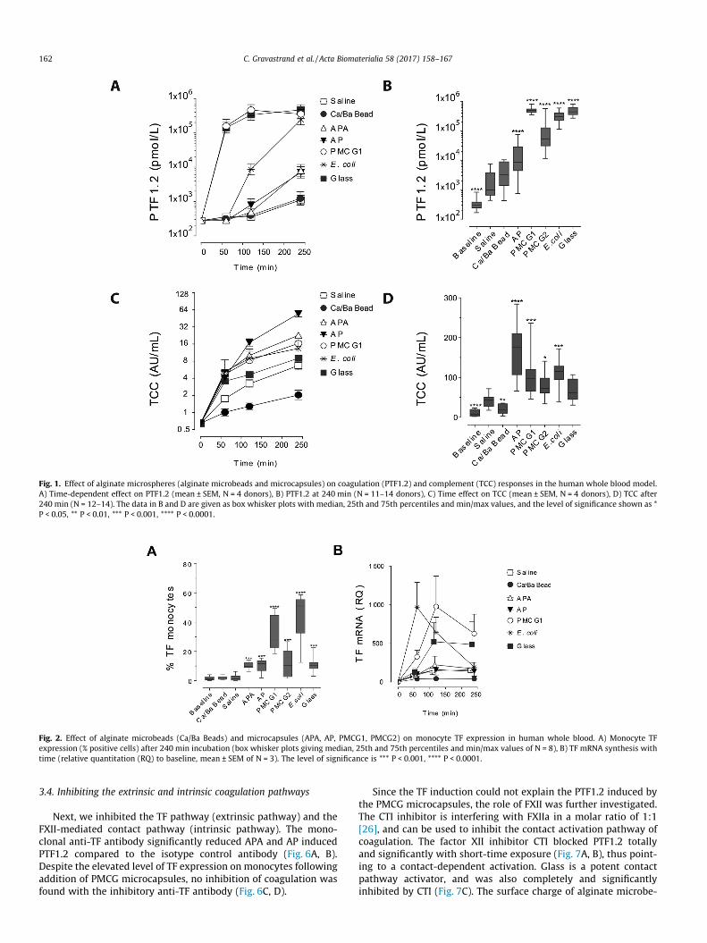
Fig. 1. Effect of alginate microspheres (alginate microbeads and microcapsules) on coagulation (PTF1.2) and complement (TCC) responses in the human whole blood model.A) Time-dependent effect on PTF1.2 (mean ± SEM, N = 4 donors), B) PTF1.2 at 240 min (N = 11–14 donors), C) Time effect on TCC (mean ± SEM, N = 4 donors), D) TCC after240 min (N = 12–14). The data in B and D are given as box whisker plots with median, 25th and 75th percentiles and min/max values, and the level of significance shown as *P < 0.05, ** P < 0.01, *** P < 0.001, **** P < 0.0001.
Fig. 2. Effect of alginate microbeads (Ca/Ba Beads) and microcapsules (APA, AP, PMCG1, PMCG2) on monocyte TF expression in human whole blood. A) Monocyte TFexpression (% positive cells) after 240 min incubation (box whisker plots giving median, 25th and 75th percentiles and min/max values of N = 8), B) TF mRNA synthesis withtime (relative quantitation (RQ) to baseline, mean ± SEM of N = 3). The level of significance is *** P < 0.001, **** P < 0.0001.
162 C. Gravastrand et al. / Acta Biomaterialia 58 (2017) 158–167
3.4. Inhibiting the extrinsic and intrinsic coagulation pathways
Next, we inhibited the TF pathway (extrinsic pathway) and theFXII-mediated contact pathway (intrinsic pathway). The mono-clonal anti-TF antibody significantly reduced APA and AP inducedPTF1.2 compared to the isotype control antibody (Fig. 6A, B).Despite the elevated level of TF expression on monocytes followingaddition of PMCG microcapsules, no inhibition of coagulation wasfound with the inhibitory anti-TF antibody (Fig. 6C, D).
Since the TF induction could not explain the PTF1.2 induced bythe PMCG microcapsules, the role of FXII was further investigated.The CTI inhibitor is interfering with FXIIa in a molar ratio of 1:1[26], and can be used to inhibit the contact activation pathway ofcoagulation. The factor XII inhibitor CTI blocked PTF1.2 totallyand significantly with short-time exposure (Fig. 7A, B), thus point-ing to a contact-dependent activation. Glass is a potent contactpathway activator, and was also completely and significantlyinhibited by CTI (Fig. 7C). The surface charge of alginate microbe-
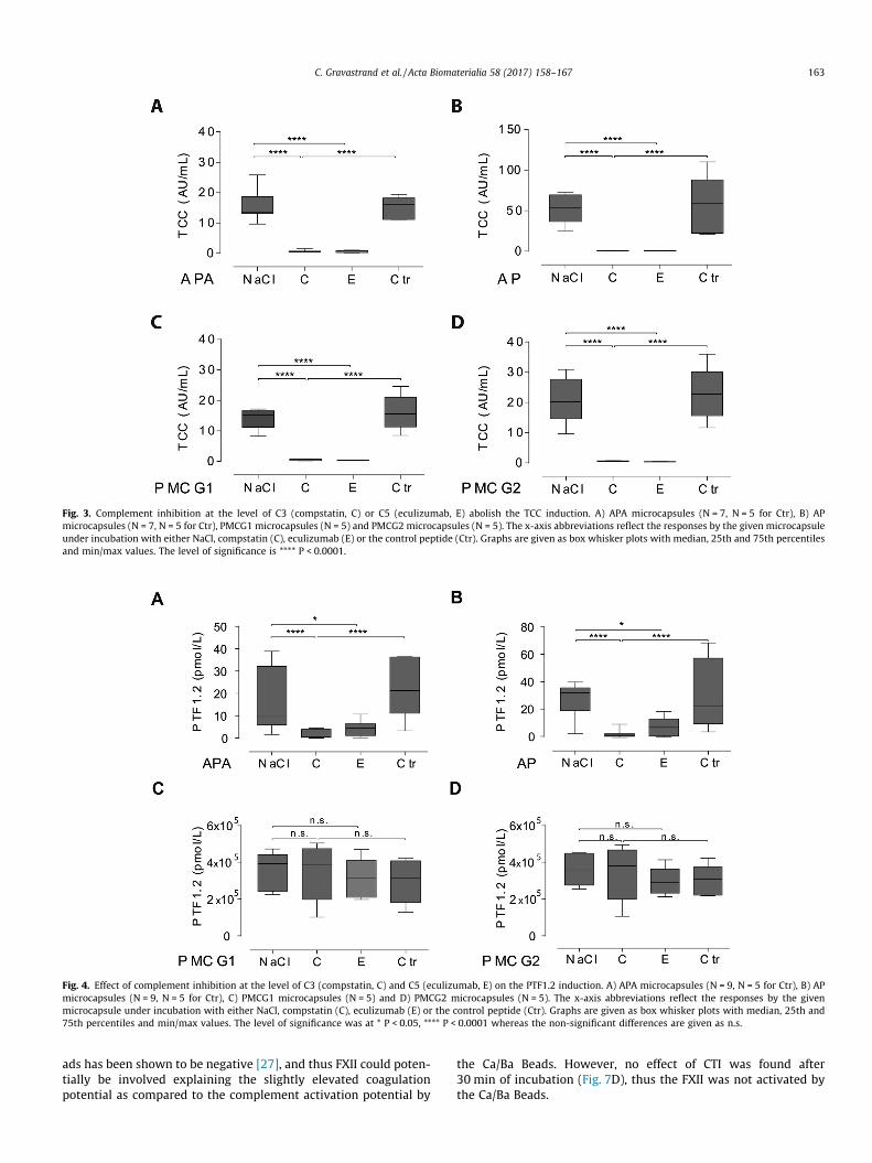
Fig. 3. Complement inhibition at the level of C3 (compstatin, C) or C5 (eculizumab, E) abolish the TCC induction. A) APA microcapsules (N = 7, N = 5 for Ctr), B) APmicrocapsules (N = 7, N = 5 for Ctr), PMCG1 microcapsules (N = 5) and PMCG2 microcapsules (N = 5). The x-axis abbreviations reflect the responses by the given microcapsuleunder incubation with either NaCl, compstatin (C), eculizumab (E) or the control peptide (Ctr). Graphs are given as box whisker plots with median, 25th and 75th percentilesand min/max values. The level of significance is **** P < 0.0001.
Fig. 4. Effect of complement inhibition at the level of C3 (compstatin, C) and C5 (eculizumab, E) on the PTF1.2 induction. A) APA microcapsules (N = 9, N = 5 for Ctr), B) APmicrocapsules (N = 9, N = 5 for Ctr), C) PMCG1 microcapsules (N = 5) and D) PMCG2 microcapsules (N = 5). The x-axis abbreviations reflect the responses by the givenmicrocapsule under incubation with either NaCl, compstatin (C), eculizumab (E) or the control peptide (Ctr). Graphs are given as box whisker plots with median, 25th and75th percentiles and min/max values. The level of significance was at * P < 0.05, **** P < 0.0001 whereas the non-significant differences are given as n.s.
C. Gravastrand et al. / Acta Biomaterialia 58 (2017) 158–167 163
ads has been shown to be negative [27], and thus FXII could poten-tially be involved explaining the slightly elevated coagulationpotential as compared to the complement activation potential by
the Ca/Ba Beads. However, no effect of CTI was found after30 min of incubation (Fig. 7D), thus the FXII was not activated bythe Ca/Ba Beads.
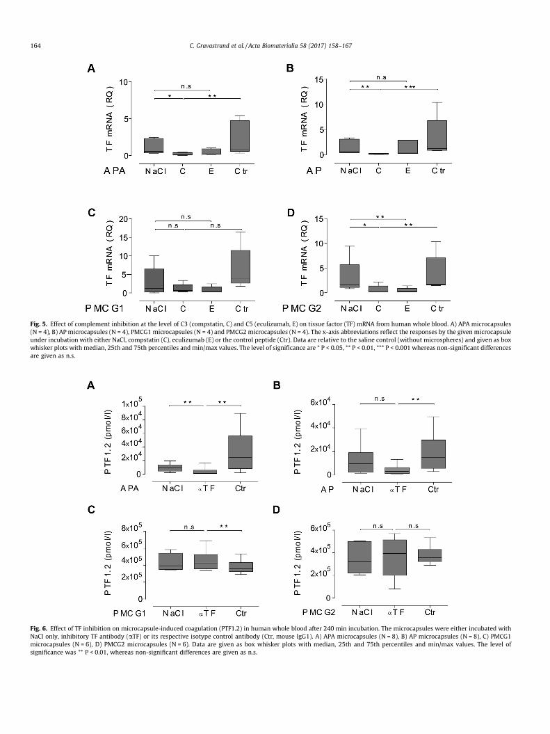
Fig. 5. Effect of complement inhibition at the level of C3 (compstatin, C) and C5 (eculizumab, E) on tissue factor (TF) mRNA from human whole blood. A) APA microcapsules(N = 4), B) AP microcapsules (N = 4), PMCG1 microcapsules (N = 4) and PMCG2microcapsules (N = 4). The x-axis abbreviations reflect the responses by the given microcapsuleunder incubation with either NaCl, compstatin (C), eculizumab (E) or the control peptide (Ctr). Data are relative to the saline control (without microspheres) and given as boxwhisker plots with median, 25th and 75th percentiles and min/max values. The level of significance are * P < 0.05, ** P < 0.01, *** P < 0.001 whereas non-significant differencesare given as n.s.
Fig. 6. Effect of TF inhibition on microcapsule-induced coagulation (PTF1.2) in human whole blood after 240 min incubation. The microcapsules were either incubated withNaCl only, inhibitory TF antibody (aTF) or its respective isotype control antibody (Ctr, mouse IgG1). A) APA microcapsules (N = 8), B) AP microcapsules (N = 8), C) PMCG1microcapsules (N = 6), D) PMCG2 microcapsules (N = 6). Data are given as box whisker plots with median, 25th and 75th percentiles and min/max values. The level ofsignificance was ** P < 0.01, whereas non-significant differences are given as n.s.
164 C. Gravastrand et al. / Acta Biomaterialia 58 (2017) 158–167
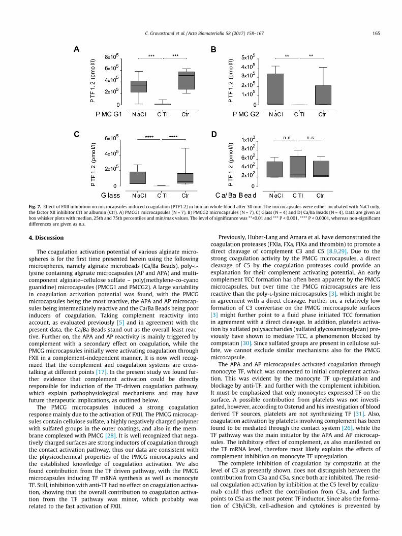
Fig. 7. Effect of FXII inhibition on microcapsules induced coagulation (PTF1.2) in human whole blood after 30 min. The microcapsules were either incubated with NaCl only,the factor XII inhibitor CTI or albumin (Ctr). A) PMCG1 microcapsules (N = 7), B) PMCG2 microcapsules (N = 7), C) Glass (N = 4) and D) Ca/Ba Beads (N = 4). Data are given asbox whisker plots with median, 25th and 75th percentiles and min/max values. The level of significance was **<0.01 and *** P < 0.001, **** P < 0.0001, whereas non-significantdifferences are given as n.s.
C. Gravastrand et al. / Acta Biomaterialia 58 (2017) 158–167 165
4. Discussion
The coagulation activation potential of various alginate micro-spheres is for the first time presented herein using the followingmicrospheres, namely alginate microbeads (Ca/Ba Beads), poly-L-lysine containing alginate microcapsules (AP and APA) and multi-component alginate–cellulose sulfate – poly(methylene-co-cyanoguanidine) microcapsules (PMCG1 and PMCG2). A large variabilityin coagulation activation potential was found, with the PMCGmicrocapsules being the most reactive, the APA and AP microcap-sules being intermediately reactive and the Ca/Ba Beads being poorinducers of coagulation. Taking complement reactivity intoaccount, as evaluated previously [5] and in agreement with thepresent data, the Ca/Ba Beads stand out as the overall least reac-tive. Further on, the APA and AP reactivity is mainly triggered bycomplement with a secondary effect on coagulation, while thePMCG microcapsules initially were activating coagulation throughFXII in a complement-independent manner. It is now well recog-nized that the complement and coagulation systems are cross-talking at different points [17]. In the present study we found fur-ther evidence that complement activation could be directlyresponsible for induction of the TF-driven coagulation pathway,which explain pathophysiological mechanisms and may havefuture therapeutic implications, as outlined below.
The PMCG microcapsules induced a strong coagulationresponse mainly due to the activation of FXII. The PMCG microcap-sules contain cellulose sulfate, a highly negatively charged polymerwith sulfated groups in the outer coatings, and also in the mem-brane complexed with PMCG [28]. It is well recognized that nega-tively charged surfaces are strong inductors of coagulation throughthe contact activation pathway, thus our data are consistent withthe physicochemical properties of the PMCG microcapsules andthe established knowledge of coagulation activation. We alsofound contribution from the TF driven pathway, with the PMCGmicrocapsules inducing TF mRNA synthesis as well as monocyteTF. Still, inhibition with anti-TF had no effect on coagulation activa-tion, showing that the overall contribution to coagulation activa-tion from the TF pathway was minor, which probably wasrelated to the fast activation of FXII.
Previously, Huber-Lang and Amara et al. have demonstrated thecoagulation proteases (FXIa, FXa, FIXa and thrombin) to promote adirect cleavage of complement C3 and C5 [8,9,29]. Due to thestrong coagulation activity by the PMCG microcapsules, a directcleavage of C5 by the coagulation proteases could provide anexplanation for their complement activating potential. An earlycomplement TCC formation has often been apparent by the PMCGmicrocapsules, but over time the PMCG microcapsules are lessreactive than the poly-L-lysine microcapsules [3], which might bein agreement with a direct cleavage. Further on, a relatively lowformation of C3 convertase on the PMCG microcapsule surfaces[3] might further point to a fluid phase initiated TCC formationin agreement with a direct cleavage. In addition, platelets activa-tion by sulfated polysaccharides (sulfated glycosaminoglycan) pre-viously have shown to mediate TCC, a phenomenon blocked bycompstatin [30]. Since sulfated groups are present in cellulose sul-fate, we cannot exclude similar mechanisms also for the PMCGmicrocapsule.
The APA and AP microcapsules activated coagulation throughmonocyte TF, which was connected to initial complement activa-tion. This was evident by the monocyte TF up-regulation andblockage by anti-TF, and further with the complement inhibition.It must be emphasized that only monocytes expressed TF on thesurface. A possible contribution from platelets was not investi-gated, however, according to Osterud and his investigation of bloodderived TF sources, platelets are not synthesizing TF [31]. Also,coagulation activation by platelets involving complement has beenfound to be mediated through the contact system [26], while theTF pathway was the main initiator by the APA and AP microcap-sules. The inhibitory effect of complement, as also manifested onthe TF mRNA level, therefore most likely explains the effects ofcomplement inhibition on monocyte TF upregulation.
The complete inhibition of coagulation by compstatin at thelevel of C3 as presently shown, does not distinguish between thecontribution from C3a and C5a, since both are inhibited. The resid-ual coagulation activation by inhibition at the C5 level by eculizu-mab could thus reflect the contribution from C3a, and furtherpoints to C5a as the most potent TF inductor. Since also the forma-tion of C3b/iC3b, cell-adhesion and cytokines is prevented by
166 C. Gravastrand et al. / Acta Biomaterialia 58 (2017) 158–167
compstatin [4], we cannot exclude also their involvement. Theinvolvement of inflammatory cytokines on monocyte TF expres-sion would however most likely be secondary to the effect of therapidly produced complement activation products [3]. Since com-plement activation also is a pre-requisite for cytokine inductionby the microcapsules [4,5], we conclude that complement activa-tion including the anaphylatoxins most probably is the main indu-cer of TF in the present investigation.
Coagulation activation with formation of fibrin may be one ofthe gateways to bio-incompatibility in a transplantation situation.The most common transplantation site for islets encapsulated inmicrobeads or microcapsules is the peritoneal cavity, due to thespace requirements for a therapeutic amount of transplanted islets.Although the continuous exposure to blood is avoided, there islikely that the transplanted microspheres are exposed to bloodparticularly during implantation. The high coagulation reactivityof the PMCGmicrocapsules and their independence of complementactivation might be a challenge during transplantation with bloodcontamination, since it could lead to surface deposition of fibrin.Since fibrin exposes epitopes serving as ligands for CR3 (CD11b/CD18) [6], the fibrin deposition is a potential starting point forleukocyte-attachment. It might therefore be of high importanceto avoid bleeding during a transplantation situation using thePMCG microcapsules. A short-time inhibition of FXII could be away of inhibiting the coagulation potential of the PMCG microcap-sules during the critical phase of implantation, since currentlythere are ongoing FXII-inhibition studies for thrombosis treat-ments [32]. Despite the coagulation reactivity, the PMCG micro-capsules can be low activators of fibrotic overgrowth reactions asshown in mice [22] and baboons [33]. This demonstrates that thesetype of microcapsules can be well tolerated and, thus, that theremay not be the lack of a direct link between the coagulation acti-vation potential and the fibrotic responses. This reported tolerationmay simply indicate the absence of microcapsule exposure toblood; a controlled bleeding experiment could contribute to clari-fying these questions. On the other hand, the reactivity of the APAand AP microcapsules, points a definite complement-dependentreactivity, probably as the most important mechanism promotingleukocyte adhesion as previously shown [4].
The additional measurements of coagulation activation in addi-tion to complement and inflammatory cytokines provide togetheran efficient screening system for the immediate host reactivitypromoted by the protein cascades in concert with the leukocytes.In transplantation setting there will also be encapsulated cellswithin the devices, which add complexity to the situation bysecreted cellular component provoking other parts of the hostimmune system as addressed in [2]. The whole blood model is, firstof all, efficient to measure the surface reactivity and the materialresponse, and, therefore, this model is strongly recommended todesign low-reactive devices. However, as the model also containsthe blood leukocytes containing the damage-associated molecularpatterns (DAMPs) receptors sensing release of dying or stress cellscomponents, the model possibly can be used to evaluate theimpact of encapsulated cells. In summary, the host immune systemevolves a complex set of mechanisms that will contribute in atransplantation setting and that we need to carefully examine inorder to enhance our understanding in the design of immunopro-tective devices. The coagulation response evaluated in this workrepresents an important part of this learning curve.
5. Conclusions
In conclusion, our data identifies for the first time the reactivityof the various alginate microspheres on coagulation, identifyingPMCGmicrocapsules as pro-coagulative, the APA and AP microcap-
sules to be weaker inducers and the Ca/Ba Beads as low/inert tocoagulation activation. Further on, we found the coagulation acti-vation of the PMCG microcapsules was strongly dependent ondirect activation by FXII, whereas by the APA and AP microcapsuleswere highly dependent on initial complement activation promot-ing TF in monocytes, thus further defining a complement-coagulation cross-talk in these interactions.
Disclosure
The authors declare no conflicts of interests. J.D.L. is the inven-tor of patents and/or patent applications that describe the use ofcomplement inhibitors for therapeutic purposes and the founderof Amyndas Pharmaceuticals, which is developing complementinhibitors for clinical applications. We confirm that all authorshave approved the final article.
Acknowledgments
This work has been financially supported by The Liaison Com-mittee for Education, Research and Innovation in Central Norway(RHA) under grants 46049600, 46056819 and 46062127 in coordi-nation with the Norwegian University of Science and Technology(NTNU). This work has also been supported by the Research Coun-cil of Norway through its Centers of Excellence funding scheme,project number 223255; the JDRF (Juvenile Diabetes ResearchFoundation) under grant number 2-SRA-2014-288-Q-R; The OddFellow Foundation [OFF-2014]; The Simon Fougner HartmannFamily Fund [SFHF-12/14], The European Community’s SeventhFramework Programme under grant agreement n� 602699 (DIR-EKT), and by National Institutes of Health Grants AI068730 andAI030040 and by the Slovak Research and Development Agencyunder contract number APVV-14-858. Dr. Gabriela Kollarikova isthanked for preparation of PMCG1 and PMCG2 microcapsules.
References
[1] J.M. Anderson, A. Rodriguez, D.T. Chang, Foreign body reaction to biomaterials,Semin. Immunol. 20 (2) (2008) 86–100.
[2] A.M. Rokstad, I. Lacik, P. de Vos, B.L. Strand, Advances in biocompatibility andphysico-chemical characterization of microspheres for cell encapsulation, Adv.Drug Deliv. Rev. 67–68 (2014) 111–130.
[3] A.M. Rokstad, O.L. Brekke, B. Steinkjer, L. Ryan, G. Kollarikova, B.L. Strand, G.Skjak-Braek, I. Lacik, T. Espevik, T.E. Mollnes, Alginate microbeads arecomplement compatible, in contrast to polycation containing microcapsules,as revealed in a human whole blood model, Acta Biomater. 7 (6) (2011) 2566–2578.
[4] P. Orning, K.S. Hoem, A.E. Coron, G. Skjak-Braek, T.E. Mollnes, O.L. Brekke, T.Espevik, A.M. Rokstad, Alginate microsphere compositions dictate differentmechanisms of complement activation with consequences for cytokine releaseand leukocyte activation, J. Control. Release 229 (2016) 58–69.
[5] A.M. Rokstad, O.L. Brekke, B. Steinkjer, L. Ryan, G. Kollarikova, B.L. Strand, G.Skjak-Braek, J.D. Lambris, I. Lacik, T.E. Mollnes, T. Espevik, The induction ofcytokines by polycation containing microspheres by a complement dependentmechanism, Biomaterials 34 (3) (2013) 621–630.
[6] W.J. Hu, J.W. Eaton, T.P. Ugarova, L. Tang, Molecular basis of biomaterial-mediated foreign body reactions, Blood 98 (4) (2001) 1231–1238.
[7] A.P. Kaplan, B. Ghebrehiwet, The plasma bradykinin-forming pathways and itsinterrelationships with complement, Mol. Immunol. 47 (13) (2010) 2161–2169.
[8] U. Amara, D. Rittirsch, M. Flierl, U. Bruckner, A. Klos, F. Gebhard, J.D. Lambris,M. Huber-Lang, Interaction between the coagulation and complement system,Adv. Exp. Med. Biol. 632 (2008) 71–79.
[9] M. Huber-Lang, J.V. Sarma, F.S. Zetoune, D. Rittirsch, T.A. Neff, S.R. McGuire, J.D.Lambris, R.L. Warner, M.A. Flierl, L.M. Hoesel, F. Gebhard, J.G. Younger, S.M.Drouin, R.A. Wetsel, P.A. Ward, Generation of C5a in the absence of C3: a newcomplement activation pathway, Nat. Med. 12 (6) (2006) 682–687.
[10] K. Ikeda, K. Nagasawa, T. Horiuchi, T. Tsuru, H. Nishizaka, Y. Niho, C5a inducestissue factor activity on endothelial cells, Thromb. Haemost. 77 (2) (1997)394–398.
[11] A. Landsem, E.W. Nielsen, H. Fure, D. Christiansen, J.K. Ludviksen, J.D. Lambris,B. Osterud, T.E. Mollnes, O.L. Brekke, C1-inhibitor efficiently inhibitsEscherichia coli-induced tissue factor mRNA up-regulation, monocyte tissue
C. Gravastrand et al. / Acta Biomaterialia 58 (2017) 158–167 167
factor expression and coagulation activation in human whole blood, Clin. Exp.Immunol. 173 (2) (2013) 217–229.
[12] A. Landsem, H. Fure, D. Christiansen, E.W. Nielsen, B. Osterud, T.E. Mollnes, O.L.Brekke, The key roles of complement and tissue factor in Escherichia coli-induced coagulation in human whole blood, Clin. Exp. Immunol. 182 (1)(2015) 81–89.
[13] R. Ovstebo, M. Hellum, H.C. Aass, A.M. Troseid, P. Brandtzaeg, T.E. Mollnes, C.E.Henriksson, Microparticle-associated tissue factor activity is reduced byinhibition of the complement protein 5 in Neisseria meningitidis-exposedwhole blood, Innate Immun. 20 (5) (2014) 552–560.
[14] K. Ritis, M. Doumas, D. Mastellos, A. Micheli, S. Giaglis, P. Magotti, S. Rafail, G.Kartalis, P. Sideras, J.D. Lambris, A novel C5a receptor-tissue factor cross-talk inneutrophils links innate immunity to coagulation pathways, J. Immunol. 177(7) (2006) 4794–4802.
[15] M.V. Ward, D.L. Conway, A severe case of acute suppurative dermatitis withdisseminated intravascular coagulation in a dog, Vet. Med. Small Anim. Clin.75 (10) (1980) 1564–1568.
[16] T. Wiedmer, C.T. Esmon, P.J. Sims, On the mechanism by which complementproteins C5b–9 increase platelet prothrombinase activity, J. Biol. Chem. 261(31) (1986) 14587–14592.
[17] E.M. Conway, Reincarnation of ancient links between coagulation andcomplement, J. Thromb. Haemost. 13 (Suppl. 1) (2015) S121–32.
[18] M.J. Rabiet, A. Blashill, B. Furie, B.C. Furie, Prothrombin fragment 1X2 X 3, amajor product of prothrombin activation in human plasma, J. Biol. Chem. 261(28) (1986) 13210–13215.
[19] H. Qu, D. Ricklin, H. Bai, H. Chen, E.S. Reis, M. Maciejewski, A. Tzekou, R.A.Deangelis, R.R. Resuello, F. Lupu, P.N. Barlow, J.D. Lambris, New analogs of theclinical complement inhibitor compstatin with subnanomolar affinity andenhanced pharmacokinetic properties, Immunobiology (2012).
[20] H. Qu, P. Magotti, D. Ricklin, E.L. Wu, I. Kourtzelis, Y.Q. Wu, Y.N. Kaznessis, J.D.Lambris, Novel analogues of the therapeutic complement inhibitor compstatinwith significantly improved affinity and potency, Mol. Immunol. 48 (4) (2011)481–489.
[21] T.E. Mollnes, H. Redl, K. Hogasen, A. Bengtsson, P. Garred, L. Speilberg, T. Lea,M. Oppermann, O. Gotze, G. Schlag, Complement activation in septic baboonsdetected by neoepitope-specific assays for C3b/iC3b/C3c, C5a and the terminalC5b–9 complement complex (TCC), Clin. Exp. Immunol. 91 (2) (1993) 295–300.
[22] I. Lacik, M. Brissova, A.V. Anilkumar, A.C. Powers, T. Wang, New capsule withtailored properties for the encapsulation of living cells, J. Biomed. Mater. Res.39 (1) (1998) 52–60.
[23] O.L. Brekke, C. Waage, D. Christiansen, H. Fure, H. Qu, J.D. Lambris, B. Osterud,E.W. Nielsen, T.E. Mollnes, The effects of selective complement and CD14inhibition on the E. coli-induced tissue factor mRNA upregulation, monocytetissue factor expression, and tissue factor functional activity in human wholeblood, Adv. Exp. Med. Biol. 734 (2013) 123–136.
[24] M. Ernofsson, A. Siegbahn, Platelet-derived growth factor-BB and monocytechemotactic protein-1 induce human peripheral blood monocytes to expresstissue factor, Thromb. Res. 83 (4) (1996) 307–320.
[25] F.J. Neumann, I. Ott, N. Marx, T. Luther, S. Kenngott, M. Gawaz, M. Kotzsch, A.Schomig, Effect of human recombinant interleukin-6 and interleukin-8 onmonocyte procoagulant activity, Arterioscler. Thromb. Vasc. Biol. 17 (12)(1997) 3399–3405.
[26] Y. Hojima, J.V. Pierce, J.J. Pisano, Hageman factor fragment inhibitor in cornseeds: purification and characterization, Thromb. Res. 20 (2) (1980) 149–162.
[27] J.O. You, S.B. Park, H.Y. Park, S. Haam, C.H. Chung, W.S. Kim, Preparation ofregular sized Ca-alginate microspheres using membrane emulsificationmethod, J. Microencapsul. (2001) 521–532.
[28] I. Lacik, A.V. Anilkumar, T.G. Wang, A two-step process for controlling thesurface smoothness of polyelectrolyte-based microcapsules, J. Microencapsul.18 (4) (2001) 479–490.
[29] U. Amara, M.A. Flierl, D. Rittirsch, A. Klos, H. Chen, B. Acker, U.B. Bruckner, B.Nilsson, F. Gebhard, J.D. Lambris, M. Huber-Lang, Molecularintercommunication between the complement and coagulation systems, J.Immunol. 185 (9) (2010) 5628–5636.
[30] O.A. Hamad, K.N. Ekdahl, P.H. Nilsson, J. Andersson, P. Magotti, J.D. Lambris, B.Nilsson, Complement activation triggered by chondroitin sulfate released bythrombin receptor-activated platelets, J. Thromb. Haemost. 6 (8) (2008) 1413–1421.
[31] B. Osterud, Tissue factor expression in blood cells, Thromb. Res. 125 (Suppl. 1)(2010) S31–S34.
[32] F. May, J. Krupka, M. Fries, I. Thielmann, I. Pragst, T. Weimer, C. Panousis, B.Nieswandt, G. Stoll, G. Dickneite, S. Schulte, M.W. Nolte, FXIIa inhibitor rHA-Infestin-4: safe thromboprotection in experimental venous, arterial andforeign surface-induced thrombosis, Br. J. Haematol. 173 (5) (2016) 769–778.
[33] M. Qi, I. Lacik, G. Kollarikova, B.L. Strand, K. Formo, Y. Wang, E. Marchese, J.E.Mendoza-Elias, K.P. Kinzer, F. Gatti, D. Paushter, S. Patel, J. Oberholzer, Arecommended laparoscopic procedure for implantation of microcapsules inthe peritoneal cavity of non-human primates, J. Surg. Res. 168 (1) (2011)e117–e123.