Algae Polar Lipids Characterized by Online Liquid
6
Published: September 26, 2011 r2011 American Chemical Society 4770 dx.doi.org/10.1021/ef201061j | Energy Fuels 2011, 25, 4770–4775 ARTICLE pubs.acs.org/EF Algae Polar Lipids Characterized by Online Liquid Chromatography Coupled with Hybrid Linear Quadrupole Ion Trap/Fourier Transform Ion Cyclotron Resonance Mass Spectrometry Huan He, † Ryan P. Rodgers, †,‡ Alan G. Marshall,* ,†,‡ and Chang Samuel Hsu* ,§ † National High Magnetic Field Laboratory, Florida State University, 1800 East Paul Dirac Drive, Tallahassee, Florida 32310, United States ‡ Department of Chemistry and Biochemistry, Florida State University, 95 Chieftain Way, Tallahassee, Florida 32306, United States § Future Fuels Institute, Florida State University, Tallahassee, Florida 32310, United States b S Supporting Information ABSTRACT: We report the first application of online LC-MS (liquid chromatography mass spectrometry) characterization of algae polar lipids by nanoscale high-performance liquid chromatography followed by electrospray ionization and mass analysis with a linear ion trap (LTQ) coupled with 14.5 T Fourier transform ion cyclotron resonance mass spectrometry (FT-ICR MS). Ultrahigh FT-ICR mass resolution provides highly accurate mass measurement and resolves monoisotopic peaks from interfering components for unique determination of lipid elemental compositions. We establish the polar lipid profile of fatty acids, glycolipids, phospholipids, and betaine lipids for a green algae, Nannochloropsis oculata, which is highly prized for its oils suitable for biodiesel production. Lipid headgroup and fatty acid identification is based on accurate mass measured by the FT-ICR MS and collision- induced dissociation (CID) MS/MS in the LTQ. Unequivocal lipid composition is further confirmed from isotopic fine structure at baseline resolution—achievable only with ultrahigh resolution FT-ICR MS. ’ INTRODUCTION Transportation fuels have overwhelmingly been obtained from fossil resources. Because of dwindling supplies and increas- ing evidence of carbon dioxide as a main culprit for global warming, fuels from biological resources have been explored and have achieved some success. First generation biofuels are produced from food sources, such as vegetable oils, starch, and sugar. 1,2 To abate the competition with food sources, second generation biofuels are produced from nonfood sources, such as lignocellulosic biomass. 1,2 Algae, specifically microalgae, are considered as third generation biofuel feedstocks designed for fuel production as an “energy crop”. 3 The use of marine algae would further reduce the competition for land and freshwater. The current focus of third generation algae biofuel research is to discover new algae strains of high oil yield and to genetically modify algae to improve the intrinsic oil yield and growth rate. Genetic modi fication and external stimuli (e.g., nitrogen starvation) profoundly influence lipid biosynthetic and metabolic pathways. The long-term goal is to generate algae lipid fingerprints that can be mapped to those pathways. The insights thus generated should have a major impact on the understanding of algae oil biosynthesis and thus help to guide future genetic modification and strain selection. Algae contain four principal biochemical classes: carbohy- drates (polysaccharides), proteins, nucleic acids, and lipids. Algae produce various types of polysaccharides that can be fermented to produce alcohols for fuels. Lipids are feedstocks for biodiesel fuel through trans-esterification processes. Proteins, on the other hand, can be developed into high value biochemicals by virtue of their amino acid composition of high nutritional quality. 4 The lipid content of microalgae is strongly influenced by growth conditions such as the effects of temperature and nitrogen content of Nannochloropsis oculata and Chlorella vulgaris for biodiesel production. 5 However, there is evidence of a negative relationship between growth rate and lipid con- tent for naturally occurring algae. Hence, high lipid yield alone cannot be used as the only criterion for optimal growth conditions. 4 Despite massive efforts on the development of biofuels from microalgae, the analysis of algae strains at the molecular level is woefully incomplete. Algae lipid analysis is especially relevant to prospective biodiesel and jet fuel production. We focus initially on Nannochloropsis oculata (green algae) be- cause of its relatively high oil yield (40 60%). 6 Most prior lipid characterization methods are bulk or indirect analyses and cannot resolve and identify intact molecular structures. 7,8 Here, we combine online liquid chromatography (LC) with electrospray ionization Fourier transform ion cyclotron reso- nance mass spectrometry 9 (ESI FT-ICR MS) and tandem mass spectrometry (MS/MS) to resolve and identify lipid elemental compositions, C c H h N n O o P p S s , head groups, fatty acid chain lengths, and unsaturation of polar lipids from green algae, as a first step in the molecular level characterization of wild type and genetically engineered (mutant) algae 7,10 of improved growth rate and lipid yield for the production of algal fuels. 11 Received: July 20, 2011 Revised: September 12, 2011
Transcript of Algae Polar Lipids Characterized by Online Liquid
Published: September 26, 2011
r 2011 American Chemical Society 4770 dx.doi.org/10.1021/ef201061j | Energy Fuels 2011, 25, 4770–4775
ARTICLE
pubs.acs.org/EF
Algae Polar Lipids Characterized by Online Liquid ChromatographyCoupled with Hybrid Linear Quadrupole Ion Trap/Fourier TransformIon Cyclotron Resonance Mass SpectrometryHuan He,† Ryan P. Rodgers,†,‡ Alan G. Marshall,*,†,‡ and Chang Samuel Hsu*,§
†National High Magnetic Field Laboratory, Florida State University, 1800 East Paul Dirac Drive, Tallahassee, Florida 32310,United States‡Department of Chemistry and Biochemistry, Florida State University, 95 Chieftain Way, Tallahassee, Florida 32306, United States§Future Fuels Institute, Florida State University, Tallahassee, Florida 32310, United States
bS Supporting Information
ABSTRACT: We report the first application of online LC-MS (liquid chromatography�mass spectrometry) characterization ofalgae polar lipids by nanoscale high-performance liquid chromatography followed by electrospray ionization andmass analysis with alinear ion trap (LTQ) coupled with 14.5 T Fourier transform ion cyclotron resonance mass spectrometry (FT-ICRMS). UltrahighFT-ICRmass resolution provides highly accuratemass measurement and resolves monoisotopic peaks from interfering componentsfor unique determination of lipid elemental compositions. We establish the polar lipid profile of fatty acids, glycolipids,phospholipids, and betaine lipids for a green algae, Nannochloropsis oculata, which is highly prized for its oils suitable for biodieselproduction. Lipid headgroup and fatty acid identification is based on accurate mass measured by the FT-ICR MS and collision-induced dissociation (CID)MS/MS in the LTQ. Unequivocal lipid composition is further confirmed from isotopic fine structure atbaseline resolution—achievable only with ultrahigh resolution FT-ICR MS.
’ INTRODUCTION
Transportation fuels have overwhelmingly been obtainedfrom fossil resources. Because of dwindling supplies and increas-ing evidence of carbon dioxide as a main culprit for globalwarming, fuels from biological resources have been exploredand have achieved some success. First generation biofuels areproduced from food sources, such as vegetable oils, starch, andsugar.1,2 To abate the competition with food sources, secondgeneration biofuels are produced from nonfood sources, such aslignocellulosic biomass.1,2 Algae, specifically microalgae, areconsidered as third generation biofuel feedstocks designed forfuel production as an “energy crop”.3 The use of marine algaewould further reduce the competition for land and freshwater.
The current focus of third generation algae biofuel research isto discover new algae strains of high oil yield and to geneticallymodify algae to improve the intrinsic oil yield and growth rate.Genetic modification and external stimuli (e.g., nitrogen starvation)profoundly influence lipid biosynthetic and metabolic pathways.The long-term goal is to generate algae lipid fingerprints that canbe mapped to those pathways. The insights thus generatedshould have a major impact on the understanding of algae oilbiosynthesis and thus help to guide future genetic modificationand strain selection.
Algae contain four principal biochemical classes: carbohy-drates (polysaccharides), proteins, nucleic acids, and lipids. Algaeproduce various types of polysaccharides that can be fermentedto produce alcohols for fuels. Lipids are feedstocks for biodieselfuel through trans-esterification processes. Proteins, on the otherhand, can be developed into high value biochemicals by virtue oftheir amino acid composition of high nutritional quality.4
The lipid content of microalgae is strongly influenced bygrowth conditions such as the effects of temperature andnitrogen content of Nannochloropsis oculata and Chlorellavulgaris for biodiesel production.5 However, there is evidenceof a negative relationship between growth rate and lipid con-tent for naturally occurring algae. Hence, high lipid yield alonecannot be used as the only criterion for optimal growthconditions.4
Despite massive efforts on the development of biofuelsfrom microalgae, the analysis of algae strains at the molecularlevel is woefully incomplete. Algae lipid analysis is especiallyrelevant to prospective biodiesel and jet fuel production. Wefocus initially on Nannochloropsis oculata (green algae) be-cause of its relatively high oil yield (40�60%).6 Most priorlipid characterization methods are bulk or indirect analysesand cannot resolve and identify intact molecular structures.7,8
Here, we combine online liquid chromatography (LC) withelectrospray ionization Fourier transform ion cyclotron reso-nance mass spectrometry9 (ESI FT-ICR MS) and tandemmass spectrometry (MS/MS) to resolve and identify lipidelemental compositions, CcHhNnOoPpSs, head groups, fattyacid chain lengths, and unsaturation of polar lipids from greenalgae, as a first step in the molecular level characterization ofwild type and genetically engineered (mutant) algae7,10 ofimproved growth rate and lipid yield for the production ofalgal fuels.11
Received: July 20, 2011Revised: September 12, 2011

4771 dx.doi.org/10.1021/ef201061j |Energy Fuels 2011, 25, 4770–4775
Energy & Fuels ARTICLE
’METHODS
Lipid Extraction. Algae polar lipids were extracted with amodified protocol for mammalian cells.12 A green algae (Nanno-chloropsis oculata, UTEX, Austin, TX) suspension in 1 mL ofErdschreiber’s medium13 was centrifuged to remove superna-tant; the residual pellet was lyophilized; and 0.4 mL of isopro-panol (IPA) was added. After sonication for 5 min, 0.4 mL ofchloroform was added, followed by another 5 min of sonication.The mixture was incubated at 48 �C, in a water bath, overnight.After centrifugation and the removal of the supernatant, 0.4 mLof 1:1 (v/v) CH3OH/CHCl3 was added to the residue, and themixture was sonicated for 5 min before centrifugation to removesupernatant. The above step was repeated one more time, andthe combined supernatant was dried. The dried lipid extract wastaken up in 0.4 mL of 3:2 (v/v) H2O/CH3OH plus 60 μL ofCHCl3. After phase separation, the upper layer containing polarlipids and lower layer with nonpolar lipids were collected, dried,and stored at �80 �C.Nano-Liquid Chromatography�Mass Spectrometry (nLC-
MS). The polar lipid fraction was redissolved in 20:80 H2O/CH3OH containing 10 mM ammonium acetate (NH4OAc) forsubsequent nLC-MS analysis with an Eksigent (Dublin, CA)nanoLC-1D system. A phenylhexyl stationary phase (Phenomenex,Torrance, CA) was selected for its dual selectivity: namely, π�πstacking interaction and reduced hydrophobic�hydrophobicinteractions between the stationary phase and the analytes.The column (75 μm i.d. � 5 cm length) was custom-packedby New Objective (Woburn, MA). For gradient elution, solutionA contained 98% H2O, 2% CH3OH, and 10 mM NH4OAc andsolution B contained 2% H2O, 98% CH3OH, and 10 mMNH4OAc, with a gradient from 80% to 98% B in 30 min, at aflow rate of 400 nL/min. The LC effluent was subjected tonegative-ion electrospray ionization (ESI) at�1.7 kV applied tothe precolumn union, and detected online with a hybrid linearion trap quadrupole (LTQ)/14.5 T Fourier transform ioncyclotron resonance (FT-ICR) mass spectrometer.14 Threemillion target ions were collected in LTQ with automatic gaincontrol (AGC)15 before transfer to the ICR cell (55 mm i.d.)through octopole ion guides (2.2 MHz, 250 Vp�p). In the ICRcell, ions were SWIFT16 excited with an m/z range from 176 to10 200. FT-ICRMS data was acquired at a mass resolving power(m/Δm50%, in which Δm50% is a mass spectral peak full width at
half-maximum peak height) of 200,000 at 400 Da with timedomain acquisition period of 767 ms. Calmix (Thermo Fisher,San Jose, CA) was used for external calibration.17,18Mass spectraldata were analyzed with commercial software (Xcalibur, ThermoFisher, San Jose, CA). Structural assignment was based on theaccurate mass of the pseudomolecular ion [M�H]� for negativeESI FT-ICR MS and corresponding fragment ions detected bycollision-induced dissociation (CID) under nitrogen (25% nor-malized collision energy) in the LTQ. Triplicate samples yieldedaverage values and standard deviations of signal magnitude.
’RESULTS AND DISCUSSION
HPLC to Address Concentration Dynamic Range. Directinfusion of the polar lipid fraction of algae yields only nonlipidsignals. Thus, LC separation prior to MS analysis is essential forlipid characterization by MS. Nano-LC requires only 2.4 μg dry
Figure 1. NanoLC-MS total ion chromatogram (TIC) showing repre-sentative algae polar lipids. Figure 2. Isotopic distribution for the deprotonated molecular ion
([M � H]�) of SQDG (16:1/16:0). The inset shows the 34S and 13C2
doublet. The observed mass difference between species differing by34S vs 32S matches perfectly with the calculated value of 1.9958 Da. Theobserved abundance of 34S relative to 32S also matches the theoreticalvalue of 4.2%. Thus, the sulfur content of the lipid species is confirmed.
Figure 3. LTQ collision-induced dissociation (CID) product ion massspectrum of the deprotonated molecular ion ([M�H]�, 791.4985 Da)from a representative sulfoquinovosyldiacylglycerol (SQDG) (16:1/16:0). Note the characteristic 225.0 Da fragment ion at 225.0 Da,characteristic of the sulfoquinovosyl group.
4772 dx.doi.org/10.1021/ef201061j |Energy Fuels 2011, 25, 4770–4775
Energy & Fuels ARTICLE
algae for each nLC-MS experiment. Two major fractions, mono-acyl and diacyl lipids, are well separated under our LC conditions(see Figure 1). LC separation enables the MS analysis for a lipidmixture of high dynamic range and increases the number ofdetected lipids. We have identified nearly 200 unique lipidspecies (not counting various adducts) with a dynamic rangegreater than 20 000:1 (Table 1 in the Supporting Information).Elemental Composition from Molecular Ion Accurate
Mass Measurement. The structure of individual lipid speciesis determined by accurate mass (typically less than 500 ppb massmeasurement error) of each pseudomolecular ion by FT-ICRMS and LTQ CID mass spectrum. For example, negative ESIFT-ICR MS of sulfoquinovosyldiacylglycerol (SQDG) yields adeprotonated molecular ion of 791.4985 Da, whose elementalcomposition may be uniquely assigned as C41H76O12S (Figure 2).That elemental composition is further confirmed by the resolu-tion of two heavy isotopic forms containing 34S or 13C2 at793.4943 Da (C41H76O12
34S) and 793.5053 Da (13C2C39H76O12S)(Figure 2 inset).Collision-Induced Dissociation MS/MS. The structure of
SQDG was determined by Collision-Induced Dissociation (CID)MS/MS of the deprotonated molecular ion (Figure 3). Themost abundant product ion (537 Da) results from the loss ofa singly unsaturated-C16 carboxyl group. Another at 535 Da is the
result of losing a saturated-C16 carboxyl group. Based on thehigher signal magnitude for ions of 537 Da relative to ions of535 Da, we are able to assign the 16:1 fatty acid at the sn-1
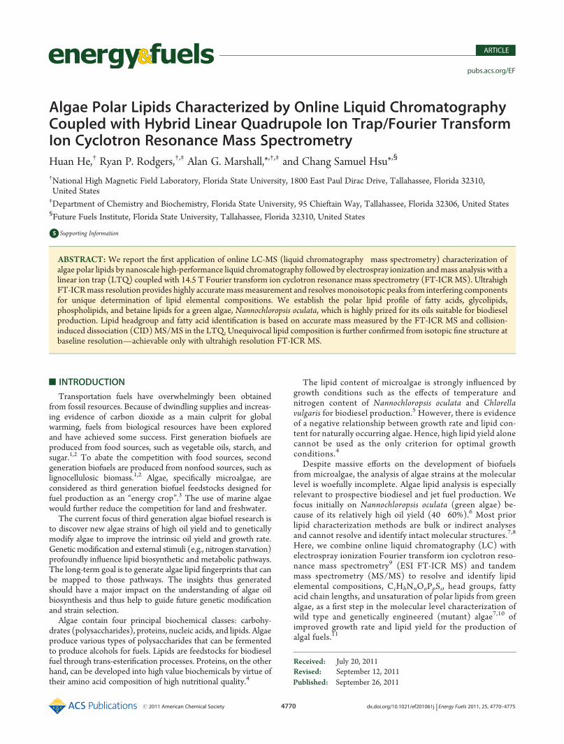
Figure 4. Representative algae lipids identified from Nannochloropsis oculata.
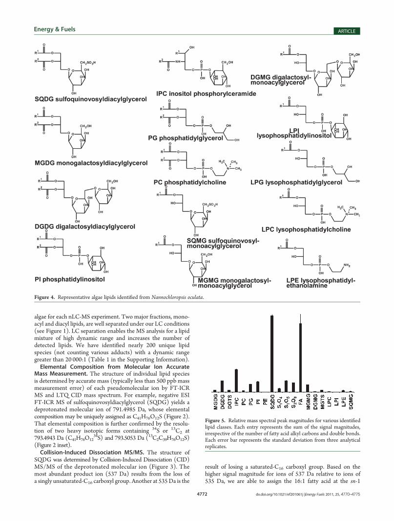
Figure 5. Relative mass spectral peak magnitudes for various identifiedlipid classes. Each entry represents the sum of the signal magnitudes,irrespective of the number of fatty acid alkyl carbons and double bonds.Each error bar represents the standard deviation from three analyticalreplicates.

4773 dx.doi.org/10.1021/ef201061j |Energy Fuels 2011, 25, 4770–4775
Energy & Fuels ARTICLE
position and the 16:0 fatty acid at the sn-2 position.19 The presenceof ions of 225 Da confirms the sulfoquinovosyl group.In this way, we identify sulfoquinovosyldiacylglycerol (SQDG),
digalactosyldiacylglycerol (DGDG), monogalactosyldiacylglycerol(MGDG), inositol phosphorylceramide (IPC), phosphatidyli-nositol (PI), phosphatidylglycerol (PG), diacylglyceryl-N,N,N-trimethylhomoserine (DGTS), phosphatidylcholine (PC),phosphatidylethanolamine (PE), fatty acids (FA), and lyso formsof the diacylglycerol lipids; structures are shown in Figure 4, andthe relative ion abundances are shown in Figure 5. SQDG, a lipid
mainly found in chloroplasts, is particularly prominent, as are freefatty acids. Two chloroplast membrane glycolipids, MGDG andDGDG, are also identified. The betaine lipid, DGTS, has beenhypothesized to transfer fatty acid from the cytoplasm tochloroplast. We identify four types of phospholipids, includingPI, PG, PC, and PE. Unlike diacylglycerol lipids, IPC contains aceramide linkage. Ceramide and inositol groups are each linkedto a phosphate group. Monoacylglycerol lipids are typically atlower concentration than diacylglycerol lipids.Sulfur-Containing Lipids. In addition to lipid species pre-
viously reported in the literature, we identified a series of novelpolar lipids containing O4S1, O5S1 and O8S2. Figure 6 shows
Figure 6. Deprotonated molecular ions of C32H62O4S, C32H64O5S, and C32H66O8S2 detected by online LC FT-ICR MS. Left: isotopic distribution.Right: near-baseline resolution of species differing by 34S vs 13C2.
Figure 7. LC selected ion chromatograms for C32H62O4S, C32H64O5S,and C32H66O8S2. The retention time order, C32H62O4S >C32H64O5S >C32H66O8S2, corresponds to the polarity order, C32H62O4S <C32H64O5S < C32H66O8S2.
Figure 8. CID product ion mass spectrum of [M � 2H]2� fromC32H66O8S2 by LTQ CID, characterized by the bisulfate fragment ion(96.7 Da) and a loss of bisulfate from the deprotonated molecular ion(543.5 Da).

4774 dx.doi.org/10.1021/ef201061j |Energy Fuels 2011, 25, 4770–4775
Energy & Fuels ARTICLE
FT-ICR MS of three representative lipid species from eachgroup, C32H62O4S, C32H64O5S, and C32H66O8S2. Elementalcomposition is established from ultrahigh mass accuracy for themonoisotopic ion (Figure 6, left) and further confirmed from theisotopic fine structure (Figure 6, right). Even with the relativelyshort ICR time-domain data acquisition period (767 ms, limitedby online LC flow rate), we are able to separate ions differing incomposition by 34S vs 13C2 with near-baseline resolution. More-over, the abundance ratio of ions differing in composition by 34Svs 32S further confirms the number of sulfur atoms. The naturalfractional abundance of 34S relative to 32S is 4.2%,20 a valueclosely approached experimentally for C32H62O4S and C32H64O5S(Figure 6, upper and middle right) The presence of two sulfuratoms in C32H66O8S2 is confirmed by the observed
34S-containingion relative abundance of ∼8% (Figure 6, bottom right). Basedon the high abundance as negative ions, it is likely that the sulfuris present in the form of sulfate.Figure 7 shows the LC elution profiles for the most abundant
lipid from each group, C32H62O4S, C32H64O5S, and C32H66O8S2.
Because phenylhexyl stationary phase behaves like a conventionalreversed-phase resin, we infer a polarity order, C32H62O4S <C32H64O5S < C32H66O8S2. Also, from the double bond equiva-lence, DBE, computed from the elemental composition,CcHhNn... (DBE = c � h/2 +
n/2 + 1 = number of rings plusdouble bonds to carbon) we postulate that C32H62O4S has onemore double bond than C32H64O5S. Similarly, C32H64O5S hasone more double bond than C32H66O8S2. The presence of asulfate group is further corroborated by the CID product ionmass spectrum of [M � 2H]2� from C32H66O8S2, shown inFigure 8. Because the sulfate group is highly labile, the domi-nant fragment ions are the bisulfate ion, [HSO4]
� (96.7 Da)and a loss of bisulfate (97 Da) from the deprotonated molecularion (543.5 Da).Polyunsaturated Fatty Acid Chains.Nannochloropsis oculata
is a rich source of highly polyunsaturated fatty acids (PUFA),including eicosahexaenoic acid (EHA, 20:6), eicosapentaenoicacid (EPA, 20:5), and eicosatetraenoic acid, (ETA, 20:4) (Table 1in the Supporting Information). Of great interest is the variation
Figure 9. Relative signal magnitudes of lipid isoforms (lipids with the same polar headgroup but different hydrophobic tails) for different lipid classes.The dominant SQDG isoform (32:1) is composedmainly of the (16:1/16:0) diacyl group. Aminor component of SQDG contains PUFA. The principalMGDG isoform (40:10) mainly contains the (20:5/20:5) diacyl group. A minor MGDG component contains saturated fatty acid. DGDG. The majorDGDG isoforms (36:6) and (36:5) mainly comprise the (16:1/20:5) and (16:0/20:5) diacyl groups. The primary IPC isoform (d36:2) contains 2hydroxyl groups, 32 total carbons, and 2 double bonds in the ceramide tails. Additional chemical labeling and/or MS/MS stage(s) would be needed todetermine the dominant fatty acid and sphingosine of the ceramide tail.

4775 dx.doi.org/10.1021/ef201061j |Energy Fuels 2011, 25, 4770–4775
Energy & Fuels ARTICLE
of the preferred diacylglycerol backbone for different lipidspecies. For example, SQDG prefers (16:1/16:0) (Figure 9),whereas MGDG and DGDG prefer PUFA. The most abundantMGDG contains (20:5/20:5) fatty acids (Figure 9) whereas thedominant DGDGcontains (16:0/20:5) and (16:1/20:5) (Figure 9).That variation suggests different enzymatic pathways in fattyacid utilization. For the ceramide-containing lipid, IPC, wedo not observe any PUFA-containing isoforms. The majorisomer contains 2 hydroxyls, 32 total carbons, and 2 doublebonds in its ceramide tail (d32:2) (Figure 9). The fatty acidchain length and the number of double bonds can profoundlyaffect the properties of lipid bilayers. Thus, such diversityin preferred lipid isoforms for different lipid species suggestsnot only complex lipid biosynthesis/metabolism networksbut also different functions/localization of different lipidspecies.
’CONCLUSION
Identification of algae polar lipids requires a combination ofextraction (to isolate the lipid fraction), HPLC (to overcome thehigh dynamic concentration range), ultrahigh resolution FT-ICRMS (for unique elemental composition), and MS/MS (to identifypolar headgroups as well as fatty acid chain lengths and degrees ofunsaturation). Lipid elemental composition is unequivocally as-signed from accurate mass measurement of the molecular ion andfrom isotopic fine structure at baseline resolution, possible only withultrahigh resolution FT-ICR MS,9 as previously demonstrated byresolution and elemental composition assignment of tens of thou-sands of constituents of petroleum crude oil.21,22 Recent improve-ments in FT-ICR mass resolution and speed of data acquisition,23
mass calibration and accuracy,24 and higher magnetic field14 furtherextend its reliability and range.
’ASSOCIATED CONTENT
bS Supporting Information. Supplementary Table 1: Polarlipids identified from Nannochloropsis oculata. This material isavailable free of charge via the Internet at http://pubs.acs.org.
’AUTHOR INFORMATION
Corresponding Author*E-mail: [email protected]; [email protected].
’ACKNOWLEDGMENT
This work was supported by the National Science FoundationDivision of Materials Research through DMR-0654118 and theState of Florida.
’REFERENCES
(1) Noweck, K. Hydrocarbon Process. 2007, 86, 83–84.(2) Naik, S. N.; Goud, V. V.; Rout, P. K.; Dalai, A. K. Renewable
Sustainable Energy Rev. 2010, 14, 578–597.(3) Singh, A.; Nigam, P. S.; Murphy, J. D. Bioresour. Technol. 2011,
102, 10–16.(4) Williams, P. J. L.; Laurens, L. M. L. Energy Environ. Sci. 2010, 3,
554–590.(5) Converti, A.; Casazza, A. A.; Ortiz, E. Y.; Perego, P.; Del Borghi,
M. Chem. Eng. Process 2009, 48, 1146–1151.(6) Algae Oil Yields. Available online: http://www.oilgae.com/
algae/oil/yield/yield.html.
(7) Lee, M. Y.; Min, B. S.; Chang, C. S.; Jin, E.Mar. Biotechnol. 2006,8, 238–245.
(8) Su, C.-H.; Fu, C.-C.; Chang, T.-C.; Nair, G. R.; Ye, J. L.Biotechnol. Bioeng. 2007, 99, 1034–1039.
(9) Marshall, A. G.; Hendrickson, C. L.; Jackson, G. S. MassSpectrom. Rev. 1998, 17, 1–35.
(10) Radakovits, R.; Jinkerson, R. E.; Darzins, A.; Posewitz, M. C.Eukaryotic Cell 2010, 9, 486–501.
(11) Hsu, C. S.; He, H.; Lu, J.; Lobodin, V. V.; Emmett, M. R.;Marshall, A. G.59th American Society for Mass Spectrometry Conference onMass Spectrometry and Allied Topics, Denver, CO, June 4�9, 2011.
(12) He, H.; Conrad, C. A.; Nilsson, C. L.; Ji, Y. J.; Schaub, T. M.;Marshall, A. G.; Emmett, M. R. Anal. Chem. 2007, 79, 8423–8430.
(13) University of Texas Austin UTEX Culture Collection of Algae.Available online: http://www.sbs.utexas.edu/utex/default.aspx.
(14) Schaub, T. M.; Hendrickson, C. L.; Horning, S.; Quinn, J. P.;Senko, M. W.; Marshall, A. G. Anal. Chem. 2008, 80, 3985–3990.
(15) Schwartz, J. C.; Senko, M. W.; Syka, J. E. P. J. Am. Soc. MassSpectrom. 2002, 13, 659–669.
(16) Guan, S.; Marshall, A. G. Anal. Chem. 1993, 65, 1288–1294.(17) Ledford, E. B., Jr.; Rempel, D. L.; Gross, M. L. Anal. Chem.
1984, 56, 2744–2748.(18) Shi, S. D.-H.; Drader, J. J.; Freitas, M. A.; Hendrickson, C. L.;
Marshall, A. G. Int. J. Mass Spectrom. 2000, 195/196, 591–598.(19) Murphy, R. C. Mass Spectrometry of Phospholipids: Tables of
Molecular and Product Ions; Illuminati Press: Denver, CO, 2002.(20) National Institute of Standards and Technology. Atomic Weights
and Isotopic Compositions. Available online: http://physics.nist.gov/cgi-bin/Compositions/stand_alone.pl?ele=&ascii=html&isotype=all.
(21) Marshall, A. G.; Rodgers, R. P. Proc. Natl. Acad. U.S.A. 2008,105, 18090–18095.
(22) Hsu, C. S.; Hendrickson, C. L.; Rodgers, R. P.;McKenna, A.M.;Marshall, A. G. J. Mass Spectrom. 2011, 46, 337–343.
(23) Xian, F.; Hendrickson, C. L.; Blakney, G. T.; Beu, S. C.;Marshall, A. G. Anal. Chem. 2010, 82, 8807–8812.
(24) Savory, J. J.; Kaiser, N. K.; McKenna, A. M.; Xian, F.; Blakney,G. T.; Rodgers, R. P.; Hendrickson, C. L.; Marshall, A. G. Anal. Chem.2011, 83, 1732–1736.