Alfonso Lopez Mayagoitia Respiratory, pneumoniapeople.upei.ca/lopez/respiratory/respirweb-4.pdf ·...
58
Alfonso Lopez Mayagoitia Patologia veterinaria Veterinary pathology Respiratory, pneumonia © 2008
Transcript of Alfonso Lopez Mayagoitia Respiratory, pneumoniapeople.upei.ca/lopez/respiratory/respirweb-4.pdf ·...
Alfonso Lopez MayagoitiaPatologia veterinariaVeterinary pathologyRespiratory, pneumonia
© 2008
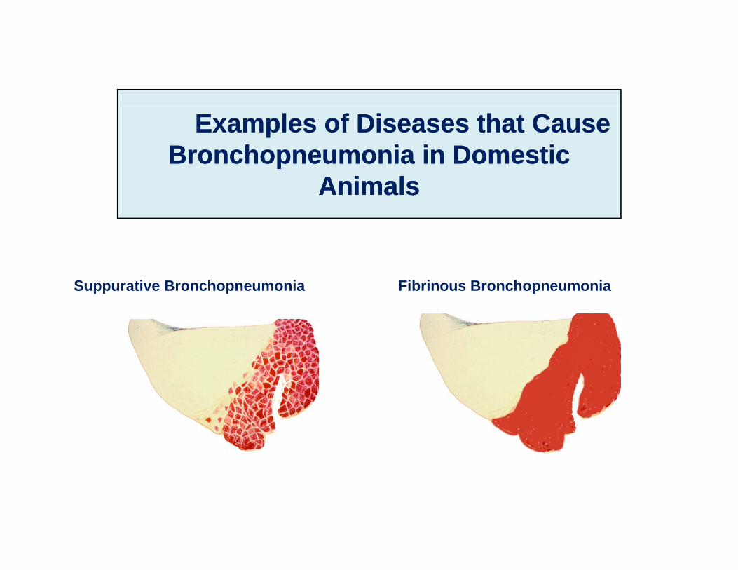
Examples of Diseases that Cause Examples of Diseases that Cause Bronchopneumonia in Domestic Bronchopneumonia in Domestic
A i lA i lAnimalsAnimals
Suppurative Bronchopneumonia Fibrinous Bronchopneumonia
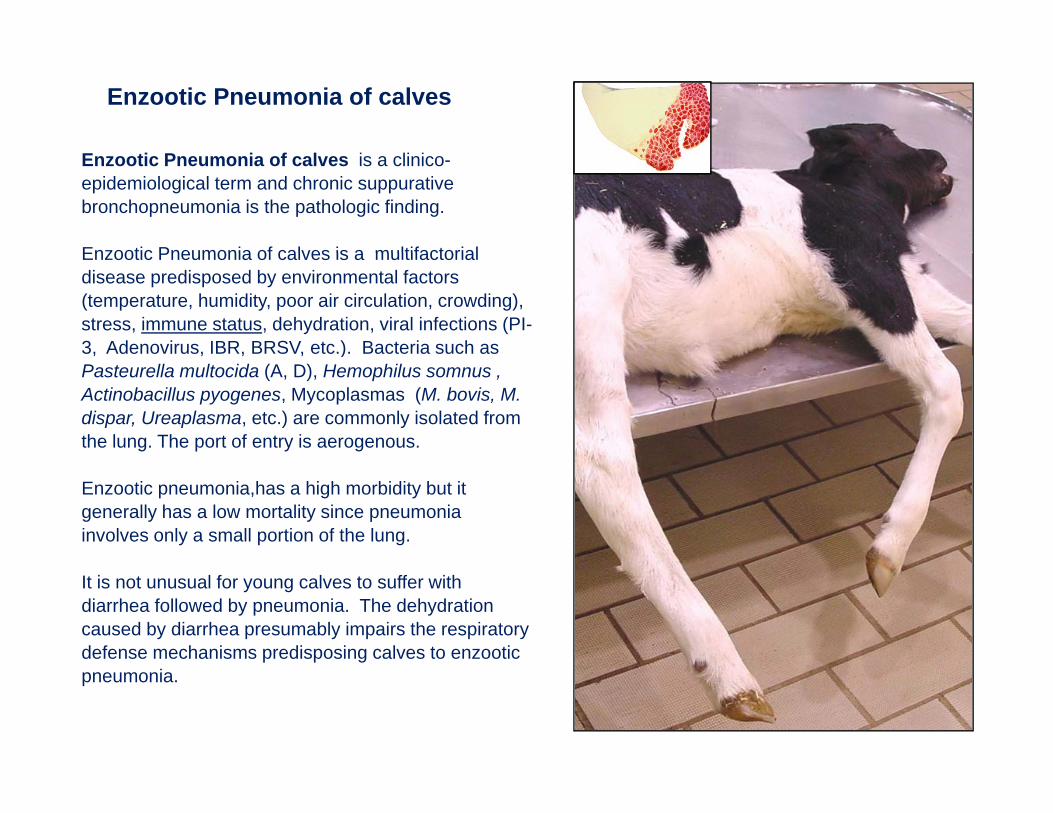
Enzootic Pneumonia of calves
Enzootic Pneumonia of calves is a clinico-epidemiological term and chronic suppurative bronchopneumonia is the pathologic finding.
Enzootic Pneumonia of calves is a multifactorialEnzootic Pneumonia of calves is a multifactorial disease predisposed by environmental factors (temperature, humidity, poor air circulation, crowding), stress, immune status, dehydration, viral infections (PI-3, Adenovirus, IBR, BRSV, etc.). Bacteria such as , , , , )Pasteurella multocida (A, D), Hemophilus somnus , Actinobacillus pyogenes, Mycoplasmas (M. bovis, M. dispar, Ureaplasma, etc.) are commonly isolated from the lung. The port of entry is aerogenous.
Enzootic pneumonia,has a high morbidity but it generally has a low mortality since pneumonia involves only a small portion of the lung.
It is not unusual for young calves to suffer with diarrhea followed by pneumonia. The dehydration caused by diarrhea presumably impairs the respiratory defense mechanisms predisposing calves to enzootic pneumonia.
Enzootic Pneumonia of calves
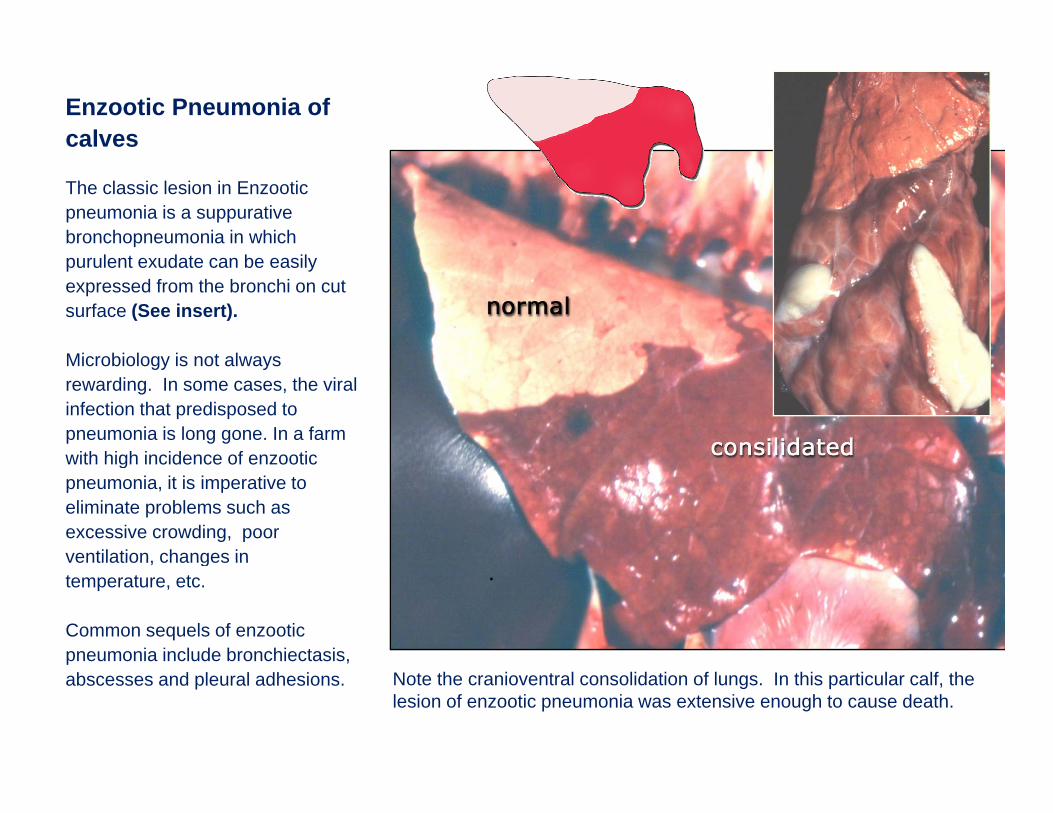
The classic lesion in Enzootic pneumonia is a suppurative bronchopneumonia in which purulent exudate can be easily expressed from the bronchi on cut surface (See insert).
Mi bi l i t lMicrobiology is not always rewarding. In some cases, the viral infection that predisposed to pneumonia is long gone. In a farm with high incidence of enzooticwith high incidence of enzootic pneumonia, it is imperative to eliminate problems such as excessive crowding, poor ventilation changes inventilation, changes in temperature, etc.
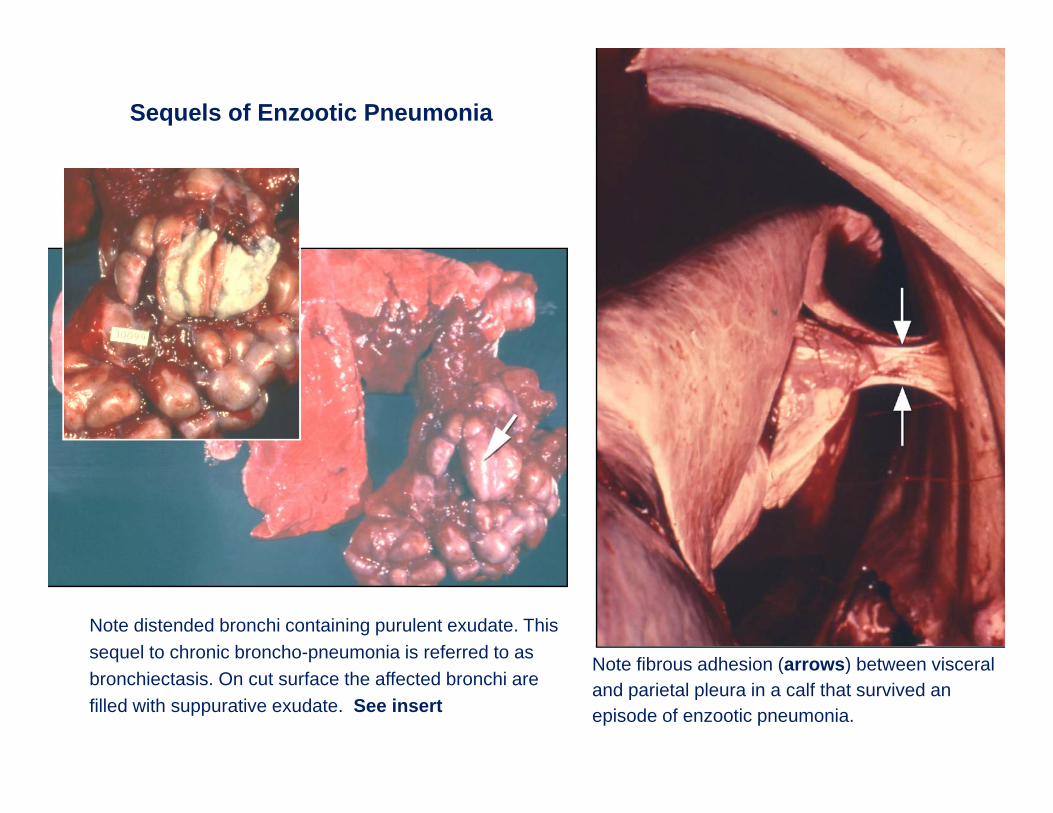
Common sequels of enzootic pneumonia include bronchiectasis,
.
p ,abscesses and pleural adhesions. Note the cranioventral consolidation of lungs. In this particular calf, the
lesion of enzootic pneumonia was extensive enough to cause death.
Enzootic Pneumonia of calves
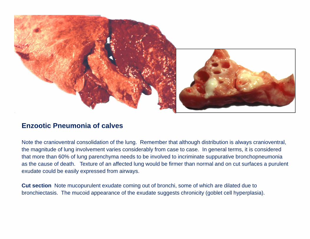
Note the cranioventral consolidation of the lung. Remember that although distribution is always cranioventral, the magnitude of lung involvement varies considerably from case to case. In general terms, it is considered that more than 60% of lung parenchyma needs to be involved to incriminate suppurative bronchopneumonia as the cause of death Texture of an affected lung would be firmer than normal and on cut surfaces a purulentas the cause of death. Texture of an affected lung would be firmer than normal and on cut surfaces a purulent exudate could be easily expressed from airways.
Cut section Note mucopurulent exudate coming out of bronchi, some of which are dilated due to bronchiectasis. The mucoid appearance of the exudate suggests chronicity (goblet cell hyperplasia).pp gg y (g yp p )
Sequels of Enzootic Pneumonia
Note distended bronchi containing purulent exudate. This sequel to chronic broncho-pneumonia is referred to as Note fibrous adhesion (arrows) between visceralbronchiectasis. On cut surface the affected bronchi are filled with suppurative exudate. See insert
Note fibrous adhesion (arrows) between visceral and parietal pleura in a calf that survived an episode of enzootic pneumonia.
Shipping Fever
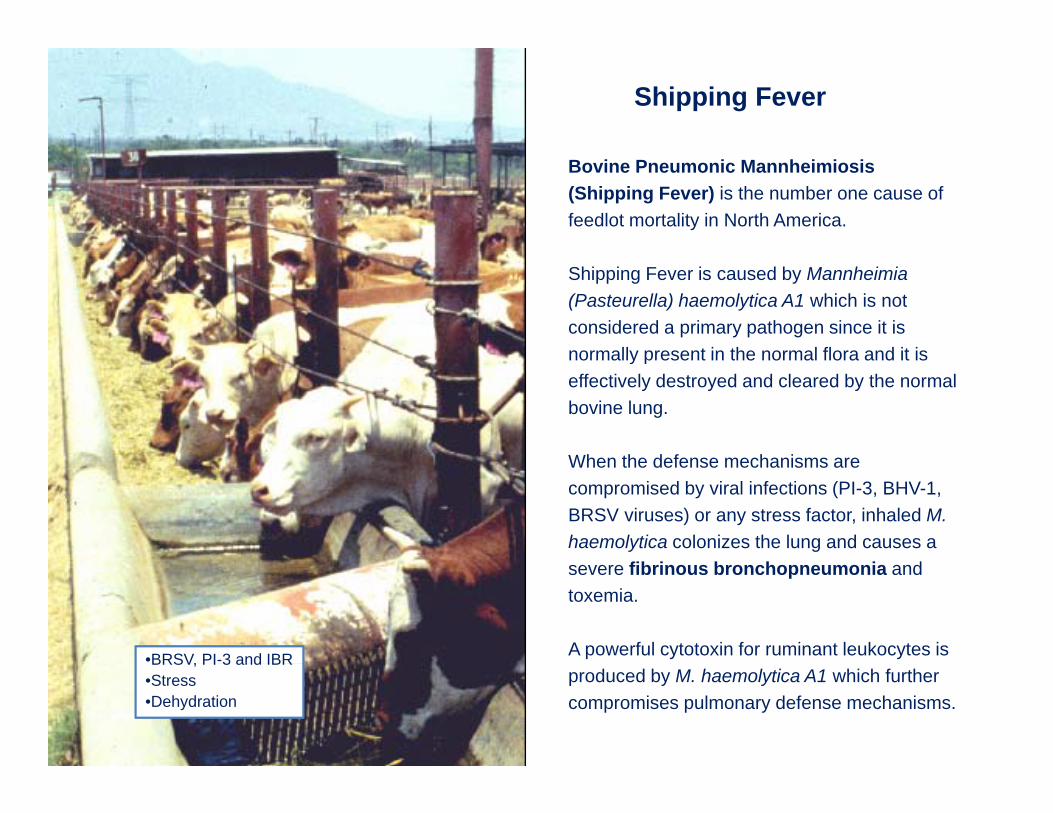
Bovine Pneumonic Mannheimiosis (Shipping Fever) is the number one cause of feedlot mortality in North America.
Shipping Fever is caused by Mannheimia (Pasteurella) haemolytica A1 which is not considered a primary pathogen since it is normally present in the normal flora and it isnormally present in the normal flora and it is effectively destroyed and cleared by the normal bovine lung.
When the defense mechanisms areWhen the defense mechanisms are compromised by viral infections (PI-3, BHV-1, BRSV viruses) or any stress factor, inhaled M. haemolytica colonizes the lung and causes a
•BRSV, PI-3 and IBR
severe fibrinous bronchopneumonia and toxemia.
A powerful cytotoxin for ruminant leukocytes is BRSV, PI 3 and IBR •Stress•Dehydration
produced by M. haemolytica A1 which further compromises pulmonary defense mechanisms.
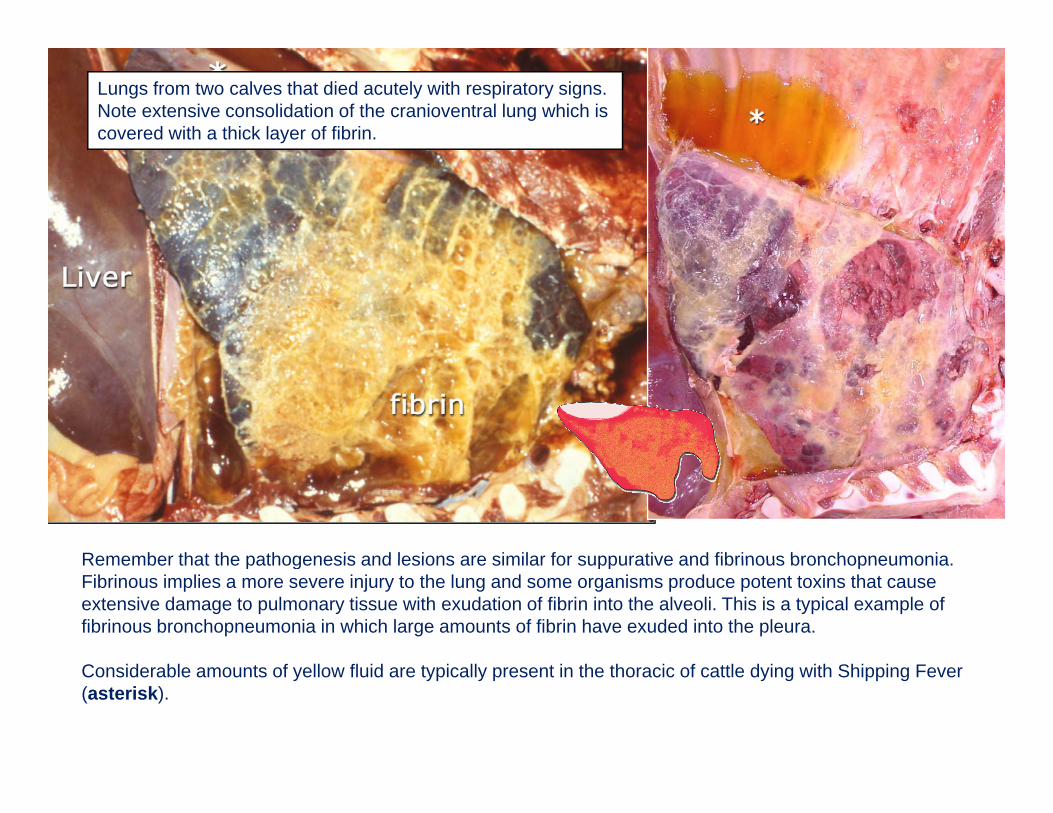
Lungs from two calves that died acutely with respiratory signs. Note extensive consolidation of the cranioventral lung which is covered with a thick layer of fibrin.
Remember that the pathogenesis and lesions are similar for suppurative and fibrinous bronchopneumoniaRemember that the pathogenesis and lesions are similar for suppurative and fibrinous bronchopneumonia. Fibrinous implies a more severe injury to the lung and some organisms produce potent toxins that cause extensive damage to pulmonary tissue with exudation of fibrin into the alveoli. This is a typical example of fibrinous bronchopneumonia in which large amounts of fibrin have exuded into the pleura.
Considerable amounts of yellow fluid are typically present in the thoracic of cattle dying with Shipping Fever (asterisk).
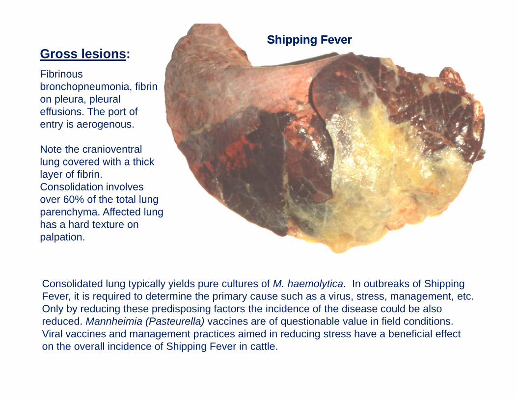
Shipping FeverShipping FeverGross lesions:FibrinousFibrinous bronchopneumonia, fibrin on pleura, pleural effusions. The port of entry is aerogenousentry is aerogenous.
Note the cranioventral lung covered with a thick layer of fibrinlayer of fibrin. Consolidation involves over 60% of the total lung parenchyma. Affected lung has a hard texture onhas a hard texture on palpation.
Consolidated lung typically yields pure cultures of M. haemolytica. In outbreaks of Shipping Fever, it is required to determine the primary cause such as a virus, stress, management, etc. Only by reducing these predisposing factors the incidence of the disease could be also reduced. Mannheimia (Pasteurella) vaccines are of questionable value in field conditions. ( ) qViral vaccines and management practices aimed in reducing stress have a beneficial effect on the overall incidence of Shipping Fever in cattle.
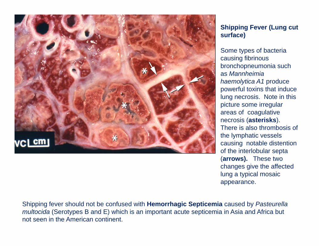
Shipping Fever (Lung cut surface)
Some types of bacteria causing fibrinous bronchopneumonia such as Mannheimia haemolytica A1 produce powerful toxins that induce lung necrosis. Note in this picture some irregular p gareas of coagulative necrosis (asterisks). There is also thrombosis of the lymphatic vessels y pcausing notable distention of the interlobular septa (arrows). These two changes give the affected g glung a typical mosaic appearance.
Shipping fever should not be confused with Hemorrhagic Septicemia caused by Pasteurella multocida (Serotypes B and E) which is an important acute septicemia in Asia and Africa but not seen in the American continent.
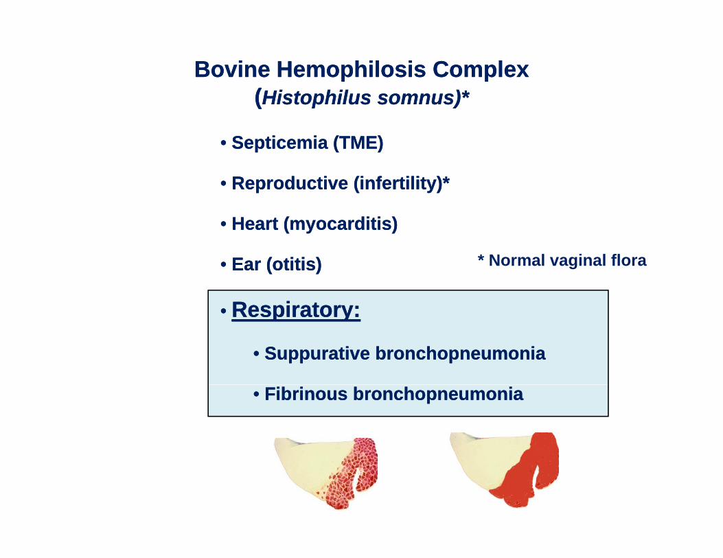
Bovine Hemophilosis ComplexBovine Hemophilosis Complex((Histophilus somnus)*Histophilus somnus)*
•• Septicemia (TME)Septicemia (TME)
((Histophilus somnus)Histophilus somnus)
•• Reproductive (infertility)*Reproductive (infertility)*
•• Heart (myocarditis)Heart (myocarditis)
•• Ear (Ear (otitisotitis))
•• Respiratory:Respiratory:
* Normal vaginal flora
Respiratory:Respiratory:
•• Suppurative bronchopneumoniaSuppurative bronchopneumonia
•• Fibrinous bronchopneumoniaFibrinous bronchopneumonia
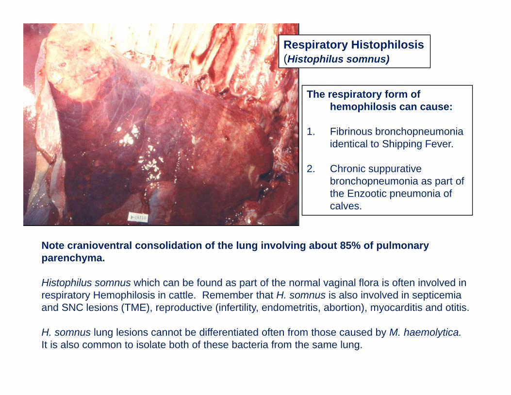
Respiratory Histophilosis(Histophilus somnus)
The respiratory form of hemophilosis can cause:
1. Fibrinous bronchopneumonia identical to Shipping Fever.
2. Chronic suppurative ppbronchopneumonia as part of the Enzootic pneumonia of calves.
Note cranioventral consolidation of the lung involving about 85% of pulmonary parenchyma.
Histophilus somnus which can be found as part of the normal vaginal flora is often involved in respiratory Hemophilosis in cattle. Remember that H. somnus is also involved in septicemia and SNC lesions (TME), reproductive (infertility, endometritis, abortion), myocarditis and otitis.
H. somnus lung lesions cannot be differentiated often from those caused by M. haemolytica.It is also common to isolate both of these bacteria from the same lung.
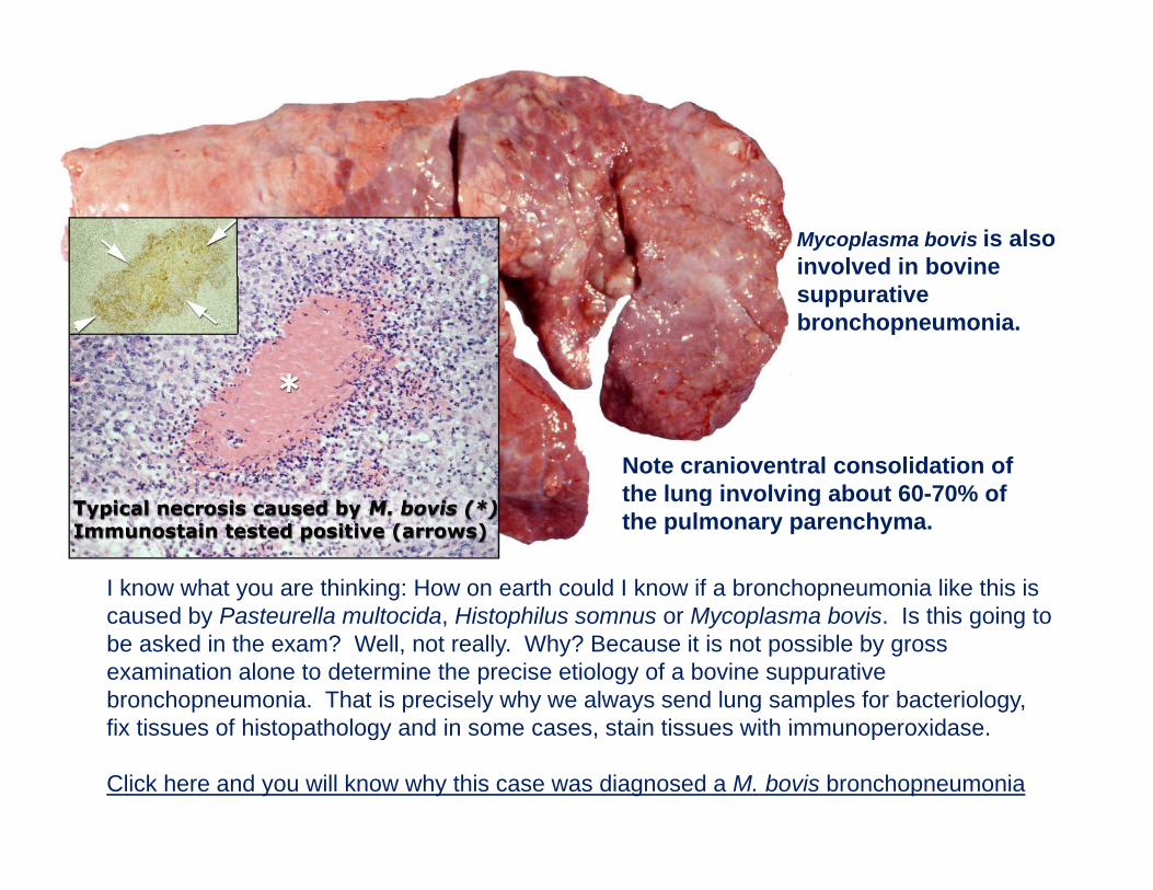
Mycoplasma bovis is also involved in bovine suppurative bronchopneumonia.
Note cranioventral consolidation of the lung involving about 60-70% of
I know what you are thinking: How on earth could I know if a bronchopneumonia like this is caused by Pasteurella multocida Histophilus somnus or Mycoplasma bovis Is this going to
t e u g o g about 60 0% othe pulmonary parenchyma.
caused by Pasteurella multocida, Histophilus somnus or Mycoplasma bovis. Is this going to be asked in the exam? Well, not really. Why? Because it is not possible by gross examination alone to determine the precise etiology of a bovine suppurative bronchopneumonia. That is precisely why we always send lung samples for bacteriology, fix tissues of histopathology and in some cases stain tissues with immunoperoxidasefix tissues of histopathology and in some cases, stain tissues with immunoperoxidase.
Click here and you will know why this case was diagnosed a M. bovis bronchopneumonia
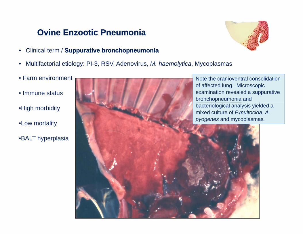
Ovine Enzootic PneumoniaOvine Enzootic Pneumonia
• Clinical term / Suppurative bronchopneumoniaSuppurative bronchopneumonia
• Multifactorial etiology: PI-3, RSV, Adenovirus, M. haemolytica, Mycoplasmas
• Farm environment
• Immune status
Note the cranioventral consolidation of affected lung. Microscopic examination revealed a suppurative bronchopneumonia and
•High morbidity
•Low mortality
bacteriological analysis yielded a mixed culture of P.multocida, A. pyogenes and mycoplasmas.
•BALT hyperplasia
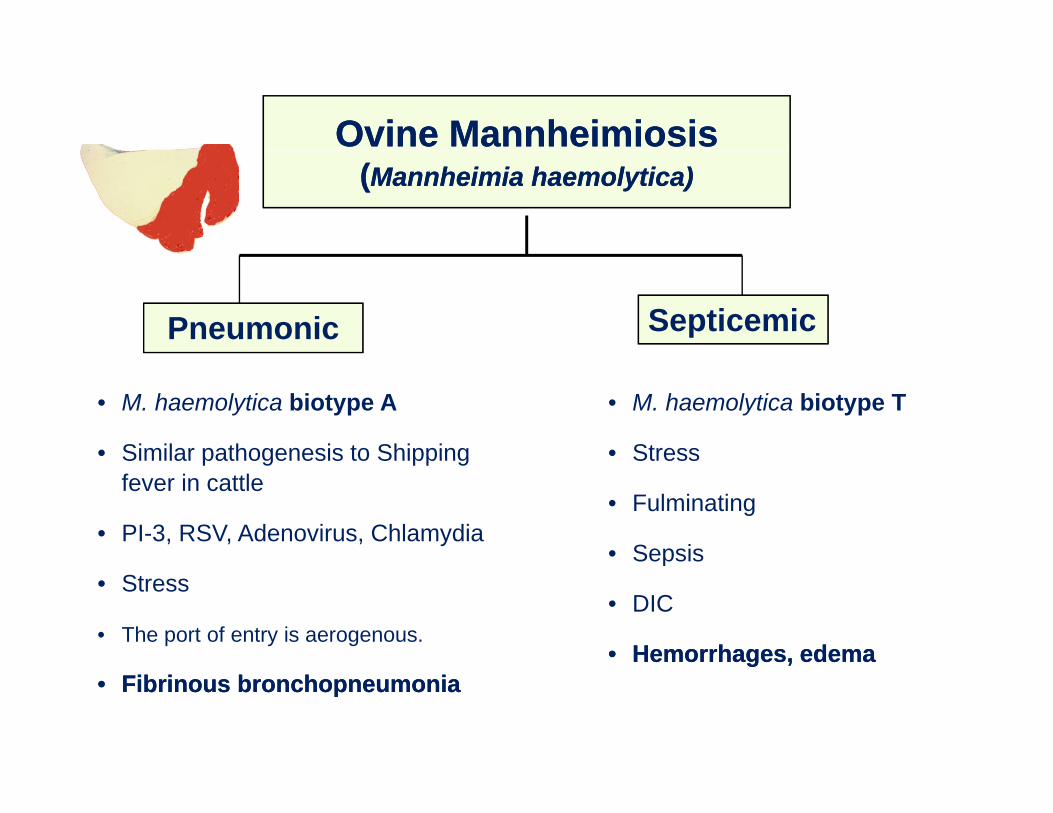
Ovine MannheimiosisOvine Mannheimiosis((Mannheimia haemolytica)Mannheimia haemolytica)
Pneumonic Septicemic
• M. haemolytica biotype A
• Similar pathogenesis to Shipping
• M. haemolytica biotype T
• StressSimilar pathogenesis to Shipping fever in cattle
• PI-3, RSV, Adenovirus, Chlamydia
Stress
• Fulminating
• Sepsis• Stress
• The port of entry is aerogenous.
Sepsis
• DIC
•• Hemorrhages, edemaHemorrhages, edema•• Fibrinous bronchopneumoniaFibrinous bronchopneumonia
g ,g ,
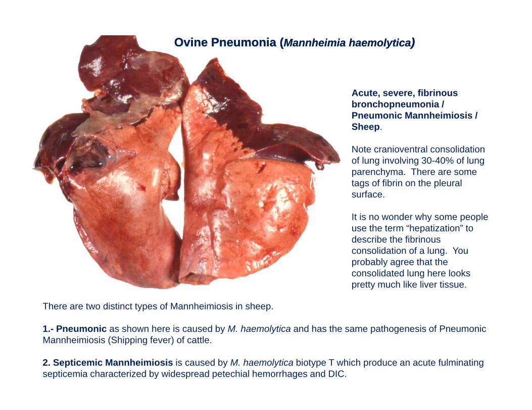
Ovine Pneumonia (Ovine Pneumonia (Mannheimia haemolyticaMannheimia haemolytica))
Acute, severe, fibrinous bronchopneumonia / Pneumonic Mannheimiosis / SheepSheep.
Note cranioventral consolidation of lung involving 30-40% of lung parenchyma. There are some tags of fibrin on the pleural surface.
It is no wonder why some people use the term “hepatization” touse the term hepatization to describe the fibrinous consolidation of a lung. You probably agree that the consolidated lung here looks prett m ch like li er tiss e
There are two distinct types of Mannheimiosis in sheep.
1.- Pneumonic as shown here is caused by M. haemolytica and has the same pathogenesis of Pneumonic
pretty much like liver tissue.
Mannheimiosis (Shipping fever) of cattle.
2. Septicemic Mannheimiosis is caused by M. haemolytica biotype T which produce an acute fulminating septicemia characterized by widespread petechial hemorrhages and DIC.
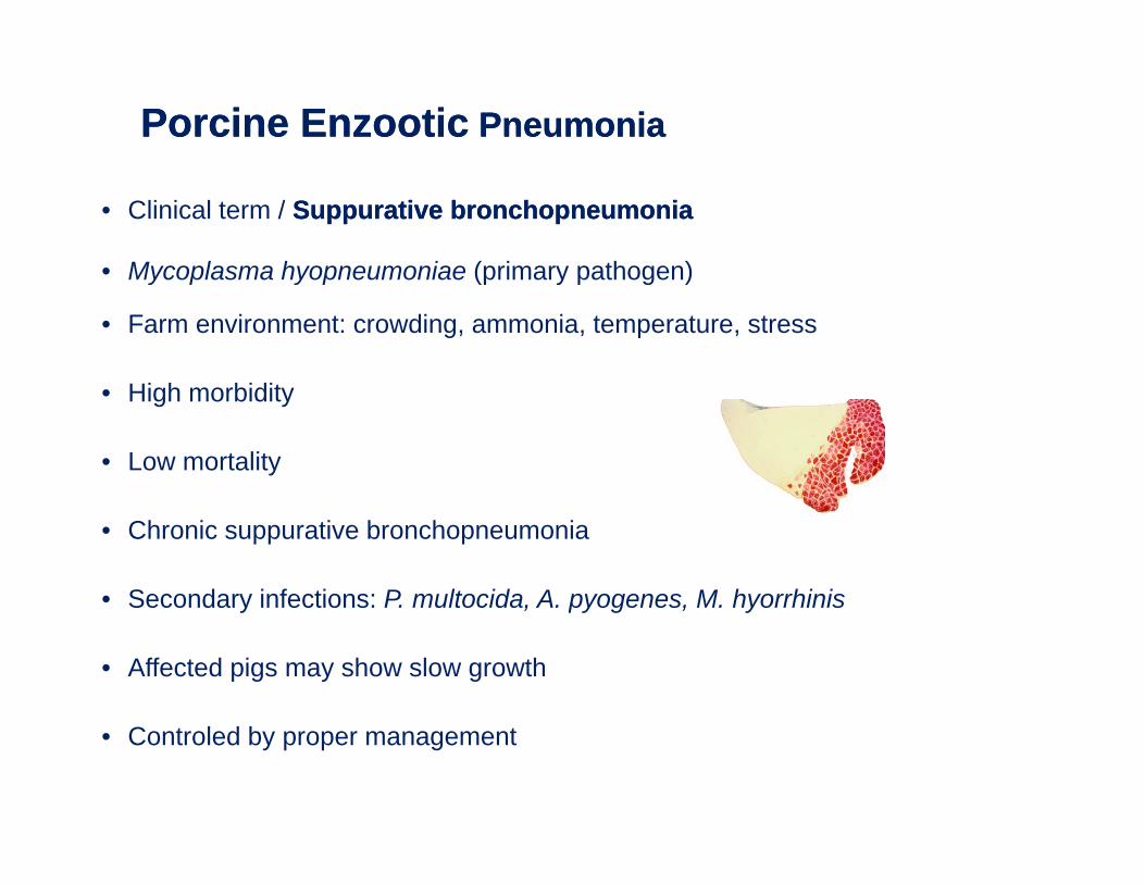
Porcine EnzooticPorcine Enzootic Pneumonia Pneumonia
• Clinical term / Suppurative bronchopneumoniaSuppurative bronchopneumonia
• Mycoplasma hyopneumoniae (primary pathogen)Mycoplasma hyopneumoniae (primary pathogen)
• Farm environment: crowding, ammonia, temperature, stress
Hi h bidit• High morbidity
• Low mortality
• Chronic suppurative bronchopneumonia
• Secondary infections: P. multocida, A. pyogenes, M. hyorrhinisy , py g , y
• Affected pigs may show slow growth
• Controled by proper management
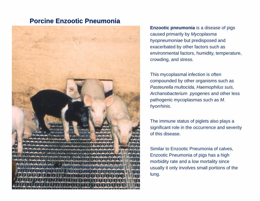
Porcine Enzootic Pneumonia Porcine Enzootic Pneumonia Enzootic pneumonia is a disease of pigs caused primarily by Mycoplasma hyopneumoniae but predisposed and exacerbated by other factors such as environmental factors, humidity, temperature, crowding, and stress.
This mycoplasmal infection is often compounded by other organisms such as Pasteurella multocida Haemophilus suisPasteurella multocida, Haemophilus suis, Archanobacterium pyogenes and other less pathogenic mycoplasmas such as M. hyorrhinis.
The immune status of piglets also plays a significant role in the occurrence and severity of this disease.
Similar to Enzootic Pneumonia of calves, Enzootic Pneumonia of pigs has a high morbidity rate and a low mortality since usually it only involves small portions of the lung.
Porcine Enzootic PneumoniaPorcine Enzootic Pneumonia
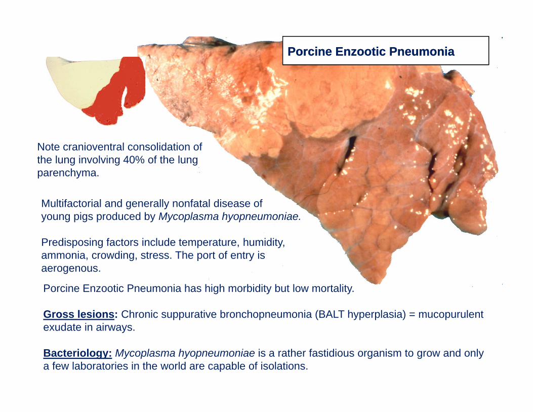
Note cranioventral consolidation of the lung involving 40% of the lung parenchyma.
Multifactorial and generally nonfatal disease of young pigs produced by Mycoplasma hyopneumoniae.
Predisposing factors include temperature, humidity, ammonia, crowding, stress. The port of entry is aerogenous.
Porcine Enzootic Pneumonia has high morbidity but low mortality.
Gross lesions: Chronic suppurative bronchopneumonia (BALT hyperplasia) = mucopurulent exudate in airways.
Bacteriology: Mycoplasma hyopneumoniae is a rather fastidious organism to grow and only a few laboratories in the world are capable of isolations.
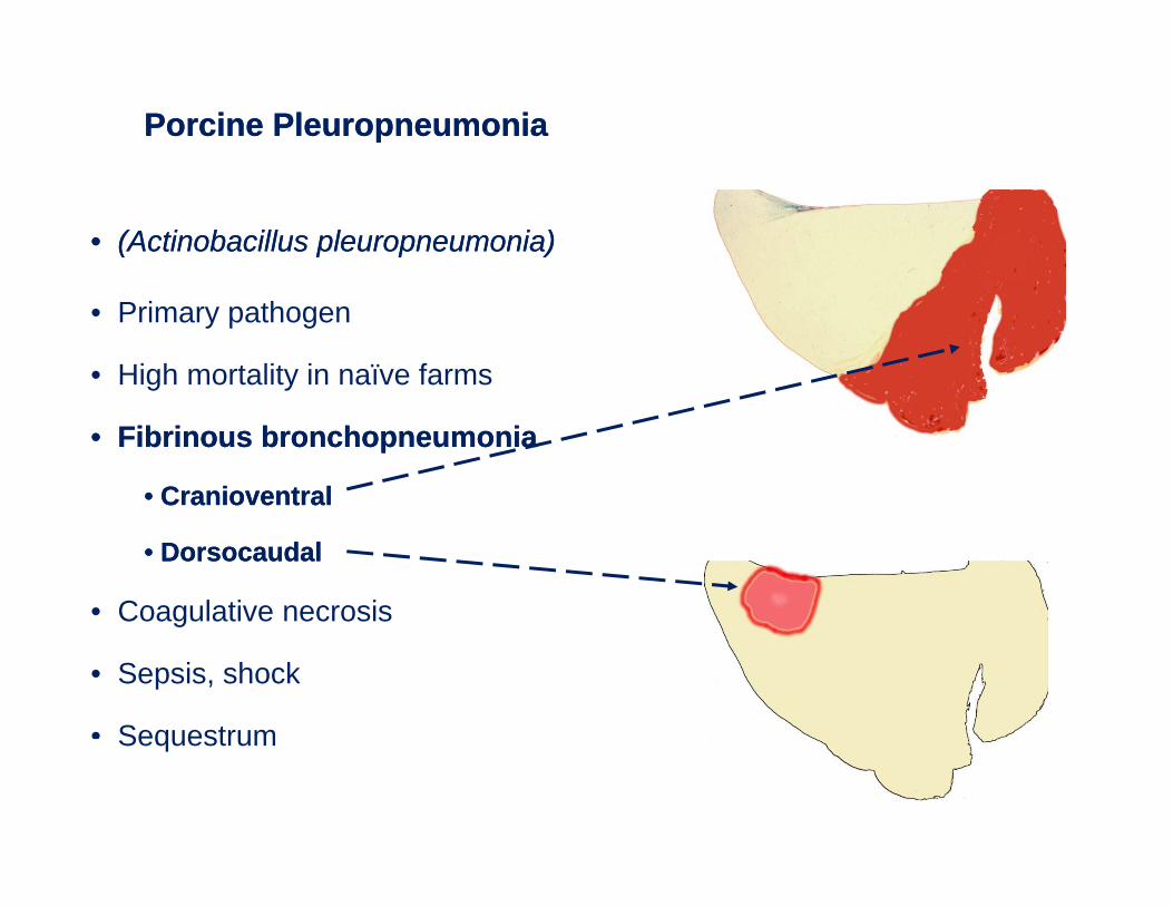
Porcine PleuropneumoniaPorcine Pleuropneumonia
•• (Actinobacillus (Actinobacillus pleuropneumoniapleuropneumonia))
• Primary pathogen
• High mortality in naïve farms
•• Fibrinous bronchopneumoniaFibrinous bronchopneumonia
•• CranioventralCranioventral
•• DorsocaudalDorsocaudal
• Coagulative necrosisg
• Sepsis, shock
• SequestrumSequestrum
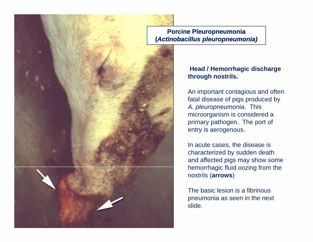
Porcine Pleuropneumonia Porcine Pleuropneumonia ((Actinobacillus Actinobacillus pleuropneumoniapleuropneumonia))
Head / Hemorrhagic discharge through nostrilsthrough nostrils.
An important contagious and often fatal disease of pigs produced by A pleuropneumonia ThisA. pleuropneumonia. This microorganism is considered a primary pathogen. The port of entry is aerogenous.
In acute cases, the disease is characterized by sudden death and affected pigs may show some hemorrhagic fluid oozing from thehemorrhagic fluid oozing from the nostrils (arrows)
The basic lesion is a fibrinous pneumonia as seen in the nextpneumonia as seen in the next slide.
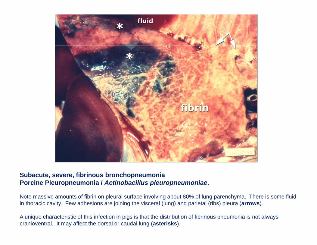
Subacute, severe, fibrinous bronchopneumoniaPorcine Pleuropneumonia / Actinobacillus pleuropneumoniae.
Note massive amounts of fibrin on pleural surface involving about 80% of lung parenchyma. There is some fluid in thoracic cavity. Few adhesions are joining the visceral (lung) and parietal (ribs) pleura (arrows).
A unique characteristic of this infection in pigs is that the distribution of fibrinous pneumonia is not always cranioventral. It may affect the dorsal or caudal lung (asterisks).
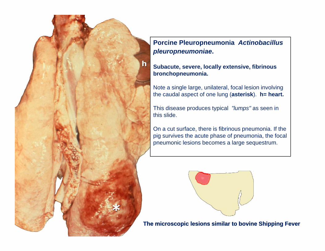
Porcine Pleuropneumonia Actinobacillus pleuropneumoniae.
Subacute, severe, locally extensive, fibrinous bronchopneumonia.
Note a single large, unilateral, focal lesion involving the caudal aspect of one lung (asterisk). h= heart.
This disease produces typical "lumps" as seen inThis disease produces typical lumps as seen in this slide.
On a cut surface, there is fibrinous pneumonia. If the pig survives the acute phase of pneumonia, the focal
i l i b l tpneumonic lesions becomes a large sequestrum.
The microscopic lesions similar to bovine Shipping FeverThe microscopic lesions similar to bovine Shipping Fever
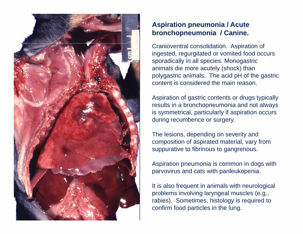
Aspiration pneumonia / Acute bronchopneumonia / Canine.
Cranioventral consolidation. Aspiration of ingested, regurgitated or vomited food occurs sporadically in all species. Monogastric animals die more acutely (shock) than polygastric animals. The acid pH of the gastric content is considered the main reason.
Aspiration of gastric contents or drugs typically results in a bronchopneumonia and not always is symmetrical, particularly if aspiration occurs during recumbence or surgery.
The lesions, depending on severity and composition of aspirated material, vary from suppurative to fibrinous to gangrenous.
Aspiration pneumonia is common in dogs with parvovirus and cats with panleukopenia.
It is also frequent in animals with neurological bl i l i l l l (problems involving laryngeal muscles (e.g.,
rabies). Sometimes, histology is required to confirm food particles in the lung.
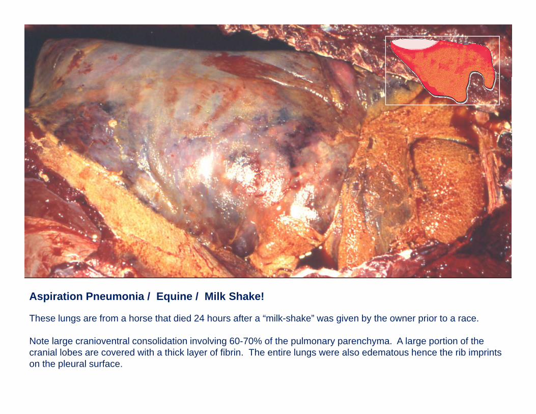
Aspiration Pneumonia / Equine / Milk Shake!
These lungs are from a horse that died 24 hours after a “milk-shake” was given by the owner prior to a race.
N t l i t l lid ti i l i 60 70% f th l h A l ti f thNote large cranioventral consolidation involving 60-70% of the pulmonary parenchyma. A large portion of the cranial lobes are covered with a thick layer of fibrin. The entire lungs were also edematous hence the rib imprints on the pleural surface.
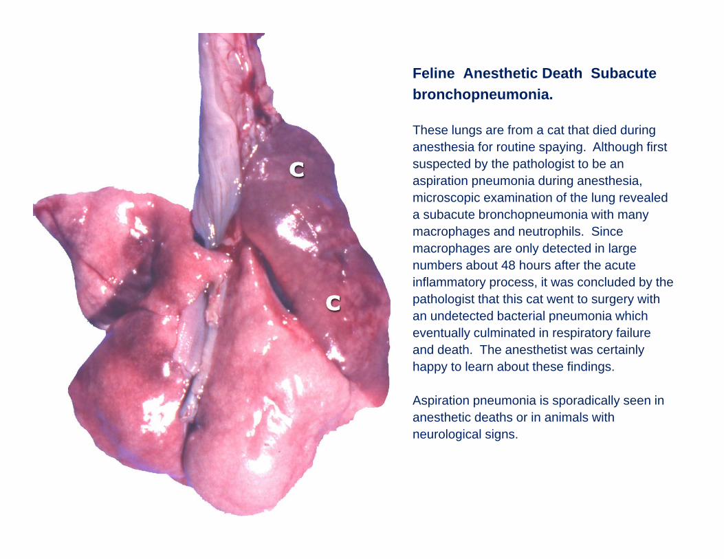
Feline Anesthetic Death Subacute bronchopneumonia.
These lungs are from a cat that died during anesthesia for routine spaying. Although first suspected by the pathologist to be an aspiration pneumonia during anesthesia, microscopic examination of the lung revealed a subacute bronchopneumonia with many macrophages and neutrophils. Since
h l d t t d i lmacrophages are only detected in large numbers about 48 hours after the acute inflammatory process, it was concluded by the pathologist that this cat went to surgery with an undetected bacterial pneumonia whichan undetected bacterial pneumonia which eventually culminated in respiratory failure and death. The anesthetist was certainly happy to learn about these findings.
Aspiration pneumonia is sporadically seen in anesthetic deaths or in animals with neurological signs.
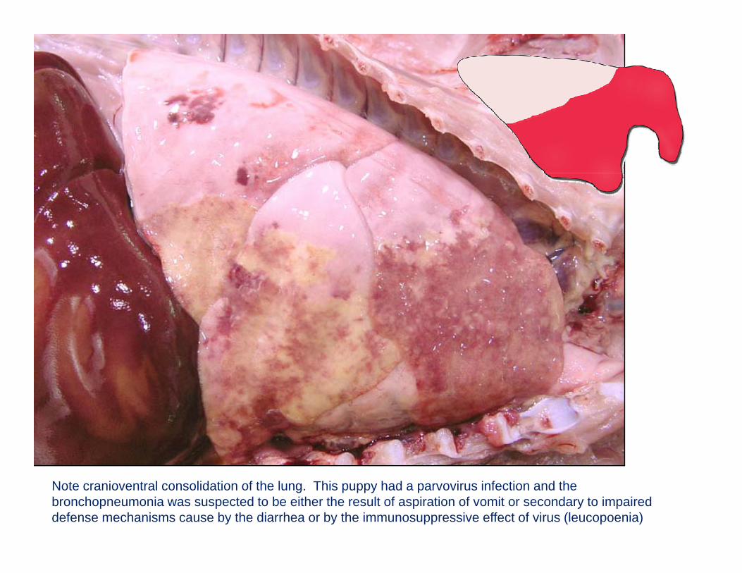
Note cranioventral consolidation of the lung. This puppy had a parvovirus infection and the bronchopneumonia was suspected to be either the result of aspiration of vomit or secondary to impaired defense mechanisms cause by the diarrhea or by the immunosuppressive effect of virus (leucopoenia)
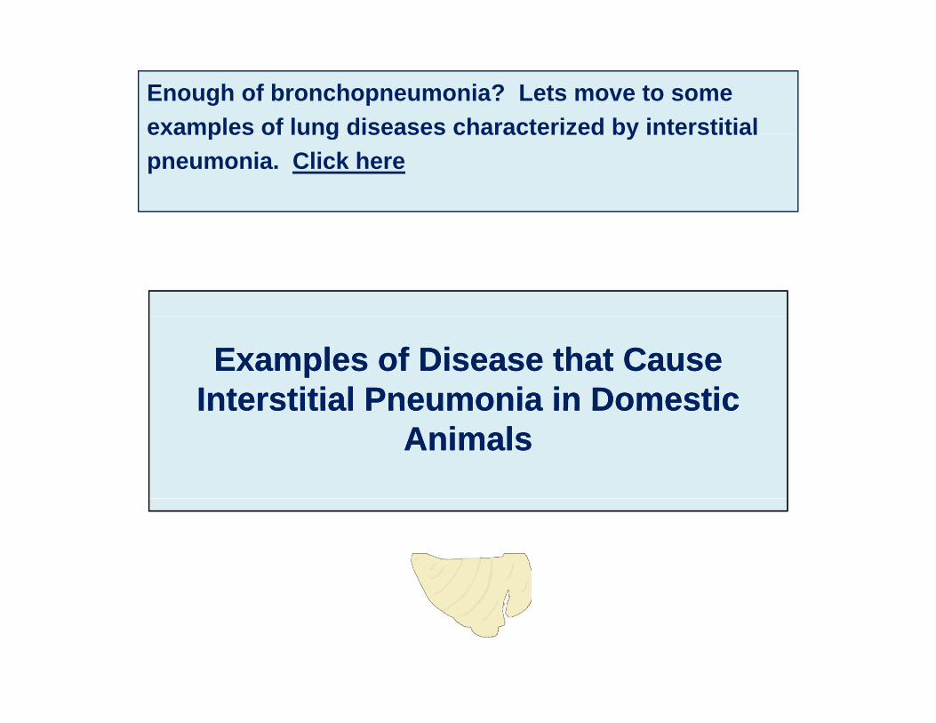
Enough of bronchopneumonia? Lets move to some examples of lung diseases characterized by interstitialexamples of lung diseases characterized by interstitial pneumonia. Click here
Examples of Disease that Cause Examples of Disease that Cause Interstitial Pneumonia in DomesticInterstitial Pneumonia in DomesticInterstitial Pneumonia in Domestic Interstitial Pneumonia in Domestic
AnimalsAnimals
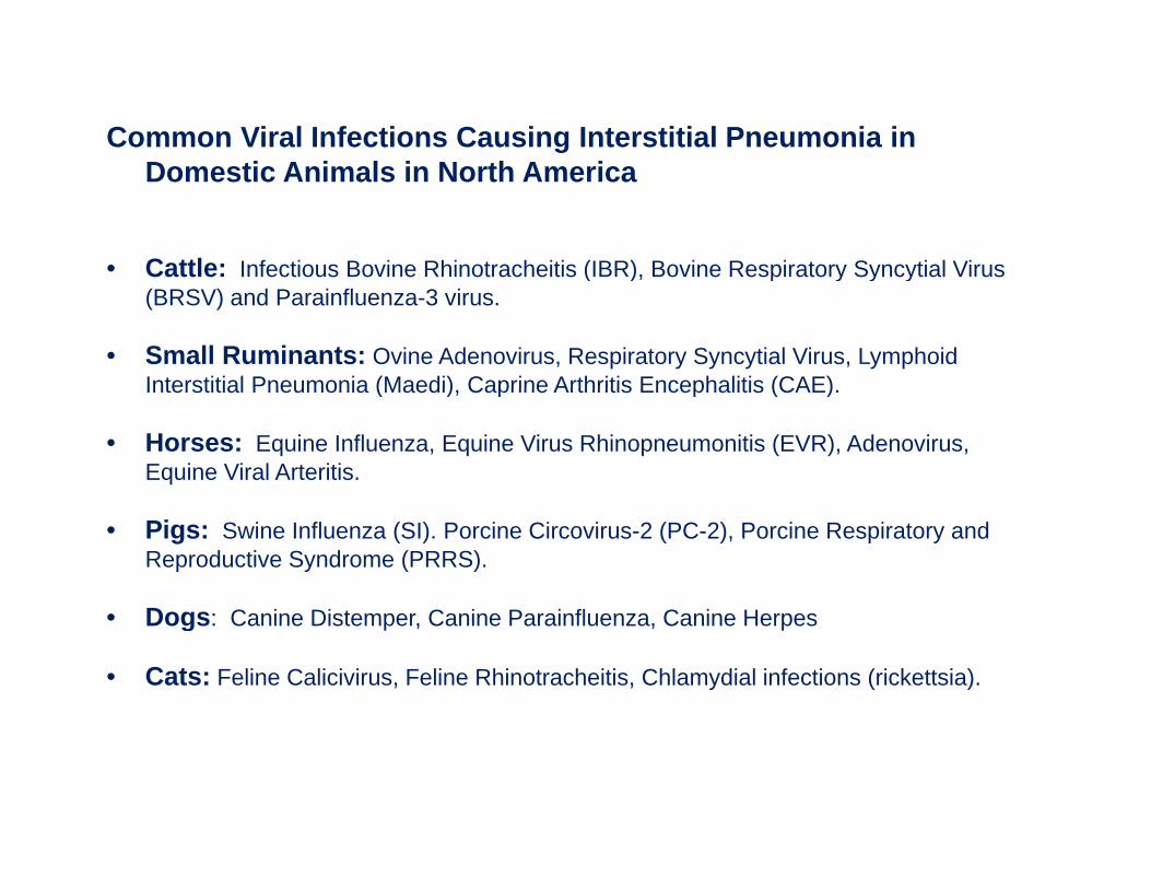
Common Viral Infections Causing Interstitial Pneumonia in D ti A i l i N th A iDomestic Animals in North America
• Cattle: Infectious Bovine Rhinotracheitis (IBR), Bovine Respiratory Syncytial VirusCattle: Infectious Bovine Rhinotracheitis (IBR), Bovine Respiratory Syncytial Virus (BRSV) and Parainfluenza-3 virus.
• Small Ruminants: Ovine Adenovirus, Respiratory Syncytial Virus, Lymphoid Interstitial Pneumonia (Maedi) Caprine Arthritis Encephalitis (CAE)Interstitial Pneumonia (Maedi), Caprine Arthritis Encephalitis (CAE).
• Horses: Equine Influenza, Equine Virus Rhinopneumonitis (EVR), Adenovirus, Equine Viral Arteritis.
• Pigs: Swine Influenza (SI). Porcine Circovirus-2 (PC-2), Porcine Respiratory and Reproductive Syndrome (PRRS).
Dogs C i Di t C i P i fl C i H• Dogs: Canine Distemper, Canine Parainfluenza, Canine Herpes
• Cats: Feline Calicivirus, Feline Rhinotracheitis, Chlamydial infections (rickettsia).
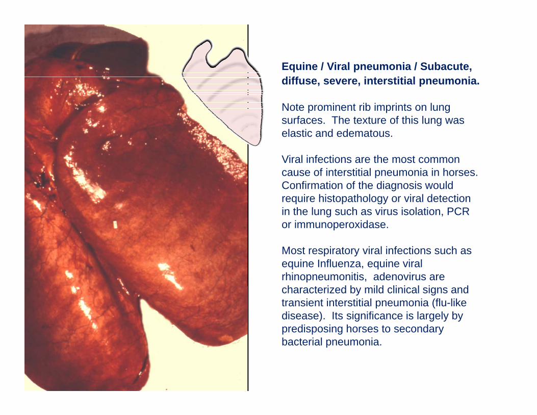
Equine / Viral pneumonia / Subacute, diff i t titi l idiffuse, severe, interstitial pneumonia.
Note prominent rib imprints on lung surfaces. The texture of this lung was l i d delastic and edematous.
Viral infections are the most common cause of interstitial pneumonia in horses. C fi ti f th di i ldConfirmation of the diagnosis would require histopathology or viral detection in the lung such as virus isolation, PCR or immunoperoxidase.
Most respiratory viral infections such as equine Influenza, equine viral rhinopneumonitis, adenovirus are h t i d b ild li i l i dcharacterized by mild clinical signs and
transient interstitial pneumonia (flu-like disease). Its significance is largely by predisposing horses to secondary b t i l ibacterial pneumonia.
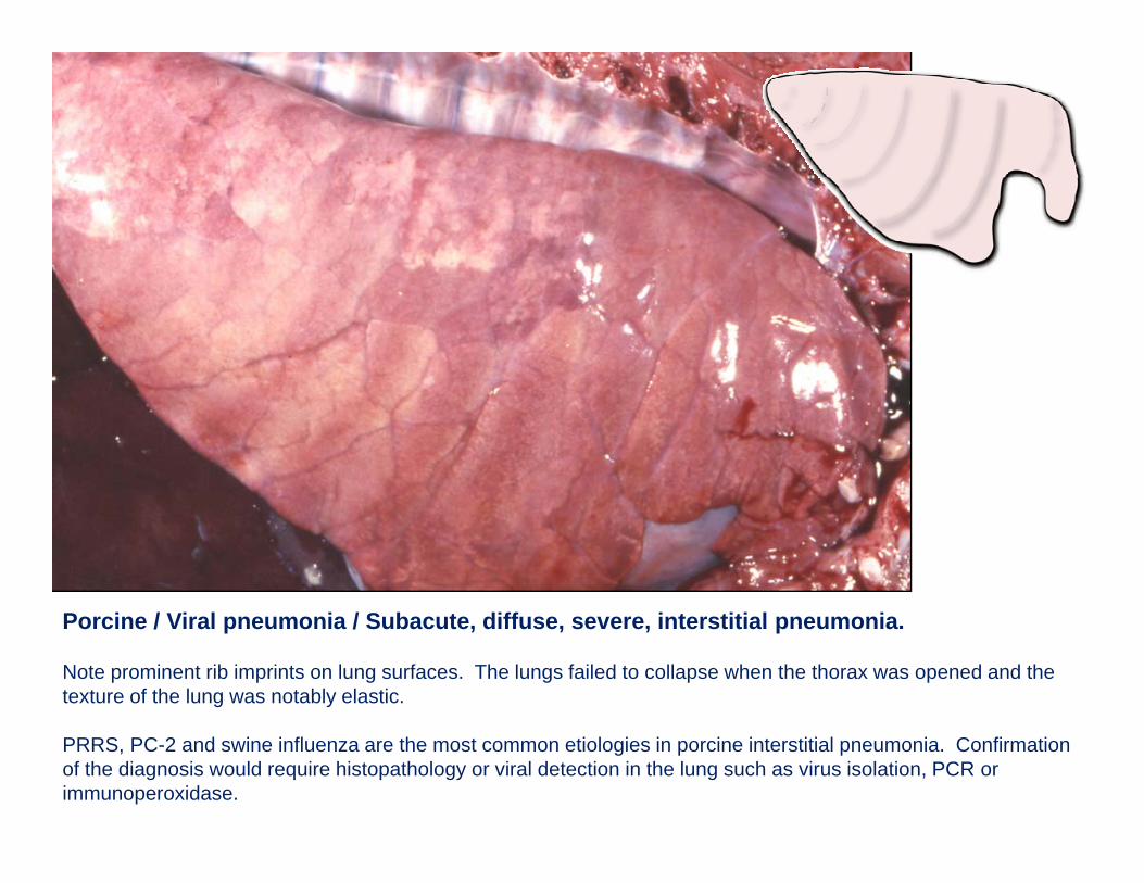
Porcine / Viral pneumonia / Subacute, diffuse, severe, interstitial pneumonia.
Note prominent rib imprints on lung surfaces. The lungs failed to collapse when the thorax was opened and the texture of the lung was notably elastic.
PRRS, PC-2 and swine influenza are the most common etiologies in porcine interstitial pneumonia. Confirmation of the diagnosis would require histopathology or viral detection in the lung such as virus isolation, PCR or immunoperoxidase.
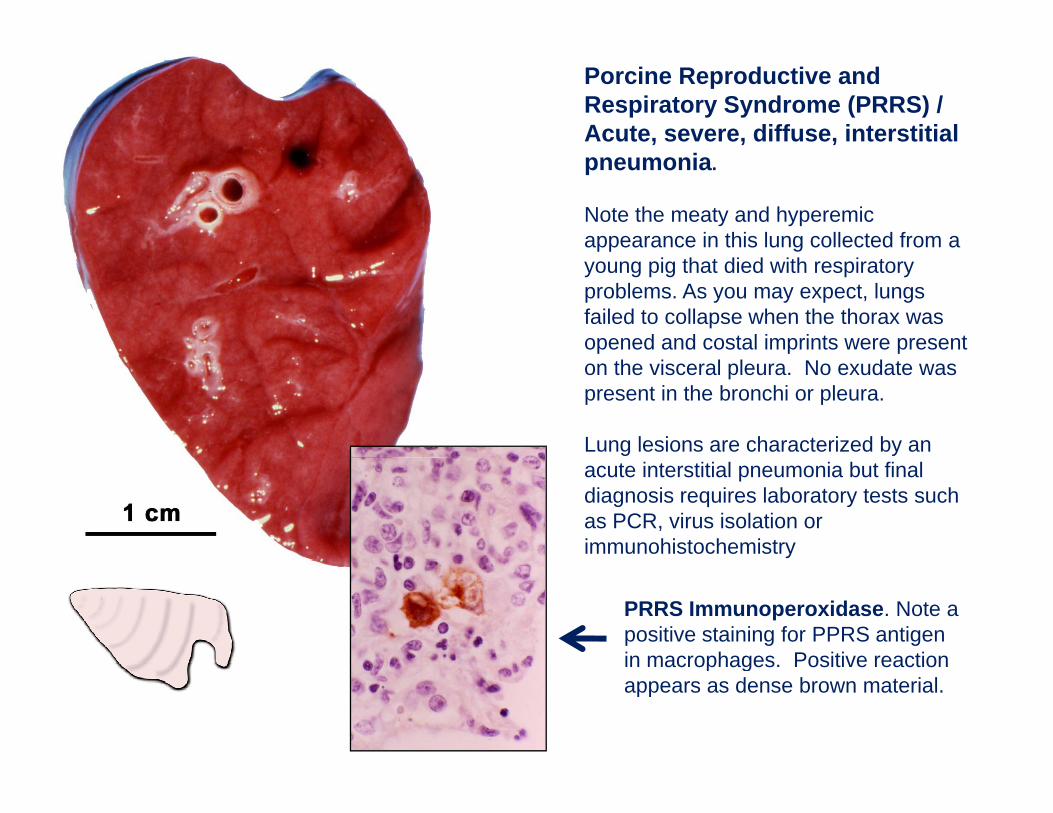
Porcine Reproductive and Respiratory Syndrome (PRRS) / Acute, severe, diffuse, interstitial pneumonia.
Note the meaty and hyperemic appearance in this lung collected from a young pig that died with respiratory problems. As you may expect, lungs failed to collapse when the thorax was opened and costal imprints were present on the visceral pleura. No exudate was present in the bronchi or pleura.
Lung lesions are characterized by an acute interstitial pneumonia but final diagnosis requires laboratory tests such as PCR, virus isolation or immunohistochemistry
PRRS Immunoperoxidase. Note a positive staining for PPRS antigen in macrophages Positive reactionin macrophages. Positive reaction appears as dense brown material.
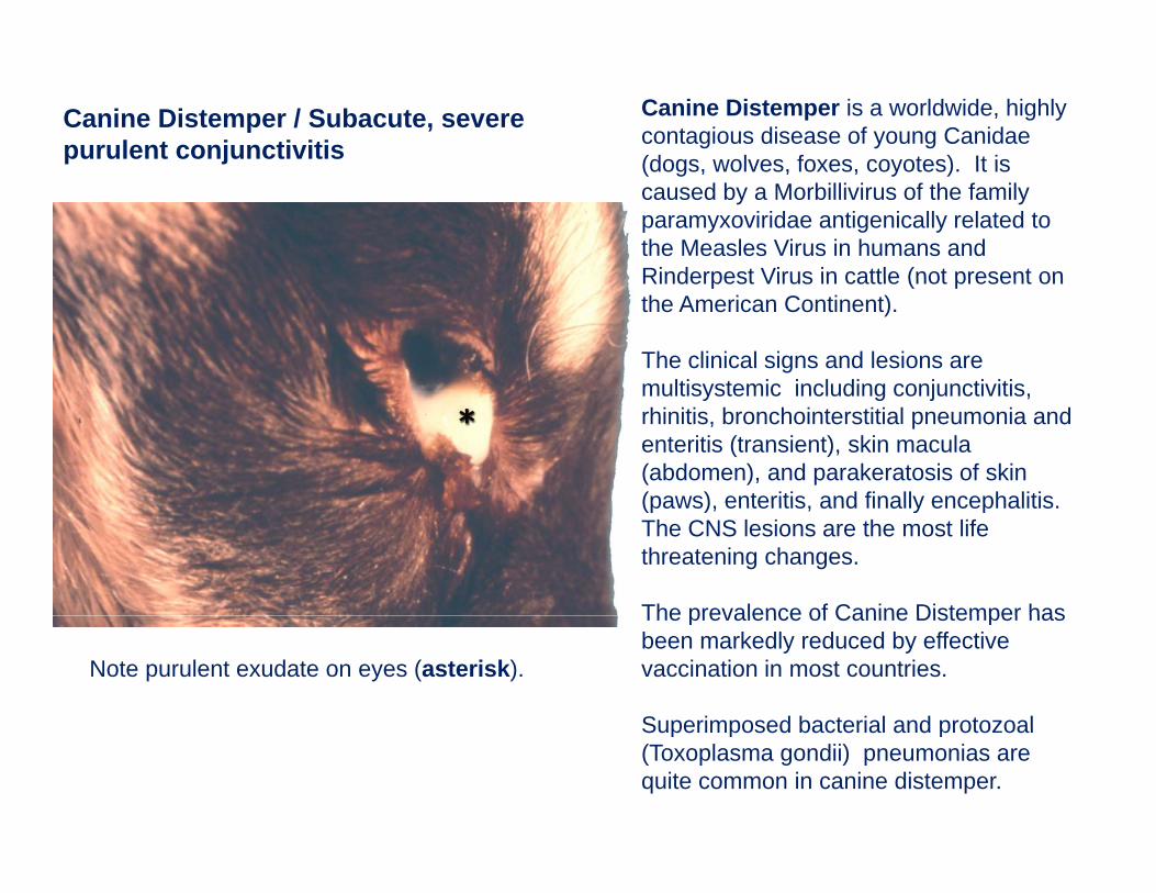
Canine Distemper is a worldwide, highly contagious disease of young Canidae (dogs wolves foxes coyotes) It is
Canine Distemper / Subacute, severe purulent conjunctivitis (dogs, wolves, foxes, coyotes). It is
caused by a Morbillivirus of the family paramyxoviridae antigenically related to the Measles Virus in humans and Rinderpest Virus in cattle (not present onRinderpest Virus in cattle (not present on the American Continent).
The clinical signs and lesions are multisystemic including conjunctivitismultisystemic including conjunctivitis, rhinitis, bronchointerstitial pneumonia and enteritis (transient), skin macula (abdomen), and parakeratosis of skin (paws) enteritis and finally encephalitis(paws), enteritis, and finally encephalitis. The CNS lesions are the most life threatening changes.
The prevalence of Canine Distemper hasThe prevalence of Canine Distemper has been markedly reduced by effective vaccination in most countries.
Superimposed bacterial and protozoal
Note purulent exudate on eyes (asterisk).
Superimposed bacterial and protozoal (Toxoplasma gondii) pneumonias are quite common in canine distemper.
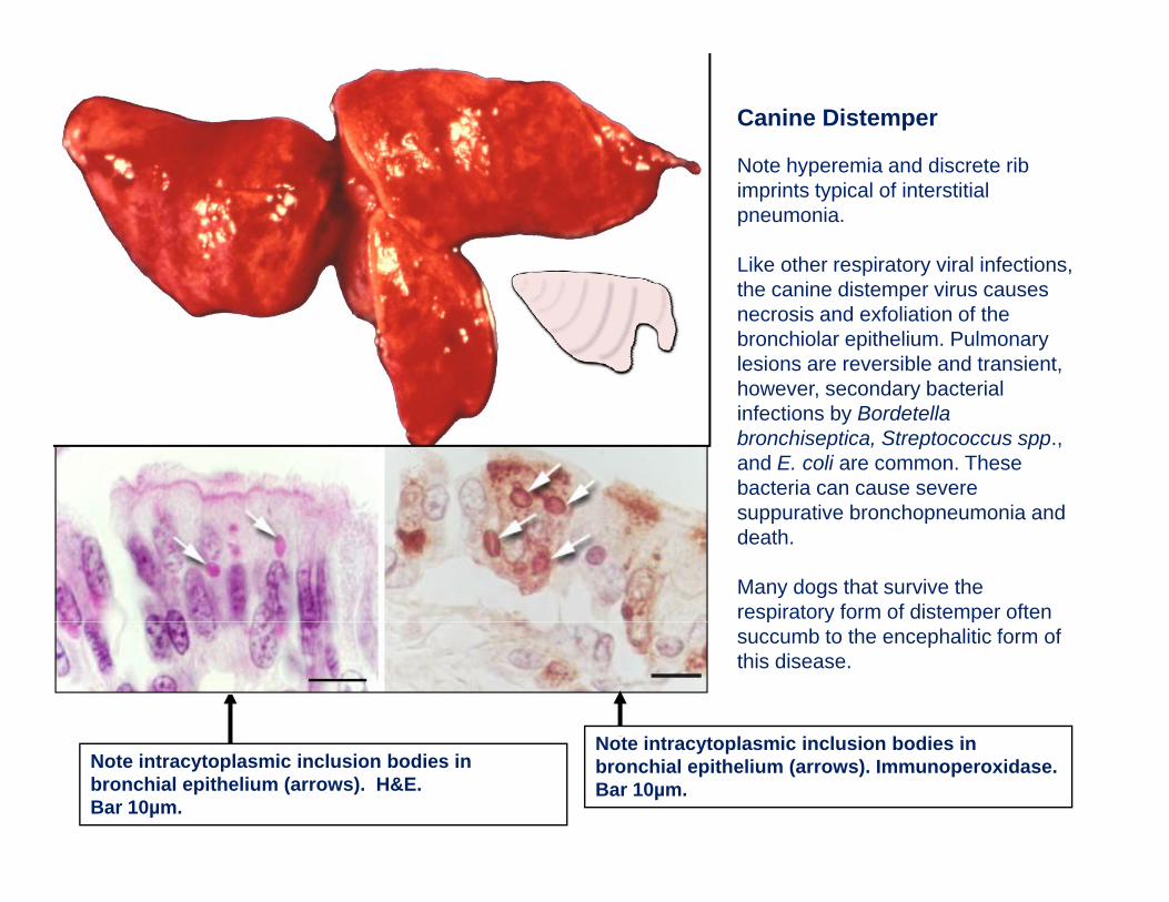
Canine Distemper
Note hyperemia and discrete ribNote hyperemia and discrete rib imprints typical of interstitial pneumonia.
Like other respiratory viral infections, the canine distemper virus causes necrosis and exfoliation of the bronchiolar epithelium. Pulmonary lesions are reversible and transient, however, secondary bacterialhowever, secondary bacterial infections by Bordetella bronchiseptica, Streptococcus spp., and E. coli are common. These bacteria can cause severe
ti b h i dsuppurative bronchopneumonia and death.
Many dogs that survive the respiratory form of distemper often p y psuccumb to the encephalitic form of this disease.
Note intracytoplasmic inclusion bodies in bronchial epithelium (arrows). H&E. Bar 10µm.
Note intracytoplasmic inclusion bodies in bronchial epithelium (arrows). Immunoperoxidase. Bar 10µm.
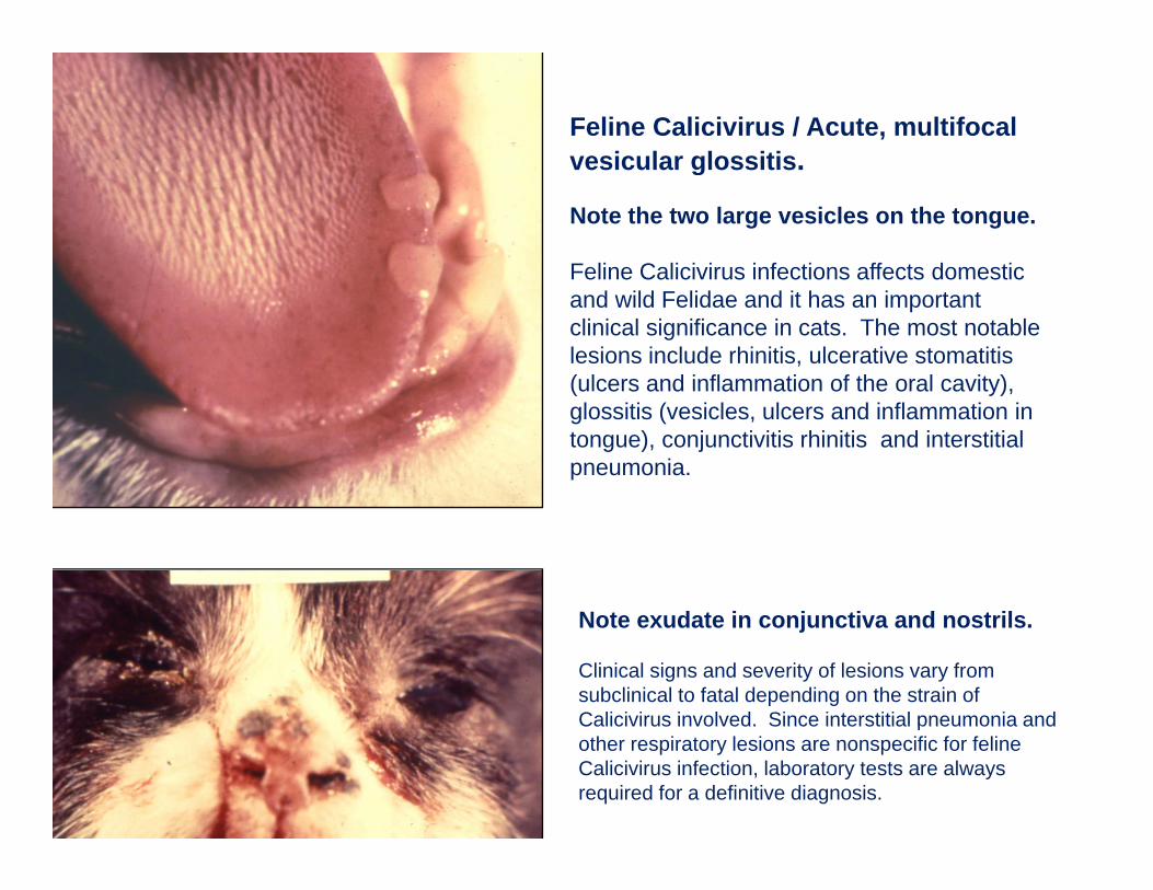
Feline Calicivirus / Acute, multifocal vesicular glossitisvesicular glossitis.
Note the two large vesicles on the tongue.
Feline Calicivirus infections affects domesticFeline Calicivirus infections affects domestic and wild Felidae and it has an important clinical significance in cats. The most notable lesions include rhinitis, ulcerative stomatitis (ulcers and inflammation of the oral cavity),(ulcers and inflammation of the oral cavity), glossitis (vesicles, ulcers and inflammation in tongue), conjunctivitis rhinitis and interstitial pneumonia.
N t d t i j ti d t ilNote exudate in conjunctiva and nostrils.
Clinical signs and severity of lesions vary from subclinical to fatal depending on the strain of Calicivirus involved. Since interstitial pneumonia andCalicivirus involved. Since interstitial pneumonia and other respiratory lesions are nonspecific for feline Calicivirus infection, laboratory tests are always required for a definitive diagnosis.
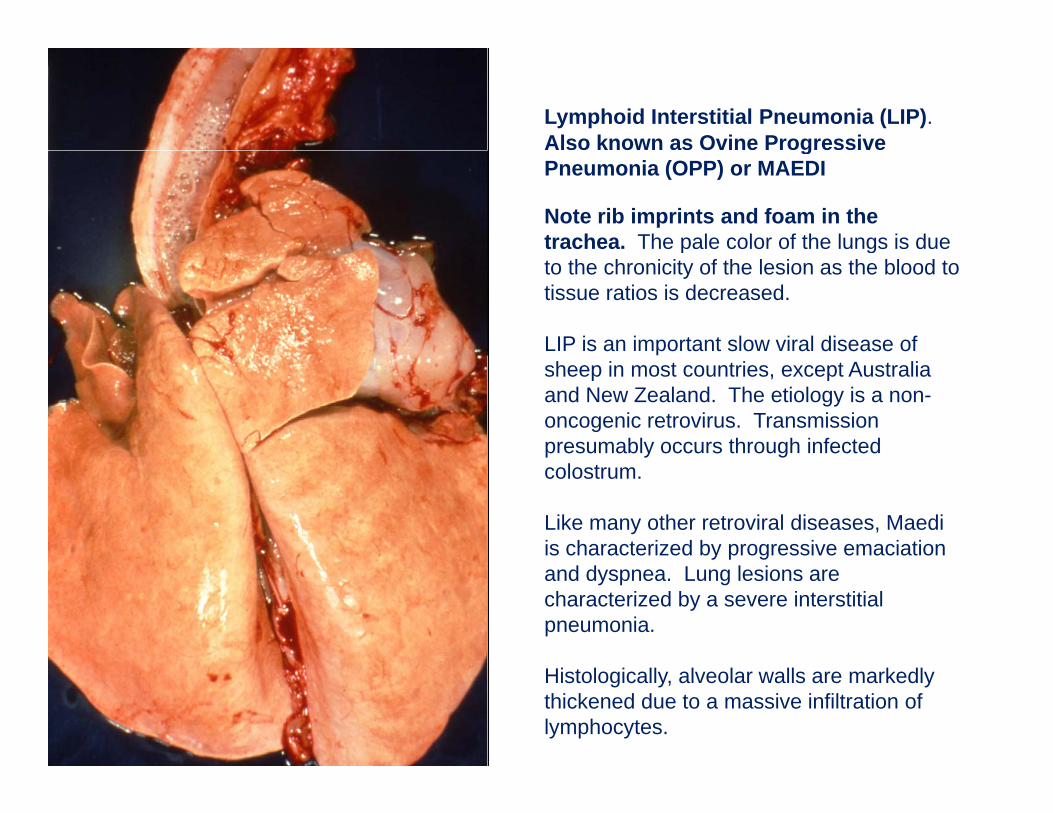
Lymphoid Interstitial Pneumonia (LIP).Also known as Ovine Progressive gPneumonia (OPP) or MAEDI
Note rib imprints and foam in the trachea. The pale color of the lungs is due p gto the chronicity of the lesion as the blood to tissue ratios is decreased.
LIP is an important slow viral disease of sheep in most countries, except Australia and New Zealand. The etiology is a non-oncogenic retrovirus. Transmission presumably occurs through infected colostrum.
Like many other retroviral diseases, Maedi is characterized by progressive emaciation and dyspnea. Lung lesions are characterized by a severe interstitial pneumonia.
Histologically, alveolar walls are markedly thickened due to a massive infiltration of lymphocytes.
Interstitial Pneumonia are also Interstitial Pneumonia are also caused by Allergic or Chemicalcaused by Allergic or Chemical--Toxic Toxic
Damage to the AirDamage to the Air--blood Barrierblood Barrier
Cattle
Atypical Interstitial Pneumonia is an archaic term that must be abandoned. What use to be atypical in now typical. The so-called "atypical interstitial pneumonia" of cattle comprises several distinct conditions characterized grossly by diffuse interstitial pneumonia:
1. Extrinsic allergic alveolitis (hypersensitivity pneumonitis or farmer's lung) is caused by inhalation of fungal spores (Micropolyspora faeni) from moldy hay ~> Ab response ~> deposition of Ag/Ab complexes in the blood air barrier ~> C'/PMN mediated injury to pneumocytes type-I ~> interstitial pneumonia. I is most commonly seen in cattle fed silage.
2. Bovine Pulmonary Edema and Emphysema (BPEE; fog fever). Ingestion of pasture (foggage) containing large amounts of L-tryptophan metabolized 3-methylindole (3-MI) ~> toxic injury to pneumocytes type 1 ~> interstitial pneumonia with severe edema and emphysema . BPEE is most commonly seen in grazing cattle. g g
3. Reinfection syndrome. It is a hypersensitivity reaction to reinfection with larvae of Dictyocaulus viviparus. Pathogenesis of the lesions similar to extrinsic allergic alveolitis. Most commonly seen in calves recently moved to pasture.
4. Bovine Respiratory Syncytial Virus. It has only been recently described. It is an acute fatal pneumonitis in feedlot cattle due to BRSV and presumably to hypersensitivity reaction against this virus. A similar condition occurs in children with the human strain of RSV.
5 Oth i t d ith i t titi l i Milk ll (t I h iti it i d i5. Other respiratory syndromes with interstitial pneumonia. Milk allergy (type I hypersensitivity in dairy cows), pit (manure) gases (H2S); "silo filler disease" (NO2), etc.
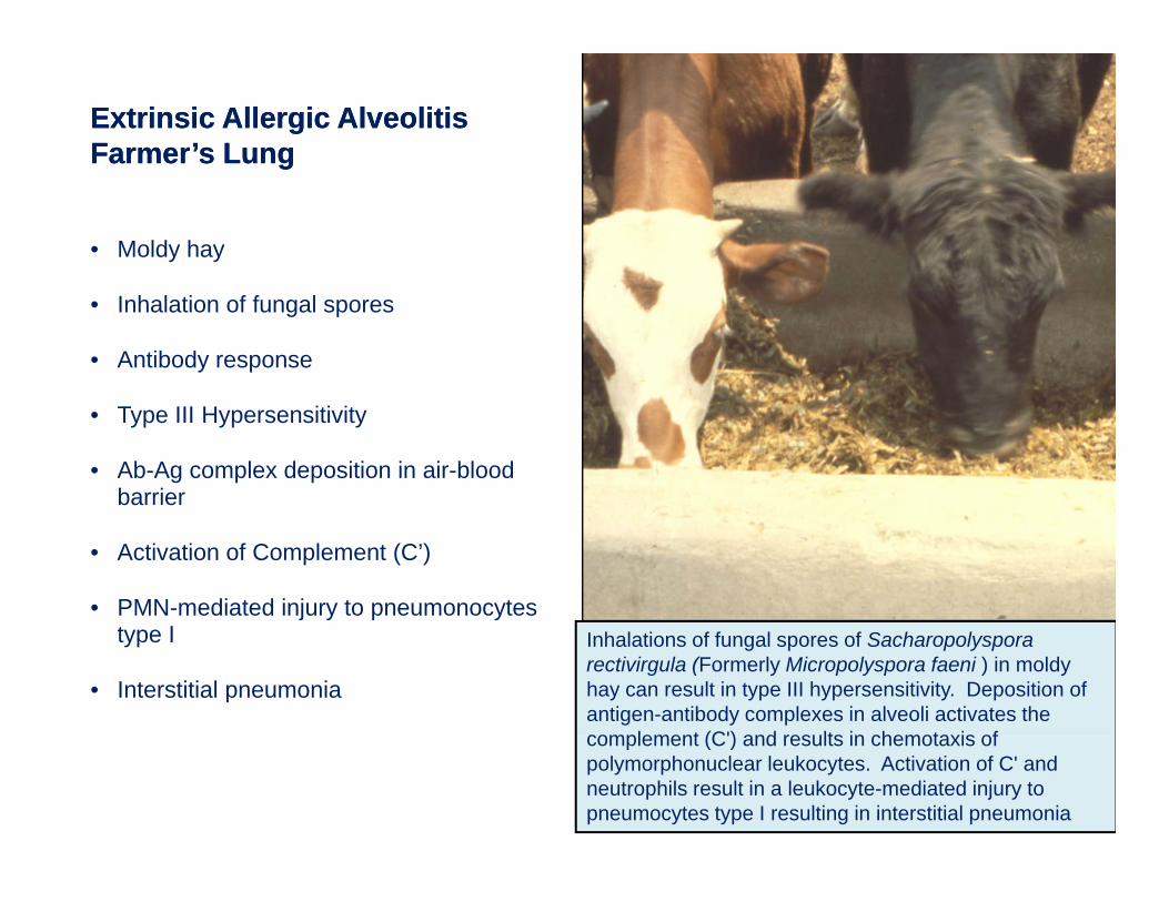
Extrinsic Allergic Alveolitis Extrinsic Allergic Alveolitis Farmer’s LungFarmer’s Lunggg
• Moldy hay
• Inhalation of fungal spores
• Antibody response
• Type III Hypersensitivity
• Ab-Ag complex deposition in air-blood barrier
• Activation of Complement (C’)
• PMN-mediated injury to pneumonocytes type I
• Interstitial pneumonia
Inhalations of fungal spores of Sacharopolysporarectivirgula (Formerly Micropolyspora faeni ) in moldy hay can result in type III hypersensitivity. Deposition of antigen-antibody complexes in alveoli activates the complement (C') and results in chemotaxis ofcomplement (C') and results in chemotaxis of polymorphonuclear leukocytes. Activation of C' and neutrophils result in a leukocyte-mediated injury to pneumocytes type I resulting in interstitial pneumonia
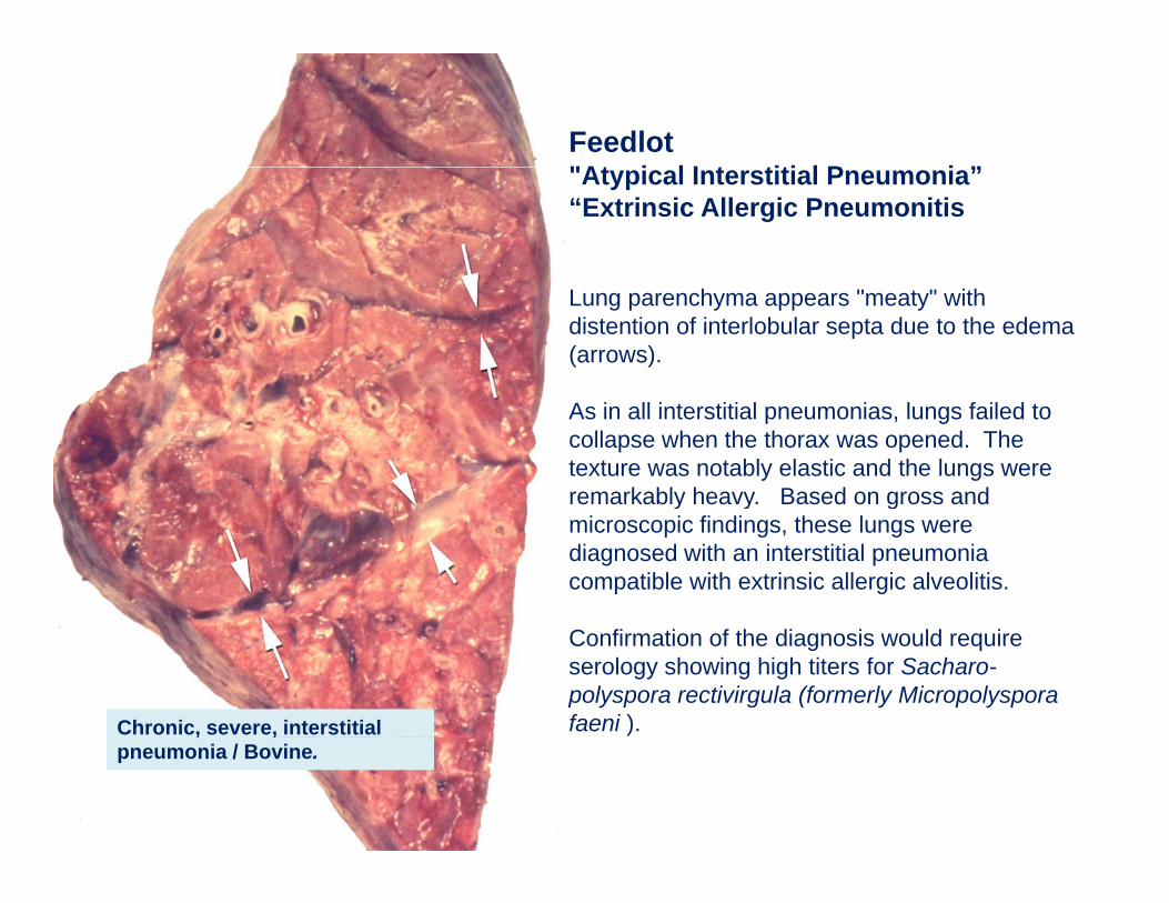
Feedlot "At i l I t titi l P i ”"Atypical Interstitial Pneumonia” “Extrinsic Allergic Pneumonitis
Lung parenchyma appears "meaty" with distention of interlobular septa due to the edema (arrows).
As in all interstitial pneumonias, lungs failed to collapse when the thorax was opened. The texture was notably elastic and the lungs were remarkably heavy. Based on gross and y y gmicroscopic findings, these lungs were diagnosed with an interstitial pneumonia compatible with extrinsic allergic alveolitis.
Confirmation of the diagnosis would require serology showing high titers for Sacharo-polyspora rectivirgula (formerly Micropolyspora faeni ).Chronic, severe, interstitial , ,
pneumonia / Bovine.
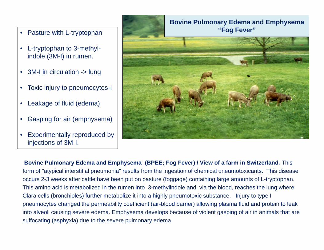
Bovine Pulmonary Edema and Emphysema “Fog Fever”• Pasture with L-tryptophan
L t t h t 3 th l• L-tryptophan to 3-methyl-indole (3M-I) in rumen.
• 3M-I in circulation -> lung
• Toxic injury to pneumocytes-I
• Leakage of fluid (edema)
• Gasping for air (emphysema)
• Experimentally reproduced by i j ti f 3M Iinjections of 3M-I.
Bovine Pulmonary Edema and Emphysema (BPEE; Fog Fever) / View of a farm in Switzerland. This form of "atypical interstitial pneumonia" results from the ingestion of chemical pneumotoxicants. This diseaseform of atypical interstitial pneumonia results from the ingestion of chemical pneumotoxicants. This disease occurs 2-3 weeks after cattle have been put on pasture (foggage) containing large amounts of L-tryptophan. This amino acid is metabolized in the rumen into 3-methylindole and, via the blood, reaches the lung where Clara cells (bronchioles) further metabolize it into a highly pneumotoxic substance. Injury to type I pneumocytes changed the permeability coefficient (air blood barrier) allowing plasma fluid and protein to leakpneumocytes changed the permeability coefficient (air-blood barrier) allowing plasma fluid and protein to leak into alveoli causing severe edema. Emphysema develops because of violent gasping of air in animals that are suffocating (asphyxia) due to the severe pulmonary edema.
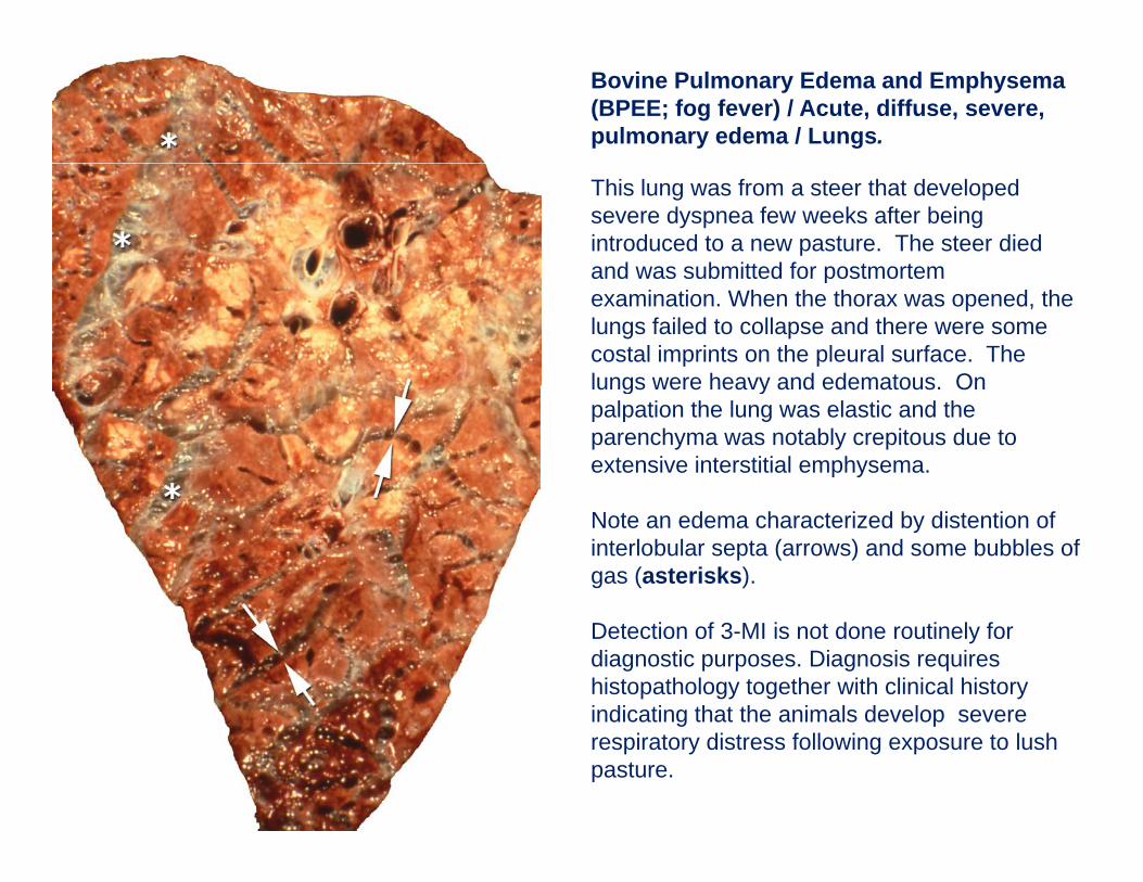
Bovine Pulmonary Edema and Emphysema (BPEE; fog fever) / Acute, diffuse, severe, pulmonary edema / Lungs.
This lung was from a steer that developed severe dyspnea few weeks after being introduced to a new pasture. The steer died and was submitted for postmortemand was submitted for postmortem examination. When the thorax was opened, the lungs failed to collapse and there were some costal imprints on the pleural surface. The lungs were heavy and edematous Onlungs were heavy and edematous. On palpation the lung was elastic and the parenchyma was notably crepitous due to extensive interstitial emphysema.
Note an edema characterized by distention of interlobular septa (arrows) and some bubbles of gas (asterisks).
Detection of 3-MI is not done routinely for diagnostic purposes. Diagnosis requires histopathology together with clinical history indicating that the animals develop severeindicating that the animals develop severe respiratory distress following exposure to lush pasture.
Examples of Disease that Cause Examples of Disease that Cause Granulomatous Pneumonia in Granulomatous Pneumonia in
Domestic AnimalsDomestic AnimalsDomestic AnimalsDomestic Animals
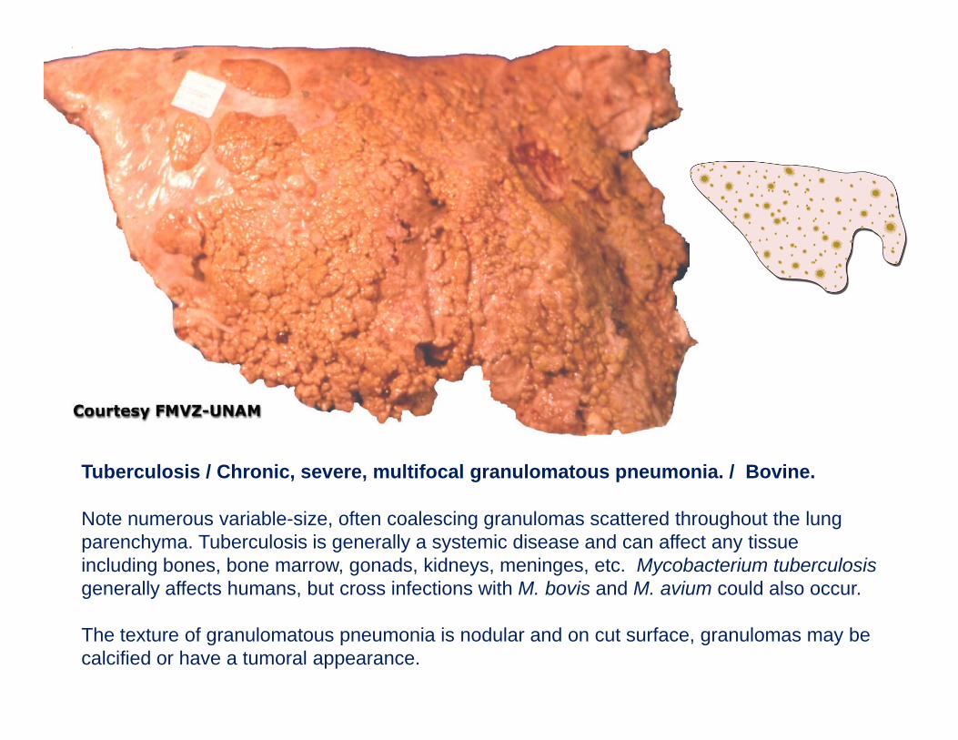
Tuberculosis / Chronic, severe, multifocal granulomatous pneumonia. / Bovine.
Note numerous variable-size, often coalescing granulomas scattered throughout the lung parenchyma. Tuberculosis is generally a systemic disease and can affect any tissue including bones, bone marrow, gonads, kidneys, meninges, etc. Mycobacterium tuberculosisgenerally affects humans, but cross infections with M. bovis and M. avium could also occur.
The texture of granulomatous pneumonia is nodular and on cut surface, granulomas may be calcified or have a tumoral appearance.
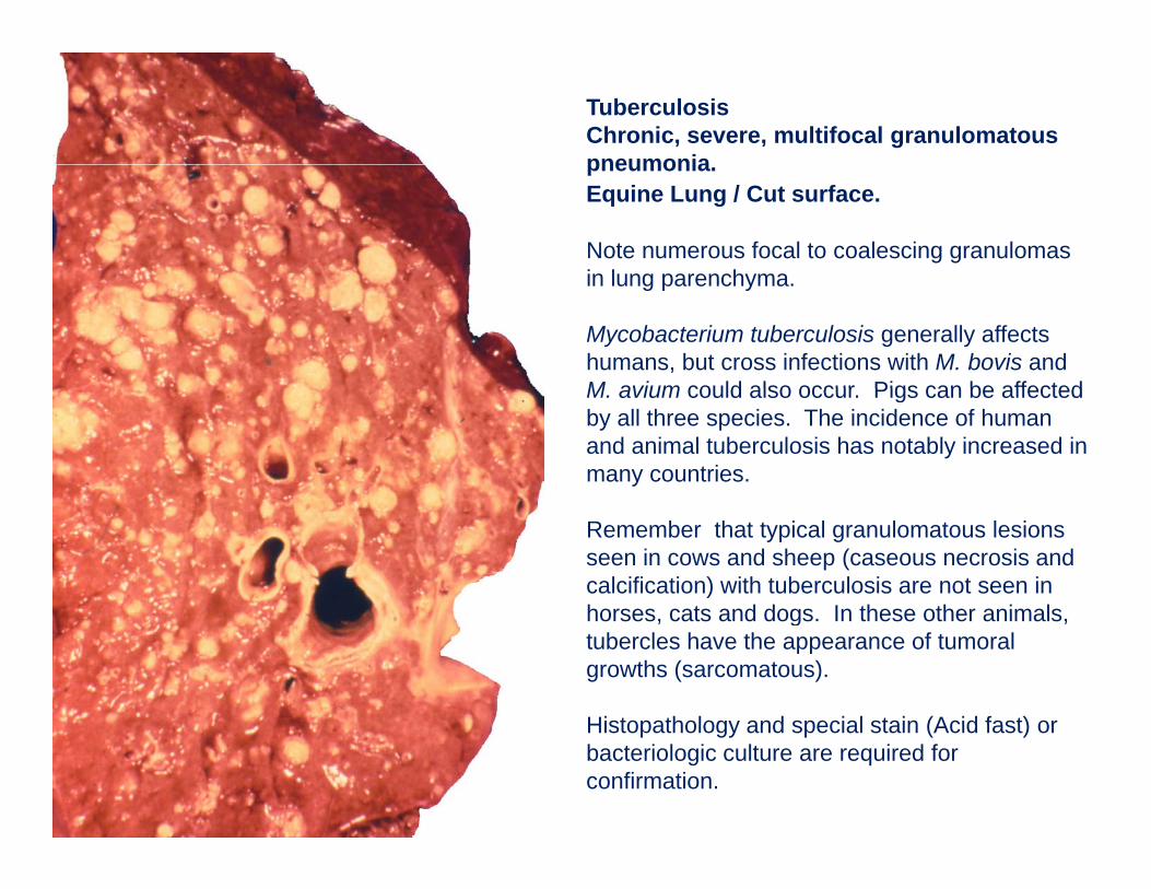
TuberculosisChronic, severe, multifocal granulomatous pneumoniapneumonia. Equine Lung / Cut surface.
Note numerous focal to coalescing granulomas in lung parenchymain lung parenchyma.
Mycobacterium tuberculosis generally affects humans, but cross infections with M. bovis and M avium could also occur Pigs can be affectedM. avium could also occur. Pigs can be affected by all three species. The incidence of human and animal tuberculosis has notably increased in many countries.
Remember that typical granulomatous lesions seen in cows and sheep (caseous necrosis and calcification) with tuberculosis are not seen in horses cats and dogs In these other animalshorses, cats and dogs. In these other animals, tubercles have the appearance of tumoral growths (sarcomatous).
Histopathology and special stain (Acid fast) orHistopathology and special stain (Acid fast) or bacteriologic culture are required for confirmation.
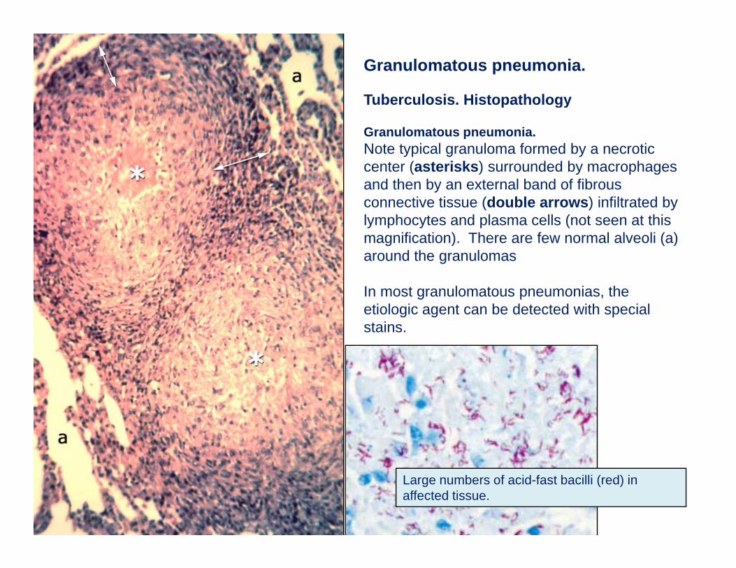
Granulomatous pneumonia.
Tuberculosis. Histopathologyp gy
Granulomatous pneumonia. Note typical granuloma formed by a necrotic center (asterisks) surrounded by macrophages and then by an external band of fibrous connective tissue (double arrows) infiltrated by lymphocytes and plasma cells (not seen at this magnification). There are few normal alveoli (a) around the granulomas
In most granulomatous pneumonias, the etiologic agent can be detected with special stains.
Large numbers of acid-fast bacilli (red) in affected tissue.
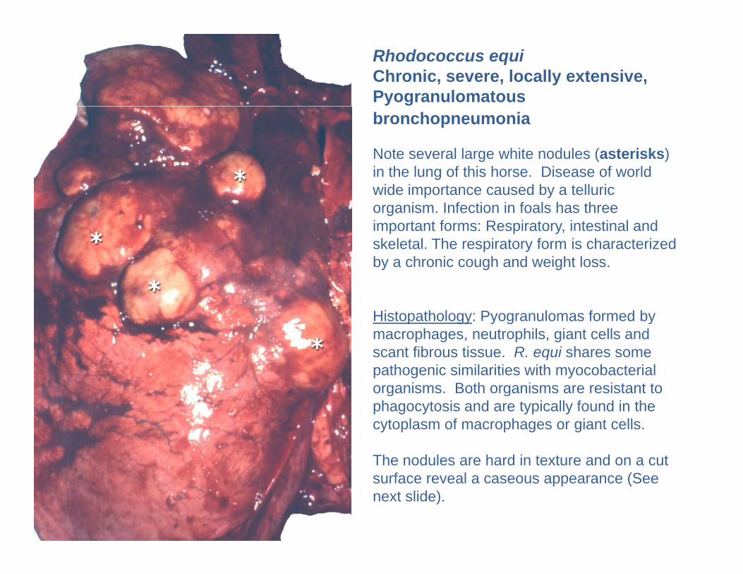
Rhodococcus equiChronic, severe, locally extensive, Pyogranulomatous bronchopneumonia
Note several large white nodules (asterisks) in the lung of this horse. Disease of world gwide importance caused by a telluric organism. Infection in foals has three important forms: Respiratory, intestinal and skeletal. The respiratory form is characterized p yby a chronic cough and weight loss.
Histopathology: Pyogranulomas formed by p gy y g ymacrophages, neutrophils, giant cells and scant fibrous tissue. R. equi shares some pathogenic similarities with myocobacterial organisms. Both organisms are resistant to g gphagocytosis and are typically found in the cytoplasm of macrophages or giant cells.
The nodules are hard in texture and on a cut surface reveal a caseous appearance (See next slide).
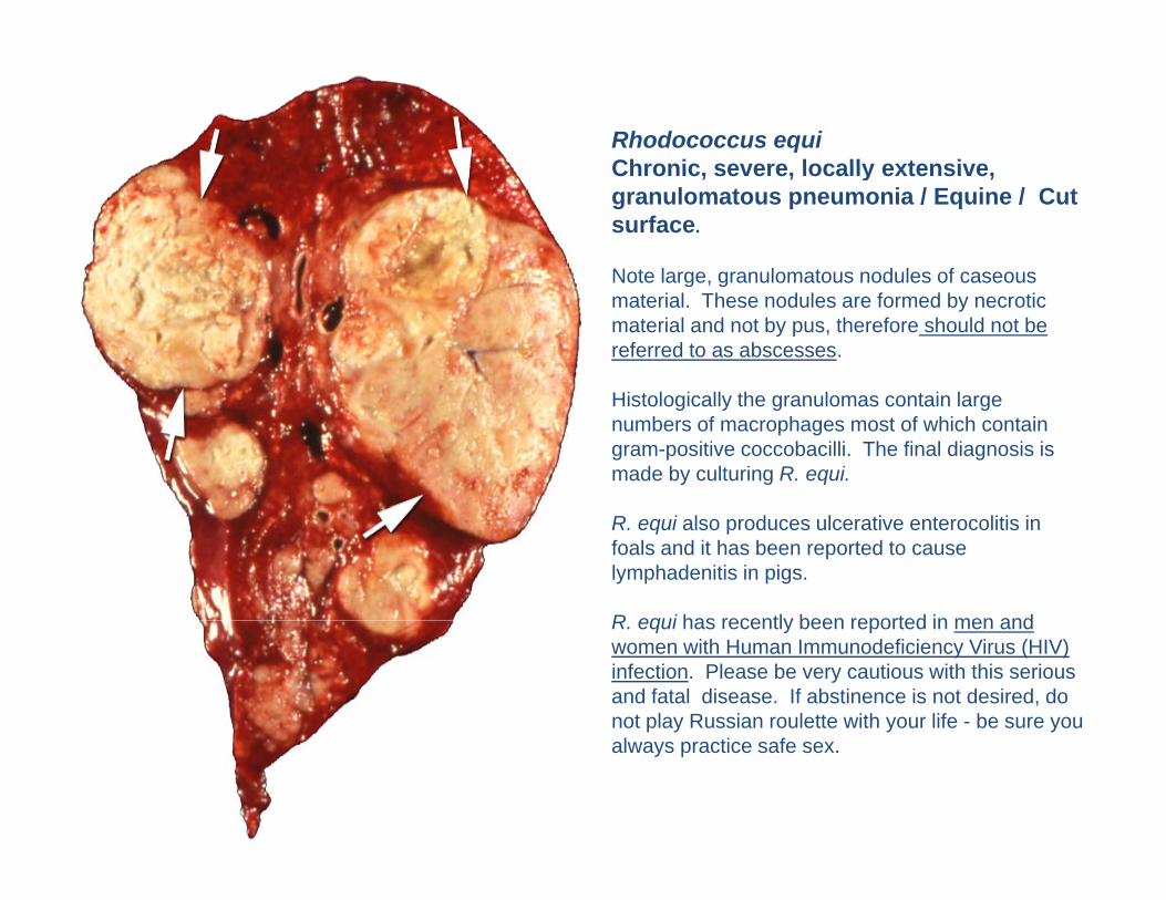
Rhodococcus equiChronic severe locally extensiveChronic, severe, locally extensive, granulomatous pneumonia / Equine / Cut surface.
Note large granulomatous nodules of caseousNote large, granulomatous nodules of caseous material. These nodules are formed by necrotic material and not by pus, therefore should not be referred to as abscesses.
Hi t l i ll th l t i lHistologically the granulomas contain large numbers of macrophages most of which contain gram-positive coccobacilli. The final diagnosis is made by culturing R. equi.
R. equi also produces ulcerative enterocolitis in foals and it has been reported to cause lymphadenitis in pigs.
R equi has recently been reported in men andR. equi has recently been reported in men and women with Human Immunodeficiency Virus (HIV) infection. Please be very cautious with this serious and fatal disease. If abstinence is not desired, do not play Russian roulette with your life - be sure you always practice safe sex.
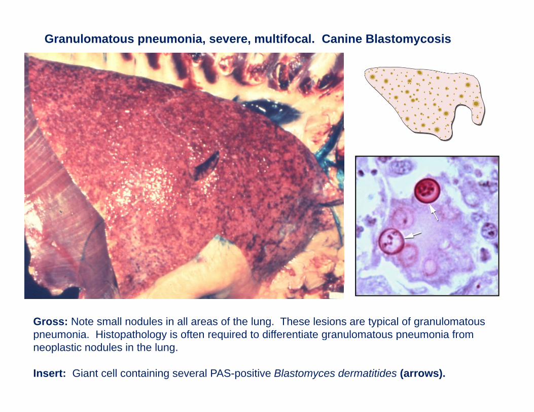
Granulomatous pneumonia, severe, multifocal. Canine Blastomycosis
Gross: Note small nodules in all areas of the lung. These lesions are typical of granulomatous pneumonia Histopathology is often required to differentiate granulomatous pneumonia frompneumonia. Histopathology is often required to differentiate granulomatous pneumonia from neoplastic nodules in the lung.
Insert: Giant cell containing several PAS-positive Blastomyces dermatitides (arrows).
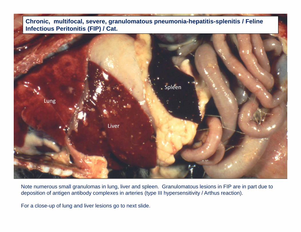
Chronic, multifocal, severe, granulomatous pneumonia-hepatitis-splenitis / Feline Infectious Peritonitis (FIP) / Cat.
Lung
Spleen
Lung
Liver
Note numerous small granulomas in lung, liver and spleen. Granulomatous lesions in FIP are in part due to g g p pdeposition of antigen antibody complexes in arteries (type III hypersensitivity / Arthus reaction).
For a close-up of lung and liver lesions go to next slide.
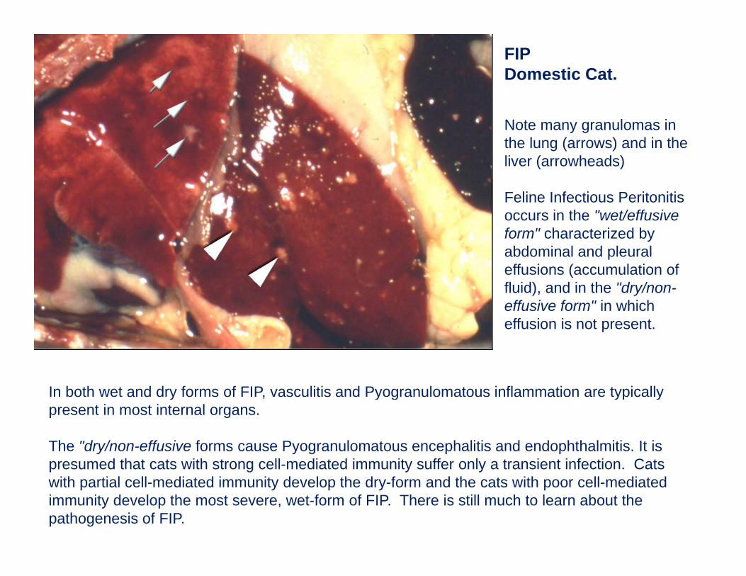
FIPDomestic Cat.
Note many granulomas in the lung (arrows) and in the liver (arrowheads)
Feline Infectious Peritonitis occurs in the "wet/effusive form" characterized by abdominal and pleuralabdominal and pleural effusions (accumulation of fluid), and in the "dry/non-effusive form" in which effusion is not presenteffusion is not present.
In both wet and dry forms of FIP, vasculitis and Pyogranulomatous inflammation are typically y , y g yp ypresent in most internal organs.
The "dry/non-effusive forms cause Pyogranulomatous encephalitis and endophthalmitis. It is presumed that cats with strong cell-mediated immunity suffer only a transient infection. Cats p g y ywith partial cell-mediated immunity develop the dry-form and the cats with poor cell-mediated immunity develop the most severe, wet-form of FIP. There is still much to learn about the pathogenesis of FIP.
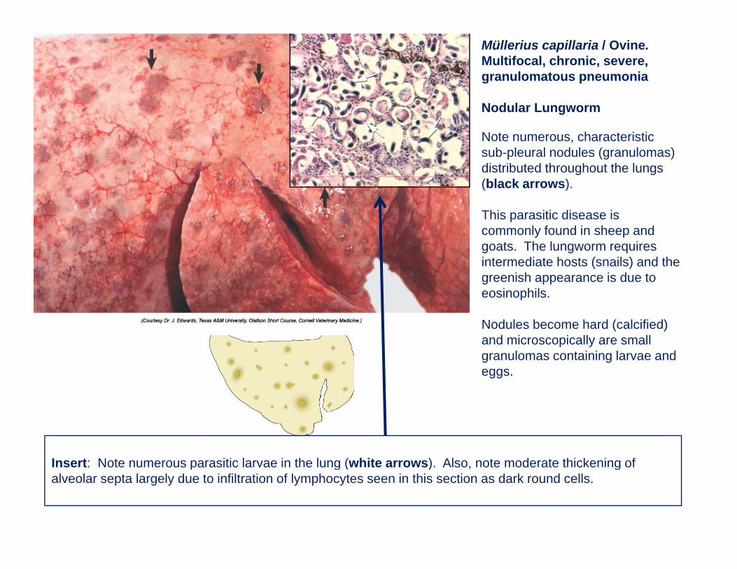
Müllerius capillaria / Ovine.Multifocal, chronic, severe, granulomatous pneumonia
Nod lar L ng ormNodular Lungworm
Note numerous, characteristic sub-pleural nodules (granulomas) distributed throughout the lungs (black arrows).
This parasitic disease is commonly found in sheep and goats The lungworm requiresgoats. The lungworm requires intermediate hosts (snails) and the greenish appearance is due to eosinophils.
N d l b h d ( l ifi d)Nodules become hard (calcified) and microscopically are small granulomas containing larvae and eggs.
Insert: Note numerous parasitic larvae in the lung (white arrows) Also note moderate thickening ofInsert: Note numerous parasitic larvae in the lung (white arrows). Also, note moderate thickening of alveolar septa largely due to infiltration of lymphocytes seen in this section as dark round cells.
FETAL PNEUMONIAFETAL PNEUMONIA
and and
ABORTIONABORTIONABORTIONABORTION
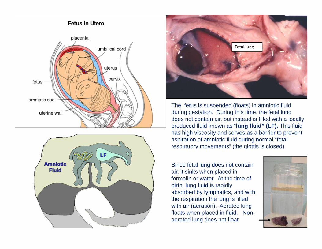
F t l lFetal lung
The fetus is suspended (floats) in amniotic fluid during gestation. During this time, the fetal lung does not contain air, but instead is filled with a locally produced fluid known as “lung fluid” (LF). This fluid has high viscosity and serves as a barrier to prevent aspiration of amniotic fluid during normal "fetalaspiration of amniotic fluid during normal fetal respiratory movements” (the glottis is closed).
Since fetal lung does not contain air it sinks when placed inair, it sinks when placed in formalin or water. At the time of birth, lung fluid is rapidly absorbed by lymphatics, and with the respiration the lung is filled with air (aeration). Aerated lung floats when placed in fluid. Non-aerated lung does not float.
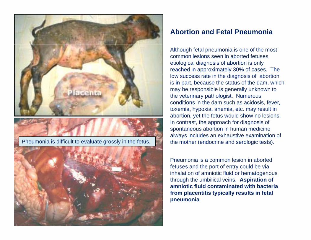
Abortion and Fetal Pneumonia
Although fetal pneumonia is one of the most common lesions seen in aborted fetuses, etiological diagnosis of abortion is only reached in approximately 30% of cases. The l t i th di i f b tilow success rate in the diagnosis of abortion is in part, because the status of the dam, which may be responsible is generally unknown to the veterinary pathologist. Numerous conditions in the dam such as acidosis, fever, , ,toxemia, hypoxia, anemia, etc. may result in abortion, yet the fetus would show no lesions. In contrast, the approach for diagnosis of spontaneous abortion in human medicine always includes an exhaustive examination ofalways includes an exhaustive examination of the mother (endocrine and serologic tests).
Pneumonia is a common lesion in aborted fet ses and the port of entr co ld be ia
Pneumonia is difficult to evaluate grossly in the fetus.
fetuses and the port of entry could be via inhalation of amniotic fluid or hematogenous through the umbilical veins. Aspiration of amniotic fluid contaminated with bacteria from placentitis typically results in fetal p yp ypneumonia.
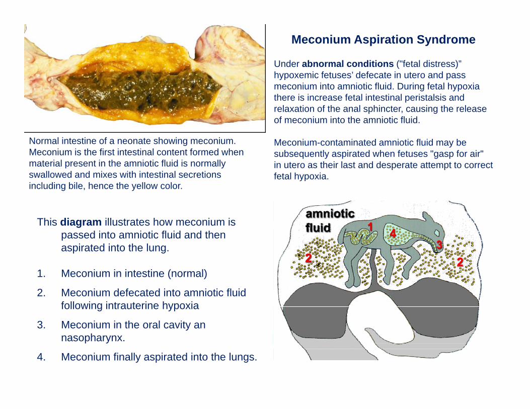
Meconium Aspiration Syndrome
Under abnormal conditions ("fetal distress)” hypoxemic fetuses’ defecate in utero and passhypoxemic fetuses defecate in utero and pass meconium into amniotic fluid. During fetal hypoxia there is increase fetal intestinal peristalsis and relaxation of the anal sphincter, causing the release of meconium into the amniotic fluid.
Meconium-contaminated amniotic fluid may be subsequently aspirated when fetuses "gasp for air" in utero as their last and desperate attempt to correct fetal hypoxia.
Normal intestine of a neonate showing meconium. Meconium is the first intestinal content formed when material present in the amniotic fluid is normally swallowed and mixes with intestinal secretions fetal hypoxia. including bile, hence the yellow color.
This diagram illustrates how meconium is passed into amniotic fluid and then aspirated into the lung.
1. Meconium in intestine (normal)
2. Meconium defecated into amniotic fluid following intrauterine hypoxia
3. Meconium in the oral cavity an hnasopharynx.
4. Meconium finally aspirated into the lungs.
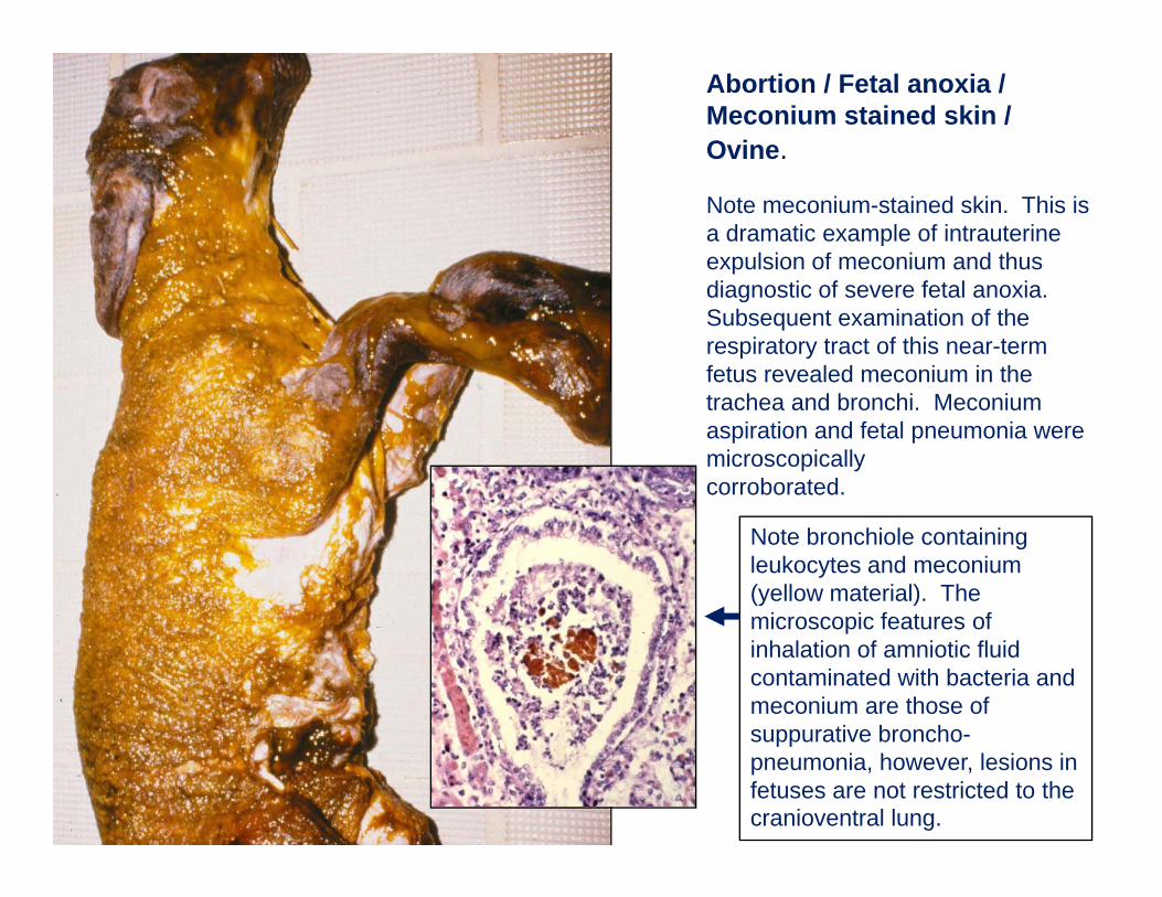
Abortion / Fetal anoxia / Meconium stained skin / Ovine.
Note meconium-stained skin. This is a dramatic example of intrauterine expulsion of meconium and thus diagnostic of severe fetal anoxia. Subsequent examination of the respiratory tract of this near-term fetus revealed meconium in the trachea and bronchi. Meconium aspiration and fetal pneumonia were microscopicallycorroborated.
Note bronchiole containing leukocytes and meconium (yellow material). The microscopic features ofmicroscopic features of inhalation of amniotic fluid contaminated with bacteria and meconium are those of suppurative bronchosuppurative broncho-pneumonia, however, lesions in fetuses are not restricted to the cranioventral lung.

THE ENDTHE END