ALEX Instruction for Use
11
5.11.2020 ALEX2 Instruction for Use English localhost:5000/pdf/alex2/English 1/11 ALEX 2 Instruction for Use INTENDED USE The Allergy Explorer 2 (ALEX 2 ) is a quantitative in vitro diagnostic test for the measurement of allergen specic IgE (sIgE) and a semi-quantitative in vitro diagnostic test for the measurement of total IgE (tIgE) in human serum or plasma (exception EDTA-plasma). It is to be used by clinical chemistry laboratories, trained laboratory personnel and medical professionals for the purpose of supporting the clinical diagnosis of IgE mediated diseases, in conjunction with other clinical ndings or diagnostic test results. SUMMARY AND EXPLANATION OF THE TEST Allergic reactions are immediate type I hypersensitivity reactions and are mediated by antibodies belonging to the IgE class of immunoglobulins. After exposure to specic allergens, IgE–mediated release of histamine and other mediators from mast cells and basophils results in clinical manifestations such as asthma, allergic rhino-conjunctivitis, atopic eczema and gastrointestinal symptoms [1]. Therefore, a detailed sensitization pattern to specic allergens assists in the evaluation of allergic patients [2-6]. All major type I allergen sources are covered by ALEX 2 . A complete list of ALEX 2 allergen extracts and molecular allergens can be found at the bottom of this instruction. Important information for the user! For the correct use of ALEX 2 , it is necessary for the user to carefully read and follow these instructions for use. The manufacturer assumes no liability for any use of this test system which is not described in this document or for modications by the user of the test system. Attention: The calibration function stored in the QR barcodes is optimized for the respective intended use. The kit variant 02-2001-01 of the ALEX 2 test is therefore exclusively intended for manual processing and kit variant 02-5001-01 exclusively for automated processing (MAX 45k). PRINCIPLE OF THE PROCEDURE ALEX 2 is a solid-phase immunoassay. Allergens extracts or molecular allergens, which are coupled to nanoparticles, are deposited in a systematic fashion onto a solid phase forming a macroscopic array. First, the particle bound allergens react with specic IgE that is present in the patient’s sample. After incubation, non-specic IgE is washed off. The procedure continues by adding an enzyme labelled anti-human IgE detection antibody which forms a complex with the particle bound specic IgE. After a second washing step, substrate is added which is converted to an insoluble, coloured precipitate by the antibody-bound enzyme. Finally, the enzyme-substrate reaction is stopped by adding a blocking reagent. The amount of precipitate is proportional to the concentration of specic IgE in the patient sample. The lab test procedure is followed by image acquisition and analysis using the ImageXplorer device. The test results are analysed with MADx’s Raptor Analysis Software and reported in IgE response units (kU A /L). Total IgE results are also reported in IgE response units (kU/L). SHIPMENT AND STORAGE The shipment of ALEX 2 takes place at ambient temperature conditions. Nevertheless, the kit must be stored immediately upon delivery at 2-8°C. Stored correctly, ALEX 2 and its components can be used until the indicated expiration date. Kit reagents are stable for 6 months after opening (at the indicated storage conditions). WASTE DISPOSAL Dispose the used ALEX 2 cartridge and unused kit components with laboratory chemical waste. Follow all national, state, and local regulations regarding disposal.
Transcript of ALEX Instruction for Use

5.11.2020 ALEX2 Instruction for Use English
localhost:5000/pdf/alex2/English 1/11
ALEX2 Instruction for Use
INTENDED USE
The Allergy Explorer2 (ALEX2) is a quantitative in vitro diagnostic test for the measurement of allergen speci�c IgE (sIgE)and a semi-quantitative in vitro diagnostic test for the measurement of total IgE (tIgE) in human serum or plasma(exception EDTA-plasma). It is to be used by clinical chemistry laboratories, trained laboratory personnel and medicalprofessionals for the purpose of supporting the clinical diagnosis of IgE mediated diseases, in conjunction with otherclinical �ndings or diagnostic test results.
SUMMARY AND EXPLANATION OF THE TEST
Allergic reactions are immediate type I hypersensitivity reactions and are mediated by antibodies belonging to the IgEclass of immunoglobulins. After exposure to speci�c allergens, IgE–mediated release of histamine and other mediatorsfrom mast cells and basophils results in clinical manifestations such as asthma, allergic rhino-conjunctivitis, atopiceczema and gastrointestinal symptoms [1]. Therefore, a detailed sensitization pattern to speci�c allergens assists in theevaluation of allergic patients [2-6].
All major type I allergen sources are covered by ALEX2. A complete list of ALEX2 allergen extracts and molecular allergenscan be found at the bottom of this instruction.
Important information for the user!
For the correct use of ALEX2, it is necessary for the user to carefully read and follow these instructions for use. Themanufacturer assumes no liability for any use of this test system which is not described in this document or formodi�cations by the user of the test system. Attention: The calibration function stored in the QR barcodes is optimizedfor the respective intended use. The kit variant 02-2001-01 of the ALEX2 test is therefore exclusively intended for manualprocessing and kit variant 02-5001-01 exclusively for automated processing (MAX 45k).
PRINCIPLE OF THE PROCEDURE
ALEX2 is a solid-phase immunoassay. Allergens extracts or molecular allergens, which are coupled to nanoparticles, aredeposited in a systematic fashion onto a solid phase forming a macroscopic array. First, the particle bound allergensreact with speci�c IgE that is present in the patient’s sample. After incubation, non-speci�c IgE is washed off. Theprocedure continues by adding an enzyme labelled anti-human IgE detection antibody which forms a complex with theparticle bound speci�c IgE. After a second washing step, substrate is added which is converted to an insoluble, colouredprecipitate by the antibody-bound enzyme. Finally, the enzyme-substrate reaction is stopped by adding a blocking reagent.The amount of precipitate is proportional to the concentration of speci�c IgE in the patient sample. The lab test procedureis followed by image acquisition and analysis using the ImageXplorer device. The test results are analysed with MADx’sRaptor Analysis Software and reported in IgE response units (kUA/L). Total IgE results are also reported in IgE responseunits (kU/L).
SHIPMENT AND STORAGE
The shipment of ALEX2 takes place at ambient temperature conditions. Nevertheless, the kit must be stored immediatelyupon delivery at 2-8°C. Stored correctly, ALEX2 and its components can be used until the indicated expiration date.
Kit reagents are stable for 6 months after opening (at the indicated storage conditions).
WASTE DISPOSAL
Dispose the used ALEX2 cartridge and unused kit components with laboratory chemical waste. Follow all national, state,and local regulations regarding disposal.
5.11.2020 ALEX2 Instruction for Use English
localhost:5000/pdf/alex2/English 2/11
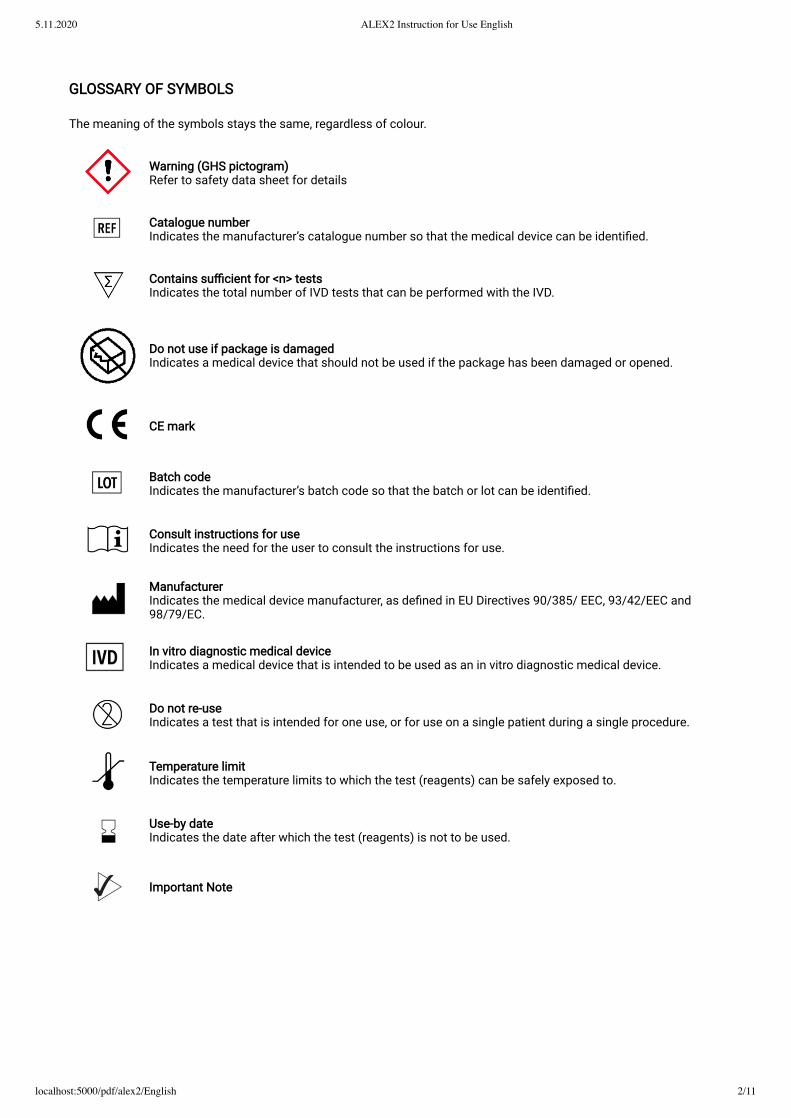
GLOSSARY OF SYMBOLS
The meaning of the symbols stays the same, regardless of colour.
Warning (GHS pictogram) Refer to safety data sheet for details
Catalogue number Indicates the manufacturer’s catalogue number so that the medical device can be identi�ed.
Contains su�cient for <n> tests Indicates the total number of IVD tests that can be performed with the IVD.
Do not use if package is damaged Indicates a medical device that should not be used if the package has been damaged or opened.
CE mark
Batch code Indicates the manufacturer’s batch code so that the batch or lot can be identi�ed.
Consult instructions for use Indicates the need for the user to consult the instructions for use.
Manufacturer Indicates the medical device manufacturer, as de�ned in EU Directives 90/385/ EEC, 93/42/EEC and98/79/EC.
In vitro diagnostic medical device Indicates a medical device that is intended to be used as an in vitro diagnostic medical device.
Do not re-use Indicates a test that is intended for one use, or for use on a single patient during a single procedure.
Temperature limit Indicates the temperature limits to which the test (reagents) can be safely exposed to.
Use-by date Indicates the date after which the test (reagents) is not to be used.
Important Note
5.11.2020 ALEX2 Instruction for Use English
localhost:5000/pdf/alex2/English 3/11
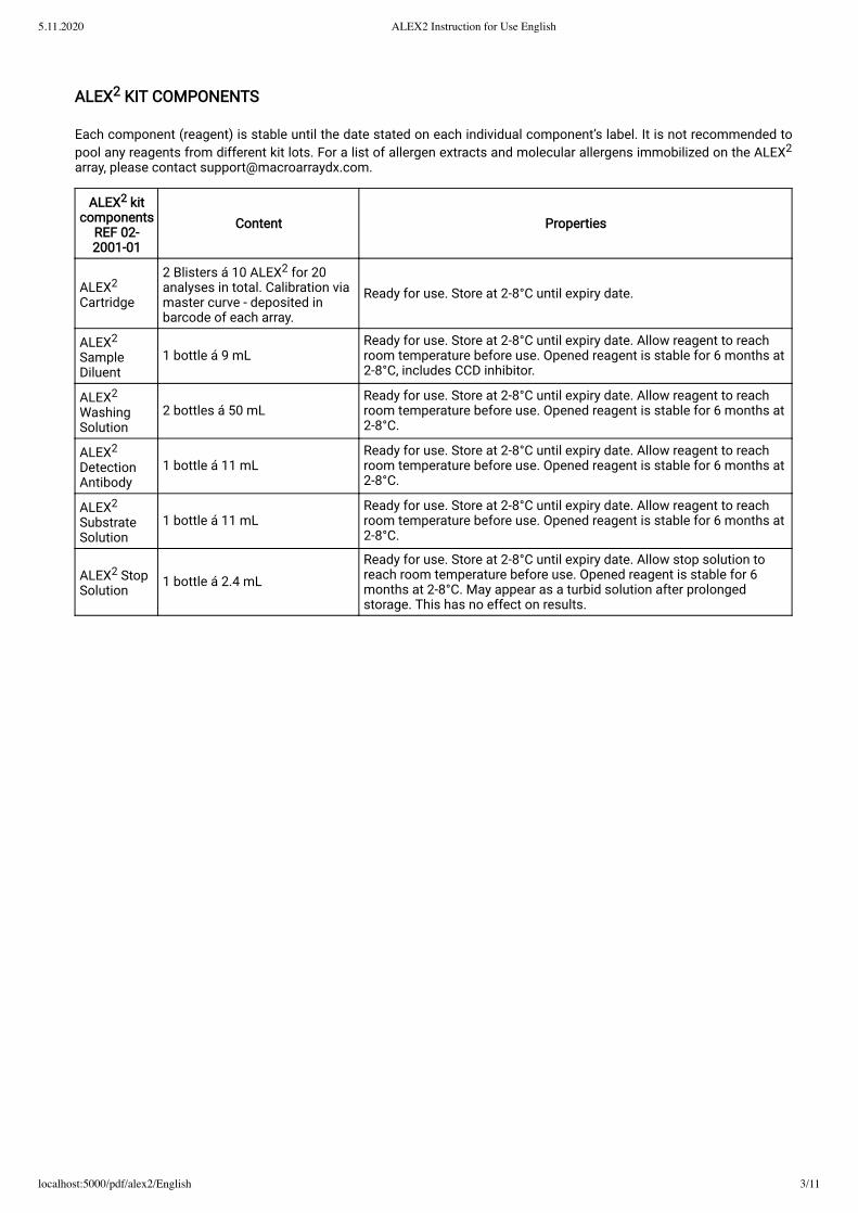
ALEX2 KIT COMPONENTS
Each component (reagent) is stable until the date stated on each individual component’s label. It is not recommended topool any reagents from different kit lots. For a list of allergen extracts and molecular allergens immobilized on the ALEX2
array, please contact [email protected].
ALEX2 kitcomponents
REF 02-2001-01
Content Properties
ALEX2
Cartridge
2 Blisters á 10 ALEX2 for 20analyses in total. Calibration viamaster curve - deposited inbarcode of each array.
Ready for use. Store at 2-8°C until expiry date.
ALEX2
SampleDiluent
1 bottle á 9 mLReady for use. Store at 2-8°C until expiry date. Allow reagent to reachroom temperature before use. Opened reagent is stable for 6 months at2-8°C, includes CCD inhibitor.
ALEX2
WashingSolution
2 bottles á 50 mLReady for use. Store at 2-8°C until expiry date. Allow reagent to reachroom temperature before use. Opened reagent is stable for 6 months at2-8°C.
ALEX2
DetectionAntibody
1 bottle á 11 mLReady for use. Store at 2-8°C until expiry date. Allow reagent to reachroom temperature before use. Opened reagent is stable for 6 months at2-8°C.
ALEX2
SubstrateSolution
1 bottle á 11 mLReady for use. Store at 2-8°C until expiry date. Allow reagent to reachroom temperature before use. Opened reagent is stable for 6 months at2-8°C.
ALEX2 StopSolution
1 bottle á 2.4 mLReady for use. Store at 2-8°C until expiry date. Allow stop solution toreach room temperature before use. Opened reagent is stable for 6months at 2-8°C. May appear as a turbid solution after prolongedstorage. This has no effect on results.
5.11.2020 ALEX2 Instruction for Use English
localhost:5000/pdf/alex2/English 4/11
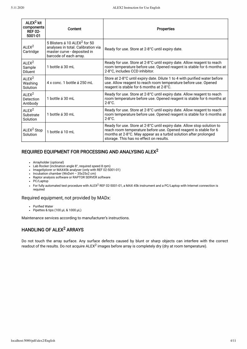
ALEX2 kitcomponents
REF 02-5001-01
Content Properties
ALEX2
Cartridge
5 Blisters á 10 ALEX2 for 50analyses in total. Calibration viamaster curve - deposited inbarcode of each array.
Ready for use. Store at 2-8°C until expiry date.
ALEX2
SampleDiluent
1 bottle á 30 mLReady for use. Store at 2-8°C until expiry date. Allow reagent to reachroom temperature before use. Opened reagent is stable for 6 months at2-8°C, includes CCD inhibitor.
ALEX2
WashingSolution
4 x conc. 1 bottle á 250 mLStore at 2-8°C until expiry date. Dilute 1 to 4 with puri�ed water beforeuse. Allow reagent to reach room temperature before use. Openedreagent is stable for 6 months at 2-8°C.
ALEX2
DetectionAntibody
1 bottle á 30 mLReady for use. Store at 2-8°C until expiry date. Allow reagent to reachroom temperature before use. Opened reagent is stable for 6 months at2-8°C.
ALEX2
SubstrateSolution
1 bottle á 30 mLReady for use. Store at 2-8°C until expiry date. Allow reagent to reachroom temperature before use. Opened reagent is stable for 6 months at2-8°C.
ALEX2 StopSolution
1 bottle á 10 mLReady for use. Store at 2-8°C until expiry date. Allow stop solution toreach room temperature before use. Opened reagent is stable for 6months at 2-8°C. May appear as a turbid solution after prolongedstorage. This has no effect on results.
REQUIRED EQUIPMENT FOR PROCESSING AND ANALYSING ALEX2
Arrayholder (optional)Lab Rocker (inclination angle 8°, required speed 8 rpm)ImageXplorer or MAX45k analyser (only with REF 02-5001-01)Incubation chamber (WxDxH – 35x25x2 cm)Raptor analysis software or RAPTOR SERVER softwarePC/LaptopFor fully automated test procedure with ALEX2 REF 02-5001-01, a MAX 45k instrument and a PC/Laptop with Internet connection isrequired
Required equipment, not provided by MADx:
Puri�ed WaterPipettes & tips (100 μL & 1000 μL)
Maintenance services according to manufacturer’s instructions.
HANDLING OF ALEX2 ARRAYS
Do not touch the array surface. Any surface defects caused by blunt or sharp objects can interfere with the correctreadout of the results. Do not acquire ALEX2 images before array is completely dry (dry at room temperature).
5.11.2020 ALEX2 Instruction for Use English
localhost:5000/pdf/alex2/English 5/11
WARNINGS AND PRECAUTIONS
It is recommended to wear hand and eye protection as well as lab coats and follow good laboratory practices when preparing andhandling reagents and samples.In accordance with good laboratory practice, all human source material should be considered potentially infectious and handled with thesame precautions as Patient samples.The sample diluent is partially prepared from human blood sources. The product was tested non-reactive for Hepatitis B Surface Antigen(HBsAg), antibodies to Hepatitis C (HCV) and antibodies to HIV-1/HIV-2.Sample diluent and washing solution contain sodium azide as a preservative and must be handled with care. Safety data sheet isavailable upon request.For in vitro diagnostic use only. Not for internal or external use in humans or animals.Only personnel trained in laboratory practice should use this kit.Upon arrival, check the kit components for damage. If one of the components is damaged (e.g. buffer bottles), contact MADx([email protected]). Do not use damaged kit components, as their use may lead to poor kit performance.Do not use reagents beyond their expiry dates.Do not mix reagents from different batches.
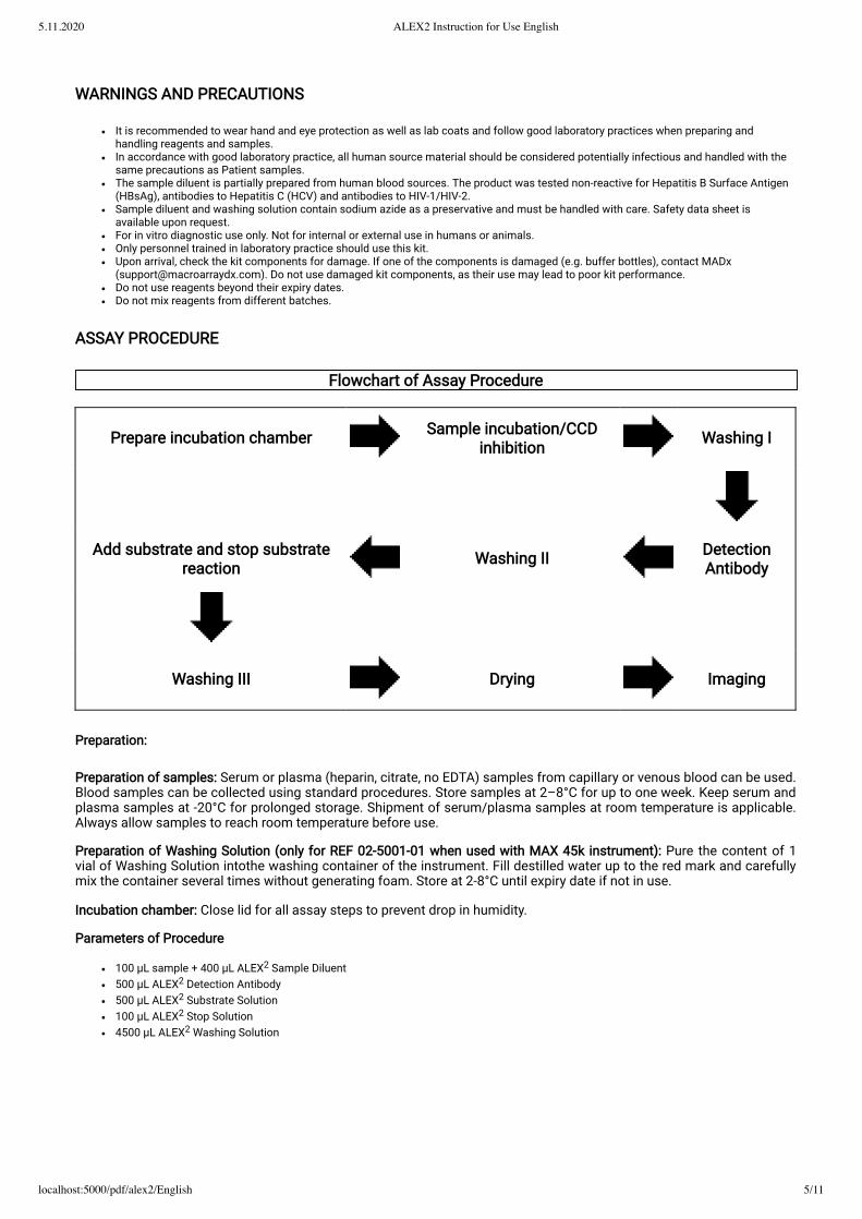
ASSAY PROCEDURE
Flowchart of Assay Procedure
Prepare incubation chamber Sample incubation/CCDinhibition Washing I
Add substrate and stop substratereaction Washing II Detection
Antibody
Washing III Drying Imaging
Preparation:
Preparation of samples: Serum or plasma (heparin, citrate, no EDTA) samples from capillary or venous blood can be used.Blood samples can be collected using standard procedures. Store samples at 2–8°C for up to one week. Keep serum andplasma samples at -20°C for prolonged storage. Shipment of serum/plasma samples at room temperature is applicable.Always allow samples to reach room temperature before use.
Preparation of Washing Solution (only for REF 02-5001-01 when used with MAX 45k instrument): Pure the content of 1vial of Washing Solution intothe washing container of the instrument. Fill destilled water up to the red mark and carefullymix the container several times without generating foam. Store at 2-8°C until expiry date if not in use.
Incubation chamber: Close lid for all assay steps to prevent drop in humidity.
Parameters of Procedure
100 μL sample + 400 μL ALEX2 Sample Diluent500 μL ALEX2 Detection Antibody500 μL ALEX2 Substrate Solution100 μL ALEX2 Stop Solution4500 μL ALEX2 Washing Solution

5.11.2020 ALEX2 Instruction for Use English
localhost:5000/pdf/alex2/English 6/11
Assay time is approximately 3 h 30 min.
It is not recommended to run more assays than can be pipetted in 8 min. All incubations are performed at roomtemperature, 20-26°C.
ALEX2 ASSAY PROCEDURE
All reagents are to be used at room temperature (20-26°C). The assay must not be performed indirect sunlight.
Prepare incubation chamber
Open incubation chamber and place paper towels on bottom part. Soak paper towels with puri�ed water until no dry partsof the paper towels are visible.
1. Sample incubation/CCD inhibition
Take out the needed number of ALEX2 cartridges and place them into the array holder(s). Add 400 μL of ALEX2 samplediluent to each cartridge. Add 100 μL patient sample to the cartridges. Ensure that the resulting solution is spread evenly.Place the cartridges onto the lab rocker and start the serum incubation with 8 rpm for 2 hours. Close incubation chamberbefore starting the lab rocker. After 2 hours, discharge the patient samples into a collection container. Carefully wipe offdroplets from the cartridge using a paper towel.
Avoid touching the array surface with the paper towel! Avoid any carry over or cross-contamination of patient samples between individual ALEX2 cartridges!
Optional or positive Hom s LF (CCD marker): with the standard CCD antibody inhibition protocol (as described inparagraph 2: sample incubation/CCD inhibition) the CCD inhibition e�ciency is 85%. If a higher rate of inhibition e�ciencyis required, prepare a 1 mL sample tube, add 400 μL sample diluent and 100 μL serum. Incubate for 30 minutes (non-shaking) and then proceed with the usual assay procedure.
Note: The extra CCD inhibition step leads in many cases to an inhibition rate for CCD antibodies of above 95%.
1a. Washing I
Add 500 μL ALEX2 Washing Solution to each cartridge and incubate on the lab rocker (at 8 rpm) for 5 minutes. Dischargethe washing solution into a collection container and vigorously tap the cartridges on a stack of dry paper towels.
Repeat this step 2 more times.

5.11.2020 ALEX2 Instruction for Use English
localhost:5000/pdf/alex2/English 7/11
2. Add detection antibody
Add 500 μL of ALEX2 Detection Antibody to each cartridge.
Make sure that the complete array surface is covered by the ALEX2 Detection Antibody solution.
Place the cartridges into the incubation chamber on the lab rocker and incubate at 8 rpm for 30 minutes. Discharge thedetection antibody solution into a collection container. Carefully wipe off remaining droplets from the cartridges using apaper towel.
2a. Washing II
Add 500 μL ALEX2 Washing Solution to each cartridge and incubate on the lab rocker at 8 rpm for 5 minutes. Dischargethe washing solution into a collection container and vigorously tap the cartridges on a stack of dry paper towels.
Repeat this step 4 more times.
3+4. Add substrate solution and stop substrate reaction
Add 500 μL of ALEX2 Substrate Solution to each cartridge. Start a timer with �lling the �rst cartridge and proceed with the�lling of the remaining cartridges. Make sure that the complete array surface is covered by the substrate solution andincubate the arrays for 8 minutes without shaking (lab rocker at 0 rpm and in horizontal position).
After exactly 8 minutes, add 100 μL of the ALEX2 Stop Solution to all cartridges, starting with the �rst cartridge to assurethat all arrays are incubated for the same time with the Substrate solution. Carefully agitate to evenly distribute the stopsolution in the array cartridges, after the stop solution was pipetted onto all arrays. Afterwards discharge substrate/stopsolution from the cartridges and wipe off any remaining droplets from the cartridges using a paper towel.
Do NOT SHAKE during substrate incubation!!
4a. Washing III
Add 500 μL ALEX2 Washing Solution to each cartridge and incubate on the lab rocker at 8 rpm for 30 seconds. Dischargethe washing solution into a collection container and vigorously tap the cartridges on a stack of dry paper towels.
5. Image analysis
After �nishing the assay procedure, air dry the arrays at room temperature until they are completely dry (can take up to 45min).
The complete drying is essential for the sensitivity of the test. Only completely dried arraysprovide an optimal signal to noise ratio.

5.11.2020 ALEX2 Instruction for Use English
localhost:5000/pdf/alex2/English 8/11
Finally, the dried arrays are scanned with the ImageXplorer and analysed with Raptor software (see details in the Raptorsoftware handbook). The Raptor software is only veri�ed in combination with the ImageXplorer instrument, thereforeMADx does not take any responsibilities for results, which have been obtained with any other image capture device (likescanners).
Assay Calibration
Systematic variations in signal levels between lots are normalized by heterologous calibration against an IgE referencecurve. A curve �t is calculated, and the resulting equation applied to transform arbitrary intensity units into quantitativeunits. Curve parameters for each lot are adjusted by in-house reference testing against a serum preparation tested onImmunoCAP (Thermo Fisher Scienti�c) for speci�c IgE against several allergens. The ALEX2 results are thereforeindirectly traceable against the WHO reference preparation 11/234 for total IgE. Lot speci�c calibration parameters areencoded in the barcode. ALEX2 sIgE test results are expressed as kUA/L. Total IgE results are semi-quantitative andcalculated from an anti-IgE measurement with lot-speci�c calibration factors encoded in the ALEX2 barcode.
Measuring Range
Speci�c IgE: 0.3-50 kUA/L quantitative Total IgE: 20-2500 kU/L semi-quantitative
QUALITY CONTROL
Record keeping for each assay
According to good laboratory practice it is recommended to record the lot numbers of all reagents used.
Control Specimens
According to good laboratory practice it is recommended that quality control samples are included within de�nedintervals.
DATA ANALYSIS
For the image analysis of processed arrays, the ImageXplorer is to be used. ALEX2 images are automatically analysedusing MADx’s Raptor analysis software and a report is generated summarising the results for the user.
RESULTS
ALEX2 is a quantitative method for speci�c IgE and semi-quantitative method for total IgE. Allergen speci�c IgEantibodies are expressed as IgE response units (kUA/L), total IgE results as kU/L. MADx’s Raptor analysis softwareautomatically calculates and reports sIgE results (quantitatively) and tIgE results (semi- quantitatively).
LIMITATIONS OF THE PROCEDURE
A de�nitive clinical diagnosis should only be made in conjunction with all available clinical �ndings by medicalprofessionals and shall not be based on results of a single diagnostic method only.
In certain areas of application (e.g. food allergy), circulating IgE antibodies may remain undetectable although a clinicalmanifestation of food allergy against a certain allergen may be present, because these antibodies may be speci�c toallergens that are modi�ed during industrial processing, cooking or digestion and hence do not exist in the original foodfor which the patient is tested.
Negative venom results only indicate undetectable levels of venom speci�c IgE antibodies (e.g. due to long term non-exposure) and do not preclude the existence of clinical hypersensitivity to insect stings.

5.11.2020 ALEX2 Instruction for Use English
localhost:5000/pdf/alex2/English 9/11
EXPECTED VALUES
The close association between allergen speci�c IgE antibody levels and allergic disease is well known and is describedthoroughly in literature [1]. Each sensitized patient will show an individual IgE pro�le when tested with ALEX2. The IgEresponse with samples from healthy non-allergic individuals will be below 0.3 kUA/L for single molecular allergens and forallergen extracts when tested with ALEX2. Good laboratory practice recommends that each laboratory establishes its ownrange of expected values.
PERFORMANCE CHARACTERISTICS
Precision (lot-lot variation)
The lot-to-lot variation was determined on 3 cartridge lots in three sperate runs. Multi-sensitized samples were included inthe study. The study comprised 319 allergen per sample combinations covering 191 individual allergens at 3 differentlevels (> 10 kUA/L, 1-10 kUA/L and 0.3-1 kUA/L [7].
Concentration - kUA/L Intra CV% Inter CV% Total CV%≥0.3 - <1.0 18.4 26.1 24.7≥1 - <10 11.6 12.7 12.1≥10 8.7 10.3 9.6≥1 10.7 12.0 11.3
Repeatability (within-run precision)
In the repeatability study, multi-sensitized samples were tested 10 times by the same operator on different days. Thestudy comprised 319 allergen per sample combinations covering 165 individual allergens at 3 different levels (>10 kUA/L,1-10 kUA/L and 0.3 - 1 kUA/L) [7].
Concentration - kUA/L Total CV%≥0.3 - <1.0 25.6≥1 - <10 13.8≥10 10.7≥1 13.5
Analytical Sensitivity
The Limit of Detection was determined in accordance with CLSI guideline EP17-A [8] for representative allergencomponents and was below 0.3 kUA/L for all allergen components and all allergen extracts.
Analytical Speci�city
There is no detectable cross-reactivity with other human Immunoglobulins (IgA, IgG1, IgG2, IgG3, IgG4 and IgM) at normalphysiological concentrations.
Interference
There is no detectable interference with bilirubin, cholesterol/triglycerides and haemoglobin at normal physiologicalconcentrations.
Neither is there an interference with tIgE which was tested in concentrations of up to 3000 kU/L.

5.11.2020 ALEX2 Instruction for Use English
localhost:5000/pdf/alex2/English 10/11
WARRANTY
The herein presented performance data were obtained using the procedure outlined in this Instructions for Use. Anychange or modi�cation in the procedure may affect the results and MacroArray Diagnostics disclaims all warrantiesexpressed (including the implied warranty of merchantability and �tness for use) in such an event. Consequently,MacroArray Diagnostics and its authorized distributors shall not be liable for damages indirect or consequential in suchan event.
Allergen list ALEX2
Allergen extracts: Aca m, Aca s, Ach d, Ail a, All c, All s, Ama r, Amb a, Ana o, Api m, Art v, Ave s, Ber e, Bos d meat, Bos dmilk, Bro p, Cam d, Can f ♂ urine, Can s, Cap a, Cap h epithelia, Cap h milk, Car c, Car i, Car p, Che a, Che q, Chi spp., Cic a,Cit s, Cla h, Clu h, Cor a pollen, Cuc p, Cup s, Cyn d, Dau c, Dol spp., Ecu c milk, Equ c meat, Fag e, Fic c, Fic b, Fra e, Gad m,Gal d meat, Gal d white, Gal d yolk, Hel a, Hom g, Hor v, Jug r, Jun a, Len c, Lit s, Loc m, Lol spp., Lup a, Mac i, Man i, Mel g,Mor r, Mus a, Myt e, Ori v, Ory meat, Ory s, Ost e, Ovi a epithelia, Ovi a meat, Ovi a milk, Pan b, Pan m, Pap s, Par j, Pas n,Pec spp., Pen ch, Per a, Pers a, Pet c, Pha v, Phr c, Pim a, Pis s, Pla l, Pol d, Pop n, Pru du, Pru av, Pyr c, Raj c, Rat n, Rudspp., Sac c, Sal k, Sal s, Sco s, Sec c �our, Sec c pollen, Ses i, Sin, Sol spp., Sola l, Sol t, Sus d epithel, Sus d meat, Ten m,Thu a, Tri fo, Tri s, Tyr p, Ulm c, Urt d, Vac m, Ves v, Zea m �our
Puri�ed natural components: nAct d 1, nAct d 10, nAct d 2, nAct d 5, nApi m 1, nAra h 1, nAra h 3, nBos d 4, nBos d 5, nBosd 6, nBos d 8, nCan f 3, nCor a 11, nCor a 9, nCup a 1, nEqu c 3, nFag e 2, nFel d 2, nGad m 1, nGad m 2 + 3, nGal d 1, nGald 2, nGal d 3, nGal d 4, nGal d 5, nGly m 6, nGly m 8, nJug r 1, nJug r 2, nJug r 4, nJug r 6, nMac i 2S Albumin, nMal d 2,nOle e 1, nPap s 2S Albumin, nPis v 2, nPis v 3, nPla a 2, nSes i 1, nTri a aA_TI
Recombinant components: rAln g 1, rAln g 4, rAlt a 1, rAlt a 6, rAmb a 1, rAmb a 4, rAna o 2, rAna o 3, rAni s 1, rAni s 3, rApig 1, rApi g 2, rApi g 6, rApi m 10, rAra h 2, rAra h 6, rAra h 8, rAra h 9, rAra h 15, rArg r 1, rArt v 1.0101, rArt v 3.0201, rAsp f1, rAsp f 3, rAsp f 4, rAsp f 6, rBer e 1, rBet v 1, rBet v 2, rBet v 6, rBla g 1, rBla g 2, rBla g 4, rBla g 5, rBla g 9, rBlo t 10, rBlo t21, rBlo t 5, rBos d 2, rCan f 1, rCan f 2, rCan f 4, rCan f 6, rCan f Fel d 1 like, rCan s 3, rCav p 1, rChe a 1, rCla h 8, rClu h 1,rCor a 1.0103, rCor a 1.0401, rCor a 8, rCor a 12 (RUO), rCor a 14, rCra c 6, rCry j 1, rCuc m 2, rCyn d 1, rCyp c 1, rDau c 1,rDer f 1, rDer f 2, rDer p 1, rDer p 10, rDer p 11, rDer p 2, rDer p 20, rDer p 21, rDer p 23, rDer p 5, rDer p 7, rEqu c 1, rEqu c 4,rFag s 1, rFel d 1, rFel d 4, rFel d 7, rFra a 1 + 3, rFra e 1, rGly d 2, rGly m 4, rGly m 5, rHev b 1, rHev b 3, rHev b 5, rHev b6.02, rHev b 8, rHev b 11, rHom s LF, rJug r 3, rLep d 2, rLol p 1, rMal d 1, rMal d 3, rMala s 11, rMala s 5, rMala s 6, rMer a1, rMes a 1 (RUO), rMus m 1, rOle e 7 (RUO), rOle e 9, rOry c 1, rOry c 2, rOry c 3, rPar j 2, rPen m 1, rPen m 2, rPen m 3,rPen m 4, rPer a 7, rPhl p 1, rPhl p 12, rPhl p 2, rPhl p 5.0101, rPhl p 6, rPhl p 7, rPho d 2, rPhod s 1, rPis v 1, rPis v 4 (RUO),rPla a 1, rPla a 3, rPla l 1, rPol d 5, rPru p 3, rPru p 7 (RUO), rRaj c Parvalbumin, rSal k 1, rSal s 1, rSco s 1, rSin a 1, rSola l 6,rSus d 1, rThu a 1, rTri a 14, rTri a 19, rTyr p 2, rVes v 1, rVes v 5, rXip g 1, rVit v 1, rZea m 14

5.11.2020 ALEX2 Instruction for Use English
localhost:5000/pdf/alex2/English 11/11
References
1. Hamilton RG. Assessment of human allergic disease. In: Rich RR et al ed. Clinical Immunology, Principles and Practice, 3:rd ed. MosbyElsevier; 2008:1471-84
2. Harwanegg C, Laffer S, Hiller R, Mueller MW, Kraft D, Spitzauer S, Valenta R. Microarrayed recombinant allergens for diagnosis of allergy.Clin Exp Allergy. 2003 Jan; 33(1):7-13
3. Hiller R, Laffer S, Harwanegg C et al. Microarrayed allergen molecules: diagnostic gatekeepers for allergy treatment. FASEB J. 2002 Mar;16(3):414-6. Epub 2002 Jan 14.
4. Molecular diagnosis in Allergology: application of the microarray technique. M Ferrer,M LSanz,J Sastre, J Bartra, A del Cuvillo, J Montoro, IJáuregui, I Dávila, J Mullol, A Valero. J Investig Allergol Clin Immunol, 2009; 19 Suppl 1:19-24
5. Allergen microarrays: a novel tool for high-resolution IgE pro�ling in adults withatopic dermatitis. Ott H., Fölster-Holst R., Mark H.F., BaronJ.M. European Journal of Dermatology, 2010, 20(1), 1-8.
6. Molecular diagnosis in allergy. SastreJ. ClinExpAllergy. 2010; 40:1442-14607. CLSI Protocols for Evaluation of Precision Performance of Quantitative Measurement Methods; Approved Guideline Second Edition CLSI
Document EP5-A2 (ISBN 1-56238-542-9) 2004.8. CLSI Protocols for Determination of Limits of Detection and Limits of Quantitation; Approved Guidelines. CLSI document EP17-A (ISBN 1-
56238-551-8), 2004.
©Copyright by MacroArray Diagnostics MacroArray Diagnostics GmbH (MADx) Lemböckgasse 59/Top 4 1230 Wien Austria +43 (0)1 865 2573 [email protected] Version number: 02-IFU-01-EN-05 Released: 09-2020