Albertus D. Keet Pyloric Sphincteric - Home - Springer978-3-642-77708...Albertus D. Keet Th e...
15
Transcript of Albertus D. Keet Pyloric Sphincteric - Home - Springer978-3-642-77708...Albertus D. Keet Th e...


Albertus D. Keet Th e Pyloric Sphincteric Cylinder in Health and Disease __ _
With 155 Figures and 7 Tables
Springer-Verlag Berlin Heidelberg New York London Paris Tokyo Hong Kong Barcelona Budapest
Dr. ALBERTUS D. KEET
25 Chavonnestreet Welgemoed Bellville 7530 South Africa
ISBN-13: 978-3-642-77710-3 001: 10.1007/978-3-642-77708-0
e-ISBN-13: 978-3-642-77708-0
Library of Congress Cataloging-in-Publication Data Keet, A. D. (Albertus D.) The pyloric sphincteric cylinder in health and diseasel A. D. Keet. p.cm. Includes bibliographical references and index.
1. Pylorus - Pathophysiology. 2. Pylorus - Diseases. 3. Pylorus - Cancer. 4. Pyloric stenosis. 5. Pylorus - Physiology. 1. Title. [DNLM: 1. Pylorus - physiology. 2. Stomach Diseases. WI 387 K26p]
This work is subject to copyright. All rights are reserved, whether the whole or part of the material is concerned, specifically the rights of translation, reprinting, reuse of illustrations, recitation, broadcasting, reproduction on microfilm or in any other way, and storage in data banks. Duplication of this publication or parts thereof is permitted only under the provisions of the German Copyright Law of September 9, 1965, in its current version, and permission for use must always be obtained from Springer-Verlag. Violations are liable for prosecution under the German Copyright Law.
© Springer-Verlag Berlin Heidelberg 1993
The use of general descriptive names, registered names, trademarks, etc. in this publication does not imply, even in the absence of a specific statement, that such names are exempt from the relevant protective laws and regulations and therefore free for general use.
Product liability: The publishers cannot guarantee the accuracy of any information about dosage and application contained in this book. In every individual case the user must check such information by consulting the relevant literature.
27/3145-5 4 3 2 1 O-Printed on acid-free paper
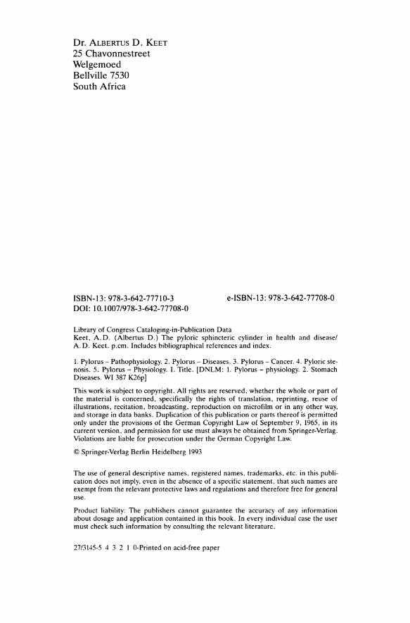
To Ilse and our Children
Nelleke, Danie, lise and Christiaan
A South African pyloros

Acknowledgements
Above all, lowe a debt of gratitude to the late Professor van Ebbenhorst Tengbergen and to Dr. P. van Exter of the University Roentgen Clinic, Amsterdam for allowing me to train as a radiologist under their supervision. I well remember my initial surprise at being told that the function of their clinic was not to teach. Rather, it was to give one the opportunity to question accepted statements and to pursue one's own line of research.
It was a great honour and privilege to be able to visit the late Professor Johan Torgersen of the University Anatomical Institute in Oslo at that time. This afforded an opportunity to discuss his monumental work on the anatomy and movements of the stomach and duodenal bulb at first hand. With characteristic modesty he took pains to explain that his findings were merely an elaboration of the work of Gbsta Forssell and that he himself had added little that was new.
This study would not have been possible without the cooperation of Professor J.J. Heydenrych (Department of Pediatric Surgery), whose advice and assistance over many years is greatly valued.
I am deeply grateful to Heads of the Department of Radiology, Professor C.J.B. Muller, Professor J.A. Beyers and Professor A.T. Scher, for their continued support and for allowing me to devote much time to the project.
My thanks are due to Professor D.J.J. Bezuidenhout and Dr. P.J. van Eeden (Department of Gastroenterology), Professor D.F. du Toit, Professor L.C. Janse van Rensburg, Dr. E.L. Bouwer and Dr. H.D. Louwrens (Department of Surgery), Professor J.F. Klopper and Professor W.J .c.J. Rosenstrauch (Department of Internal Medicine), Professor D.J. Rossouw (Department of Anatomy), Professor J.J. van der Walt and Professor B.D. Middlecote (Department of Anatomical Pathology) and Dr. D.H. Jamieson (Department of Radiology) for their help with specific problems.
Dr. R.H. Hewlett (Department of Anatomical Pathology) and Dr. R.D.J. Jamieson (Department of Radiology) read individual chapters and suggested improvements; for this I thank them.
VIII Acknowledgements
I gratefully record my thanks to Professor T.G. Schwar (Department of Forensic Medicine) and to Professor H-J. Ehrlein of the Institute for Zoophysiology, University of Hohenheim, Germany, for assistance with publication of the manuscript.
Special thanks are due to Mr. B. Reichenthaler, Ms Hanna Hensler-Fritton and Ms Gabi Schroder-Djeiran of SpringerVerlag, Heidelberg for their patience and kind cooperation.
I am very indebted to Ms I.L. Jansen van Vuuren, L.A. Bindeman, I.H. Snyders, B. Rademeyer and S.P. Joerdens for secretarial assistance, to Ms E. Geldenhuys and L.M. Giildenpfennig of the Medical Library for their kind assistance, and to Mr. J. Hough for help with the medical records.
Reproduction of radiographic films was in the capable hands of Mr. M.Y. Jooste of the Faculty of Dentistry.
All case reports published here are from Tygerberg Hospital, teaching hospital of the Medical Faculty, University of Stellenbosch. I wish to thank the Medical Superintendent of the hospital and the Dean of the Faculty for permission to publish.
The Editors of the following journals have kindly given permission to reproduce illustrations, tables and in some instances paragraphs previously published in the volumes indicated:
A.M.A. Archives of Pathology 1956, Vol. 61, pp 20-23, Fig. 1; Acta Radiologica 1957, Vol. 48, pp 413-424, Fig. ID, 2B, 3B, 9, 10; Acta Radiologica 1958, Vol. 50, pp 413-429; Acta Radiologica 1962, Vol. 57, pp 31-44, Fig. 13; American Journal of Roentgenology, Radium Therapy and Nuclear Medicine 1971, Vol. 113, pp 217-228, Fig. 3, 4, 5, 6, 7, 8; American Journal of Gastroenterology 1978, Vol. 69, pp 144-148, Fig. 1; South African Medical Journal 1953, Vol. 27, pp 117-123; South African Medical Journal 1960, Vol. 34, pp 881-884; South African Medical Journal 1974, Vol. 48, pp 441-448, Fig. 1,2; South African Medical Journa11982, Vol. 61, pp 78-81, Fig. 1,2,3,4; South African Medical Journal 1982, Vol. 62, pp 15-18, Fig. 2, 3, 4, 5; South African Medical Journal 1982, Vol. 62, pp 329-333, Fig. 1, 2, 3; South African Medical Journal 1984, Vol. 66, pp 740-742, Fig. 1,2,3.
Tygerberg, South Africa A. D. KEET
Contents
Chapter 1
Chapter 2
Chapter 3
Chapter 4
Introduction. . . . . . . . . . . . . . . . . . . . . . . .. 1
Some Uncertain Concepts . 5
The Pyloric Antrum . Discussion. . . . . . References. . . . . . What is a Sphincter? Discussion . References. Peristalsis . Discussion . References.
The Walls of the Stomach and Duodenum
Serous Coat . . . . . . . . . . . . . . • Muscular Coat or Muscularis Externa.
Morbid Anatomical Study . . Development of Musculature . . .
Discussion. . . . . . . . . . . . . . Sphincteric Mechanism at Pylorus Terminology
References. . . . . . . . . . . . . .
The Submucous Coat.
Stomach .. Duodenum. Discussion . References.
5 9 9
11 13 13 15 17 18
21
21 22 30 31 32 33 34 34
35
35 36 38 39
X Contents
Chapter 5
Chapter 6
Chapter 7
ChapterS
The Mucous Membrane or Mucosa .
Stomach ....... . Muscularis Mucosae Lamina Propria. . . Epithelial Lining . . Three Mucosal Zones Anatomical Extent and Boundaries of the Pyloric Mucosal Zone.
Duodenum ...... . Muscularis Mucosae Lamina Propria. . . Epithelial Lining . . Additional Features of the Mucosa.
Discussion . References. . . . . . . . . . . . . . .
Electrical Potential Difference at the Gastroduodenal Mucosal Junction .
Discussion . References.
Arteries, Veins, Lymphatics
Arterial Supply . . . Venous Drainage . . Lymphatic Drainage. Discussion . References. . . . . .
Nerves . ........... .
Parasympathetic Nerve Supply Anterior Vagus . . . . . . Posterior Vagus. . . . . . Parasympathetic Ganglia.
Discussion. . . . . . . . . Sympathetic Nerve Supply.
Sympathetic Ganglia . Discussion. . . . . Peptidergic System References. . . . .
41
41 41 41 42 42
44 47 47 47 48 48 49 50
53
54 54
55
55 56 57 59 59
61
61 61 64 65 66 67 67 68 68 68
Chapter 9
Chapter 10
Chapter 11
Chapter 12
Regulatory Peptides
Gastrin ....... . Somatostatin. . . . . Vasoactive Intestinal Peptide Substance P Enkephalin Galanin .. Neurotensin Discussion . References.
Ultrasonography of Normal Anatomy.
Contents
Conventional Surface Ultrasonography of the Normal Infantile Pylorus. Discussion . References. Endoscopic Ultrasonography of the Layer Structure of the Gastric Walls Discussion . References.
Anatomy of the Pyloric Ring
Radiographic Anatomy Present Investigations
Microscopic Anatomy . Present Investigations
Sonographic Anatomy. Present Investigations
Discussion . References.
The Pylorus at Rest: Open or Closed?
Present Investigations . Patients and Methods Results.
Discussion . References.
XI
71
71 74 75 77 78 79 79 80 81
85
85 86 87
88 89 90
91
92 92 93 94 95 96 96 97
99
99 99
100 103 104
XII Contents
Chapter 13 Radiographic Examination of Normal Motility. 105
Validation Studies. . . . . . . . . . 106 Intraluminal Pressure Profiles. . . . 107
Patients, Materials and Methods. 107 Results in Stomach. . 108 Conclusion . . . . . . 108 Results In Duodenum 108 Conclusion . . . . . . 109
Living Anatomical Studies. 109 Patients, Materials and Methods. 110 Results . . . . . . . . . . 111 Conclusion . . . . . . . . 111
Motor Divisions of Stomach. 111 Fornix . . . . . . 113 Corpus and Sinus. . . . . 113 Distal 3-4 cm. . . . . . . 114 Do Gastric Peristaltic Waves Progress as Far as the Pyloric Aperture? . . . . . . . . . . . . . . . 115
Discussion. . . . . . . . . . . . . . . . . . . . . 116 Contraction Patterns of Distal 3-4 cm of Stomach 116
Radiological Studies . 117 Patients and Methods 117 Results . . . . . . . . 117 Mucosal Movements 129
References. . . . . . . . 136
Chapter 14 The Pylorus at Gastroscopy .
Discussion . References.
Chapter 15 Manometry at the Gastroduodenal Junction .
Discussion . References.
139
142 144
147
154 155
Chapter 16 Myoelectric Activity at the Gastroduodenal Junction 157
Discussion . References.
164 165
Chapter 17 Ultrasonography of Pyloric Motility and Gastric Emptying
Discussion . References.
Chapter 18 Radionuclides in the Investigation of Gastric Emptying . . . . . .
Emptying of Liquids and Solids. Discussion . References. . . . . . . . . . . .
Contents XIII
167
169 170
171
171 175 177
Chapter 19 Gastric Tone and the Pyloric Sphincteric Cylinder. 179
Determination of Tone Discussion. . . .
Hypotonicity . Hypertonicity.
References. . . .
Chapter 20 Pylorospasm....
Radiological Features Problems of Definition Patients and Methods Discussion. . . . . Pathogenesis. . . . . Ultrasonic Features . Operative and Experimental Features. Discussion . References. . . . . . . . . . . . . . .
Chapter 21 Congenital Anomalies
Pyloric Atresia. Discussion. . References. . Duplications. Discussion. . References. . Congenital Double Pylorus Discussion. . . . . . . . .
179 181 181 182 182
183
183 183 184 186 186 188 189 192 194
197
197 197 198 199 199 199 200 200
XIV Contents
References. . . . . . . . . . . . . . . . Pyloric Membrane, Web or Diaphragm . Discussion. . . . . . . . References. . . . . . . . Ectopic Pancreatic Tissue Discussion . References. . . . . . . .
Chapter 22 Partial or Intramural Gastric Diverticulum .
Discussion . References.
200 202 203 203 205 205 206
207
210 211
Chapter 23 Infantile Hypertrophic Pyloric Stenosis. . . . . . . 213
Anatomical Localization and Radiographic Features . 213 Anatomical Localization and Ultrasonic Features. 218 Pathogenesis and Etiology. 222 Discussion. . . 226
Pathogenesis 227 Etiology 227
Conclusion. 229 References. 229
Chapter 24 Adult Hypertrophic Pyloric Stenosis 233
Discussion. . . . . . . 239 Types of AHPS. . . 239 Associated Lesions. 241 Relationship to Infantile Hypertrophic Pyloric Stenosis. . . . . . . . . . . . . 241 Anatomical Localization and Operative Features 242 Radiographic Features . . . . . 242 The Pathogenesis and Etiology. 243
References. . . . . . . . . . . . . 243
Chapter 25 Focal Hypertrophy and Focal Spasm of the Pyloric Musculature in Adults
Discussion . References.
245
249 250
Chapter 26 Nausea, Retching and Vomiting .
Nausea .. Retching. Vomiting. Discussion . References.
Chapter 27 Duodenogastric Reflux.
Previous Tests . . . . . . A Double-Contrast Radiographic Test for Duodenogastric Reflux
Advantages. . . . . . . . . Disadvantages . . . . . . . Results in Normal Subjects. Results in Patients
Subsequent Tests. Discussion . References. . . .
Chapter 28 Gastritis and Erosions in the Pyloric Sphincteric Cylinder .
Present Investigations . Patients and Methods
Discussion . References. . . . . . . .
Chapter 29 Gastric Ulceration and the Pyloric Sphincteric Cylinder . . . . . . . .
Gastric Ulceration Proximal to the Pyloric Sphincteric Cylinder. . . . . . . . . . Discussion. . . . . . . . . . . . . . . Gastric Ulceration Within the Pyloric Sphincteric Cylinder. Discussion . References. . . . . .
Contents XV
251
251 254 256 257 258
259
259
262 263 264 264 266 268 268 272
275
280 280 283 285
........ 287
288 295
299 305 306

XVI Contents
Chapter 30 Duodeual Ulceration and the Pyloric Sphincteric Cylinder . 309
Present Investigations . 312 Patients and methods. 312
Discussion . 313 References. . . . . . . . 315
Chapter 31 Pyloroduodenal Fistula or Acquired Double Pylorus 317
Discussion . 323 References. 323
Chapter 32 Hiatus Hernia and the Pyloric Sphincteric Cylinder . 325
Infants ........ 325 Adults ........ 326 Radiographic Studies 328
Patients and Methods 328 Experimental Studies . . 333
Material and Methods 334 Results. 335
Discussion . 336 References. 339
Chapter 33 Pyloric Carcinoma . . . 341 Present Investigations. . 346
Patients and Methods 346 Results ...... 352
Pyloric region. 352 Results ..... 353
Duodenum .. 353 Discussion. . . . 358
Emptying of Solids and Liquids 359 Duodenal Spread. . . . 359
Brunner's glands and EGF 362 References. . . . . . . . . 363
Chapter 34 Malignant Lymphoma 365
Discussion . 369 References. 370

Contents XVII
Chapter 35 Malignancy at the Gastro-oesophageal Junction. 371
Discussion . 374 References. 374
Chapter 36 Sessile Polyps in the Sphincteric Cylinder. 375
Peutz-Jeghers Syndrome. 376 Discussion . 378 References. . . . . . . . 378
Chapter 37 Diabetes Mellitus. 379
Discussion . 385 References. 387
Chapter 38 Prolapse of Gastric Mucosa into the Duodenum . 389
Normal Mobility of the Mucosa. 390 Pathoanatomical Diagnosis 391 Surgical Diagnosis. . . 392 Gastroscopic Diagnosis 393 Radiological Diagnosis 393 Discussion. . . . . . . 397
Types and Grades of Prolapse 399 Relationship to Cyclical Activity of Sphincteric Cylinder . . . . . . . . 399 The Radiological Differential Diagnosis . 400 Associated Gastroduodenal Lesions . . . 401 Possible True Complications . . . . . . . 402 Malignancy and Prolapse of Gastric Mucosa. 403
References. . . . . . . . . . . . . . . . . . . . 403
Chapter 39 Acid Corrosive Injuries and the Pyloric Sphincteric Cylinder 407
Discussion . 410 References. 410
Subject Index. . . . . . . . . . . . . . . . . . . . . .. 411


![70 Albertus AUSTRALIA PARLIAMENT HOUSE[1] - OK](https://static.fdocuments.in/doc/165x107/616a484d11a7b741a350ceeb/70-albertus-australia-parliament-house1-ok.jpg)