Akt-activated endothelium constitute the niche for...
36
Akt-activated endothelium and bevacizumab in ovarian cancer Akt-activated endothelium constitute the niche for residual disease and resistance to bevacizumab in ovarian cancer Bella S. Guerrouahen 1,2 , Jennifer Pasquier 1,2 , Nadine Abu Kaoud 1 , Mahtab Maleki 1 , Marie-Claude Beauchamp 3 , Amber Yasmeen 3 , Pegah Ghiabi 1 , Raphael Lis 2 , Fabien Vidal 1 , Ahmed Saleh 4 , Walter H. Gotlieb 3 , Shahin Rafii 2 & Arash Rafii 1,2,5 From the 1 Stem Cell and Microenvironment Laboratory, Weill Cornell Medical College in Qatar, Education City, Qatar Foundation, Doha, Qatar, 2 Department of Genetic Medicine, Weill Cornell Medical College, NY, USA, 3 Department of Gynecologic Oncology, Lady Davis Institute for Medical Research. Jewish General Hospital, McGill University, Montreal, Quebec, Canada. 4 National Center for Cancer Care and Research, Doha, Qatar 5 Department of Gynecologic oncology, University Montpellier 1, France § Address correspondence to: Dr Arash Rafii. Department of Genetic Medicine and Obstetrics and Gynecology, Weill Cornell Medical College, Stem cell and microenvironment laboratory Weill Cornell Medical College in Qatar, Qatar- Foundation, P.O Box: 24144, Doha, Qatar. Phone: +974 3313 5828. Fax: +974 4492 8422. Email: [email protected] Keywords: Ovarian cancer, anti-angiogenic therapy, endothelial cell heterogeneity, resistance, bevacizumab. Running Title: Akt-activated endothelium and bevacizumab in ovarian cancer Financial support: This work was founded by Qatar National Research Fund under its National Priorities Research Program award number: 09-1174-3- 291, 4-640-1-096, 6-1131-3-268 (A. Rafii) and JSREP No: 4-013-3-005 (BS. Guerrouahen). The content of this work is solely the responsibility of the authors and does not necessarily represent the views of the Qatar National Research Fund. No conflict of interest to disclose. Abstract: 196 words Figures: 6 Supplementary Figures: 5 figures and supplementary table on May 22, 2018. © 2014 American Association for Cancer Research. mct.aacrjournals.org Downloaded from Author manuscripts have been peer reviewed and accepted for publication but have not yet been edited. Author Manuscript Published OnlineFirst on October 15, 2014; DOI: 10.1158/1535-7163.MCT-13-1053
Transcript of Akt-activated endothelium constitute the niche for...
Akt-activated endothelium and bevacizumab in ovarian cancer
Akt-activated endothelium constitute the niche for residual disease and
resistance to bevacizumab in ovarian cancer
Bella S. Guerrouahen1,2, Jennifer Pasquier1,2, Nadine Abu Kaoud1, Mahtab
Maleki1, Marie-Claude Beauchamp3, Amber Yasmeen3, Pegah Ghiabi1,
Raphael Lis2, Fabien Vidal1, Ahmed Saleh 4, Walter H. Gotlieb3, Shahin Rafii2
& Arash Rafii1,2,5
From the 1Stem Cell and Microenvironment Laboratory, Weill Cornell Medical College in Qatar, Education City, Qatar Foundation, Doha, Qatar, 2Department of Genetic Medicine, Weill Cornell Medical College, NY, USA, 3Department of Gynecologic Oncology, Lady Davis Institute for Medical Research. Jewish General Hospital, McGill University, Montreal, Quebec, Canada. 4National Center for Cancer Care and Research, Doha, Qatar 5 Department of Gynecologic oncology, University Montpellier 1, France §Address correspondence to: Dr Arash Rafii. Department of Genetic Medicine and Obstetrics and Gynecology, Weill Cornell Medical College, Stem cell and microenvironment laboratory Weill Cornell Medical College in Qatar, Qatar-Foundation, P.O Box: 24144, Doha, Qatar. Phone: +974 3313 5828. Fax: +974 4492 8422. Email: [email protected] Keywords: Ovarian cancer, anti-angiogenic therapy, endothelial cell heterogeneity, resistance, bevacizumab. Running Title: Akt-activated endothelium and bevacizumab in ovarian cancer
Financial support: This work was founded by Qatar National Research Fund
under its National Priorities Research Program award number: 09-1174-3-
291, 4-640-1-096, 6-1131-3-268 (A. Rafii) and JSREP No: 4-013-3-005 (BS.
Guerrouahen). The content of this work is solely the responsibility of the
authors and does not necessarily represent the views of the Qatar National
Research Fund.
No conflict of interest to disclose.
Abstract: 196 words Figures: 6 Supplementary Figures: 5 figures and supplementary table
on May 22, 2018. © 2014 American Association for Cancer Research. mct.aacrjournals.org Downloaded from
Author manuscripts have been peer reviewed and accepted for publication but have not yet been edited. Author Manuscript Published OnlineFirst on October 15, 2014; DOI: 10.1158/1535-7163.MCT-13-1053

Akt-activated endothelium and bevacizumab in ovarian cancer
2
ABSTRACT
Ovarian cancer is the second leading cause of cancer-related death in women
worldwide. Despite optimal cytoreduction and adequate adjuvant therapies,
initial tumor response is often followed by relapse suggesting the existence of
a tumor niche. Targeted therapies have been evaluated in ovarian cancer to
overcome resistant disease. Among them anti-angiogenic therapies inhibit
new blood vessel growth, induce endothelial cell apoptosis, and block the
incorporation of haematopoietic and endothelial progenitor cells into new
blood vessels. Despite in vitro and in vivo successes, anti-vascular therapy
with bevacizumab targeting VEGF-A has limited efficacy in ovarian cancer.
The precise molecular mechanisms underlying clinical resistance to anti-
VEGF therapies are not yet well understood. Among them, tumor and stromal
heterogeneity might determine the treatment outcomes. The present study
investigates whether abnormalities in the tumor endothelium may contribute to
treatment resistance to bevacizumab and promote a residual microscopic
disease. Here we showed that ovarian cancer cells (OCC) activate Akt
phosphorylation in endothelial cells inducing resistance to bevacizumab
leading to an autocrine loop based on FGF-2 secretion. Altogether our results
point out the role of an activated endothelium in the resistance to
bevacizumab and in the constitution of a niche for a residual disease.
on May 22, 2018. © 2014 American Association for Cancer Research. mct.aacrjournals.org Downloaded from
Author manuscripts have been peer reviewed and accepted for publication but have not yet been edited. Author Manuscript Published OnlineFirst on October 15, 2014; DOI: 10.1158/1535-7163.MCT-13-1053
Akt-activated endothelium and bevacizumab in ovarian cancer
3
Introduction
Neo-angiogenesis is a primordial step for tumor growth and metastasis and its
targeting is under investigation in many solid tumors (1-4). Human cancer
cells express elevated levels of proangiogenic factors (5). Among them the
role of vascular endothelial growth factor (VEGF-A) in tumor progression has
been clearly established (6). Elevated expression of circulating VEGF-A is
associated with poor prognosis in metastatic colorectal, lung, and renal
cancer (7). Subsequently the first anti-angiogenic molecule developed to
impair neo-angiogenesis targeted VEGF. Bevacizumab (Avastin, Genentech
Inc., San Francisco, CA) is a humanized monoclonal antibody directed
against all isoforms of VEGF-A (8). Bevacizumab prevents the binding of
VEGF-A to its receptor VEGF-R1 (Flt-1) and R2 (Flt-2, or KDR). Although the
function of VEGF-R2 in tumor angiogenesis has been characterized
thoroughly, the function of VEGF-R1 is not yet well defined (9). Bevacizumab
has been found clinically active in many solid tumors, such as colon, non-
small cell lung, metastatic renal cell carcinoma or glioblastoma (10-13).
Currently the Food and Drug Administration approves its use in combination
with chemotherapy/immunotherapy in colon, lung and renal cancers and in
glioblastoma as a single agent. The mechanisms of action of bevacizumab
are not perfectly understood. It could act by sequestering VEGF-A in the
blood and/or tumor interstitium or induce normalization of VEGF-A
concentration in these compartments (14). Despite initial efficacy, the
withdrawal of bevacizumab can be associated to rapid tumor re-growth or a
‘‘rebound’’ phenomenon with accelerated clinical decline in patients (15, 16).
Moreover, in case of tumor progression under bevacizumab, salvage
on May 22, 2018. © 2014 American Association for Cancer Research. mct.aacrjournals.org Downloaded from
Author manuscripts have been peer reviewed and accepted for publication but have not yet been edited. Author Manuscript Published OnlineFirst on October 15, 2014; DOI: 10.1158/1535-7163.MCT-13-1053

Akt-activated endothelium and bevacizumab in ovarian cancer
4
chemotherapy has been quite inefficient, suggesting the emergence of a
treatment-resistant phenotype (17). Most tumors will develop resistance to
bevacizumab on a long term (18). While vessels were perceived as passively
conducting nutrients to tumor cells, recent reports underline the perfusion-
independent role of the endothelium. In such context endothelial cells (ECs)
participate to an active cross-talk with tumor cells, resulting in the modification
of their phenotype (19). ECs isolated from various tumors acquired genotype
alterations as aneuploidie, or abnormal multiple chromosomes (20) or
displayed an activated profile through Pi3/Akt or MAPK pathway (21).
Ovarian cancer usually presents as locally advanced disease with
involvement of the peritoneal cavity and the use of bevacizumab is under
investigation in several clinical trials. Here we hypothesized that the cross-talk
between cancer and endothelial cells would induce an activated ECs profile
responsible for bevacizumab resistance and the constitution of a vascular
niche. We demonstrated that the cross-talk between ovarian cancer cells and
established human umbilical vein endothelial cell line (HUVECs), resulted in
an activation of Akt pathway among others. To model tumor endothelium, we
used the Akt-activated endothelial cells (E4+ECs) to study the involvement of
the endothelial component in the resistance to anti-angiogenic therapy (22).
We demonstrated that in the setting of tumor-endothelial cross-talk, ECs
activation of Pi3k/Akt pathway induces an autocrine loop through the pro-
angiogenic factor fibroblast growth factor 2 (FGF-2), bypassing the VEGF-R
pathway. We demonstrated that blocking FGF-2 would efficiently reverse the
resistance to bevacizumab. These results propose the use of stroma directed
synthetic therapy to overcome resistance in ovarian cancer.
on May 22, 2018. © 2014 American Association for Cancer Research. mct.aacrjournals.org Downloaded from
Author manuscripts have been peer reviewed and accepted for publication but have not yet been edited. Author Manuscript Published OnlineFirst on October 15, 2014; DOI: 10.1158/1535-7163.MCT-13-1053

Akt-activated endothelium and bevacizumab in ovarian cancer
5
Material and methods
Reagents and chemicals. Bevacizumab (trade name Avastin, Genentech) or
100 mg / 16.6 ml paclitaxel (Ebetaxel®, EBEWE Pharma, Unterach, Austuria)
were obtained from National Center for Cancer Care and Research (NCCCR,
Doha, Qatar) pharmacy. The bevacizumab doses used for patients’ treatment
(5-15 mg/kg), correlates to a mean plasma concentrations value between 0.1
and 0.5 mg/mL; we diluted bevacizumab in serum free medium to obtain final
concentrations of 0.5/ 1.0/ 1.5/ 2/ and 2.5 mg/mL. Ly294002 (L9908), FGF
receptor (FGFR) inhibitors PD17034 (P2499) and SU-5402 (SML0443) were
purchased from Sigma-Aldrich and resuspended in DMSO.
Immunohistochemistry. Immunohistochemistry (IHC) was performed at the
Segal Cancer Centre Research Pathology Facility (Jewish General Hospital,
Montreal, Quebec, Canada). Tissue samples were cut at 4-µm, placed on
Superfrost Plus stain slides (Fisher), and dried overnight at 37°C. The slides
were then loaded onto the Discovery XT Autostainer (Ventana Medical
System). All solutions used for automated immunohistochemistry were from
Ventana Medical System unless otherwise specified. Immunostaining for
phospho-Akt was performed using a heat protocol. Briefly, rabbit monoclonal
anti-phospho-Akt (Ser473) (D9E) (Cell Signaling Technology) diluted 1:50 in
Antibody diluent solution, was manually applied for 30 min, and then followed
by the appropriate detection kit (Omnimap anti-Rabbit HRP). Slides were
counterstained with hematoxylin for four minutes, blued with Bluing Reagent
for four minutes, removed from the autostainer, washed in warm soapy water,
on May 22, 2018. © 2014 American Association for Cancer Research. mct.aacrjournals.org Downloaded from
Author manuscripts have been peer reviewed and accepted for publication but have not yet been edited. Author Manuscript Published OnlineFirst on October 15, 2014; DOI: 10.1158/1535-7163.MCT-13-1053

Akt-activated endothelium and bevacizumab in ovarian cancer
6
dehydrated through graded alcohols, cleared in xylene, and mounted with
Permount. Sections were analyzed by conventional light microscopy.
Cell culture. Established Human umbilical vein endothelial cells (HUVECs)
were purchased and grown as recommended by the American Type Culture
Collection (ATCC, Manassas, VA, USA). E4+ECs were obtained as previously
described (22). Briefly, isolated HUVEC were transduced with the early 4
region (E4) of the adenoviral vectors (AdE4+) to generate durable PEC
feeders. HUVECs and E4+EC cells were cultured in M199 medium
supplemented with 20% fetal bovine serum (FBS), 1% penicillin ⁄
streptomycin, 0.1% heparin and 0.1% endothelial cell growth supplement
(ECGS) and maintained at 37 °C in a humidified 5% CO2 atmosphere.
HUVECs cells were seeded on plates coated with 0.1% gelatin and allowed to
grow. Treatments were performed in serum- growth factor- free medium over
24 hours. Human Ovarian Microvascular Endothelial Cells (HOMEC) were
purchased from ScienCell research Laboratories (#7300) and grown
according to the manufacturer instructions. Ovarian cancer cell lines SKOV-3
(HTB-77), OVCAR-3 (HTB-161) were purchased from ATCC (Manassas, VA,
USA). Cells were grown in DMEM high glucose (Hyclone, Thermo Scientific)
supplemented with 10% FBS (Hyclone, Thermo Scientific), 1% Penicillin-
Streptomycin-Amphotericyn B solution (Sigma), 2 mM L-glutamine (Sigma),
1X Non-Essential Amino-Acid (Hyclone, Thermo Scientific). All cell lines were
obtained between 2011 and 2013 from American Type Culture Collection
(ATCC) or ScienCell research laboratory. Upon receipt, cells were expanded
and aliquots of cells at passage number <10 were stored frozen in liquid
on May 22, 2018. © 2014 American Association for Cancer Research. mct.aacrjournals.org Downloaded from
Author manuscripts have been peer reviewed and accepted for publication but have not yet been edited. Author Manuscript Published OnlineFirst on October 15, 2014; DOI: 10.1158/1535-7163.MCT-13-1053

Akt-activated endothelium and bevacizumab in ovarian cancer
7
nitrogen. Cells from one aliquot were kept in culture for less than 2 months.
The authors did not authenticate identity of each cell line purchased. GOC-2
and GOC-A2 are primary cell lines that were derived by Dr Gotlieb’s
laboratory. GOC-2 cells were isolated from a papillary serous ovarian cancer
obtained after 4 courses of neo-adjuvant chemotherapy with carboplatin /
taxol; and GOC-A2 from a stage IIIc serous ovarian cancer.
Cell proliferation assay. Cellular viability and proliferation was analyzed by
both trypan blue exclusion and tetrazolium dye assay. HUVECs and E4+ECs
cells were allowed to grow to 80% confluence in 96 well plates. Cells were
then incubated with the different concentrations of bevacizumab in serum,
cytokine free medium for 24 hours and subjected to XTT assay as previously
described by the manufacturer (TACS XTT cell proliferation assay kit,
Trevigen, Inc.). Optical density was measured at 540 nm. Alternatively,
treated cells were then collected and diluted in trypan blue dye (Sigma) for
manual counting with hemocytometer.
Wound Healing and Tube Formation Assays. Cells were grown to
confluency in starvation medium (serum- growth factor- free). Using a pipette
tip, cells were scraped from the culture dish leaving a void space. Cells were
washed with PBS and then incubated for 36 hours in the presence of the
treatment. After incubation, wound closure was imaged by phase contrast
microscope and analyzed using Image J software. Five random fields per well
were examined. For the tube formation assay, 24-well culture plates were
coated with Matrigel according to the manufacturer's instructions. HUVECs
on May 22, 2018. © 2014 American Association for Cancer Research. mct.aacrjournals.org Downloaded from
Author manuscripts have been peer reviewed and accepted for publication but have not yet been edited. Author Manuscript Published OnlineFirst on October 15, 2014; DOI: 10.1158/1535-7163.MCT-13-1053
Akt-activated endothelium and bevacizumab in ovarian cancer
8
and E4+ECs were seeded on coated plates at 5.104 cell/well in serum- growth
factor- free medium, treated and incubated at 37 °C for 8 hours. Cells were
then analyzed for tube formation with an inverted light microscope. The
criteria taken into account were the branching point number being formed.
Five random fields per well were examined. Cells seeded on coated plate in
complete medium served as positive control.
Generation of a treatment-resistant HUVEC line. A treatment-refractory
HUVEC line was developed by continuous exposure to bevacizumab. Briefly,
HUVECs were exposed to stepwise increases in bevacizumab
concentrations. The initial bevacizumab exposure was at a concentration of
0.1 mg/mL. After cells had regained their exponential growth rate,
bevacizumab concentration was doubled; this procedure was repeated until
selection of a clone resistant to 1 mg/mL of bevacizumab. The resulting
bevacizumab-refractory cell line was sub-cultured every 5 days and treated
every 15 days with 1mg/mL of bevacizumab to maintain a high level of anti
angiogenesis resistance.
Sphere formation assay. E4+EC-eGFP and SKOV-3 were dissociated into
single cell suspension by trypsinization and further sieving through 40-µm cell
strainers. E4+ECeGFP and SKOV-3 cells (ratio 10/1) were then resuspended
in 3D media consisting of DMEM F-12 supplemented with 2% B27, 20 ng/mL
EGF, 20 ng/mL bFGF, and 5μg/mL insulin in ultralow attachment plates.
Primary cancer spheroids and angiospheres (SKOV-3 / E4+ECeGFP, ratio
1:10) started to form at day 3 and maintained up to day 7. Primary spheres
on May 22, 2018. © 2014 American Association for Cancer Research. mct.aacrjournals.org Downloaded from
Author manuscripts have been peer reviewed and accepted for publication but have not yet been edited. Author Manuscript Published OnlineFirst on October 15, 2014; DOI: 10.1158/1535-7163.MCT-13-1053

Akt-activated endothelium and bevacizumab in ovarian cancer
9
were dissociated into single cell suspension and re-plated as mentioned
above to form secondary spheres. 7 days old secondary spheres were used
for inducing tertiary spheres. Sphere formation was evaluated by ImageJ64
software based on the area of the sphere covered by SKOV-3. To evaluate
E4+EC-eGFP survival under bevacizumab treatment, the area of the sphere
covered by E4+EC-eGFP was majored.
Analysis of viability and apoptosis detection by flow cytometry. Viability
was assessed by flow cytometry evaluation of calcein AM staining as
described by the manufacturer (Live Dead Viability/cytotoxicity Kit, Molecular
Probes, Invitrogen). Cells treated with bevacizumab (1.5mg/mL), were
harvested after 24h. Analyses were performed on SORP FACS Aria2 (BD
Biosciences). Calcein AM fluorescence was acquired with 488 nm blue laser
and 525/50 nm emission, ethidium homodimer was acquired with 640 nm red
laser and 670/14 nm emission. Cytotoxicity was assessed by flow cytometry
evaluation of annexin V, propidium iodide (PI) staining cells as described by
the manufacturer (FITC Annexin V Apoptosis Detection Kit I, BD
Pharmingen). Cells were treated overnight with varying concentrations of
Bevacizumab (0.1, 1, 1.5, 2, 2.5 mg/ml) and/or Ly294002 (10 µM). PI and
Annexin V were added and cells incubated at room temperature for 15 min.
Stained cells were analyzed on SORP FACS Aria2 using excitation at 535 nm
and emission at 582/15 nm for PI and 488 nm excitation, 525/50 nm emission
for annexin-FITC. Data were processed with FACSDiva 6.3 (BD Biosciences).
Doublets were excluded by FSC-W × FSC-H and SSC-W × SSC-H analysis,
single stained channels were used for compensation, and fluorophore minus
on May 22, 2018. © 2014 American Association for Cancer Research. mct.aacrjournals.org Downloaded from
Author manuscripts have been peer reviewed and accepted for publication but have not yet been edited. Author Manuscript Published OnlineFirst on October 15, 2014; DOI: 10.1158/1535-7163.MCT-13-1053

Akt-activated endothelium and bevacizumab in ovarian cancer
10
one (FMO) controls were used for gating (23). Single stained channels were
used for compensation and fluorophore minus one (FMO) controls were used
for gating.
Phospho-flow analysis and intracellular detection. After treatment or co-
culture, cells were fixed using BD CytofixTM Fixation Buffer (BD Biosciences),
followed by permeabilization at 4oC with BDTM Phosflow Perm Buffer III (BD
Biosciences). Conjugated antibodies were added to the cells for 45 min 4oC.
Phospho-Akt S473 PE-conjugated antibody (BD Biosciences) and VEGF-A
PE-conjugated antibody (R&D Systems) was acquired with 535 nm green
laser and 582/15 nm emission. Phospho-STAT-3 (Y705) alexa Fluor 647 (BD
Biosciences) was acquired with 640 nm red laser and 670/14 nm emission.
50000 events were acquired per sample. Single stained channels were used
for compensation and FMO controls were used for gating.
Western blot analyses, antibodies and immunoprecipitation. Immunoblot
analysis were performed using the following antibodies from Cell Signaling
Technology (Beverly, MA): phospho-Akt (S473) (#9271), Akt isoform sampler
kit (#9940), FGF-2 (#3196), PARP (Poly [ADP-Ribose] Polymerase, #9532),
cleaved PARP (#9541), phospho-FGFR1 (#3472), phospho STAT-3 (Y705)
(#9145), phospho-STAT-3 (S727) (#9134), STAT-3 (#4904), phospho-ERK1/2
(#9101), phospho-Src (Y416) (#2101), actin (#3700) and VEGF-A from Santa
Cruz Biotechnology (#SC-152). Immunoreactive bands were visualized by
using a developing solution (Western Lightening, Perkin Elmer LAS, NEL104,
Boston). For immunoprecipitation: equal amount of cell lysate were
on May 22, 2018. © 2014 American Association for Cancer Research. mct.aacrjournals.org Downloaded from
Author manuscripts have been peer reviewed and accepted for publication but have not yet been edited. Author Manuscript Published OnlineFirst on October 15, 2014; DOI: 10.1158/1535-7163.MCT-13-1053

Akt-activated endothelium and bevacizumab in ovarian cancer
11
preabsorbed with 50 μL of protein A/G Plus agarose (Santa Cruz, Sc-2003)
and then incubated overnight at 4°C with anti-Akt1, anti-Akt2, and anti-Akt3
antibody. Immune complexes were precipitated with 50 μL of Protein A/G Plus
agarose and then washed 3 times with lysis buffer. Immunoprecipitated
proteins were eluted in Laemmli sample buffer and subjected to western blot
analysis.
RT-PCR analysis. Total RNA was isolated by TRIzol reagent (Invitrogen,
Carlsbad, CA, USA). Briefly, addition of chloroform, after the centrifugation,
separates the solution into aqueous and organic phases. RNA remains only in
the aqueous phase. After transferring the aqueous phase, RNA is recovered
by precipitation with isopropyl alcohol. A quantitative analysis of RNA was
performed using a two-step reverse transcription-quantitative PCR (GoTaq®
2-Step qRT-PCR System qRT-PCR, Promega, USA) protocol according to the
manufacturer’s instructions. cDNA was synthesized from 2µg total RNA, using
the GoScript™ Reverse Transcription System and amplified using the
GoTaq® qPCR Master Mix. Cycling conditions were 95°C for 2 minutes, 40
cycles of 95°C for 15 seconds, 60°C for 1 minute, 72°C for 10 seconds, 72°C
for 5 minutes and cooled to 4°C. The final extension step was continued for 5
min. Variation in cDNA loading was normalized to GAPDH expression and
relative expression values were determined using ∆∆Ct method of relative
quantification. Primer sequences are listed as supplementary table and were
purchased from IDT.
on May 22, 2018. © 2014 American Association for Cancer Research. mct.aacrjournals.org Downloaded from
Author manuscripts have been peer reviewed and accepted for publication but have not yet been edited. Author Manuscript Published OnlineFirst on October 15, 2014; DOI: 10.1158/1535-7163.MCT-13-1053
Akt-activated endothelium and bevacizumab in ovarian cancer
12
Neutralizing anti FGF-2 assay. A monoclonal neutralizing mouse anti-FGF-2
antibody (Millipore, clone bFM-1) was used for FGF-2 neutralization at 1 µg/ml
and 5 µg/ml with or without bevacizumab. Cell viability was determined by
XTT assay after 2 days neutralization. E4+ECs stimulated with 20 ng/ml of
FGF-2 were used as positive controls. Isotypic IgG control was used as
internal control, and serum-free medium as negative control.
FGF-2 and VEGF-A secretion. We used a Human VEGF-A Quantikine
ELISA Kit (DVE00, R&D Systems) and Human FGF-2 Quantikine ELISA Kits
(DFB50, R&D Systems). Plates were read at 450 nm. Concentrations were
interpolated from a standard curve.
Statistical analysis. All quantitative data were expressed as mean ±
standard error of the mean (SEM). Statistical analysis was performed, using
SigmaPlot 11 (Systat Software Inc., Chicago, IL, USA). A Shapiro-Wilk
normality test, with a p=0.05 rejection value, was used to test normal
distribution of data prior further analysis. All pairwise multiple comparisons
were performed by one way ANOVA followed by Sidak-Holm Post Hoc tests
for data with normal distribution or by Kruskal-Wallis analysis of variance on
ranks followed by Tukey Post Hoc tests, in case of failed normality test.
Paired comparisons were performed by Student's t-tests or by Mann-Whitney
rank sum tests in case of unequal variance or failed normality test. Statistical
significance was accepted for p < 0.05 (*), p < 0.01 (**) or p < 0.001 (***).
on May 22, 2018. © 2014 American Association for Cancer Research. mct.aacrjournals.org Downloaded from
Author manuscripts have been peer reviewed and accepted for publication but have not yet been edited. Author Manuscript Published OnlineFirst on October 15, 2014; DOI: 10.1158/1535-7163.MCT-13-1053

Akt-activated endothelium and bevacizumab in ovarian cancer
13
Results
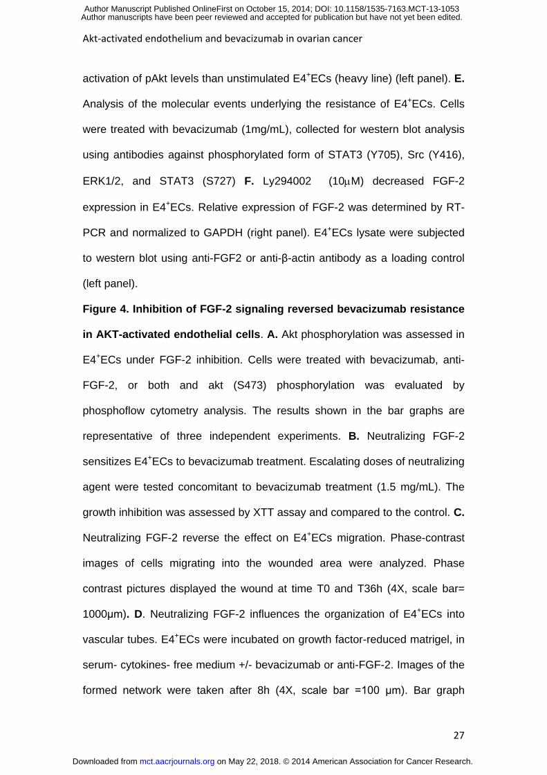
Cancer cell cross-talk with endothelial cells induces pro-survival
signaling. Tumor endothelial heterogeneity and in particular endothelial Akt
activation has been previously reported (24). Ovarian tumor vessels displayed
heterogenous phospho-Akt staining (Fig. 1A). To investigate the role of
soluble factors in tumor-endothelial cells communication, we established a
trans-well co-culture system of HUVEC and ovarian cancer cells (OCCs). We
used (i) established OCCs lines (OVCAR-3, SKOV-3), (ii) and two primary
OCCs isolated from papillary serous ovarian cancers (GOC-2 and GOC-A2).
As shown in Figure 1B, we observed heterogeneous Akt phosphorylation in
HUVECs. OVCAR-3 cells induced less Akt phosphorylation than the other cell
lines tested. SKOV-3, which are highly resistant to chemotherapy and have
metastatic features, induced the higher level of Akt activation. We confirmed
the 'activated' phenotype in HUVEC by western blot (Supplementary Fig. S1).
E4+ECs display resistance to bevacizumab. We hypothesize that
endothelium activation might play a role in bevacizumab resistance. The use
of serum and cytokines to maintain HUVECs in culture might hinder cell
autonomous effect. Thus to model a tumor associated endothelium, we used
our recently generated endothelial cell line E4+ECs that can survive in serum-
cytokine- free media (22) to study further mechanisms of resistance to
bevacizumab. We first verified the presence of VEGF-A and VEGFR (mRNA
and protein levels) in HUVECs and E4+ECs (supplementary Fig. S2-A and B).
We conducted several functional assays with E4+ECs, their control HUVEC
and HUVEC co-cultured with ovarian cancer cells (Figure 2A). We evaluated
on May 22, 2018. © 2014 American Association for Cancer Research. mct.aacrjournals.org Downloaded from
Author manuscripts have been peer reviewed and accepted for publication but have not yet been edited. Author Manuscript Published OnlineFirst on October 15, 2014; DOI: 10.1158/1535-7163.MCT-13-1053

Akt-activated endothelium and bevacizumab in ovarian cancer
14
the cell viability by XTT Cell proliferation assay and FACS, after bevacizumab
treatment. Doses of bevacizumab between 1 and 2.5 mg/mL did not
significantly affect the number of viable E4+ECs contrary to HUVECs at 24h
and 48h (Fig. 2B, 2C and data not shown). Addition of VEGF-A (10, 25, 50
ng/mL) could not completely protect HUVEC from the decrease of viability
(supplementary Fig. S3-A, upper panel). Interestingly, HUVEC pretreated with
VEGF-A before treatment with Bevacizumab displayed higher survival
(supplementary Fig. S3-A, bottom panel). We could illustrate the absence of
an apoptotic phenomenon in E4+ECs and HUVEC co-cultured with SKOV-3
conditioned medium by the absence of cleaved PARP upon bevacizumab
treatment (Fig. 2D). Besides the pro-survival signaling, VEGF-A also induces
endothelial cell migration and vessel formation (25). Under serum- growth
factor- free condition, we were not able to evaluate the motility of HUVEC in
the presence or absence of VEGF-A (10, 25, 50ng/mL), or bevacizumab
treatment. But we observed that in E4+ECs, or HUVEC co-cultured with
SKOV-3 condition medium, bevacizumab was not able to prevent wound
closure (Fig. 2E and supplementary Fig. S3-B, S3-C).
In a tube formation assay, while HUVECs treated by bevacizumab alone or
supplemented with VEGF-A resulted in unramified tube-like structures with
dead ends, E4+ECs or HUVEC co-cultured with SKOV-3 condition medium
maintained the capacity to form assembled capillary-like structures (Figure 2F
and Supplementary Fig. S3-D and S3-E). Concordantly, E4+ECs displayed
the same profile of resistance than HUVECs co-cultured with SKOV-3
supernatants, confirming the use of E4+ECs as a model. Interestingly, we also
showed that bevacizumab had a profound effect on HOMEC compared to
on May 22, 2018. © 2014 American Association for Cancer Research. mct.aacrjournals.org Downloaded from
Author manuscripts have been peer reviewed and accepted for publication but have not yet been edited. Author Manuscript Published OnlineFirst on October 15, 2014; DOI: 10.1158/1535-7163.MCT-13-1053

Akt-activated endothelium and bevacizumab in ovarian cancer
15
E4+ECs (Supplementary Fig. S3-F).
Inhibition of Akt-mediated survival pathways increases bevacizumab
effect. We first showed that Akt phosphorylation was abrogated as early as
10 minutes after treatment with the pan-PI3K inhibitor LY294002 (10 µM) (Fig.
3A and Supplementary Fig.S4-A). LY294002 treatment alone did not
decrease E4+ECs viability, but co-treatment with bevacizumab reversed
E4+ECs resistance to anti-angiogenic therapies (Fig. 3B and supplementary
S4-B). This underlines the central role of Akt pathway in the resistance to
bevacizumab treatment. To illustrate the essential role of Akt, we developed a
bevacizumab-refractory HUVEC line. Treatment of this cell line with Ly294002
decreased Akt phosphorylation and restored the sensitivity to bevacizumab
(Supplementary Fig. S4-C). Western blot analysis and immunoprecipitation
showed that Akt1 is the predominant isoform expressed in E4+ECs as well as
the main form phosphorylated (supplementary Fig. S4-D). Additional
angiogenic factors such as FGF-2 (26) can compensate the blockade of
VEGF (27). Interestingly, transcriptomic and RT-PCR analysis showed that
FGFR1 mRNA was increased by 2.5 fold in E4+ECs compared to HUVECs
(Supplementary Fig. S4-E). In all OCCs used in this study, while bevacizumab
efficiently neutralizes VEGF-A, the expression of FGF-2 could still be detected
(Supplementary Fig. S4-F). We observe that E4+ECs treatment by
bevacizumab is accompanied by a three-fold increase in FGF-2 mRNA and
protein level. We also observed an increase in FGFR1 mRNA and in FGFR1
phosphorylation (Fig. 3C). We then verified if FGF-2 was able to induce Akt
activation and found that Akt phospohorylation could be detected in HUVEC
on May 22, 2018. © 2014 American Association for Cancer Research. mct.aacrjournals.org Downloaded from
Author manuscripts have been peer reviewed and accepted for publication but have not yet been edited. Author Manuscript Published OnlineFirst on October 15, 2014; DOI: 10.1158/1535-7163.MCT-13-1053

Akt-activated endothelium and bevacizumab in ovarian cancer
16
as soon as 10 minutes after treatment with FGF-2 (10ng/mL) (Fig. 3D, left
panel). An overnight treatment with FGF-2 shows that Akt phosphorylation in
HUVEC cells is similar to the Akt phosphorylation observed in E4+EC as
shown by phospho-flow analysis (Fig. 3D, right panel). Concordantly, the
treatment of HUVEC by FGF-2 induced resistance to bevacizumab and
reversed the inhibition of angiogenesic attributes (Supplementary Fig.S5-A, B,
C). We investigated the molecular events underlying the potential
mechanisms of resistance through FGF-2 expression in endothelial cells. As
previously reported (28), endothelial FGF-2 synthesis is regulated by a
signaling cascade involving ERK1/2 and STAT-3 activation. Under
bevacizumab treatment, FGF-2/FGFR1 axis mediates Src activation and
downstream activation of the pro-angiogenic ERK1/2-STAT-3 signaling
pathway as shown by the increase phosphorylation of Src (Y416), STAT3 and
ERK1/2 (Fig. 3E). The secretion of FGF-2 has been mainly attributed to
cancer cells (27, 29), we showed the existence of an autocrine loop
maintaining Akt activation in E4+ECs in the context of VEGF-A inhibition. Akt
inhibition by Ly294002 resulted in FGF-2 inhibition at mRNA and protein
levels suggesting that Akt activation is responsible for FGF-2 secretion (Fig.
3F).
Inhibition of FGF2 signaling reversed bevacizumab resistance in
E4+ECs. To investigate if the inhibition of FGF-2 signaling would reverse
bevacizumab resistance, we used a FGF-2 blocking antibody. We showed
that neutralization of FGF-2 activity decreases Akt phosphorylation in E4+EC.
We then showed a synergistic effect when anti-FGF-2 was added to
on May 22, 2018. © 2014 American Association for Cancer Research. mct.aacrjournals.org Downloaded from
Author manuscripts have been peer reviewed and accepted for publication but have not yet been edited. Author Manuscript Published OnlineFirst on October 15, 2014; DOI: 10.1158/1535-7163.MCT-13-1053

Akt-activated endothelium and bevacizumab in ovarian cancer
17
bevacizumab treatment. This result suggests that Akt activation of E4+EC
depend on both VEGF-A and FGF-2 secretion (Figure 4A). We verified that
blocking FGF-2 alone did not impact E4+ECs survival. However, in the context
of FGF-2 deprivation, bevacizumab resistance was reverted (Fig. 4B). Finally,
we also investigated the role of FGF-2 in the angiogenic attributes. Blocking
FGF-2 alone has a minor effect on motility, but a drastic effect on tube
formation, confirming the role of FGF-2 as a potent angiogenic factor (Fig.4C
and 4D). We then assessed the effect of the inhibition of FGFR activity using
two FGFR-selective inhibitors, PD17034 (30) and SU5402 (31) in combination
with bevacizumab. While treatment with PD17034 (10nM), or SU5402 (1µM)
alone resulted in a modest decrease of E4+EC growth, a bevacizumab-dose-
dependent reduction of E4+EC proliferation was observed after co-targeting
VEGF-A and FGFR, confirming the synergistic role of the two cytokines (Fig.
4E). These data suggests that the FGF/FGFR axis influences the sensitivity of
the E4+ECs to bevacizumab therapy.
Residual Disease. The rebound effect described in the literature and
observed in the clinical setting can from anendothelial niche. We set-up a
model of co-culture with endothelial and cancer cells and tried to reproduce a
residual disease. Both HUVECs and E4+ECs integrated tumor nodules of
SKOV-3 produced on matrigel. We then sequentially treated these nodules
with the microtubule-damaging agent taxol (10µM) and bevacizumab
(1mg/mL). After taxol treatment, few cancer and endothelial cells survived.
E4+ECs presented significant resistance to the treatment. The treatment with
bevacizumab completely eliminated all tumor and endothelial cells in the
on May 22, 2018. © 2014 American Association for Cancer Research. mct.aacrjournals.org Downloaded from
Author manuscripts have been peer reviewed and accepted for publication but have not yet been edited. Author Manuscript Published OnlineFirst on October 15, 2014; DOI: 10.1158/1535-7163.MCT-13-1053
Akt-activated endothelium and bevacizumab in ovarian cancer
18
HUVEC control group. However E4+ECs and SKOV-3 were able to grow and
produce a “recurrent nodule” (Fig. 5A). We went further and created
structures called angiospheres. We have recently illustrated the ability of the
endothelium to form 3D structures that can sustain tissue specific cell growth
(32). We hypothesize that the same phenomenon could be relevant to cancer
biology where within 3D structures the endothelial cells can support the
expansion of the cancer propagating cells. We were able to demonstrate an
increase ability of cancer cells co-cultured with E4+ECs to form spheres
comparing to cancer cells alone. The results were similar when secondary
and tertiary spheres were induced (Figure 5B). To confirm that E4+ECs are
responsible for the expansion of the spheroids under bevacizumab treatment,
we treated daily the spheroids and showed that after 7 days, E4+ECs can still
support sphere formation (Figure 5C).
Discussion
This study demonstrates that an endothelial FGF-2 autocrine loop induce
resistance to bevacizumab and lead the creation of an Akt-dependant
endothelial niche.
Angiogenesis supports tumor progression through delivery of oxygen and
nutrients and provides a point of entry into circulation that enables blood born
metastases (33). Bevacizumab is the first anti-angiogenic agent available in
the clinical setting. Despite a number of clinical results and reports showing
improvements in progression-free survival in patients with advanced ovarian
cancer (34), some patients do not respond to bevacizumab or gradually
develop resistance. Converging data support that tumor microenvironment
on May 22, 2018. © 2014 American Association for Cancer Research. mct.aacrjournals.org Downloaded from
Author manuscripts have been peer reviewed and accepted for publication but have not yet been edited. Author Manuscript Published OnlineFirst on October 15, 2014; DOI: 10.1158/1535-7163.MCT-13-1053
Akt-activated endothelium and bevacizumab in ovarian cancer
19
modulates cancer cells sensitivity to treatment. Our study describes a
comprehensive observational and functional investigation on the pivotal role
played by the endothelium in the resistance to bevacizumab. Anti-angiogenic
agents are able to induce or aggravate hypoxia, which up-regulates the
production of other pro-angiogenic factors, leading to a ‘VEGF-independent’
revascularization. This phenomenon is called evasive resistance (35). Le
Page et al. reported that FGF-2 levels are elevated in the serum of patients
with ovarian cancer compared to cancer-free individuals and in tumoral tissue
compared to non-tumoral tissue (36). Concordantly Lin et al. and Zhang et al.
reported that FGF-2 stimulates proliferation, migration, angiogenesis and
invasion in ovarian cancer cell lines OVCAR-3 and SKOV-3 (37-39). They
also reported that treatment with an FGF-2 antibody could inhibit FGF-2
dependent proliferation and angiogenesis. Furthermore, Gan et al. reported
that high FGF-2 tumor levels reduced drug sensitivity, in part due to the direct
effects of FGF-2 on proliferation and apoptosis (40). FGF-2 activates the
phosphatidylinositol-3 kinase (PI3-K) in endothelial cells (41), which regulate
Akt or protein kinase B (PKB) activity (42). Our data indicate that FGF-2 is
able to revert the effect of bevacizumab on HUVEC and highlight FGF-2
participation to E4+EC survival under bevacizumab treatment. We
demonstrated that FGF-2 blocking would efficiently reverse the resistance to
bevacizumab in E4+EC. We showed that primary ovarian cancer cells and
ovarian cancer cell lines express FGF-2, this data supports the FGF-2/FGFR
mediated cross-talk between cancer and endothelium.
The cross-talk between the tumor and endothelium supports the tumor
vasculature after bevacizumab treatment in a paracrine manner. Butler and
on May 22, 2018. © 2014 American Association for Cancer Research. mct.aacrjournals.org Downloaded from
Author manuscripts have been peer reviewed and accepted for publication but have not yet been edited. Author Manuscript Published OnlineFirst on October 15, 2014; DOI: 10.1158/1535-7163.MCT-13-1053

Akt-activated endothelium and bevacizumab in ovarian cancer
20
colleagues recently proposed that endothelial cells produce “angiocrine”
factors that could enable tumor growth, motility, and ultimately metastasis
(26). Autocrine circuit for FGF-2 as well other angiocrine factors may be
responsible for the acquisition of autonomous proliferation of endothelial cells
in contact with the tumor. It has been suggested that activated endothelium
provide a vascular ‘niche’ inducing the angiogenic switch in micrometastases
and allowing the persistence of a residual disease. These data are compatible
with our findings, as the activated endothelium resists to anti-angiogenic
therapy. The concept that a physical niche protects tumor cells during drug
treatment has also been suggested for the perivascular space within the
tumors. Indeed, recent work has highlighted a tumor re-initiating cells along
tumor vessels suggesting that these locations have a predictive role in tumor
sensitivity to treatment (43, 44). Bissel group demonstrated that ECs and
factors deposited within their surrounding basement membrane may be a
prime player within the dormant niche, and revealed that stable
microvasculature constitutes a dormant niche whereas sprouting
neovasculature sparks micrometastatic outgrowth (45). Moreover,
bevacizumab-treated spheroid cells could form more spheres than the control,
indicating that they have an increased self-renew capacity. Patient’s
therapeutic responses remain a significant challenge, and therapeutic
resistance to bevacizumab reflects active tumor evolution and also
microenvironmental mediated resistance.
In summary, our study used the E4+ECs as a surrogate for tumor associated
endothelium. The tumor-protective effect of the endothelium can be attributed
to the ability to support tumor vasculature after bevacizumab treatment,
on May 22, 2018. © 2014 American Association for Cancer Research. mct.aacrjournals.org Downloaded from
Author manuscripts have been peer reviewed and accepted for publication but have not yet been edited. Author Manuscript Published OnlineFirst on October 15, 2014; DOI: 10.1158/1535-7163.MCT-13-1053

Akt-activated endothelium and bevacizumab in ovarian cancer
21
leading to the relapse of the tumor. An FGF-2/PI3K-Akt autocrine loop is
required in ECs to perturb bevacizumab treatment and involve the pro-
angiogenic ERK1/2-STAT-3 signaling pathway (Figure 6). The FGF/FGFR/Akt
signaling pathway is involved in an angiocrine switch and may be a target for
therapeutic strategies against bevacizumab resistant ovarian cancer.
Acknowledgments
We would like to acknowledge Renuka Gupta, Jessica Hoarau-Vechot, Asha
Elmi and Idil Aigha for their technical support.
References
1. Folkman J. Role of angiogenesis in tumor growth and metastasis. Semin Oncol. 2002;29:15-8. 2. Cao Y. Tumor angiogenesis and molecular targets for therapy. Front Biosci. 2009;14:3962-73. 3. Jain RK. Normalization of tumor vasculature: an emerging concept in antiangiogenic therapy. Science. 2005;307:58-62. 4. Cao Y, Zhong W, Sun Y. Improvement of antiangiogenic cancer therapy by understanding the mechanisms of angiogenic factor interplay and drug resistance. Semin Cancer Biol. 2009;19:338-43. 5. Folkman J, Klagsbrun M. Angiogenic factors. Science. 1987;235:442-7. 6. Chekhonin VP, Shein SA, Korchagina AA, Gurina OI. VEGF in tumor progression and targeted therapy. Curr Cancer Drug Targets. 2013;13:423-43. 7. Hegde PS, Jubb AM, Chen D, Li NF, Meng YG, Bernaards C, et al. Predictive impact of circulating vascular endothelial growth factor in four phase III trials evaluating bevacizumab. Clin Cancer Res. 2013;19:929-37. 8. Los M, Roodhart JM, Voest EE. Target practice: lessons from phase III trials with bevacizumab and vatalanib in the treatment of advanced colorectal cancer. Oncologist. 2007;12:443-50. 9. Roskoski R, Jr. Vascular endothelial growth factor (VEGF) signaling in tumor progression. Crit Rev Oncol Hematol. 2007;62:179-213. 10. Yang JC, Haworth L, Sherry RM, Hwu P, Schwartzentruber DJ, Topalian SL, et al. A randomized trial of bevacizumab, an anti-vascular endothelial growth factor antibody, for metastatic renal cancer. N Engl J Med. 2003;349:427-34.
on May 22, 2018. © 2014 American Association for Cancer Research. mct.aacrjournals.org Downloaded from
Author manuscripts have been peer reviewed and accepted for publication but have not yet been edited. Author Manuscript Published OnlineFirst on October 15, 2014; DOI: 10.1158/1535-7163.MCT-13-1053

Akt-activated endothelium and bevacizumab in ovarian cancer
22
11. Hurwitz H, Fehrenbacher L, Novotny W, Cartwright T, Hainsworth J, Heim W, et al. Bevacizumab plus irinotecan, fluorouracil, and leucovorin for metastatic colorectal cancer. N Engl J Med. 2004;350:2335-42. 12. Sandler A, Gray R, Perry MC, Brahmer J, Schiller JH, Dowlati A, et al. Paclitaxel-carboplatin alone or with bevacizumab for non-small-cell lung cancer. N Engl J Med. 2006;355:2542-50. 13. Kreisl TN, Kim L, Moore K, Duic P, Royce C, Stroud I, et al. Phase II trial of single-agent bevacizumab followed by bevacizumab plus irinotecan at tumor progression in recurrent glioblastoma. J Clin Oncol. 2009;27:740-5. 14. Grothey A, Galanis E. Targeting angiogenesis: progress with anti-VEGF treatment with large molecules. Nat Rev Clin Oncol. 2009;6:507-18. 15. Cacheux W, Boisserie T, Staudacher L, Vignaux O, Dousset B, Soubrane O, et al. Reversible tumor growth acceleration following bevacizumab interruption in metastatic colorectal cancer patients scheduled for surgery. Ann Oncol. 2008;19:1659-61. 16. Zuniga RM, Torcuator R, Jain R, Anderson J, Doyle T, Schultz L, et al. Rebound tumour progression after the cessation of bevacizumab therapy in patients with recurrent high-grade glioma. J Neurooncol. 2010;99:237-42. 17. Norden AD, Young GS, Setayesh K, Muzikansky A, Klufas R, Ross GL, et al. Bevacizumab for recurrent malignant gliomas: efficacy, toxicity, and patterns of recurrence. Neurology. 2008;70:779-87. 18. Bergers G, Hanahan D. Modes of resistance to anti-angiogenic therapy. Nat Rev Cancer. 2008;8:592-603. 19. Pasquier J, Guerrouahen BS, Al Thawadi H, Ghiabi P, Maleki M, Abu-Kaoud N, et al. Preferential transfer of mitochondria from endothelial to cancer cells through tunneling nanotubes modulates chemoresistance. J Transl Med. 2013;11:94. 20. Hida K, Hida Y, Amin DN, Flint AF, Panigrahy D, Morton CC, et al. Tumor-associated endothelial cells with cytogenetic abnormalities. Cancer Res. 2004;64:8249-55. 21. Dimmeler S, Fleming I, Fisslthaler B, Hermann C, Busse R, Zeiher AM. Activation of nitric oxide synthase in endothelial cells by Akt-dependent phosphorylation. Nature. 1999;399:601-5. 22. Seandel M, Butler JM, Kobayashi H, Hooper AT, White IA, Zhang F, et al. Generation of a functional and durable vascular niche by the adenoviral E4ORF1 gene. Proc Natl Acad Sci U S A. 2008;105:19288-93. 23. Roederer M. Spectral compensation for flow cytometry: visualization artifacts, limitations, and caveats. Cytometry. 2001;45:194-205. 24. Bussolati B, Assenzio B, Deregibus MC, Camussi G. The proangiogenic phenotype of human tumor-derived endothelial cells depends on thrombospondin-1 downregulation via phosphatidylinositol 3-kinase/Akt pathway. J Mol Med (Berl). 2006;84:852-63. 25. Zhang F, Cheng J, Hackett NR, Lam G, Shido K, Pergolizzi R, et al. Adenovirus E4 gene promotes selective endothelial cell survival and angiogenesis via activation of the vascular endothelial-cadherin/Akt signaling pathway. J Biol Chem. 2004;279:11760-6. 26. Butler JM, Kobayashi H, Rafii S. Instructive role of the vascular niche in promoting tumour growth and tissue repair by angiocrine factors. Nat Rev Cancer. 2010;10:138-46.
on May 22, 2018. © 2014 American Association for Cancer Research. mct.aacrjournals.org Downloaded from
Author manuscripts have been peer reviewed and accepted for publication but have not yet been edited. Author Manuscript Published OnlineFirst on October 15, 2014; DOI: 10.1158/1535-7163.MCT-13-1053

Akt-activated endothelium and bevacizumab in ovarian cancer
23
27. Cao R, Brakenhielm E, Pawliuk R, Wariaro D, Post MJ, Wahlberg E, et al. Angiogenic synergism, vascular stability and improvement of hind-limb ischemia by a combination of PDGF-BB and FGF-2. Nat Med. 2003;9:604-13. 28. Finetti F, Donnini S, Giachetti A, Morbidelli L, Ziche M. Prostaglandin E(2) primes the angiogenic switch via a synergic interaction with the fibroblast growth factor-2 pathway. Circulation research. 2009;105:657-66. 29. Fujimoto J, Hori M, Ichigo S, Hirose R, Sakaguchi H, Tamaya T. Plausible novel therapeutic strategy of uterine endometrial cancer with reduction of basic fibroblast growth factor secretion by progestin and O-(chloroacetyl-carbamoyl) fumagillol (TNP-470; AGM-1470). Cancer Lett. 1997;113:187-94. 30. Mohammadi M, Froum S, Hamby JM, Schroeder MC, Panek RL, Lu GH, et al. Crystal structure of an angiogenesis inhibitor bound to the FGF receptor tyrosine kinase domain. The EMBO journal. 1998;17:5896-904. 31. Mohammadi M, McMahon G, Sun L, Tang C, Hirth P, Yeh BK, et al. Structures of the tyrosine kinase domain of fibroblast growth factor receptor in complex with inhibitors. Science. 1997;276:955-60. 32. Ding BS, Nolan DJ, Guo P, Babazadeh AO, Cao Z, Rosenwaks Z, et al. Endothelial-derived angiocrine signals induce and sustain regenerative lung alveolarization. Cell. 2011;147:539-53. 33. Chung AS, Lee J, Ferrara N. Targeting the tumour vasculature: insights from physiological angiogenesis. Nat Rev Cancer. 2010;10:505-14. 34. Perren TJ, Swart AM, Pfisterer J, Ledermann JA, Pujade-Lauraine E, Kristensen G, et al. A phase 3 trial of bevacizumab in ovarian cancer. N Engl J Med. 2011;365:2484-96. 35. Casanovas O, Hicklin DJ, Bergers G, Hanahan D. Drug resistance by evasion of antiangiogenic targeting of VEGF signaling in late-stage pancreatic islet tumors. Cancer Cell. 2005;8:299-309. 36. Le Page C, Ouellet V, Madore J, Hudson TJ, Tonin PN, Provencher DM, et al. From gene profiling to diagnostic markers: IL-18 and FGF-2 complement CA125 as serum-based markers in epithelial ovarian cancer. Int J Cancer. 2006;118:1750-8. 37. Lin W, Peng Z, Wang G, Bi J, Liu S, Wang H. [An experimental research in the inhibiting effect of bFGF-MAb on the growth of ovarian cancer cells and transplanted tumor]. Sichuan Da Xue Xue Bao Yi Xue Ban. 2003;34:625-7, 93. 38. Lin W, Peng ZL, Zheng A, Bi JH, Huang ZY. [The effect of basic fibroblast growth factor in ovarian cancer growth and angiogenesis]. Zhonghua Yi Xue Yi Chuan Xue Za Zhi. 2003;20:532-5. 39. Zhang Y, Shang H, Sun LG, Liu N, Yu HY. [Expression of aFGF and bFGF in ovarian cancer and their effect on ovarian cancer cell proliferation]. Ai zheng = Aizheng = Chinese journal of cancer. 2003;22:1162-5. 40. Gan Y, Wientjes MG, Au JL. Expression of basic fibroblast growth factor correlates with resistance to paclitaxel in human patient tumors. Pharm Res. 2006;23:1324-31. 41. Maffucci T, Raimondi C, Abu-Hayyeh S, Dominguez V, Sala G, Zachary I, et al. A phosphoinositide 3-kinase/phospholipase Cgamma1 pathway regulates fibroblast growth factor-induced capillary tube formation. PLoS One. 2009;4:e8285.
on May 22, 2018. © 2014 American Association for Cancer Research. mct.aacrjournals.org Downloaded from
Author manuscripts have been peer reviewed and accepted for publication but have not yet been edited. Author Manuscript Published OnlineFirst on October 15, 2014; DOI: 10.1158/1535-7163.MCT-13-1053

Akt-activated endothelium and bevacizumab in ovarian cancer
24
42. Altomare DA, Testa JR. Perturbations of the AKT signaling pathway in human cancer. Oncogene. 2005;24:7455-64. 43. Calabrese C, Poppleton H, Kocak M, Hogg TL, Fuller C, Hamner B, et al. A perivascular niche for brain tumor stem cells. Cancer Cell. 2007;11:69-82. 44. Krishnamurthy S, Dong Z, Vodopyanov D, Imai A, Helman JI, Prince ME, et al. Endothelial cell-initiated signaling promotes the survival and self-renewal of cancer stem cells. Cancer Res. 2010;70:9969-78. 45. Ghajar CM, Peinado H, Mori H, Matei IR, Evason KJ, Brazier H, et al. The perivascular niche regulates breast tumour dormancy. Nat Cell Biol. 2013;15:807-17.
Figure legends
Figure 1. Akt pathway is activated in vivo and in vitro in ovarian cancer
endothelium. A. Representative immunohistochemical staining of phospho-
Akt (Ser473) and CD31 in papillary serous adenocarcinomas specimens.
Sections exhibit intense CD31 immunostaining co-localizing with pAkt staining
(arrows) 20X. B. Co-culture with established OCCs lines and isolated primary
OCCs induced Akt activation in HUVECs. HUVECs co-cultured 24h with
ovarian cancer cell lines were stained for phospho-flow cytometry analysis
targeting Akt (S473) phosphorylation. A representative experiment depicting
phosphorylation of Akt at S473 after co-culture with OCCs is shown. The
percentage of cells scoring positive for pAkt-Ser473 (red area) was
determined by comparing co-cultured HUVECs with their control (non-co-
cultured HUVECs).
Figure 2. Akt-activated endothelial cells (E4+ECs) display resistance to
bevacizumab. A. Schematic representation of the experimental design used
B. Effect of bevacizumab on the viability of E4+ECs; their control HUVECs
and HUVECs incubated with SKOV-3 conditioned media (CM) after 24h
treatment with bevacizumab. Growth inhibition was assessed by XTT assay
on May 22, 2018. © 2014 American Association for Cancer Research. mct.aacrjournals.org Downloaded from
Author manuscripts have been peer reviewed and accepted for publication but have not yet been edited. Author Manuscript Published OnlineFirst on October 15, 2014; DOI: 10.1158/1535-7163.MCT-13-1053
Akt-activated endothelium and bevacizumab in ovarian cancer
25
and normalized to the control (non treated cells). C. E4+ECs and HUVECs
monolayers were incubated 24h in serum and growth factor free medium and
treated with bevacizumab (2 mg/mL). The number of cells was visualized by
phase-contrast microscopy (4X, Scale bar= 500μm). Viability was evaluated
by flow cytometry using Live/Dead kit. Live cells are represented in green. D.
Effect of bevacizumab on cleavage of PARP in HUVECs, E4+ECs, and
HUVECs incubated with SKOV-3 CM. Cells were treated 24h with
bevacizumab (1.5 mg/mL), harvested, and lysates were subjected to western
blotting using anti-PARP, anti-cleaved PARP or anti-β-actin antibody as a
loading control. E. Effect of bevacizumab on E4+EC cells migration. E4+EC
cells were grown as monolayer in triplicate. Confluent cultures were starved
overnight, scratched with a pipette tip and incubated in serum free media
alone (control) or with bevacizumab (1.5mg/mL). Pictures displayed the
wound at time T0 and T36h (4X, scale bar= 1000μm). F. Organization of
endothelial cells into vascular tubes. HUVECs and E4+ECs were incubated on
growth factor-reduced matrigel, in serum- cytokines- free medium +/-
bevacizumab (1.5 mg/mL). Phase contrast evaluation of the networks was
performed after 8h. Five random fields were analyzed based on the branching
point number. Data were means from three independent experiments (4X,
scale bar =100 μm).
Figure 3. Akt inhibition restores sensibility to bevacizumab and
activation of FGF-2 signaling contributes to bevacizumab resistance via
an activation of the Akt pathway. A. Flow cytometry and western blot
analysis of Akt phosphorylation in E4+ECs and its inhibition by Ly294002
(10M, 1h). Histograms represent mean fluorescence intensity (MFI) of pAkt
on May 22, 2018. © 2014 American Association for Cancer Research. mct.aacrjournals.org Downloaded from
Author manuscripts have been peer reviewed and accepted for publication but have not yet been edited. Author Manuscript Published OnlineFirst on October 15, 2014; DOI: 10.1158/1535-7163.MCT-13-1053
Akt-activated endothelium and bevacizumab in ovarian cancer
26
(S473). HUVECs (heavy line) used as a negative control, E4+ECs treated with
Ly294002 (red) display an inhibition in pAkt levels as shown by the leftward
shift of the histogram when compared to unstimulated E4+ECs (grey filled
histogram). The SEM was calculated from 3 different experiments (HUVEC:
1% 0.5%, E4+ECs control: 26.3% 3% and E4+ECs treated with Ly294002:
5.2% 3.7%). Cell lysates from HUVECs, E4+ECs control or treated with
Ly294002 (10M) were subjected to western blot analysis using anti-pAkt
(S473) or anti-β-actin antibody as a loading control. B. Involvement of Akt in
E4+ECs resistance to bevacizumab. E4+ECs were treated 24h with
bevacizumab +/- Ly294002 (10M). Phase-contrast microscopy
representative pictures displayed cells after treatment (4X, scale bar=
500M). Survival determined by XTT assay. C. E4+ECs treatment with
bevacizumab induces an increase in FGF-2 and FGFR1 expression. Cells
were treated overnight with bevacizumab (1mg/mL). RNA was extracted and
relative expression of FGF-2 or FGFR1 was determined by RT-PCR and
normalized to GAPDH. Lysates were subjected to western blotting using anti-
FGF2, anti-phospho FGFR1 or anti-β-actin antibody as a loading control. D.
FGF-2 induces Akt activation in HUVEC. HUVECs incubated in 96-well plates
were incubated for 10 and 60 minutes with FGF-2 (10 ng/mL).
Phosphorylation of Akt on S473 was determined using phospho-Akt (S473)
cell-based ELISA. Values represent the mean the range of triplicate
determinations (right panel). Flow cytometry analysis of Akt phosphorylation in
HUVEC incubated overnight with FGF-2 (10 ng/mL). Histogram represent
mean fluorescence intensity of pAkt (S473) in HUVECs (grey) used as a
negative control, HUVECs in the presence of FGF-2 (red) display a similar
on May 22, 2018. © 2014 American Association for Cancer Research. mct.aacrjournals.org Downloaded from
Author manuscripts have been peer reviewed and accepted for publication but have not yet been edited. Author Manuscript Published OnlineFirst on October 15, 2014; DOI: 10.1158/1535-7163.MCT-13-1053
Akt-activated endothelium and bevacizumab in ovarian cancer
27
activation of pAkt levels than unstimulated E4+ECs (heavy line) (left panel). E.
Analysis of the molecular events underlying the resistance of E4+ECs. Cells
were treated with bevacizumab (1mg/mL), collected for western blot analysis
using antibodies against phosphorylated form of STAT3 (Y705), Src (Y416),
ERK1/2, and STAT3 (S727) F. Ly294002 (10M) decreased FGF-2
expression in E4+ECs. Relative expression of FGF-2 was determined by RT-
PCR and normalized to GAPDH (right panel). E4+ECs lysate were subjected
to western blot using anti-FGF2 or anti-β-actin antibody as a loading control
(left panel).
Figure 4. Inhibition of FGF-2 signaling reversed bevacizumab resistance
in AKT-activated endothelial cells. A. Akt phosphorylation was assessed in
E4+ECs under FGF-2 inhibition. Cells were treated with bevacizumab, anti-
FGF-2, or both and akt (S473) phosphorylation was evaluated by
phosphoflow cytometry analysis. The results shown in the bar graphs are
representative of three independent experiments. B. Neutralizing FGF-2
sensitizes E4+ECs to bevacizumab treatment. Escalating doses of neutralizing
agent were tested concomitant to bevacizumab treatment (1.5 mg/mL). The
growth inhibition was assessed by XTT assay and compared to the control. C.
Neutralizing FGF-2 reverse the effect on E4+ECs migration. Phase-contrast
images of cells migrating into the wounded area were analyzed. Phase
contrast pictures displayed the wound at time T0 and T36h (4X, scale bar=
1000μm). D. Neutralizing FGF-2 influences the organization of E4+ECs into
vascular tubes. E4+ECs were incubated on growth factor-reduced matrigel, in
serum- cytokines- free medium +/- bevacizumab or anti-FGF-2. Images of the
formed network were taken after 8h (4X, scale bar =100 μm). Bar graph
on May 22, 2018. © 2014 American Association for Cancer Research. mct.aacrjournals.org Downloaded from
Author manuscripts have been peer reviewed and accepted for publication but have not yet been edited. Author Manuscript Published OnlineFirst on October 15, 2014; DOI: 10.1158/1535-7163.MCT-13-1053
Akt-activated endothelium and bevacizumab in ovarian cancer
28
showing the branching point numbers. Data were means from three
independent experiments. E. Involvement of FGFR1 in E4+ECs resistance to
bevacizumab. E4+ECs were treated 24h with bevacizumab and PD17034
(10nM) or SU5402 (1µM). Survival was assessed by XTT assay and
normalized to the control.
Figure 5. Residual disease and re-growth of the nodule due to Akt-
activated endothelium. A. Residual disease model. Endothelial cells
(E4+EC-eGFP and HUVECs labeled with PKH26 tracker dye) were co-
cultured with SKOV-3 on top of growth factor-reduced matrigel. The nodules
were successively treated with taxol (24h), followed by bevacizumab (5 days).
Images of formed-structures were taken by phase-contrast microscopy (4X)
(Taxol: 10M, bevacizumab: 1.5mg/mL) B. E4+EC-eGFP increase the ability
of SKOV-3 to form 3D structures comparing to SKOV-3 alone. Confocal
imaging representing cancer cells spheroids and the interaction of SKOV-3
with E4+EC-eGFP (angiospheres). Scale bar= 20 μm. Primary spheres
(cancer spheroids or angiospheres alone) were maintained up to 7 days.
Pictures of the entire well were taken and the surface area of SKOV-3
integrated in the angiospheres or SKOV-3 spheroids alone was determined by
ImageJ64 software. Secondary and tertiary spheres were produced as
indicated in Material and Methods and the surface area measured as
described. Graph bars display the values representing the mean the range
of triplicate determinations. C. E4+EC-eGFP supports sphere formation under
bevacizumab treatment. E4+EC-eGFP and SKOV-3 dissociated into single
cell suspension by trypsinization were suspended in 3D media in the
presence or absence of bevacizumab (1.5mg/mL). Spheroids were formed
on May 22, 2018. © 2014 American Association for Cancer Research. mct.aacrjournals.org Downloaded from
Author manuscripts have been peer reviewed and accepted for publication but have not yet been edited. Author Manuscript Published OnlineFirst on October 15, 2014; DOI: 10.1158/1535-7163.MCT-13-1053
Akt-activated endothelium and bevacizumab in ovarian cancer
29
and bevacizumab was added daily to the culture during 7 days. E4+EC-eGFP
survival was analyzed by ImageJ64 software based on the area covered by
E4+EC-eGFP spheroids. Graph bars display the values representing the
mean ± the range of triplicate determinations (4X, scale bar= 500 μm).
Figure 6. Schematic representation of the FGF-2/PI3K-Akt autocrine loop
in E4+ECs. Cross-talk between Akt and FGF-2 mediates an autocrine
pathway leading to E4+ECs survival and angiogenesis during bevacizumab
treatment.
on May 22, 2018. © 2014 American Association for Cancer Research. mct.aacrjournals.org Downloaded from
Author manuscripts have been peer reviewed and accepted for publication but have not yet been edited. Author Manuscript Published OnlineFirst on October 15, 2014; DOI: 10.1158/1535-7163.MCT-13-1053
Figure 1.
B.
A.
on May 22, 2018. © 2014 American Association for Cancer Research. mct.aacrjournals.org Downloaded from
Author manuscripts have been peer reviewed and accepted for publication but have not yet been edited. Author Manuscript Published OnlineFirst on October 15, 2014; DOI: 10.1158/1535-7163.MCT-13-1053

Figure 2.
A.
E.
F.
B.
D.
C.
Ctrl
Beva
HUVEC
Beva
Ctrl
E4+EC
on May 22, 2018. © 2014 American Association for Cancer Research. mct.aacrjournals.org Downloaded from
Author manuscripts have been peer reviewed and accepted for publication but have not yet been edited. Author Manuscript Published OnlineFirst on October 15, 2014; DOI: 10.1158/1535-7163.MCT-13-1053

Figure 3.
B. A.
E. F.
C. D.
on May 22, 2018. © 2014 American Association for Cancer Research. mct.aacrjournals.org Downloaded from
Author manuscripts have been peer reviewed and accepted for publication but have not yet been edited. Author Manuscript Published OnlineFirst on October 15, 2014; DOI: 10.1158/1535-7163.MCT-13-1053
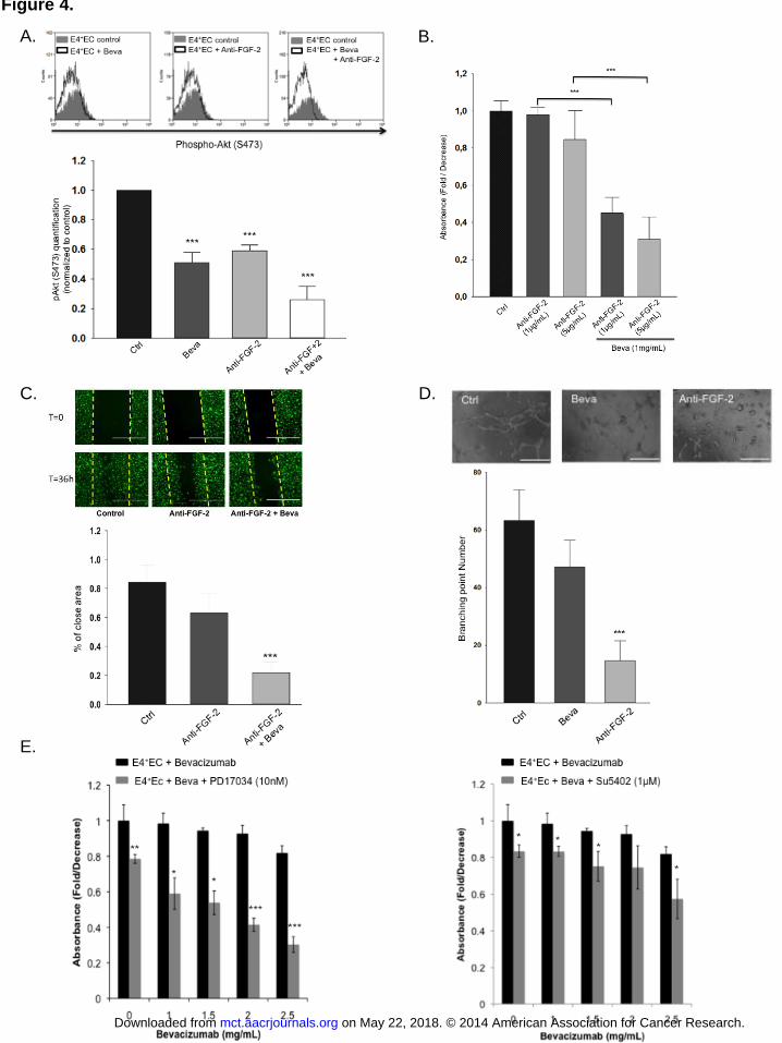
Figure 4.
A. B.
C. D.
E.
on May 22, 2018. © 2014 American Association for Cancer Research. mct.aacrjournals.org Downloaded from
Author manuscripts have been peer reviewed and accepted for publication but have not yet been edited. Author Manuscript Published OnlineFirst on October 15, 2014; DOI: 10.1158/1535-7163.MCT-13-1053
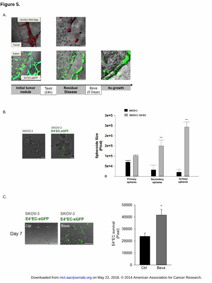
Figure 5.
A.
B.
C.
on May 22, 2018. © 2014 American Association for Cancer Research. mct.aacrjournals.org Downloaded from
Author manuscripts have been peer reviewed and accepted for publication but have not yet been edited. Author Manuscript Published OnlineFirst on October 15, 2014; DOI: 10.1158/1535-7163.MCT-13-1053

Figure 6, Schematic presentation of the FGF-2/PI3K-Akt autocrine loop in endothelial cells
Angiogenic Factors
(VEGF-A, PDGFB, FGF1, FGF2…)
Tumor
Activated-vascular
Endothelial cell
FGFR
FGF-2
Blood vessels
VEGFR
VEGF-A
Bevacizumab
Gene
Expression
FGF-2
AKT
Y705
Y705
on May 22, 2018. © 2014 American Association for Cancer Research. mct.aacrjournals.org Downloaded from
Author manuscripts have been peer reviewed and accepted for publication but have not yet been edited. Author Manuscript Published OnlineFirst on October 15, 2014; DOI: 10.1158/1535-7163.MCT-13-1053

Published OnlineFirst October 15, 2014.Mol Cancer Ther Bella S. Guerrouahen, Jennifer Pasquier, Nadine Abu Kaoud, et al. disease and resistance to bevacizumab in ovarian cancerAkt-activated endothelium constitute the niche for residual
Updated version
10.1158/1535-7163.MCT-13-1053doi:
Access the most recent version of this article at:
Material
Supplementary
http://mct.aacrjournals.org/content/suppl/2014/10/15/1535-7163.MCT-13-1053.DC1
Access the most recent supplemental material at:
Manuscript
Authorbeen edited. Author manuscripts have been peer reviewed and accepted for publication but have not yet
E-mail alerts related to this article or journal.Sign up to receive free email-alerts
Subscriptions
Reprints and
To order reprints of this article or to subscribe to the journal, contact the AACR Publications
Permissions
Rightslink site. Click on "Request Permissions" which will take you to the Copyright Clearance Center's (CCC)
.http://mct.aacrjournals.org/content/early/2014/10/15/1535-7163.MCT-13-1053To request permission to re-use all or part of this article, use this link
on May 22, 2018. © 2014 American Association for Cancer Research. mct.aacrjournals.org Downloaded from
Author manuscripts have been peer reviewed and accepted for publication but have not yet been edited. Author Manuscript Published OnlineFirst on October 15, 2014; DOI: 10.1158/1535-7163.MCT-13-1053