AJD regular cover art (reversed with text in dark blue) Issues/66542 AJOD Nov_New.pdfAJD regular...
49
Volume 31, Special Issue B, (0nline) November, 2018 - p. 1B-48B American Journal of Dentistry, Vol. 31, Special Issue B, November, 2018 Antibacterial and Bioactive Dental Restorative Materials: Do They Really Work?
Transcript of AJD regular cover art (reversed with text in dark blue) Issues/66542 AJOD Nov_New.pdfAJD regular...

Volume 31, Special Issue B, (0nline) November, 2018 - p. 1B-48B
Am
erican Journal of Dentistry, V
ol. 31, Special Issue B, N
ovember, 2018
AJD regular cover art
(reversed with text in dark blue) Note – overall background PMS blue gray –
PMS 5415
Antibacterial and Bioactive Dental Restorative Materials: Do They Really Work?

EDITOR Franklin García-Godoy University of Tennessee HSC, USA MANAGING EDITOR Katherine J. García-Godoy EDITORIAL BOARD Michael C. Alfano New York University, USA Thomas Attin University of Zurich, Switzerland Eugenio Brambilla University of Milan, Italy Daniel C.N. Chan University of Washington, USA Gordon J. Christensen Practical Clinical Courses, USA Sebastian G. Ciancio SUNY at Buffalo, USA Gary A. Crim University of Louisville, USA Jaime Cury University of Campinas, Brazil Kevin J. Donly University of Texas HSCSA, USA Albert J. Feilzer ACTA, The Netherlands Jack L. Ferracane Oregon Health & Science University, USA Marco Ferrari University of Siena, Italy Catherine M. Flaitz Nationwide Children’s Hospital, USA Roland Frankenberger University of Marburg, Germany Robert W. Gerlach Procter & Gamble Co., USA Reinhard Hickel University of Munich, Germany M. John Hicks Baylor College of Medicine, USA Mark E. Jensen University of Tennessee HSC Andrej M. Kielbassa Danube Private University, Austria Norbert Krämer University of Erlangen, Germany Ivo Krejci University of Geneva, Switzerland John F. McCabe Newcastle University, UK Peter E. Murray Nova Southeastern University, USA Raquel Osorio University of Granada, Spain Cornelis H. Pameijer University of Connecticut, USA Rade D. Paravina University of Texas HSC at Houston, USA Jorge Perdigão University of Minnesota, USA John M. Powers Dental Advisors, USA Mark S. Putt Indiana University-Purdue University, USA James C. Ragain, Jr University of Tennessee HSC Hans-Jörg Staehle University of Heidelberg, Germany Edward J. Swift, Jr. University of North Carolina, USA Junji Tagami Tokyo Medical and Dental University, USA Daranee Tantbirojn University of Tennessee HSC Franklin Tay Augusta University Health, USA Manuel Toledano University of Granada, Spain Bart Van Meerbeek University of Leuven, Belgium Antheunis Versluis University of Tennessee HSC Anthony R. Volpe Private consultant, USA Ann Wennerberg Göteborg University, Sweden STATISTICAL CONSULTANT Daniel L. Jones Texas A&M University, USA
Volume 31, Special Issue B, November, 2018 - p. 1B – 48B
www.amjdent.com
CONTENTS
Antibacterial and bioactive dental restorative materials: Do they really work?
ForewardD.C.N. Chan & A. Sadr 2B
Introduction ArticleAntibacterial and bioactive dental restorative materials: Do they really work? D.C.N. Chan, A.K.H. Chung & A. Paranjpe 3B
Review Articles Antibacterial dental restorative materials: A review. L. Chen, B.I. Suh & J. Yang 6B
Development of an antibacterial bioactive dental adhesive: Simplicity and innovation. Y. Fujimura, D. Weerasinghe & M. Kawashima 13B
Special Issue Articles Synthesis, antibacterial activity, and biocompatibility of new antibacterial dental monomers. Y. Wang, S. Costin, J-f. Zhang, S. Liao, Z.T. Wen, T. Lallier, Q. Yu & X. Xu 17B
Dental cements: Bioactivity, bond strength and demineralization progression around restorations. A. Turkistani, S. Islam, Y. Shimada, J. Tagami & A. Sadr 24B Reactions: Antibacterial and bioactive dental restorative materials: Do they really work? D.C.N. Chan, W. Hu, K-H. Chung, R. Larsen, S. Jensen, D. Cao, L. Gaviria, J.L. Ong, K. Whang & T. Eiampongpaiboon 32B Antibacterial-containing dental adhesives’ effects on oral pathogens and on Streptococcus mutans biofilm: Current perspectives. C.B. André, D.C.N. Chan & M. Giannini 37B Release of calcium ions from particulate monosodium titanates for dental mineralization applications J.L. Drury, Y-W. Chen, B.J. Plancich, K.M.L. Taylor-Pashow, D.T. Hobbs & J.C. Wataha 42B
Volume 31, Special Issue B, November, 2018 - p. 1B – 48B
www.amjdent.com
CONTENTS
Antibacterial and bioactive dental restorative materials: Do they really work?
ForewardD.C.N. Chan & A. Sadr 2B
Introduction ArticleAntibacterial and bioactive dental restorative materials: Do they really work? D.C.N. Chan, A.K.H. Chung & A. Paranjpe 3B
Review Articles Antibacterial dental restorative materials: A review. L. Chen, B.I. Suh & J. Yang 6B
Development of an antibacterial bioactive dental adhesive: Simplicity and innovation. Y. Fujimura, D. Weerasinghe & M. Kawashima 13B
Special Issue Articles Synthesis, antibacterial activity, and biocompatibility of new antibacterial dental monomers. Y. Wang, S. Costin, J-f. Zhang, S. Liao, Z.T. Wen, T. Lallier, Q. Yu & X. Xu 17B
Dental cements: Bioactivity, bond strength and demineralization progression around restorations. A. Turkistani, S. Islam, Y. Shimada, J. Tagami & A. Sadr B42 Reactions: Antibacterial and bioactive dental restorative materials: Do they really work? D.C.N. Chan, W. Hu, K-H. Chung, R. Larsen, S. Jensen, D. Cao, L. Gaviria, J.L. Ong, K. Whang & T. Eiampongpaiboon 32B Antibacterial-containing dental adhesives’ effects on oral pathogens and on Streptococcus mutans biofilm: Current perspectives. C.B. André, D.C.N. Chan & M. Giannini 37B Release of calcium ions from particulate monosodium titanates for dental mineralization applications J.L. Drury, Y-W. Chen, B.J. Plancich, K.M.L. Taylor-Pashow, D.T. Hobbs & J.C. Wataha 42B
_______________________________________________________________________________________________________________________________________________________________
Special Issue Foreward _______________________________________________________________________________________________________________________________________________________________
On behalf of the University of Washington School of Dentistry Department of Restorative Dentistry, we would like to welcome you to the proceedings publication on “Antibacterial and Bioactive Dental Restorative Materials: Do They Really Work?” We believe it was high time that such an event took place to focus on and discuss some of the basic questions related to the topic. We made a special effort to invite presentations from a diversity of research back-grounds. The symposium, supported by the Dental Materials Group under the same title, was held during the 94th General Session & Exhibition of the IADR in Seoul, Korea on June 24, 2016, and provided a valuable opportunity for research scientists, industry specialists, clinicians and academicians to share experiences. We would like to thank the International Association for Dental Research and the Dental Materials Group for providing such a platform. We are especially grateful to the many experts who shared their knowledge via presentation, discussion and feedback during the proceedings.
We also thank all the participants and reviewers of the Special Issue and the IADR for allowing us to conduct the Symposium in the first place. Such a large-scale international project is impossible without the support and help from organizations, foundations, and sponsors. We are very grateful to WDS Endowment, Japanese Society for Promo-tion of Science, Bisco Inc., Cao Group, and Kuraray Noritake Dental for their financial support towards the publication of this Special Issue. The American Journal of Dentistry editorial staff also provided invaluable suggestions and help to make this Special Issue a reality. With the limits of time and resources, we are certain that several topics and experts have been left out and there are still many unanswered questions. Our goal was to initiate a fruitful and rewarding exchange with this proceedings. We look forward to a successful outcome and continued research, discussion, and debate on the subject, with all of our esteemed global colleagues.
Daniel C.N. Chan Alireza Sadr
University of Washington School of Dentistry Seattle, Washington
_______________________________________________________________________________________________________________________________________________________________
Special Issue Introduction _______________________________________________________________________________________________________________________________________________________________
Antibacterial and bioactive dental restorative materials: Do they really work? DANIEL C.N. CHAN, DMD, DDS, ALBERT K.H. CHUNG, DDS, PHD & AVINA PARANJPE, BDS, PHD
ABSTRACT: Purpose: This proceedings reviews current antibacterial and bioactive dental materials and new agents in development. Methods: Experts from across academia, industry and clinical practice were invited to present, discuss, and work together to develop solutions to the challenge of formulating and applying antibacterial dental materials in a symposium in Seoul, Korea in June, 2016. (Am J Dent 2018;31:3B-5B). : Dr. Daniel Chan, Department of Restorative Dentistry, University of Washington School of Dentistry, Box 357456, Seattle, WA 98195-7456, USA. E-: [email protected]
Introduction
The publication of the symposium proceedings is very timely. The National Institute of Health reports that bacteria growing as a biofilm cause 80% of infections in the body. Biofilms at the margin of an existing restoration give rise to secondary caries (NIH).1 One approach for overcoming the development of dental biofilms and secondary caries is to develop antibacterial and bioactive dental restorative materials. This proceedings reviewed current dental materials and new agents in development. We invited experts from across academia, industry and clinical practice working together to develop solutions to this challenge. Bioactive or antibacterial? The first paper (Chen et al2) sets the tone of the pro-ceedings. This current manuscript and others by the same group looked at both bioactive and antibacterial materials. The terms bioactivity and bioactive material both have recently emerged in the dental literature. On the surface, bioactive is defined as having a biological effect. As such, all dental materials fit into that category. The term bioactive material appears to have originated with Dr. Larry Hench, the developer of the calcium silicophosphate glass.3 Drury et al4 (in this Special Issue) did touch on the calcium-mediated mineralization or re-minerali-zation and the fact that particulate MST-Ca(II) complexes exhibit sustained release of calcium, and that release might be customized by conditions of pH and ionic strength. Regrettably, bioactive glass is a topic we hardly explored in this symposium. On the other hand, an antibacterial is an agent that kills bacteria or stops their growth. The determination of the antibacterial activity is described in international norms. Not all dental materials can be antibacterial. In the oral cavity, mixed microbial biofilms can accumulate on hard and soft tissue, and are involved in the pathogenesis of caries and periodontitis. A biofilm is an accumulation of bacteria, fungi, or protozoa on solid surfaces. Two popular approaches in dentistry to prevent biofilm formation are: (A) to design a biomaterial that slowly releases an agent that is lethal to approaching bacterial cells; and (B) to develop a non-adhesive surface by modifying the surface chemistry of restoration materials. Various chemical agents can affect bacterial adhesion indirectly by disrupting bacterial cell metabolism. Numerous materials have been impregnated with various antibiotics only to have most of the agent released over a very short time, thus providing no long-term effect. Recent
studies5 have shown that sub-lethal doses of antibiotics can induce bacterial resistance and counteractively actually enhance biofilm formation. The potential negative consequences of bacterial resistance to antibiotics are dire because they put all of society at risk. The question of whether it is more advantageous to be bioactive or antibacterial probably will never be settled. Usually dental bioactive materials can improve mechanical integrity and offer protective bioactivity e.g. in the form of fluoride release. One must remember that not all bacteria are bad, and to be antibacterial indiscriminately may cause more harm than good. Bioactive and antibacterial strategies - Metal or non-metal based? Two papers6,7 (in this Special Issue) dealt with development and synthesis of new antibacterial monomers. Both new agents are organic in nature and can be classified as non-metal. MDPD is commercially available and much has been published on MDPD. Fujimura6 gave us a historical perspective and serves as an excellent blueprint for translation of basic science from laboratory to clinical use. Wang et al7 presented a small library of antibacterial dental monomers based on quaternary ammonium salts. Quaternary ammonium polyethylenimine (QAS-PEI) nanoparticles (NPs) have been incorporated into restorative materials to improve antibacterial activity and further reduce adverse effects on mechanical properties.3 Incorporation of QAS-PEI NPs into dental resin composites at 1 wt% concentration has been effective against Streptococcus mutans (SM) as well as against biofilm formation in vivo. However, given the increasing resistance of bacteria to or-ganic antibacterials, metal-based antibacterials are a promising alternative. Our group here at University of Washington took a different approach. We looked at metal-based antibacterials since metal-based antibacterials such as silver and zinc are an attractive alternative to antibiotics. Metal ions have chemical properties that inhibit bacterial growth. The unique binding, coordination, and redox properties make development of bacterial resistance less likely, and predict effectiveness across a broad bacterial spectrum. Unfortunately, development of new metal-based antibacterials has been severely impeded due to previous controversies and fears. If systemic toxicity could be limited and therapeutic indices were optimized, metal ions and their associated compounds could emerge as a new powerful class of antibacterial agents.

4B Chan et al Fig. 1. Possible applications of dental materials in terms of bioactivity include incorporation into the restorative materials, the adhesive system or as part of a base and liner. In addition, they can be used inside an endodontic sealer or as coating for implants. The most recent addition to the growing list of metal-based antibacterials are our gold-titanate nanoparticles. Our team has developed micro-particulate metal-titanate complexes as a new class of antibacterial agents. The micro-particulate gold (III)-loaded titanate complexes inhibit growth of oral bacteria at micro molar concentrations. We have shown that nano-particulate metal-titanate complexes are even more effective than micro-particulate complexes at inhibiting oral bacteria growth as these nano-particulate complexes have a significantly greater surface-to-volume ratio, resulting in more effective ion-exchange characteristics.8,9 Our current approach is to incorporate the gold-titanate nanoparticles in adhesive systems because of manufacturing availability issues, but in the long term, gold-titanate nanopar-ticles may also be incorporated into restorative materials as fillers, or as coating on implant systems.10 Additionally, be-cause these complexes are not organic, degradation is not an issue and thus they can have long-term effectiveness, and also may be less likely than organic antibacterial agents to contri-bute to bacterial resistance. Wang et al7 and Giannini & Andre11 (both in this Special Issue) evaluated a few species of bacteria. It will be ideal to analyze a standard microbial mixed culture such as the one developed by Guggenheim12 and is considered by the research community as a “model” of dental caries microbial flora. Applications of antibacterial and bioactive dental restorative materials Regardless of the types of materials, clinicians would even-tually hope to apply the science and technology in real life situations. Based on the discussions in this Issue’s articles, most of the applications seem to be focused on usage as restorative materials, cements, and adhesives (Fig. 1a-d). We will attempt to expand the discussions to root canal sealers and bioactive denture resins. Another important area not to be forgotten is implant coating, especially when usage of implants is on the rise and peri-implantitis is getting more common. Root canal sealers Successful endodontic treatment depends on the effective-ness of the cleaning and shaping of the root canal system. Any remaining tissue, bacteria, or debris can contribute to endodontic failure. Elimination of bacteria from the root canal system can be done via chemomechanical debridement and also placement of intracanal medicament for reduction and
American Journal of Dentistry, Vol. 31, Special Issue B, November, 2018 Fig. 2. Gold-titanate is a promising antibacterial nanoparticle material with a light sensitivity side-effect. Its use thus is limited to a thin layer as adhesive (A) or as a sealer inside the root canal (B). In terms of its compatibility with titanates, application as an implant coating can also be entertained. elimination of any residual bacteria remaining post-instru-mentation. Calcium hydroxide is routinely placed as an inter-appoint-ment intracanal medicament for non-surgical endodontic procedures. Despite efficacy of calcium hydroxide, this medicament has some limitations in its antimicrobial efficacy and due to the fact that certain bacteria can withstand a high pH environment. Other alternatives to calcium hydroxide have been investigated and used. Examples of historic medicaments are phenolic compounds, essential oils, aldehydes, halogens, and quarternary ammonium compounds. The use of these materials has been discontinued due to their cytotoxicity and limited antimicrobial efficacy. Antibiotics have been and may be used as intracanal medicaments, however they may produce resistant microbes and cause host sensitization. Therefore, their routine use is not recommended. Steroids that have been used as intracanal medicaments can prevent the inflammatory response and subsequent pain. However they have limited antimicrobial efficacy. Due to these reasons, antibiotics and steroids are not routinely placed. The antimicrobial activity of gold nanoparticles (AuNPs) has been studied in both Gram-positive and Gram-negative bacteria such as Staphylococcus aureus, Escherichia coli and Pseudomonas aeruginosa. Based on the bacterial protein assay, nano-sized monosodium titanate (nanomonosodium titanate) nMST-Au(III) showed the best effectiveness among titanates and gold-titanates to decrease bacterial protein concentrations. A Univeristy of Washington Master’s thesis study13 examined the efficacy of gold-titanates on E. faecalis when used in intracanal medicament separately or in addition to calcium hydroxide and compared it with calcium hydroxide alone. Unfortunately, it cannot yet be concluded that gold-titanate nanoparticles have antimicrobial efficacy against Gram-positive E. faecalis, most likely due to insensitivity of culturing technique. The study did find that gold-titanate nanoparticles mixed with sterile water or added to calcium hydroxide leave residual crystals in the canal system, which may occlude the dentin tubules and bacteria (Fig. 2). Bioactive fillers in denture resin Much attention has been paid to the development of direct dental materials that are antibacterial. Surface pre-reacted glass-ionomer fillers (S-PRG) are a newly introduced bioactive material and is of high potential interest to address the problem of caries in denture-wearing populations. S-PRG fillers are a novel class of particle that can be incorporated into resinous
Chan Overview figures
Fig. 1. Possible applications of dental materials in terms of bioactivity include incorporation into the restorative materials, the adhesive system or as part of a base and liner. In addition, they can be used inside an endodontic sealer or as coating for implants.
Fig. 2. Anatomy of S-PRG filler. It forms by a type of stable glass-ionomer, which allows ion release and recharge to take place.
Fig. 3. Anatomy of S-PRG filler. It forms by a type of stable glass-ionomer, which allows ion release and recharge to take place.
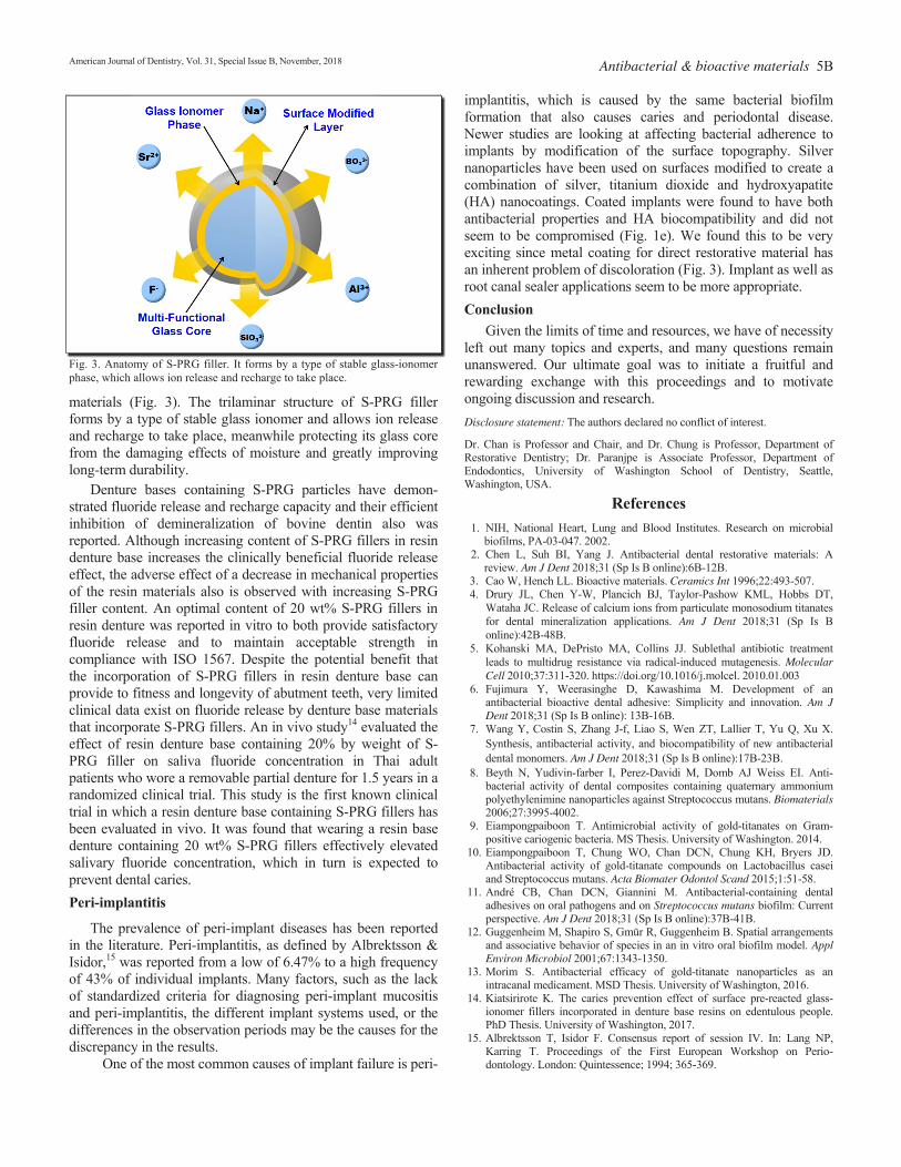
American Journal of Dentistry, Vol. 31, Special Issue B, November, 2018 Fig. 3. Anatomy of S-PRG filler. It forms by a type of stable glass-ionomer phase, which allows ion release and recharge to take place. materials (Fig. 3). The trilaminar structure of S-PRG filler forms by a type of stable glass ionomer and allows ion release and recharge to take place, meanwhile protecting its glass core from the damaging effects of moisture and greatly improving long-term durability. Denture bases containing S-PRG particles have demon-strated fluoride release and recharge capacity and their efficient inhibition of demineralization of bovine dentin also was reported. Although increasing content of S-PRG fillers in resin denture base increases the clinically beneficial fluoride release effect, the adverse effect of a decrease in mechanical properties of the resin materials also is observed with increasing S-PRG filler content. An optimal content of 20 wt% S-PRG fillers in resin denture was reported in vitro to both provide satisfactory fluoride release and to maintain acceptable strength in compliance with ISO 1567. Despite the potential benefit that the incorporation of S-PRG fillers in resin denture base can provide to fitness and longevity of abutment teeth, very limited clinical data exist on fluoride release by denture base materials that incorporate S-PRG fillers. An in vivo study14 evaluated the effect of resin denture base containing 20% by weight of S-PRG filler on saliva fluoride concentration in Thai adult patients who wore a removable partial denture for 1.5 years in a randomized clinical trial. This study is the first known clinical trial in which a resin denture base containing S-PRG fillers has been evaluated in vivo. It was found that wearing a resin base denture containing 20 wt% S-PRG fillers effectively elevated salivary fluoride concentration, which in turn is expected to prevent dental caries. Peri-implantitis The prevalence of peri-implant diseases has been reported in the literature. Peri-implantitis, as defined by Albrektsson & Isidor,15 was reported from a low of 6.47% to a high frequency of 43% of individual implants. Many factors, such as the lack of standardized criteria for diagnosing peri-implant mucositis and peri-implantitis, the different implant systems used, or the differences in the observation periods may be the causes for the discrepancy in the results.
One of the most common causes of implant failure is peri-
Antibacterial & bioactive materials 5B implantitis, which is caused by the same bacterial biofilm formation that also causes caries and periodontal disease. Newer studies are looking at affecting bacterial adherence to implants by modification of the surface topography. Silver nanoparticles have been used on surfaces modified to create a combination of silver, titanium dioxide and hydroxyapatite (HA) nanocoatings. Coated implants were found to have both antibacterial properties and HA biocompatibility and did not seem to be compromised (Fig. 1e). We found this to be very exciting since metal coating for direct restorative material has an inherent problem of discoloration (Fig. 3). Implant as well as root canal sealer applications seem to be more appropriate. Conclusion Given the limits of time and resources, we have of necessity left out many topics and experts, and many questions remain unanswered. Our ultimate goal was to initiate a fruitful and rewarding exchange with this proceedings and to motivate ongoing discussion and research. Disclosure statement: The authors declared no conflict of interest. Dr. Chan is Professor and Chair, and Dr. Chung is Professor, Department of Restorative Dentistry; Dr. Paranjpe is Associate Professor, Department of Endodontics, University of Washington School of Dentistry, Seattle, Washington, USA.
References
1. NIH, National Heart, Lung and Blood Institutes. Research on microbial biofilms, PA-03-047. 2002.
2. Chen L, Suh BI, Yang J. Antibacterial dental restorative materials: A review. Am J Dent 2018;31 (Sp Is B online):6B-12B.
3. Cao W, Hench LL. Bioactive materials. Ceramics Int 1996;22:493-507. 4. Drury JL, Chen Y-W, Plancich BJ, Taylor-Pashow KML, Hobbs DT,
Wataha JC. Release of calcium ions from particulate monosodium titanates for dental mineralization applications. Am J Dent 2018;31 (Sp Is B online):42B-48B.
5. Kohanski MA, DePristo MA, Collins JJ. Sublethal antibiotic treatment leads to multidrug resistance via radical-induced mutagenesis. Molecular Cell 2010;37:311-320. https://doi.org/10.1016/j.molcel. 2010.01.003
6. Fujimura Y, Weerasinghe D, Kawashima M. Development of an antibacterial bioactive dental adhesive: Simplicity and innovation. Am J Dent 2018;31 (Sp Is B online): 13B-16B.
7. Wang Y, Costin S, Zhang J-f, Liao S, Wen ZT, Lallier T, Yu Q, Xu X. Synthesis, antibacterial activity, and biocompatibility of new antibacterial dental monomers. Am J Dent 2018;31 (Sp Is B online):17B-23B.
8. Beyth N, Yudivin-farber I, Perez-Davidi M, Domb AJ Weiss EI. Anti-bacterial activity of dental composites containing quaternary ammonium polyethylenimine nanoparticles against Streptococcus mutans. Biomaterials 2006;27:3995-4002.
9. Eiampongpaiboon T. Antimicrobial activity of gold-titanates on Gram-positive cariogenic bacteria. MS Thesis. University of Washington. 2014.
10. Eiampongpaiboon T, Chung WO, Chan DCN, Chung KH, Bryers JD. Antibacterial activity of gold-titanate compounds on Lactobacillus casei and Streptococcus mutans. Acta Biomater Odontol Scand 2015;1:51-58.
11. André CB, Chan DCN, Giannini M. Antibacterial-containing dental adhesives on oral pathogens and on Streptococcus mutans biofilm: Current perspective. Am J Dent 2018;31 (Sp Is B online):37B-41B.
12. Guggenheim M, Shapiro S, Gmür R, Guggenheim B. Spatial arrangements and associative behavior of species in an in vitro oral biofilm model. Appl Environ Microbiol 2001;67:1343-1350.
13. Morim S. Antibacterial efficacy of gold-titanate nanoparticles as an intracanal medicament. MSD Thesis. University of Washington, 2016.
14. Kiatsirirote K. The caries prevention effect of surface pre-reacted glass-ionomer fillers incorporated in denture base resins on edentulous people. PhD Thesis. University of Washington, 2017.
15. Albrektsson T, Isidor F. Consensus report of session IV. In: Lang NP, Karring T. Proceedings of the First European Workshop on Perio-dontology. London: Quintessence; 1994; 365-369.
_______________________________________________________________________________________________________________________________________________________________
Special Issue Review Article _______________________________________________________________________________________________________________________________________________________________
Antibacterial dental restorative materials: A review LIANG CHEN, PHD, BYOUNG IN SUH, PHD & JIE YANG, PHD
ABSTRACT: Purpose: To provide updated summary information about antibacterial dental materials, primarily covering the literature from 2012 through 2017. Methods: A key-worded search was conducted of peer-reviewed literature (Titles/Abstracts) indexed by PubMed databases, constrained to “English” and “dental” publications between the years 2012 and 2017. Key words applied to the search included: antimicrobial, antibacterial, primer, bonding agent, adhesive, cement, composite, liner, sealant, etchant, and core-build-up. Titles and abstracts of the articles returned by the search were reviewed and evaluated for appropriateness for inclusion in this review. Results: A variety of antibacterial agents have been incorporated into experimental and commercial dental restorative materials to provide antibacterial activity in dental applications. No new antibacterial compounds were introduced in this review period (2012-2017), since the last review of period of 1980-2012. Antibacterial agents include leachable compounds (e.g. benzalkonium chloride, chlorhexidine), polymerizable monomers (e.g. quaternary ammonium methacrylates), and filler particles (e.g. silver nanoparticle). During the 2012-2017 review period, many antibacterial agents were tested in experimental formulations, but only four agents (benzalkonium chloride, chlorhexidine, glutaraldehyde, and MDPB) were used in commercial products. (Am J Dent 2018;31(Sp Is B):6B-12B). CLINICAL SIGNIFICANCE: Leachable antibacterial agents are the most frequently used type of antibacterial dental materials, but their efficacy may be short-lived due to their characteristic burst effect. Solid filler particles appear to be effective antibacterial agents, especially given their ability to reduce biofilm formation, but the color stability of their component metal particles is unfavorable for use in a commercial product. Polymerizable antibacterial agents (MDPB) are theoretically a good choice of material because they are very effective at killing any residual bacteria in a cavity preparation prior to polymerization, however, apart from their proven effect on reduction of biofilm formation, their long-term clinical performance is still questionable. : Dr. Liang Chen, Department of Research and Development, Bisco Inc., 1100 W. Irving Park Road, Schaumburg, IL 60193, USA. E-: [email protected]
Introduction
For both patients and dentists, longevity is one of the most important aspects of dental restorations. In the United States, 50-70% of all dental restorations placed every year are replacements of failed restorations.1 The most common reason for restoration failure is secondary caries,2 which are mainly caused by oral bacteria.3 In recent years, numerous research studies have been conducted with the common goal of developing antibacterial dental restorative materials to be used to eradicate the cause of dental caries.4 Two comprehensive reviews on antibacterial dental materials were published in the past two decades. The first such review was published in 2003 and focused on antibacterial features and their benefits in dental bonding agents and resin composites.4 The second review article covered the literature from 1980 to 2012, and focused on the antibacterial effects of dental composites, cement, primers, and adhesives.5 This review article will provide updated information about antibacterial dental materials, primarily covering the literature from 2012 through 2017. The materials discussed in the review will include those that have both direct contact and no direct contact with tooth structures.
Material and Methods A search of peer-reviewed literature (Titles/Abstracts) indexed by PubMed databases was conducted and limited to the “English” and “dental” publications between the years 2012 and 2017. Key words used included: antimicrobial, antibac-terial, primer, bonding agent, adhesive, cement, composite, liner, sealant, etchant, and core-build-up. Titles and abstracts of
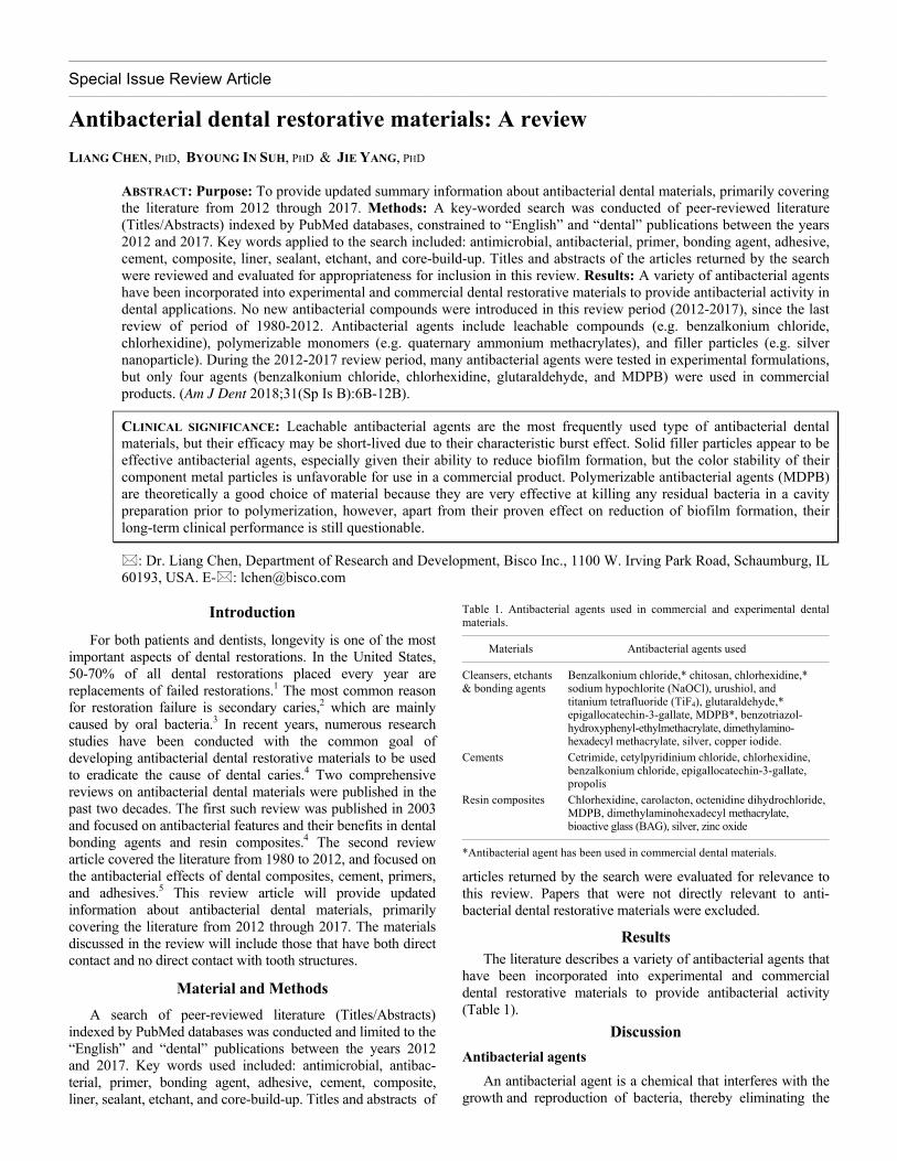
Table 1. Antibacterial agents used in commercial and experimental dental materials. ____________________________________________________________________________________________________
Materials Antibacterial agents used ____________________________________________________________________________________________________
Cleansers, etchants Benzalkonium chloride,* chitosan, chlorhexidine,* & bonding agents sodium hypochlorite (NaOCl), urushiol, and titanium tetrafluoride (TiF4), glutaraldehyde,* epigallocatechin-3-gallate, MDPB*, benzotriazol- hydroxyphenyl-ethylmethacrylate, dimethylamino- hexadecyl methacrylate, silver, copper iodide. Cements Cetrimide, cetylpyridinium chloride, chlorhexidine, benzalkonium chloride, epigallocatechin-3-gallate, propolis Resin composites Chlorhexidine, carolacton, octenidine dihydrochloride, MDPB, dimethylaminohexadecyl methacrylate, bioactive glass (BAG), silver, zinc oxide ____________________________________________________________________________________________________
*Antibacterial agent has been used in commercial dental materials. articles returned by the search were evaluated for relevance to this review. Papers that were not directly relevant to anti-bacterial dental restorative materials were excluded.
Results The literature describes a variety of antibacterial agents that have been incorporated into experimental and commercial dental restorative materials to provide antibacterial activity (Table 1).
Discussion Antibacterial agents An antibacterial agent is a chemical that interferes with the growth and reproduction of bacteria, thereby eliminating the
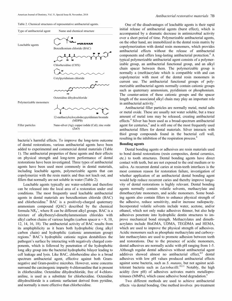
American Journal of Dentistry, Vol. 31, Special Issue B, November, 2018 Table 2. Chemical structures of representative antibacterial agents. ____________________________________________________________________________________________________
Type of antibacterial agent Name and chemical structure ____________________________________________________________________________________________________
Leachable agents Benzalkonium chloride (BAC)
Chlorhexidine (CHX)
Cetylpyridinium chloride
Octenidine dihydrochloride Polymerizable monomers
12-methacryloyloxydodecypyridinium bromide (MDPB) Filler particles Nano-silver (Ag); copper iodide (CuI); zinc oxide (ZnO) ____________________________________________________________________________________________________
bacteria’s harmful effects. To improve the long-term outcome of dental restorations, various antibacterial agents have been added to experimental and commercial dental materials (Table 1). The antibacterial properties of these agents and their effects on physical strength and long-term performance of dental restorations have been investigated. Three types of antibacterial agents have been used most commonly in dental materials, including leachable agents, polymerizable agents that can copolymerize with the resin matrix and thus not leach out, and fillers that normally are not soluble in water (Table 2). Leachable agents typically are water-soluble and therefore can be released into the local area of a restoration under oral conditions. The most frequently used leachable antibacterial agents in dental materials are benzalkonium chloride (BAC) and chlorhexidine.4 BAC is a positively-charged quaternary ammonium compound (QAC) described by the chemical formula NR4
+, where R can be different alkyl groups. BAC is a mixture of alkylbenzyl-dimethylammonium chlorides with alkyl carbon chains of various lengths (carbon spacer n = 8, 10, 12, 14, 16, 18). The antibacterial activity of BAC results from its amphiphilicity as it bears both hydrophobic (long alkyl carbon chain) and hydrophilic (cationic ammonium group) regions.6 BAC’s hydrophilic cationic region destabilizes the pathogen’s surface by interacting with negatively charged com-ponents, which is followed by penetration of the hydrophobic long alky group into the bacterial hydrophobic bilayer leading to cell leakage and lysis. Like BAC, chlorhexidine also is a broad spectrum antibacterial agent, effective against both Gram-negative and Gram-positive microbes. However, some concerns surround the carcinogenic impurity 4-chloroaniline that is present in chlorhexidine. Octenidine dihydrochloride, free of 4-chloro-aniline, is used as a substitute for chlorhexidine. Octenidine dihydrochloride is a cationic surfactant derived from pyridine, and normally is more effective than chlorhexidine.
Antibacterial restorative materials 7B One of the disadvantages of leachable agents is their rapid initial release of antibacterial agents (burst effect), which is accompanied by a dramatic decrease in antimicrobial activity over a short period of time. Polymerizable antibacterial agents, on the other hand, are immobilized in the dental resin matrix by copolymerization with dental resin monomers, which provides antibacterial effects without the release of antibacterial components and offers long-lasting antibacterial protection.4 A typical polymerizable antibacterial agent consists of a polymer-izable group, an antibacterial functional group, and an alkyl chain spacer between them. The polymerizable group is normally a (meth)acrylate which is compatible with and can copolymerize with most of the dental resin monomers in current use. The antibacterial functional groups of poly-merizable antibacterial agents normally contain cationic groups such as quaternary ammonium, pyridinium or phosphonium. The counter-anion of these cationic groups and the spacer length of the associated alkyl chain may play an important role in antibacterial activity.7 Antibacterial filler particles are normally metal, metal salts or metal oxide. These are usually not water soluble, but a trace amount of metal ions may be released, creating antibacterial effects.4 Silver has been used as a broad-spectrum antibacterial agent for centuries,8 and is still one of the most frequently used antibacterial fillers for dental materials. Silver interacts with thiol group compounds found in the bacterial cell wall, resulting in the inhibition of the respiration process.8 Bonding agents Dental bonding agents or adhesives are resin materials used to bond dental restorations (resin composites, dental ceramics, etc.) to tooth structures. Dental bonding agents have direct contact with teeth, but are not exposed to the oral medium or to saliva. As recurrent dental caries at resin-teeth interfaces is the most common reason for restoration failure, investigation of whether application of an antibacterial dental bonding agent would help reduce recurrent caries and thereby improve longe-vity of dental restorations is highly relevant. Dental bonding agents normally contain volatile solvents, methacrylate and dimethacrylate monomers, and acidic monomers. Some bond-ing agents also contain fillers to enhance physical strength of the adhesive, reduce sensitivity, and/or increase radiopacity. Incorporated volatile solvents include water, acetone, and/or ethanol, which not only make adhesives thinner, but also help adhesives penetrate into hydrophilic dentin structures to im-prove mechanical bond strength. Methacrylates and dimeth-acrylates include BisGMA, UDMA, TEGDMA and HEMA, which are used to improve the physical strength of adhesives. Acidic monomers such as phosphate methacrylate and carboxy-late methacrylates are used to promote adhesion between teeth and restorations. Due to the presence of acidic monomers, dental adhesives are normally acidic with pH ranging from 1-5. Although regular dental adhesives without antibacterial agent additives showed almost no antibacterial effect,4,9 dental adhesives with low pH values produced antibacterial effects against some bacteria, such as S. mutans, but not against acid-tolerant bacteria such as Lactobacilli.10,11 Notably, the high acidity (low pH) of adhesives activates matrix metallopro-teinases (MMPs), which cause adhesive bond degradation.6 Two different methods are used to achieve antibacterial effects via dental bonding. One method involves pre-treatment

8B Chen et al Table 3. Methods used to achieve antibacterial dental bonding agents. ____________________________________________________________________________________________________
Method Antibacterial agents used ____________________________________________________________________________________________________
Pre-treatment of teeth Benzalkonium chloride, chlorhexidine, sodium with antibacterial agents hypochlorite, urushiol, and titanium tetrafluoride Incorporation of leachable Benzalkonium chloride, glutaraldehyde, antibacterial agents into chlorhexidine, epigallocatechin-3-gallate dental adhesives Incorporation of 12-methacryloyloxydodecypyridinium bromide polymerizable antibacterial (MDPB), benzotriazol-hydroxyphenyl- agents into dental ethylmethacrylate, dimethylaminohexadecyl adhesives methacrylate Incorporation of Nano-silver, copper iodide antibacterial filler particles into dental adhesives ____________________________________________________________________________________________________
of tooth structures using antibacterial etchants or disinfectants and the other method is to incorporate antibacterial agents (leachable agent, polymerizable agent, or filler particle) into dental adhesives (Table 3). BAC and chlorhexidine are the most frequently used antibacterial agents for pre-treatment of teeth. BAC is stable in acidic media and has been added into commercial phosphoric acid etchants to a final concentration of 1%. Examples of such products include EtCH-37a w/BAC or UNI-ETCHa w/BAC, which exhibited zone inhibitions of bacteria, without compro-mising bond strength. In addition, BAC can also inhibit MMPs, thus preserving the dentin-resin bonded interface.6 Unlike BAC, chlorhexidine is not stable in phosphoric acid and cannot be added to etchants. Chlorhexidine digluconate (2%) has been added to commercial dental disinfectants, such as Cavity Cleanser.a Both in vivo and in vitro studies demonstrated that Cavity Cleanser reduced microorganisms in contaminated dentin.12 Pre-treatment of dentin with chlorhexidine maintained resin-dentin bond strength for up to 14 months, while a control group without chlorhexidine pre-treatment experienced signi-ficant bond strength reduction in vivo;13 the observed enhanced stability was mainly due to inhibition of the degradation of hybrid layers by chlorhexidine.14 Some other agents added to experimental products, including 6% sodium hypochlorite (NaOCl), 0.01% urushiol, and 2.5% titanium tetrafluoride (TiF4), also showed antibacterial capability in pre-treatment of dentin, but studies15,16 suggested that higher bond strength was obtained when the disinfectants were rinsed away. Leachable agents have been incorporated into both comer-cial and experimental dental adhesives. For instance, glutaral-dehyde was incorporated into Gluma 2 Bondb and chlor-hexidine was incorporated into Peak Universal Bond.c André et al17 demonstrated that Gluma 2 Bond required at least 24 hours for killing microorganisms, and that Peak Universal Bond killed only strict anaerobic microorganisms after 24 hours. Sabatini et al18 added BAC into All-Bond Universal,a a universal dental adhesive, to create experimental adhesives with final BAC concentrations of 0.5%, 1%, and 2% (wt/ wt).19 These BAC-containing adhesives delivered higher bond strength than did the control after 1-year storage in artificial saliva, probably because of their ability to inhibit MMPs.19 Du et al20 reported that an experimental dental adhesive containing 0.02% epigallocatechin-3-gallate (EGCG) exhibited inhibitory effect on the growth of S. mutans, and demonstrated higher
American Journal of Dentistry, Vol. 31, Special Issue B, November, 2018 bond strength than the control without EGCG after 6 months. Some concerns persist regarding the “burst effect” of leachable agents and more research is needed to investigate the long-term performance of antibacterial adhesives containing leachable agents. In an attempt to overcome the disadvantage (burst effect) of leachable agents, polymerizable antibacterial agents have been incorporated into dental adhesives. Polymerizable agents are immobilized in the resin matrix system upon polymerization, presumably enabling long-lasting antibacterial effects.4 One such polymerizable agent is 12-methacryloyloxydodecypyri-dinium bromide (MDPB), which has been incorporated into a commercial dental adhesive (5% MDPB in Clearfil Protect Bondd and used in clinical practice. One study21 showed that Clearfil Protect Bond inhibited growth of S. mutans and L.gasseri. In a 14-day in situ study, Pinto et al22 reported that Clearfil Protect Bond resulted in lower counts of total Streptococci as well as S. mutans and smaller lesion depths than did a non-MDPB containing adhesive for enamel and dentin restorations, but Clearfil Protect Bond did not prevent demineralization or bacteria growth.22 In contrast, Vasconcelos et al23 found no statistically significant difference between Clearfil Protect Bond and a non-antibacterial dental adhesive (All-Bond SEa) either in enamel demineralization or in dental biofilm formation, suggesting that Clearfil Protect Bond was unable to inhibit secondary caries in situ. Other studies24,25 also showed that the performance of Clearfil Protect Bond was similar to that of other non-MDPB containing adhesives in terms of caries formation, and that it did not inhibit secondary caries in a simulated high caries challenge. Polymerizable antibacterial agents such as MDPB are designed to immobilize in the resin matrix, in hopes of producing long-lasting antibacterial effects. However, Clearfil Protect Bond exerted only a short-term antibacterial effect (for 7 days), and lost the antibacterial activity after storage in phosphate-buffered saline for 14 days,26,27 in direct contrast to the expectation of long-lasting antibacterial effects of polymerizable agents. A possible explanation for this is that immobilization/polymerization of antibacterial agents reduces their antibacterial activity substantially, and that the observed short-term antibacterial effects were mainly a result of unpolymerized MDPB mono-mers (a resin typically experiences 70-80% polymerization conversion). The antibacterial effects disappeared after all unpolymerized MDPB monomers had leached out.27 Some other polymerizable antibacterial monomers have been evaluated in experimental dental adhesives. For instance, 5% 2-[3-(2H-benzotriazol-2-YL)-4-hydroxyphenyl] ethyl methacrylate in a dental adhesive showed higher antibacterial activity than did the negative control.28 A new antibacterial monomer, dimethylaminohexadecyl methacrylate (carbon chain length 16) was synthesized and added (5%) into a dental adhesive. The experimental adhesive showed a great ability to reduce biofilm accumulation and to decrease lactic acid production without impairing bond strength.29,30 Some filler particles have been added to experimental dental adhesives to improve their antibacterial activity. One of the most frequently used antibacterial particles is nano-silver. Studies31,32 have shown that the addition of 0.05% silver nanoparticle (particle size 2.7 nm) into dental adhesives significantly reduces biofilm viability, colony-forming unit

American Journal of Dentistry, Vol. 31, Special Issue B, November, 2018 (CFU) counts, and lactic acid production, without compro-mising dentin bond strength. One of the biggest issues for silver particles is color stability.4 Antibacterial fillers that demonstrate better color stability than silver also have been incorporated into experimental dental adhesives. For example, the addition of polyacrylic acid-modified copper iodide particles (1 mg/ml) into adhesives reduced Streptococcus mutans viable cell counts by 79-99% even after aging for 1 year in vitro and no signi-ficant differences in bond strength or cytotoxicity were detected between these experimental adhesives and their corresponding controls.18 Chitosan has long been known for its antimicrobial activity and is also a promising additive in dental materials. A recent study reported that total-etch adhesive systems supple-mented with chitosan (at concentrations of 0.2% and 0.5%) displayed similar inhibitory effects on S. mutans and L. casei as a commercial conventional 2-step adhesive system (Adper Single Bond 2e). The antimicrobial activity of chitosan may be derived from a combination of factors including pH, metal chelating capacity, and the positive charge of its gluco-samine groups interacting with the negative charge of the bacteria cell surface.33,34 Cements Dental cements function in luting or adhesion of indirect restorations with tooth structures, and can be classified as four different types: (1) water-based acid-base cements, including glass ionomer cement (GIC), resin-modified glass ionomer cement (RM-GIC), and zinc phosphate cement; (2) oil-based acid-based cements, such as zinc oxide eugenol and non-eugenol zinc oxide; (3) self-adhesive resin cements; and (4) non-self-adhesive resin cements. The first three types of cements have direct contact with tooth structures whereas the 4th type has no direct contact with tooth structures and requires the application of separate primers and/or adhesives. Among the above four types of cements, zinc oxide-based cements possess antibacterial properties without the addition of a separate antibacterial agent35 whereas the third and fourth types of cements normally do not display antibacterial activities. GIC and RM-GIC release fluoride for a long period, but their antibacterial activity is usually low.36,37 Additives have been included to enhance the antimicrobial activity of these cements, and the physical properties of the resulting cements have been studied. Propolis, a natural resinous substance produced by honeybees, improved antimicrobial effects of GIC but significantly decreased the compressive strength and increased solubility of the cement.38 Conventional luting cements, such as zinc phosphate (ZP), zinc polycarboxylate (PC), and GIC, containing 5% chlorhexidine diacetate/ cetrimide demonstrated long-lasting antibacterial effects for up to 180 days despite reduced physical strength and increased solubility of the cements.39 Similarly, the addition of a paste of chlorhexidine-hexametaphosphate into GIC exhibited a sustained release of chlorhexidine for at least 14 months, accompanied by compromised cement strength.40 Addition of different antibacterial agents (1-2%), such as cetrimide, cetyl-pyridinium chloride, chlorhexidine and BAC, to conventional GIC also impaired the cement’s microhardness during 90-day water storage.41 Nonetheless, incorporation of a lower concentration (0.5%) of chlorhexidine seemed to produce an optimum favorable outcome as it increased antibacterial
Antibacterial restorative materials 9B Table 4. Methods used to produce antibacterial resin composite ____________________________________________________________________________________________________
Method Antibacterial agents used ____________________________________________________________________________________________________
Incorporation of Chlorhexidine, carolacton, octenidine leachable antibacterial dihydrochloride agents into composites Incorporation of MDPB, dimethylaminohexadecyl methacrylate polymerizable antibacterial agents into composites Blending of antibacterial Bioactive glass (BAG), silver, zinc oxide filler particles with existing composite fillers ____________________________________________________________________________________________________
activity without adversely affecting physical-mechanical pro-perties.42 Therefore, using low concentrations of additives might be a promising approach for enhancing conventional cements with antibacterial activity. For example, 0.1% epigallo-catechin-3-gallate in GIC increased not only its antibacterial activities, but also its flexural strength and surface hardness43 and GIC supplemented with a quaternary ammonium mono-mer, DMADDM (dimethylaminododecyl methacrylate), even at the high concentrations of 1.1% and 2.2%, showed improved material performance and antibacterial properties.44 Unlike GIC, the physical strength of other types of cements seemed to be less sensitive to additives. For example, the incorporation of up to 4.5% doxycycline hyclate into RM-GIC or the addition of 7.5% chlorhexidine diacetate to provisional cements did not compromise their physical strength.45,46 Resin composites Dental resin composites are used as restorative materials. Resin composites are normally placed on top of dental adhesives and usually are not in direct contact with caries or tooth structures. Some dental composites are used for enamel restorations and as such are exposed to the oral medium and to saliva. Resin composites are composed mainly of inert inorganic fillers and organic monomers. Unlike amalgam which has antibacterial activities by virtue of releasing a trace amount of metal ions, cured resin composites typically lack anti-bacterial activity, resulting in bacterial adherence and plaque accumulation on their surfaces.4,47,48 The reason for the lack of antibacterial activity exhibited by dental resin composites is that the quantity of monomers and other components leached out from composites is much lower than the minimum concen-tration required for bacterial inhibition. The fillers used in composites are normally inert silica fillers with no antibacterial activity, as opposed to the metal-containing fillers described above. To produce an antibacterial resin composite, an antibacterial agent could be dissolved in the composite’s resin monomers, or, if the antibacterial agent is not soluble in resin monomers, could be blended with filler particles (Table 4). Many leachable antibacterial agents have been incorporated into experimental dental resin composites (Table 4). Chlorhexidine, one of the most frequently used antibacterial agents, was released faster in media of lower pH values due to its higher solubility at lower pH.49 Release rate also may be influenced by hydrophilicity of resin. Composites with hydrophilic resin tended to release chlorhexidine faster as chlorhexidine-containing resin lost antibacterial activities after storage in water for 2 weeks.50 To improve its long-term release, chlorhexidine has been encapsulated using mesoporous
10B Chen et al silica nanoparticles, and composites containing encapsulated chlorhexidine showed controlled release of chlorhexidine over a long period of time.51 Due to concerns surrounding the carcinogenic impurity 4-chloroaniline present in chlorhexidine, octenidine dihydrochloride has been considered as an alter-native to chlorhexidine. Addition of 3 wt% of octenidine dihydrochloride into dental composites significantly reduced biofilm formation.1 Furthermore, carolacton was found to be a more effective antibacterial agent than chlorhexidine and triclosan when incorporated into resin composites. A small amount of carolacton (0.002%,w/w) in experimental resin composite reduced biofilm viability by up to 64% and reduced CFUs by 98%, with no adverse effects on physical properties. The anti-biofilm activity of carolacton-containing composite was stable over a period of 42 days.52 Incorporation of a polymerizable antibacterial monomer into a dental composite is another way to produce antibacterial composites. After antibacterial monomers copolymerize with resin composites, the antibacterial agents are not expected to leach out from the composite matrix, presumably resulting in long-lasting antibacterial effects via inhibition of bacterial growth on the composite surface upon contact. Imazato et al53 reported that MDPB-containing composites demonstrated significant antibacterial effects even after 90 days of immersion in water. Another antibacterial monomer, dimethylamino-hexadecyl methacrylate, also was incorporated into experi-mental dental composites and demonstrated good biofilm inhibition.54 One of the disadvantages of immobilization of polymerizable agents is that these then can kill bacteria only upon contact. In addition, the immobilization of antibacterial agent limits their capacity for penetration into bacterial cell membranes, which may reduce antibacterial functionality. Blending of antibacterial particles into composites is one more way to produce an antibacterial dental composite. Anti-bacterial particles include polymer nanoparticles, bioactive glass (BAG), and metal/metal oxide. Compared to leachable antibacterial agents, polymeric antibacterial particles have many advantages, including nonvolatility, chemically stability, long-term activity, and non-permeability through skin.55,56 Incorporation of cross-linked quaternary ammonium poly-ethylenimine nanoparticles into dental resin composites induced antibacterial activity without affecting mechanical properties.55,56 Bioactive glass (BAG) is known to possess antibacterial properties due to its alkalinity and incorporation of alkali-ion substituted calcium phosphate fillers into experi-mental dental composites which resulted in a reduction of the bacterial population by 25-70%.57 Khvostenko et al58 reported that incorporation of 15% BAG into composites reduced biofilm penetration into marginal gaps of simulated tooth restorations and had no adverse effects on the physical properties of the composite.59 Addition of nano-silver particles (0.5-1%) to composite resin significantly reduced bacterial growth.60 However, nano-silver increased monomer elution from composites61 and silver has poor color stability due to oxidation. Addition of zinc oxide (0-5%) into composite significantly reduced bacterial growth without adversely affecting physical strength, but also significantly lowered depth of cure due to the opacity of zinc oxide.62 Antibacterial agents have been added to other experimental
American Journal of Dentistry, Vol. 31, Special Issue B, November, 2018 products, such as pit and fissure sealants, orthodontic materials, and core build-up materials. Some commercial varnish products that have short body contact duration also contain antibacterial agents. For instance, EC 40f contains 35% chlorhexidine, and Cervitecg and Cervitec Plusg contain 1% chlorhexidine plus 1% thymol. An in vitro study showed that EC40 killed 100% of all bacteria strains except for E. faecalis ATCC 29212 (98.78% kill). Cervitec and Cervitec Plus showed antimicrobial activity against all oral bacteria strains, but with lower efficacy (30-40% kill). EC40 completely inhibited the formation of biofilm, while Cervitec and Cervitec Plus achieved 76-92% of biofilm reduction.63 Recent research64 suggests that the development of secondary caries might be influenced by restorative materials. However, other factors such as patient and clinic-related factors also are very important determinants of secondary caries.
Conclusions No new antibacterial compounds were introduced in the period 2012-2017, since the previous review period of 1980-2012. Antibacterial agents include leachable compounds (e.g. BAC and chlorhexidine), polymerizable monomers (e.g. quar-ternary ammonium methacrylates), and filler particles (e.g. silver nanoparticle). Many antibacterial agents have been tested in experimental formulations, but only four agents (BAC, chlorhexidine, glutaraldehyde, and MDPB) are used in commercial products currently. Leachable antibacterial agents are most frequently used despite their potential short-lived efficacy (a result of their characteristic burst effect). Solid filler particles appear to be effective antibacterial agents, especially in reducing biofilm formation, but the color stability of their component metal particles is unfavorable for use in a commercial product. Polymerizable antibacterial agents (MDPB) are theoretically a good choice of material because they are very effective at eliminating residual bacteria in a cavity preparation prior to polymerization, however, apart from their proven effect on reduction of biofilm formation, their long-term clinical performance is still unknown. a. Bisco, Schaumburg, IL USA. b. Heraeus Kulzer GmbH, Hanau, Germany. c. Ultradent Products, South Jordan, UT, USA. d. Kuraray, Kurashiki, Japan. e. 3M ESPE, St. Paul, MN, USA. f. Bio-Dent, Amsterdam, The Netherlands. g. Ivoclar Vivadent, Schaan, Liechtenstein. Disclosure statement: The authors are employees of Bisco Inc. Dr. Chen is Chief Scientist and Director of R&D, Department of Research and Development, Dr. Suh is President and Dr. Yang is a Postdoctoral Researcher, Bisco Inc., Schaumburg, Illinois, USA.
References 1. Rupf S, Balkenhol M, Sahrhage TO, Baum A, Chromik JN, Ruppert K,
Wissenbach DK, Maurer HH, Hannig M. Biofilm inhibition by an experimental dental resin composite containing octenidine dihydrochloride. Dent Mater 2012;28:974-984.
2. Featherstone JD. The continuum of dental caries – Evidence for a dynamic disease process. J Dent Res 2004;83(Sp Is C):C39-C42.
3. Sakaguchi RL. Review of the current status and challenges for dental posterior restorative composites: clinical, chemistry, and physical behavior considerations. Dent Mater 2005;21:3-6.
4. Imazato S. Antibacterial properties of resin composites and dentin bonding systems. Dent Mater 2003;19:449-457.
5. Chen L, Shen H, Suh BI. Antibacterial dental restorative materials: A state-of-the-art review. Am J Dent 2012;25:337-346.

American Journal of Dentistry, Vol. 31, Special Issue B, November, 2018 6. Tezvergil-Mutluay A, Mutluay MM, Gu LS, Zhang K, Agee KA, Carvalho
RM, Manso A, Carrilho M, Tay FR, Breschi L, Suh BI, Pashley DH. The anti-MMP activity of benzalkonium chloride. J Dent 2011;39:57-64.
7. Kenawy ER, Worley SD, Broughton R. The chemistry and applications of antimicrobial polymers: A state-of-the-art review. Biomacromolecules 2007;8:1359-1384.
8. Rai M, Yadav A, Gade A. Silver nanoparticles as a new generation of antimicrobials. Biotech Adv 2009;27:76-83.
9. Imazato S, Kuramoto A, Kaneko T, Ebisu S, Russell RR. Comparison of antibacterial activity of simplified adhesive systems. Am J Dent 2002;15:356-360.
10. Herrera M, Carrión P, Bravo M, Castillo A. Antibacterial activity of four dentin bonding systems. Int J Antimicrob Agents 2000;15:305-309.
11. Imazato S, Imai T, Ebisu S. Antibacterial activity of proprietary self-etching primers. Am J Dent 1998;11:106-108.
12. Borges FM, de Melo MA, Lima JP, Zanin IC, Rodrigues LK. Antimicrobial effect of chlorhexidine digluconate in dentin: In vitro and in situ study. J Conserv Dent 2012;15:22-26.
13. Carrilho MR, Geraldeli S, Tay F, de Goes MF, Carvalho RM, Tjäderhane L, Reis AF, Hebling J, Mazzoni A, Breschi L, Pashley DH. In vivo preservation of the hybrid layer by chlorhexidine. J Dent Res 2007;86:529-533.
14. Hebling J, Pashley DH, Tjäderhane L, Tay FR. Chlorhexidine arrests subclinical degradation of dentin hybrid layers in vivo. J Dent Res 2005;84:741-746.
15. Cha HS, Shin DH. Antibacterial capacity of cavity disinfectants against Streptococcus mutans and their effects on shear bond strength of a self-etch adhesive. Dent Mater J 2016;35:147-152.
16. Bridi EC, Amaral Flávia Lucisano Botelho, França Fabiana Mantovani Gomes, Turssi Cecilia Pedroso, Florio FM, Basting RT. In vitro effects of 2.5% titanium tetrafluoride on Streptococcus mutans and Lactobacillus casei in dentin followed by self-etching adhesive systems. Eur J Prosthodont Restor Dent 2015;23:179-186.
17. André CB, Gomes BP, Duque TM, Stipp RN, Chan DC, Ambrosano GM, Giannini M. Dentine bond strength and antimicrobial activity evaluation of adhesive systems. J Dent 2015;43:466-745.
18. Sabatini C, Mennito AS, Wolf BJ, Pashley DH, Renné WG. Incorporation of bactericidal poly-acrylic acid modified copper iodide particles into adhesive resins. J Dent 2015;43:546-555.
19. Sabatini C, Pashley DH. Aging of adhesive interfaces treated with benzalkonium chloride and benzalkonium methacrylate. Eur J Oral Sci 2015;123:102-107.
20. Du X, Huang X, Huang C, Wang Y, Zhang Y. Epigallocatechin-3-gallate (EGCG) enhances the therapeutic activity of a dental adhesive. J Dent 2012;40:485-492.
21. Jacobo C, Torrella F, Bravo-González LA, Ortiz AJ, Vicente A. In vitro study of the antibacterial properties and microbial colonization susceptibility of four self-etching adhesives used in orthodontics. Eur J Orthod 2014;36:200-206.
22. Pinto CF, Berger SB, Cavalli V, Da Cruz SE, Gonçalves RB, Ambrosano GM, Giannini M. In situ antimicrobial activity and inhibition of secondary caries of self-etching adhesives containing an antibacterial agent and/or fluoride. Am J Dent 2015;28:167-173.
23. Vasconcelos SM, Melo MA, Wenceslau JP, Zanin IC, Beltrao HC, Fernandes CA, Almeida PC, Rodrigues LK. In situ assessment of effects of the bromide- and fluoride-incorporating adhesive systems on biofilm and secondary caries. J Contemp Dent Pract 2014;15:142-148.
24. Lobo MM, Gonçalves RB, Pimenta LA, Bedran-Russo AK, Pereira PN. In vitro evaluation of caries inhibition promoted by self-etching adhesive systems containing antibacterial agents. J Biomed Mater Res B Appl Biomater 2005;75:122-127.
25. de Carvalho FG, Puppin-Rontani RM, Soares LE, Santo AM, Martin AA, Nociti-Junior FH. Mineral distribution and CLSM analysis of secondary caries inhibition by fluoride/MDPB-containing adhesive system after cariogenic challenges. J Dent 2009;37:307-314.
26. Hegde MN, Hegde P, Shetty V, Sampath PB. Assessment of antibacterial activity of self-etching dental adhesive systems: An in vitro study. JConserv Dent 2008;11:150-153.
27. Feuerstein O, Matalon S, Slutzky H, Weiss EI. Antibacterial properties of self-etching dental adhesive systems. J Am Dent Assoc 2007; 138:349-354.
28. Centenaro CC, Rostirolla FV, Leitune VC, Parolo CF, Ogliari FA, Samuel SM, Collares FM. Influence of addition of 2-[3-(2H-benzotriazol-2-YL)- 4-hydroxyphenyl] ethyl methacrylate to an experimental adhesive system. Acta Odontol Latinoam 2015;28:72-78.
29. Zhang K, Wang S, Zhou X, Xu HH, Weir MD, Ge Y, Li M, Wang S, Li Y,
Antibacterial restorative materials 11B Xu X, Zheng L, Cheng L. Effect of antibacterial dental adhesive on
multispecies biofilms formation. J Dent Res 2015;94:622-629. 30. Li F, Weir MD, Xu HH. Effects of quaternary ammonium chain length on
antibacterial bonding agents. J Dent Res 2013;92:932-938. 31. Cheng L, Zhang K, Melo MA, Weir MD, Zhou X, Xu HH. Anti-biofilm
dentin primer with quaternary ammonium and silver nanoparticles. J Dent Res 2012;91:598-604.
32. Zhang K, Melo MA, Cheng L, Weir MD, Bai Y, Xu HH. Effect of quaternary ammonium and silver nanoparticle-containing adhesives on dentin bond strength and dental plaque microcosm biofilms. Dent Mater 2012;28:845-852.
33. Labato MF, Turssi CP, Amaral FL, França FM, Basting RT. Chitosan incorporated in a total-etch adhesive system: antimicrobial activity against Streptococcus mutans and Lactobacillus casei. Gen Dent 2017;65:62-66.
34. Elsaka S, Elnaghy A. Effect of addition of chitosan to self-etching primer: Antibacterial activity and push-out bond strength to radicular dentin. JBiomed Res 2012;26:288-294.
35. Tchaou WS, Turng BF, Minah GF, Coil JA. In vitro inhibition of bacteria from root canals of primary teeth by various dental materials. Pediatr Dent 1995;17:351-355.
36. Unosson E, Cai Y, Jiang X, Lööf J, Welch K, Engqvist H. Antibacterial properties of dental luting agents: Potential to hinder the development of secondary caries. Int J Dent 2012:529495.
37. van Dijken JW, Kalfas S, Litra V, Oliveby A. Fluoride and mutans streptococci levels in plaque on aged restorations of resin-modified glass ionomer cement, compomer and resin composite. Caries Res 1997;31:379-383.
38. Subramaniam P, Girish Babu KL, Neeraja G, Pillai S. Does addition of propolis to glass ionomer cement alter its physicomechanical properties? An in vitro study. J Clin Pediatr Dent 2017;41: 62-65.
39. Korkmaz FM, Tüzüner T, Baygin O, Buruk CK, Durkan R, Bagis B. Antibacterial activity, surface roughness, flexural strength, and solubility of conventional luting cements containing chlorhexidine diacetate/cetrimide mixtures. J Prosthet Dent 2013;110:107-115.
40. Bellis CA, Nobbs AH, O'Sullivan DJ, Holder JA, Barbour ME. Glass ionomer cements functionalised with a concentrated paste of chlorhexidine hexametaphosphate provides dose-dependent chlorhexidine release over at least 14 months. J Dent 2016;45:53-58.
41. Tüzüner T, Ulusu T. Effect of antibacterial agents on the surface hardness of a conventional glass-ionomer cement. J Appl Oral Sci 2012;20:45-49.
42. Marti LM, Mata Md, Ferraz-Santos B, Azevedo ER, Giro EM, Zuanon AC. Addition of chlorhexidine gluconate to a glass ionomer cement: A study on mechanical, physical and antibacterial properties. Braz Dent J 2014;25:33-37.
43. Hu J, Du X, Huang C, Fu D, Ouyang X, Wang Y. Antibacterial and physical properties of EGCG-containing glass ionomer cements. J Dent 2013;41:927-934.
44. Wang SP, Ge Y, Zhou XD, Xu HH, Weir MD, Zhang KK, Wang HH, Hannig M, Rupf S, Li Q, Cheng L. Effect of anti-biofilm glass-ionomer cement on Streptococcus mutans biofilms. Int J Oral Sci 2016;8:76-83.
45. Lewinstein I, Zenziper E, Block J, Kfir A. Incorporation of chlorhexidine diacetate in provisional cements: Antimicrobial activity against Streptococcus mutans and the effect on tensile strength in vitro. Int Endod J 2012; 45:1010-1017.
46. de Castilho AR, Duque C, Negrini Tde C, Sacono NT, de Paula AB, Sacramento PA, de Souza Costa CA, Spolidorio DM, Puppin-Rontani RM. Mechanical and biological characterization of resin-modified glass-ionomer cement containing doxycycline hyclate. Arch Oral Biol 2012;57:131-138.
47. Svanberg M, Mjor IA, Orstavik D. Mutans streptococci in plaque from margins of amalgam, composites, and glass-ionomer restorations. J Dent Res 1990;69:861-864.
48. Al Ghadban A, Al Shaarani F. Antibacterial properties of amalgam and composite resin materials used as cores under crowns. Eur J Prosthodont Restor Dent 2012;20:71-76.
49. Anusavice KJ, Zhang NZ, Shen C. Controlled release of chlorhexidine from UDMA-TEGDMA resin. J Dent Res 2006;85:950-954.
50. Hiraishi N, Yiu CK, King NM, Tay FR, Pashley DH. Chlorhexidine release and water sorption characteristics of chlorhexidine-incorporated hydrophobic/hydrophilic resins. Dent Mater 2008;24:1391-1399.
51. Zhang JF, Wu R, Fan Y, Liao S, Wang Y, Wen ZT, Xu X. Antibacterial dental composites with chlorhexidine and mesoporous silica. J Dent Res 2014;93:1283-1289.
52. Apel C, Barg A, Rheinberg A, Conrads G, Wagner-Döbler I. Dental composite materials containing carolacton inhibit biofilm growth of Streptococcus mutans. Dent Mater 2013;29:1188-1199.

12B Chen et al 53. Imazato S, Torii M, Tsuchitani Y, McCabe JF, Russell RR. Incorporation
of bacterial inhibitor into resin composite. J Dent Res 1994;73:1437-1443. 54. Wu J, Zhou H, Weir MD, Melo MA, Levine ED, Xu HH. Effect of
dimethylaminohexadecyl methacrylate mass fraction on fracture tough-ness and antibacterial properties of CaP nanocomposite. J Dent 2015;43:1539-1546.
55. Shvero DK, Zatlsman N, Hazan R, Weiss EI, Beyth N. Characterisation of the antibacterial effect of polyethyleneimine nanoparticles in relation to particle distribution in resin composite. J Dent 2015;43:287-294.
56. Beyth N, Yudovin-Farber I, Perez-Davidi M, Domb AJ, Weiss EI. Polyethyleneimine nanoparticles incorporated into resin composite cause cell death and trigger biofilm stress in vivo. Proc Natl Acad Sci USA 2010;107:22038-22043.
57. Herzlieb W, Köhler KM, Ewald A, Hofmann N, Gbureck U. Antimicrobial and physicochemical properties of experimental light curing composites with alkali-substituted calcium phosphate fillers. Dent Mater 2012;28:597-603.
58. Khvostenko D, Hilton TJ, Ferracane JL, Mitchell JC, Kruzic JJ. Bioactive glass fillers reduce bacterial penetration into marginal gaps for composite restorations. Dent Mater 2016;32:73-81.
59. Khvostenko D, Mitchell JC, Hilton TJ, Ferracane JL, Kruzic JJ. Mechani-
American Journal of Dentistry, Vol. 31, Special Issue B, November, 2018 cal performance of novel bioactive glass containing dental restorative
composites. Dent Mater 2013;29:1139-1148. 60. Azarsina M, Kasraei S, Yousef-Mashouf R, Dehghani N, Shirinzad M. The
antibacterial properties of composite resin containing nanosilver against Streptococcus mutans and Lactobacillus. J Contep Dent Pract 2013;14:1014-1018.
61. Durner J, Stojanovic M, Urcan E, Hickel R, Reichl FX. Influence of silver nano-particles on monomer elution from light-cured composites. Dent Mater 2011;27:631-636.
62. Tavassoli Hojati S, Alaghemand H, Hamze F, Ahmadian Babaki F, Rajab-Nia R, Rezvani MB, Kaviani M, Atai M. Antibacterial, physical and mechanical properties of flowable resin composites containing zinc oxide nanoparticles. Dent Mater 2013;29:495-505.
63. Arias-Moliz MT, Ferrer-Luque CM, González-Rodríguez MP, Navarro-Escobar E, de Freitas MF, Baca P. Antimicrobial activity and enterococcus faecalis biofilm formation on chlorhexidine varnishes. Med Oral Patol Oral Cir Bucal 2012;17:e705-e709.
64. Nedeljkovic I, Teughels W, De Munck J, Van Meerbeek B, Van Landuyt KL. Is secondary caries with composites a material-based problem? Dent Mater 2015;31:e247-e277.

_______________________________________________________________________________________________________________________________________________________________
Special Issue Review Article _______________________________________________________________________________________________________________________________________________________________
Development of an antibacterial bioactive dental adhesive: Simplicity and innovation YUSUKE FUJIMURA, MS, DINESH WEERASINGHE, DDS & MITSUNOBU KAWASHIMA, MS
ABSTRACT: Purpose: Synthetic resins were originally used for esthetic purposes but have evolved as restorative materials. Achieving a strong, durable resin tooth adhesion has always been a topic of interest in the field of dentistry. This article demonstrates a review of a manufacturer’s efforts to realize this goal through development of functional monomers since the 1970s. These functional monomers are thought to promote chemical adhesion to the dental substrate to prevent failure of restorations and to reduce the post-operative sensitivity. Methods: This review focuses on functional monomer with antibacterial properties to avert caries around restorations and improve durability of the bond. Results: This product is presented and discussed as bioactive adhesive. (Am J Dent 2018;31 (Sp Is B:13B-16B). CLINICAL SIGNIFICANCE: Development of an antibacterial monomer that would polymerize and remain antibacterial over time can be clinically important to prevent secondary caries at the adhesive-tooth interface.
: Mr. Yusuke Fujimura, Kuraray Noritake Dental Inc., Tokyo, Japan. E-: [email protected]
Introduction
Dental caries is a multifactorial condition that results in cariogenic bacterial activity on the tooth surface destroying the dental tissue through acid production and enzymatic activity. Caries is considered to be a current oral health problem in many societies. In order to treat a decayed tooth, traditional dentistry used to eliminate the decayed tissue and healthy structure around it to create mechanical retention of predominantly metal-based materials for direct treatments. This was achieved through preparing a tapered box-shaped cavity, which could potentially result in excessive removal of sound tissues. Such excessive tissue removal would in turn result in structural weakness of the tooth. Leakage at the interface between restoration and tooth has been another frequent problem, which could lead to secondary caries, defined as demineralization of dental tissues around existing restorations. Bacteria would penetrate the dental structure through these interfacial defects. The lack of seal and bacterial leakage also resulted in other problems, such as increased risk of mechanical failure or dislodgement of the filling material and hypersensitivity of vital teeth, and patient discomfort after the treatment. Dentistry evolved with the introduction of adhesive dentistry, where resin-based mate-rials could be bonded to the tooth. The bond was originally solely a “micro-mechanical” retention concept, achieved through acid-etching of the tooth to increase surface and available surface area, then a low-viscosity and hydrophobic resin diffused into enamel, whereby upon polymerization of the resin, adhesion was achieved to the enamel by inter-locking of monomers into the enamel. However, bonding to dentin has proven to be more challenging, considering its inhomogeneous nature and high organic substance compared to enamel. Our first total-etch bonding system was developed in the 1970s; “Clearfil Bond System” by Kuraray Co., Ltd.a (cur-rently Kuraray Noritake Dental Inc.) based in Tokyo, Japan. In this polymer-based restorative system the phosphoric acid solution was applied to enamel and dentin simultaneously. Phenyl-P functional monomer was used as the adhesive resin
monomer for this product, and indicated to apply phosphoric acid to both dentin and enamel, even when phosphoric acid etching to dentin had not been widely recognized inter-nationally. The work to improve dental adhesives continued and Kuraray scientists incorporated an antibacterial adhesive property and developed a new monomer called methacryl-oyloxydodecylpyridinium bromide (MDPB), which was included in the primer of Clearfil SE Protect,a the two-step self-etch adhesive system. This approach can provide high chemical bond to the tooth substrate and eliminate any bacterial activity in the prepared cavity and prevent future microleakage. The formulation of MDPB and its mechanism of action will be further discussed. Advancement of technology Prior to the total-etching technique, the concept of acid etching was applied just to the enamel. It was only after the 1990s that the total etching system became well known and accepted in the world. Kuraray continued to improve tech-nologies, since total-etching was an innovative method in which phosphoric acid was applied to both dentin and enamel simultaneously to prepare the tooth substrate for resin monomer penetration. With earlier adhesives, post-operative sensitivity, second-ary caries, and marginal discoloration were frequently reported. The gaps and clinical failures were likely caused by the low bond strength to tooth structure. Therefore, Kuraray developed a new original adhesive monomer “MDP,” Methacryloyloxy-decyl dihydrogen phosphate, in their adhesives, which achieved excellent adhesive bond strength. By incorporating MDP in their adhesives, long-term reliability and a very simple proce-dure were achieved. The self-etch system was introduced from Kuraray first in 1993. It was Clearfil Liner Bond2a which had a mild pH and an adhesive monomer incorporated in a primer. Compared to the total-etching system, the enhanced self-etching system resulted in a reduced technique sensitivity since it has fewer steps and requires no rinsing or blot drying. Also, bonding with the self-etching showed much less postoperative sensitivity. Another clinically relevant issue was identified. In
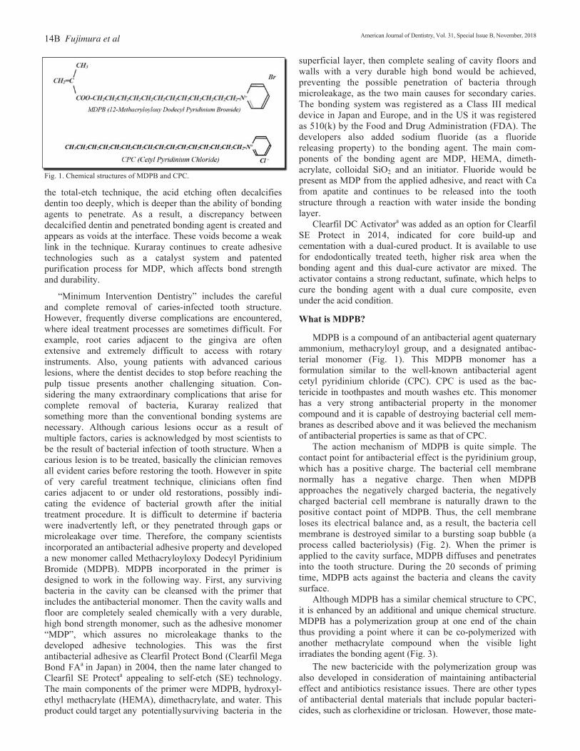
14B Fujimura et al Fig. 1. Chemical structures of MDPB and CPC. the total-etch technique, the acid etching often decalcifies dentin too deeply, which is deeper than the ability of bonding agents to penetrate. As a result, a discrepancy between decalcified dentin and penetrated bonding agent is created and appears as voids at the interface. These voids become a weak link in the technique. Kuraray continues to create adhesive technologies such as a catalyst system and patented purification process for MDP, which affects bond strength and durability. “Minimum Intervention Dentistry” includes the careful and complete removal of caries-infected tooth structure. However, frequently diverse complications are encountered, where ideal treatment processes are sometimes difficult. For example, root caries adjacent to the gingiva are often extensive and extremely difficult to access with rotary instruments. Also, young patients with advanced carious lesions, where the dentist decides to stop before reaching the pulp tissue presents another challenging situation. Con-sidering the many extraordinary complications that arise for complete removal of bacteria, Kuraray realized that something more than the conventional bonding systems are necessary. Although carious lesions occur as a result of multiple factors, caries is acknowledged by most scientists to be the result of bacterial infection of tooth structure. When a carious lesion is to be treated, basically the clinician removes all evident caries before restoring the tooth. However in spite of very careful treatment technique, clinicians often find caries adjacent to or under old restorations, possibly indi-cating the evidence of bacterial growth after the initial treatment procedure. It is difficult to determine if bacteria were inadvertently left, or they penetrated through gaps or microleakage over time. Therefore, the company scientists incorporated an antibacterial adhesive property and developed a new monomer called Methacryloyloxy Dodecyl Pyridinium Bromide (MDPB). MDPB incorporated in the primer is designed to work in the following way. First, any surviving bacteria in the cavity can be cleansed with the primer that includes the antibacterial monomer. Then the cavity walls and floor are completely sealed chemically with a very durable, high bond strength monomer, such as the adhesive monomer “MDP”, which assures no microleakage thanks to the developed adhesive technologies. This was the first antibacterial adhesive as Clearfil Protect Bond (Clearfil Mega Bond FAa in Japan) in 2004, then the name later changed to Clearfil SE Protecta appealing to self-etch (SE) technology. The main components of the primer were MDPB, hydroxyl-ethyl methacrylate (HEMA), dimethacrylate, and water. This product could target any potentially surviving bacteria in the
American Journal of Dentistry, Vol. 31, Special Issue B, November, 2018 superficial layer, then complete sealing of cavity floors and walls with a very durable high bond would be achieved, preventing the possible penetration of bacteria through microleakage, as the two main causes for secondary caries. The bonding system was registered as a Class III medical device in Japan and Europe, and in the US it was registered as 510(k) by the Food and Drug Administration (FDA). The developers also added sodium fluoride (as a fluoride releasing property) to the bonding agent. The main com-ponents of the bonding agent are MDP, HEMA, dimeth-acrylate, colloidal SiO2 and an initiator. Fluoride would be present as MDP from the applied adhesive, and react with Ca from apatite and continues to be released into the tooth structure through a reaction with water inside the bonding layer. Clearfil DC Activatora was added as an option for Clearfil SE Protect in 2014, indicated for core build-up and cementation with a dual-cured product. It is available to use for endodontically treated teeth, higher risk area when the bonding agent and this dual-cure activator are mixed. The activator contains a strong reductant, sufinate, which helps to cure the bonding agent with a dual cure composite, even under the acid condition. What is MDPB? MDPB is a compound of an antibacterial agent quaternary ammonium, methacryloyl group, and a designated antibac-terial monomer (Fig. 1). This MDPB monomer has a formulation similar to the well-known antibacterial agent cetyl pyridinium chloride (CPC). CPC is used as the bac-tericide in toothpastes and mouth washes etc. This monomer has a very strong antibacterial property in the monomer compound and it is capable of destroying bacterial cell mem-branes as described above and it was believed the mechanism of antibacterial properties is same as that of CPC. The action mechanism of MDPB is quite simple. The contact point for antibacterial effect is the pyridinium group, which has a positive charge. The bacterial cell membrane normally has a negative charge. Then when MDPB approaches the negatively charged bacteria, the negatively charged bacterial cell membrane is naturally drawn to the positive contact point of MDPB. Thus, the cell membrane loses its electrical balance and, as a result, the bacteria cell membrane is destroyed similar to a bursting soap bubble (a process called bacteriolysis) (Fig. 2). When the primer is applied to the cavity surface, MDPB diffuses and penetrates into the tooth structure. During the 20 seconds of priming time, MDPB acts against the bacteria and cleans the cavity surface. Although MDPB has a similar chemical structure to CPC, it is enhanced by an additional and unique chemical structure. MDPB has a polymerization group at one end of the chain thus providing a point where it can be co-polymerized with another methacrylate compound when the visible light irradiates the bonding agent (Fig. 3). The new bactericide with the polymerization group was also developed in consideration of maintaining antibacterial effect and antibiotics resistance issues. There are other types of antibacterial dental materials that include popular bacteri-cides, such as clorhexidine or triclosan. However, those mate-
Fujimura Figures
Fig. 1. Chemical structures of MDPB and CPC.
Fig. 2. Schematic antibacterial function of MDPB in three steps; (B) The positively charged MDPB molecule approaches the cell membrane; (B) The bacterial membrane structure loses the electrical balance; (C) The cell membrane is destroyed.
Fig. 3. Proposed functions of MDPB in the two-step self-etch adhesive; (A) When the primer is applied MDPS kills the remaining bacteria; (B) After polymerization of the adhesive, MDPB become a part of the bonding polymer layer and is immobilized in the resin matrix.

American Journal of Dentistry, Vol. 31, Special Issue B, November, 2018 Antibacterial bioactive dental adhesive 15B Fig. 2. Schematic antibacterial function of MDPB in three steps; (1) The positively charged MDPB molecule approaches the cell membrane; (2) The bacterial membrane structure loses the electrical balance; (3) The cell membrane is destroyed. Fig. 3. Proposed functions of MDPB in the two-step self-etch adhesive; (A) When the primer is applied MDPS kills the remaining bacteria; (B) After polymerization of the adhesive, MDPB become a part of the bonding polymer layer and is immobilized in the resin matrix. rials do not have any modifications to stop the continuous action of bacteria and release of the bactericide. Moreover, these bactericide materials will not polymerize with another monomer. Therefore, there is a risk of continuous dispersion into the oral cavity as well as the pulp tissue with its heavy vascular supply. Many scientists believe that continuous dispersion of a bactericide could result in the development of tolerance and resistance. Thus, MDPB was designed to prevent any dispersion of the bactericide after the restoration was cured and therefore, it is a very safe system. Overview of research findings: (1) Antibacterial properties of MDPB before polymerization - There are some research results for antibacterial properties against MDPB monomer and/or the product including MDPB, which are before polymerization of the monomer. Antibacterial effects of MDPB monomer for various caries related bacteria were evaluated by Imazato et al.1,2 They test-ed minimum bactericidal concentration (MBC) values for MDPB against a range of microorganisms detected in coronal caries lesions, including oral streptococci, lactobacilli, and a number of obligated anaerobic bacteria. The value range was from 15.6 to 125 µg/mL. It has been proven that MDPB has strong killing activity against various oral bacteria measured by its minimum bactericidal concentration values. Antibacterial properties of the product including MDPB were evaluated by Turkun et al.3 They investigated the properties of MDPB by the diameter of inhibition zone (millimeters) tested with agar well technique. In addition to that, they evaluated a number of recovered bacteria (CFU/ml) tested by the cavity model technique.3 They compared that primer with the following three cavity disinfectants: chlor-
hexidine gluconate base, benzalkonium chloride based pro-ducts, and 3% hydrogen peroxide. For cavity model technique, cylindrical cavities were prepared in the flat occlusal dentin of a human molar. The teeth were left in a broth culture of Streptococcus mutans allowing bacteria to invade, then tested materials were applied. After temporarily sealing and storing in saline, the dentin chips were collected and bacterial recovery was measured. Using the agar well technique, the MDPB-containing primer exhibited a greater inhibition zone than all three cavity disinfectants. When tested by the cavity method, the system showed significantly less bacterial recovery than all disinfectants. In addition, Imazato4 reported similar results evaluating the inhibitory effects of seven commercially-available adhesives/primers against caries associated bacteria: S mutans, L. casei and A. viscosus, with the MDPB-containing primer showing more inhibition zones than the others. Both articles concluded that the MDPB-containing system could inactivate the bacteria in the cavity more effectively than the tested cavity disinfectants or other adhesives. (2) MDPB for long term durability - The immediate bond strength of contemporary adhesives is quite high, however, those bond strengths gradually weaken with aging, decreasing at rates of 35-40% in 6-12 months.5-7 This is because of the degradation of the hybrid layer between resin adhesive and dentin interface.1 Pashley et al8 reported that endogenous matrix metalloproteinases (MMPs) bound to dentin contribute to the degradation of collagen fibrils in hybrid layers. The loss of collagen fibrils within the hybrid layer causes a loss of continuity with the underlying dentin, and decreases the bond strength to dentin. Therefore, they looked for compounds which were able to inhibit the activities of the enzyme.
Fujimura Figures
Fig. 1. Chemical structures of MDPB and CPC.
Fig. 2. Schematic antibacterial function of MDPB in three steps; (B) The positively charged MDPB molecule approaches the cell membrane; (B) The bacterial membrane structure loses the electrical balance; (C) The cell membrane is destroyed.
Fig. 3. Proposed functions of MDPB in the two-step self-etch adhesive; (A) When the primer is applied MDPS kills the remaining bacteria; (B) After polymerization of the adhesive, MDPB become a part of the bonding polymer layer and is immobilized in the resin matrix.

16B Fujimura et al Another study9 revealed that the experimental 5 wt% MDPB, which is the same as the concentration of commer-cially available product, showed great inhibition of soluble recombinant human MMP-9 (rhMMP-9) and matrix-bound MMPs. Chlorhexidine is also reported as an anti-MMP compound, however, it is water-soluble and it does not have polymerizable functional groups in its chemical structure, meaning that it may reach out from its bonding interface. On the other hand, MDPB is polymerizable and it may work as an inhibitor for years. It can also be copolymerized and retained in the hybrid layer. Several studies have compared the durability of MDPB-containing adhesive systems to other adhesives. The reported results indicated improved long term durability in the MDPB-containing system compared to other adhesives in vivo and in vitro.10,11 This may be partially explained by MDPB’s anti-MMPs function. (3) Clinical research results of the MDPB adhesive – A study12 evaluating post-operative sensitivity of the adhesive system reported that no postoperative sensitivity was experienced at the 1-year evaluation period. The bonding system also showed excellent clinical performance in high stress bearing areas for at least 5 years.13
Conclusions The incorporation of MDPB into the self-etching primer of the self-etching adhesive is a fine example of a marketed “bioactive adhesive”. This new class of dental adhesives can do far more than simply bond to dentin. Such adhesives provide specific and robust function, inactivating residual bacteria in caries-infected dentin. They work for long term in the mouth to inhibit any endogenous MMPs that are activated by the caries process or that are exposed and activated by the self-etching adhesive system. a. Kuraray Noritake Dental Inc., Tokyo, Japan. Acknowledgements: To Dr. Alireza Sadr, Department of Restorative Dentistry, University of Washington, Seattle, Washington, for his advice on
American Journal of Dentistry, Vol. 31, Special Issue B, November, 2018 preparing this manuscript. The help of Selene Sarraf, University of Washington, Seattle, Washington, is appreciated in editing the text.
Disclosure statement: The author is an employee of Kuraray Noritake Dental Inc. Mr. Fujimura is Technical Manager, Kuraray Noritake Dental Inc., Tokyo, Japan.
References 1. Imazato S, Ebi N, Tarumi H, Russell RRB, Kaneko T, Ebisu S.
Bactericidal activity and cytotoxicity of antibacterial monomer MDPB. Biomaterials 1999;20:899-903.
2. Imazato S, Torii Y, Takatsuka T, Inoue K, Ebi N, Ebisu S. Bactericidal effect of dentin primer containing antibacterial monomer methacryloyloxy-dodecylpyridinium bromide (MDPB) against bacteria in human carious dentin. J Oral Rehabil 2001;28:314-319.
3. Turkun M, Turkun LS, Erguch Z., Ates M. Is an antibacterial adhesive system more effective than cavity disinfectants? Am J Dent 2006; 19:166-170.
4. Imazato S. Bioactive restorative materials with antibacterial effects: new dimension of innovation in restorative dentistry. Dent Mater J 2009;28:11-19.
5. Breschi L, Mazzoni A, Ruggeri A, Cadenaro M, Di Lenarda R, DeStephano Dorigo E. Dental adhesion review: Aging and stability of the bonded interface. Dent Mater 2008;24:90-101.
6. Zhang SC, Kern M. The role of host-derived dentinal matrix metalloproteinases in reducing dentin bonding of resin adhesives. Int J Oral Sci 2009;1:163-176.
7. Tam L, Jokstad A. The bond between resin composite restorations and dentin may degrade in the mouth over time. J Evid Based Dent Pract 2010;10:21-22.
8. Pashley DH, Tay FR, Yiu C, Hashimoto M, Breschi L, Carvalho RM, Ito S. Collagen degradation by host-derived enzymes during aging. J Dent Res 2004;83:216-221.
9. Pashley DH, Tay FR, Imazato S. How to increase the durability of resin-dentin bonds. Compend Contin Educ Dent 2011;32:60-64.
10. Donmez N, Belli S, Pashley DH, Tay FR. Ultrastructural correlates of in vivo/in vitro bond degradation in self-etch adhesives. J Dent Res 2005;84:355-359.
11. Ansari ZJ, Sadr A, Moezizadeh M, Aminian R, Ghasemi A, Shimada Y, Tagami J, Effects of one-year storage in water on bond strength of self-etching adhesives to enamel and dentin. Dent Mater J 2008;27: 266-272.
12. Turkun LS. The clinical performance of one- and two-step self-etching adhesive systems at one year. J Am Dent Assoc 2005;136:656-664.
13. Turkun LS. Five year clinical performance of an antibacterial adhesive resin. J Dent Res 2008;87(Sp Iss C):0623.

_______________________________________________________________________________________________________________________________________________________________
Special Issue Article _______________________________________________________________________________________________________________________________________________________________
Synthesis, antibacterial activity, and biocompatibility of new antibacterial dental monomers YAPIN WANG, PHD, STEPHEN COSTIN, PHD, JIAN-FENG ZHANG, PHD, SUMEI LIAO, PHD, ZEZHANG T. WEN, PHD, THOMAS LALLIER, PHD, QINGZHAO YU, PHD & XIAOMING XU, PHD
ABSTRACT: Purpose: To synthesize a small library of antibacterial dental monomers based on quaternary ammonium salts and to test their antibacterial activity against cariogenic bacteria. Methods: Five new antibacterial monomers were synthesized and characterized by NMR, IR and HRMS. Results: Cytotoxicity assays using human gingival fibroblast cells showed that these new antibacterial monomers were biocompatible at concentrations of 10-5 M and displayed less cytotoxicity than BisGMA, a common dental monomer. When analyzed in vitro, all new monomers demonstrated strong inhibitory activity against biofilm formation by cariogenic Streptococcus mutans and Lactobacillus casei. Results indicated that antibacterial monomers containing a long alkyl (i.e. hexadecyl) chain are superior to their shorter-chain counterparts. The cross-linking monomers based on glycerol dimethacrylate also consistently outperformed their monomethacrylate analogs. Finally, the ammonium salts containing the dimethylbenzyl moiety were superior to the similar structures containing 1,4-diazabicyclo[2.2.2]octane (DABCO) in some cases. (Am J Dent 2018;31(Sp Is B):17B-23B). CLINICAL SIGNIFICANCE: All five new monomers were deemed biocompatible at concentrations of 10-5 M or less, and most had better biocompatibility than BisGMA. Dimethacrylate monomers 5 and 6 generally demonstrated high antibacterial activities, with the highest activity shown for the most lipophilic monomer 6, and these new antibacterial monomers have potential future application in dental composites and bonding agents. : Dr. Xiaoming Xu, Department of Comprehensive Dentistry & Biomaterials, Louisiana State University Health, New Orleans, School of Dentistry, 1100 Florida Ave., New Orleans, LA 70119, USA. E-: [email protected]
Introduction
Resin-based dental composites consisting of BisGMA and other methacrylate dental monomers have been widely used in dentistry to restore decayed teeth. Composite restorations have limited service life (typically 5-7 years). The occurrence of secondary (recurrent) caries caused by bacterial biofilms accumulated at the restoration margin is the leading cause of failure and replacement of dental restorations. To inhibit bacterial biofilms and reduce recurrent caries, new composites and bonding agents that exhibit antibacterial activity have been developed.1-5 Antibacterial restorative dental materials gen-erally fall into two categories: those with releasable agents and those with non-releasable antibacterial monomers. Common releasable antibacterial agents in dental materials include silver6 and chlorhexidine.2 Materials with releasable agents often show very high antibacterial activity over a short time span (< 1 week) followed by little to no activity as the material leaches out. The release of compounds such as chlorhexidine can also result in a significant reduction of mechanical properties over time, likely due to the formation of a porous structure and increased water sorption.7 As a result, the probability of restoration failure due to fracture is increased. Dental materials containing non-releasable antibacterial monomers have been under investigation.3,4,9 Many of these monomers contain a methacrylate group and a long-chain alkyl ammonium or pyridinium salt. These monomers show bactericidal activity in the uncured state and a bacteriostatic and/or bactericidal (contact-kill) effect in the cured state against oral pathogens including Streptococcus mutans.4,10 Since the antibacterial functional group is immobilized (polymerized) in the material, such materials usually have long-term antibacterial effect without significant adverse effect on the physical and
Fig. 1. Structure of antibacterial monomer methacryloyloxyundecyldimethyl-benzylammonium fluoride 1.12
mechanical properties. For example, the monomer methacryl-oyloxydodecylpyridinium bromide (MDPB) shows bactericidal activity against S. mutans in the uncured state, and the composites containing MDPB at concentrations of up to 2.83 wt% show antibacterial activity with no adverse effects on mechanical properties. Increasing the concentration of MDPB in the composite beyond this wt% results in a deterioration of mechanical properties.3,11 Thus, striking a balance between (maximizing) antibacterial capability of the monomers and (minimizing) detrimental effects on the mechanical properties of the material is of great importance. The synthesis of a fluoride-releasing antibacterial monomer, methacryloyloxyundecyldimethylbenzylammonium fluoride 1 (Fig. 1), which exhibits antibacterial activity against S. mutans was previously reported.12 This new monomer, which incorpor-ates the dimethylbenzylammonium moiety, exhibited overall better bactericidal activity against S. mutans biofilm than did the corresponding pyridinium salt and a dodecyltrimethyl-ammonium methacrylamide monomer. This new monomer also can serve as a fluoride source and counter ion for antibacterial fluoride-releasing dental monomers.13 Additionally, composites containing this monomer maintained good mechanical proper-ties with antibacterial monomer concentrations of up to 3 wt%. Unfortunately, at high concentration (6 wt%), mechanical pro-perties of the composite were significantly decreased over time.
18B Wang et al To improve the overall performance of antibacterial com-posites, we sought to improve both the efficacy of this antibac-terial monomer and the mechanical properties of composites containing a higher amount of this monomer. Observed differ-ences in the activity of antibacterial dental monomers based on the structure of the ammonium group led us to examine a broader structure-activity relationship for this class of compounds, covering varied alkyl chain lengths, ammonium salts based on 1,4-diazabicyclo[2.2.2]octane (DABCO) and cross-linking anti-bacterial monomers. The alkyl chain length of ammonium salts has a significant impact on bactericidal activity, with longer chains (up to 18 C atoms) conferring the best effects.14 Moreover, alkyl ammonium salts derived from DABCO have been synthe-sized previously and have demonstrated antibacterial effects.15,16 However, to the best of our knowledge, DABCO based ammo-nium salts have not been incorporated into dental monomers. Furthermore, cross-linking antibacterial dental monomers are rare in comparison with their monounsaturated counterparts (monomethacrylates).17,18 This is of particular importance because monounsaturated antibacterial monomers can increase water sorption and decrease mechanical properties of composite. As a result, the useful concentration of such antibacterial mono-mers in dental composites is very limited (ca. 3%).11,12 Therefore, new cross-linking antibacterial monomers would be desirable for dental composites because they would allow a higher content of the antibacterial component while maintaining physical and mechanical properties of the material. The cytotoxicity and the bactericidal activity of the antibacterial monomers changes after polymerization. How-ever, determination of antibacterial activity in monomer form is important because removal of carious material from the tooth structure can be incomplete, leaving behind cariogenic bacteria such as S. mutans.19 During the restoration process before polymerization, uncured monomer can potentially kill bacteria that are still present in the existing tooth structure, thus decreasing the likelihood of restoration failure due to secondary caries formation.20 Building upon our previous results, we report here the synthesis of five new (three cross-linking dimethacrylate) antibacterial monomers and the comparison of the structure-activity relationships of these and the previously reported monomer 1 in terms of cytotoxicity and antibacterial activity against four bacteria species: S. mutans, L. casei, Staphylo-coccus aureus and Pseudomonas aeruginosa. Among these four species, S. mutans and L. casei are known for their role in caries formation, and S. aureus and P. aeruginosa are oppor-tunistic pathogens involved in various systemic infections, especially in aging and immunocompromised patients.3,21 The antibacterial activities of the synthesized monomers against the latter two bacteria will explore their potential applications in other biomedical materials such as implants, feeding tubes and catheters.
Materials and Methods Monomer synthesis - All solvents were dried over 3Ǻ molecular sieves and reactions were run under N2 atmosphere. All synthesized intermediates and products were purified by column chromatography. 1H- and 13C-NMR spectra were recorded at room temperature with a Varian Unity Plus 400
American Journal of Dentistry, Vol. 31, Special Issue B, November, 2018
MHza instrument. High resolution mass spectra were obtained with a Waters Synapt HDb mass spectrometer with a nano-electrospray source. FI-IR spectra were recorded with a Thermo-Nicolet 670 FT-IRc spectrometer (resolution: 4 cm-1, number of scans: 128). 2-(1,3-dimethacryloyloxy)propyl 10-bromodecanoate (2). To a 50 mL round bottom flask containing 1,3-glycerol-dimethacryate (1.9302 g, 8.4569 mmol), 10-bromodecanoic acid (0.5339 g, 2.216 mmol) and 4-dimethylaminopyridine (DMAP) (0.0250 g, 0.205 mmol) under N2 atmosphere, 5 mL dichloromethane was added followed by dicyclohexyl-carbodiimide (DCC) (0.4839 g, 2.345 mmol). A white precipitate formed immediately. After 3-hour stirring, the slurry was filtered over a coarse (60 M) frit and the filtrate collected. The solvent was removed under reduced pressure. Purification by chromatography (2 × 16 cm silica) and elution with acetone/hexanes 1:19-1:9 v/v, Rf ~ 0.45 (1:9) yielded the product as a yellow oil (0.7490 g, 1.623 mmol, yield 76%). 1H NMR (CDCl3, δ)24 6.11 (br, 2H, 2CHH’), 5.61-5.59 (m, 2H, 2CHH’), 5.44-5.34 (m, 1H, (CH2)2CHOR), 4.44-4.22 (m, 4H, (CH2)2CHOR), 3.40 (t, 3JHH = 6.8 Hz, 2H, CH2Br), 2.32 (pseudo td, 3JHH = 7.5 Hz, 3JHH = 2.7 Hz, 2H, CH2CO2R), 1.94 (s, 6H, 2CH3), 1.85 (pent, 3JHH = 7.5 Hz, 2H, CH2CH2Br), 1.64-1.57 (m, 2H, CH2CH2CH2Br), 1.46-1.37 (m, 2H, CH2CH2CH2CH2Br), 1.29 (br, 8H, 4CH2); 13C{1H} 173.4, 173.0, 166.9, 166.5, 136.0, 135.91, 135.90, 126.6, 126.53, 126.51, 69.5, 69.0, 62.8, 62.6, 62.2, 34.3, 34.2, 34.1, 32.9, 29.4, 29.3, 29.2, 29.1, 28.8, 25.03, 24.99, 18.42, 18.40. IR (cm-1) 2928(m), 2855(w), 1720(s, C=O), 1638(w, C=C), 1453(m), 1292(m), 1144(s), 941(m). HRMS calculated for C21H32O6BrNa+: 483.1353; found: 483.1369. 2-(1,3-dimethacryloyloxy)propyl 16-bromohexadecanoate (3). To a 50 mL round bottom flask containing 1,3-glycerol-dimethacryate (4.0808 g, 17.879 mmol), 16-bromohexa-decanoic acid (3.0068 g, 8.9670 mmol) and DMAP (0.0560 g, 0.458 mmol) under N2 atmosphere, 20 mL dichloro-methane was added and the solution cooled to 0°C. DCC (2.0251 g, 9.8149 mmol) was added dropwise as a solution in dichloromethane (4 mL) and a white precipitate formed. After 5-hour stirring, the slurry was filtered over a coarse (60 M) frit and the filtrate collected. The solvent was removed under reduced pressure. Purification by chromatography (4 × 15 cm silica) and elution with acetone/hexanes 1:19 v/v, Rf ~ 0.5 (1:9) yielded the product as an oily white solid (4.1082 g, 7.5304 mmol, yield 84%). 1H NMR (CDCl3, δ)23 6.12 (br, 2H, 2CHH’), 5.61-5.59 (m, 2H, 2CHH’), 5.42-5.35 (m, 1H, (CH2)2CHOR), 4.42-4.22 (m, 4H, (CH2)2CHOR), 3.41 (t, 3JHH = 6.9 Hz, 2H, CH2Br), 2.32 (pseudo td, 3JHH = 7.6 Hz, 3JHH = 2.8 Hz, 2H, CH2CO2R), 1.94 (br, 6H, 2CH3), 1.85 (pent, 3JHH = 7.6 Hz, 2H, CH2CH2Br), 1.64-1.57 (m, 2H, CH2CH2CH2Br), 1.45-1.38 (m, 2H, CH2CH2CH2CH2Br), 1.33-1.23 (m, 20H, 10CH2); 13C{1H} 173.5, 173.0, 166.9, 166.5, 136.0, 135.91, 135.89, 126.6, 126.5, 126.4, 69.6, 69.0, 62.8, 62.6, 62.2, 34.4, 34.2, 34.1, 33.0, 29.79, 29.77, 29.76, 29.7, 29.61, 29.60, 29.4, 29.25, 29.21, 28.9, 28.3, 25.1, 25.0, 18.40, 18.38.

American Journal of Dentistry, Vol. 31, Special Issue B, November, 2018 IR (cm-1) 2922(s), 2852(m), 1722(s, C=O), 1655(m, C=C), 1453(m), 1293(m), 1148(s), 941(m). HRMS calculated for C27H45O6Br: 567.2292; found: 567.2291. 2-(1,3-dimethacryloyloxy)propyl 10-(1-(1-azonia-4-azabicylco- [2.2.2]octyl))decanoate bromide (4). To a 50 mL round bottom flask containing 2 (1.2948 g, 2.8063 mmol) and 1,4-diazabicyclo[2.2.2]octane (DABCO) (0.3169 g, 2.8249 mmol) under N2 atmosphere, 3 mL dichloromethane was added and the solids dissolved. After 18.5 hours, the solvent was removed under vacuum. Purification by chromatography (2 × 15 cm silica) and elution with dichloromethane/methanol 1:9 v/v, Rf ~ 0.1 yielded the product as a clear oil (0.5961 g, 1.039 mmol, yield 37%). 1H NMR (CDCl3, δ)23 6.10 (br, 2H, 2CHH’), 5.61-5.59 (m, 2H, 2CHH’), 5.39-5.32 (m, 1H, (CH2)2CHOR), 4.40-4.20 (m, 4H, (CH2)2CHOR), 3.65 (t, 3JHH = 7.3 Hz, 6H, 3N+CH2CH2N), 3.54-3.49 (m, 2H, N+CH2), 3.26 (t, 3JHH = 7.3 Hz, 6H, 3N+CH2CH2N), 2.31 (pseudo td, 3JHH = 7.5 Hz, 3JHH = 2.8 Hz, 2H, CH2CO2R), 1.92 (s, 6H, 2CH3), 1.75 (br, 2H, CH2), 1.62-1.54 (m, 2H, CH2), 1.36-1.32 (m, 4H, CH2), 1.27 (br, 6H, 3CH2); 13C{1H} 173.5, 173.1, 167.0, 166.6, 135.85, 135.83, 135.81, 126.8, 126.7, 126.6, 69.5, 68.9, 64.8, 62.7, 62.7, 62.2, 52.7, 45.5, 34.3, 34.1, 29.3, 29.2, 29.1, 29.0, 26.5, 25.0, 24.9, 22.3, 18.4, 18.42. IR (cm-1) 3411(m, br, H2O), 2927(m), 2856(w), 1719(s, C=O), 1637(w, C=C), 1455(m), 1293(m), 1149(s), 943(m). HRMS calculated for C27H45O6N2
+: 493.3272; found: 493.3283 2-(1,3-dimethacryloyloxy)propyl 16-(1-(1-azonia-4-azabicylco-[2.2.2]octyl))hexadecanoate bromide (5). To a 50 mL round bottom flask containing 3 (1.0061 g, 1.8442 mmol) and DABCO (0.3169 g, 2.8249 mmol) under N2 atmosphere, 3 mL ethyl acetate was added and the solids dissolved. After 6 days, the solvent was removed under vacuum. Purification by chromatography (2 × 15 cm silica) and elution with dichloro-methane/methanol 1:9 v/v, Rf ~ 0.1 yielded the product as a clear oil (0.8000 g, 1.216 mmol, yield 66%). 1H NMR (CDCl3, δ )23 6.06 (br, 2H, 2CHH’), 5.55 (br, 2H, 2CHH’), 5.36-5.28 (m, 1H, (CH2)2CHOR), 4.36-4.15 (m, 4H, (CH2)2CHOR), 3.62 (t, 3JHH = 7.1 Hz, 6H, 3N+CH2CH2N), 3.46-3.38 (m, 2H, N+CH2), 3.24 (t, 3JHH = 7.1 Hz, 6H, 3N+CH2CH2N), 2.30-2.23 (m, 2H, CH2CO2R), 1.88 (s, 6H, 2CH3), 1.71 (br, 2H, CH2), 1.59-1.50 (m, 2H, CH2), 1.33-1.26 (m, 4H, 2CH2), 1.19 (br, 16H, 8CH2); 13C{1H} 173.5, 173.1, 167.0, 166.5, 135.89, 135.86, 135.8, 126.7, 126.6, 126.5, 69.5, 68.9, 64.8, 62.8, 62.6, 62.2, 53.7, 52.7, 45.6, 34.4, 34.2, 29.82, 29.79, 29.7, 29.6, 29.4, 29.3, 29.2, 26.6, 25.1, 25.0, 22.4, 18.43, 18.42. IR (cm-1) 3402(m, br, H2O), 2922(m), 2852(m), 1721(s, C=O), 1637(w, C=C), 1456(w), 1293(m), 1152(s), 941(m). HRMS calculated for C33H57O6N2
+: 577.4211; found: 577.4190. 2-(1,3-dimethacryloyloxy)propyl 16-N,N-dimethylbenzyl-ammoniumhexadecanoate bromide (6). To a 50 mL round bottom flask containing 3 (1.0288 g, 1.8858 mmol) and dimethylbenzylamine (0.285 mL, 0.256 g, 1.90 mmol) under
New adhesive dental monomers 19B N2 atmosphere, 2 mL acetonitrile was added and the mixture heated to 50°C. After 48 hours, the reaction was allowed to cool to room temperature and the solvent was removed under vacuum. Purification by chromatography (2 × 15 cm silica) and elution with dichloro-methane/methanol gradient, 3%-10% v/v, Rf ~ 0.5 yielded the product as a clear oil (1.0920 g, 1.6041 mmol, 85%). 1H NMR (CDCl3, δ )23 7.64 (t, 3JHH = 7.9 Hz, 2H, Ph), 7.52-7.40 (m, 3H, Ph), 6.10 (br, 2H, 2CHH’), 5.59 (br, 2H, 2CHH’), 5.41-5.33 (m, 1H, (CH2)2CHOR), 5.03 (s, 2H, CH2Ph), 4.41-4.20 (m, 4H, (CH2)2CHOR), 3.54-3.49 (m, 2H, CH2N+), 3.28 (s, 6H, N+(CH3)2), 2.31 (pseudo td, 3JHH = 7.5 Hz, 3JHH = 2.8 Hz, 2H, CH2CO2R), 1.79 (br, 2H, CH2), 1.67 (s, 6H, 2CH3), 1.63-1.54 (m, 2H, CH2), 1.36-1.29 (m, 4H, 2CH2), 1.23 (br, 16H, 8CH2); 13C{1H} 173.5, 173.1, 166.9, 166.5, 135.83, 135.80, 135.77, 133.4, 130.8, 129.3, 127.6, 126.7, 126.6, 126.5, 69.5, 68.9, 67.5, 63.9, 62.7, 62.6, 62.1, 49.8, 34.3, 34.2, 29.72, 29.70, 26.6, 29.5, 29.4, 29.3, 29.2, 29.1, 26.4, 25.0, 24.9, 23.0, 18.4, 18.3. IR (cm-1) 3404(w, br, H2O), 2923(m), 2852(m), 1720(s, C=O), 1637(w, C=C), 1455(m), 1293(m), 1151(s), 940(m). HRMS calculated for C36H58O6N+: 600.4259; found: 600.4247. 16-bromohexadecanol (7b). A 100 mL round bottom flask equipped with magnetic stirring bar was charged with 16-bromohexadecanoic acid (1.68 g, 5 mmol) in THF (20 mL) and BH3/THF was added dropwise at 0°C. The reaction mixture was allowed to slowly warm to room temperature and was stirred overnight. 30 mL water was added then the product was extracted using ether (3 × 25 mL). The organic layer was washed by water and brine, dried over anhydrous Na2SO4, filtered and the solvent was removed under vacuum to give 7b as a white solid (1.472 g, 4.6 mmol, 92%). A similar reaction starting with 11-bromoundecanoic acid yielded 11-bromoundecanol (7a, 97%) 1H NMR (CDCl3, δ) 3.62 (t, 2H, CH2OH), 3.39 (t, 2H, CH2Br), 1.86-1.82 (m, 2H, CH2CH2OH), 1.55-1.41 (m, 2H, CH2CH2Br), 1.30-1.25 (m, 24H, 12CH2); 13C{1H} 63.3, 34.3, 33.1, 30.1, 29.9, 29.8, 29.7, 29.0, 28.4, 26.0. IR (cm-1) 3277(m, OH), 2916(s), 2848(s), 1473(m), 1462(m), 1122(w), 731(m). 16-(1-(1-azonia-4-azabicylco[2.2.2]octyl))-1-hexadecanol bromide (9). A 100 mL round bottom flask equipped with magnetic stirring bar was charged with 1,4-diazabicyclo[2.2.2]-octane (DABCO, 4 mmol), 16-bromohexadecanol (7 b, 1.28 g, 4 mmol) and EtOAc (30 mL). A white solid precipitated and was collected by filtration, washed with cold EtOAc and dried under vacuum to give 9 as a white solid (1.32 g, 3.06 mmol, 77%). A similar reaction of DABCO with 7a yielded 8 (83%). 1H NMR (CDCl3, δ) 3.55-3.52 (t, 2H, CH2OH), 3.40-3.36 (t, 6H, 3CH2N+), 3.27-3.17 (m, 8H, 3CH2N, CH2N+), 1.72-1.48 (m, 4H, 2CH2), 1.39-1.24 (m, 24H, 12CH2); 13C{1H} 61.8, 52.33, 52.27, 52.2, 44.9, 32.5, 29.6, 29.5, 29.44, 29.39, 29.2, 29.0, 25.8, 21.6. IR (cm-1) 3282(m, OH), 2916(s), 2847(s), 1470(m, C=C), 1462(m), 1056(s), 720(m). HRMS calculated for C22H45ON2: 353.3526; found: 353.3562.

20B Wang et al American Journal of Dentistry, Vol. 31, Special Issue B, November, 2018 Fig. 2. Synthesis of the new dimethacrylate monomers. 16-(1-azonia-4-azabicylco[2.2.2] octyl)hexadecylmethacrylate bromide (11). A 100 mL round flask equipped with magnetic stirring bar was charged with 1-(16-(hydroxyhexadecyl-4-azaoniabicyclo[2.2.2]octane)) bromide (9) 1.3 g, 3 mmol and dichloromethane (30 mL) and was placed in an ice bath. After the reaction flask was cooled for 15 minnutes, methacryloyl chloride (3.2 mmol) was added via syringe over 10 minutes. The reaction mixture was stirred at 0°C for 2 hours and then room temperature overnight. The reaction mixture was quenched by adding saturated aqueous K2CO3 (150 mL). The aqueous layer was extracted with chloroform (3 × 30 mL). The combined organic extract was washed sequentially with saturated aqueous NaHCO3 (2 × 20 mL) and brine (2 × 20 mL), dried over anhydrous MgSO4, filtered and concentrated under reduced pressure. The crude product was purified on silica gel column with EtOAc:MeOH (3:1) as mobile phase. After removal of the solvent under vacuum, 11 was isolated as a waxy, white solid (1.14 g, 2.28 mmol, 76%). A similar reaction of 8 with methacryloyl chloride yielded monomer 10 (70%). 1H NMR (CDCl3, δ) 6.08 (s, 1H, C=CHH’), 4.14 (t, 2H, CH2O), 5.60 (s, 1H, C=CHH’), 3.40-3.36 (m, 6H, 3CH2), 3.27-3.18 (m, 8H, 4CH2), 1.93 (s, 3H, CH3), 1.71-1.64 (m, 4H, 2CH2), 1.39-1.30 (m, 24H, 12CH2); 13C{1H} 167.6, 137.0, 124.8, 64.8, 62.4, 52.3, 52.22, 52.18, 29.54, 29.51, 29.48, 29.46, 29.4, 29.3, 29.13, 29.09, 28.5, 25.9, 21.6, 17.2. IR (cm-1) 3365(m, br, H2O), 2923(m), 2850(m), 1723(s, C=O), 1635(w, C=C), 1467(m) 1152(s), 905(w). HRMS calculated for C26H49O2N2: 421.3789; found: 421.3792. Cytotoxicity test - Human gingival fibroblasts were obtained from extracted molars from patients with healthy gingiva following informed consent as prescribed in an approved IRB protocol. Gingival fibroblasts were maintained in MEMα containing 10% fetal calf serum (FCS) and 200 units/mL penicillin and 200 µg/mL streptomycin. Cells were grown in
48-well plates for 24 hours prior to exposure to the synthesized antibacterial monomers. Growth media containing 0.1% dimethylsulfoxide (DMSO) were supplemented with 10-4 M, 10-5 M, 10-6 M and 10-7 M concentrations of the five newly synthesized monomers (4-6, 10, 11) and added to the cells for 24 hours. MEMα served as a control for cytotoxicity. Cell survival was visualized using a fluorescent esterase substrate (Calcein-AMd) and a Nikon TE2000e inverted fluorescent microscope. Cell survival was quantified using a BioTek Synergy 2f fluorescent multi-well plate reader. Evaluation of antimicrobial activity - S. mutans UA159 and L. casei ATCC 4646, two major cariogenic bacteria, were used for antibacterial activity assessment. S. mutans was grown in brain heart infusion broth (BHIg), and L. casei was grown in MRS medium.g In an effort to find out the breadth of the antibacterial activity of the monomers and their potential in other medical applications, S. aureus ATCC 25923 and P. aeruginosa ATCC 27853, two bacteria com-monly associated with a range of medical conditions such as wounds and abscesses, were also tested. S. aureus and P.aeroginosa were grown in Tryptic Soy Broth (TSBg). All bacteria were maintained under static conditions in a 37°C aerobic chamber with (for S. mutans only) or without 5% CO2. For antibacterial activity assay, these bacteria were cultivated using a semi-defined medium (BM) with glucose (18 mM) and sucrose (2 mM) (BMGS) as supplemental carbohydrate sources. Antimicrobial efficacy was measured using a Bioscreen C,h which is an automated system that provides constant temperature and automatic optical density (OD) measurement.24 Overnight cultures were transferred to fresh BMGS medium and allowed to grow to mid-exponential phase, at which point they were properly diluted in BMGS and allowed to grow in Bioscreen C with and without inclusion of different concentrations of antimicrobial monomers. All antimicrobial
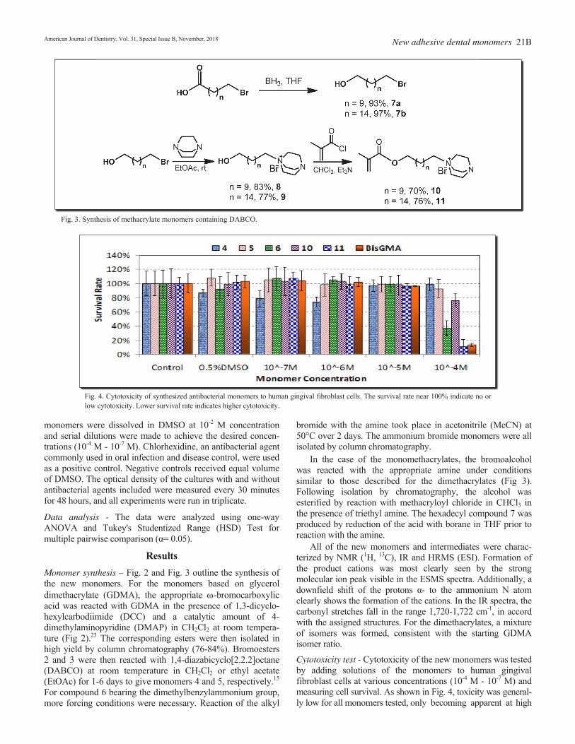
American Journal of Dentistry, Vol. 31, Special Issue B, November, 2018
New adhesive dental monomers 21B
Fig. 3. Synthesis of methacrylate monomers containing DABCO.
Fig. 4. Cytotoxicity of synthesized antibacterial monomers to human gingival fibroblast cells. The survival rate near 100% indicate no or low cytotoxicity. Lower survival rate indicates higher cytotoxicity.
monomers were dissolved in DMSO at 10-2 M concentration and serial dilutions were made to achieve the desired concen-trations (10-4 M - 10-7 M). Chlorhexidine, an antibacterial agent commonly used in oral infection and disease control, were used as a positive control. Negative controls received equal volume of DMSO. The optical density of the cultures with and without antibacterial agents included were measured every 30 minutes for 48 hours, and all experiments were run in triplicate. Data analysis - The data were analyzed using one-way ANOVA and Tukey's Studentized Range (HSD) Test for multiple pairwise comparison (α= 0.05).
Results Monomer synthesis – Fig. 2 and Fig. 3 outline the synthesis of the new monomers. For the monomers based on glycerol dimethacrylate (GDMA), the appropriate -bromocarboxylic acid was reacted with GDMA in the presence of 1,3-dicyclo-hexylcarbodiimide (DCC) and a catalytic amount of 4-dimethylaminopyridine (DMAP) in CH2Cl2 at room tempera-ture (Fig 2).23 The corresponding esters were then isolated in high yield by column chromatography (76-84%). Bromoesters 2 and 3 were then reacted with 1,4-diazabicyclo[2.2.2]octane (DABCO) at room temperature in CH2Cl2 or ethyl acetate (EtOAc) for 1-6 days to give monomers 4 and 5, respectively.15 For compound 6 bearing the dimethylbenzylammonium group, more forcing conditions were necessary. Reaction of the alkyl
bromide with the amine took place in acetonitrile (MeCN) at 50°C over 2 days. The ammonium bromide monomers were all isolated by column chromatography. In the case of the monomethacrylates, the bromoalcohol was reacted with the appropriate amine under conditions similar to those described for the dimethacrylates (Fig 3). Following isolation by chromatography, the alcohol was esterified by reaction with methacryloyl chloride in CHCl3 in the presence of triethyl amine. The hexadecyl compound 7 was produced by reduction of the acid with borane in THF prior to reaction with the amine. All of the new monomers and intermediates were charac-terized by NMR (1H, 13C), IR and HRMS (ESI). Formation of the product cations was most clearly seen by the strong molecular ion peak visible in the ESMS spectra. Additionally, a downfield shift of the protons α- to the ammonium N atom clearly shows the formation of the cations. In the IR spectra, the carbonyl stretches fall in the range 1,720-1,722 cm-1, in accord with the assigned structures. For the dimethacrylates, a mixture of isomers was formed, consistent with the starting GDMA isomer ratio. Cytotoxicity test - Cytotoxicity of the new monomers was tested by adding solutions of the monomers to human gingival fibroblast cells at various concentrations (10-4 M - 10-7 M) and measuring cell survival. As shown in Fig. 4, toxicity was general- ly low for all monomers tested, only becoming apparent at high
Fig. 4. Cytotoxicity of synthesized antibacterial monomers to human gingival fibroblast cells. The survival rate near 100% indicate no or low cytotoxicity. Lower survival rate indicates higher cytotoxicity.
Fig. 5. Effects of antibacterial monomers on the growth of four bacteria: (a) S. mutans, (b) L. casei, (3) P. aeruginosa, and (d) S. aureus. Bacteria were grown in Bioscreen C with and without inclusion of monomers (1, 4, 5, 6, 10, 11,) or chlorhexidine (CHX) as positive control. Bar graphs represent the average maximum optical densities of the cultures. Those with *and ** indicate significant difference at the level of P< 0.05 when compared to the control (CTRL). Those with ** also indicate significant difference from those with *.
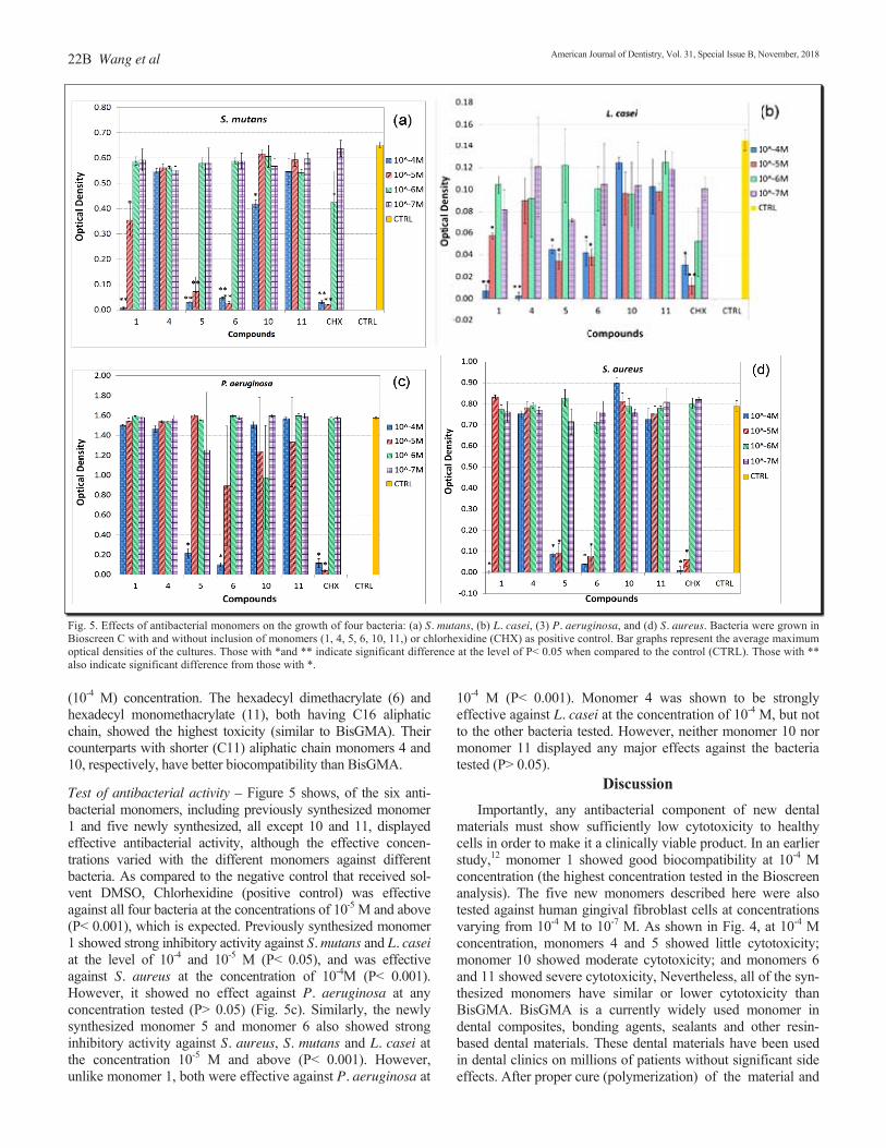
22B Wang et al
American Journal of Dentistry, Vol. 31, Special Issue B, November, 2018
Fig. 5. Effects of antibacterial monomers on the growth of four bacteria: (a) S. mutans, (b) L. casei, (3) P. aeruginosa, and (d) S. aureus. Bacteria were grown in Bioscreen C with and without inclusion of monomers (1, 4, 5, 6, 10, 11,) or chlorhexidine (CHX) as positive control. Bar graphs represent the average maximum optical densities of the cultures. Those with *and ** indicate significant difference at the level of P< 0.05 when compared to the control (CTRL). Those with ** also indicate significant difference from those with *. (10-4 M) concentration. The hexadecyl dimethacrylate (6) and hexadecyl monomethacrylate (11), both having C16 aliphatic chain, showed the highest toxicity (similar to BisGMA). Their counterparts with shorter (C11) aliphatic chain monomers 4 and 10, respectively, have better biocompatibility than BisGMA. Test of antibacterial activity – Figure 5 shows, of the six anti-bacterial monomers, including previously synthesized monomer 1 and five newly synthesized, all except 10 and 11, displayed effective antibacterial activity, although the effective concen-trations varied with the different monomers against different bacteria. As compared to the negative control that received sol-vent DMSO, Chlorhexidine (positive control) was effective against all four bacteria at the concentrations of 10-5 M and above (P< 0.001), which is expected. Previously synthesized monomer 1 showed strong inhibitory activity against S. mutans and L. casei at the level of 10-4 and 10-5 M (P< 0.05), and was effective against S. aureus at the concentration of 10-4M (P< 0.001). However, it showed no effect against P. aeruginosa at any concentration tested (P> 0.05) (Fig. 5c). Similarly, the newly synthesized monomer 5 and monomer 6 also showed strong inhibitory activity against S. aureus, S. mutans and L. casei at the concentration 10-5 M and above (P< 0.001). However, unlike monomer 1, both were effective against P. aeruginosa at
10-4 M (P< 0.001). Monomer 4 was shown to be strongly effective against L. casei at the concentration of 10-4 M, but not to the other bacteria tested. However, neither monomer 10 nor monomer 11 displayed any major effects against the bacteria tested (P> 0.05).
Discussion Importantly, any antibacterial component of new dental materials must show sufficiently low cytotoxicity to healthy cells in order to make it a clinically viable product. In an earlier study,12 monomer 1 showed good biocompatibility at 10-4 M concentration (the highest concentration tested in the Bioscreen analysis). The five new monomers described here were also tested against human gingival fibroblast cells at concentrations varying from 10-4 M to 10-7 M. As shown in Fig. 4, at 10-4 M concentration, monomers 4 and 5 showed little cytotoxicity; monomer 10 showed moderate cytotoxicity; and monomers 6 and 11 showed severe cytotoxicity, Nevertheless, all of the syn-thesized monomers have similar or lower cytotoxicity than BisGMA. BisGMA is a currently widely used monomer in dental composites, bonding agents, sealants and other resin-based dental materials. These dental materials have been used in dental clinics on millions of patients without significant side effects. After proper cure (polymerization) of the material and
Fig. 4. Cytotoxicity of synthesized antibacterial monomers to human gingival fibroblast cells. The survival rate near 100% indicate no or low cytotoxicity. Lower survival rate indicates higher cytotoxicity.
Fig. 5. Effects of antibacterial monomers on the growth of four bacteria: (a) S. mutans, (b) L. casei, (3) P. aeruginosa, and (d) S. aureus. Bacteria were grown in Bioscreen C with and without inclusion of monomers (1, 4, 5, 6, 10, 11,) or chlorhexidine (CHX) as positive control. Bar graphs represent the average maximum optical densities of the cultures. Those with *and ** indicate significant difference at the level of P< 0.05 when compared to the control (CTRL). Those with ** also indicate significant difference from those with *.
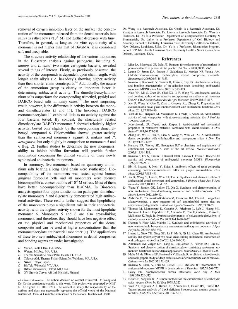
American Journal of Dentistry, Vol. 31, Special Issue B, November, 2018 removal of oxygen inhibition layer on the surface, the concen-tration of the monomers released from the dental materials into saliva is rather low (<10-5 M) and further decreases with time. Therefore, in general, as long as the vitro cytotoxicity of a monomer is not higher than that of BisGMA, it is considered safe and acceptable. The structure-activity relationship of the various monomers in the Bioscreen analysis against pathogens, including S.mutans and L. casei, two major cariogenic bacteria, revealed several things of interest. Firstly, as previously observed, the activity of the compounds is dependent upon chain length, with longer chain alkyls (i.e. hexadecyl) showing higher activity than their shorter chain counterparts.14 Additionally, the nature of the ammonium group is clearly an important factor in determining antibacterial activity. The dimethylbenzylammo-nium salts outperform the corresponding trimethyl, pyridyl and DABCO based salts in many cases.12 The most surprising result, however, is the difference in activity between the mono- and dimethacrylates (5 and 11). The hexadecyl DABCO monomethacrylate 11 exhibited little to no activity against the four bacteria tested. By contrast, the structurally related dimethacrylate DABCO monomer 5 showed relatively higher activity, bested only slightly by the corresponding dimethyl-benzyl compound 6. Chlorhexidine showed greater activity than the synthesized monomers against S. mutans and P. aeruginosa, but only slightly in comparison to monomers 5 and 6 (Fig. 2). Further studies to determine the new monomers’ ability to inhibit biofilm formation will provide further information concerning the clinical viability of these newly synthesized antibacterial monomers. In summary, five monomers based on quaternary ammo-nium salts bearing a long alkyl chain were synthesized. Bio-compatibility of the monomers was tested against human gingival fibroblast cells and all monomers were deemed biocompatible at concentrations of 10-5 M or less. Most of them have better biocompatibility than BisGMA. In Bioscreen analysis against four opportunistic human pathogens, dimethac-rylate monomers 5 and 6 generally demonstrated high antibac-terial activities. These results further suggest that lipophilicity of the monomers plays a significant role in their antibacterial activity, with the highest activity shown for the most lipophilic monomer 6. Monomers 5 and 6 are also cross-linking monomers, and therefore, they should have less negative effect on the physical and mechanical properties of the dental composite and can be used at higher concentrations than the monomethacrylate antibacterial monomer (1). The applications of these two new antibacterial monomers in dental composites and bonding agents are under investigation. a. Varian, Santa Clara, CA, USA. b. Waters, Milford, MA, USA. c. Thermo Scientific, West Palm Beach, FL, USA. d. Calcein-AM, Thermo Fisher Scientific, Waltham, MA, USA. e. Nikon, Tokyo, Japan. f. BioTek, Winooski, VT, USA. g. Difco Laboratories, Detroit, MI, USA. h. OY Growth Curves AB Ltd, Helsinki, Finland. Disclosure statement: The authors declared no conflict of interest. Dr. Wang and Dr. Costin contributed equally to this work. This project was supported by NIH/ NIDCR grant R01DE019203. The content is solely the responsibility of the authors and does not necessarily represent the official views of the National Institute of Dental & Craniofacial Research or the National Institutes of Health.
New adhesive dental monomers 23B Dr. Wang is a Research Associate, Dr. Costin is a Research Associate, Dr. Zhang is a Research Associate, Dr. Liao is a Research Associate, Dr. Wen is a Professor, Dr. Xu is a Professor, Department of Comprehensive Dentistry & Biomaterials; Dr. Lallier is a Professor Department of Cell Biology and Anatomy, School of Dentistry, Louisiana State University Health-New Orleans, New Orleans, Louisiana, USA. Dr. Yu is a Professor, Biostatistics Program, School of Public Health, Louisiana State University Health - New Orleans, New Orleans, Louisiana, USA.
References 1. Mjör IA, Moorhead JE, Dahl JE. Reasons for replacement of restorations in
permanent teeth in general dental practice. Int Dent J 2000;50:361-366. 2. Leung D, Spratt DA, Pratten J, Gulabivala K, Mordan NJ, Young AM.
Chlorhexidine-releasing methacrylate dental composite materials. Biomaterials 2005;26:7145-7153.
3. Imazato S, Kinomoto Y, Tarumi H, Ebisu S, Tay FR. Antibacterial activity and bonding characteristics of an adhesive resin containing antibacterial monomer MDPB. Dent Mater 2003;19:313-319.
4. Xiao YH, Ma S, Chen JH, Chai ZG, Li F, Wang YJ. Antibacterial activity and bonding ability of an adhesive incorporating an antibacterial monomer DMAE-CB. J Biomed Mater Res B: Appl Biomater 2009;90B:813-817.
5. Xie D, Weng Y, Guo X, Zhao J, Gregory RL, Zheng C. Preparation and evaluation of a novel glass-ionomer cement with antibacterial functions. DentMater 2011;27:487-496.
6. Yoshida K, Tanagawa M, Matsumoto S, Ymada T. Atsuta M. Antibacterial activity of resin composites with silver-containing materials. Eur J Oral Sci 1999;107:290-296.
7. Jedrychowski JR, Caputo AA, Kerper S. Anti-bacterial and mechanical properties of restorative materials combined with chlorhexidines. J Oral Rehabil 1983;10:373-381.
8. Zhang JF, Wu R, Fan Y, Liao S, Wang Y, Wen ZT, Xu X Antibacterial dental composites with chlorhexidine and mesoporous silica. J Dent Res 2014;93:1283-1289.
9. Kenawy ER, Worley SD, Broughton R.The chemistry and applications of antimicrobial polymers: A state of the art review. Biomacromolecules 2007;8:1359-1384.
10. Imazato S, Ebi N, Tarumi H, Russell RRB, Kaneko T, Ebisu S. Bactericidal activity and cytotoxicity of antibacterial monomer MDPB. Biomaterials 1999;20:899-903.
11. Ebi N, Imazato S, Noiri Y, Ebisu S. Inhibitory effects of resin composite containing bactericide-immobilized filler on plaque accumulation. Dent Mater 2001;17:485-491.
12. Xu X, Wang Y, Liao S, Wen ZT, Fan Y. Synthesis and characterization of antibacterial dental monomers and composites. J Biomed Mater Res B Appl Biomater 2012;100B:1151-1162.
13. Wang Y, Samoei GK, Lallier TE, Xu X. Synthesis and characterization of new antibacterial fluoride-releasing monomer and dental composite. ACS Macro Letters 2012;2:59-62.
14. Ahlström B, Chelminska-Bertilsson M, Thompson RA, Edebo L. Long-chain alkanoylchoines, a new category of soft antimicrobial agents that are enzymatically degradable. Antimicrob Agents Chemother 1995;39:50-55.
15. Thomas M, Montenegro D, Castaño A, Friedman L, Leb J, Huang ML, Rothman L, Lee H, Capodiferro C, Ambinder D, Cere E, Galante J, Rizzo JL, Melkonian K, Engle R. Synthesis and properties of polycationic derivatives of carbohydrates. Carbohydr Res 2009;344:1620-1627.
16. Dizman B, Elasri MO, Mathias LJ. Synthesis and antimicrobial activities of new water-soluble bis-quaternary ammonium methacrylate polymers. J Appl Polym Sci 2004;94:635-642.
17. Huang L, Xiao YH. Xing XD, Li F, Ma S, Qi LL, Chen JH. Antibacterial activity and cytotoxicity of two novel cross-linking antibacterial monomers on oral pathogens. Arch Oral Biol 2011;56:367-373.
18. Antonucci JM, Zeiger DN, Tang K, Lin-Gibson S, Fowler BO, Lin NJ. Synthesis and characterization of dimethacrylates containing quaternary am-monium functionalities for dental applications. Dent Mater 2012;28:219-228.
19. Maltz M, de Oliveria EF, Fontanella V, Bianchi R. A clinical, microbiologic, and radiographic study of deep caries lesions after incomplete caries removal. Quintessence Int 2002;33:151-159.
20. Imazato S, Ehara A, Torii M, Russell RRB, McCabe JF. Incorporation of antibacterial monomer MDPB in dentin primer. J Dent Res 1997;76:768-772.
21. Lowy FD. Staphylococcus aureus infections. New Eng J Med 1998;339:520-532.
23. Neises B, Steglich W. A simple method for the esterification of carboxylic acids. Angew Chem Int Ed Eng 1978;7:522.
24. Wen ZT, Nguyen AH, Bitoun JP, Abranches J, Baker HV, Burne RA. Transcriptome analysis of LuxS-deficient Streptococcus mutans grown in biofilms. Mol Oral Microbiol 2011;26:2-18.
_______________________________________________________________________________________________________________________________________________________________
Special Issue Article _______________________________________________________________________________________________________________________________________________________________
Dental cements: Bioactivity, bond strength and demineralization progression around restorations ALAA TURKISTANI, BDS, PHD, SOFIQUL ISLAM, DDS, PHD, YASUSHI SHIMADA, DDS, PHD, JUNJI TAGAMI, DDS, PHD & ALIREZA SADR, DDS, PHD
ABSTRACT: Purpose: To evaluate demineralization progression around indirect restorations placed with various cements using swept-source optical coherence tomography (OCT) and microshear bond strength (MSBS) to enamel and dentin. Methods: Resin inlays in cervical preparations (4×2 mm) were luted with two glass ionomer luting cements, Fuji I (FI) and RelyX Luting Cement (RL) and two adhesive cements, Adshield RM (AD) and RelyX Unicem 2 (UC). After 7-day artificial saliva incubation and 10,000 thermal cycles, specimens were demineralized (pH 4.5). Lesion progression at enamel and dentin margins was measured on OCT images after 1, 3 and 5 weeks demineralization (n= 8). Results: Repeated-measures ANOVA showed that demineralization period, cement type, and their interaction had a significant effect on lesion size in both substrates (P< 0.001). Enamel lesion progression was slower in RL, FI and AD, and was significantly different from UC and control (P< 0.001). RL dentin lesions were significantly different from FI and AD lesions (P< 0.05), which in turn were significantly different than UC and control lesions (P< 0.001). MSBS means of AD and UC were significantly higher than those of FI and RL (P< 0.001). (Am J Dent 2018;31(Sp Is B):24B-31B). CLINICAL SIGNIFICANCE: A bioactive cement combining bioavailable calcium, functional monomer and glass-ionomer formulations showed better lesion progression inhibition around restorations than the adhesive resin cement, and higher bond strength than the resin-modified and conventional glass-ionomer cements. : Dr. Alireza Sadr, Department of Restorative Dentistry, School of Dentistry, University of Washington, 1959 Northeast Pacific Street, Box 357456, Seattle, WA 98195, USA. E-: [email protected]
Introduction
Caries lesion formation around restoration margins is still a concern in clinical practice.1-3 Marginal microgaps contribute significantly to the progression of demineralization around the margins, while fluoride release may decrease the rate of progression of this process.1 Indirect restorations are considered as viable alternatives of direct restorations for cases with more extensive dental structure loss.3 For indirect restorations, long-term clinical success somewhat depends on the luting cement, which con-tributes retention, marginal integrity and longevity of the indirect restoration as well as the integrity of the dental substrate.4,5 Resin-based cements are the material of choice for adhesive luting allowing for more conservative restorative techniques as well as the ability to achieve excellent esthetic appearance and adequate strength. Among resin cements, self-adhesive resin cements were recently introduced and exhibit some advantages, including reduction of technique sensitivity and single clinical step application, similar to conventional cements. While the progression of caries around restorations would still mainly depend on patient’s caries risk, the preventive aspect of current dentistry compels the use of materials that provide protective effects, hence fluoride-releasing resin based cements are increasingly used in dental practice. Glass ionomer cements have long been recognized for their ability to release fluoride and therefore benefit the hard tissue. Resin modified glass ionomer cements (RMGI) were developed to combine the desirable properties of fluoride release from glass ionomer cements (GIC) with composite resin bond strength and low solubility. Traditional cement classification places GICs and adhesive
cements into separate categories, but advances such as the addition of adhesive monomers into resin-modified glass-ionomer formulations and the addition of particles or fillers releasing calcium, fluoride or other elements have created hybrid material categories. These materials have been termed bioactive, as they actively interact with the biological substrate on a molecular scale. This definition is quite broad and includes cases ranging from interaction of the monomer with the hard tissue substrate, for example the chemical bonding of 10-methacryloyloxydecyl dihydrogen phosphate (MDP) with apatite, to incorporation of a bioavailable ion such as fluoride, calcium or calcium analogues into the crystalline structure. In fact, GIC could be considered a classic bioactive cement due to its ion-exchange interaction with dentin. More commonly, mineral trioxide aggregate (MTA) or calcium-silicate-based restorative materials are termed bioactive, given their ability to form minerals adjacent to dentin.6 While there have been several reports on the interfacial and demineralization inhibitory properties of traditional GIC and RMGI restoratives,6,7 there are few studies on efficacy of the newer adhesive cements. Besides, the rate of formation of demineralized lesions was not assessed in these previous studies; rather, interfaces were evaluated based on radiographic or microscopic findings on cross-cut specimens, not longi- tudinally over time. However, rate of progression of marginal demineralization is an important factor affecting the longevity of the restoration. Clinically, longitudinal monitoring of margins and adjacent tooth structure is of high importance. Nevertheless, sensitivity and specificity of visual and tactile criteria for clinical detection are usually low.2 Despite the feasibility of evaluation at the microscopic level in laboratory studies, these conventional tests are often destructive and thus unsuitable for analysis over time.

American Journal of Dentistry, Vol. 31, Special Issue B, November, 2018 Dental cements 25B Table. Materials used in the study. _______________________________________________________________________________________________________________________________________________________________________________________________________________
Material (Abbreviation) Composition Application method _______________________________________________________________________________________________________________________________________________________________________________________________________________
Clearfil Majesty Silanated glass ceramics, surface treated alumina microfillers, Bis-GMA, Dispense in layers up to 2 mm in thickness, light cure Posterior TEGDMA, hydrophobic aromatic dimethacrylate, dl-camphorquinone. for 20 seconds. GC Fuji I (FI) Powder: Alumino-fluoro-silicate glass(amorphous) 95%, polyacrylic Dispense powder and liquid 1:2. Add all the powder to acid 5%. the liquid and mix rapidly for 20 seconds, coat the in- Liquid: Distilled water 50-55%, polyacrylic acid 30-40%. ternal surface of the restoration and seat immediately. Maintain moderate pressure, remove excess cement when rubbery. RelyX Unicem 2 (UC) Base paste: Phosphoric acid methacrylate monomer, methacrylate mono- Mix for 20 seconds, light cure for 20 seconds. Remove mers, silanated fillers, initiator components, stabilizers, rheological the excess after 2 seconds. Additives. Catalyst paste: Methacrylate monomers, alkaline fillers, silanated fillers, initiators, stabilizers, pigments, rheological additives. RelyX Luting Powder: fluoro-aluminosilicate glass, microencapsulated potassium Mix powder aggressively into the liquid about 30 Cement (RL) persulfate, ascorbic acid, catalyst, opacifiers. seconds. Spread the cement on interior surface of Liquid: aqueous solution of polycarboxylic acid modified with pendant restoration and seat in pace. Remove the excess after methacrylate groups, HEMA, water, tartaric acid. 3 minutes. Adshield RM (AD) Powder: fluoro-aluminosilicate glass, polycarboxylic acids, POs-Ca, Mix powder aggressively into the liquid about 20-30 zirconium oxide, tetracalcium phosphate-calcium hydrogen phosphate seconds. Spread the cement on interior surface of anhydride, persulfate, chemical polymerization catalyst, silica micro restoration and seat in place. Light cure for 10 seconds. fillers, pigments. Remove the excess after 2-3 seconds. Liquid: MDP, Bis-GMA, HEMA, other methacryalte monmomer, water, catalyst, accelerator. _______________________________________________________________________________________________________________________________________________________________________________________________________________
Bis-GMA: bisphenol-A diglycidylether dimethacrylate; TEGDMA: triethyleneglycol dimethacrylate; HEMA: 2-hydroxyethyl methacrylate; POs-Ca: phosphoryl oligosaccharide of calcium; MDP: 10-methacryloyloxydecyl dihydrogen phosphate. Optical coherence tomography (OCT) can provide real time, noninvasive, high-resolution cross-sectional images based on light backscattering from within a structure. OCT showed potential for assessment of occlusal, interproximal and caries around restorations, as well as dental materials.8-10 Demineralization progression around direct resin restorations has been investigated using this technique.1,11,12 However, no reports have evaluated indirect restorations and luting cements. The current laboratory study utilized swept-source OCT to monitor lesion progression around indirect composite restorations, aiming to investigate the effect of luting cements on demineralized lesion progression around enamel and dentin margins, and to compare the bonding performance of these luting cements to enamel and dentin by measuring microshear bond strength (MSBS). The null hypotheses tested were as follows: (1) no difference exists in marginal lesion extent among the tested groups at different demineralization periods; (2) no correlation exists between cement type and demin-eralization period on lesion size for both enamel and dentin; and (3) bond strength does not vary with type of luting cement in either enamel or dentin.
Materials and Methods Specimen preparation - A schematic drawing of the study procedure is shown in Fig. 1. The cervical one-third of 40 freshly extracted bovine incisors was lightly polished with 1,000-grit silicon carbide (SiC) paper to obtain a flat cervical surface. Standard tapered cervical cavities (4 mm diameter, 2 mm depth, 135° cavosurface angle) were prepared using a regular diamond bur attached to a high-speed air turbine under water coolant (100-μm grita), followed by finishing diamond bur (25-μm grita). Specimens were randomly divided into five groups of eight specimens each. In the control group, cavities were directly filled with one increment of composite (Clearfil Majesty Posteriorb) and light cured for 40 seconds using a
Fig. 1. The study design at a glance; (a) Tapered cavities were prepared while OCT was used to ensure standardization of measurements. Resin inlays were cemented in the tapered cavities and specimens were thermal cycled for 10,000 cycles. Specimens were then demineralized for 5 weeks. (b) Fixed cross-sections were subjected to OCT monitoring at each week and CLSM was used for confirmation of OCT findings after polishing selected specimens. (c) Microshear bond test was applied on cylinders of cements bonded to human enamel and dentin surfaces. halogen light unit with 600-mW∕cm2 output power density (Optilux 501c). For the other four groups, impressions were taken using polyvinylsiloxane material and composite inlays were fabricated on the poured stone casts from these impressions. The internal surfaces of the composite inlays were sandblasted with 50 µm alumina (Jet Blast IIId) for 10 seconds, cleaned ultrasonically in distilled water for 2 minutes, treated with 37% phosphoric acid for 10 seconds, rinsed and dried. Four cements were used to cement the resin inlays, as described in the Table; RelyX Unicem2e (UC), Fuji If (FI), Adshield RMb (AD), and RelyX Luting Cemente (RL). After marginal finishing with 2,000-grit SiC paper to remove any excess cement, specimens were stored for 7 days in standard artificial
Turkistani Figures
Fig. 1. The study design at a glance; (a) Tapered cavities were prepared while OCT was used to ensure standardization of measurements. Resin inlays were cemented in the tapered cavities and specimens were thermal cycled for 10,000 cycles. Specimens were then demineralized for 5 weeks. (b) Fixed cross-sections were subjected to OCT monitoring at each week and CLSM was used for confirmation of OCT findings after polishing selected specimens. (c) Microshear bond test was applied on cylinders of cements bonded to human enamel and dentin surfaces.
Fig. 2. Representative OCT B-scans of UC group after1 week (a), after 3 weeks (b) and after 5 weeks of demineralization (c) and corresponding confirmatory CLSM images under magnifications of ×125 and ×250 (d, e, f). (a) After 1 week, demineralization has resulted in formation of lesions with exposure of dentin margin. (b) and (c) OCT image of same cross-section showing progression of demineralized lesions during the proceeding weeks of demineralization, as confirmed by CLSM (d). (e) and (f) Higher magnification images of enamel and dentin lesions. EN: enamel, CE: cement, CR: composite restoration, DE: dentin.

26B Turkistani et al American Journal of Dentistry, Vol. 31, Special Issue B, November, 2018 Fig. 2. Representative OCT B-scans of UC group after 1 week (a), after 3 weeks (b) and after 5 weeks of demineralization (c) and corresponding confirmatory CLSM images under magnifications of ×125 and ×250 (d, e, f). (a) After 1 week, demineralization has resulted in formation of lesions with exposure of dentin margin. (b) and (c) OCT image of same cross-section showing progression of demineralized lesions during the proceeding weeks of demineralization, as confirmed by CLSM (d). (e) and (f). Higher magnification images of enamel and dentin lesions. EN: enamel, CE: cement, CR: composite restoration, DE: dentin. saliva (37°C, pH = 6.5) composed of 1 mM CaCl2, 3 mM NaH2PO4, 100 mM NaCl, and 2% NaN3, which was refreshed every day. Finally, specimens were thermocycled for 10,000 cycles between 5°C and 55°C water baths, with a dwell time of 30 seconds and transfer time of 2 seconds. Demineralization challenge - For the acidic challenge, all surfaces were covered with two coats of nail polish, with the exception of 0.5 mm of peripheral area around the margins. Each specimen was immersed in 1 mL of a demineralizing gel (pH = 4.5) containing 50 mM acetic acid CH3COOH, 1.5 mM CaCl2, 0.9 mM KH2PO4, 0.02%NaN3, and 3% hydroxyethyl-cellulose (HEC), and stored in an incubator at 37°C for 5 weeks. Every 2 days, specimens were removed from the gel, thoroughly rinsed with deionized water and blotted by tissue paper, then returned to refreshed gel. OCT imaging - Specimens were subjected to OCT evaluation to detect progression of demineralization after 1 week, 3 weeks and 5 weeks. A swept-source OCT system (IVS-2000g) was used. This system utilizes a high-speed scanning laser, sweeping 1,260- to 1,360-nm (center: 1,310 nm) wavelength at a 20-kHz rate. The optical resolution is 20 μm transversally and 12 μm
axially in air (7-8 μm in tissues with a refractive index around 1.5).8 At the time of scanning, specimens were washed with deionized water and positioned on a micrometer metal stage with 5° tilt to decrease specular surface reflections. To standardize the hydration condition of scanned surfaces, a thin film of water-based gel containing 2% HEC was applied. For each specimen, four cross-sectional images were acquired at 0°, 45°, 90°, and 135° planes across the cavity (Fig. 1). To replicate the imaging location each time, each specimen was marked by a pen and placed in the same orientation as for previous scans. Raw OCT data, 2,000 × 1,000 pixels corresponding to 7 mm × 7.48 mm for each cross-section, were imported to ImageJ softwareh and cross-sectional areas of tissue loss due to demineralization at enamel and dentin margins were outlined measured in mm2. Confocal laser scanning microscopy (CLSM) - After 5-week demineralization, randomly selected specimens were embedded in epoxy resin, cross-sectioned by a diamond sawi and polished with SiC papersi followed by diamond pastes of particle size down to 0.25 μm. The same OCT cross-sectional slice was observed under CLSM (1LM21H/Wj) at ×125 and ×250 magnifications.
Turkistani Figures
Fig. 1. The study design at a glance; (a) Tapered cavities were prepared while OCT was used to ensure standardization of measurements. Resin inlays were cemented in the tapered cavities and specimens were thermal cycled for 10,000 cycles. Specimens were then demineralized for 5 weeks. (b) Fixed cross-sections were subjected to OCT monitoring at each week and CLSM was used for confirmation of OCT findings after polishing selected specimens. (c) Microshear bond test was applied on cylinders of cements bonded to human enamel and dentin surfaces.
Fig. 2. Representative OCT B-scans of UC group after1 week (a), after 3 weeks (b) and after 5 weeks of demineralization (c) and corresponding confirmatory CLSM images under magnifications of ×125 and ×250 (d, e, f). (a) After 1 week, demineralization has resulted in formation of lesions with exposure of dentin margin. (b) and (c) OCT image of same cross-section showing progression of demineralized lesions during the proceeding weeks of demineralization, as confirmed by CLSM (d). (e) and (f) Higher magnification images of enamel and dentin lesions. EN: enamel, CE: cement, CR: composite restoration, DE: dentin.
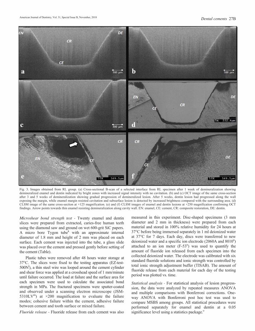
American Journal of Dentistry, Vol. 31, Special Issue B, November, 2018 Dental cements 27B Fig. 3. Images obtained from RL group. (a) Cross-sectional B-scan of a selected interface from RL specimen after 1 week of demineralization showing demineralized enamel and dentin indicated by bright zones with increased signal intensity with no cavitation. (b) and (c) OCT image of the same cross-section after 3 and 5 weeks of demineralization showing gradual progression of demineralized lesion. After 5 weeks, dentin lesion had progressed along the wall exposing the margin, while enamel margin resisted cavitation and subsurface lesion is detected by increased brightness compared with the surrounding area. (d) CLSM image of the same cross-section at ×125 magnification. (e) and (f) CLSM images of enamel and dentin lesions at ×250 magnification confirming OCT findings. Arrow points towards thin enamel resisting demineralization along cavity wall. EN: enamel, CE: cement, CR: composite restoration, DE: dentin. Microshear bond strength test - Twenty enamel and dentin slices were prepared from extracted, caries-free human teeth using the diamond saw and ground on wet 600-grit SiC papers. A micro bore Tygon tubek with an approximate internal diameter of 1.8 mm and height of 2 mm was placed on each surface. Each cement was injected into the tube, a glass slide was placed over the cement and pressed gently before setting of the cement (Table). Plastic tubes were removed after 48 hours water storage at 37°C. The slices were fixed to the testing apparatus (EZ-test-500Nl), a thin steel wire was looped around the cement cylinder and shear force was applied at a crosshead speed of 1 mm/minute until failure occurred. The load at failure and the surface area for each specimen were used to calculate the associated bond strength in MPa. The fractured specimens were sputter-coated and observed under a scanning electron microscope (JSM-5310LVm) at ×200 magnification to evaluate the failure modes; cohesive failure within the cement, adhesive failure between cement and tooth surface or mixed failure. Fluoride release - Fluoride release from each cement was also
measured in this experiment. Disc-shaped specimens (3 mm diameter and 2 mm in thickness) were prepared from each material and stored in 100% relative humidity for 24 hours at 37°C before being immersed separately in 1 ml deionized water at 37°C for 7 days. Each day, discs were transferred to new deionized water and a specific ion electrode (2060A and 8010n) attached to an ion meter (F-53n) was used to quantify the amount of fluoride ion released from each specimen into the collected deionized water. The electrode was calibrated with six standard fluoride solutions and ionic strength was controlled by total ionic strength adjustment buffer (TISAB). The amount of fluoride release from each material for each day of the testing period was plotted vs. time. Statistical analysis - For statistical analysis of lesion progress-sion, the data were analyzed by repeated measures ANOVA and multiple comparisons with Bonferroni corrections. One-way ANOVA with Bonferroni post hoc test was used to compare MSBS among groups. All statistical procedures were performed separately for enamel and dentin at a 0.05 significance level using a statistics package.o
Fig. 3. Images obtained from RL group. (a) Cross-sectional B-scan of a selected interface from RL specimen after 1 week of demineralization showing demineralized enamel and dentin indicated by bright zones with increased signal intensity with no cavitation. (b) and (c) OCT image of the same cross-section after 3 and 5 weeks of demineralization showing gradual progress of demineralized lesion. After 5 weeks, dentin lesion had progressed along the wall exposing the margin, while enamel margin resisted cavitation and subsurface lesion is detected by increased brightness compared with the surrounding area. (d) CLSM image of the same cross-section at ×125 magnification. (e) and (f) CLSM images of enamel and dentin lesions at ×250 magnification confirming OCT findings. Arrow points towards thin enamel resisting demineralization along cavity wall. EN: enamel, CE: cement, CR: composite restoration, DE: dentin.

28B Turkistani et al American Journal of Dentistry, Vol. 31, Special Issue B, November, 2018 Fig. 4. (a), (b) and (c) B-scans of selected interface from AD showing the gradual progression of demineralized lesions. After 1 week, demineralization resulted in shallow dentin cavitation, which progressed gradually during the following weeks, exposing dentin margin after 5 weeks, while enamel margin remained attached to the cement despite the progression of an adjacent subsurface lesion. CLSM images (×125, ×250) from the same section confirming the SS-OCT findings. EN: enamel, CE: cement, CR: composite restoration, DE: dentin.
Results Demineralization progression - Representative OCT and confirmatory CLSM images are shown in Figs. 2, 3 and 4. Demineralization of enamel and dentin, appearing as bright zones under SS-OCT, occasionally progressed toward cavitation and tissue loss appearing as dark zones at the margins. In enamel, the lesion patterns of FI, RL and AD were different from UC and the control. Demineralization resulted in subsurface enamel lesions or shallow cavitation with the former cements, whereas in UC and control, deep cavitated wall lesions were formed (Figs. 2a-c, 3a-c, 4a-c). In addition, zones of inhibition of demineralization were detected in close proximity to the cement-enamel interface of restorations luted with FI, RL and AD (Fig. 3). Lesion size was significantly influenced by both demin-eralization period (P< 0.001) and cement type (P< 0.001) in enamel and dentin. The interaction between these two factors was also significant (P< 0.001) in both substrates. Results of repeated measures ANOVA suggested that in enamel, lesion progression over time in FI, RL and AD was significantly different from that in UC (P< 0.001), which in turn was significantly different from the control (P< 0.001). No significant differences were detected between FI, RL and AD
(P> 0.05) (Fig. 5a). In contrast, in dentin, lesions forming around the margins of RL were significantly different from those in FI and AD (P< 0.05), which in turn were significantly different from UC and control (P< 0.001), but not from each other (P> 0.05). The difference between UC and the control in dentin was also significant (P< 0.05) (Fig. 5b). Microshear bond strength - The means and standard deviations of MSBS are presented in Figs. 5c and d. ANOVA results showed that in enamel, MSBS of AD and UC were significantly higher than FI and RL (P< 0.001), with no difference between AD and UC (P> 0.05) or between FI and RL (P> 0.05). In dentin, however, UC showed a significantly higher mean value than AD (P< 0.001), which in turn was higher than those for FI and RL (P< 0.001). No significant difference was detected between FI and RL for MSBS in dentin (P> 0.05). Specimens bonded with UC and FI recorded mainly adhesive failure in both enamel and dentin. In contrast, the predominant mode of failure in AD and RL was mixed in enamel and cohesive in dentin (Figs. 5e and f). Typical SEM images of the bonded area after the test are shown in Fig. 6. Fluoride release - The amount of fluoride release from each material at each day of the testing period was plotted vs. time in Fig. 7.
Fig. 4. (a), (b) and (c) B-scans of selected interface from AD showing the gradual progress of demineralized lesions. After 1 week, demineralization resulted in shallow dentin cavitation, which progressed gradually during the following weeks, exposing dentin margin after 5 weeks, while enamel margin remained attached to the cement despite the progress of an adjacent subsurface lesion. CLSM images (×125, ×250) from the same section confirming the SS-OCT findings. EN: enamel, CE: cement, CR: composite restoration, DE: dentin.

American Journal of Dentistry, Vol. 31, Special Issue B, November, 2018
Dental cements 29B
RL, FI and AD showed some fluoride release, while the concentrations were below the detection limit (therefore considered 0 ppm) at all times for composite and UC. Both RL and FI released comparable amounts of fluoride that were higher and more long lasting than that of AD. Note that the presence of Ca in the formulation of AD could promote the formation of Ca-F complexes, thereby preventing detection of fluoride by the ion-specific electrode, as a technical limitation of this measurement method.
Discussion This study investigated dental cement effects on the demineralization progression in adjacent enamel and dentin. The experimental design of deminer-alization testing in the present study was based on previous work,1 in which aggressive demineralization using acidified gel was used to promote the formation of standard, comparable lesions in all groups to facilitate objective comparison of lesions by measuring cross-sectional size of the cavitation formed due to demineralization. Also, OCT was used as a non-destructive objective method to monitor deminerali-zation at the same location over time. OCT has shown potential for quantitative estimation of lesion depth and mineral loss in demineralized lesions in many previous studies.8,11-13 In SS-OCT, demineralized enamel and dentin can be distinguished from sound tissue based on increased light scattering in porous demineralized tissue, which causes increased bright-ness in the corresponding SS-OCT image.8 When the demineralization progresses and results in cavity forma-tion, tissue loss removes scatterers completely resulting in the appearance of lesions as background or air.
Fig. 5. Lesion progression and bond strength results. Enamel (a) and dentin (b) lesion size at each week for various tested groups; similar italicized, lowercase letters indicate no significant difference between denoted groups (P> 0.05). Shear bond strength of various luting agents to enamel (c) and dentin (d) with failure mode in each test for enamel (e) and dentin (f).
Demineralized lesions showed different patterns in enamel versus dentin. Dentin margins were more susceptible to demineralization than was enamel, which is in accordance with common knowledge regarding acid-resistance and mineral content of these substrates. On the other hand, a narrow zone of demineralization-resistant enamel was found adjacent to the cavity wall when RL, AD and FI were used. Lesions progressed in slower rates around RL, AD and FI when compared to the self-adhesive resin cement UC. The slower progression of demineralization was attributed to fluoride release; fluoride has been found to increase enamel and dentin resistance to acid attack. Remineralization is also expected to be accelerated or enhanced by the effect of fluoride, conventional glass ionomer and resin-modified glass ionomer have been shown to inhibit demineralization adjacent to restoration margins and to remineralize enamel and dentin.7 The zone of demineralization-resistant enamel found adjacent to the cavity wall may have formed due to uptake of released fluoride and formation of CaF2 or fluoridated apatite, increasing resistance to demin-eralization. When the amount of fluoride released from luting cements was compared (Fig. 7), RL and FI released the largest amount. Fluoride release from AD showed an initial burst, but concentration tapered off quickly after the first day. FI and RL on the other hand exhibited a gradual decrease in released fluoride over time. Despite this, dentin lesions adjacent to FI
were deeper than those of RL and AD. Moreover, previous studies14,15 on RMGIs-dentin interfacial microstructure have revealed a submicron hybrid layer and tag-like structures of RMGI penetrating dentin. This hybrid layer formed by infiltration of resin into dental substrate might act as an acid-resistant layer, thereby inhibiting demineralization. The microshear bond testing used in this study was developed to enable measurement of bond strengths to small areas of substrate. Compared to micro-tensile bond testing, specimen preparation for MSBS is simpler. Also, little stress is produced during preparation because no trimming is needed after the bonding procedure. Furthermore, the lower probability of inducing a crack opening relative to load applied allows the evaluation of brittle materials with low modulus of elasticity such as GIC.16 Variations in the observed bond strengths of the cements examined in this study may be explained by their individual material compositions. The superior mechanical properties of UC may be attributed to a high degree of crosslinking of the methacrylate monomers, which also link firmly to fillers forming a highly cross-linked three-dimensional network of resin matrix.17,18 Nevertheless, the high bond strength and tight seal of the UC could not prevent rapid lesion progression around the restoration under acidic conditions. This finding is a highlight of the current study, emphasizing the importance of material bioactivity in terms of fluoride-release.
Fig. 5. Lesion progress and bond strength results. Enamel (a) and dentin (b) lesion size at each week for various tested groups; similar italicized, lowercase letters indicate no significant difference between denoted groups (P> 0.05). Shear bond strength of various luting agents to enamel (c) and dentin (d) with failure mode in each test for enamel (e) and dentin (f).
Fig. 6. Shear test failure modes in enamel (a-d) and dentin (e-h). Adhesive failure of UC can be seen in both enamel (a) and dentin (e). (b) and (f) represents mixed failure of AD in enamel and cohesive failure in dentin, respectively. In FI, adhesive failure in enamel is shown in (c) with its mixed failure in dentin (g). (d) represents a mixed failure of RL in enamel while (h) shows cohesive failure of the same material in dentin.

30B Turkistani et al American Journal of Dentistry, Vol. 31, Special Issue B, November, 2018
Fig. 6. Shear test failure modes in enamel (a-d) and dentin (e-h). Adhesive failure of UC can be seen in both enamel (a) and dentin (e). (b) and (f) represents mixed failure of AD in enamel and cohesive failure in dentin, respectively. In FI, adhesive failure in enamel is shown in (c) with its mixed failure in dentin (g). (d) represents a mixed failure of RL in enamel while (h) shows cohesive failure of the same material in dentin.
Fig. 7. Fluoride release from each material. On the other hand, superficial interaction and limited micro-mechanical retention might have been responsible for the relatively low bond strength to enamel of UC measured in this study.4,5 Despite the resinous component and expected dual mechanism of adhesion of RMGIC, bond strength values of RL were not significantly different than those of FI. One reason for this may be the low cohesive strength of both materials, under the small surface area for MSBS testing. The bonding per-formance of AD could be attributed to chemical bonding to calcium and enhanced wetting and demineralization by the MDP acidic monomer and resin infiltration promoted by the hydrophilic monomer HEMA and other resinous monomers into the substrate, improving the bond strengths.19,20 Also, light curing, in addition to the self-curing setting reaction, may have increased the overall cohesive strength of the material.21 In addition to MDP monomer and fluoride release, phosphoryl oligosaccharides of calcium (POs-Ca)p added to AD release bioavailable calcium and phosphate ions, enhancing remin-eralization of dental tissue.22 The initial extent of marginal gaps at the interface have shown a correlation with the rate of lesion progression around a
restoration.1 An inferior sealing ability has been reported for conventional GIC compared to RMGI,23 which could explain the dentin lesion progression. Desiccation after setting and brittleness of GIC may have exposed the margin, accelerating demineralization around the restoration. Based on the results of this study, the null hypotheses were rejected. The results for the cements differed in terms of size and progression of demineralized lesions. Bond strengths varied with the type of luting cement, but did not show a clear relationship to lesion progression around the restoration. New bioactive-adhesive formulations are a highly attractive category of materials, which can potentially deliver benefits to dental patients, especially with regard to high caries-risk situations and populations. Cements with higher bond strength values have become increasingly popular, particularly in situations and conservative preparation designs wherein restoration retention largely depends on the adhesive strength of the applied cement. There is an apparent benefit in restorative materials in combining bioactive ion-release and state-of-the-art adhesion technology. In conclusion, a bioactive cement combining bioavailable calcium, functional monomer and glass-ionomer formulations showed better demineralization inhibition when compared with adhesive resin cement and superior bond strength when compared with resin-modified and conventional glass-ionomer cements. a. Shofu, Kyoto, Japan. b. Kuraray Noritake Dental, Tokyo, Japan. c. Kerr, Orange, CA, USA. d. J Morita Corporation, Tokyo, Japan. e. 3M, St. Paul, MN, USA. f. GC Co., Tokyo, Japan. g. Santec Co., Komaki, Japan. h. National Institutes of Health, Bethesda, MD, USA. i. Buehler, Lake Bluff, IL, USA. j. Lasertec Co., Yokohama, Japan. k. Performance Plastics, Nagano, Japan. l. Shimazu Co, Kyoto, Japan. m. JEOL, Tokyo, Japan. n. Horiba, Kyoto, Japan. o. SPSS, Chicago, IL, USA. p. Ezaki Glico, Osaka, Japan.
Fig. 5. Lesion progress and bond strength results. Enamel (a) and dentin (b) lesion size at each week for various tested groups; similar italicized, lowercase letters indicate no significant difference between denoted groups (P> 0.05). Shear bond strength of various luting agents to enamel (c) and dentin (d) with failure mode in each test for enamel (e) and dentin (f).
Fig. 6. Shear test failure modes in enamel (a-d) and dentin (e-h). Adhesive failure of UC can be seen in both enamel (a) and dentin (e). (b) and (f) represents mixed failure of AD in enamel and cohesive failure in dentin, respectively. In FI, adhesive failure in enamel is shown in (c) with its mixed failure in dentin (g). (d) represents a mixed failure of RL in enamel while (h) shows cohesive failure of the same material in dentin.
Fig. 7. Fluoride release from each material.

American Journal of Dentistry, Vol. 31, Special Issue B, November, 2018 Acknowledgement: To Professor Daniel C Chan for his support towards the publication of this report. Disclosure statement: The authors declared no conflict of interest. Dr. Turkistani is Assistant Professor, Department of Operative Dentistry, Faculty of Dentistry, King Abdulaziz University, Jeddah, Saudi Arabia. Dr. Islam is Assistant Professor, RAK Medical and Health Sciences University, Ras al-Khaimah, United Arab Emirates. Dr. Shimada is Associate Professor, Department of Operative Dentistry, Graduate School of Medicine and Dentistry, Okayama University, Okayama, Japan. Dr. Tagami is Professor and Chair, Cariology and Operative Dentistry Department, Graduate School of Medical and Dental Sciences, Tokyo Medical and Dental University, Tokyo, Japan. Dr. Sadr is Associate Professor, Department of Restorative Dentistry, School of Dentistry, University of Washington, Seattle, WA, USA.
References 1. Turkistani A, Nakashima S, Shimada Y, Tagami J, Sadr A. Microgaps and
demineralization progress around composite restorations. J Dent Res 2015;94:1070-1077.
2. Nedeljkovic I, Teughels W, De Munck J, Van Meerbeek B, Van Landuyt KL. Is secondary caries with composites a material-based problem? Dent Mater 2015;31:e247-e277.
3. Morimoto S, Rebello de Sampaio FB, Braga MM, Sesma N, Ozcan M. Survival rate of resin and ceramic inlays, onlays, and overlays: A systematic review and meta-analysis. J Dent Res 2016;95:985-994.
4. De Munck J, Vargas M, Van Landuyt K, Hikita K, Lambrechts P, Van Meerbeek B. Bonding of an auto-adhesive luting material to enamel and dentin. Dent Mater 2004;20:963-971.
5. Hikita K, Van Meerbeek B, De Munck J, Ikeda T, Van Landuyt K, Maida T, Lambrechts P, Peumans M. Bonding effectiveness of adhesive luting agents to enamel and dentin. Dent Mater 2007;23:71-80.
6. Atmeh AR, Chong EZ, Richard G, Festy F, Watson TF. Dentin-cement interfacial interaction: Calcium silicates and polyalkenoates. J Dent Res 2012;91:454-459.
7. Wiegand A, Buchalla W, Attin T. Review on fluoride-releasing restorative materials - Fluoride release and uptake characteristics, antibacterial activity and influence on caries formation. Dent Mater 2007;23:343-362.
8. Shimada Y, Sadr A, Sumi Y, Tagami J. Application of Optical Coherence Tomography (OCT) for diagnosis of caries, cracks, and defects of restorations. Curr Oral Health Rep 2015;2:73-80.
9. Turkistani A, Sadr A, Shimada Y, Nikaido T, Sumi Y, Tagami J. Sealing performance of resin cements before and after thermal cycling: Evaluation by optical coherence tomography. Dent Mater 2014;30:993-1004.
10. Dao Luong MN, Shimada Y, Turkistani A, Tagami J, Sumi Y, Sadr A. Fractography of interface after microtensile bond strength test using swept-
Dental cements 31B source optical coherence tomography. Dent Mater 2016;32:862-869. 11. Horie K, Shimada Y, Matin K, Ikeda M, Sadr A, Sumi Y, Tagami, J.
Monitoring of cariogenic demineralization at the enamel-composite interface using swept-source optical coherence tomography. Dent Mater 2016;32:1103-1112.
12. Zhou Y, Shimada Y, Matin K, Sadr A, Sumi Y, Tagami J. Assessment of bacterial demineralization around composite restorations using swept-source optical coherence tomography (SS-OCT). Dent Mater 2016; 32:1177-1188.
13. Natsume Y, Nakashima S, Sadr A, Shimada Y, Tagami J, Sumi Y. Estimation of lesion progress in artificial root caries by swept source optical coherence tomography in comparison to transverse microradio-graphy. J Biomed Opt 2011;16:071408.
14. Coutinho E, Yoshida Y, Inoue S, Fukuda R, Snauwaert J, Nakayama Y, De Munck J, Lambrechts P, Suzuki K, Van Meerbeek B. Gel phase formation at resin-modified glass-ionomer/tooth interfaces. J Dent Res 2008;86:656-661.
15. Mitra SB, Lee CY, Bui HT, Tantbirojn D, Rusin RP. Long-term adhesion and mechanism of bonding of a paste-liquid resin-modified glass-ionomer. Dent Mater 2009;25:459-466.
16. McDonough WG, Antonucci JM, He J, Shimada Y, Chiang MY, Schumacher GE, Schultheisz, CR. A microshear test to measure bond strengths of dentin-polymer interfaces. Biomaterials 2009;23:3603-3608.
17. Holderegger C, Sailer I, Schuhmacher C, Schlapfer R, Hammerle C, Fischer J. Shear bond strength of resin cements to human dentin. Dent Mater 2008;24:944-950.
18. Gerth HU, Dammaschke T, Zuchner H, Schafer E. Chemical analysis and bonding reaction of RelyX Unicem and Bifix composites - A comparative study. Dent Mater 2006;22:934-941.
19. Yoshida Y, Nagakane K, Fukuda R, Nakayama Y, Okazaki M, Shintani H, Inoue S, Tagawa Y, Suzuki K, De Munck J, Van Meerbeek B. Comparative study on adhesive performance of functional monomers. JDent Res 2004;83:454-458.
20. Van Landuyt KL, Snauwaert J, De Munck J, Peumans M, Yoshida Y, Poitevin A, Coutinho E, Suzuki K, Lambrechts P, Van Meerbeek B. Systematic review of the chemical composition of contemporary dental adhesives. Biomaterials 2007;28:3757-3785.
21. Lawson NC, Cakir D, Beck P, Ramp L, Burgess JO. Effect of light activation on resin-modified glass ionomer shear bond strength. Oper Dent 2012;37:380-385.
22. Kitasako Y, Sadr A, Hamba H, Ikeda M, Tagami J. Gum containing calcium fluoride reinforces enamel subsurface lesions in situ. J Dent Res 2012;91:370-375.
23. Pontes DG, Guedes-Neto MV, Cabral MF, Cohen-Carneiro F. Microleakage evaluation of class V restorations with conventional and resin-modified glass ionomer cements. Oral Health Dent Manag 2014;13:642-646.
_______________________________________________________________________________________________________________________________________________________________
Special Issue Article _______________________________________________________________________________________________________________________________________________________________
Reactions: Antibacterial and bioactive dental restorative materials: Do they really work? DANIEL C.N. CHAN, DMD, MS, DDS, WENJIE HU, DDS, PHD, KWOK-HUNG CHUNG, DDS, PHD, ROBERT LARSEN, MS, STEVEN JENSEN, PHD, DENSEN CAO, PHD, LAURA GAVIRIA, PHD, JOO L. ONG, PHD, KYUMIN WHANG, PHD & TRINUCH EIAMPONGPAIBOON, DDS, MS, PHD.
ABSTRACT: Purpose: The study and development of antibacterial materials for use in dental applications is growing with the development of novel materials and procedures. Examination of the effects of such antibacterial materials on oral pathogens as well as on stability and longevity of dental restorations is of paramount importance to the field. Results: This review addressed the range of topics covered by the manuscripts presented at the Seoul symposium on antibacterial dental materials. (Am J Dent 2018;31Sp Is B:32B-36B). CLINICAL SIGNIFICANCE: Based on the presented works, it seems that the emerging antibacterial and bioactive mate-rials can potentially benefit restorative dentistry; however, like many other subjects in clinical dentistry, good quality evidence on their effectiveness under clinical situations is yet to be accumulated. : Dr. Daniel Chan, Department of Restorative Dentistry, University of Washington School of Dentistry, Box 357456, Seattle, WA 98195-7456, USA. E-: [email protected]
Introduction
The general need for anti-microbials stems from the overall procedure of placing a dental restoration to treat an infectious disease; caries. An existing pervasive weakness of resin-based dental restorations lies in their ability to intimately adapt to the tooth structures on which they are placed, followed by polymerization shrinkage. Thus, even if complete adaptation is achieved initially, when the material is polymerized, gaps are produced as the materials shrink towards their center of gravity, away from the tooth surface. Both adhesives and composites are susceptible to polymerization shrinkage. The resulting gaps allow infiltration of bacteria, which thrive and flourish under the protection of the restoration and away from tooth brushing and brief oral rinses. It is for this very reason that the selection of antimicrobials requires careful consideration. Antibacterial dental adhesives Because of inherent polymerization shrinkage of resin-based composite materials, detachment of cured composite fillings from the cavity wall and formation of microgaps are almost inevitable. Although recently developed resin-based composite/adhesive systems bond to dentin with bond strength values greater than enamel (20 MPa) in vitro, recurrent or secondary caries along the microgap between restorations and cavity walls is still a major issue for resin-based composite fillings. Previous studies reported that resin-based composites tend to accumulate more bacteria or plaque than other restorative materials in vitro. Antimicrobial agents available for dental adhesive systems Although some commercially available resin-based com-posites/adhesive systems show antibacterial activity, this is normally only an adverse reaction of the components of the composites, and the inhibitory effect against bacteria is unlikely to be reliable. Besides developing stronger and more durable adhesive systems, incorporation of antibacterial agents into resin-based composite/adhesive systems has been investigated for decades. Attempts have been made to prevent plaque ac-
cumulation on the tooth and restorative surfaces by incorpor-ation of antibacterial agents such as glutaraldehyde, chlorhexi-dine digluconate, and 12-methacryloyloxy dodecyl pyridinium bromide (MDPB) monomer into restorative materials. Certain synthesized monomers similar to MDPB also have shown antibacterial activity when immobilized in a resin-based composite material and their effectiveness has been supported by results from in vitro tests. MDPB is a compound of quaternary ammonium plus a methacrylate group. In an unpolymerized state, this monomer acts only as a disinfectant. When the material is polymerized, the copolymerization of MDPB with other monomers from the composite material immobilizes the antibacterial agent in the polymer matrix, and inhibits the growth of bacteria with which it has direct contact. Through the advancement of dental adhesive systems, incorporation of the antibacterial monomer MDPB enhanced the antibacterial effect of a proprietary dentin primer before curing and showed no adverse influence on bond strength to dentin and polymerization of the adhesive system. Particulate silver is well known for its low toxicity and good biocompatibility with human cells. Silver nanoparticles (AgNPs) have been extensively explored over the last decade and are a potent antibacterial agent. Incorporation of AgNPs alone or combined with synthesized quaternary ammonium di-methacrylates into either dental resins or adhesive systems has been observed to inhibit microcosm biofilm growth, metabolic activity, and lactic acid production. Biological methods are available for the synthesis of AgNPs with active antibacterial potency and to make AgNPs more biocompatible with human tissues and cells. Propolis, a natural non-toxic beehive product, has been shown to reduce the incidence of dental caries in rats, and the accumulation of supragingival plaque in vivo. Two compounds, apigenin and tt-Farnesol, have been identified as potential anti-plaque/anti-caries agents. Apigenin and tt-farnesol, alone or in combination, showed cariostatic properties in rats without sig- nificant effect on microbial viability in the rats’ mouths. Results

American Journal of Dentistry, Vol. 31, Special Issue B, November, 2018 of a recent in vitro study revealed no changes in dentin bond strength, resin-dentin interfacial morphology, or total amount of protein and soluble polysaccharide with the additions of the above anti-caries agents. Effect of antibacterial dental adhesives on oral pathogens Recent studies have examined the effects of the above antibacterial adhesive systems primarily on Streptococcus mutans bacteria. The majority of initial effectiveness studies were conducted in vitro using the agar diffusion method. The applied commercial adhesive had MDPB incorporated into the primer and demonstrated an antibacterial effect on infected cavities in dog teeth as well as reduction of enamel deminerali-zation around orthodontic brackets after 30 days. The current trend in this area of study is the use of a microcosm model because it offers the advantage of coming closer to the physico-chemical, microbiological and nutrient conditions of in vivo plaques, in addition to maintaining complexity and hetero-geneity. However, the only in situ study of these commercial adhesive systems demonstrated that none of the antibacterial materials tested reduced caries formation in dentin. What have we learned from published studies? Most studies that aim to develop new antibacterial agents are in vitro studies that focus mainly on caries-related oral pathogens. However, the geometric factor of the actual adhesive layer including primer and bonding agent for resin-based composite restorations may not be designed correctly in most of these in vitro studies. According to examination by scanning electron microscope of the resin-based composite/ adhesive systems’ bond to dentin, the adhesive layer or hybrid layer is usually only 2 to 5 µm-thick between the dentin wall and the bonding surface of resin-based composite restoration. This thin layer of adhesive exists rarely at the cavosurface and proximal enamel margin, but mostly at the gingival margin on the root surface of composite restorations. This means that only a very limited surface area of dental adhesive is exposed to the oral cavity in clinical situations. The antimicrobial effectiveness results observed in some bench studies therefore could be misinterpreted due to improper design, specifically due to the surface area mismatch between specimen and in situ clinical condition. In particular, the antimicrobial effect is over-estimated and magnified by the high surface area design of specimen exposure to high agent concentration, especially by the direct contact test method. If an in vitro test will be conducted to determine the efficacy of an antibacterial agent, the most correct testing methodology will present a clinically relevant design for the tested specimens. Such a procedure should be developed and adopted accordingly in future studies. When antimicrobial agents are added to primer solution or to a self-adhesive system, the final adhesive layer will be polymerized which results in the added agent being trapped inside the cross-linked network of polymer matrix. Many parameters, such as permeability of the resinous matrix, and driven force factors determine the releasing rate of some releasing agents such as chlorhexidine and AgNPs. In order to achieve efficacy and reaction longevity for the antimicrobial agent, the antimicrobial mechanisms and the releasing factors of each agent must be verified.
Antibacterial and bioactive materials 33B Future perspectives As the world’s population ages, an increasing incidence of root caries will be observed among the elderly who retain more of their teeth and who are at a higher risk for root caries. Gingi-val recession leads to increased exposure of tooth root surfaces, which have a higher solubility to biofilm acids than does coronal enamel. Root surface restorations with subgingival margins are difficult to clean and may develop pockets that facilitate perio-dontal bacterial growth. Thus, the need exists to develop anti-bacterial dental adhesives that can inhibit cariogenic and periodontal pathogens at root surface restorations. To validate these materials for this application, periodontitis-related bacteria such as Aggregatibacter actinomycetemcomitans, Porphyro-monas gingivalis, Prevotella intermedia, and Fusobacterium nucleatum, etc. must to be tested in relevant research studies, in addition to the cariogenic bacteria already tested. Finally, in vivo studies are absolutely necessary to validate the efficacy of antibacterial dental agents applied to the dy-namic environment of the living oral cavity, rather than to the limited static environment simulated by in vitro studies. In addition, optimization of the microecologic regulative effects of antimicrobial materials has received little attention to date and should be further examined. Antibacterial dental restorative materials Chen et al1 conducted a literature review of published information in a single online database, PubMed, restricting the search criteria to publications issued within a very recent 4-year period, from 2012-2016. Journal entries were limited to publi-cations written in English, and although the majority of scien-tific publications are presented in the English language, valuable and clinically relevant research is presented in the non-English native language of the study authors. Of particular note are publications recently coming from Eastern Europe, specifically from Turkey and Hungary. Because of high research costs in the U.S. and other mature scientific markets, some clinical research is favored in these or other locations where oversight and/or cost of research is not as substantial and the universal adoption or education of the scientific community to English is not fully established, yet the quality and reliability of the research is sound and trustworthy. Accordingly, some clinically relevant information may have been omitted by restricting the search parameters to only publications presented in English. Three classes of antimicrobials were reviewed by Chen et al:1 leachables, suspended particles, and polymerizable mono-mers. In addition to the drawbacks mentioned by the authors, other negatives should be mentioned for each of these three types. For leachables, a concern exists where the antimicrobial, in escaping the dental restoration, becomes a systemic com-pound that can be ingested, absorbed into the circulatory system, or otherwise have reactive effects with other tissues in other locations of the body. The risk of systemic effects to the patient bears consideration. Generally, these effects are miti-gated by the dilution effect of the leached compound relative to the sheer size of the human body. Suspended particles by nature have a limited physical reach - only colonization directly on the surface of the particle is disrupted. Growth adjacent to the particles, up to a respectable inhibition zone, is still expected. Thus, suspended particles are better described as microbial
34B Chan et al static - preventing further growth against the restoration. Poly-merizable agents, which may be better defined as bacteriocidal ligands, experience the same weaknesses as the other two groups with potential additional limitations. If the ligand is ca-pable of detaching from the monomer chain after polymeri-zation, then the ligand is in reality a leachable although an advantage may exist in that the detached lingand bound within the polymer might take more time to migrate to the surface of the dental material and thus prolong the overall activity of the ligand added to the dental material. If the ligand remains bound to the polymer, then the ligand itself acts as a captured particle with the added disadvantage that numerous ligands would be wholly bound within the polymer, unable to interact with the microbes at the polymer surface. Further, unless the monomer is relatively small or short-chained, the ratio of active ligands to overall polymer may be less than the presented surface area of bound particles, providing a lowered anti-microbial effect. This behavior is noted by the authors, where the activity of only the monomer is noted as effective. The authors present several anti-microbial agents indicated in the searched literature. Some, such as benzalkonium chloride and chlorhexidine have a well-known record of use. Other suggested compounds are relatively unknown in dentistry and certainly unknown to us, particularly the referenced use or research into urushiol and copper iodide. Urushiol is an oily extract obtained from several plants of the Toxicodendron genus, whose members include poison oak, poison sumac, and the Japanese urushi tree. The literature suggests a rapid onset of dermal edema when the compound is absorbed into the skin, and also suggests that a large portion of the population would present some level of allergic reaction to the compound. Urushiol is also reported to oxidize to a black-colored com-pound, which would further make the compound unsuitable for use in dental restorations. Copper iodide also may be subject to a similar concern. Some oral bacteria have demonstrated a re-ducing effect on metal ions. For example, ferric ions left over from some hemostatic preparations are known to reduce to metallic iron, producing a black discoloration. Copper ions are expected to do the same, reducing to an unsightly black-to-brown discoloration. Likewise, our experience with zinc com-pounds provides further evidence of metal redoxoxidation reactions in which zinc compounds (not necessarily zinc oxide) were observed to react in the presence of saliva to produce a gray color. Because gaps are produced due to polymerization shrink-age, the selection of antimicrobials requires careful considera-tion. Leachables can quickly fill a marginal gap to achieve short-term, effective protection. However, this margin also pro-vides a conduit for the leachable compound to escape and, in time, the situation becomes the same as if the leachable were never present to begin with. This marginal gap also limits the effectiveness of particles or active ligand polymers. Microbes may be inhibited at the surface of the dental material, but if the gap is sufficient the microbes may thrive on the tooth surface regardless, and the effectiveness of these materials is nullified. If a marginal gap cannot be addressed, then the ideal antimi-crobial is a suspended compound with a zone of inhibition large enough to exceed the common distance of the marginal gap. Any efforts to pre-cleanse the treatment site may achieve a
American Journal of Dentistry, Vol. 31, Special Issue B, November, 2018 short-term reduction in microbial activity, but so long as the margin exists, a pathway is available for subsequent infiltration of microbes long after the restoration is placed. Little debate exists regarding effective materials and strategies for microbial control up to 1-2 years, however the ideal would be to achieve long term (5-10 years and more) microbial control. Bioactive dental adhesives The topic of bioactive dental adhesives has been discussed in the past and the development of new generations of multi-functional dental adhesives is still an area of great interest to many dental clinicians as well as dental materials researchers. This interest is reflected in Fujimura’s article2 which reviews the development of antibacterial bioactive dental adhesives from the manufacturer Kuraray Noritake Dental Inc., since the 1970s. Supporting literature for the review ranges from the 1990s to 2011. Although the focus of this article2 is the review of Kuraray’s developments, newer generations of bioactive dental adhesives have been studied in the past 5 years and are worth reviewing as well as comparing against Kuraray’s products. The Fujimura et al2 review article focused primarily on the properties of methacryloyloxydodecylpryridinium bromide (MD PB), a newly developed monomer in dental adhesives and supported Kuraray’s findings with a review of in vitro studies which evaluate antibacterial properties, long-term durability and post-operative sensitivity. Providing more details about these studies in terms of groups compared, time ranges and comer-cially available and commonly used adhesives would be useful for clinicians and readers to better understand and evaluate the capabilities of Kuraray’s products. The biggest challenge in the development of antibacterial dental adhesives is leakage at the interface between tooth surface and restorative material because such leakage is the primary cause of secondary caries and failure of the tooth due to structural weakness. Moreover, bonding of dentin to the restoration has been shown to be even more challenging in addressing the successful restoration. In the review article,2 Fujimura highlights three important properties of Kuraray’s dental adhesives: (1) antibacterial properties of MDPB before polymerization, (2) MDPB long-term durability and (3) no post-operative sensitivity. Bacterial inactivation has been studied with a number of bacteria and results have indicated inhibition of different types of bacteria compared to other adhesives. However, the article does not specify differences between other adhesives compared or the duration of inhibition of bacterial growth. In general, one of the most important needs addressed by Kuraray’s products is the bonding of dental adhesive to dentin, which throughout the years has proven to be one of the biggest challenges to achieving a successful restoration. With the development of MDPB, long term durability was demonstrated to exceed that of the other adhesives when compared in several in vitro and in vivo studies. As explained in the review, this property was the result of polymerization of the adhesive as well as the inhibition of matrix metalloproteinases which lead to degradation and subsequently affect bonding at the interface. Finally, post-operative sensitivity was reported to be improved over a 6-week period and over 1 year in two different studies. However, no further details were given regarding factors that might have influenced this effect and the differences between

American Journal of Dentistry, Vol. 31, Special Issue B, November, 2018 the two studies that led to such discrepancies. Several antibacterial dental methods have been investigated over the past decades. One such approach was the incorporation into dental materials of silver particles and other antibacterial agents proven to be highly effective in inhibiting bacterial growth in many applications. Kuraray has maintained interest on multifunctional adhesives capable not only of achieving antibacterial activity by bacteriolysis, but also by polymerizing and sealing the area of interest. This underscores the advantages of Kuraray’s adhesives by eliminating the addition of multiple components and by having one component platform with multiple capabilities. Although the studies mentioned in the Fujimura et al article2 highlighted the antibacterial properties of Kuraray’s adhesives, literature in the field still addresses the concern of a long-term antibacterial effect and the stability of the current commercially available adhesives in the market. With current commercially available adhesives attributed to more than half of all restora-tions failing within 10 years, the studies mentioned in the review article did not address or demonstrate longer survival of their adhesives. Additionally, the effect of remineralizing agents also needs to be considered. Synthesis of novel antibacterial dental monomers Antibacterial monomers bear various disadvantages in the field of dental materials. Firstly, antibacterial monomers tend to exhibit antibacterial activity only in the uncured state and only show bacteriostatic activity in the cured state. Secondly, mono-functional antibacterial monomers tend to weaken the mechani-cal properties of the cured resin at the higher concentrations needed for strong antibacterial effect. Wang et al3 sought to develop new, more effective antimicrobial monomers and to improve the performance of these antibacterial monomers by increasing chain length for increased antibacterial activity and increasing monomer functionality to allow crosslinking with the resin to prevent the deterioration of mechanical properties. Wang et al3 synthesized five new antibacterial monomers, characterized them using NMR, IR and HRMS, tested their in vitro biocompatibility using a human gingival fibroblasts cytotoxicity assay, and determined their in vitro antibacterial activity against Streptococcus mutans, Lactobacillus casei, Staphylococcus aureus and Pseudomonas aeruginosa. The differences among these monomers include alkyl chain length and the number of reactive vinyl groups (mono- vs. dimeth-acrylate). The study found that none of the monomers was cytotoxic compared to BisGMA; that longer alkyl chains and dimethacrylates tended to produce stronger antibacterial effects; and that ammonium salts containing the dimethylbenzyl moiety had stronger antibacterial effects than structures containing 1,4diazabicyclo[2.2.2]octane (DABCO). Several drawbacks of this study are noteworthy. Wang et al3 did not attempt to test the ability of their new monomers to form polymers with dental resins, and no mechanical tests were done on dental resins containing these monomers. This is important, especially given the disadvantages of current anti- bacterial monomers. Only one antibacterial test was presented in this study, and this was with monomers only. Many mono-mers so-called antimicrobial are not actually bactericidal but only bacteriostatic once cured; therefore, the antibacterial acti- vity of polymers cured from these new monomers must be
Antibacterial and bioactive materials 35B tested, otherwise no improvement over what is already in the literature has been presented. A ring of inhibition assay could have been done with resin discs cured from these monomers, or better yet, dental resins cured with different concentrations of these monomers. Even when just testing monomers, which would be more potent than the cured form, the author states, “Compound 6 has an inhibitory effect against P. aeruginosa at 10-4 M and 10-5 M concentrations, but the bacterium grows after a significant delay (although to a much lesser extent compared to the con-trol). Monomer 5 causes an extended lag in growth for P. aeruginosa at 10-4 M concentration, but after 24 hours the total growth is equal to that of the control.” These results indicate that when a biofilm begins to form, these monomers, and most likely the polymers made from or with these monomers, were not as antibacterial. It can thus be extrapolated that when cured, these monomers may not be effective once the surface is covered with proteins and/or biofilm. This is one of the disad-vantages of antibacterial monomers upon which the authors were trying to improve. Finally, the authors of this article3 state, “The results indi-cate that the lipophilicity of the monomers plays a significant role in the antibacterial activity, with the highest activity shown for the most lipophilic monomer 6.” However, the lipophilicity of these specific monomers was never determined, thus this statement cannot be corroborated until the authors test it. A strong need exists to develop novel antimicrobial materials, and this study is intriguing in that regard. However, based on this presentation, it is not clear that the authors have indeed improved upon the disadvantages of antibacterial mono-mers. It seems that a combination of the use of antimicrobial monomers/polymers and releasing antibacterial agents may be needed. Furthermore, while a severe disadvantage of releasing antimicrobial agents is their limited time of efficacy, some publications have shown that the antimicrobial activity of Ag+ ions released from Ag nanoparticles in dental resins can last 4 weeks. However, whether the Ag+ ions would be depleted if the surface is bonded to dentin or enamel is unknown. Their release may be delayed until a marginal gap is formed and their release is needed, but this will need to be tested. Anti-demineralization activity of cements Turkistani et al4 report on the possible inhibition of demineralization around a restoration made with a so-called bioactive new glass ionomer cement that releases calcium and fluoride. Of special note in the study by Turkistani et al4 is the authors’ application of optical coherence tomography (OCT) in their research. OCT is an optical diagnostic tool based on interferometers, and uses a low coherence broadband near-infrared light source. Excellent spatial resolution (~20 μm) and real-time images are obtained by OCT. Application of OCT in dentistry has become very popular, especially for early detec-tion of caries, periodontal disease and oral cancer which are quite difficult to detect early (and often with ambiguous results) based on clinical examination or radiographs alone. OCT is a noninvasive, nondestructive, non-radiated, and real-time moni-toring method with three-dimensional imaging ability that can help clinicians locate problem areas accurately and rapidly. OCT is not without limitations, however. Firstly, cost and availability of the instrument can be a drawback, and even with

36B Chan et al Fig. 1. Physical craze lines or cracks on the surface of anterior central incisors. Cracks are defined as gaps in the tooth surface, such as enamel cracks. OCT can be applied for non-invasive detection of cracks (fractures) and microleakage. access to the instrument, OCT has limited penetration depth and scanning range. Because the scanning range is usually several millimeters, hundreds or thousands of pictures may be necessary to visualize a whole lesion. Wavelength choice may be another important consideration for specific types of tissue substrates. Given that OCT is a relatively new technology, comparison of its results with those of other dental diagnostic methods is important to assist researchers in interpreting results. In the results presented by Turkistani et al,4 we would have preferred to see images from both OCT and CLSM from all groups including the control, but results for only three groups were shown. Other limitations of the tests performed by Turkistani et al4 included choice of tooth substrate and in vitro demineralization as a means to simulate the actual carious process. Holding de-mineralized test specimens at pH 4.5, for example, may not accurately represent the condition of the oral cavity condition, even though it does result in somewhat accelerated deminerali-zation progress. Demineralization and remineralization is a cyclic process which could result from the presence of ions not only in dental cement, but also from ions in saliva. What are the future perspectives? As evidenced by Figs. 1 and 2, physical cracks and gaps can be aptly evaluated by OCT, but bioactivity of antibacterial dental materials around restorations may not be as evident with this technique. Nonetheless, such effects can be indirectly observed under clinical situation using this technology. Conclusion The investigation of anti-microbials in dental restorative materials is not new. Whether intrinsically present in metal amalgams that have been in use for a century, or in resin-based restoratives that have found wide acceptance for over 30 years, the topic of anti-microbial properties in dental restoratives has been foremost on the minds of researchers and manufacturers for decades. Many technologies and additive compounds that have been in use for decades continue to be in use today. A contemporary challenge that researchers face in the development of antibacterial dental adhesives is the need for more accurate and robust in vitro and in vivo models that can provide reliable information and matching to clinical scenarios. This implies the need to collect data from studies with longer durations (up to 10 years) in order to fully address concerns on survival of restorations and persistence of antibacterial activity.
American Journal of Dentistry, Vol. 31, Special Issue B, November, 2018 Fig. 2.A. CLSM from a central incisor specimen with an invisible crack. Fluorescein dye penetration revealed the presence of a major crack and of lateral cracks. B. A SS-OCT image of a sample visualized as a deep enamel crack transillumination. This crack extended beyond the DEJ. Current research on minimally invasive dental restorations suggests the use of composite materials that are typically bio-inert to replace missing volume. Future developments should focus on dental adhesives with synergistic effects that will not only replace missing volume but also will have bioactive and therapeutic properties. More robust and longer studies proving information about the properties and their mechanisms of action are required both in vitro and in vivo. Inclusion of opti-mization studies for antimicrobial materials will be required to fully understand possible induction of drug resistance. Through multidisciplinary efforts, the development of antimicrobial dental adhesives promises tremendous advances in oral health. Identification of compounds that have long-term bacterial inhibition effect and that are compatible with restorative agents like adhesives and cement is a challenging task. When such a material is applied to the restorative interface, bacteria will not grow at the dental structure and a long term antibacterial effect will be achieved. Advances from different groups as well as collaborations among different groups and technologies will result in truly long-term antimicrobial dental materials that are biocompatible and capable of preventing secondary caries and restoration failure. With efforts from the community, we expect this to be accomplished in the near future. Disclosure statement: The authors declared no conflict of interest with regard to the authorship of this manuscript. Dr. Chan is Professor and Chair, and Dr. Chung is Professor, Department of Restorative Dentistry, School of Dentistry, University of Washington, Seattle, WA, USA. Dr. Hu is Professor, Department of Periodontology, Peking University School and Hospital of Stomatology, Beijing, China. Mr. Larsen and Dr. Jensen are employees, and Dr. Cao is President and CEO, CAO Group, Inc., West Jordan, UT. Dr. Gaviria is Senior Lecturer, and Dr. Ong is Professor, Department of Biomedical Engineering, University of Texas, San Antonio, TX, USA. Dr. Whang is Associate Professor, Comprehensive Dentistry, School of Dentistry, UT Health, San Antonio, TX, USA. Dr. Eiampongpaiboon is Assistant Dr., Department of Prosthodontics, Faculty of Dentistry, Mahidol University, Bangkok, Thailand.
References 1. Chen L, Suh BI, Yang J. Antibacterial dental restorative materials: A
review. Am J Dent 2018;31 (Sp Is B online): 6B-12B. 2. Fujimura Y, Weerasinghe, M. Kawashima. Development of an antibacterial
bioactive dental adhesive: Simplicity and innovation. Am J Dent 2018;31 (Sp Is B online):13B-16B.
3. Wang Y, Costin S, Zhang J-f, Liao S, Wen ZT, Lallier T, Yu Q, Xu X. Synthesis, antibacterial activity, and biocompatibility of new antibacterial dental monomers. Am J Dent 2018;31 (Sp Is B online):17B-23B.
4. Turkistani A, Islam S, Shimada Y, Tagami J, Sadr A. Dental cements: Bioactivity, bond strength and demineralization progress around restorations. Am J Dent 2018;31 (Sp Is B online):24B-31B.
Chan Hu Figs
Fig. 1. Physical craze lines or cracks on the surface of anterior central incisors. Cracks are defined as gaps in the tooth surface, such as enamel cracks. OCT can be applied for non-invasive detection of cracks (fractures) and microleakage.
Fig. 2. A. CLSM from a central incisor specimen with an invisible crack. Fluorescein dye penetration revealed the presence of a major crack and of lateral cracks. B. A SS-OCT image of a sample visualized as a deep enamel crack transillumination. This crack extended beyond the DEJ.

_______________________________________________________________________________________________________________________________________________________________
Special Issue Article _______________________________________________________________________________________________________________________________________________________________
Antibacterial-containing dental adhesives’ effects on oral pathogens and on Streptococcus mutans biofilm: Current perspectives CAROLINA BOSSO ANDRÉ, DDS, PHD, DANIEL C.N. CHAN, DDS, MSC, PHD & MARCELO GIANNINI, DDS, PHD
ABSTRACT: Purpose: To describe the literature findings regarding commercially available antibacterial-containing dental adhesives and the futures perspectives of this field. Results: High-risk caries patients could yield benefits from restorative materials containing antibacterial properties in order to reduce the recurrent caries formation. Dental adhesives with antibacterial agents may reduce restoration replacement, as recurrent caries is still one of the major reasons for replacing a resin restoration. Literature results of three commercially available adhesives: Gluma 2Bond, Clearfil SE Protect and Peak Universal Bond, containing glutaraldehyde, MDPB and chlorhexidine, respectively indicates that Clearfil SE Protect seems to have better results against oral pathogens and on Streptococcus mutans biofilm. Besides the promising findings, clinical studies are still necessary in order to validate the clinical efficacy when exposed to a more complex environment and the long-term effect of either commercially available materials, experimental antibacterial monomers or antibacterial incorporations. As a suggestion of this article and according to the current scientific trends in this specific field, future directions should focus on restorative materials with therapeutic components targeting the virulence factors of cariogenic biofilm with minimal toxicity and side effects, and long-term action. (Am J Dent 2018;31:(Sp Is B):37B-41B). CLINICAL SIGNIFICANCE: Antibacterial-containing dental adhesives may have therapeutic effects, working as an additional source to reduce recurrent caries development in patients with high-risk of caries, and consequently the reduction in restoration replacements. Dr. Marcelo Giannini, Department of Restorative Dentistry, Operative Dentistry Division, Piracicaba Dental School, State University of Campinas, Av. Limeira, 901, Bairro Areião, Zip Code: 13414-903, São Paulo, Brazil. E-: [email protected]
Introduction
Practitioners have spent a lot of time replacing or per-forming resin restorations due to recurrent caries formation, tooth fractures, restoration fractures, loss of marginal integrity or lack of marginal sealing and non-carious cervical lesions, such as erosion, abrasion and abfraction.1-6 To restore small and middle-size cavities, resin-based composites have been used due to their outstanding esthetic appeal7 and excellent adhesive strength to dentin and enamel in combination with bonding agents. Several dental adhesive systems are commercially available for clinical use and are classified according to their application mode.8 Etch-and-rinse adhesives can be applied in two or three steps and their main characteristic is the application of an adhesive after phosphoric acid etching in wet demineralized dentin. Three-step etch-and-rinse adhesives use a primer, which is generally an aqueous solution containing HEMA (2-hydroxyethyl metha-crylate), while two-step etch-and-rinse adhesives present a combination of primer and bonding resin in a single bottle, which contains organic solvents, such as alcohol or acetone.9 Self-etch adhesives are applied in one or two steps and the main compositional characteristic is the presence of functional monomers, which are responsible to etch and infiltrate into mineralized tooth structures. Two-step self-etch adhesives use an acidic primer followed by a bonding or hydrophobic resin. Single-step or all-in-one self-etching systems are user-friendly bonding agents; however, many studies have criticized this category of adhesives regarding clinical durability.10 Besides resin monomers, chemical initiators and organic solvents, dental adhesives may contain filler, fluoride, desensi-
tizing or antimicrobial agents.8 Many compounds and sub-stances, such as triclosan, dimethylaminododecyl methacrylate (DMADDM), silver nanoparticles, doxycycline-encapsulated halloysite nanotube, zinc methacrylate, methacryloxylethyl cetyl dimethyl ammonium chloride (DMAE-CB) have been incorporated into dental bonding agents in order to promote antibacterial activity.11-15 Antibacterial properties in adhesive systems or composites are considered a viable option to reduce the bacterial colonization around dental restorations, prevent recurrent caries by suppressing biofilm formation and acid production, and thereby reduce restoration replacement.16-18 Although extensive research on antibacterial agents incorporated into dental adhesives or antibacterial monomer syntheses is available, just a few commercial adhesives contains antimicrobial agents, such as Clearfil SE Protecta (methacryloyloxydodecylpyridinium bromide, MDPB), Gluma 2Bondb (glutaraldehyde) and Peak Universal Bondc (chlorhexidine).19,20 The most well-known adhesive with antimicrobial activity is Clearfil SE Protect, a two-step self-etch system that contains MDPB in the primer solution. MDPB is a polymerizable quaternary ammonium methacrylate that copolymerizes with other adhesive monomers and disrupts the bacterial cell membrane when bacterium is in direct contact with the adhesive layer (by contact of the negatively charged bacteria with positively charged quaternary ammonium).21,22 Antibac-terial monomers that copolymerize with other adhesive mono-mers may provide long-term antibacterial activity.23 After the development of MDPB, several other monomers with quater-nary ammonium have been synthesized and incorporated into dental materials as antibacterial agents.24-26 Despite the in-
38B André et al creased development and evaluation of experimental antibac-terial monomers, containing or based on substances with broad antimicrobial action, as antibacterial agents, the focus of this article is to discuss commercially available dental adhesives and their future perspectives. Regarding the commercially available dental adhesives containing antibacterial agents, using a direct contact method the Clearfil SE Protect was tested against four facultative bacteria and four strict anaerobic microorganisms and had a bactericidal effect against Fusobacterium nucleatum after 10 minutes, against Streptococcus mutans, Porphyromonas gingivalis, Prevotella intermedia and Prevotella nigrescens after 30 minutes and against Staphylococcus aureus and Lactobacillus casei after 24 hours.19 Another study20 showed antimicrobial effects against oral pathogens by inhibition halo method and the decrease of viability of S. mutans biofilm grown on top of the adhesive layer, compared to Clearfil SE Bond. The same adhesive was tested in simulated Class I restorations and a significant reduction in formation of biofilm of S. mutans was also achieved, when compared to an adhesive without antibacterial agent.27 In situ studies28,29 indicate that Clearfil SE Protect is capable of controlling the caries progression in enamel at the restoration interface under conditions of high cariogenic challenge, compared to an adhesive with fluoride in its composition. Likewise, an in vivo study30 showed a reduction in caries formation around brackets after 30 days compared to conventional methods. In addition, it was reported31 that E. faecalis and S. mutans were not able to adapt to MDPB, which may suggest a lower risk of producing drug resistance. A two-step etch-and-rinse adhesive, Gluma 2Bond, contains 5% glutaraldehyde, which is a desensitizing and strong antibacterial agent.32-34 This adhesive showed bactericidal contact activity against Staphylococcus aureus, Enterococcus faecalis, Lactobacillus casei, Streptococcus mutans, Prevotellanigrescens and Fusobacterium nucleatum after 24 hours and against Porphyromonas gingivalis and Prevotella intermedia after 1 hour.19 The qualitative analysis of S. mutans biofilm using scanning electron microscopy showed a decrease of colonies when using Gluma 2Bond compared to a similar adhesive without glutaraldehyde; a result that was confirmed by colony counting.20 Another study35 also investigated dental adhesives containing glutaraldehyde (Gluma Primerb and Syntac Classic Systemc) and glutaraldehyde present in Gluma Primerb and Syntac Adhesivec appears to be effective against infected dentin. An in vivo study36 also showed the dentin disinfecting capacity of a glutaraldehyde-containing adhesive compared to an adhesive without antibacterial agent. Glutar-aldehyde-containing bonding agents have been criticized due to toxicity and mutagenic potential of this type of aldehyde. These effects were already described.37,38 Peak Universal Bond contains 0.2% chlorhexidine di(acetate), which is a cationic polybiguanide, bisphenol component containing chlorine that reacts with the negatively charged microbial cell surface, destroying its membrane. Chlorhexidine has a wide spectrum of action against gram-positive and gram-negative organisms, facultative, anaerobes, aerobes and fungi.39-41 This two-step etch-and-rinse adhesive demonstrated bactericidal contact activity only for strict anaero-
American Journal of Dentistry, Vol. 31, Special Issue B, November, 2018 bic microorganisms (Porphyromonas gingivalis, Prevotella intermedia, Prevotella nigrescens and Fusobacterium nucleatum after 24 hours).19 No effect against Streptococcus mutans biofilm was observed for this adhesive, compared to the same adhesive without chlorhexidine. However, these adhe-sives (with and without chlorhexidine) presented a reduction in biofilm of S. mutans similar to Clearfil SE Protect, which implies that other components, such as adhesive monomers and solvents may have antibacterial activity.20 These results suggest that chlorhexidine may stay trapped in the polymer chain, without the release properties.19,20 In another study,42 Peak Universal Bond presented a lower S. mutans biofilm formation compared to the same adhesive version without chlorhexidine; however the specimen preparation was different and the incubation time was lower. In addition, this non-light-cured adhesive presented an inhibition halo against some bacteria, suggesting that it may work as a cavity disinfectant.20 Also, for Peak Universal Bond, an inhibition halo for S. mutans was identified when it was not light-cured.43 The complex interactions between the specific oral bacteria, salivary constituents, dietary carbohydrate, and tooth surface modulates the transition from a condition of health to a diseased state by the establishment of cariogenic biofilms and conse-quently surface cavitation by acid dissolution, resulting in dental caries.44 Regarding the role of the aforementioned bac-teria at the pathogenesis of caries disease, S. mutans is considered the main pathogen involved in caries formation.45 S.mutans is not always the most predominant at the initial colonizing community, however the primary role of S. mutans resides with its ability to assemble an insoluble polymeric matrix, forming the core of the matrix-scaffold in cariogenic biofilms.46 Besides the extracellular polysaccharides produc-tion, the virulence of S. mutans is also associated to the pro-duction of weak acids from sugars, to adapt to large fluctua-tions in pH, oxygen tension and nutrient availability.47,48 Other microorganisms present in the complex oral micro-biota also play an important role in caries disease development and progression.20 Lactobacillus casei is an acidogenic and acid tolerant bacteria that can grow and survive in an acidic environment;49,50 Staphylococcus aureus is found in individuals with aggressive periodontitis51 and Enterococcus faecalis is associated with chronic periodontitis and frequently is the only species that persists in endodontically treated teeth.20,52,53 Strict anaerobic bacteria are more related to periodontal disease and can be found in cariogenic biofilm around the gingival margin.54 Due to further accumulation of biofilm, the number of obligatory anaerobic bacteria increase, changing the antimicrobial biofilm composition from streptococcus-domi-nated to Actinomyces spp. that is involved in root caries, and P. gingivalis involved and periodontal disease.19,54,55 P. intermedia is also a periodontal pathogen found in patients with early periodontitis, advanced periodontitis, and acute necrotizing ulcerative gingivitis.56,57 P. nigrescens also plays a role in the pathogenesis of periodontal disease, gingivitis and some odontogenic infections.58,59 F. nucleatum is frequently asso-ciated with periodontal diseases and is commonly found in human dental plaque with a crucial role in plaque develop-ment.19,60-62 Clearfil SE Protect, Gluma 2Bond and Peak Universal Bond
American Journal of Dentistry, Vol. 31, Special Issue B, November, 2018 present dentin bond strengths around 40 MPa and did not differ among them when the specimens were analyzed after artificial saliva storage for 1 year. These adhesives form a hybrid layer and resin tags, which represent the bonding mechanism of con-temporary bonding agents. Thus, the presence of antimicrobial components in the composition of adhesives seems to not interfere in the bond strength and bonding mechanism.19,20 Bonding agents containing antibacterial compounds are indicated for patients with very poor oral health, due to the high probability of recurrent caries development. Elderly patients with greater incidence of root caries and patients who have limitations to promote their own oral hygiene may also benefit when restorations are performed with materials containing antibacterial agents. Although some advantages have been extensively reported in the dental literature, there are concerns regarding the side effects produced by antibacterial agents and little clinical evidence that supports the in vitro findings has been reported.24,33 Also, the antibacterial activity in multi-species biofilm may be lower compared to results with planktonic bacteria,63 considering that the bacteria are protected by a diffusion barrier, the extracellular matrix.64 Another concern regarding in vitro tests remains on the interaction between the adhesive layer and the saliva pellicle. Some publications suggested that the saliva pellicle could attenuate antibacterial properties of underlying surfaces.65,66 However, the antibacterial effect of Gluma 2Bond and Clearfil Protect Bond was expressed in the biofilm of S. mutans, even covered with clarified saliva.20 One of the major side effects related to substances with broad antimicrobial spectra into restorative materials is the oral health resident bacterial interference and the promotion of bacterial resistance, producing undesirable outcomes on oral health.67 In order to reduce these side effects, the incorporation of natural products are been proposed, as a result of the lower probability of producing bacterial resistance. Natural products are considered a potential alternative approach to the current chemotherapeutic strategies, owing to the fact that natural products are a safer technology, biologically and environ-mentally, when compared to compounds synthetized by chemi-cal or physical methods.68,69 Propolis is a natural product composed of a resinous substance collected by Apis mellifera bees from various plant sources. It is considered a nontoxic natural product with a complex chemical composition and exhibits a wide range of biological activities, including antimicrobial, anti-inflamma-tory, anesthetic, and cytostatic properties.70,71 Two components were isolated from a Brazilian propolis, apigenin and tt-farnesol, and may represent an important alternative to current antibacterial agents, seeing that they can reduce the expression of virulence of S. mutans without necessarily suppressing the resident oral microbiota.68 Apigenin (4′,5,7-Trihydroxyflavone) is a potent inhibitor of water-insoluble glucan synthesis (inhibitor of glucosyltrans-ferases B and C), while tt-farnesol (trans,trans-3,7,11-trimethyl-2,6,10-dodecatrien-1-ol) changes the permeability and fluidity of the cell membrane by its lipophilic properties, affecting its glycolytic activity, production-secretion of glucosyltransferases and acidurance.72,73 They can be used separately or together, and seem to be more effective in the presence of fluoride.68
Dental adhesives on oral pathogens 39B One study67 incorporated these components into comercial bonding agents that contain fluoride (Patent: BR 10 2014 024497 5): Clearfil S3 Bond Plus, a single-step self-etch adhesive and Optibond S,e a two-step etch-and-rinse adhesive. The results were promising and may represent a novel alternative to decrease the cariogenicity of the biofilm around dental restorations, without suppressing the target micro-organism. The addition of apigenin or appigenin and tt-farnesol to Clearfil S3 Bond Plus were more efficient regarding the reduction of virulence of S. mutans compared to Optibond S and they did not interfere on the adhesion mechanism of both adhesives.67 Clearfil S3 Bond Plus con-taining apigenin reduced the amount of insoluble and intra-cellular polysaccharides of S. mutans biofilm grown for 5 days on top of the adhesive layer covered with clarified saliva.67 The new approach of incorporating anti-caries agents that are less likely to induce bacterial resistance into restorative materials could yield benefits in terms of enhanced durability of composite restorations, mainly in areas where biofilms accu-mulate, such as the interproximal and cervical regions of the teeth, by targeting the main virulence factors of S. mutans biofilm, namely the insoluble polysaccharides and intracellular polysaccharides.67 The reduction of both polysaccharides could affect the S. mutans ability to colonize the tooth surface and become the dominant bacteria and expressing it’s virulence.74 Although this approach is considered promising, further studies are necessary to clarify the effect on multispecies biofilm, on long-term action, and in vivo conditions (animal studies or long-term clinical trials). Following the same trend of incorporating natural products that have antibacterial properties into dental materials, chitosan and Epigallocatechin-3-gallate (EGCG) were also investigated when added to dental adhesives. The antibacterial activity of chitosan remains on the interaction between the positively charged chitosan and the negatively charged bacteria cell surface, causing the cell wall rupture.75,76 When added to dental adhesives the antibacterial effect has been reported.76,77 Conversely one study78 showed the absence of antibacterial activity of chitosan into a dental adhesive. EGCG, a flavonoid produced by Camellia sinensis plant (green tea), may be capable of suppressing gtf B, C, and D gene expression, disrupting S. mutans biofilm formation. This compound was able to express antibacterial activity when incorporated into dental adhesives in some concentrations.79 In addition, the increased research in natural products brings new alternative formulations to oral health care, including antibacterial, antifungal, and anti-caries properties, still poorly explored in the dental biomaterials field.
Conclusions and future perspectives Dental adhesive systems containing antibacterial or anti-caries agents show remarkable results against oral pathogens in in vitro studies. MDPB containing adhesives had greater results and is extensively explored in the dental literature. These antibacterial findings suggest a favorable indication of anti-bacterial dental adhesives for patients with high caries risk. Incorporation of natural products into restorative materials that can act on the S. mutans virulence factors can be considered a new approach in order to reduce recurrent caries formation,
40B André et al without killing the target organism. Besides the promising findings, clinical studies are still necessary in order to validate the clinical efficacy when exposed to a more complex environ-ment and the long-term effect of either commercially available materials, experimental antibacterial monomers or antibacterial incorporations. Future directions in research should focus on restorative materials with therapeutic components targeting the virulence factors of cariogenic biofilm with minimal toxicity, side effects, and with long-term action. a. Kuraray Noritake Dental Inc., Tainai City, Niigata, Japan. b. Heraeus Kulzer GmbH, Hanau, Hessen, Germany. c. Ivoclar Vivadent, Schaan. Liechtenstein. d. Ultradent Products Inc., South Jordan, UT, USA. e. Kerr Corp., Orange, CA, USA. Acknowledgements: The authors thank the financial support from Brazilian Financial Agencies: FAPESP: 2010/13599-0, 2011/17841-2 and 2014/17543-0; CNPq: 140698/2013-2. Funding sources were not involved in study design; in the collection, analysis and interpretation of data; in the writing of the report; or in the decision to submit the article for publication. Disclosure statement: The authors declared no conflict of interest. Dr. André is a postdoctoral researcher, Dr. Giannini is Associate Professor, Department of Restorative Dentistry, Piracicaba Dental School, State University of Campinas, São Paulo, Brazil. Dr. Chan is Professor, Department of Restorative Dentistry, School of Dentistry, University of Washington, Seattle, Washington, USA.
References 1. Gordan VV, Garvan CW, Blaser PK, Mondragon E, Mjör IA. A long-term
evaluation of alternative treatments to replacement of resin-based composite restorations: Results of a seven-year study. J Am Dent Assoc 2009;140:1476-1484.
2. Demarco FF, Corrêa MB, Cenci MS, Moraes RR, Opdam NJ. Longevity of posterior composite restorations: Not only a matter of materials. Dent Mater 2012;28:87-101.
3. Opdam NJ, van de Sande FH, Bronkhorst E, Cenci MS, Bottenberg P, Pallesen U, Gaengler P, Lindberg A, Huysmans MC, van Dijken JW. Longevity of posterior composite restorations: A systematic review and meta-analysis. J Dent Res 2014;93:943-949.
4. Makishi P, Thitthaweerat S, Sadr A, Shimada Y, Martins AL, Tagami J, Giannini M. Assessment of current adhesives in class I cavity: Nondestructive imaging using optical coherence tomography and microtensile bond strength. Dent Mater 2015;31:e190-e200.
5. Nedeljkovic I, Teughels W, De Munck J, Van Meerbeek B, Van Landuyt KL. Is secondary caries with composites a material-based problem? Dent Mater 2015;31:e247-e277.
6. Pena CE, Rodrigues JA, Ely C, Giannini M, Reis AF. Two-year randomized clinical trial of self-etching adhesives and selective enamel etching. Oper Dent 2016;41:249-257.
7. Ferracane JL. Resin-based composite performance: Are there some things we can't predict? Dent Mater 2013;29:51-58.
8. Van Landuyt KL, Snauwaert J, De Munck J, Peumans M, Yoshida Y, Poitevin A, Coutinho E, Suzuki K, Lambrechts P, Van Meerbeek B. Systematic review of the chemical composition of contemporary dental adhesives. Biomaterials 2007;28:3757-3785.
9. Pashley DH, Tay FR, Breschi L, Tjaderhane L, Carvalho RM, Carrilho M, Tezvergil-Mutluay A. State of the art etch-and-rinse adhesives. Dent Mater 2011;27:1-16.
10. Giannini M, Makishi P, Ayres AP, Vermelho PM, Fronza BM, Nikaido T, Tagami J. Self-etch adhesive systems: A literature review. Braz Dent J 2015;26:3-10.
11. Chai Z, Li F, Fang M, Wang Y, Ma S, Xiao Y, Huang L, Chen J. The bonding property and cytotoxicity of a dental adhesive incorporating a new antibacterial monomer. J Oral Rehabil 2011;38:849-856.
12. Henn S, Nedel F, de Carvalho RV, Lund RG, Cenci MS, Pereira-Cenci T, Demarco FF, Piva E. Characterization of an antimicrobial dental resin adhesive containing zinc methacrylate. J Mater Sci Mater Med 2011; 22:1797-1802.
13. Feitosa SA, Palasuk J, Kamocki K, Geraldeli S, Gregory RL, Platt JA, Windsor LJ, Bottino MC. Doxycycline-encapsulated nanotube-modified dentin adhesives. J Dent Res 2014;93:1270-1276.
American Journal of Dentistry, Vol. 31, Special Issue B, November, 2018 14. Li F, Weir MD, Fouad AF, Xu HHK. Effect of salivary pellicle on
antibacterial activity of novel antibacterial dental adhesives using a dental plaque microcosm biofilm model. Dent Mater 2014;30:182-191.
15. Melinte V, Buruiana T, Aldea H, Matiut S, Silion M, Buruiana EC. Photopolymerizable phosphate acrylates as comonomers in dental adhesives with or without triclosan monomer units. Mater Sci Eng C Mater Biol Appl 2014;34:176-185.
16. Spencer P, Ye Q, Park J, Topp EM, Misra A, Marangos O, Wang Y, Bohaty BS, Singh V, Sene F, Eslick J, Camarda K, Katz JL. Adhesive/ dentin interface: The weak link in the composite restoration. Ann Biomed Eng 2010;38:1989-2003.
17. Zhang K, Cheng L, Imazato S, Antonucci JM, Lin NJ, Lin-Gibson S, Bai Y, Xu HH. Effects of dual antibacterial agents MDPB and nano-silver in primer on microcosm biofilm, cytotoxicity and dentine bond properties. JDent 2013;41:464-474.
18. Imazato S, Ma S, Chen JH, Xu HH. Therapeutic polymers for dental adhesives: Loading resins with bio-active components. Dent Mater 2014;30:97-104.
19. André CB, Gomes BP, Duque TM, Stipp RN, Chan DC, Ambrosano GM, Giannini M. Dentine bond strength and antimicrobial activity evaluation of adhesive systems. J Dent 2015;43:466-475.
20. André CB, Gomes BPFA, Duque TM, Rosalen PL, Chan DCN, Ambrosano GMB, Giannini M. Antimicrobial activity, effects on Streptococcus mutans biofilm and interfacial bonding of adhesive systems with and without antibacterial agent. Int J Adhes Adhes 2017;72:123-129.
21. Imazato S, McCabe JF. Influence of incorporation of antibacterial monomer on curing behavior of a dental composite. J Dent Res 1994;73:1641-1645.
22. Imazato S, Russell RR, McCabe JF. Antibacterial activity of MDPB polymer incorporated in dental resin. J Dent 1995;23:177-181.
23. Cheng L, Weir MD, Zhang K, Arola DD, Zhou X, Xu HH. Dental primer and adhesive containing a new antibacterial quaternary ammonium monomer dimethylaminododecyl methacrylate. J Dent 2013;41:345-355.
24. Cocco AR, Rosa WL, Silva AF, Lund RG, Piva E. A systematic review about antibacterial monomers used in dental adhesive systems: Current status and further prospects. Dent Mater 2015;31:1345-1362.
25. Liang X, Huang Q, Liu F, He J, Lin Z. Synthesis of novel antibacterial monomers (UDMQA) and their potential application in dental resin. J Appl Polym Sci 2013;129:3373-3381.
26. Zhou H, Liu H, Weir MD, Reynolds MA, Zhang K, Xu HH. Three-dimensional biofilm properties on dental bonding agent with varying quaternary ammonium charge densities. J Dent 2016;53:73-81.
27. Brambilla E, Ionescu A, Fadini L, Mazzoni A, Imazato S, Pashley D, Breschi L, Gagliani M. Influence of MDPB-containing primer on Streptococcus mutans biofilm formation in simulated class I restorations. JAdhes Dent 2013;15:431-438.
28. Pinto CF, Paes Leme AF, Ambrosano GM, Giannini M. Effect of a fluoride- and bromide-containing adhesive system on enamel around composite restorations under high cariogenic challenge in situ. J Adhes Dent 2009;11:293-297.
29. Pinto CF, Berger SB, Cavalli V, Da Cruz SE, Goncalves RB, Ambrosano GM, Giannini M. In situ antimicrobial activity and inhibition of secondary caries of self-etching adhesives containing an antibacterial agent and/or fluoride. Am J Dent 2015;28:167-173.
30. Uysal T, Amasyali M, Ozcan S, Koyuturk AE, Sagdic D. Effect of antibacterial monomer-containing adhesive on enamel demineralization around orthodontic brackets: An in-vivo study. Am J Orthod Dentofacial Orthop 2011;139:650-656.
31. Kitagawa H, Izutani N, Kitagawa R, Maezono H, Yamaguchi M, Imazato S. Evolution of resistance to cationic biocides in Streptococcus mutans and Enterococcus faecalis. J Dent 2016;47:18-22.
32. Schupbach P, Lutz F, Finger WJ. Closing of dentinal tubules by Gluma desensitizer. Eur J Oral Sci 1997;105:414-421.
33. Imazato S. Antibacterial properties of resin composites and dentin bonding systems. Dent Mater 2003;19:449-457.
34. Ergucu Z, Hiller KA, Schmalz G. Influence of dentin on the effectiveness of antibacterial agents. J Endod 2005;31:124-129.
35. Schmidlin PR, Zehnder M, Gohring TN, Waltimo TM. Glutaraldehyde in bonding systems disinfects dentin in vitro. J Adhes Dent 2004;6:61-64.
36. Felton D, Bergenholtz G, Cox CF. Inhibition of bacterial growth under composite restorations following GLUMA pretreatment. J Dent Res 1989;68:491-495.
37. Manabe A, Hasegawa T, Chigira H, Itoh K, Wakumoto S, Nakayama S, Tachikawa T. Morphological changes of rabbit skin by application of dentin primer. Dent Mater J 1990;9:147-152.
38. Schweikl H, Schmalz G, Göttke C. Mutagenic activity of various dentine

American Journal of Dentistry, Vol. 31, Special Issue B, November, 2018 bonding agents. Biomaterials 1996;17:1451-1456. 39. Fardal O, Turnbull RS. A review of the literature on use of chlorhexidine in
dentistry. J Am Dent Assoc 1986;112:863-869. 40. Löe H, Rindom Schiøtt C. The effect of mouthrinses and topical
application of chlorhexidine on the development of dental plaque and gingivitis in man. J Periodontal Res 1970;5:79-83.
41. Siqueira JF, Paiva SSM, Rocas IN. Reduction in the cultivable bacterial populations in infected root canals by a chlorhexidine-based antimicrobial protocol. J Endod 2007;33:541-547.
42. Brambilla E, Ionescu AC, Cazzaniga G, Ottobelli M, Mazzoni A, Cadenaro M, Gagliani M, Tay FR, Pashley DH, Breschi L. In vitro Streptococcus mutans biofilm formation on surfaces of chlorhexidine-containing dentin bonding systems. Int J Adhes Adhes 2017;75:23-30.
43. Atalayin C, Turkun LS, Ates M, Kemaloglu H, Turkun M. Are antibacterial component additions in etchants and adhesives effective against Streptococcus Mutans? J Adhes Sci Technol 2018;32:197-206.
44. Xiao J, Klein MI, Falsetta ML, Lu B, Delahunty CM, Yates JR, 3rd, Heydorn A, Koo H. The exopolysaccharide matrix modulates the interaction between 3D architecture and virulence of a mixed-species oral biofilm. PLoS Pathog 2012;8:e1002623.
45. Loesche WJ. Role of Streptococcus mutans in human dental decay. Microbiol Rev 1986;50:353.
46. Koo H, Falsetta ML, Klein MI. The exopolysaccharide matrix: A virulence determinant of cariogenic biofilm. J Dent Res 2013;92:1065-1073.
47. Galvao LC, Rosalen PL, Rivera-Ramos I, Franco GC, Kajfasz JK, Abranches J, Bueno-Silva B, Koo H, Lemos JA. Inactivation of the spxA1 or spxA2 gene of Streptococcus mutans decreases virulence in the rat caries model. Mol Oral Microbiol 2016;32:142-153.
48. Lemos JA, Abranches J, Burne RA. Responses of cariogenic streptococci to environmental stresses. Curr Issues Mol Biol 2005;7:95-107.
49. van Houte J. Role of micro-organisms in caries etiology. J Dent Res 1994;73:672-681.
50. Borgstrom MK, Sullivan A, Granath L, Nilsson G. On the pH-lowering potential of lactobacilli and mutans streptococci from dental plaque related to the prevalence of caries. Community Dent Oral Epidemiol 1997;25:165-169.
51. Fritschi BZ, Albert-Kiszely A, Persson GR. Staphylococcus aureus and other bacteria in untreated periodontitis. J Dent Res 2008;87:589-593.
52. Dahlen G, Samuelsson W, Molander A, Reit C. Identification and antimicrobial susceptibility of enterococci isolated from the root canal. Oral Microbiol Immunol 2000;15:309-312.
53. Souto R, Colombo AP. Prevalence of Enterococcus faecalis in subgingival biofilm and saliva of subjects with chronic periodontal infection. Arch Oral Biol 2008;53:155-160.
54. Koo H, Gomes BP, Rosalen PL, Ambrosano GM, Park YK, Cury JA. In vitro antimicrobial activity of propolis and Arnica montana against oral pathogens. Arch Oral Biol 2000;45:141-148.
55. Schupbach P, Osterwalder V, Guggenheim B. Human root caries: Microbiota in plaque covering sound, carious and arrested carious root surfaces. Caries Res 1995;29:382-395.
56. Dorn BR, Leung KL, Progulske-Fox A. Invasion of human oral epithelial cells by Prevotella intermedia. Infect Immun 1998;66:6054-6057.
57. Marcotte H, Lavoie MC. Oral microbial ecology and the role of salivary immunoglobulin A. Microbiol Mol Biol Rev 1998;62:71-109.
58. Yakob M, Soder B, Meurman JH, Jogestrand T, Nowak J, Soder PO. Prevotella nigrescens and Porphyromonas gingivalis are associated with signs of carotid atherosclerosis in subjects with and without periodontitis. JPeriodontal Res 2011;46:749-755.
59. Stingu CS, Schaumann R, Jentsch H, Eschrich K, Brosteanu O, Rodloff AC. Association of periodontitis with increased colonization by Prevotella nigrescens. J Investig Clin Dent 2013;4:20-25.
Dental adhesives on oral pathogens 41B 60. Diaz PI, Zilm PS, Rogers AH. Fusobacterium nucleatum supports the
growth of Porphyromonas gingivalis in oxygenated and carbon-dioxide-depleted environments. Microbiology 2002;148:467-472.
61. Zilm PS, Rogers AH. Co-adhesion and biofilm formation by Fusobac-terium nucleatum in response to growth pH. Anaerobe 2007;13:146-152.
62. Signat B, Roques C, Poulet P, Duffaut D. Fusobacterium nucleatum in periodontal health and disease. Curr Issues Mol Biol 2011;13:25-36.
63. Mah T-FC, O'Toole GA. Mechanisms of biofilm resistance to antimicrobial agents Trends Microbiol 2001;9:34-39.
64. Flemming H-C, Wingender. J The biofilm matrix. Nat Rev Microbiol 2010;8:623.
65. Imazato S, Ebi N, Takahashi Y, Kaneko T, Ebisu S, Russell RR. Antibacterial activity of bactericide-immobilized filler for resin-based restoratives. Biomaterials 2003;24:3605-3609.
66. Müller R, Eidt A, Hiller K-A, Katzur V, Subat M, Schweikl H, Imazato S, Ruhl S, Schmalz G. Influences of protein films on antibacterial or bacteria-repellent surface coatings in a model system using silicon wafers. Biomaterials 2009;30:4921-4929.
67. André CB, Rosalen PL, Galvao LCC, Fronza BM, Ambrosano GMB, Ferracane JL, Giannini M. Modulation of Streptococcus mutans virulence by dental adhesives containing anti-caries agents. Dent Mater 2017;33:1084-1092.
68. Koo H, Schobel B, Scott-Anne K, Watson G, Bowen WH, Cury JA, Rosalen PL, Park YK. Apigenin and tt-farnesol with fluoride effects on S. mutans biofilms and dental caries. J Dent Res 2005;84:1016-1020.
69. Mashwani ZUR, Khan T, Khan MA, Nadhman A. Synthesis in plants and plant extracts of silver nanoparticles with potent antimicrobial properties: Current status and future prospects. Appl Microbiol Biotechnol 2015;99:9923-9934.
70. Park YK, Alencar SM, Aguiar CL. Botanical origin and chemical composition of Brazilian propolis. J Agric Food Chem 2002;50:2502-2506.
71. Simone-Finstrom M, Spivak M. Propolis and bee health: The natural history and significance of resin use by honey bees. Apidologie 2010; 41:295-311.
72. Koo H, Pearson SK, Scott-Anne K, Abranches J, Cury JA, Rosalen PL, Park YK, Marquis RE, Bowen WH. Effects of apigenin and tt-farnesol on glucosyltransferase activity, biofilm viability and caries development in rats. Oral Microbiol Immunol 2002;17:337-343.
73. Koo H, Hayacibara MF, Schobel BD, Cury JA, Rosalen PL, Park YK, Vacca-Smith AM, Bowen WH. Inhibition of Streptococcus mutans biofilm accumulation and polysaccharide production by apigenin and tt-farnesol JAntimicrob Chemother 2003;52:782-789.
74. Jeon JG, Klein MI, Xiao J, Gregoire S, Rosalen PL, Koo H. Influences of naturally occurring agents in combination with fluoride on gene expression and structural organization of Streptococcus mutans in biofilms. BMC Microbiol 2009;9:228.
75. Kong M, Chen XG, Xing K, Park HJ. Antimicrobial properties of chitosan and mode of action: A state of the art review. Int J Food Microbiol 2010;144:51-63.
76. Elsaka SE. Antibacterial activity and adhesive properties of a chitosan-containing dental adhesive. Quintessence Int 2012;43:603-613.
77. Elsaka S, Elnaghy A. Effect of addition of chitosan to self-etching primer: Antibacterial activity and push-out bond strength to radicular dentin. JBiomed Res 2012;26:288-294.
78. Lobato MF, Turssi CP, Amaral FL, Franca FM, Basting RT. Chitosan incorporated in a total-etch adhesive system: Antimicrobial activity against Streptococcus mutans and Lactobacillus casei. Gen Dent 2017; 65:62-66.
79. Du X, Huang X, Huang C, Wang Y, Zhang Y. Epigallocatechin-3-gallate (EGCG) enhances the therapeutic activity of a dental adhesive. J Dent 2012;40:485-492.
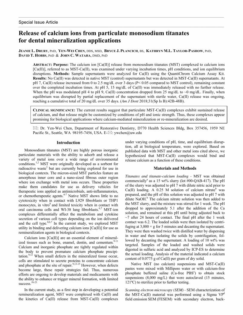
_______________________________________________________________________________________________________________________________________________________________
Special Issue Article _______________________________________________________________________________________________________________________________________________________________
Release of calcium ions from particulate monosodium titanates for dental mineralization applications JEANIE L. DRURY, PHD, YEN-WEI CHEN, DDS, MSD, BRYCE J. PLANCICH, BS, KATHRYN M.L. TAYLOR-PASHOW, PHD, DAVID T. HOBBS, PHD & JOHN C. WATAHA, DMD, PHD
ABSTRACT: Purpose: The calcium ion [Ca(II)] release from monosodium titanates (MST) complexed to calcium ions [Ca(II)], referred to as MST-Ca(II), was examined under varying incubation times, pH conditions, and ion equilibrium disruptions. Methods: Sample supernatants were analyzed for Ca(II) using the QuantiChrom Calcium Assay Kit. Results: No Ca(II) was detected in native MST (control) supernatants but was detected in MST-Ca(II) supernatants. At pH 7, Ca(II) release increased from 0 to 2.5 mg/dL over 3 days (P< 0.05 compared to MST control), remaining constant over the completed incubation times. At pH 5, 15 mg/dL of Ca(II) was immediately released with no further release. When the pH was modulated pH 4 to pH 9, Ca(II) concentration dropped from 25 mg/dL to ~0 mg/dL. Finally, when equilibrium was disrupted by partial replacement of the supernatant with sterile water, Ca(II) release was ongoing, reaching a cumulative total of 20 mg/dL over 35 days. (Am J Dent 2018;31(Sp Is B):42B-48B). CLINICAL SIGNIFICANCE: The current results suggest that particulate MST-Ca(II) complexes exhibit sustained release of calcium, and that release might be customized by conditions of pH and ionic strength. Thus, these complexes appear promising for biological applications where calcium-mediated mineralization or re-mineralization are desired. : Dr. Yen-Wei Chen, Department of Restorative Dentistry, D770 Health Sciences Bldg, Box 357456, 1959 NE Pacific St., Seattle, WA 98195-7456, USA. E-: [email protected]
Introduction
Monosodium titanates (MST) are highly porous inorganic particulate materials with the ability to adsorb and release a variety of metal ions over a wide range of environmental conditions.1,2 MST were originally developed as a sorbent for radioactive waste3 but are currently being explored for use in biological contexts. The micron-sized MST particles feature an amorphous inner core and a nano-sized fibrous outer region where ion exchange with metal ions occurs. These properties make them candidates for use as delivery vehicles for therapeutic ions applied as antimicrobials, anti-inflammatories, or chemotherapeutic agents.4-7 Native MST shows little to no cytotoxicity when in contact with L929 fibroblasts or THP1 monocytes, in vitro8 and limited toxicity when in contact with oral carcinoma cells and WI-38 lung fibroblasts.6,7 MST-ion complexes differentially affect the metabolism and cytokine secretion of various cell types depending on the ion delivered and the cell type.4,6,7 In the current study, we explored MST utility in binding and delivering calcium ions [Ca(II)] for use as remineralization agents in biological contexts. Calcium ions [Ca(II)] are an essential element of mineral-ized tissues such as bone, enamel, dentin, and cementum.9-11 Calcium and inorganic phosphate are tightly regulated within the body to prevent premature calcium phosphate precipi-tation.9,12 When small defects in the mineralized tissue occur, cells are stimulated to secrete proteins to concentrate calcium and phosphate at the site of repair.9,13-15 However, when defects become large, these repair strategies fail. Thus, numerous efforts are ongoing to develop materials and medicaments with the ability to enhance or trigger mineral formation, with limited success.16-25 In the current study, as a first step in developing a potential remineralization agent, MST were complexed with Ca(II) and the kinetics of Ca(II) release from MST-Ca(II) complexes
under varying conditions of pH, time, and equilibrium disrup-tion, all at biological temperature, were explored. Based on published data with MST and other metal ions cited above, we hypothesized that MST-Ca(II) complexes would bind and release calcium as a function of these conditions.
Materials and Methods Titanates and titanate-calcium loading - MST was obtained commerciallya as a 15 wt% slurry (lot #00-QAB-417). The pH of the slurry was adjusted to pH 7 with dilute nitric acid prior to Ca(II) loading. A 0.25 M solution of calcium nitrateb was prepared, and the pH of this solution was adjusted to pH 7 with dilute NaOH.b The calcium nitrate solution was then added to the MST slurry, and the mixture was stirred for 1 week. The pH dropped to approximately 5.1 after the addition of the Ca solution, and remained at this pH until being adjusted back to ~7 after 24 hours of contact. The final pH after the 1 week contact was 6.2. The loaded solids were then isolated by centri-fuging at 3,000 × g for 5 minutes and decanting the supernatant. They were then washed twice with distilled water by dispersing in water and then isolating the solids by centrifugation, fol-lowed by decanting the supernatant. A loading of 10 wt% was targeted. Samples of the loaded and washed solids were digested in sulfuric acid and analyzed by ICP-ES to determine the actual loading. Analysis of the material indicated a calcium content of 0.0775 g of Ca(II) per gram of dry solid. Native MST (no calcium) suspensions and MST-Ca(II) pastes were mixed with Millipore water or with calcium-free phosphate buffered saline (Ca-free PBSc) to obtain stock suspensions (8,000 mg/L) that were autoclaved (15 minutes, 121ºC) to sterilize prior to further testing. Scanning electron microscopy (SEM) - SEM characterization of the MST-Ca(II) material was performed using a Sigma VPd field emission SEM (FESEM) with secondary electron, back-

American Journal of Dentistry, Vol. 31, Special Issue B, November, 2018 Release of calcium ions 43B
Fig. 1. (a) SEM of MST-Ca(II) showing typical particle morphology and size to that of the original MST with no visible evidence of a separate calcium-containing phase precipitated onto the surface of the particles. (b) High magnification SEM of MST-Ca(II), (c) EDS Elemental Map for calcium, and (d) EDS Elemental Map for titanium. Taken together, these provide strong evidence that Ca(II) is exchanged into the crystalline surface of MST, resulting in MST-Ca(II).
scattered electron, and in lens secondary electron detectors. It has imaging capability up to ×500,000. Energy dispersive spectroscopy (EDS) (X-Max 20e) was performed using an X-Max 20 silicon drift detector (SDD) to detect elements greater than atomic number 3 (Z > 3). EDS data and maps were analyzed using the INCA 4.15e data analysis software. Samples of the powdered MST-Ca(II) were mounted in epoxy and either carbon or palladium coated to reduce charging. MST-Ca(II) release of calcium into water - Under sterile conditions, 100 μL aliquots of Millipore water stock suspension were distributed into sterile microtubes and incubated at 37°C, 100% humidity for time points ranging from 0 hours to 8 weeks depending on the trial (n= 3-6). During incubation, each sample was vortexed once a week to redistribute settled particles. Supernatant and particulates were collected by removing aliquots from incubation and vortexing for 15 seconds. Aliquots were then centrifuged for 30 seconds and the top 50 μL of supernatant were pipetted and transferred to new microtubes. The remaining MST and MST-Ca(II) suspensions (particulates) were also reserved. All samples were stored at 4°C until analy-sis (section 2.7). MST-Ca(II) release of calcium into Ca-free PBS - Under sterile conditions, 100 μL aliquots of Ca-free PBS stock suspensions were distributed into sterile microtubes and incubated at 37°C, 100% humidity for time points ranging from 0 hours to 28 days (n= 3). During incubation, each sample was vortexed once a week to redistribute settled particles. Supernatant and particu-
lates were collected by removing aliquots from incubation and vortexing for 15 seconds. Aliquots were then centrifuged for 30 seconds and the top 50 μL of supernatant were pipetted and transferred to new microtubes. The remaining MST and MST-Ca(II) suspensions (particulates) were separately reserved. All samples were stored at 4°C until analysis (section 2.7). Effect of equilibrium disruption on calcium release - Under sterile conditions, 100 μL aliquots of Millipore water stock suspensions were distributed into sterile microtubes and incubated at 37°C, 100% humidity (n= 3-6). Supernatant samples were collected as described in Section 2.3. Following supernatant collection, new Millipore water (sterile, 50 µL) was added to each aliquot. The aliquots were then vortexed for 15 seconds to redistribute the particles and placed back into incubation. Supernatant collection and water exchange took place every 24 hours (n= 3-6 supernatant samples per day, days 0-35), and particulates were collected every 7 days (n= 3). All supernatant and particulate samples were stored at 4°C for later analysis. Effect of pH on calcium release - To determine the effect of pH on MST-Ca(II) release of calcium ions, the procedures outlined in Section 2.1 and 2.3 were altered as follows. Prior to the sterilization of the Millipore water stock suspensions, the pH of the suspensions were adjusted using 1.0 N HCl or 1.0 N NaOH (reagents from Sigma Aldrich) until a pH of 4 to 9 was obtained (pH meter, MP220f). Supernatant and particulate samples were collected (n=3) for each stock suspension (pH 4,

44B Drury et al
American Journal of Dentistry, Vol. 31, Special Issue B, November, 2018
Fig. 2. Ca(II) release into sterile water from MST-Ca(II) during 21 and 56 days of incubation. Ca(II) increased during the initial 3 days of incubation before reaching an equilibrium level of ~ 2.5 mg/dL. No Ca(II) was measured in supernatant samples collected from incubated MST. Ca(II) release from MST-Ca was significantly greater than Ca(II) released from MST (Differences indicated by A, B; P< 0.05, n= 3).
pH 5, pH 6, pH 7, pH 8, and pH 9) and assayed for Ca(II) content (section 2.7). MST and MST-Ca(II) stock suspensions at pH 5 were then sterilized and tested for calcium release following the procedure outlined in Section 2.3, for time points ranging from 0 hours to 7 days (n= 3). Measurement of calcium - QuantiChrom Calcium Assay Kitg (DICA-500) was used according to manufacturer’s protocol to measure concentrations of calcium ions. This assay was chosen because it was indicated for detecting calcium in water based and biologically based samples, because of the detection range of the assay, and because of the simplicity of the assay protocol. To assess supernatant samples, 5 μL of each sample or standard were plated in 96-well plates in triplicate. Standards were generated (0-20 mg/dL) by diluting a 20 mg/dL standard solution (provided, BioAssay Systems) with Millipore water. Following plating, 200 μL of the mixed test reagent were added to each well. The plate was incubated at room temperature for 3 minutes and a SpectraMax M2h plate reader was used to determine the optical density (OD) of the standards and samples at 612 nm. The procedure to measure the amount of calcium on the particulates was altered slightly because of known OD inter-ference by MST7. To mitigate particulate interference, particulate samples were reacted with test reagents in a round-bottom 96-well plate. The plates were then centrifuged and 100 μL of developed assay solution, free of particulates (samples and standards), were transferred to new 96-well plate and the OD assessed (612 nm). All optical densities were converted to calcium concen-trations in mg/dL following generation of a standard linear curve to the known standards. Statistical significance and differences were assessed utilizing a Student t-test (α-= 0.05).
Results MST was reacted with a calcium nitrate solution under conditions such that 100% of the theoretically available sodium was exchanged for calcium ions (equivalent to 10 wt% Ca). Elemental analysis indicated a calcium content of 0.0775 g of Ca(II) per g of dry solid. SEM (Fig. 1a) revealed the calcium-exchanged MST had the same particle size, shape, and morphology of the native MST material and no evidence of precipitated calcium-containing phase on the surface of or otherwise present in the material. Higher magnification in conjunction with elemental mapping (Figs. 1b - d) revealed that both calcium (Fig. 1c) and titanium (Fig. 1d) are evenly distributed over the particles with no regions where calcium is present without titanium. This suggests that the Ca(II) has been incorporated into the MST structural framework by exchange of Ca(II) for sodium ions. If Ca(II) were precipitated, the surface details and spaces between particulates would not have been visible. Following confirmation that calcium precipitation was not occurring, samples were tested for their ability to release the loaded Ca(II) ions. MST-Ca(II) and native MST were incubated in water in two separate trials spanning 21 and 56 days. For both trials, the initial Ca(II) concentration was 0 mg/dL for both MST and MST-Ca(II). The MST-Ca(II) supernatant Ca(II) concentration increased over Days 1-3, reaching a steady-state concentration of ~2.5 mg/dL (Fig. 2). Within statistical error, this concen-tration was maintained over the remaining duration of the 21-day and 56-day time periods of the respective studies. No Ca(II) was detected in native MST supernatants during either trial (Fig. 2). Under biological conditions, the system would presumably be dynamic in that the biological fluids in contact with the MST-Ca(II) would be changing, thus we sought to determine if
Drury Figures 2-6
Fig. 2. Ca(II) release into sterile water from MST-Ca(II) during 21 and 56 days of incubation. Ca(II) increased during the initial 3 days of incubation before reaching an equilibrium level of ~ 2.5 mg/dL. No Ca(II) was measured in supernatant samples collected from incubated MST. Ca(II) release from MST-Ca was significantly greater than Ca(II) released from MST (Differences indicated by A, B; P< 0.05, n= 3).
Fig. 3. Daily replacement of 50% of the volume of supernatant over MST-Ca(II) particulates with water. Y-axis indicates the cumulative Ca(II) detected in supernatants. Ca(II) was released by the MST-Ca(II) following each replacement, re-establishing equilibrium. Release continued over 35 days of testing.

American Journal of Dentistry, Vol. 31, Special Issue B, November, 2018 Fig. 3. Daily replacement of 50% of the volume of supernatant over MST-Ca(II) particulates with water. Y-axis indicates the cumulative Ca(II) detected in supernatants. Ca(II) was released by the MST-Ca(II) following each replacement, re-establishing equilibrium. Release continued over 35 days of testing. Fig. 4. Ca(II) release into Ca-free PBS from MST-Ca(II) during 28 days of incubation. Ca(II) release was immediate with an average equilibrium of 1.88 mg/dL maintained over the 28 days of incubation. additional Ca(II) would be released under controlled alterations in conditions. Upon daily disruption of steady-state, Ca(II) was released and measured to range from 0 - 3.0 mg/dL per day. The cumulative Ca(II) released was calculated and plotted against time (Fig. 3). After 35 days, the cumulative Ca(II) released reached 20 mg/dL. At no time was any Ca(II) detected in MST control samples. In addition to being dynamic, biological conditions are also ionically complex, thus Ca(II) release from MST-Ca(II) was studied with Ca-free PBS as the bathing solution. Ca(II) release into Ca-free PBS was immediate and attained an average steady-state of 1.88 mg/dL (Fig. 4). This steady-state was maintained over 28 days in incubation. Ca(II) release from MST-Ca(II) was found to be highly dependent on sample pH. Adjustments to pH resulted in imme-
Release of calcium ions 45B Fig. 5. (a) Short-term (< 5 minutes) Ca(II) release from MST-Ca(II): dependence on pH (pH 4 to 9). The amount of Ca(II) detected ranged from 25.4 mg/dL at pH 4 to ~0 mg/dL at pH 9. There was a nearly exponential relationship between pH and calcium concentration (black fitted line). * denotes statistically significant release (P< 0.001). (b) Ca(II) measured on MST-Ca(II) particulates (34.4 mg/dL) following release. There was no significant difference in Ca(II) detected on the particulates, suggesting a large reservoir of Ca(II) available for release that exceeded the ability of the assay to detect all the calcium on these particles. diate (less than 5 minutes) release of Ca(II) into the supernatant. The quantity of Ca(II) released decreased from 25.7 mg/dL at pH 4 to 0.41 mg/dL at pH 9 (Fig. 5a). The pH versus Ca(II) concentration data had an exponential association: [Ca] = 81.5*exp(-0.988*pH). Despite the pH-dependent calcium concentration in the supernatant, the Ca(II) measured on the particulates was statistically constant and in excess of the standards (Fig. 5b), suggesting a large remaining reservoir of Ca(II) on the MST-Ca(II) particles with saturation of the assay. When samples of pH = 5 were incubated for a period of 7 days (Fig. 6), an initial, steady-state Ca(II) concentration of 15 mg/dL was attained and remained statistically constant over the 7-day trial. Ca(II) steady-state at pH 5 was significantly greater
Drury Figures 2-6
Fig. 2. Ca(II) release into sterile water from MST-Ca(II) during 21 and 56 days of incubation. Ca(II) increased during the initial 3 days of incubation before reaching an equilibrium level of ~ 2.5 mg/dL. No Ca(II) was measured in supernatant samples collected from incubated MST. Ca(II) release from MST-Ca was significantly greater than Ca(II) released from MST (Differences indicated by A, B; P< 0.05, n= 3).
Fig. 3. Daily replacement of 50% of the volume of supernatant over MST-Ca(II) particulates with water. Y-axis indicates the cumulative Ca(II) detected in supernatants. Ca(II) was released by the MST-Ca(II) following each replacement, re-establishing equilibrium. Release continued over 35 days of testing.
Fig. 4. Ca(II) release into Ca-free PBS from MST-Ca(II) during 28 days of incubation. Ca(II) release was immediate with an average equilibrium of 1.88 mg/dL maintained over the 28 days of incubation.
Fig. 4. Ca(II) release into Ca-free PBS from MST-Ca(II) during 28 days of incubation. Ca(II) release was immediate with an average equilibrium of 1.88 mg/dL maintained over the 28 days of incubation.
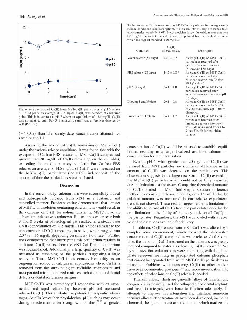
46B Drury et al Fig. 6. 7-day release of Ca(II) from MST-Ca(II) particulates at pH 5 versus pH 7. At pH 5, an average of ~15 mg/dL Ca(II) was detected at each time point. This is in contrast to pH 7 where an equilibrium of ~2.5 mg/dL Ca(II) was not attained until Day 3. Statistically significant differences denoted by A,B (P< 0.05). (P< 0.05) than the steady-state concentration attained for samples at pH 7. Assessing the amount of Ca(II) remaining on MST-Ca(II) under the various release conditions, it was found that with the exception of Ca-free PBS release, all MST-Ca(II) samples had greater than 20 mg/dL of Ca(II) remaining on them (Table), exceeding the maximum assay standard. For Ca-free PBS release, an average of 14.5 mg/dL of Ca(II) were measured on the MST-Ca(II) particulates (P< 0.05), independent of the amount of time the particulates were incubated.
Discussion In the current study, calcium ions were successfully loaded and subsequently released from MST in a sustained and controlled manner. Previous testing demonstrated that contact of MST with a solution containing calcium ions would result in the exchange of Ca(II) for sodium ions in the MST;2 however, subsequent release was unknown. Release into water over both 3 and 8 weeks at physiological pH resulted in a steady-state Ca(II) concentration of ~2.5 mg/dL. This value is similar to the concentration of Ca(II) measured in saliva, which ranges from 2.07 to 4.16 mg/dL depending on salivary flow rate.26 Further tests demonstrated that interrupting this equilibrium resulted in additional Ca(II) release from the MST-Ca(II) until equilibrium was reestablished. Additionally, a large quantity of Ca(II) was measured as remaining on the particles, suggesting a large reservoir. Thus, MST-Ca(II) has conceivable utility as an ongoing ion source of calcium in applications where Ca(II) is removed from the surrounding microfluidic environment and incorporated into mineralized matrices such as bone and dental defects or dental restoration margins. MST-Ca(II) was extremely pH responsive with an expo-nential and rapid relationship between pH and measured released Ca(II). This attribute has potential therapeutic advan-tages. At pHs lower than physiological pH, such as may occur during infection or under overgrown biofilms;27-29 a greater
American Journal of Dentistry, Vol. 31, Special Issue B, November, 2018 Table. Average Ca(II) measured on MST-Ca(II) particles following various release conditions (see description). * indicates statistically difference from other samples tested (P< 0.05). Note: precision is low for calcium concentrations >20 mg/dL because these values are extrapolated from a standard curve in which the highest standard is 20 mg/dL. ____________________________________________________________________________________________________
Ca(II) Condition (mg/dL) ± SD Description ____________________________________________________________________________________________________
Water release (56 days) 44.0 ± 2.2 Average Ca(II) on MST-Ca(II) particulates reserved after extended release into water (21 days and 56 days) PBS release (28 days) 14.5 ± 0.8 * Average Ca(II) on MST-Ca(II) particulates reserved after extended release into Ca-free PBS (28 days) pH 5 (7 days) 36.1 ± 1.0 Average Ca(II) on MST-Ca(II) particulate reserved after extended release in water at pH 5 (7 days) Disrupted equilibrium 29.1 ± 0.8 Average Ca(II) on MST-Ca(II) particulates reserved after 35 days release, daily equilibrium disruption Immediate pH release 34.4 ± 1.7 Average Ca(II) on MST-Ca(II) particulates reserved after immediate release into water when pH was varied from 4 to 9 (see Fig. 5b for individual values). ____________________________________________________________________________________________________
concentration of Ca(II) would be released to establish equili-brium, resulting in a large localized available calcium ion concentration for remineralization. Even at pH 4, when greater than 20 mg/dL of Ca(II) was released from MST particles, no significant difference in the amount of Ca(II) was detected on the particulates. This observation suggests that a large reservoir of Ca(II) existed on the MST-Ca(II) particles which could not be fully measured due to limitations of the assay. Comparing theoretical amounts of Ca(II) loaded on MST (utilizing a solution difference method) to measured calcium amounts, only 1/3 of the loaded calcium amount was measured in our release experiments (results not shown). These results suggest either a limitation in the ability to release all Ca(II) from the particulates once loaded or a limitation in the ability of the assay to detect all Ca(II) on the particulates. Regardless, the MST was loaded with a reser-voir of calcium ions available for delivery. In addition, Ca(II) release from MST-Ca(II) was altered by a complex ionic environment, which reduced the steady-state concentration of Ca(II) compared to water release. At the same time, the amount of Ca(II) measured on the materials was greatly reduced compared to materials releasing Ca(II) into water. We hypothesize that calcium ions were interacting with the phos-phate reservoir resulting in precipitated calcium phosphate that cannot be separated from white MST-Ca(II) particulates or measured. Problems with measuring Ca(II) in other buffers have been documented previously30 and more investigation into the effects of other ions on Ca(II) release is needed. Titanium alloys, which are generally alloys of titanium and oxygen, are extensively used for orthopedic and dental implants and need to integrate with bone to function adequately. In attempts to improve this integration and interface, numerous titanium alloy surface treatments have been developed, including chemical, heat, and micro-arc treatments which oxidize the
Fig. 5. (a) Short-term (< 5 minutes) Ca(II) release from MST-Ca(II): dependence on pH (pH 4 to 9). The amount of Ca(II) detected ranged from 25.4 mg/dL at pH 4 to ~0 mg/dL at pH 9. There was a nearly exponential relationship between pH and calcium concentration (black fitted line). * denotes statistically significant release (P< 0.001). (b) Ca(II) measured on MST-Ca(II) particulates (34.4 mg/dL) following release. There was no significant difference in Ca(II) detected on the particulates, suggesting a large reservoir of Ca(II) available for release that exceeded the ability of the assay to detect all the calcium on these particles.
Fig. 6. 7-day release of Ca(II) from MST-Ca(II) particulates at pH 5 versus pH 7. At pH 5, an average of ~15 mg/dL Ca(II) was detected at each time point. This is in contrast to pH 7 where an equilibrium of ~2.5 mg/dL Ca(II) was not attained until Day 3. Statistically significant differences denoted by A,B (P< 0.05).

American Journal of Dentistry, Vol. 31, Special Issue B, November, 2018 titanium, resulting in a titanate surface.31-33 In this context, titanate is a general term used to describe oxides of titanium on the alloy surfaces. Calcium also has been integrated into these treated surfaces resulting in a layer commonly referred to as calcium titanate.32-34 However, studies suggest that little calcium is released from the calcium-titanate alloy surfaces.35 Treated titanium alloy surfaces are fundamentally different than the MST and MST-Ca(II) particulates used in the current study. MST particulates have, by design and synthesis, highly crystalline surfaces with substantial surface area designed for ion exchange. Unlike titanium alloy surfaces treated with calcium, we have shown in the current work that MST-Ca(II) releases Ca(II); this result is consistent with previous studies that have reported release of other ions under physiological conditions.4-7 The release of calcium ions from MST-Ca(II) complexes offers several potential therapeutic applications. MST particulates also have similarities to titanium dioxide nanoparticles (anatase). Both MST and titanium dioxide nanoparticles are oxides of titanium, however MST is larger than anatase (on the order of microns versus nanometers, respectively). By definition anatase is crystalline whereas MST has an amorphous core with a well-defined crystalline surface.4 Both materials have been shown to successfully deliver metals and chemotherapeutic agents in biological settings.36,37 How-ever, no literature has reported that calcium ions have been delivered from titanium dioxide nanoparticles. Overall, we demonstrated that Ca(II) can be exchanged onto MST (denoted as MST-Ca(II)) and subsequently released into solution in a controlled and sustained manner. In the future, we intend to investigate the biocompatibility and mineralization capabilities of this material in biological contexts as a prelude to therapeutic applications. a. Optima Chemical Group, LLC, Douglas, GA, USA. b. Sigma-Aldrich, St. Louis, MO, USA. c. Life Technologies, Carlsbad, CA, USA. d. Carl Zeiss Microscopy LLC, Thornwood, NY, USA. e. Oxford Instruments, Abingdon, Oxfordshire, UK. f. Mettler-Toledo, Columbus, OH, USA. g. BioAssay Systems, Hayward, CA, USA. h. Molecular Devices, Sunnyvale, CA, USA. Acknowledgements: To the Spencer Endowment at the University of Washington, School of Dentistry for their support of this work. Disclosure statement: The authors declared no conflict of interest. Dr. Drury is Research Scientist, Dr. Chen is Assistant Professor, Mr. Plancich is a senior dental student, Dr. Wataha is Professor, Department of Restorative Dentistry, School of Dentistry, University of Washington, Seattle, Washington, USA. Dr. Taylor-Pashow is the LDRD Program Manager, and Dr. Hobbs is Senior Advisory Scientist, Savannah River National Laboratory, Aiken, South Carolina, USA.
References 1. Hobbs, DT, Barnes, MJ, Pulmano, RL, Marshall, KM, Edwards TB,
Bronikowski MG, Fink, SD. Strontium and actinide separation from high-level nuclear waste solutions using monosodium titanate 1. Simulant testing. Separation Sci Technol 2005;40:3093-3111.
2. Elvington MR, Click DR, Hobbs DT. Sorption behavior of monosodium titanate and amorphous peroxotitanate materials under weakly acidic conditions. Separation Sci & Technol 2009;45:66-72.
3. Lynch RW, Dosch RG, Kenna BT, Johnstone JK, Nowak EJ. The Sandia solidification process - A broad range aqueous waste solidification method. IAEA 1976;207:361-373.
4. Hobbs DT, Messer RLW, Lewis JB, Click DR, Lockwood PE, Wataha JC. Adsorption of biometals to monosodium titanate in biological environ-
Release of calcium ions 47B ments. J Biomed Mater Res Part B 2006;78:296-301. 5. Chung WO, Wataha JC, Hobbs DT, An J, Wong JJ, Park CH, Dogan S,
Elvington MC, Rutherford RB. Peroxotitanate- and monosodium metal-titanate compounds as inhibitors of bacterial growth. J Biomed Mater Res Part A 2011;97:348-354.
6. Drury JL, Chen Y-W, Wong JJ, Elvington MC, Rutherford RB, Hobbs DT, Wataha JC. Titanates deliver metal compounds to suppress cell metabolism. J Exp Clin Med 2014; 6:21-27.
7. Drury JL, Jang Y, Taylor-Pashow KML, Elvington M, Hobbs DT, Wataha JC. In vitro biological response of micro- and nano-sized monosodium titanates and titanate-metal compounds. J Biomed Mater Res Part B 2015;103B:254-260.
8. Davis RR, Lockwood PE, Hobbs DT, Messer RLW, Price RJ, Lewis JB, Wataha JC. In vitro biological effects of sodium titanate materials. JBiomed Mat Res B 2007;83B:505-511.
9. Kawasaki K, Buchanan AV, Weiss KM. Biomineralization in humans: Making the hard choices in life. Annu Rev Genet 2009;43:119-142.
10. Veis A, Dorvee JR. Biomineralization mechanisms: A new paradigm for crystal nucleation in organic matrices. Calcif Tissue Int 2013; 93:307-315.
11. Kardos TB. Cellular responses to metal ions released from implants. J Oral Implantol 2014;40:294-298.
12. Bonjour J-P. Calcium and phosphate: A duet of ions playing for bone health. J Am Coll Nutrition 2011;30:438S-448S.
13. Hao J, Ramachandran A, George A. Temporal and spatial localization of the dentin matrix proteins during dentin biomineralization. J Histochem Cytochem 2009;57:227-237.
14. Cameron JA, Milner DJ, Lee JS, Cheng J, Fang NX, Jasiuk IM. Employing the biology of successful fracture repair to heal critical size bone defects. Curr Top Microbiol Immunol 2013;367:113-132.
15. Gorski JP. Biomineralization of bone: A fresh view of the roles of non-collagenous proteins. Front Biosci 2015;16:2598-2621.
16. Chen, L, Shen H, Suh, BI. Bioactive dental restorative materials: A review. Am J Dent 2013;26:219-227.
17. Skrtic D and Antonucci JM. Bioactive polymeric composites for tooth mineral regeneration: Physicochemical and cellular aspects. J Funct Biomater 2011;2:271-307.
18. Cao Y, Song M, Kim E, Shon W, Chugal N, Bogen G, Lin L, Kim RH, Park N-H, and Kang MK. Pulp-dentin regeneration: Current state and future prospects. J Dent Res 2015;1-8.
19. Khan Y, Yaszemski MJ, Mikos AJ, Laurencin CT. Tissue engineering of bone: Material and matrix considerations. J Bone Joint Surg Am 2008;90(Suppl 1):36-42.
20. Pérez-Sánchez MJ, Ramírez-Glindon E, Lledó-Gil M, Calvo-Guirado JL, Pérez-Sánchez C. Biomaterials for bone regeneration. Med Oral Patol Oral Cir Bucal 2010;15:e517-e522.
21. Ginebra M-P, Canal C, Espanol M, Pastorino D, Montufar EB. Calcium phosphate cements as drug delivery materials. Adv Drug Delivery Rev 2012;64:1090-1110.
22. Hoppe A, Güldal NS, Boccaccini AR. A review of the biological response to ionic dissolution products from bioactive glasses and glass-ceramics. Biomaterials 2011; 32: 2757-2774.
23. Chai YC, Carlier A, Bolander J, Roberts SJ, Geris L, Schrooten J, Van Oosterwyck H, Luyten FP. Current views on calcium phosphate osteogenicity and the translation into effective bone regeneration strategies. Acta Biomaterialia 2012;8:3876-3887.
24. Cochrane NJ, Shen P, Yuan Y, Reynolds EC. Ion release from calcium and fluoride containing dental varnishes. Austr Dent J 2014;59:100-105.
25. Deb S, Chana S. Biomaterials in relation to dentistry. Front Oral Biol 2015;17:1-12.
26. Shannon IL, Suddick RP, and Dowd FJ. Saliva: Composition and secretion. In: Monographs in Oral Science, Vol 2. Basel, Switzerland: S. Karger AG. 1974;1-103.
27. Marsh PD. Microbial ecology of dental plaque and its significance in health and disease. Adv Dent Res 1994;8:263-271.
28. Costalonga M and Herzberg MC. The oral microbiome and the immunobiology of periodontal disease and caries. Immunology Letters 2014;162:22-38.
29. Kianoush N, Adler CJ, Nguyen KA, Browne GV, Simonian M, Hunter N. Bacterial profile of dentine caries and the impact of pH on bacterial population diversity. PLoS One 2014; 9(3):e92940.
30. Crowell JA and Bowers GN. Apparent binding of ionized calcium by various buffers. Clin Chem 1985;31:267-280.
31. Kawanabe K, Ise K, Goto K, Akiyama H, Nakamura T, Kaneuji A, Sugimori T, Matsumoto T. A new cementless total hip arthroplasty with bioactive titanium porous-coating by alkaline and heat treatment: Average 4.8-year

48B Drury et al results. Biomed Mater Res Part B: Appl Biomater 2009;90B: 476-481. 32. Kang B-S, Sul Y-T, Johansson CB, Oh S-J, Lee H-J, Albrektsson T. The
effect of calcium ion concentration on the bone response to oxidized titanium implants. Clin Oral Impl Res 2012;23:690–697.
33. Kaluđerović MR, Schreckenbach JP, Graf H-L. First titanium dental implants with white surfaces: Preparation and in vitro tests. Dent Mater 2014;30:759-768.
34. Kizuki T, Takadama H, Matsushita T, Nakamura T, Kokubo T. Preparation of bioactive Ti metal surface enriched with calcium ions by chemical treat-
American Journal of Dentistry, Vol. 31, Special Issue B, November, 2018 ment. Acta Biomaterialia 2010; 6:2836–2842. 35. Yamaguchi S, Nath S, Matsushita T, Kokubo T. Controlled release of
strontium ions from a bioactive Ti metal with a Ca-enriched surface layer. Acta Biomaterialia 2014; 10:2282-2289.
36. Chen Y-W, Drury JL, Chung WO, Hobbs DT and Wataha JC. Titanates and titanate-metal compounds in biological contexts. Int J Med Nano Res 2015;2:1. Pii009.
37. Yin ZF, Wu L, Yang HG, Su YH. Recent progress in biomedical appli-cations of titanium dioxide. Phys Chem Chem Phys 2013;15:4844-4858.