Aiyush Bansal Sandra Escandor - McMaster Universityibruce/courses/EE3BA3_2008/EE3BA3_2007... ·...
36
Aiyush Bansal Sandra Escandor
Transcript of Aiyush Bansal Sandra Escandor - McMaster Universityibruce/courses/EE3BA3_2008/EE3BA3_2007... ·...

Aiyush
BansalSandra Escandor
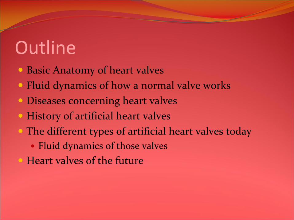
OutlineBasic Anatomy of heart valvesFluid dynamics of how a normal valve worksDiseases concerning heart valvesHistory of artificial heart valvesThe different types of artificial heart valves today
Fluid dynamics of those valvesHeart valves of the future

The flow of bloodRight Side
Right atrium ‐> tricuspid‐> right ventricle ‐> pulmonic valve ‐>pulmonary arteries
Left SideLung ‐> left atrium ‐> mitral valve ‐> left ventricle ‐> aortic valve ‐> aorta
Thrombogenesis – the creation of blood clotsEmbolism – movement of those blood clots
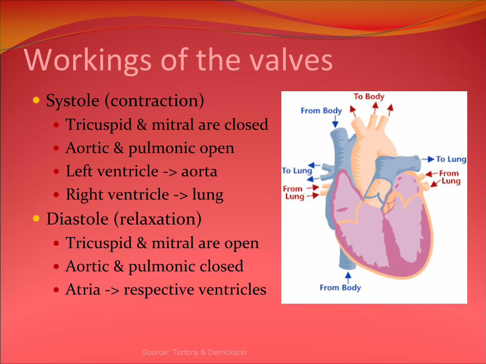
Workings of the valvesSystole (contraction)
Tricuspid & mitral are closedAortic & pulmonic openLeft ventricle ‐> aortaRight ventricle ‐> lung
Diastole (relaxation)Tricuspid & mitral are openAortic & pulmonic closedAtria ‐> respective ventricles
Source: Tortora
& Derrickson

Fluid Dynamics “Basics”Fluid flows from areas of high pressure to areas of low pressureHeart valves open and close in response to pressure gradients
valves open when pressure in the preceding chamber is higher andclose when the gradient reversesThese one‐way valves are important in ensuring that the blood flows in the proper direction
Central flow – blood moves through the center of the valveTurbulence – fluid flowing randomlyShear stress – force exerted by the movement of blood on walls (stress exerted parallel)
5

Fluid mechanics cont’d
Goal:minimal pressure drops=>minimize QLower R=>higher Q=>more turbulence=>more clots
Q= ∆p /RR~1/r^4
Q –
flow rateR –
resistance to flow of blood∆p -
pressure differenceP1 P2
∆p=P1 -
P2 > 0
Q
What??
r

Did you know?Valvular
heart disease is a life‐threatening disease
that afflicts millions of people worldwide and leads to approximately 250,000 valve repairs and
replacements each year.

Source: http://www.sjm.com/conditions/condition.aspx?na
me=Heart+Valve+Disease
Heart Valve DiseasesStenosis – does not fully open
If affecting mitral valve, left ventricle will not receive as much blood =>less oxygenated blood is supplied by heart If affecting tricuspid valve, right atrium will not receive as much blood =>less blood becomes oxygenated
Incompetence – does not fully close
If affecting mitral valve, left ventricle will back up blood to left atrium If affecting tricuspid valve, right ventricle will back up blood to right atrium

Heart Valve DiseasesName Description Endocarditis Occurs when germs (i.e. from another part
of your body) enter blood stream and infect damaged areas of heart. If untreated, may destroy heart valves – life threatening
Calcific degeneration Buildup of calcium in mitral or aortic valves => causes valves to thicken
Regurgitation Blood is leaking backwards because the valve does not close properly
Source: http://www.sjm.com/procedures/procedure.
aspx?name=Heart+Valve+Replacement
Heart valve repairVideo break!! Video about mitral clip.http://youtube.com/watch?v=F0aj6TkQbxc

1952
–
The first valve, the
Hufnagel
valve was developed and
was made out of plexiglass
and
silcone‐coated nylon. Later, it was
implanted; patient lived for 14
hours.
Problem:
•High rate of blood clot formation
•Sounded like a ticking time bomb•Lack of central flow => causes
turbulence => creates blood clots
1961
–
Starr‐Edwards Ball and
Cage is developed. Only 6/8
patients lived.
Problem:•Initial design was susceptible to
creating blood clots!•Cloth covered version induces
blood clots since cloth tears
Upside: Overall reduction in
rate of embolization, but
permanent blood thinner
therapy was needed.
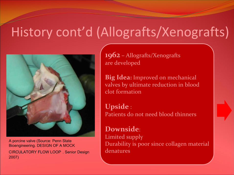
History cont’d (Allografts/Xenografts)
1962
–
Allografts/Xenograftsare developed
Big Idea: Improved on mechanical
valves by ultimate reduction in blood
clot formation
Upside
:
Patients do not need blood thinners
Downside:Limited supplyDurability is poor since collagen material
denatures
A porcine valve (Source: Penn State Bioengineering. DESIGN OF A MOCK
CIRCULATORY FLOW LOOP
. Senior Design 2007)

Late 1960s –Bioprosthetic
valves (hybrids) also
developed
Big Idea: Improved on the cage and ball design by
removing the restriction of central flow of blood
•Reduced turbulence•Reduced resistance (in forward flow direction)•Reduced shear stress
History cont’d

Carpentier
did this:
1.Washed porcine (pig) aortic valve with Hank's solution and
an oxidizing agent =>hides valve's antigenic components 2.Treated porcine aortic valve with glutaraldehyde
=> prevents
collagen denaturing by stabilizing cross‐links3.Combined the treated valve with a fabric covered metal
frame => keeps the three dimensional shape of the valve; easy
to implant
Upside: •No Need for blood thinners!•Central flow achieved
Downside:•Valves have a harder time opening due to stiffness•Calcium deposits easily on valves due to preservative used =>
strokes are more prevalent since calcium deposits chip off
History cont’d (The Bioprosthesis)

Late 1960s –
Tilting disc is discovered. Also pyrolytic
carbon is discovered by the space program
Big Idea: Improved on the cage and ball design by
removing the restriction of central flow of bloodReduced turbulenceReduced resistance (in forward flow direction)Reduced shear stress
Upside: Need for blood thinners was greatly reduced
Downside:blood clots formation was not totally eliminated

1976
‐
St. Jude’s introduces the bileaflet
valve. Today, this design is more than 25
years old
Big Idea: Improved on the tilting disk
design by removing the restriction of central
flow of blood even more than the tilting
disk!
Upside: •Central flow achieved!
Downside:•Still need blood thinners for life•More regurgitation than tilting disk
History cont’d (Bileaflet)
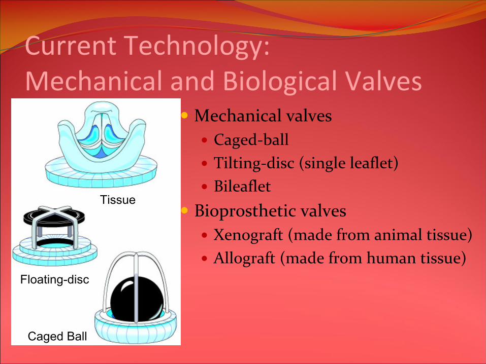
Current Technology: Mechanical and Biological Valves
Mechanical valvesCaged‐ballTilting‐disc (single leaflet)Bileaflet
Bioprosthetic valvesXenograft (made from animal tissue)Allograft (made from human tissue)
Caged Ball
Floating-disc
Tissue

18
Ideal heart valve requirements:
Produce minimal pressure dropsA larger pressure drop on a prosthetic valve means a even larger pressure from the ventricle to drive the flow of blood through.
Small regurgitation volumes (reverse flow) Equal to the sum of the closing volume and leakage volume. The closing volume is the volume of retrograde flow through the valve during valve closure. Leakage volume is any fluid volume accumulation after valve closure.
Minimize turbulence and high shear stresses Not create stagnation or flow separation regions in the flow fields.
Flow cycle divided into forward flow, closing volume, and leakage volume
Fluid Mechanics of heart valves

Caged‐ball valvePros
Very little regurgitationCons
Ball blocks central flow i.e. blood has to move around the ball.Increases collisions of blood cells => blood clotsThe weight and wear resistance of the occluderaffect the opening and closing of the valve

Fluid Mechanics of Caged‐BallMaximum velocity of 2.2 m/s at peak systolePeak reverse velocities in the wake region can be as high as 0.25 m/sRegurgitation volumes are around 5ml/beatTurbulent shear stresses are around 1850 – 3500 dynes/cm2
20
Velocity during systole(cm/s)
Turbulent shear stresses(dynes/cm2)

Tilting‐disc (single leaflet)Pros
Uses a tilting occluder disk to better mimic natural flow patterns through the heartTilting pattern allows more central flow while still preventing backflow
ConsSome damage still occurs to blood cellsReduces thrombosis and infection, but does not eliminate either problem

Fluid Mechanics of Tilting discHas major and minor orifices divided by a tilting disc.The peak velocities are 2.1 m/sand 2 m/s in the major and minor orifice regions, respectivelyMaximum reverse velocity of 0.25 m/s in the minor orifice.Regurgitation volumes are around 9 ml/beatTurbulent shear stresses are 1200‐1500 dynes/cm2
22
Velocity during systole(cm/s)
Turbulent shear stresses(dynes/cm2)
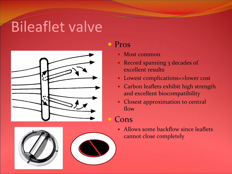
Bileaflet valve
ProsMost commonRecord spanning 3 decades of excellent results Lowest complications=>lower costCarbon leaflets exhibit high strength and excellent biocompatibilityClosest approximation to central flow
ConsAllows some backflow since leaflets cannot close completely

Fluid mechanics of bileaflet
Two semicircular leaflets divide into three regions: two lateral orifices and a central orificeTurbulent shear stress levels exceeding 1150‐1700 dynes/cm2.The lateral and central orifice jets reach maximum velocities of 2.2 m/s and 2 m/s, respectivelyRegurgitation volume >9 ml/beat
Velocity during systole(cm/s)
Turbulent shear stresses(dynes/cm2)

Summary of Mechanical ValvesLeast Turbulence
Bileaflet>>Tilting disc>>Caged ballLeast Regurgitation
Caged ball>>Tilting disc>>Bileaflet

Animal tissue valvesHeterograft/Xenograft – animal to humanPorcine valves – from pig
Has good durability and and good hemodynamicsMaterials: Porcine valve tissue, Elgiloy(cobalt‐nickel alloy), sewing ring‐knitted Teflon
Bovine pericardial valves – from cowLasts as long as standard porcine valves at 10 yearsThe pericardial valve has excellent hemodynamics, and has gained a large market share (about 40% of US tissue valves) in this group of patients
Biggest problem is biocompatibility between animal and human tissue i.e. the immune response of the human system

Fluid mechanics of tissue valves
Bovine pericardial valves Porcine valves
Bovine valves are better hemodynamically
because there’s less resistance to flow
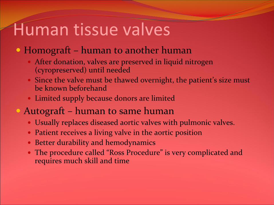
Human tissue valvesHomograft – human to another human
After donation, valves are preserved in liquid nitrogen (cyropreserved) until neededSince the valve must be thawed overnight, the patient’s size must be known beforehandLimited supply because donors are limited
Autograft – human to same humanUsually replaces diseased aortic valves with pulmonic valves.Patient receives a living valve in the aortic positionBetter durability and hemodynamicsThe procedure called “Ross Procedure” is very complicated and requires much skill and time

Animal VS Human Tissue Valves
Animal valves Less biocompatibleMore supply
Human valvesMore biocompatibleLess supplyMore complicated procedure (autograft)
30
Mechanical Vs Tissue ValvesMechanical valves
ProsExtremely durable. The struts and occluders are made out of either pyrolytic carbon or titanium coated with pyrolytic carbon, and the sewing ring cuff is Teflon, polyester or dacronTypically last about 20 ‐ 30 yearsPreferred choice for children, teens, and adults age 60 and younger
ConsThe hard mechanical tissue of the valve causes blood cells to tear as they pass through, causing clots to form.People must constantly take anticoagulants
Biological valvesPros
Typically last about 10 ‐ 15 yearsVirtually no regurgitation volumeMuch lower risk of blood clotting than mechanical valvesBetter for people who are older than 60 and cannot take anticoagulants
ConsCan also tear or become infectedMay fail for the same reason the original one did

Heart valves of the future!!!Engineers are still trying to make better artificial heart valves using better materials and designs.Stem cells may provide the answer!
Scientists for the first time have now grown human heart valves using stem cells from the amniotic fluid that cushions babies in the womb.The world’s leading surgeon, Sir Magdi found a way to turn adult stem cells from bone marrow into 31


Sourceshttp://womenshealth.aetna.com/WH/ihtWH/r.W===23/st.36134/t.36479.htmlhttp://en.wikipedia.org/wiki/Heart_valveshttp://www.biomed.metu.edu.tr/courses/term_papers/artif‐heart‐valves_erol.htmhttp://www.bookrags.com/research/artificial‐heart‐valve‐woi/http://cape.uwaterloo.ca/che100projects/heart/files/testing.htmhttp://www.ece.mcmaster.ca/~ibruce/courses/EE3BA3_2007.htmhttp://heart.health.ivillage.com/heartvalve/artificialheartvalve2.cfmhttp://www.nytimes.com/2006/11/18/health/18stem.htmlhttp://www.dailymail.co.uk/pages/live/articles/technology/technology.html?in_article_id=479481&in_page_id=1965http://youtube.com/watch?v=WXwYYsi6z7Qhttp://youtube.com/watch?v=4Fq3hVaUQbQhttp://www.youtube.com/watch?v=aNzkANIlI5chttp://cardiacsurgery.ctsnetbooks.org/cgi/content/full/2/2003/951?ck=nckhttp://www.ctv.ca/servlet/ArticleNews/story/CTVNews/20061116/heartvalves_wombfluid_061116http://news.bbc.co.uk/2/hi/health/6517645.stmhttp://www.timesonline.co.uk/tol/news/uk/health/article2374079.ece

Sources cont’dhttp://www.pubmedcentral.nih.gov/articlerender.fcgi?artid=1281395http://www.clevelandclinic.org/heartcenter/pub/guide/disease/congenital/congenvalve.htmhttp://www.mayoclinic.com/health/endocarditis/DS00409http://www.texasheartinstitute.org/HIC/Topics/Cond/valvedis.cfmhttp://heartlab.robarts.ca/what.is.1.htmlhttp://www.nlm.nih.gov/medlineplus/ency/imagepages/1056.htmhttp://www.sjm.com/conditions/condition.aspx?name=Heart+Valve+Diseasehttp://www.sjm.com/procedures/procedure.aspx?name=Heart+Valve+Replacementhttp://content.nejm.org.libaccess.lib.mcmaster.ca/cgi/content/full/357/14/1368#F1D Dutta, MB MRCP and CE Ashton, MB FRCP. “Dementia with a prosthetic aortic valve”.