Airway Management: Part 2paramedfac.tbzmed.ac.ir/uploads/11/CMS/user/file/28/Airway Manag… ·...
90
Airway Management: Part 2 EMS Professions Temple College
Transcript of Airway Management: Part 2paramedfac.tbzmed.ac.ir/uploads/11/CMS/user/file/28/Airway Manag… ·...

Airway Management:
Part 2
EMS Professions
Temple College

Risks/Protective Measures
Be prepared for:Coughing
Spitting
Vomiting
Biting
Body Substance IsolationGloves
Face, eye shields
Respirator, if concern for airborne disease
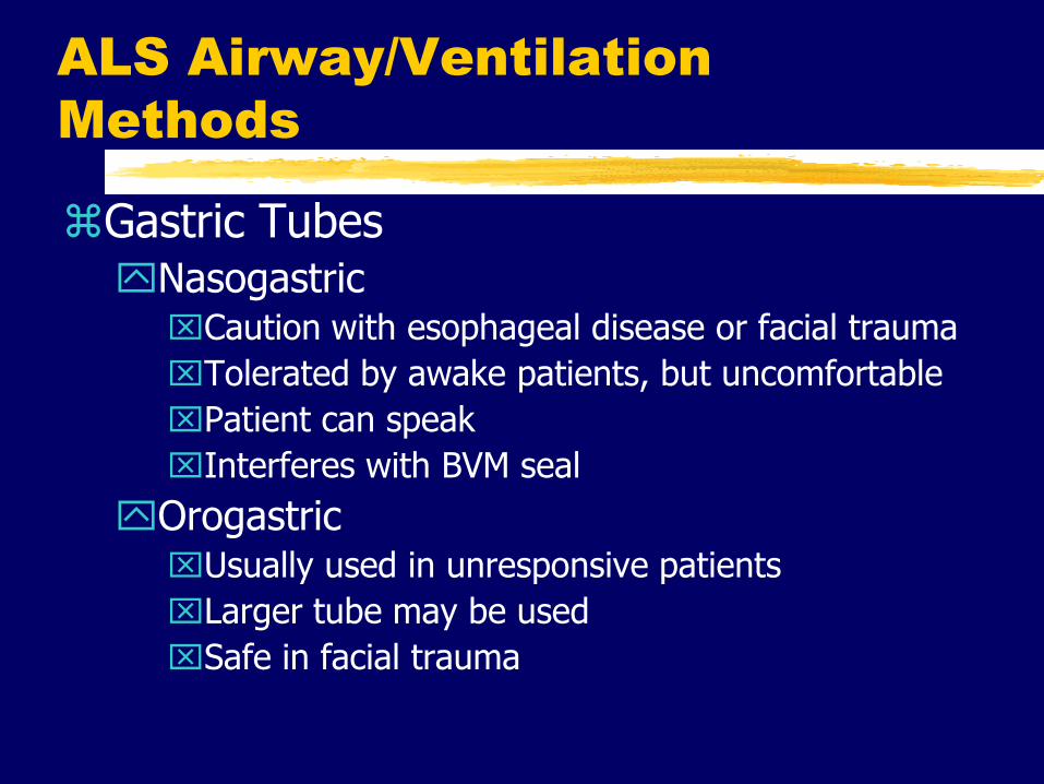
ALS Airway/Ventilation
Methods
Gastric TubesNasogastric Caution with esophageal disease or facial trauma
Tolerated by awake patients, but uncomfortable
Patient can speak
Interferes with BVM seal
OrogastricUsually used in unresponsive patients
Larger tube may be used
Safe in facial trauma

ALS Airway/Ventilation
Methods
Nasogastric Tube Insertion
Select size (French)
Measure length (nose to ear to xiphoid)
Lubricate end of tube (water soluble)
Maintain aseptic technique
Position patient sitting up if possible

ALS Airway/Ventilation
Methods
Nasogastric Tube InsertionInsert into nare towards angle of jaw
Advance gradually to measured length
Have patient swallow
Assess placementInstill air, ausculate
aspirate gastric contents
Secure
May connect to low vacuum (80-100 mm Hg)

ALS Airway/Ventilation
Methods
Orogastric Tube InsertionSelect size (French)
Measure length
Lubricate end of tube
Position patient (usually supine)
Insert into mouth
Advance gradually but steadily
Assess placement (instill air or aspirate)
Secure
Evacuate contents as needed
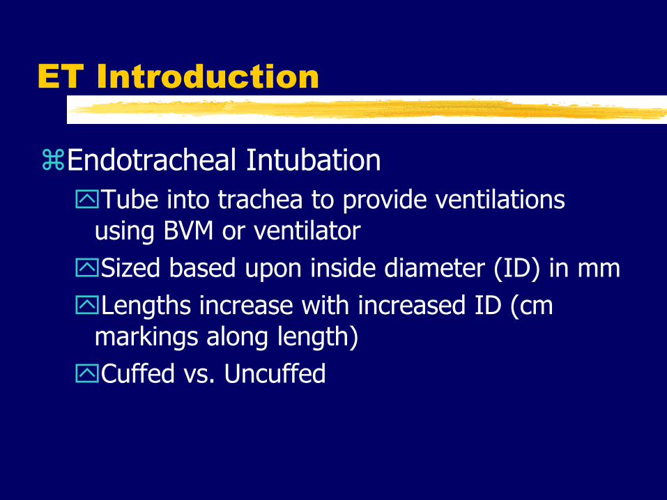
ET Introduction
Endotracheal Intubation
Tube into trachea to provide ventilations using BVM or ventilator
Sized based upon inside diameter (ID) in mm
Lengths increase with increased ID (cm markings along length)
Cuffed vs. Uncuffed
Endotracheal Intubation
Advantages
Secures airway
Route for a few medications (LANE)
Optimizes ventilation, oxygenation
Allows suctioning of lower airway

Endotracheal Intubation
Indications
Present or impending respiratory failure
Apnea
Unable to protect own airway

Endotracheal Intubation
These are NOT Indications
Because I can intubate
Because they are unresponsive
Because I can’t show up at the hospital without it
Endotracheal Intubation
ComplicationsSoft tissue trauma/bleeding
Dental injury
Laryngeal edema
Laryngospasm
Vocal cord injury
Barotrauma
Hypoxia
Aspiration
Esophageal intubation
Mainstem bronchus intubation

Endotracheal Intubation
Insertion Techniques
Orotracheal Intubation (Direct Laryngoscopy)
Blind Nasotracheal Intubation
Digital Intubation
Retrograde Intubation
Transillumination

Orotracheal Intubation
Technique
Position, ventilate patient
Monitor patient
ECG
Pulse oximeter
Assess patient’s airway for difficulty
Assemble, check equipment (suction)
Hyperventilate patient (30-120 sec)
ALS Airway/Ventilation
Methods
Orotracheal IntubationPosition patient
Open mouth
Insert laryngoscope blade on right side
Sweep tongue to left
Identify anatomical landmarks
Advance laryngoscope bladeVallecula for curved (Miller) blade
Under epiglottis for straight (Miller) blade

ALS Airway/Ventilation
Methods
Orotracheal Intubation
Elevate epiglottis
Directly with straight (Miller) blade
Indirectly with curved (Macintosh) blade
Visualize vocal cords, glottic opening
Enter mouth with tube from corner of mouth

ALS Airway/Ventilation
Methods
Orotracheal IntubationAdvance tube into glottic opening about 1/2
inch past vocal cords
Continue to hold tube, note location
Ventilate, ausculateEpigastrium
Left and right chest
Inflate cuff until air leak around cuff stops
Reassess tube placement

ALS Airway/Ventilation
Methods
Orotracheal Intubation
Secure tube
Reassess tube placement, ventilation effectiveness
Intubation
Total time between ventilations
should not exceed
30 seconds!

Intubation
Death occurs from failure to Ventilate,
not failure to Intubate

ALS Equipment
Equipment
Laryngoscope Handle (lighted) & Blades
Stylet
Syringe
Magills
Lubricant
Suction
BVM
BAAM (Blind Nasal)
Selection
Typical Adult ET Tube Sizes
Male - 8.0, 8.5
Female - 7.0, 7.5, 8.0
Blade
Mac - 3 or 4
Miller - 3
Tube Depth
Usually 20 - 22 cm at the teeth

ALS Equipment

ALS Equipment
From AHA PALS

ALS Equipment

Pediatric ET Intubation
Pediatric Equipment Differences
Uncuffed tube < 8 yoa
Miller blade preferred
Tube Size
Premie: 2.0, 2.5
Newborn: 3.0, 3.5
1 year: 4
Then: (age/4)+4
Pediatric Differences
Anatomic Differences
Depth (cm)
Tube ID x 3
12 + (age/2)
easily dislodged
Intubation vs BVM
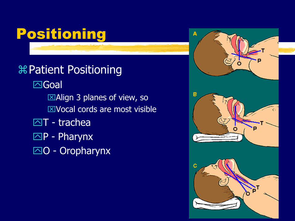
Positioning
Patient Positioning
Goal
Align 3 planes of view, so
Vocal cords are most visible
T - trachea
P - Pharynx
O - Oropharynx

Airway Assessment
Cervical Spine
Temporal Mandibular Joint
A/O Joint
Neck length, size and muscularity
Mandibular size in relation to face
Over bite
Tongue size

Assessment Acronym
M Mandible
O Opening
U Uvula
T Teeth
H Head
S Silhouette

The Lemon Law
L Look externally
E Evaluate the 3-3-2 rule
M Mallampati score
O Obstruction?
N Neck Mobility
Look
Morbidly obese
Facial hair
Narrow face
Overbite
Trauma

Evaluate 3-3-2
Temporal Mandibular Joint
Should allow 3 fingers between incisors
3-4 cm

Evaluate 3-3-2
Mandible
3 fingers between mentum & hyoid bone
Less than three fingers
Proportionately large tongue
Obstructs visualization of glottic opening
Greater than three fingers
Elongates oral axis
More difficult to align the three axis

Evaluate 3-3-2
Larynx
Adult located C5,6
If higher, obstructive view of glottic opening
Two fingers from floor of mouth to thyroid cartilage

Mallampati Score
Evaluates ability to visualize glottic opening
Patient seated with neck extended
Open mouth as wide as possible
Protrude tongue as far as possible
Look at posterior pharynx
Grade based on visual field
Grades 1,2 have low intubation failure rates
Grades 3,4 have higher intubation failure rates

Mallampati Score
Not useful in emergent situations
Informal version
Use tongue blade to visualize pharynx

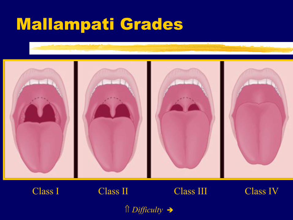
Mallampati Grades
Difficulty
Class I Class II Class III Class IV

Obstruction
Know or suspected
Foreign bodies
Tumors
Abscess
Epiglottitis
Hematoma
Trauma

Neck Mobility
Align axis to facilitate orotracheal intubation
Decreased mobility from
C-Spine immobilization
Rheumatoid arthritis
Quick Test
Put chin on chest then move toward ceiling

Curved Blade (Macintosh)
Insert from right to left
Visualize anatomy
Blade in vallecula
Lift up and away DO NOT PRY ON TEETH
Lift epiglottis indirectly
From AHA ACLS

Straight Blade (Miller)
Insert from right to left
Visualize anatomy
Blade past vallecula and over epiglottis
Lift up and away DO NOT PRY ON TEETH
Lift epiglottis directly
From AHA ACLS
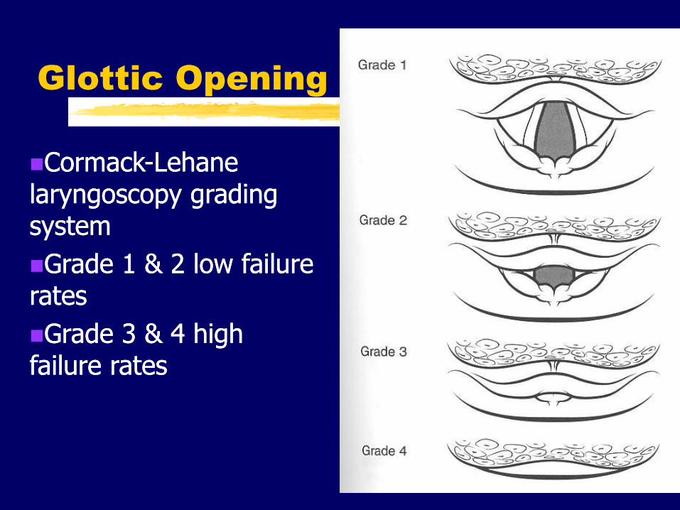
Glottic Opening
CormackCormack--Lehane Lehane laryngoscopy grading laryngoscopy grading system system
Grade Grade 1 1 & & 2 2 low failure low failure ratesrates
Grade Grade 3 3 & & 4 4 high high failure ratesfailure rates

Tube Placement
From TRIPP, CPEM

Confirmation of Placement

Placement of the ETT within the esophagus is an accepted
complication.
However, failure to recognize and correct is not!

Traditional Methods
Observation of ETT passing through vocal cords.
Presence of breath sounds
Absence of epigastric sounds
Symmetric rise and fall of chest
Condensation in ETT
Chest Radiograph
All of these methods have failed in the clinical setting

Additional Methods
Pulse Oximetry
Aspiration Techniques
End Tidal CO2
Confirming ETT Location
Fail Safe
Near Fail Safe
Non-Fail Safe

Fail Safe
Improvement in Clinical Signs
ETT visualized between vocal cords
Fiberoptic visualization of
Cartilaginous rings
Carina

Near Failsafe
CO2 detection
Rapid inflation of EDD
Non-Failsafe
Presence of breath sounds
Absence of epigastric sounds
Absence of gastric distention
Chest Rise and Fall
Large Spontaneous Exhaled Tidal Volumes

Non Failsafe
Condensation in tube disappearing and reappearing with respiration
Air exiting tube with chest compression
Bag Valve Mask having the appropriate compliance
Pressure on suprasternal notch associated with pilot balloon pressure

ALS Airway/Ventilation
Methods
Blind Nasotracheal Intubation
Position, oxygenate patient
Monitor patient
ECG monitor
Pulse oximeter

ALS Airway/Ventilation
Methods
Blind Nasotracheal Intubation
Assess for difficulty or contraindication
Mid-face fractures
Possible basilar skull fracture
Evidence of nasal obstruction, septal deviation
Assemble, check equipment
Lubricate end of tube; do not warm
Attach BAAM (if available)
ALS Airway/Ventilation
Methods
Blind Nasotracheal IntubationPosition patient (preferably sitting upright)
Insert tube into largest nare
Advance slowly, but steadily
Listen for sound of air movement in tube or whistle via BAAM
Advance tube
Assess placement
Inflate cuff, reassess placement
Secure, reassess placement

ALS Airway/Ventilation
Methods
Digital Intubation
Blind technique
Variable probability of success
Using middle finger to locate epiglottis
Lift epiglottis
Slide lubricated tube along index finger
Assess tube placement/depth as with orotracheal intubation

ALS Airway/Ventilation
Methods
Digital Intubation
From AMLS, NAEMT

ALS Airway Ventilation
Methods
Surgical Cricothyrotomy
Indications
Absolute need for definitive airway, AND
• unable to perform ETT due for structural or anatomic reasons, AND
• risk of not securing airway is > than surgical airway risk
OR
Absolute need for definitive airway AND
• unable to clear an upper airway obstruction, AND
• multiple unsuccessful attempts at ETT, AND
• other methods of ventilation do not allow for effective ventilation, respiration

ALS Airway/Ventilation
Methods
Surgical Cricothyrotomy
Contraindications (relative)
No real demonstrated indication
Risks > Benefits
Age < 8 years (some say 10, some say 12)
Evidence of fractured larynx or cricoid cartilage
Evidence of tracheal transection

ALS Airway/Ventilation
Methods
Surgical Cricothyrotomy
Tips
Know anatomy
Short incision, avoid inferior trachea
Incise, do not saw
Work quickly
Nothing comes out until something else is in
Have a plan
Be prepared with backup plan

ALS Airway/Ventilation
Methods
Needle Cricothyrotomy/Transtracheal Jet Ventilation
Indications
Same as surgical cricothyrotomy with
Contraindication for surgical cricothyrotomy
Contraindications
None when demonstrated need
Caution with tracheal transection

ALS Airway/Ventilation
Methods
Jet Ventilation
Usually requires high-pressure equipment
Ventilate 1 sec then allow 3-5 sec pause
Hypercarbia likely
Temporary: 20-30 mins
High risk for barotrauma

ALS Airway/ Ventilation
Methods
Alternative Airways
Multi-Lumen Devices (CombiTube, PTLA)
Laryngeal Mask Airway (LMA)
Esophageal Obturator Airways (EOA, EGTA)
Lighted Stylets

ALS Airway/ Ventilation
Methods
Pharyngeal Tracheal Lumen Airway
(PTLA)
From AMLS, NAEMT

ALS Airway/ Ventilation
Methods
No. 1
100 ml
No. 215 m
l
No. 2
No. 1
No. 1
100 ml
No. 2
15 ml
No. 2
No. 1
Combitube®
From AMLS, NAEMT

ALS Airway/ Ventilation
Methods
Combitube®
Indications
Contraindications
Height
Gag reflex
Ingestion of corrosive or volatile substances
Hx of esophageal disease

ALS Airway/ Ventilation
Methods
Laryngeal Mask Airway (LMA)
use in OR
Gaining use out-of-hospital
Not useful with high airway pressure
Not replacement for endotracheal tube
Multiple models, sizes

LMA

ALS Airway/ Ventilation
Methods

BLS & ALS Airway/ Ventilation
Methods
Esophageal Obturator Airway, Esophageal Gastric Tube Airway
Used less frequently today
Increased complication rate
Significant contraindications
Patient height
Caustic ingestion
Esophageal/liver disease
Better alternative airways are now available

Esophageal Gastric Tube
Airway (EGTA)
From AHA ACLS

ALS Airway/ Ventilation
Methods
Lighted Stylette
Not yet widely used
Expensive
Another method of visual feedback about placement in trachea
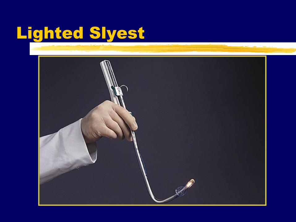
Lighted Slyest

ALS Airway/Ventilation
Methods

Pharmacologic Assisted
Intubation “RSI”
Sedation
Reduce anxiety
Induce amnesia
Depress gag reflex, spontaneous breathing
Used for
induction
anxious, agitated patient
Contraindications
hypersensitivity
hypotension

Pharmacologic Assisted
Intubation “RSI”
Common Medications for Sedation
Benzodiazepines (diazepam, midazolam)
Narcotics (fentanyl)
Anesthesia Induction Agents
Etomidate
Ketamine
Propofol (Diprivan®)

Pharmacologic Assisted
Intubation
Neuromuscular Blockade
Temporary skeletal muscle paralysis
Indications
When intubation required in patient who:
• is awake,
• has gag reflex, or
• is agitated, combative

Pharmacologic Assisted
Intubation
Neuromuscular Blockade
Contraindications
Most are specific to medication
Inability to ventilate once paralysis induced
Advantages
Enables provider to intubate patients who otherwise would be difficult, impossible to intubate
Minimizes patient resistance to intubation
Reduces risk of laryngospasm

Pharmacologic Assisted
Intubation
NMB Agent Mechanism of Action
Acts at neuromuscular junction where ACh normally allows nerve impulse transmission
Binds to nicotinic receptor sites on skeletal muscle
Depolarizing or non-depolarizing
Blocks further action by ACh at receptor sites
Blocks further depolarization resulting in muscular paralysis
Pharmacologic Assisted
Intubation
Disadvantages/Potential Complications
Does not provide sedation, amnesia
Provider unable to intubate, ventilate after NMB
Aspiration during procedure
Difficult to detect motor seizure activity
Side effects, adverse effects of specific drugs

Pharmacologic Assisted
Intubation
Common Used NMB Agents
Depolarizing NMB agents
succinylcholine (Anectine®)
Non-depolarizing NMB agents
vecuronium (Norcuron®)
rocuronium (Zemuron®)
pancuronium (Pavulon®)
Pharmacologic Assisted
Intubation
Summarized Procedure
Prepare all equipment, medications while ventilating patient
Hyperventilate
Administer induction/sedation agents and pretreatment meds (e.g. lidocaine or atropine)
Administer NMB agent
Sellick maneuver
Intubate per usual
Continue NMB and sedation/analgesia prn
Pharmacologic Assisted
Intubation
Failure is not an option!

ALS Airway/Ventilation
Methods
Needle Thoracostomy
Indications
Positive signs/symptoms of tensionpneumothorax
Cardiac arrest with PEA or asystole with possible tension pneumothorax
Contraindications
Absence of indications

ALS Airway/Ventilation
Methods
Tension Pneumothorax Signs/Symptoms
Severe respiratory distress
or absent lung sounds (usually unilateral)
resistance to manual ventilation
Cardiovascular collapse (shock)
Asymmetric chest expansion
Anxiety, restlessness or cyanosis (late)
JVD or tracheal deviation (late)

ALS Airway/Ventilation
Methods
Needle Thoracostomy
Prepare equipment
Large bore angiocath
Locate landmarks: 2nd intercostal space at midclavicular line
Insert catheter through chest wall into pleural space over top of 3rd rib (blood vessels, nerves follow inferior rib margin)
Withdraw needle, secure catheter like impaled object
ALS Airway/Ventilation
Methods
Chest Escharotomy
Indications
Presence of severe edema to soft tissue of thorax as with circumferential burns
inability to maintain adequate tidal volume, chest expansion even with assisted ventilation
Considerations
Must rule out upper airway obstruction
Rarely needed

ALS Airway/Ventilation
Methods
Chest Escharotomy
Procedure
Intubate if not already done
Prepare site, equipment
Vertical incision to anterior axillary line
Horizontal incision only if necessary
Cover, protect

Airway & Ventilation Methods
Saturday’s class
Practice using equipment
orotracheal intubation
nasotracheal intubation
gastric tube insertion
surgical airways
needle thoracostomy
combitube
retrograde intubation