Aim Clinical indication Manufacturing scan prosthesis CT scan protocol Double scan registration...
13
• Aim • Clinical indication • Manufacturing scan prosthesis • CT scan protocol • Double scan registration wizard Double scan
-
Upload
kenneth-cannon -
Category
Documents
-
view
235 -
download
4
Transcript of Aim Clinical indication Manufacturing scan prosthesis CT scan protocol Double scan registration...
• Aim
• Clinical indication
• Manufacturing scan prosthesis
• CT scan protocol
• Double scan registration wizard
Double scan
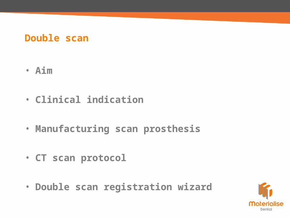
• Visualize tooth setup within CT imagesImplant planning can be based on both clinical and aesthetic considerations
• Visualize mucosa surfaceNecessary for manufacturing of mucosa-supported SurgiGuide
Aim
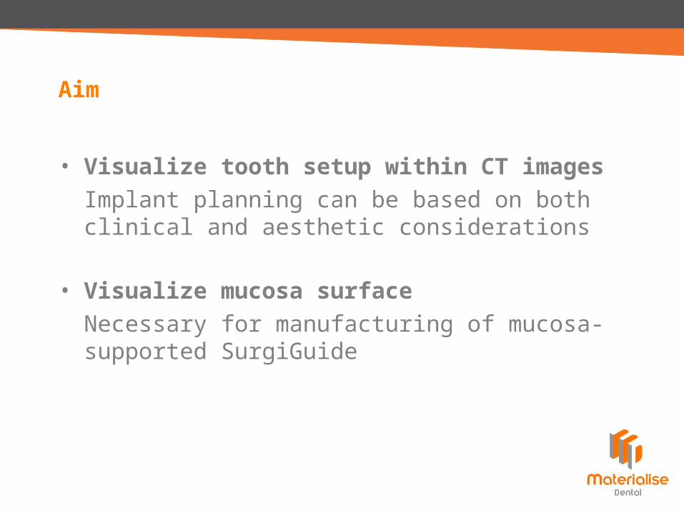
Fully edentulous cases– Maxilla– Mandible
Clinical indication
• Good fitting removable dentureNo air between gingiva and prosthesisto avoid
=> false indication mucosa thickness
=> malfitting mucosa-supported SurgiGuide
• Prosthesis– Material: acrylic resin– No radio-opaque teeth– No metallic parts– Base plate minimally 2 mm thick
Manufacturing scan prosthesis
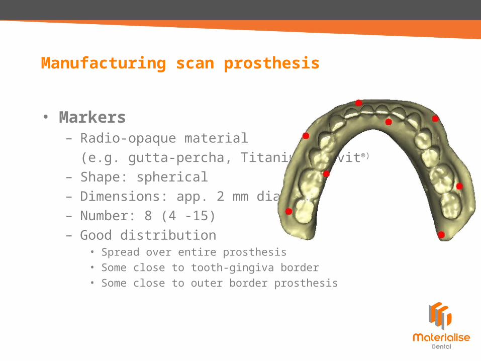
• Markers– Radio-opaque material
(e.g. gutta-percha, Titanium, Cavit®)
– Shape: spherical– Dimensions: app. 2 mm diameter– Number: 8 (4 -15) – Good distribution
• Spread over entire prosthesis• Some close to tooth-gingiva border• Some close to outer border prosthesis
Manufacturing scan prosthesis
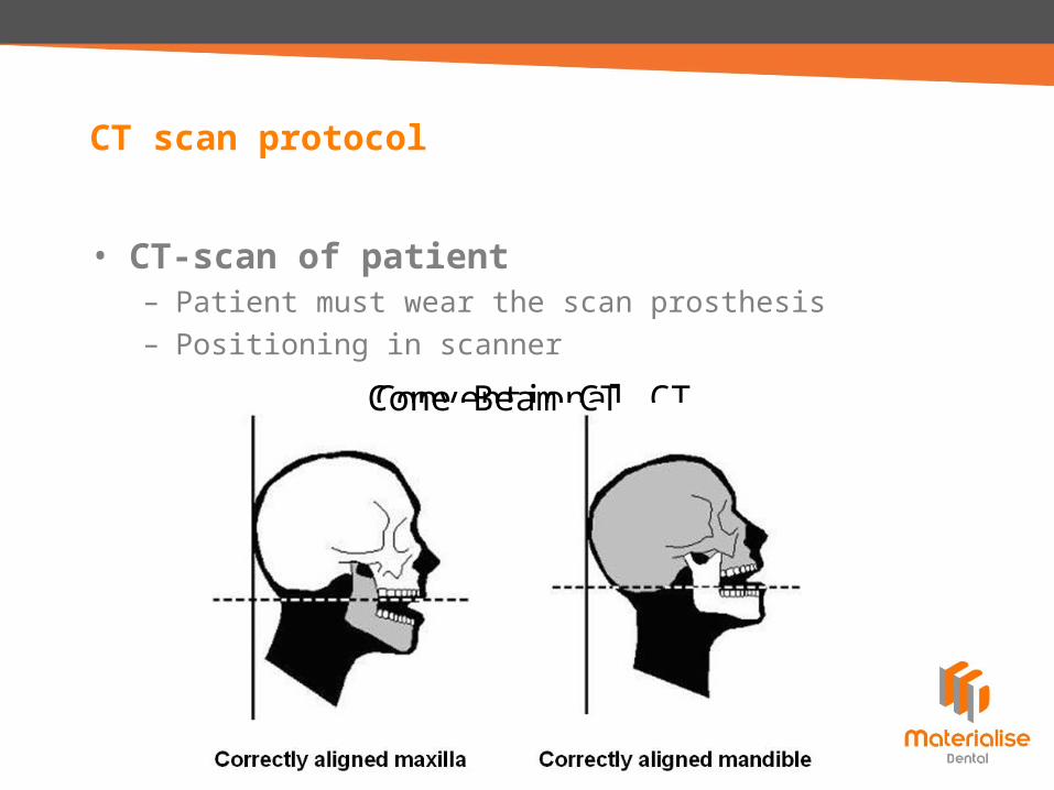
• CT-scan of patient– Patient must wear the scan prosthesis– Positioning in scanner
CT scan protocol
Conventional CTCone Beam CT
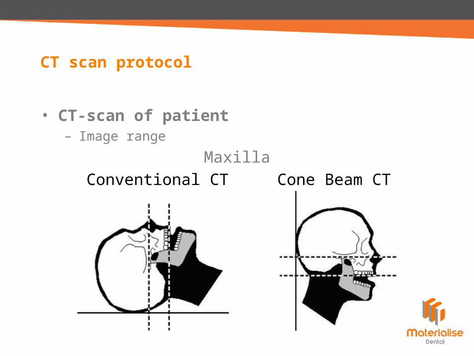
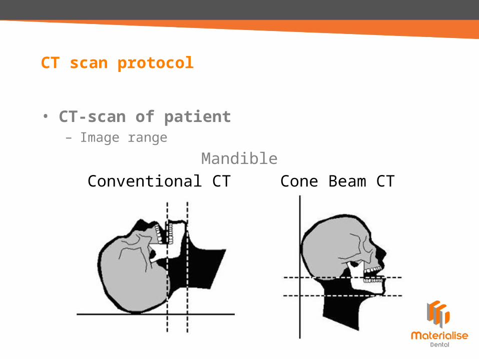
• CT-scan of patient– Image range
CT scan protocol
Conventional CT Cone Beam CTMaxilla
• CT-scan of patient– Image range
CT scan protocol
Conventional CT Cone Beam CTMandible
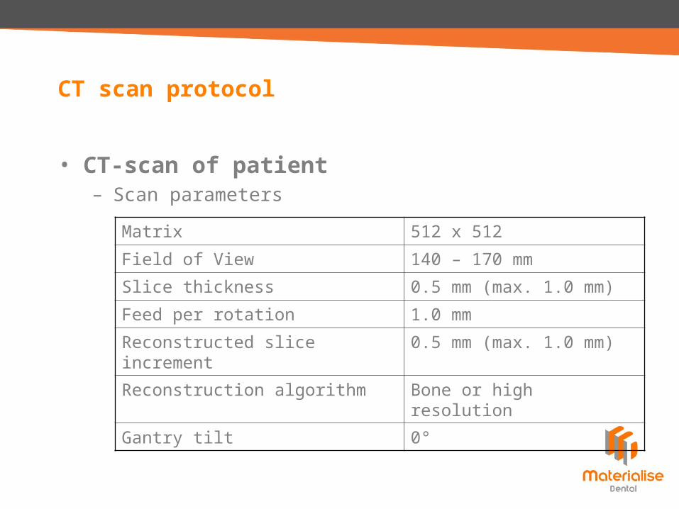
• CT-scan of patient– Scan parameters
CT scan protocol
Matrix 512 x 512
Field of View 140 – 170 mm
Slice thickness 0.5 mm (max. 1.0 mm)
Feed per rotation 1.0 mm
Reconstructed slice increment 0.5 mm (max. 1.0 mm)
Reconstruction algorithm Bone or high resolution
Gantry tilt 0°
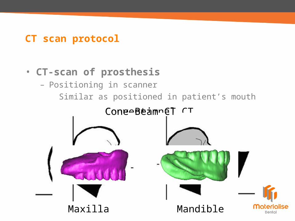
• CT-scan of prosthesis– Positioning in scanner
Similar as positioned in patient’s mouth
CT scan protocol
Maxilla Mandible
Conventional CTCone Beam CT
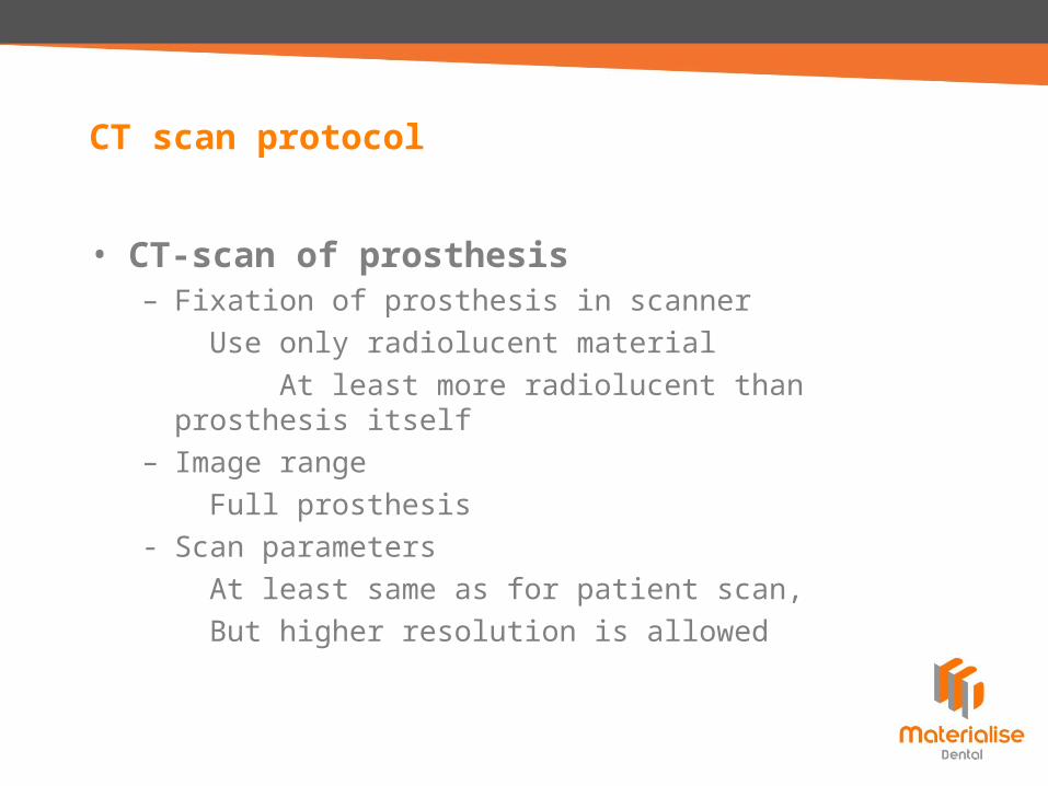
• CT-scan of prosthesis– Fixation of prosthesis in scanner
Use only radiolucent materialAt least more radiolucent than prosthesis
itself– Image range
Full prosthesis- Scan parameters
At least same as for patient scan, But higher resolution is allowed
CT scan protocol
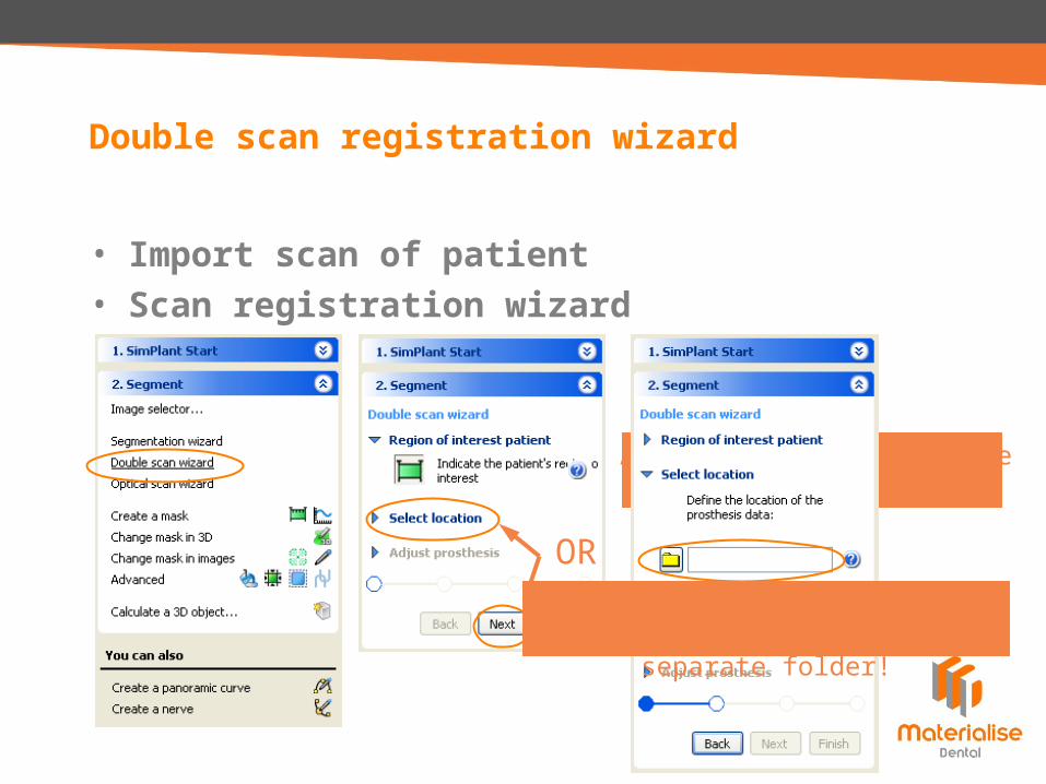
• Import scan of patient• Scan registration wizard
Double scan registration wizard
Adapt box to only include radio-opaque markers!
ORDICOM images of scan of prosthesis
preferably in separate folder!
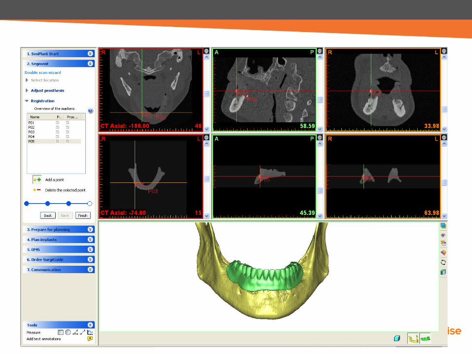
• Import scan of patient• Scan registration wizard
Double scan registration wizard
Possible to changethreshold for prosthesis
Possible to adapt box toonly include prosthesis


![INDEX [microdentsystem.com] · 2015-11-24 · INDEX PRESENTATION. INTRODUCTION MULTIPLE PROSTHESIS. REMOVABLE AND IMMEDIATE PROSTHESIS. SINGLE PROSTHESIS CEMENTED PROSTHESIS. Microdent](https://static.fdocuments.in/doc/165x107/5facd9ee77a5ed547a36b19c/index-2015-11-24-index-presentation-introduction-multiple-prosthesis-removable.jpg)


![Intelligent Prosthesis - tams. · PDF fileI Electrooculography (EOG) I Electrocorticogram (EcoG) [ ] Irina Intelligent Prosthesis 4/21. ... Irina Intelligent Prosthesis 21/21](https://static.fdocuments.in/doc/165x107/5aab10c57f8b9aa9488b839d/intelligent-prosthesis-tams-electrooculography-eog-i-electrocorticogram-ecog.jpg)