Adventures With a Hand Lens
222
A project of Volunteers in Asia AdventuE: PTitha &nd Lens by: Richard Headstrom Published by: Dover Publications, Inc. 180 Varick Street New York, NY MO14 USA Paper copies are $2.75. For a free catalogue of Dover publications, please write to the address given below. Available from: Dover Publications, Inc. 130 Varick Street New York, NY 10014 USA Reproduced by permission of Dover Publications, Reproduction of this microfiche document in any form is subject to the same restrictions as those of the original document.
-
Upload
neil-bigelow -
Category
Documents
-
view
222 -
download
2
description
Instructions for using a hand lens
Transcript of Adventures With a Hand Lens
A project of Volunteers in Asia
AdventuE: PTith a &nd Lens
by: Richard Headstrom
Published by: Dover Publications, Inc. 180 Varick Street New York, NY MO14 USA
Paper copies are $2.75. For a free catalogue of Dover publications, please write to the address given below.
Available from: Dover Publications, Inc. 130 Varick Street New York, NY 10014 USA
Reproduced by permission of Dover Publications,
Reproduction of this microfiche document in any form is subject to the same restrictions as those of the original document.

DOVER BOOKS EXPLAINING SCIENCE
1 HE Evot ~‘TION OF SCIENTIFIC THOIGHT FRC>M NEU’TON TO
EISSTEIS. A. d’,4hro. ( ZOOO2-7) SF.00.
THE RUSE OF THE SE\\’ ~%YSICS. A. d’Abro. (20003-5. 20004-3) Two volume set $10.00
STAR NAMES: THEIR LORE AND MEANING, Richard H. Allen. ( 2 1079-O) $5.00
LUNAR ATLAS. Dinsmorc Alter. ( 2 1701-9 I “6.00 WOW CRYSTALS. W. A. Bentley, W. I. Hurrphreys. (20287-9)
$6.00 FINSTEIN’S THEORY OF RELATIVITY. Max Born. (60;69-0) $3.50 THE RESTLESS UNIVERSE. Max Born. (20412-X) $4.50 THE WORLD OF SOUND, Sir William Bragg. (21853-s) $2.00 CLIM.%TE THROUGH THE AGES, C. E. P. Brooks. (22245-4) $3.50 ~RSOL~TEI Y MAD INVENTIONS. A. E. Brown, H. A. Jeffcott.
(22596-S! $1.50 \~:H.\T Is SCIENCE? Norman R. Campbell. (60043-2) $2.50 PLANETS, STARS AND GALAXIES, A. E. Fanning. ( 21680-2) $2.50 A HANDBOOK FOR THE TECHNKAL AND SCIENTIFIC SECRETARY,
George ;reedman. ( 23024-4 ) $3.50 FADS AND FALLACIES IN THE NAME OF SCIENCE, Martin Gardner.
( 20394-S ) $3.50 MATHEMATICS. MAGIC AND MYSTERY, Martin Gardner.
( 20335-2 ) $2.00 EXPLORING MATHEMATICS ON YOUR OWN, William H. Glenn,
Donovan A. Johnson. (20383-2) $2.50 INVITATION TO MATHEMATICS, William H. Glenn, Donovan A.
Johnson. (22906-8) $4.00 SAFE AND SIMPLE ELECTRICAL EXPERIMENTS, Rudolf F. Graf.
(22950-5) $2.50 LISTENING IN THE DARK, Donald R. Griffin. (21714-O) $4.00 BIOLOGY EXPERIMENTS FOR CHILDREN, Ethel Hanauer.
( 22032-X) $1.75 THE STRANGE STORY OF THE QUANTUM, Banesh Hoffmann.
( 205 18-5 ) $3.00 THE DIVINE PROPORTION: A STUDY IN MATHEMATICAL BEAUTY,
H. E. Huntley. (22254-3 ) $2.50 ART AND GEOMETRY: A STUDY IN SPACE INTUITIONS, William
M. Ivins. (20941-5) $2.00 SCIENCE AND MUSIC, Sir James Jeans. (21964-8) $3.00 GEMS AND PRECIOUS STONES OF NORTH AMERICA, George F.
Kunz. ( 2 1855-4) $4.50 PRACTICAL STATISTICS SIMPLY EXPLAINED, Dr. Russell A.
Langley. (22729-4) $4.00

With an ordinary magnifying glass and this book as your guide, 50 adventures in close observation await you. These entertaining nature studies take you on field trips in and around your home, calling attention to interesting features of dozens of familiar or overlooked plants, insects and other animals, and common materials like cloth, quartz and the paper on which this book is printed.
A great deal of basic natural-sdence thtory and detail is presented in this delightful narrative. Flowers and bgrasses, fish scales, moth and insect wings, egg cases, buds, feathers, seeds, leaf scars, moss, molds, ferns, common crystals are among the many structures exam- ined, often comparatively. Many ,natural prti-:sses and behavior patterns are observed- dispers,al and o?i. , methods of repro- duction, protective coloration, rustin?. symbios;i. fertilization of the soil, breathing and case-buiiding :.;. :,nsects, and many others, all with only an inexpensive hand ien* as equipment and with “speci- mens” you probably pass by going for a walk. More than 260 labeled illustrations accompany the text.
The author is a former teacher and associate curator of the New England lluxum of Natural History. No previous science back- ground is assumed of readers, and curious readers of almost any age will find this book an interesting introduction to numerous facets of nature study.
Unabridged, slightly revised republication of 1962 edition. 209 illus- trations by the author. 220~~. 5s/, x 8. 23330-8 Paperbound
A DOVER EDITION DESIGNED FOR YEARS OF USE!
We have made every effort to make this the best book possible. Our paper is opaque, with minimal show-through; it will not discolor or become brittle with age. Pages are sewn in signatures, in the method traditionally used for the best books, and will not drop out, as often happens with paperbacks held together with glue. Rooks open flat for easy reference. The binding will not crack or split. This is a permanent book.
d DOVER B
$2.50 in U.S.A. $2.95 in Canacln

ANIMALS PARAW ;’ IN SIAN. Geoffrey Lapage. (21047-2) $3.50
THREE CENTURIES OF M ICRO~IOLOGY. Hubert A. Lechevalier, !Uorris Sol.~tcrovsky. (2303S-X) $5.00
CH.~NCE. Lb’cx I:‘LD STAPISTICS, Horace C. Levinson. (21007-3) 52 50
.4 kr.!~r I-O ML~CAL Ac~~~TKs. H. Lowery. (21628-4) $2.00 Snc?r.c~rc%r RHYTHMS IN HUMAN AND ANIMAL PHYSIOLOGY,
Gay Gaer Lute. ( 225860) $3.00 VISUAL ILL~~I~N~. Matthew Luckiesh. (21530-X) $2.50 PHYSICS E~PERI~IENTS FOR CHILDREN, Muriel Mandell.
4 22033-s ) S 1.75 THE FRIENDLY STARS, Martha E. Martin. ( 21099-5) $2.00 MATHEMATICAL EXCURSIONS, Helen A. Merrill. (20350-6) $1.75 THE NATURE OF LIGHT AND COLOUR IN THE OPEN AIR,
M. Minnaert. (20196-l ) $4.00 THE CONCEPT OF ENERGY SIMPLY EXPL.~INED. Morton Mott-
Smith. (210?1-5) $2.75 T?% CONCEPT OF HEA-T AND ITS WORKINGS SIMPLY EXPLJNFD,
31orton Mott-Smith. f 20978-4) $2.00 PRINCIPLES OF MKHANCS SIMPLY EXPLAINED, Morton Motr-
Smith. ( 2 1067-7) $2.00 CHEMISTRY EXPERIMENTS FOR CHILDREN, Virginia L. Mullen.
(22031-l) 51.75 M~src OF THE SPHERES: THE MATERIAL. UNNERSE FROM ATOM
TO QUASAR SIMPLY EXPLAINED, Guy Murchie. (2 1809-0, 2 18 10-4) Two volume set $7.50
MUSIC, PHYSICS AND ENGINEERING, Harry F. Olson. (2 1769-8 ) $4.50
THE ORIGINS OS LIFE, A. I. Oparin. (60213-3) $3.00 THE GENTLE ART OF MATHEMATICS, Dan Pedoe. (22949-l )
$2.50 SPINNIF‘G TOPS AND GYROSCOPIC MOTION, John Perry.
(20416-2) $1.50 ELECTRICITY EXPERIMENTS FOR CHILDREN, Gabriel Reuben.
(22030-3) $1.50 GEOMETRIC EXERCISES IN PAPER FOLDING, T. Sundara Row
(21594-6) $2.00
Paperbound unless otherwise indicated. Prices subject to change without notice. Available at your book dealer or write for free catalogues to Dept. 42, Dover Publications, Inc., 180 Varick St., N.Y., N-Y. 10014. Please indicate your field of interest. Each year Dover publishes over 150 books on music, fine art, science, mathematics, languages, chess, puzzles, literature, nature, anthropology, antiques, folklore, art instruction, crafts and needlework, children’s books, and other areas.
Manufactuwd in the U.S.A.
ITH HAND LEN
RICHARD HEADSTROM Illustrated by the Author
Dover Publications, Inc., New York

Copyright @ 1962.1976 by Richard Headstrom. Ail rights reserved under Pan American and Inter-
national Copyright Conventions.
Published in tinada by General Publishing Cafil- pany. Ltd., 30 Lesmill Road, Don Mills, Toror\to, Ontario.
Published in the United Kingdom by Constable and Company, Ltd., 10 Orange Street, London WQ 2.
This Dover edition, first published in 1976, is an unabridged and slightly revised republication of the work originally published by J. B. Lippincott Cam- pany. New York, in 1962.
International Standard Book Number: O-486-2333@ tibrasy of Congress Catalog Card Nunlbet: ?fGIt?910
Manufactured in the United States of America Dover Publications, Inc.
180 Varick Street New York. N. Y. 10014

TO MY WIFE
In Appreciation of Her Help and Devotion
.

INTRODUCTION
ADVENTURE
1
2
3
4
5
6
7
8
9
10
11
12
13
14
15
16
17
18
19
20
21
22
23
IVe Peer through Our Lens at Some Familiar 06jects
We Listen to Some Nrcsic and Discover How It Is Played
We Give Seeds More Than a Passing Glance
We Become Better Acquainted with the Snail
We Irlquire into the Sature of Rusting
We Meet the Insect Brownies
We Visit Fairy/an
We Examine a Feather
We Learn the Meaning of the Word Catkin
We Study the Wings of Insects
We Watch Sane Acrobats Perform
We Go Botanizing
We Compare Insect Antennae
We See How Ants Keep Clean
We Find out how the Earthworm Moves
We Discover that There Are Different Kinds of Hairs
We Spy on the Aphids
We Become Familiar with the Eating Habits of Insects
We Look a Spider in the Eye
We Investigate the Structure of Grass Flowers
We Hunt a Lion
We Trace a Tadpole’s Development
Wre Encounter Some Hitchhikers
9
12
16
18
23
27
30
34
41
43
51
57
59
65
74
77
82
85
89
93
99
103
107
115
24
25
26
27
28
29
30
31
32
33
34
35
36
37
38
39
40
41
42
43
44
45
46
47
48
49
50
We Make the Acquaintavce of Some Housebuilders
We Come CTpon Some L’ilore Housebuilders
We Ascertain how the Leech Sttrks Blood
We Identify Some Ferns
We View the Scale Insects
We Undertake the Study of Minerals
We Consider Some Diagnostic Characters
We Inspect the Breathing Apparatus of Some Aquatic Insects
We Do Some Fishing
We Turn Our Attention to Buds
We Trace Some ‘Tunnels
We Continue Our Study of Minerals
We Scrutinize a Miniature Pepper Box
We Invade the Privacy of Gall Makers
We Are Introduced i~) Some Queer Plants
We Stand Corrected that Eggs Are h’ot Always Egg-Shaped
We Play Amateur Petrologists
We Marvel at h’nture’s Ingenuity
We Breed Some Flies
We Explore the Subject of Variation
We Get to Know the Barnacle
We Seek the Liverwort
We Set Some Traps
We Quest for Oil
We Assume the Role of Farmers
We Are Intrigued by an Ingenious Mechanism
We Probe into a Complicated Life History
I39
I?4
149
I52
I58
I62
I&6
169
I74
I77
180
I84
189
194
198
200
204
207
209
212
217
UN-IL ONLY A FEW YEARS i\co, as time is
measured, man had lived on the earth un- aware of what existed in the heavens, un- aware of how plants and animals are put together, unaware of the countless numbers of tiny plant;i and animals that exist every- where but are invis;ble to the naked eye. He could see and study only what was ap- parent. When his eyesight faded he was doomed to grope about in a world he could but dimly perceive.
Then, somewhere, someone happened to look through a piece of curved glass. Today we need not grope our way when eyesight begins to fade, nor suffer the results of de- fective vision. We can peer into the far reaches of the heavens and look at faraway stars and planets, we can study microscopic plants and animals, we can examin- the cells of our bodies, we can record for our children and our grandchildren happenings in our own lives, and for posterity world-wide events as they occur, we can sit in our homes and be transported to places miles away and watch a sports spectacle or a political ccinvention or some other event in progress. By means of a piece of curved glass, which came to be called a lens because it is shaped like a len- til, we can look at countless things we never suspected existed, or, if we do know of their existence, what they actually look like, for
9
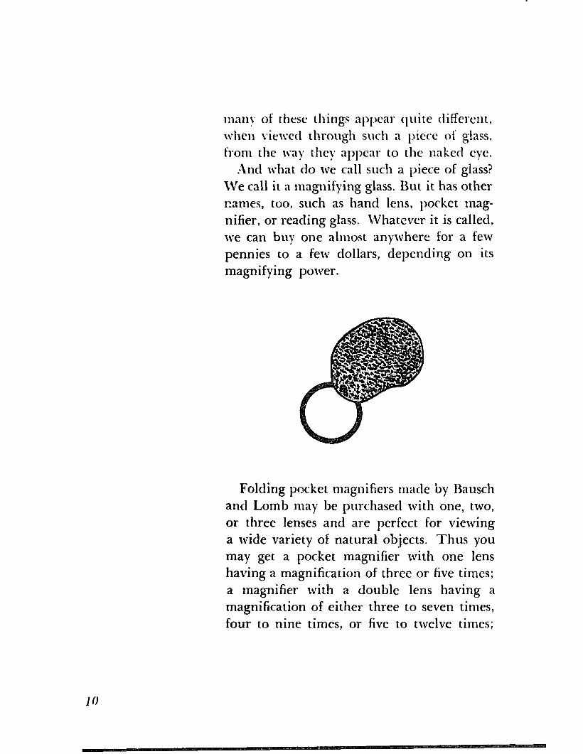
.-Inct what do we call such a piece OF glass?
We call it a magnifying glass. But it has other
tames, too, such as hand lens, pocket mag-
nifier, or reading glass. Whatever it is called,
we can buy one almost anywhere for a few
pennies to a few dollars, depending on its
magnifying power.
Folding pocket magnifiers made by Bausch
and Lomb may be purchased with one, two,
or three lenses and are perfect for viewing
a wide variety of natural objects. Thus you
may get a pocket magnifier with one lens
having a magnification of three or five times;
a magnifier with a double lens having a
magnification of either three to seven times,
four to nine times, or five to twelve times;
or a magnifier with a triple lens having a
magnification of five to twenty times.
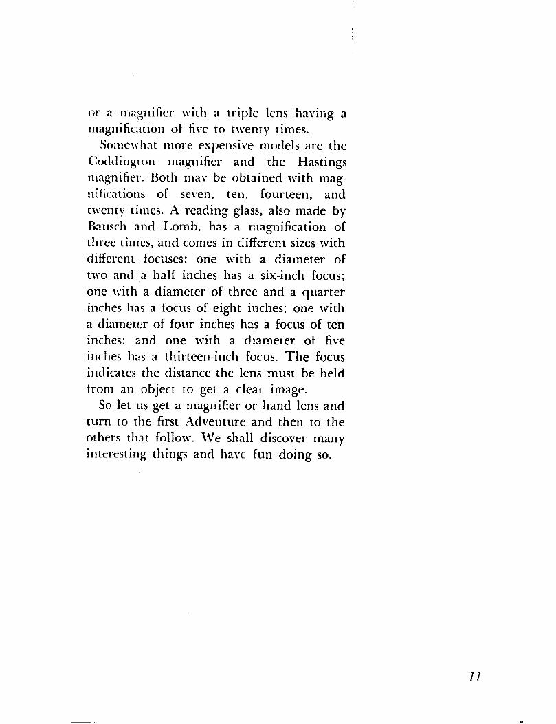
Sonmvhat more expensive models are the
Codrlingr on magnifier and the Hastings
magnifier. Roth may be obtained with mag-
niflications of seven, ten, fourteen, and
twenty times. ,A reading glass, also made by
Bausch and Lomb, has a magnification of
three times, and comes in different sizes with
different. focuses: one with a diameter of
two and ,a half inches has a six-inch focus;
one with a diameter of three and a quarter
inches has a focus of eight inches; one with
a diameter of four inches has a focus of ten
inches: and one with a diameter of five
inches has a thirteen-inch focus. The focus
indicates the distance the lens must be held
from an object to get a clear image.
So let us get a magnifier or hand lens and
turn to the first Adventure and then to the
others that follow. ‘IVe shall discover many
interestin.g things and have fun doing so.

ADVENTURE 1
We Peer through Our Lens at Some Familiar 0 bjects
FOR OUR FIRST ADVENTURE we shall examine
a few things we can find around the house.
And what can we better start with than the
paper on which these words are printed?
Examine it with your hand lens or pocket
magnifier. Next examine a piece of news-
paper and then a piece of glossy paper from
one of the better magazines, and finally com-
pare all three with a piece of blotting paper.
In which paper do you find a network of
loose fibers? I think you will agree that in the
blotting paper the fibers are more in eii-
dence and that there are larger spaces be-
tween them. This is the secret of the blotting
paper’s action in absorbing ink. It all has to
I2
do with capillarity, lvhich we can define sim-
ply as the creeping of a liquid into a \rery
narrow space, and that is just about what the
ink dots when it passes into the blotting
paper.
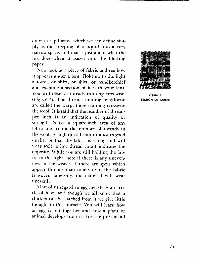
Sow look at a piece of fabric and see how
it al)~xars under a lens. I-?old up to the light a towel, ur shirt, or skirt, or handkerchief
and examine a section of it ixith your lens.
\‘ou \\-ill obserive threads running crosswise.
(f-igritc- 1). The thread5 running lengthwise
are called the warp; those running crosswise
the ivoof. It is said that the number of threads
per inch is an indication of quality or
strength. Select a square-inch area of any
fabric and count the number of threads in
the woof. A high thread count indicates good
quality or that the fabric is strong and will
It-ear well; a low thread count indicates the
opposite. il’hile you are still holding the fab-
.ric to the light, note if there is any uneven-
ness in the 1ueaI.e. If there are spots which
appear thinner than others or if the fabric
is \voi.e~l iine\,enly, the material will wear i’rilil’b’c”Ii i’. 1
\l.xt of us regard an egg merely as an arti-
cle of food, and though we all know that a
chicken can be hatched from it we give little
thought to this miracle. You will learn how
all egg is put together and how a plant or
animal tlei*elops from it. For the present all
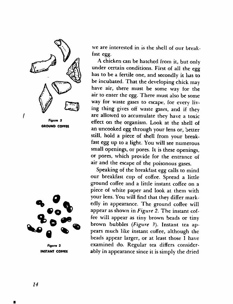
Figure 2
GROUND COFFEE
your lens. You will find that they differ mark-
oe*
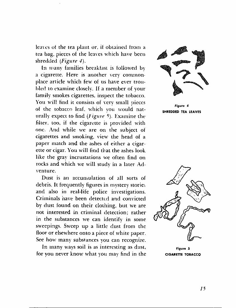
edly in appearance. The ground coffee will
Qo@* appear as shown in Figure 2. The instant cof-
fee will appear as tiny brown beads or tiny w .
Bo w
1 a
brown bubbles (Figure 3). Instant tea ap-
0 pears much like instant coffee, although the
beads appear larger, or at least those I have
we are interested in is the shell of our break-
fast egg.
A chicken can be hatched from it, but only
under certain conditions. First of all the egg
has to be a fertile one, and secondly it has to
be incubated. That the developing chick may
have air, there must be some way for the
air to enter the egg. There must also be some
way for waste gases to escape, for every liv-
ing thing gives oti waste gases, and if they
are allowed to accumulate they have a toxic
effect on the organism. Look at the shell of
an uncooked egg through your lens or, better
still, hold a piece of shell from your break-
fast egg up to a light. You will see numerous
small openings, or pores. It is these openings,
or pores, which provide for the entrance of
air and the escape of the poisonous gases.
Speaking of the breakfast egg calls to mind
our breakfast cup of coffee. Spread a little
ground coffee and a little instant coffee on a
piece of white paper and look at them with
figure 3 examined do. Regular tea differs consider- INSTANT COFFEE ably in appearance since it is simply the dried
I4
lca\~s of the tea plant or, if obtained from a
tea bag, pieces of the lea\ves which halme been
shredded (Figlclu -I).
In many families breakfast is followed b)
a cigarette. Here is another very common-
place article which few of us have ever trou-
bletf to examine closely. if a member of your
family smokes cigarettes, inspect the tobacco. You will find it consists of leery small pieces
of the tobacco leaf, which you would nat-
urally expect to find (Figlcre 5). Examine the
filter, too, if the cigarette is provided with
one. And while we are on the subject of
cigarettes and smoking, view the head of a
paper match and the ashes of either a cigar-
ette or cigar. You will find that the ashes look
like the gray incrustations we often find on
rocks and which we will study in a later Ad-
\‘en ture.
Figure 4
SHREDDED TEA LEAVES
Dust is an accumulation of all sorts of
debris. It frequently figures in mystery stories
and also in real-life police inL*estigations.
Criminals nave been detectcrl and convicted
by clust found on their clothing, but we are
not interested in criminal detection; rather
in the substances we can identify in some
sweepings. Sweep up a little dust from the
floor or elsewhere onto a piece of white paper.
See how many substances you can recognize.
In many ways soil is as interesting as dust, Figure 5 for you never know what you may find in the CIGARETTE TOBACCO
I5
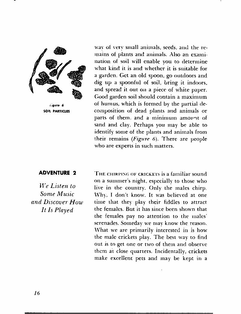
wa\ of i.ery small animals, seeds, and the re-
mains of plants ant1 animals. Also an exami-
nation of soil will enable you to determine
what kind it is and whether it is suitable for
a garden. Get an old spoon, go outdoors and
dig up a spoonful of soil, bring it indoors,
and spread it out oil a piece of white paper.
Good garden soil should contain a maximum
of humus, which is formed by the partial de-
composition of dead plants and animals or
parts of them, and a minimum amovqt of
sancl and clay. Perhaps you may be able to
identify some of the plants and animals from
their remains (Figllre 6). There are people
who are experts in such matters.
ADVENTURE 2
We Listen to
Some Music
and Lliscover How
It Is Played
THE CHIRPING OF CRICKETS is a familiar sound
on a summer’s night, especially to those who
live in the country. Only the males chirp.
\Vll~., I don’t know. It was believed at one
time that they play their fiddles to attract
the females. But it has since been shown that
the females pay no attention to the males’ serenades. Someday we may know the reason.
\Vhat we are primarily interested in is how
the male crickets play. The best way to find
out is to get one or two of them and observe
them at close quarters. Incidentally, crickets
make excellent pets and may be kept in a
16
large jar or similar container with a little soil
on the bottom. They can be fed bits of melon
and other fruits, lettuce, and moist bread.
A little bone meal should also be supplied
to reduce cannibalism. If eggs are laid and
you want to hatch them, sprinkle the soil
with water as you would for plants. The fe-
male, by the way, can be distinguished from
the male by the presence of a long, swordlike
ovipositor, or egg-laying apparatus, extending
from the end of her body.
To return to our question of how a male
cricket plays, observe that when he does so he lifts his wing covers at an angle of forty-
five degrees and then rubs them together.
Actually it isn’t quite as simple as that. If you
examine one of the wing covers with your
lens, you will note that the venarion is of a
peculiar scroll pattern which probably serves
as a framework for the purpose of making a
sounding board of the wing membrane by
stretching it out as a drumhead is stretched.
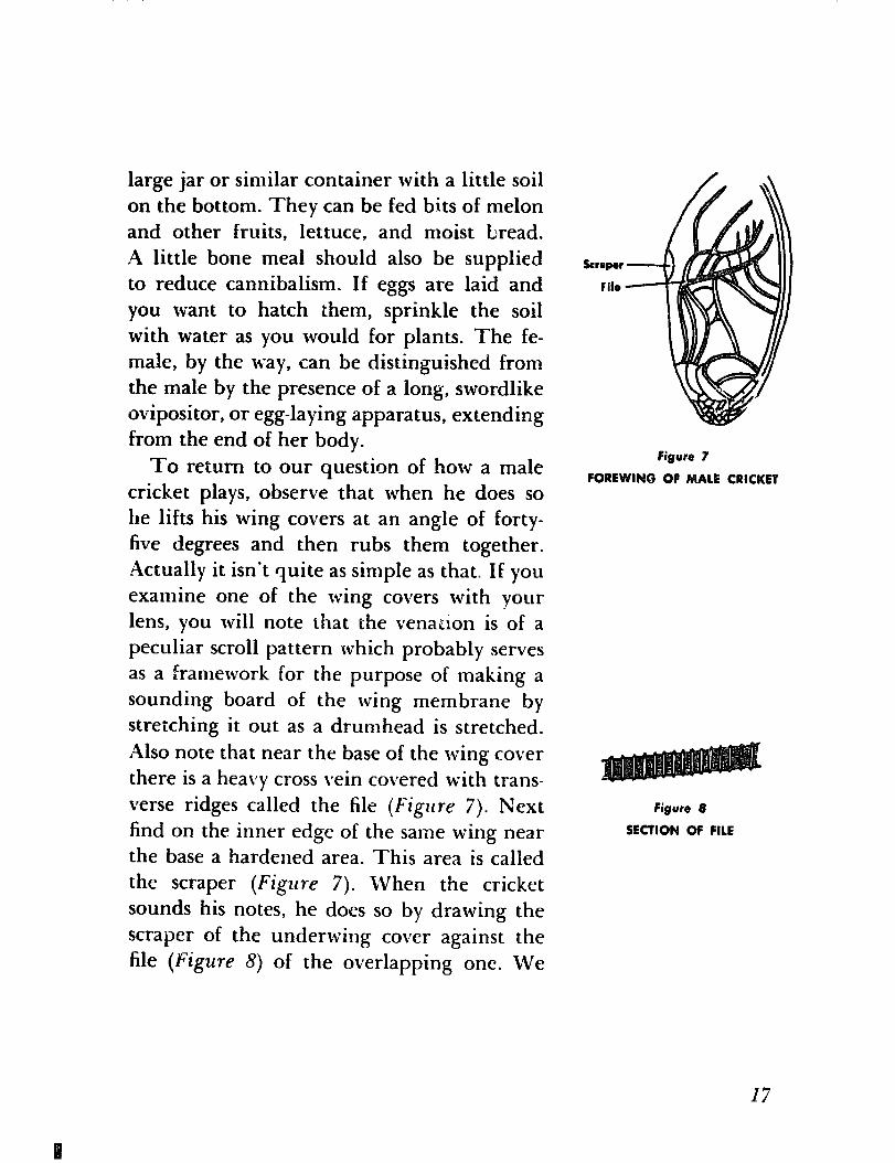
Also note that near the base of the wing cover
there is a heavy cross vein covered with trans-
verse ridges called the file (Figure 7). Next
find on the inner edge of the same wing near
the base a hardened area. This area is called
the scraper (Figure 7). When the cricket
sounds his notes, he does so by drawing the
scraper of the underwing cover against the
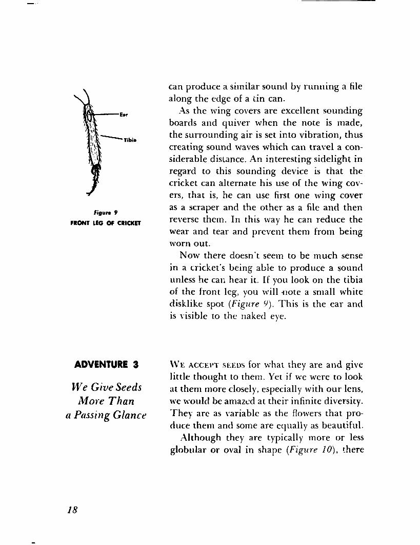
file (Figure 8) of the overlapping one. We
krapar
lit0
figure 7
FOREWINQ OP MALE CRICKET
figure 8
SECTION OF FILE
I7
fiRwe 9
mow LEG of CRICKET
ADVENTURE 3
We Give Seeds More Than
a Passing Glance
can produce a similar sound by running a file
along the edge of a tin can. .\s the wing covers are excellent sounding
boards and quiver when the note is made,
the surrounding air is set into vibration, thus
creating sound waves which can travel a con-
siderable distance. An interesting sidelight in
regard to this sounding device is that the
cricket can alternate his use of the wing cov-
ers, that is, he can use first one wing cover
as a scraper and the other as a file and then
reverse them. In this way he can reduce the
wear and tear and prevent them from being worn out.
Now there doesn’t seem to be much sense
in a cricket’s being able to produce a sound
unless he car; hear it. If you look on the tibia
of the front leg, you will note a small white
disklike spot (Figllre 9). This is the ear and
is ivisible to the naked eye.
LVe ACCEPI SEEDS for what they are and give
little thought to them. Yet if we were to look
at them more closely, especially with our lens,
we would be amazed at their infinite diversity.
They are as variable as the flowers that pro-
duce them and some are equally as beautiful.
Although they are typically more or less
globular or oval in shape (Figure IO), there
figure 10
ROUNClNC RET
figure 1 I
SQUASH
Figure 12
MARIGOLD
f$jurr 13
POUR O‘CLOCK
are seeds that are extremely thin and flat (Figwe II) or greatly elongated (Fa’gwe 12). Seeds, too, may be smooth or wrinkled or pitted or angled or furrowed (Figure 13). There are seeds that are twisted or coiled
I9

Figure 14
MOONSEED
(Figure 14) or otherwise irregularly distorted (Figure 15). Then there are seeds that are more or less covered with hairs or supplied with broad and extremely delicate mem- branous wings to make them wind-borne. In size the variations are equally pronounced. Some seeds are as fine as dust; others several inches in diameter; and, of course, there are seeds that represent all the gradations in be- tween. But it is in the variety of color and color patterns that they show the most con- spicuous external differentiation. I daresay we can find them matching every known color, from shining jet black through the gamut of blue, red, yellow, and other bright tints to the less striking and more somber brown and gray, to say nothing of the mani- fold designs produced by a blending of these colors.
Colors, needless to say, are not without some purpose. Seeds that are scattered by wind and water are, for the most part, in- conspicuously or neutrally colored, but the bright and showy ones are designed to appeal to animal agents of dissemination. Although we may find the numberless variations some- what surprising, what is even more surprising is that such an infinite variety should be pro- duced under conditions that appear to be more or less uniform and constant. What may also seem incongruous is that plants that may
20
appear superficially similar often produce seeds that are strikingly different, or that plants that are wholly unlike often produce seeds that are very much alike. Generally seeds are so characteristic that they serve as useful agents in classification, in some cases being so characteristically differentiated as to be an infallible clue to the identity of the plant that produced them.
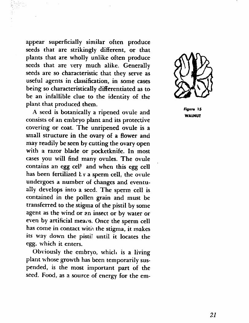
A seed is botanically a ripened ovule and consists of an embryo plant and its protective covering or coat. The unripened ovule is a small structure in the ovary of a flower and may readily be seen by cutting the ovary open with a razor blade or pocketknife. In most cases you will find many ovules. The ovule contains an egg cell and when this egg cell ‘I has been fertilized E Y a sperm cell, the ovule undergoes a numb er of changes and eventu- ally develops into a seed. The sperm cell is contained in the pollen grain and must be transferred to the stigma of the pistil by some agent as the wind or an insect or by water or even by artificial mealIs. Once the sperm cell has come in contact witi\ the stigma, it makes its way down the pistii until it locates the egg, which it enters.
Obviously the embryo, which is a living plant whose growth has been temporarily sus- pended, is the most important part of the seed. Food, as a source of energy for the em-
Figure IS
WALNUT
21

figure 16
BABY’S BREATH
bryo plant until it has developed to the stage where it can manufacture its own food, is stored within the seed. Long ago man discov- ered that he could make use of this reserve food for his own use. The seeds of wheat and rice are today the principal items of diet for millions of human beings, and countless others consume daily large quantities of such seeds as those of corn and barley, oats and rye. Beans and peas, which are also seeds, are eaten extensively. In addition to their value as food, man has found other uses for them. I might mention cottonseed oil, linseed oil, and coconut oil, which are used in the man- ufacture of substitutes for butter and lard, soap, and a variety of other products.
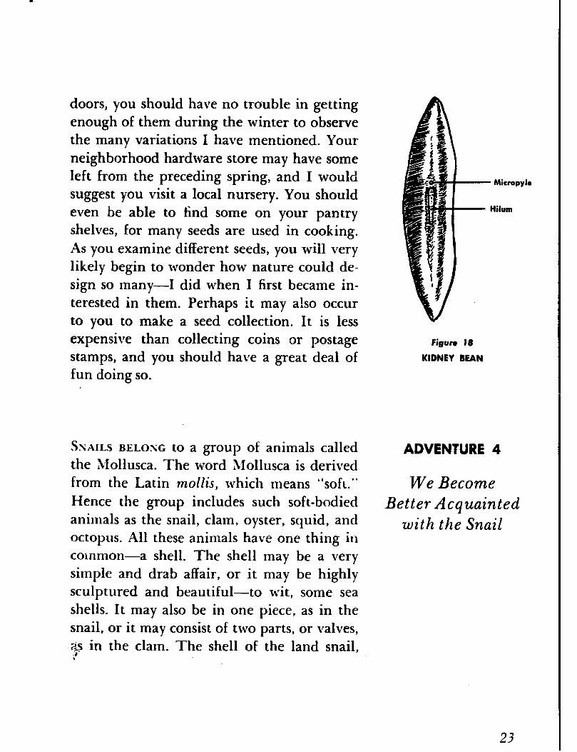
Although seeds exhibit a great diversity of surface markings (Figure 16) and configura- tions (Figure 17), they all agree in showing a minute pore or pit and a scar called the hilum. The pore or pit marks the position of the micropyle, an opening in the ovule through which the sperm cell entered on its \+-a). to the egg cell, and the h.jlum marks the place where the ovule was attached to the ovary. You should be able to find both of these structures with your hand lens (Figure 18).
Figure 17
SPIDERWBRT
Although seeds are more readily available during the summer and early fall, when they may be obtained from almost any plant out-
22
doors, you should have no trouble in getting enough of them during the winter to observe the many variations I have mentioned. Your neighborhood hardware store may have some left from the preceding spring, and I would suggest you visit a local nursery. You should even be able to find some on your pantry shelves, for many seeds are used in cooking. As you examine different seeds, you will very likely begin to wonder how nature could de- sign so many-1 did when I first became in- terested in them. Perhaps it may also occur to you to make a seed collection. It is less expensive than collecting coins or postage stamps, and you should have a great deal of fun doing so.
SNAILS BELONG to a group of animals called the Mollusca. The word hlollusca is derived from the Latin mollis, which means “sofC Hence the group includes such soft-bodied animals as the snail, clam, oyster, squid, and octopus. All these animals have one thing in common -a shell. The shell may be a very simple and drab affair, or it may be highly sculptured and beautiful-to wit, some sea shells. It may also be in one piece, as in the snail, or it may consist of two parts, or valves, &s in the clam. The shell of the land snail, .+- \
Figure 18
KIDNEY BEAN
- Micropyla
Hilum
We Become Better Acquainted
with the Snail
23

which we often find in our gardens, is a spiral cone made from a substance secreted by cer- tain cells and that hardens on exposure to the air. The snail’s soft body is twisted and coiled like th.e shell and extends into the apex, but usually does not completely fill the shell to the tip. .
A snail may seem such an insignificant sort of animal and its actions may not seem to bear watching, but I think you will be in for a surprise if you find one and observe its be- havior through your lens. In this instance
.-..__ a reading glass will prove to be of greater advantage than a pocket magnifier. Place the snail in a tumbler and watch it climb up the glass sides. As you do so you will find that the snail’s foot is one of the most wonderful means of locomotion ever devised by nature. Observe how the foot stretches out and holds on, then contracts, and then again stretches out. All this is accomplished by muscles. Ob- serve, too, how a slime gland at the anterior end of the foot deposits a film of mucus on which the animal travels. It lays down a side- walk, as it were, ahead of itself, and this side- walk is always the same whether the path is rough or smooth, uphill or downhill.
Although the shell provides the snail with a maximum amount of protection, it would seem to be something of a disadvantage to carry it around wherever it goes. Yet in spite
24
-% its cumbersome burden the little animal gets about remarkably ~~31 even though it will never set any speed records. Watch it for .a while and you will discover that its pace is always about the same. It may be two inches per minute, tell feet per hour, or two hundred and forty feet per day if the animal keeps constantly on the move. It also appears to progress forward without any apparent muscular effort.
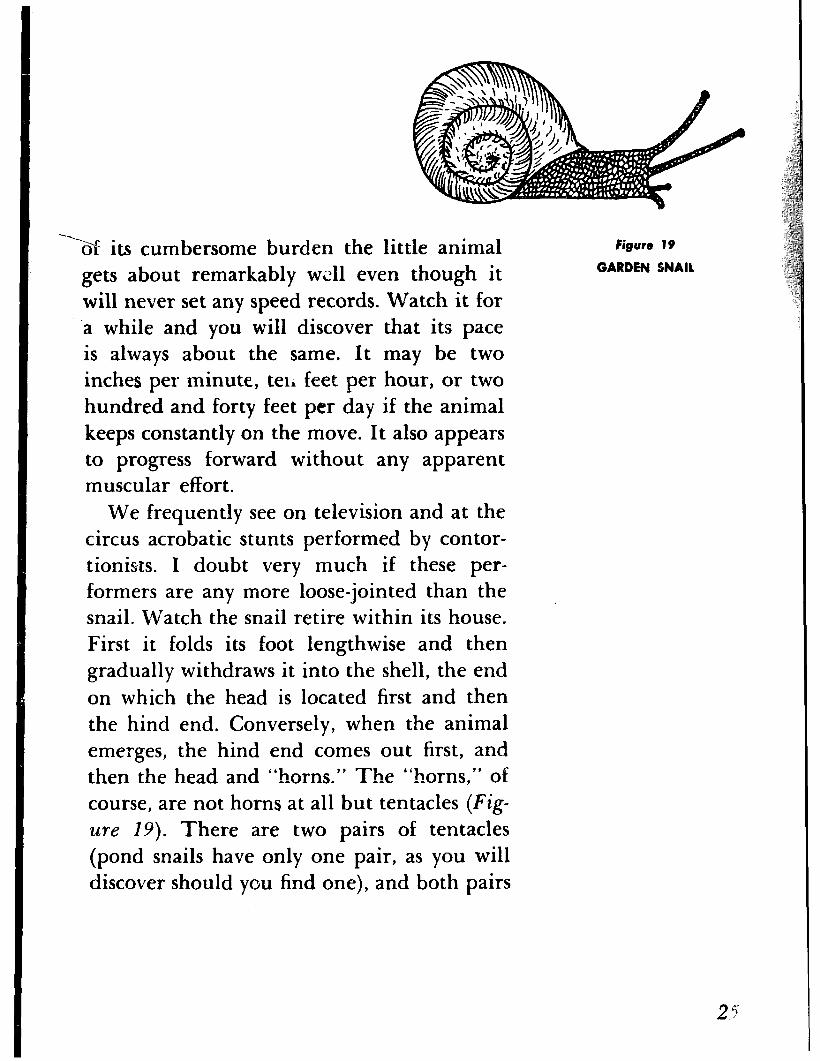
We frequently see on television and at the circus acrobatic stunts performed by contor- tionists. I doubt very much if these per- formers are any more loose-jointed than the snail. Watch the snail retire within its house. First it folds its foot lengthwise and then gradually withdraws it into the shell, the end on which the head is located first and then the hind end. Conversely, when the animal emerges, the hind end comes out first, and then the head and “horns.” The “horns,” of course, are not horns at all but tentacles (Fig- ure 19). There are two pairs of tentacles (pond snails have only one pair, as you will discover should you find one), and both pairs
Figure I9
GARDEN 4NAll
are provided with little round, knoblike tips. On the longer pair the tips serve as eyes; on the shorter pair it is believed they function as organs of smell. It is said that a snail can detect food as, far away as twenty inches.
You can determine this for yourself if you get a piece of apple or other soft food and place it at various distances. Observe how the animal uses its eyes to explore its surround- ings and what happens if you place an object too close to it. Once it has located the piece of apple or fruit, you will find that it won’t be long before it has made a good-sized hole in it. You will also observe that its table man- ners could be improved upon. It is a hopeless slobberer. The mouth is located directly be- low the tentacles and is furnished with one or more chitinous jaws and a long, ribbonlike tongue (the radula) that is covered with horny teeth. The snail uses its tongue as a rasp to file the surfaces of leaves or other food. The teeth vary in number, form, size, and arrangement in the different species of snails and are of considerable value in classification. If you want to examine these teeth more closely, you will need a microscope.
Before you return the snail to its natural cnGronment, examine the skin with your pocket magnifier. It will remind you of an alligator’s skin-rough and divided into plates with a surface like pebbled leather.

THE RUSTING OF IRON is a common occur- ADVENTURE 5 rence, and we see evidence of it everywhere. Few of us have not sustained some loss or damage through rust, and perhaps a few of us have had the painful experience of having stepped on a rusty nail. The tendency of iron .-s,. -
We Inquire into the Nature
of Rusting
to rust is its most unfavorable property, and millions of dollars are spent annually to fight this enemy who, silently and unseen, is con- stantly bent on destruction.
You are taught in school that rusting is due to the action of oxygen in the air on the iron, which results in the formation of a reddish- brown substance called iron oxide. Actually it is not quite so simple. Furthermore, iron is not the only metal that can “rust.” Most metals, when exposed to the air, are attacked by the elements and compounds present in the atmosphere. The phenomenon is gen- erally known as corrosion; when applied to iron it is called rusting.
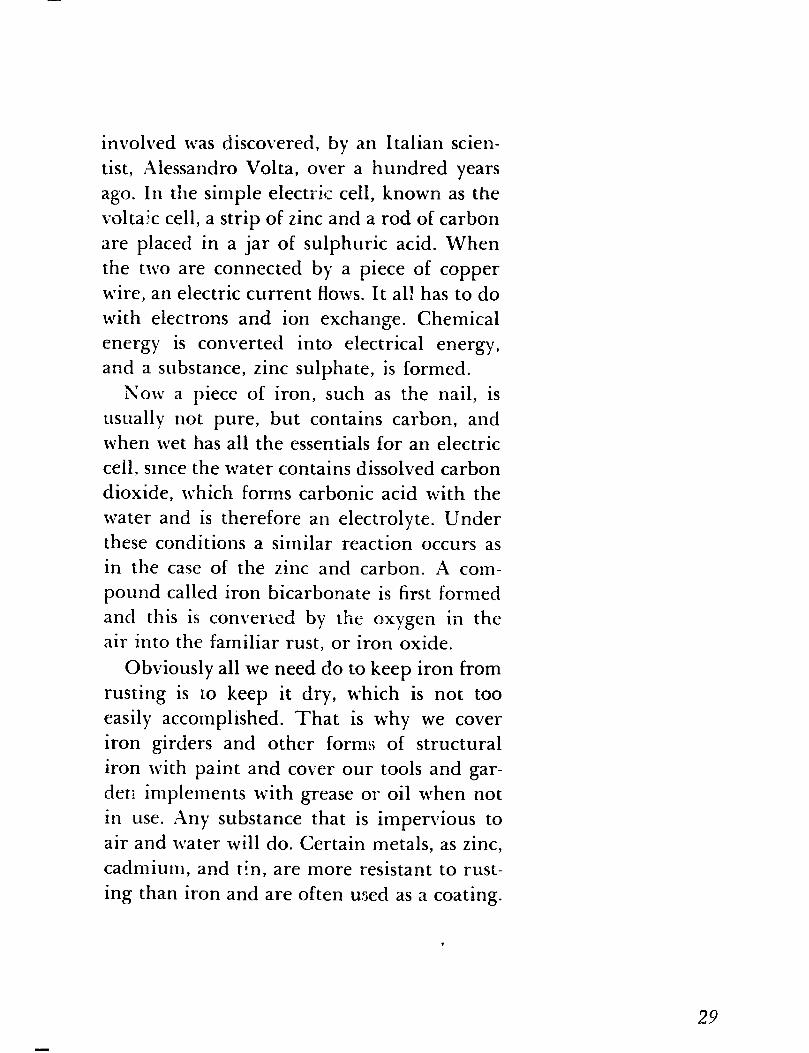
Examine a new and clean nail and note its appearance. Now wet it and leave it outdoors for a few days or until you observe brown spots on it. Look at these brown spots through your lens and you will find that they appear as incrustations that you can break off into small pieces with your fingernail. Next examine a really rusty nail-one that has been exposed to the air for a long time and is completely covered with rust. You will
27
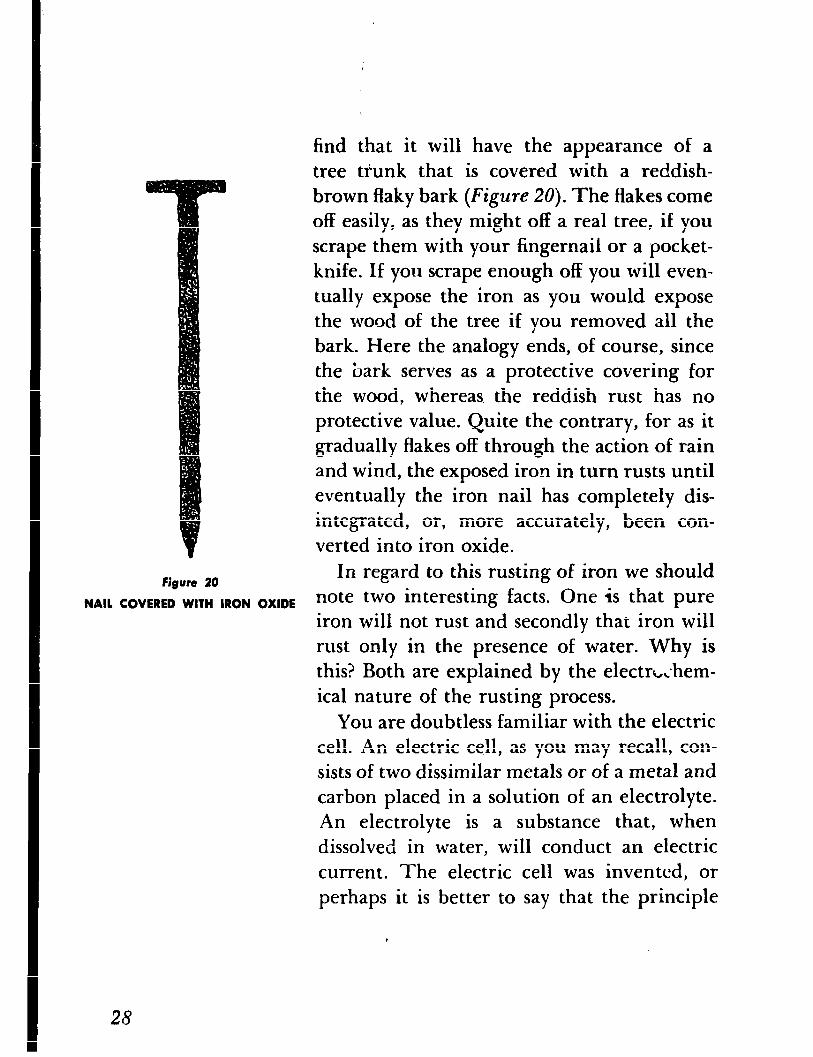
Figure 20
NAIL COVERED WITH IRON OXIDE
,
find that it will have the appearance of a
tree tfunk that is covered with a reddish-
brown flaky bark (Figure 20). The flakes come
off easily, as they might off a real tree, if you
scrape them with your fingernail or a pocket-
knife. If you scrape enough off you will even-
tually expose the iron as you would expose
the wood of the tree if you removed all the
bark. Here the analogy ends, of course, since
the bark serves as a protective covering for
the wood, whereas. the reddish rust has no
protective value. Quite the contrary, for as it
gradually flakes off through the action of rain
and wind, the exposed iron in turn rusts until
eventually the iron nail has completely dis-
integrated, or, more accurately, been con-
verted into iron oxide.
In regard to this rusting of iron we should
note two interesting facts. One is that pure
iron will not rust and secondly that iron will
rust only in the presence of water. Why is
this? Both are explained by the electrL;hem-
ical nature of the rusting process.
You are doubtless familiar with the electric
cell. 14n electric cell, as you may recall, con-
sists of two dissimilar metals or of a metal and
carbon placed in a solution of an electrolyte.
An electrolyte is a substance that, when
dissolved in water, will conduct an electric
current. The electric cell was invented, or
perhaps it is better to say that the principle
,
25
involved was discovered, by an Italian scien-
tist, Alessandro Volta, over a hundred years
ago. In the simple electric cell, known as the
voltajc cell, a strip of zinc and a rod of carbon
are placed in a jar of sulphuric acid. When
the two are connected by a piece of copper
wire, an electric current flows. It all has to do
with electrons and ion exchange. Chemical
energy is converted into electrical energy,
and a substance, zinc sulphate, is formed.
Now a piece of iron, such as the nail, is
usually not pure, but contains carbon, and
when wet has all the essentials for an electric
cell, smce the water contains dissolved carbon
dioxide, which forms carbonic acid with the
water and is therefore an electrolyte. Under
these conditions a similar reaction occurs as
in the case of the zinc and carbon. A com-
pound called iron bicarbonate is first formed
and this is converisd by the rixygen in the air into the familiar rust, or iron oxide.
Obviously all we need do to keep iron from
rusting is to keep it dry, which is not too
easily accomplished. That is why we cover
iron girders and other forms of structural
iron with paint and cover our tools and gar-
den implements with grease or oil when not
in use. Xny substance that is impervious to
air and water will do. Certain metals, as zinc,
cadmium, and tin, are more resistant to rust-
ing than iron and are often used as a coating.
29
The iron is dipped into the melted metal and
a thin layer of it adheres to the iron. Iron
thus coated is called galvanized iron. Another
method of attacking this age-old problem of
rusting is to alloy iron with other metals. An
alloy is merely a mixture of two or more
metals. Stainless steel, for instance, which is
useful in building streamlined trains, for
metal trim on buildings and automobiles,
and for kitchenware, is a mixture of chro-
mium, nickle, and iron.
ADVENTURE 6 THE INSECT BROWNIES are very small insects
We Meet and are more appropriately known, at least
to entomologists, as tree hoppers. They be-
the Insect Brownies long to the family of insects called the Mem-
bracidae and are very well named, since most
of them live on trees and hop vigorously
when disturbed. Not all of them, however,
live on trees; some live on bushes and still
others on grasses and other herbaceous plants.
If these insects are more correctly known
as tree hoppers, why are they also called in-
s&t brownies? To answer this question, you
first have to know what brownies are. In folk-
lore they are believed to be good-natured
goblins who are supposed to do various
household chores by night. They have been
pictured as whimsical little people with
30
quaint, if not grotesque, faces. If you can
picture in your mind these little imaginary
people and then look a few of the tree hop-
pers full in the face, I think you will see the
reason for calling them insect brownies.
A well-known entomologist once remarked
that “Nature must have been in a joking
mood when tree hoppers were developed.”
But: I am not so sure that she was in a joking
mood; on the contrary, I think she was quite
serious when she designed these little insects,
for their grotesque and bizarre appearance is
not without value. They appear so much like
spines and other plant structures in shape or
form that they are not readily seen and thus
escape detection by their enemies. The tree
hoppers provide us with innumerable exam-
ples of what we call protective resemblance.
I might mention just one-the common lit-
tle tree hopper of the bittersweet. Its thorn-
like process resembles a thorn so closely that
the insect can be distinguished from a real
thorn only with difficulty.
How aud where can we obtain these in-
sects? You need an insect net, which you can
make yourself or buy for a nominal sum, and
a killing jar. The latter is merely a bottle
with a screw cover with a piece of cotton at-
tached to it so that the cotton will hang down-
ward in the bottle. The killing jar serves as
a lethal chamber, and in order for it to func-
31
tion all we need do is to soak the cotton in
a fluid that will evaporate quickly. Carbon
tetrachloride, which is used as a cleaning
fluid and w,hich can be purchased at a drug-
store, will do nicely. It is not inflammable
like alcohol or some other fluids we could use,
but since it will kill the insects it is best not
to inhale too much of it.
The best way to get tree hoppers is to walk
through the grass of a field and swish the
insect net back and forth through the grass
or the wild flowers growing there. Do not
swish your net among the branches of a shrub
or tree, as it is very apt to get caught in them
and tear. When you have passed your net
through the grass a few times, examine it.
You should find a large number of various
insect5 crawling about in it. Now remove
the cover from the killing jar and empty the
contents of the net into the bottle, then re-
place the cover and wait a few moments until
the fumes of the carbon tetrachloride have
done their work. When all the insects are
dead, remove the cover and shake the insects
out onto a piece of paper or into a cardboard
Figure 21
TREE HOPPER
box where you can pick them over for tree
hoppers. A pair of forceps or tweezers, such as
you can get a< the five-and-ten-cent store, will
facilitate your handling the insects and will
help you in holding them up when you exarn-
ine them.
32
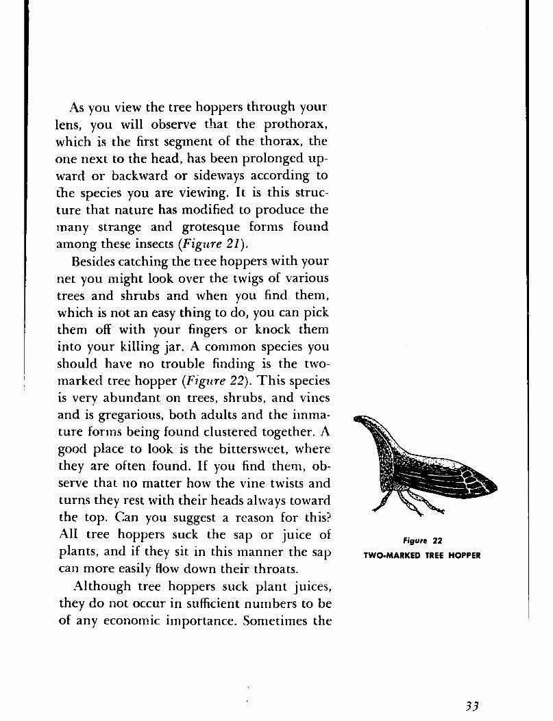
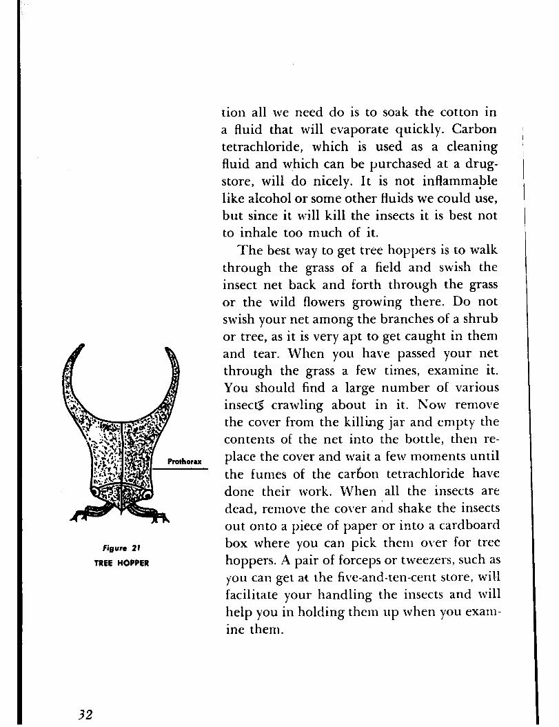
As you view tl le tree hoppers through your
lens, you will observe that the prothorax,
which is the first segment of the thorax, the
one next to the head, has been prolonged up-
ward or backward or sideways according to
the species you are viewing. It is this struc-
ture that nature has modified to produce the
many strange and grotesque forms found
among these insects (Figure 21).
Besides catching the tree hoppers with your
net you might look over the twigs of various
trees and shrubs and when you find them,
which is not an easy thing to do, you can pick
them off with your fingers or knock them
into your killing jar. A common species you
should have no trouble finding is the two-
marked tree hopper (Figure 22). This species
is very abundant on trees, shrubs, and vines
and is gregarious, both adults and the imma-
ture forms being found clustered together. A
good place to look is the bittersweet, where
they are often found. If you find them, ob-
serve that no matter how the vine twists and
turns they rest with their heads always toward
the top. Can you suggest a reason for this?
All tree hoppers suck the sap or juice of
plants, and if they sit in this manner the sap
can more easily flow down their throats.
Although tree hoppers suck plant juices,
they do not occur in sufficient numbers to be
of any economic importance. Sometimes the
Figure 22
TWO-MARKED TREE HOPPER
33
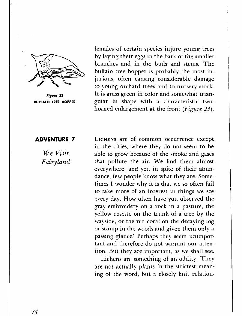
Figure 23
BUFFALO TREE HOPPER
ADVENTURE 7
We Visit Fuiryland
females of certain species injure young trees
by laying their eggs in the bark of the smaller
branches and in the buds and stems. The
buffalo tree hopper is probably the most in-
jurious, often causing considerable damage
to young orchard trees and to nursery stock.
It is grass green in color and somewhat trian-
gular in shape with a characteristic two-
horned enlargement at the front (Figjlre 23).
LICHENS are of common occurrence except
in the cities, where they do not seem to be
able to grow because of the smoke and gases
that pollute the air. We find them almost
everywhere, and yet, in spite of their abun-
dance, few people know what they are. Some-
times I wonder why it is that we so often fail
to take more of an interest in things we see
every day. How often have you observed the
gray embroidery on a rock in a pasture, the
yellow rosette on the trunk of a tree by the
wayside, or the red coral on the decaying log
or stump in the woods and given them only a
passing glance? Perhaps they seem unimpor-
tant and therefore do not warrant our atten-
tion. But they are important, as we shall see.
Lichens are something of an oddity. They
are not actually plants in the strictest mean-
ing of the word, but a closely knit relation-
34
ship of two entirely dissimilar plants living
together for mutual benefit, a sort of partner-
ship that biologists call mutualism. The two
plants are an alga and a fungus. The alga is
a relative of the simple green plants that are
found on damp stones or on the shady sides
of houses and trees. The fungus is a relative
of the mushrooms that appear magically in
our gardens after a heavy rain and the molds
that so often appear on our foodstuffs, such
as bread and jellies and cheese. The fungus
has lost the ability, if it ever did have it, of
manufacturing its own food, but instead has
acquired the power of absorbing large quan-
tities of water. If exposed to dry air the alga
will perish, but when kept moist is capable
of taking various elements from the air and
converting them into food. So it is the function
of the fungus to provide the partnership with
water, and the alga to furnish the food.
Though the partnership may seem a
strange one, it is a most successful one, for
lichens can exist where no other plants can
grow-on a bare alpine peak, in the arctic
wastes, in a tropical desert. They need no soil
-a bare rock will do or any kind of surface
on which they can obtain a foothold. Because
of their ability to live in such inhospitable
places it would appear that they must be
rather remarkable, as indeed they are. The
mechanical contrivances that permit them to
35

survive under what appears to be the most
precarious of conditions have a strong appeal
to the physicist, and the chemical processes
that take place within them are of no less in-
terest to the chemist, for, among other things,
they secrete powerful acids that etch the
rocks and break them up, thereby providing
needed minerals for their own survival. The
biologist regards the lichens with even
greater respect because they are able to cre-
ate soil in which other plants can grow. They
represent the pioneer stage in a series of
stages called ecological succession, a term
given to the transition of a barren area into
the climax forest through the intervening
steps of a small pool, then a larger pond,
followed by the swamp and marsh, and then
the meadow and field. You will learn more
about this when you study biology or, more
specifically, that phase of biology called
ecology.
Apart from their primary purpose of con-
verting inhospitable waste ground into a soil
suitable for other plants to grow in, some
species also serve as food for various animals,
such as the reindeer and caribou. Perhaps
you have heard of reindeer moss, which is a
lichen and not a moss. Not to be outdone by
the animals, man has also made use of lichens
as food. The people in Sweden at one time
made a bread from one of them, and another
species was once used in a Siberian monastery for a beer. The manna of the Israelites is sup- posed to have been a lichen. Drugs as well as various dyes have been obtained from certain species. The litmus we use in our chemical laboratories is obtained from a lichen.
But I think we have talked enough about the lichens. Let us go outdoors and look at them. Where shall we go? To some neglected pasture? To the woods? It doesn’t matter. Wherever we turn, whether it is the marsh, the shore of a pond, the bank of a river, the roadside, we find them decorating trees, stumps, fallen logs, fence rails, rocks, almost any surface that will provide them with a foothold. And how may we recognize them? Almost any flat or ruffled and rootless growth of almost any color is likely to be a lichen, particularly if it bears flattened dish-shaped or saucer-shaped colored disks or cushions or if it branches like a coral or if it hangs like fringes from the branches of a tree. Best of all, we can find them at any time of the y:ar, though they are at .tht:ir best in spring and fall or even in the winter, for they like mois- ture and in the dry atmosphere of summer tend to dry out. In the summer, too, many of them are hidden by the foliage and other leafy vegetation and sometimes are rather difficult to find.
The first lichen to attract our attention will
37
FQpJro 25
SCARLET CRESTED CLADONIA
38
Figure 24
GRAY ETAR LICHEN
Figure 26
GOBLET LICHEN
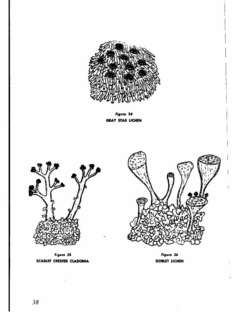
probably be the gray star lichen since it is fairly common on trees and rocks. It is a small but rather conspicuous silvery-gray or slate-gray rosette (Figure 24). Let us look closely at it with our hand lens and we will find small, flat disks, dark brown or black or frosted pale gray, with pale gray rims that may be smooth, toothed, or broken (Figure 24). The flat disks are the fruiting structures in which spores are produ.ced, which perform the same function as the seeds of the higher plants.
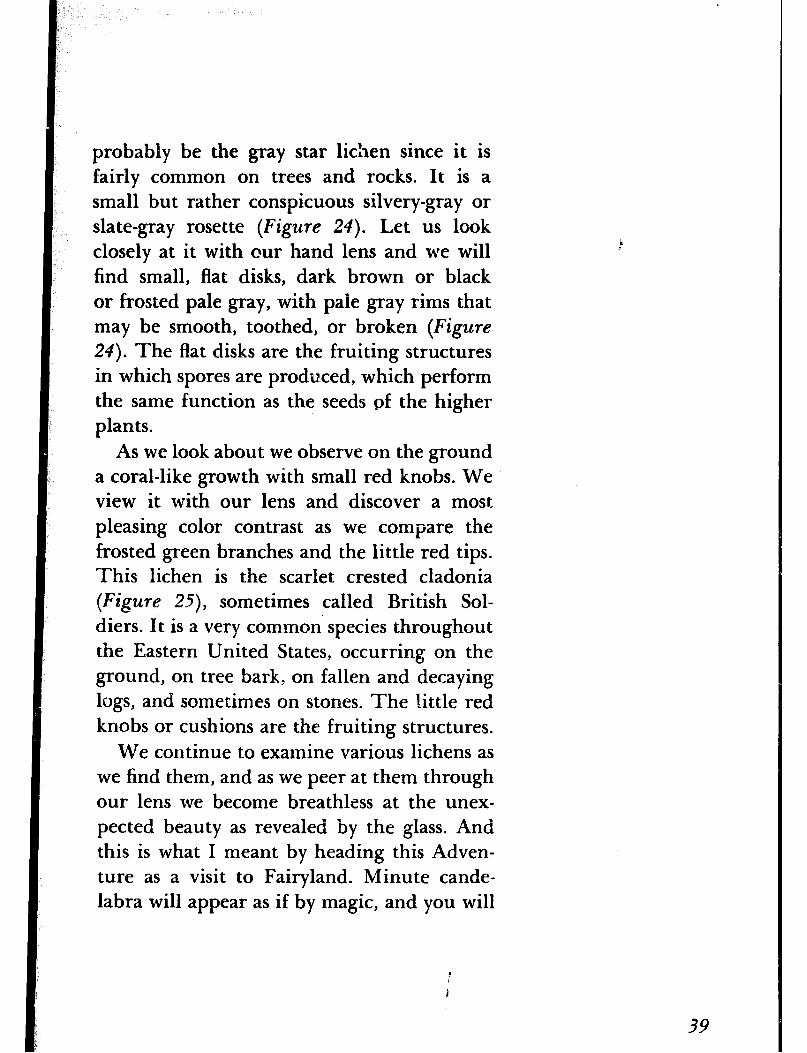
As we look about we observe on the ground a coral-like growth with small red knobs. We view it with our lens and discover a most pleasing color contrast as we compare the frosted green branches and the little red tips. This lichen is the scarlet crested cladonia (Figure 25) sometimes called British Sol- diers. It is a very common species throughout the Eastern United States, occurring on the ground, on tree bark, on fallen and decaying logs, and sometimes on stones. The little red knobs or cushions are the fruiting structures.
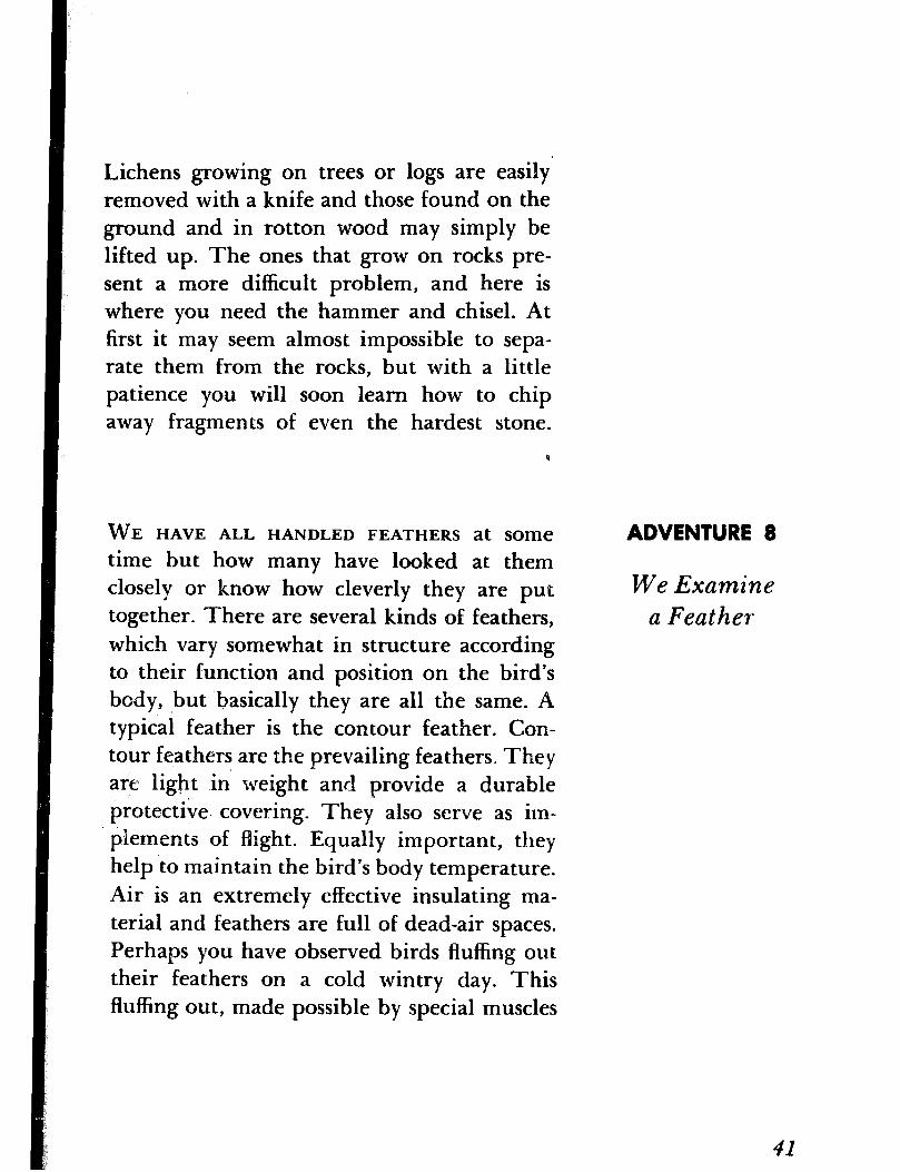
We continue to examine various lichens as we find the.m, and as we peer at them through our lens we become breathless at the unex- pected beauty as revealed by the glass. And this is what I meant by heading this Adven- ture as a visit to Fairyland. Minute cande- labra will appear as if by magic, and you will
39

see tiny goblets that may have served the pot-
ter as models for his art (Figure 26). Examine
the unkempt and rather dirty incrustations
on the surface of a rock and you may well
wonder if some ancient sculptor did not find
inspiration in the beautiful designs traced by
them. The intricate tracery that many of the
lichens have embroidered on a bare and
rather forbidding surface are no less remark-
able than those you will find in the ornamen-
tation of some marble temple. But I will let
you discover all this for yourself.
As you become better acquainted with the
lichens, I think you will become fascinated,
too, with the many curious names that have
been given to them. Old Man’s Beard, the
Dog Peltigera, the Crumpled Bat’s Wing,
and the Mustard Seed are only a few. They
are all well named, too, as you will observe
when you find them. The most interesting of
all the lichens is, perhaps, the Rock Tripe.
With every change in humidity the lichen
curls and uncurls, writhes and twists, alter-
nately covering the rock on which it grows
with a green and black coating.
Once you have become interested in these
odd plants, you may want to build up i col-
lection. They keep well, for the most part,
and even when dry may be made fresh look-
ing by adding a little water. All you need is
a knife and a hammer and a cold chisel.
40
Lichens growing on trees or logs are easily removed with a knife and those found on the ground and in rotton wood may simply be lifted up. The ones that grow on rocks pre- sent a more difficult problem, and here is where you need the hammer and chisel. At first it may seem almost impossible to sepa- rate them from the rocks, but with a little patience you will soon learn how to chip away fragments of even the hardest stone.
WE HAVE ALL HANDLED FEATHERS at SOITle
time but how many have looked at them closely or know how cleverly they are put together. There are several kinds of feathers, which vary somewhat in structure according to their function and position on the bird’s body, ,but basically they are all the same. A typical feather is the contour feather. Con- tour feathers are the prevailing feathers. They are light ih weight and provide a durable protective. covering. They also serve as im- plements of flight. Equally important, they help to maintain the bird’s body temperature. Air is an extremely effective insulating ma- terial and feathers are full of dead-air spaces. Perhaps you have observed birds fluffing out their feathers on a cold wintry day. This fluffing out, made possible by special muscles
ADVENTURE 8
We Examine a Feather
41
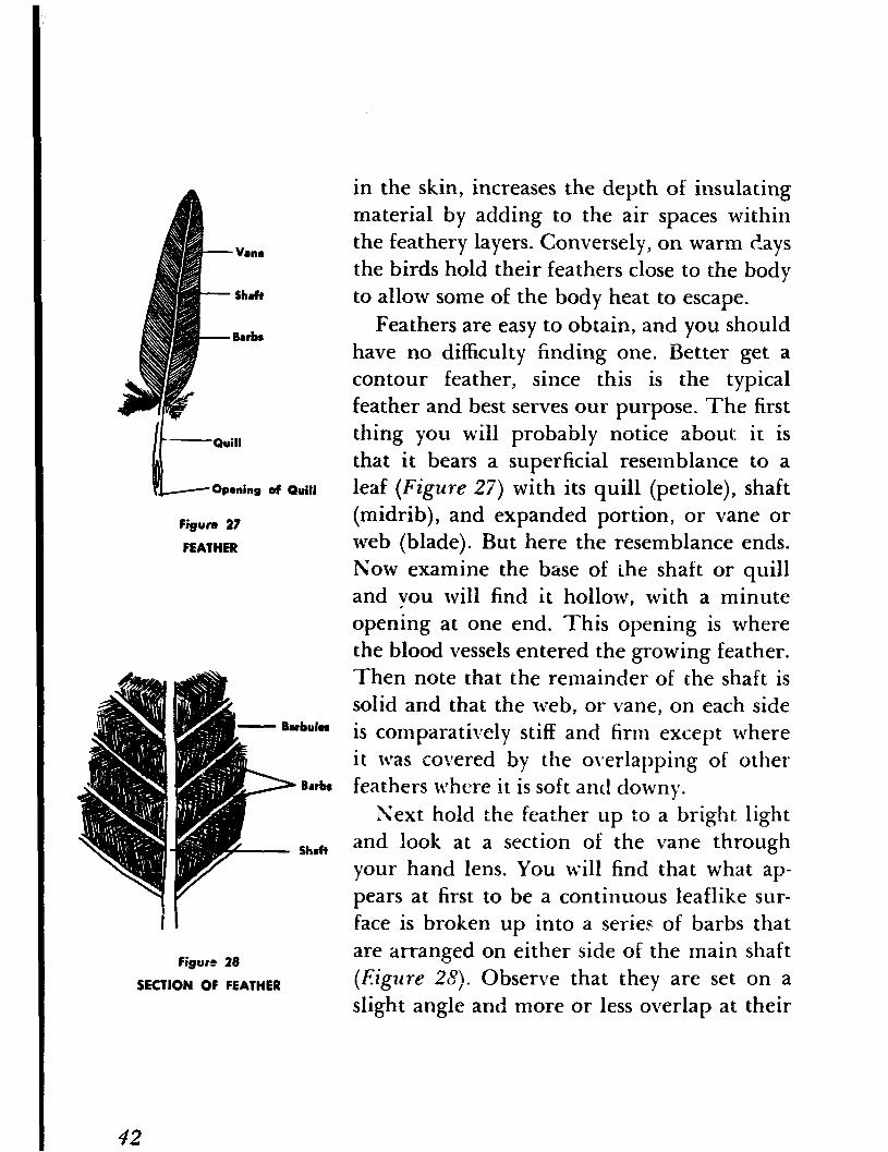
. Vane
Shaft
6L Quill
Opening of Quill
Figure 27
FEATHER
Figure 28
SECTION OF FEATHER
in the skin, increases the depth of insulating
material by adding to the air spaces within
the feathery layers. Conversely, on warm ?.ays
the birds hold their feathers close to the body
to allow some of the body heat to escape.
Feathers are easy to obtain, and you should
have no difficulty finding one. Better get a
contour feather, since this is the typical
feather and best serves our purpose. The first
thing you will probably notice about it is
that it bears a superficial resemblance to a
leaf (Figure 27) with its quill (petiole), shaft
(midrib), and expanded portion, or vane or
web (blade). But here the resemblance ends.
Now examine the base of the shaft or quill
and you will find it hollow, with a minute
opening at one end. This opening is where
the blood vessels entered the growing feather.
Then note that the remainder of the shaft is
solid and that the web, or vane, on each side
is comparatively stiff and firm except where
it was covered by the overlapping of other
feathers where it is soft and downy.
Next hold the feather up to a bright light
and look at a section of the vane through
your hand 1 ens. You will find that what ap-
pears at first to be a continuous leaflike sur-
face is broken up into a series of barbs that
are arranged on either side of the main shaft
(Eigzlre 28). Observe that they are set on a
slight angle and more or less overlap at their
42
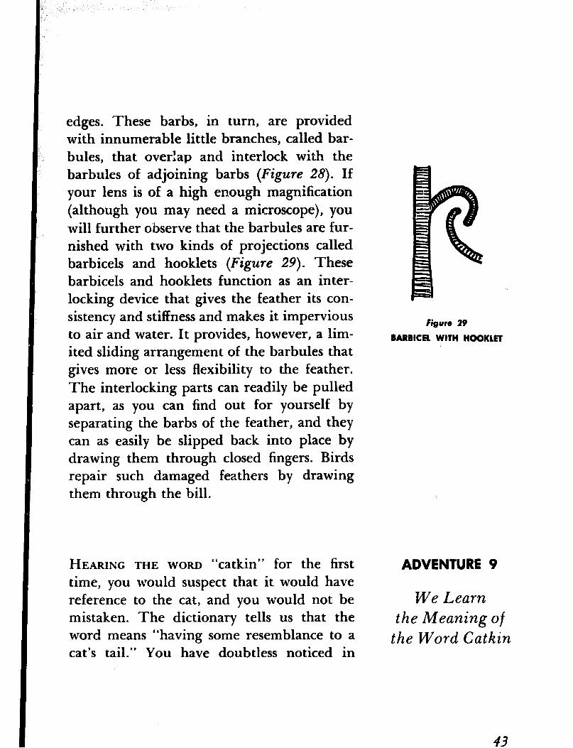
edges. These barbs, in turn, are provided with innumerable little branches, called bar- bules, that overlap and interlock with the barbules of adjoining barbs (Figure 28). If your lens is of a high enough magnification (although you may need a microscope), you will further observe that the barbules are fur- nished with two kinds of projections called barbicels and hooklets (Figure 29). These barbicels and hooklets function as an inter- locking device that gives the feather its con- sistency and stiffness and makes it impervious to air and water. It provides, however, a lim- ited sliding arrangement of the barbules that gives more or less flexibility to the feather. The interlocking parts can readily be pulled apart, as you can find out for yourself by separating the barbs of the feather, and they can as easily be slipped back into place by drawing them through closed fingers. Birds repair such damaged feathers by drawing them through the bill.
Figure 29
BARBICRU WITH HOOKLET
HEARING THE WORD “catkin” for the first time, you would suspect that it would have reference to the cat, and you would not be mistaken. The dictionary tells us that the word means “having some resemblance to a cat’s tail.” You have doubtless noticed in
ADVENTURE 9
We Learn the Meaning of
the Word Catkin
43

early spring countless tassels hanging from the twigs and branches of certain trees and shrubs. From their fancied resemblance to the cat’s tail they were given the name “cat- kin,” and though they are also known bo- tanically as aments, they are today more gen- erally called catkins. Actually they are flower clusters.
Flowers such as the rose and lily and tulip are borne singly- at the end of a long flower stalk and are called single or solitary flowers. In most flowering plants the flowers are borne in groups or clusters. A group or cluster re- sults from the branching of the main flower stalk and is known as an inflorescence. There are different kinds of inflorescences, as the raceme, spike, cyme, corymb, umbel and, of course, the catkin, depending on the manner in which the flowers are arranged.
The catkins of the speckled alder” are among the first to appear in early spring. This small tree, so named because its stems are sprinkled or speckled with numerous and conspicuous light gray spots that are actually breathing pores called lenticels, is a water-
* The speckled alder is a shrub of the ncrthern states but a closely related species, the smooth alder, is widely distributed throughout the south. The two, which may often be found growing together where their ranges overlap, are very much alike in habit ‘and the catkins of either may be examined since they are similar.
44
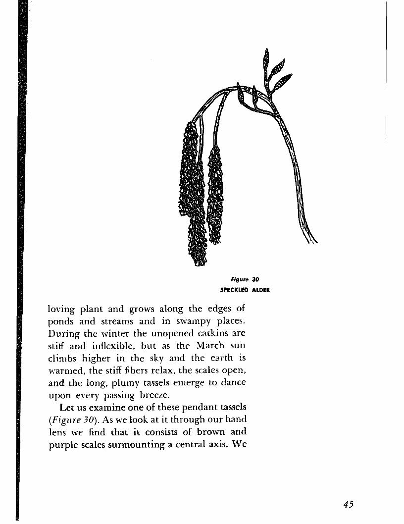
Figure! 30
SPECKLED ALDER
loving plant and grows along the edges of
ponds and streams and in swampy places.
During the winter the unopened catkins are
stii-f and inflexible, but as the March sun
climbs higher in the sky and the earth is
warmed, the stiff fibers relax, the scales open,
and the long, plumy tassels emerge to dance
upon every passing breeze.
Let us examine one of these pendant tassels
(Figure ?O). As we look at it through our hand
lens we find that it consists of brown and purple scales surmounting a central axis. We
45

Anther -
Stigma
Pistil
FiBwe 3OA
DIAGRAM OF A SIMPLE FLOWER
further observe that the scales are set on short stalks and that beneath each scale are three flowers, each having a three-to-five lobed calyx cup and three-to five sta’mens whose anthers are covered with yellow pollerl.
Let us pause at this point and recall our knowledge of botany. In spite of their strik- ing external multiformity flowers are com- paratively simple and uniform in their mode of construction. Figure 3OA shows the dia- gram of a typical flower to which you can compare almost any blossom you have at
46
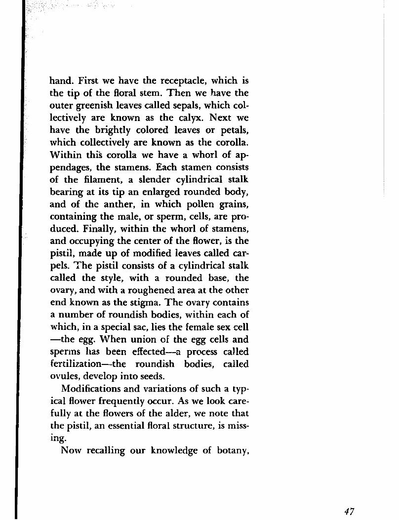
hand. First we have the receptacle, which is the tip of the floral stem. Then we have the outer greenish leaves called sepals, which col- lectively are known as the calyx. Next we have the brightly colored leaves or petals, which collectively are known as the corolla. Within this corolla we have a whorl of ap- pendages, the stamens. Each stamen consists of the filament, a slender cylindrical stalk bearing at its tip an enlarged rounded body, and of the anther, in which pollen grains, containing the male, or sperm, cells, are pro- duced. Finally, within the whorl of stamens, and occupying the center of the flower, is the pistil, made up of modified leaves called car- pels. The pistil consists of a cylindrical stalk called the style, with a rounded base, the ovary, and with a roughened area at the other end known as the stigma. The ovary contains a number of roundish bodies, within each of which, in a special sac, lies the female sex cell -the egg. When union of the egg cells and sperms has been elected---a process called fertilization-the roundish bodies, called ovules, develop into seeds.
Modifications and variations of such a typ- ical flower frequently occur. As we look care- fully at the flowers of the alder, we note that the pistil, an essential floral structure, is miss- ing.
Now recalling our knowledge of botany,
47

we discover that the pistil, an essential floral
structure, is missing. The absence of a pistil
at first appears to be something of a puzzle un-
til we examine one of the shorter erect cat-
kins. Here we find that each of the fleshy
scales, with which the catkin is provided, en-
closes two flowers, each having. a pistil with
a scarlet style. So there are two kinds of cat-
kins on the alder, one having flowers with
stamens only and the other with flowers hav-
ing only pistils. In other words, we have what
are known as staminate and pistillate catkins.
Where both are found on the same tree, as in
the alder, the tree is said to be monoecious.
Examining the flowers still further, we also
observe the absence of petals. As the pollen
grains are transferred from one flower to an-
other by the wind, the petals would only be
a hindrance and impede the wind from pick-
ing up the grains and acting as an effective
agent of pollination. Here is an adaptation of
distinct advantage to the plant.
Qnce pollination has been carried out, the
pistils develop into small cones that resem-
ble miniature pine cones. They consist of
woody scales that protect the seeds formed
beneath them. When the seeds are fully ma-
tured, the scales open and release the seeds,
which are also scattered by the wind.
Next let us turn our attention to the pussy
willow, whose furry catkins are a familiar
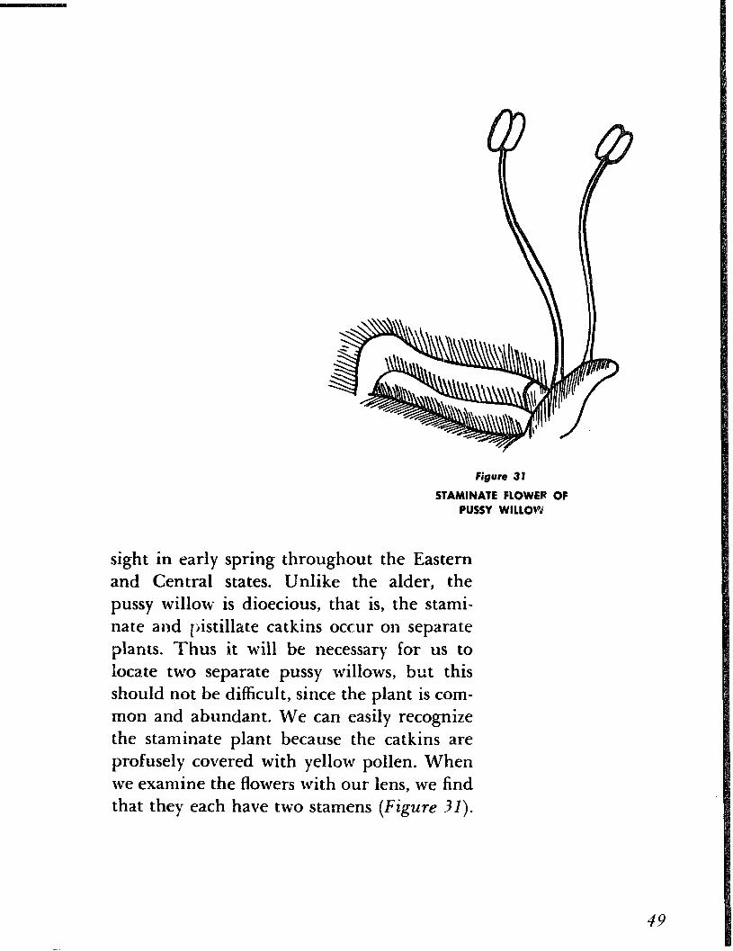
Figure 31
STAMINATE FLOWER OF PUSSY WlLLOvrd
sight in early spring throughout the Eastern
and Central states. Unlike the alder, the
pussy willow is dioecious, that is, the stami-
nate and pistillate catkins occur on separate
plants. Thus it will be necessary for us to
locate two separate pussy willows, but this
should not be difficult, since the plant is com-
mon and abundant. We can easily recognize
the staminate plant because the catkins are
profusely covered with yellow pollen. When
we examine the flowers with our lens, we find
that they each have two stamens (Figure 31).

Figure 32
PlSTlLlAtE FLOWER OF PUSSY WILLOW
Now let us examine a pistillate catkin, which
we can identify by the absence of the yellow
pollen and by the very shape of the flowers
themselves. When we do so we find that each
flower has only a single pistil, with two more
or less divided stigmas (Figure ?Z). Pf we keep
the pistils under observation for a few days,
we will find that they develop into conic-
shaped capsules. These capsules contain the
maturing seeds, and when the latter become
fully developed the capsules will open. The
seeds are furnished with long silky down that
catches in the wind, an effective means that
the pussy willow, in common with other
plants, has evolved to ensure a wide distribu-
tion of its seeds.
Figure 33
STAMINATE FLOWER OF ASPEN
The aspen is said to be the most widely
distributed tree in Norrh America. Most of
us know it as the quaking aspen, because its
leaves quiver or tremble in the slightest
breeze. The catkins appear before the leaves,
are furry, and show a touch of pink. The tree
may be readily identified by its gray-green
bark. Like the willow, the aspen is also dioe-
cious. Note how the scales of the staminate
flowers are deeply cut into three to four linear
divisions and how they are fringed with long,
soft gray hairs (FigzLre 3?). The stamens nurn-
ber from six to twelve. The stigma is two-
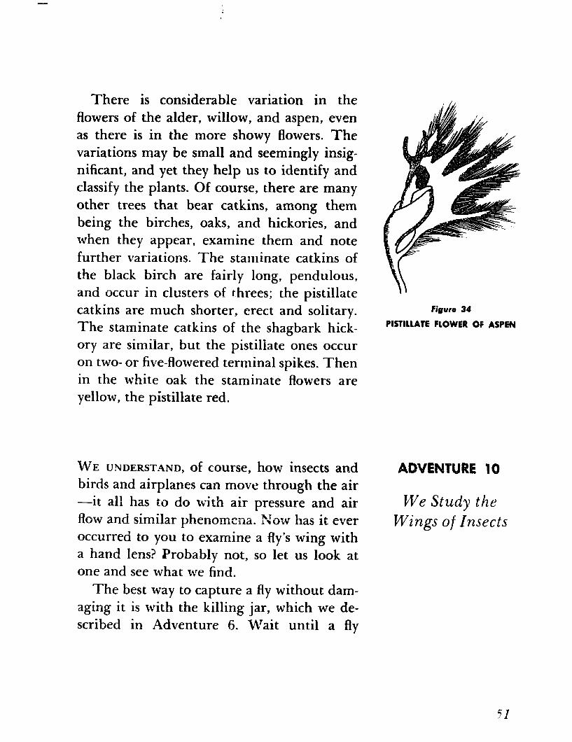
lobed (Figure ?4), and the ovary is sur-
rounded by a broad, oblique disk.
50
There is considerable variation in the
flowers of the alder, willow, and aspen, even
as there is in the more showy flowers. The
variations may be small and seemingly insig- nificant, and yet they help us to identify and classify the plants. Of course, there are many
other trees that bear catkins, among them
being the birches, oaks, and hickories, and
when they appear, examine them and note
further variations. The staminate catkins of
the black birch are fairly long, pendulous,
and occur in clusters of threes; the pistillate
catkins are much shorter, erect and solitary.
The staminate catkins of the shagbark hick-
ory are similar, but the pistillate ones occur
on two- or five-flowered terminal spikes. Then
in the white oak the staminate flowers are
yellow, the pistillate red.
Figure 34
PISTILLATE FLOWER OF ASPEN
WE UNDERST4ND, of course, how insects and
birds and airplanes can move through the air -it all has to do wi.th air pressure and air
flow and similar phenomena. X0-w has it ever
occurred to you to examine a fly’s wing with
a hand lens? Probably not, so let us look at
one and see what we find.
ADVENTURE 10
We Study the Wings of Insects
The best way to capture a fly without dam-
aging it is with the killing jar, which we de-
scribed in Adventure 6. Wait until a fly
arights on a flat surface, since it will then have the least chance of escaping, and then ap- proach it stealthily with the uncovered jar and quickly place the jar over it. The startled insect will fly into the jar, and then you can replace the cover.
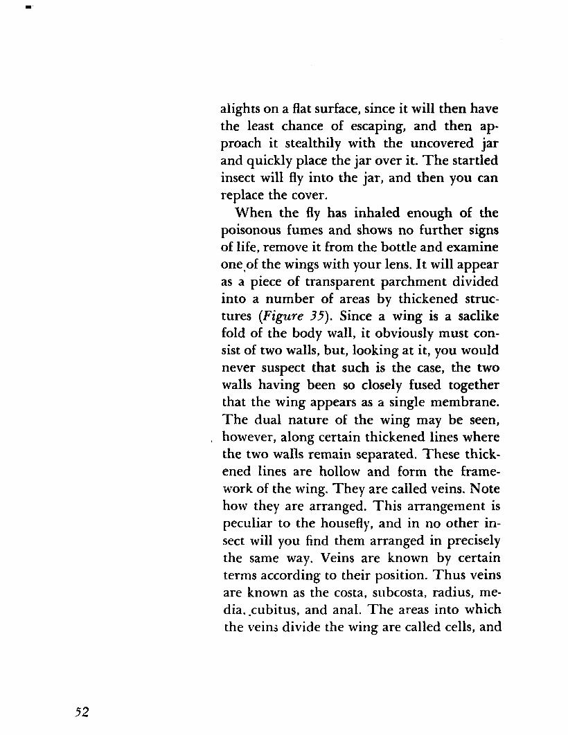
When the fly has inhaled enough of the poisonous fumes and shows no further signs of life, remove it from the bottle and examine one,of the wings with your lens. It will appear as a piece of transparent parchment divided into a number of areas by thickened struc- tures (Figure 35). Since a wing is a saclike fold of the body wall, it obviously must con- sist of two walls, but, looking at it, you would never suspect that such is the case, the two walls having been so closely fused together that the wing appears as a single membrane. The dual nature of the wing may be seen, however, along certain thickened lines where the two walls remain separated. These thick- ened lines are hollow and form the frame- work of the wing. They are called veins. Note how they are arranged. This arrangement is peculiar to the housefly, and in no other in- sect will you find them arranged in precisely the same way. Veins are known by certain terms according to their position. Thus veins are known as the costa, subcosta, radius, me- dia, -cubitus, and anal. The areas into which the veins divide the wing are called cells, and
52
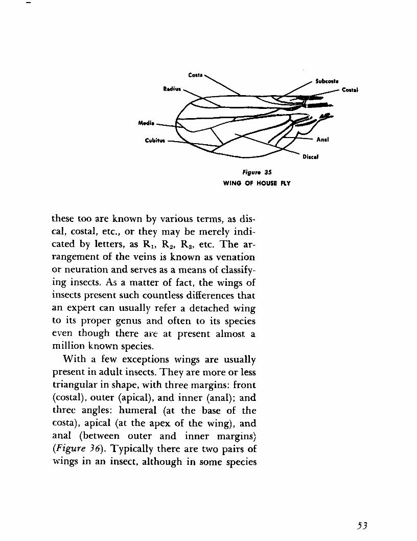
Figure 35
WING OF HOUSE FLY
these too are known by various terms, as dis- cal, costal, etc., or they may be merely indi- cated by letters, as RI, R2, R3, etc. The ar- rangement of the veins is known as venation or neuration and serves as a means of classify- ing insects. As a matter of fact, the wings of insects present such countless differences that an expert can usually refer a detached wing to its proper genus and often to its species even though there are at present almost a million known species.
With a few exceptions wings are usually present in adult insects. They are more or less triangular in shape, with three margins: front (costal), outer (apical), and inner (anal); and three angles: humeral (at the base of the Costa), apical (at the apex of the wing), and anal (between outer and inner margins) (Figure ?6). Typically there are two pairs of wings in an insect, although in some species
53
Figure 34
DIAGRAM OF A WING SHOWING ANGLES AND MARGINS
the females are wingless and in the vast group of true flies (Diptera) the second pair has been lost, having been replaced by knobbed, threadlike organs called halteres. The hal- teres appear to function as balancers, for the fly can no longer maintain its equilibrium if one of them is removed. See if you can locate them.
Next examine the wing of some other kind of insect-a butterfly, dragonfly, grasshopper, a beetle, a bee-better still, a number of them. As you do so, you will find that the front wings are variously modified, in some insects being more useful for protection than for flight. In grasshoppers (Orthoptera) the front wings are leathery and are called teg- mina; in the beetles (Coleoptera) they are
54
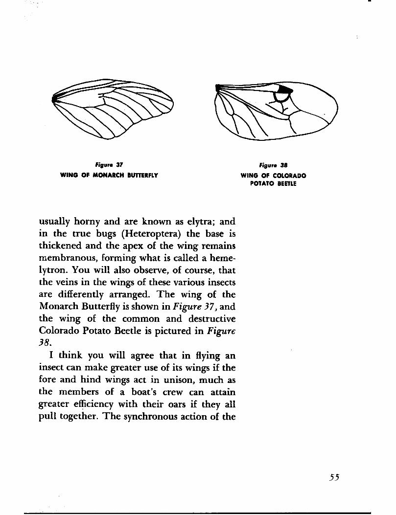
Figure 37
WING OF MONARCH BUTTERFLY
Figure 38
WING OF COLORADO POTATO BEETLE
usually horny and are known as elytra; and in the true bugs (Heteroptera) the base is thickened and the apex of the wing remains membranous, forming what is called a heme- lytron. You will also observe, of course, that the veins in the wings of these various insects are differently arranged. The wing of the Monarch Butterfly is shown in Figure 37, and the wing of the common and destructive Colorado Potato Beetle is pictured in Figure 38.
I think you will agree that in flying an insect can make greater use of its wings if the fore and hind wings act in unison, much as the members of a boat’s crew can attain greater efficiency with their oars if they all pull together. The synchronous action of the
55
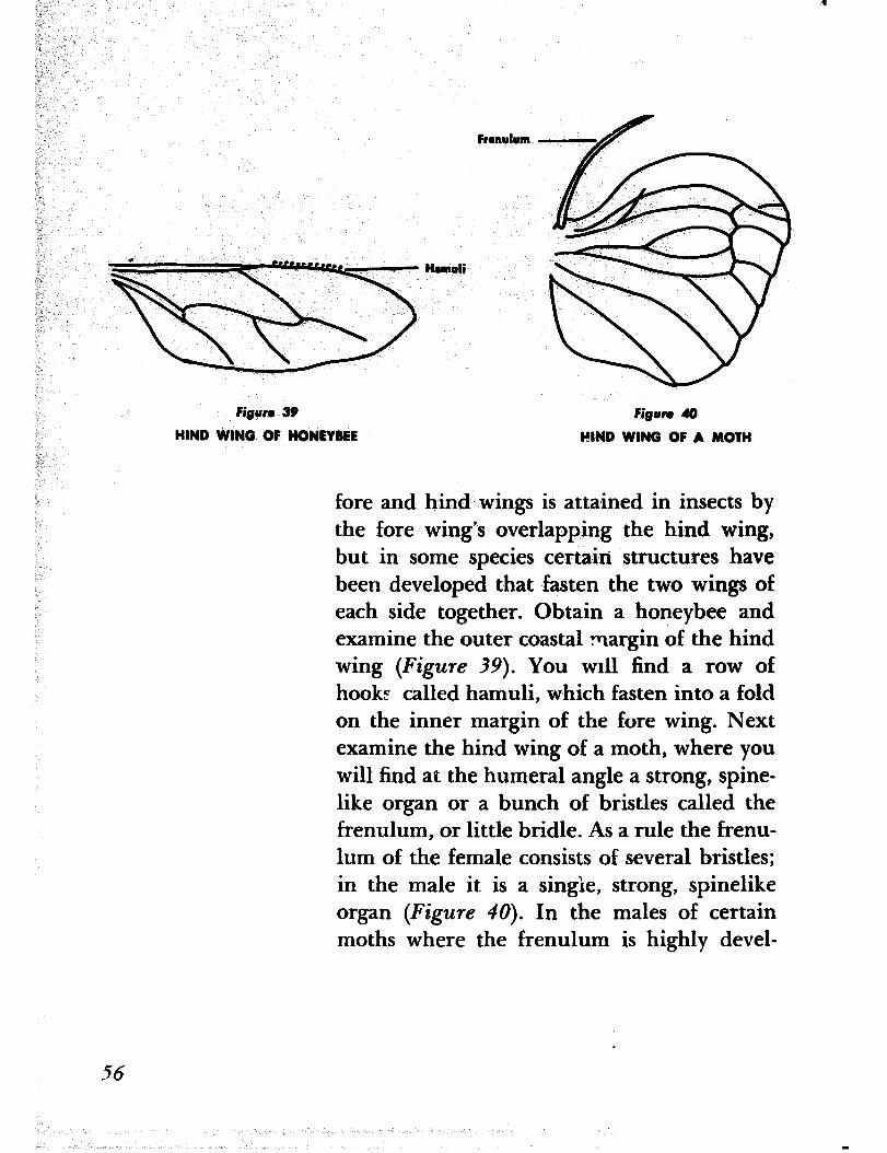
Figure 39 Figure 40
HIND WING OF HONEYBEE HIND WING OF A MOTH
fore and hind wings is attained in insects by the fore wing’s overlapping the hind wing, but in some species certain structures have been developed that fasten the two wings of each side together. Obtain a honeybee and examine the outer coastal margin of the hind wing (Figure 39). You ~111 find a row of hook? called hamuli, which fasten into a fold on the inner margin of the fore wing. Next examine the hind wing of a moth, where you will find at the humeral angle a strong, spine- like organ or a bunch of bristles called the frenulum, or little bridle. As a rule the frenu- lum of the female consists of several bristles; in the male it is a single, strong, spinelike organ (Figure 40). In the males of certain moths where the frenulum is highly devel-
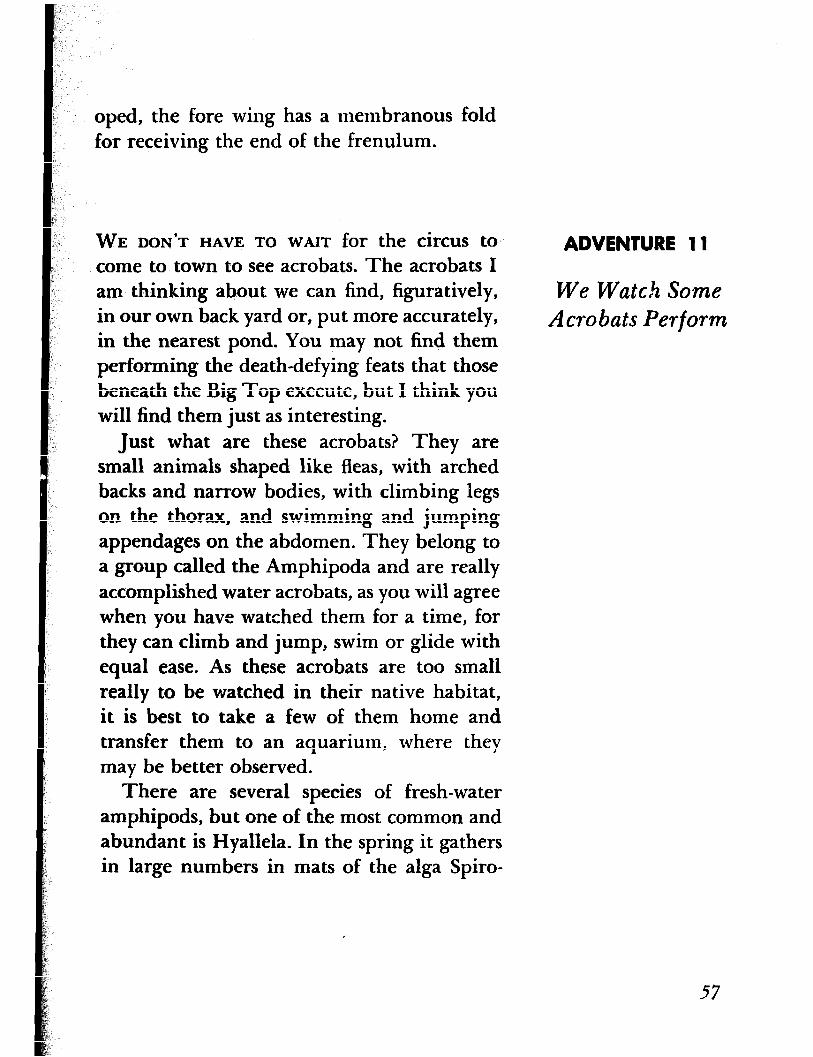
oped, the fore wing has a membranous fold for receiving the end of the frenulum.
WE DON'T HAVE TO WAIT for the circus to. come to town to see acrobats. The acrobats I am thinking about we can find, figuratively, in our own back yard or, put more accurately, in the nearest pond. You may not find them performing the death-defying feats that those beneath the Big Top execute, but I think you will find them just as interesting.
ADVENTURE 11
We Watch Some Acrobats Perform
Just what are these acrobats? They are small animals shaped like fleas, with arched backs and narrow bodies, with climbing legs on the thorax, and swimming and jumping appendages on the abdomen. They belong to a group called the Amphipoda and are really accomplished water acrobats, as you will agree when you have watched them for a time, for they can climb and jump, swim or glide with equal ease. As these acrobats are too small really to be watched in their native habitat, it is best to take a few of them home and transfer them to an aquarium, where they may be better observed.
There are several species of fresh-water amphipods, but one of the most common and abundant is Hyallela. In the spring it gathers in large numbers in mats of the alga Spiro-
57
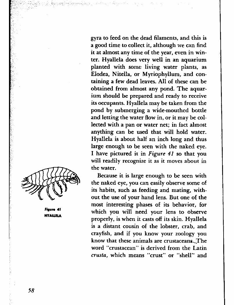
58
gyra to feed on the dead filaments, and this is a good time to collect it, although we can find it at almost any time of the year, even in win- ter. Hyallela does very well in an aquarium planted with some living water plants, as EIodea, Nitella, or Myriophyllum, and con- taining a few dead leaves. All of these can be obtained from almost any pond. The aquar- ium should be prepared and ready to receive its occupants. Hyallela may be taken from the pond by submerging a wide-mouthed bottle and letting the water flow in, or it may be col- lected with a pan or water net; in fact almost anything can be used that will hold water. Hyallela is about half an inch long and thus large enough to be seen with the naked eye. I have pictured it in Figure 41 so that you will readily recognize it as it moves about in the water.
Because it is large enough to be seen with the naked eye, you can easily observe some of its habits, such as feeding and mating, with- out the use of your hand lens. But one of the most interesting phases of its behavior, for which you will need your lens to observe properly, is when it casts off its skin. Hyallela is a distant cousin of the lobster, crab, and crayfish, and if you know your zoology you know that these animals are crustaceans.,The word “crustacean” is derived from the Latin crusta, which means “crust” or “shell” and
.
refers to the hard outside covering possessed by these animals. This hard outer covering is made largely of a hard, inelastic material called chitin. Since this material cannot stretch, these animals periodically reach a point where growth can no longer take place beneath the hard covering. So it must be dis- carded. The casting off of this outer covering is called molting and is repeated a number of times, depending on the species, until full growth is attained. The molting process, which is in itself interesting to watch, can very well be observed in the case of Hyallela simply because it occurs so slowly. I don’t want to describe it-1 would rather have you watch it yourself and see how it takes place.
THE STUDY OF FLOWERS can be made highly absorbing because of their great diversity. Let us, for instance, examine the dandelion with our hand lens. Its appearance when viewed through the glass will be quite un- like what you saw with your naked eye. You will find that instead of being a single flower, which many suppose it to be, it is composed of many small flowers, and if you were to count them you would find that there would be somewhere between 150 and 200 of them. Each one of them is a perfect flower or floret;
ADVENTURE 12
We Go Botanizing
59
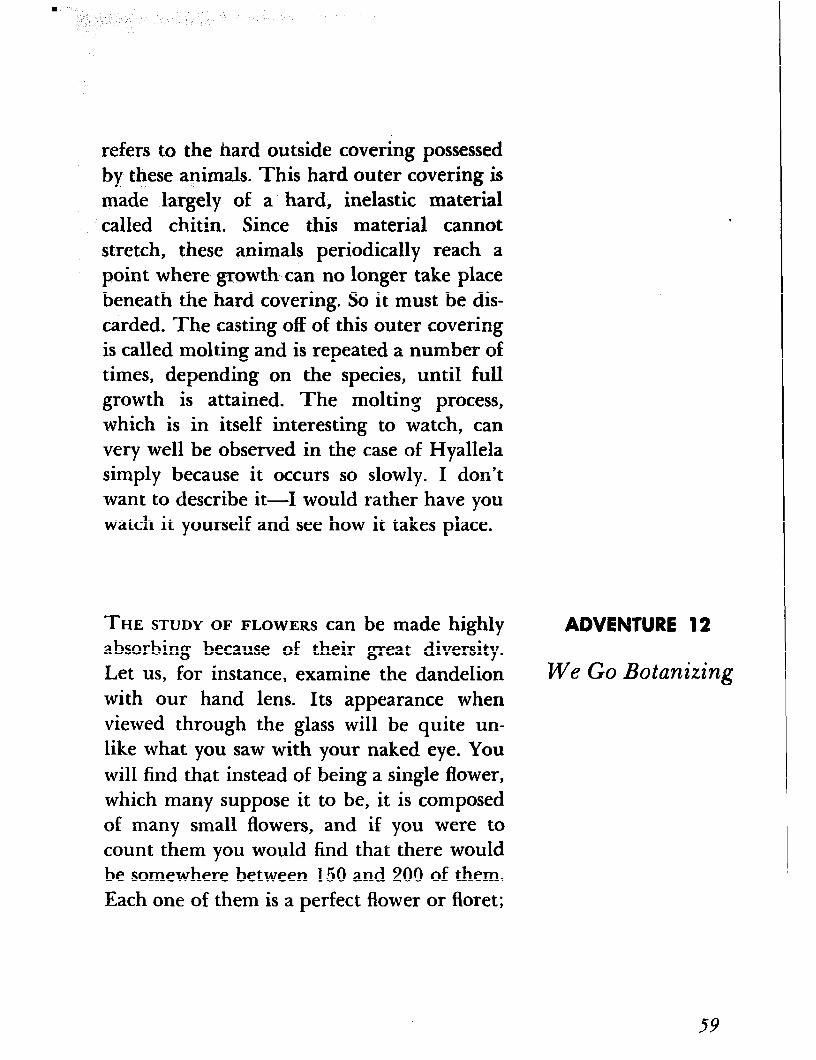
that is, each has both stamens and a pistil, but with a corolla consisting of a tube and a ray upon one side only. This corolla is straplike, with five teeth at the apex (Figure 42).
Once upon a time, this flower may have been a five-petaled blossom, for the five teeth at the top and the five lines descending from them would seem to indicate that once-dis- tinct parts had been welded together to form
Corolla a more showy and suitable corolla. Next see if you can find the five anthers that form a
Stigma tube from which the pistil extends with its two-lobed stigma. You will note as you exam-
Pistil ine several of the florets that they may all be
AntIwe in various stages of development. The florets in the outer row of the dandelion head blos- som first. After a corolla has opened, there first appears the anther tube, and then later the pistil, which gradually rises out of the anther tube and extends above it, when the stigma lobes quirl back.
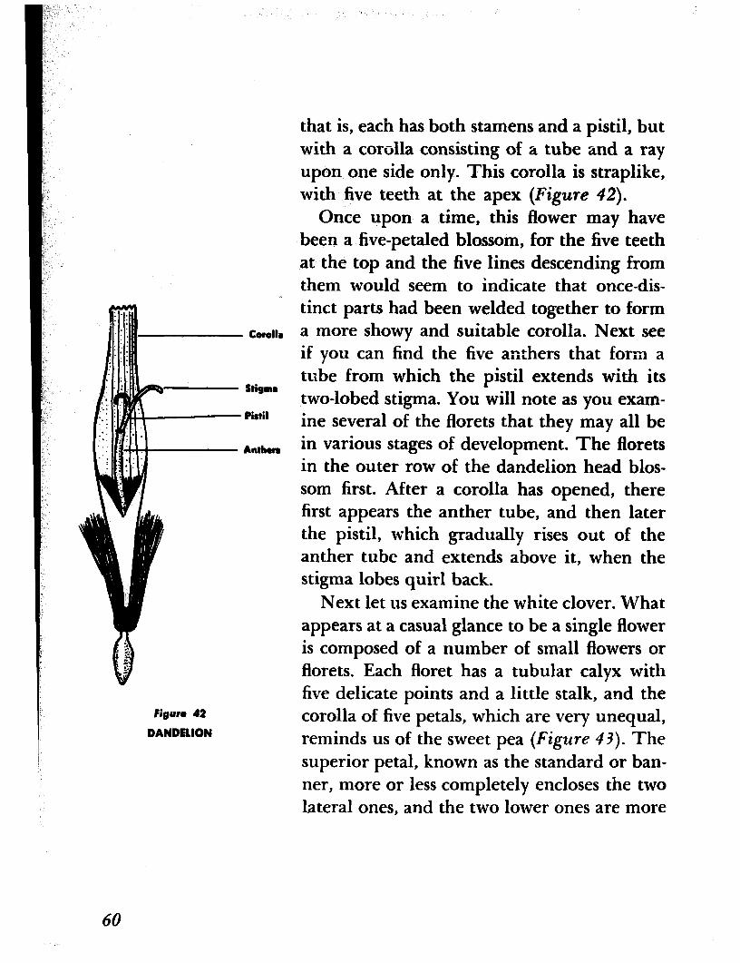
Next let us examine the white clover. What appears at a casual glance to be a single flower is composed of a number of small flowers or florets. Each floret has a tubular calyx with five delicate points and a little stalk, and the corolla of five petals, which are very unequal, reminds us of the sweet pea (Figure 43). The superior petal, known as the standard or ban- ner, more or less completely encloses the two lateral ones, and the two lower ones are more
Figure 42
DANDELION
60
or less united into what is known as the keel. There are ten stamens, nine of which are united and one is free (see if you can find them), and one pistil. Incidentally, both the dandelions and the clover represent the type of inflorescence (see Adventure 9) called the head, which can be defined as a dense cluster of sessile or nearly sessile, flowers on a very short axis. (Sessile flowers are flowers that are attached directly to the main stem without having a stem or stalk of their own.) Such a dense cluster is obviously more showy than the small florets and therefore more effective in attracting insect visitors.
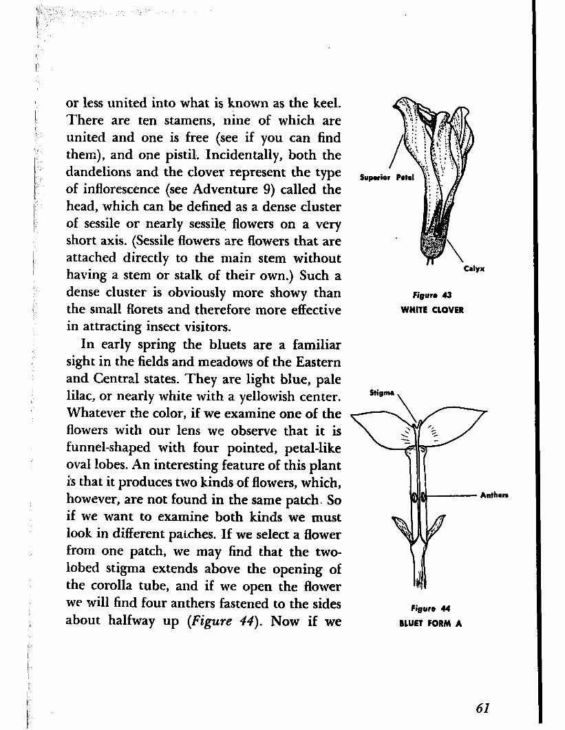
In early spring the bluets are a familiar sight in the fields and meadows of the Eastern and Central states. They are light blue, pale lilac, or nearly white with a yellowish center.
Figure 43
WHITE CLOVER
Stigma \
Whatever the color, if we examine one of the flowers with our lens we observe that it is funnel-shaped with four pointed, petal-like oval lobes. An interesting feature of this plant is that it produces two kinds of flowers, which, however, are not found in the same patch. So if we want to examine both kinds we must look in different patches. If we select a flower from one patch, we may find that the two- lobed stigma extends above the opening of the corolla tube, and if we open the flower we will find f our anthers fastened to the sides about halfway up (Figure 44). Now if we
Figure 44
BLUET FORM A
61
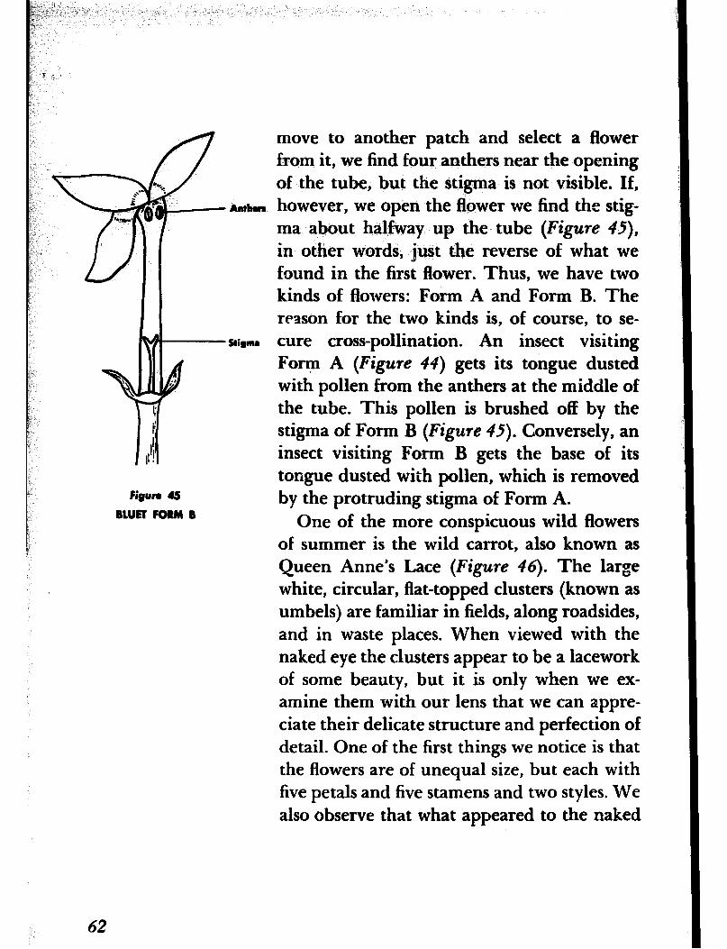
Figure 45 BLUR FORM B
Al&US
Stigma
move to another patch and select a flower from it, we find four anthers near the opening of the tube, but the stigma is not visible. If, however, we open the flower we find the stig- ma about halfway up the tube (Figure 43,
in other words, just the reverse of what we found in the first flower. Thus, we have two kinds of flowers: Form A and Form B. The reason for the two kinds is, of course, to se- cure cross-pollination. An insect visiting Form A (Figure 44) gets its tongue dusted with pollen from the anthers at the middle of the tube. This pollen is brushed off by the stigma of Form B (Figure 45). Conversely, an insect visiting Form B gets the base of its tongue dusted with pollen, which is removed by the protruding stigma of Form A.
One of the more conspicuous wild flowers of summer is the wild carrot, also known as Queen Anne’s Lace (Figure 46). The large white, circular, flat-topped clusters (known as umbels) are familiar in fields, along roadsides, and in waste places. When viewed with the naked eye the clusters appear to be a lacework of some beauty, but it is only when we ex- amine them with our lens that we can appre- ciate their delicate structure and perfection of detail. One of the first things we notice is that the flowers are of unequal size, but each with five petals and five stamens and two styles. We also observe that what appeared to the naked
62

F&n 46
WILO CARROT
eye to be a single cluster is actually composed of a number of small clusters. A very odd feature of the cluster (I am now speaking of the entire cluster) is that at the very center there is a large floret with delicate, wine- colored petals. This floret, as you will note, is not a part of any of the smaller clusters but is entirely separate and is set upon its own isolated stalk. The presence of this striking floret in the very center of the wide, circular flower cluster is a mystery-I know of no one who has been able to account for it. Even-
63
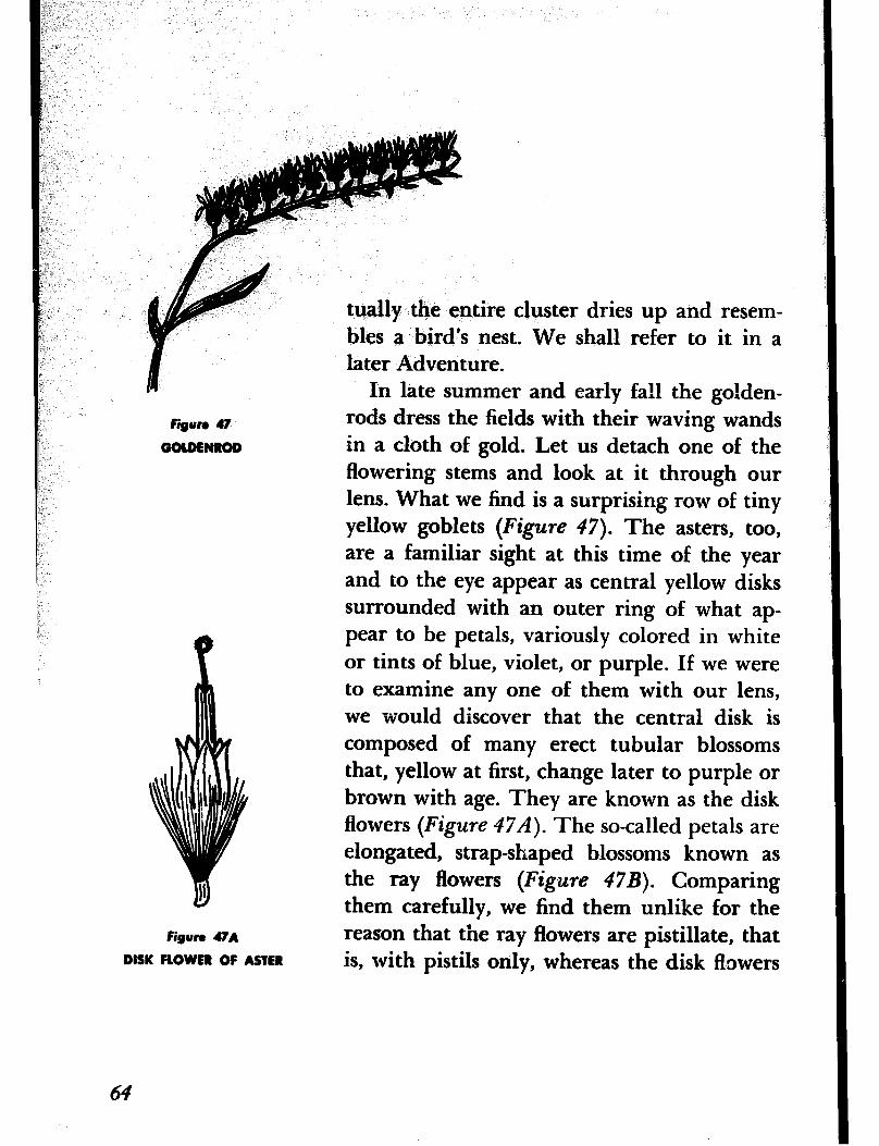
r
tually the entire cluster dries up and resem- bles a bird’s nest. We shall refer to it in a later Adventure.
figure 47
GOLDENROD
n 1
Figun #A
DISK FLOWRR OF ASTER
In late summer and early fall the golden- rods dress the fields with their waving wands in a cloth of gold. Let us detach one of the flowering stems and look at it through our lens. What we find is a surprising row of tiny yellow goblets (Figure 47). The asters, too, are a familiar sight at this time of the year and to the eye appear as central yellow disks surrounded with an outer ring of what ap- pear to be petals, variously colored in white or tints of blue, violet, or purple. If we were to examine any one of them with our lens, we would discover that the central disk is composed of many erect tubular blossoms that, yellow at first, change later to purple or brown with age. They are known as the disk flowers (Figure 47A). The so-called petals are elongated, strap-shaped blossoms known as the ray flowers (Figure 47B). Comparing them carefully, we find them unlike for the reason that the ray flowers are pistillate, that is, with pistils only, whereas the disk ftswers
64
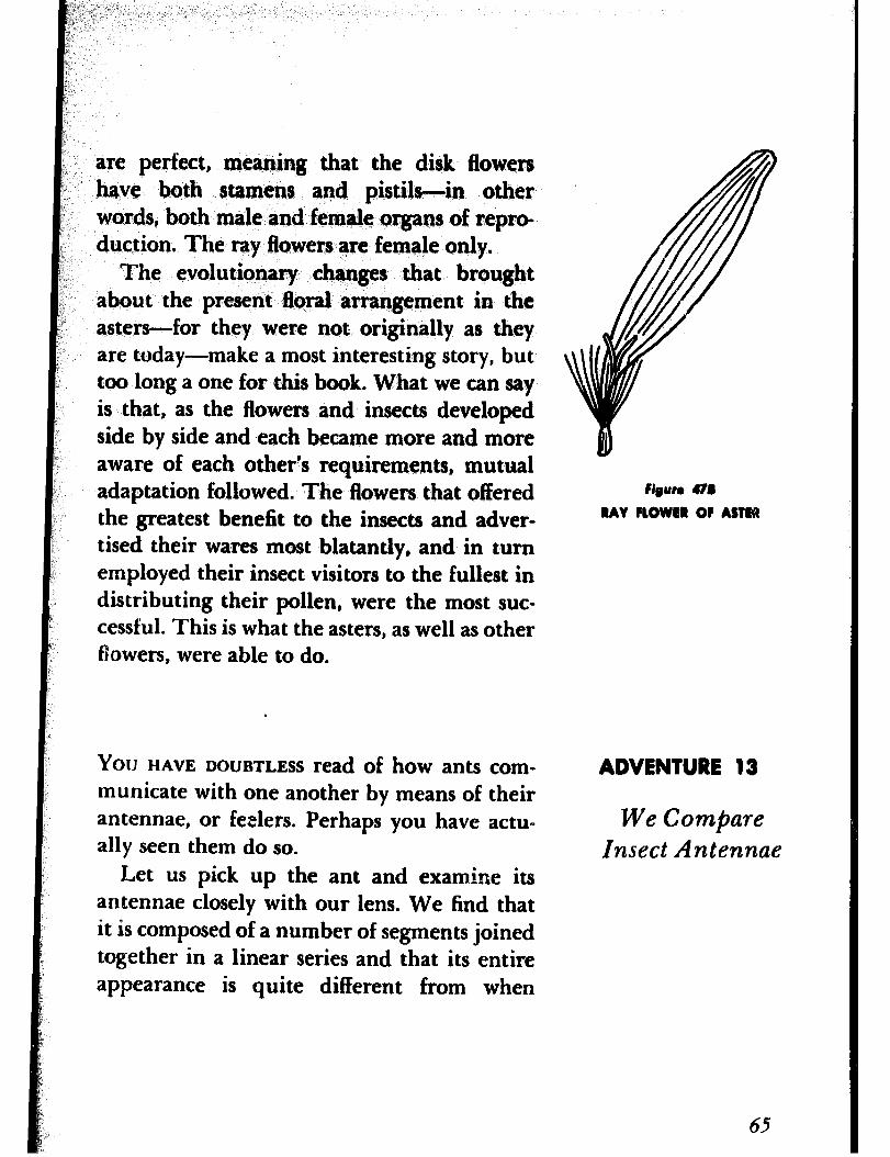
are perfect, meaning that the disk flowers have both stamens and pistils-in other ’ words, both male and female organs of repro- duction, The ray fiowers are female only.
The evolutionary changes that brought about the present floral arrangement in the asters-for they were not originally as they are today-make a most interesting story, but too long a one for this book. What we can say is that, as the flowers and insects developed side by side and each became more and more aware of each other’s requirements, mutual adaptation followed. The flowers that offered the greatest benefit to the insects and adver- tised their wares most blatantly, and in turn employed their insect visitors to the fullest in distributing their pollen, were the most suc- cessful. This is what the asters, as well as other flowers, were able to do.
Figure 478
RAY FLOWER OF ASTER
You HAVE DOUBTLESS read of how ants com- municate with one another by means of their antennae, or feelers. Perhaps you have actu- ally seen them do so.
Let us pick up the ant and examine its
ADVENTURE 13
We Compare Insect Antennae
antennae closely with our lens. We find that it is composed of a number of segments joined together in a linear series and that its entire appearance is quite different from when
65
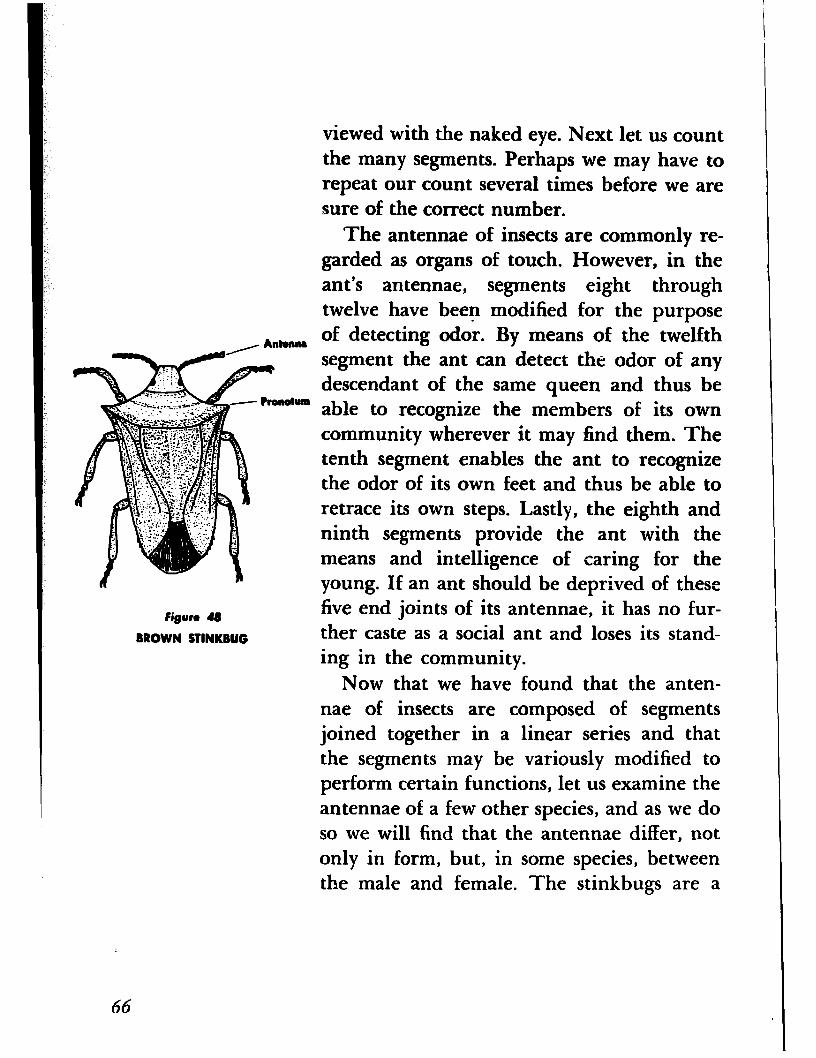
viewed with the naked eye. Next let us count the many segments. Perhaps we may have to repeat our count several times before we are sure of the correct number.
The antennae of insects are commonly re- garded as organs of touch. However, in the ant’s antennae, segments eight through twelve have been modified for the purpose of detecting odor. By means of the twelfth segment the ant can detect the odor of any descendant of the same queen and thus be able to recognize the members of its own community wherever it may find them. The tenth segment enables the ant to recognize the odor of its own feet and thus be able to retrace its own steps. Lastly, the eighth and ninth segments provide the ant with the means and intelligence of caring for the young. If an ant should be deprived of these
figure 4g
BROWN STINKBUG
five end joints of its antennae, it has no fur- ther caste as a social ant and loses its stand- ing in the community.
Now that we have found that the anten- nae of insects are composed of segments joined together in a linear series and that the segments may be variously modified to perform certain functions, let us examine the antennae of a few other species, and as we do so we will find that the antennae differ, not only in form, but, in some species, between the male and female. The stinkbugs are a
66
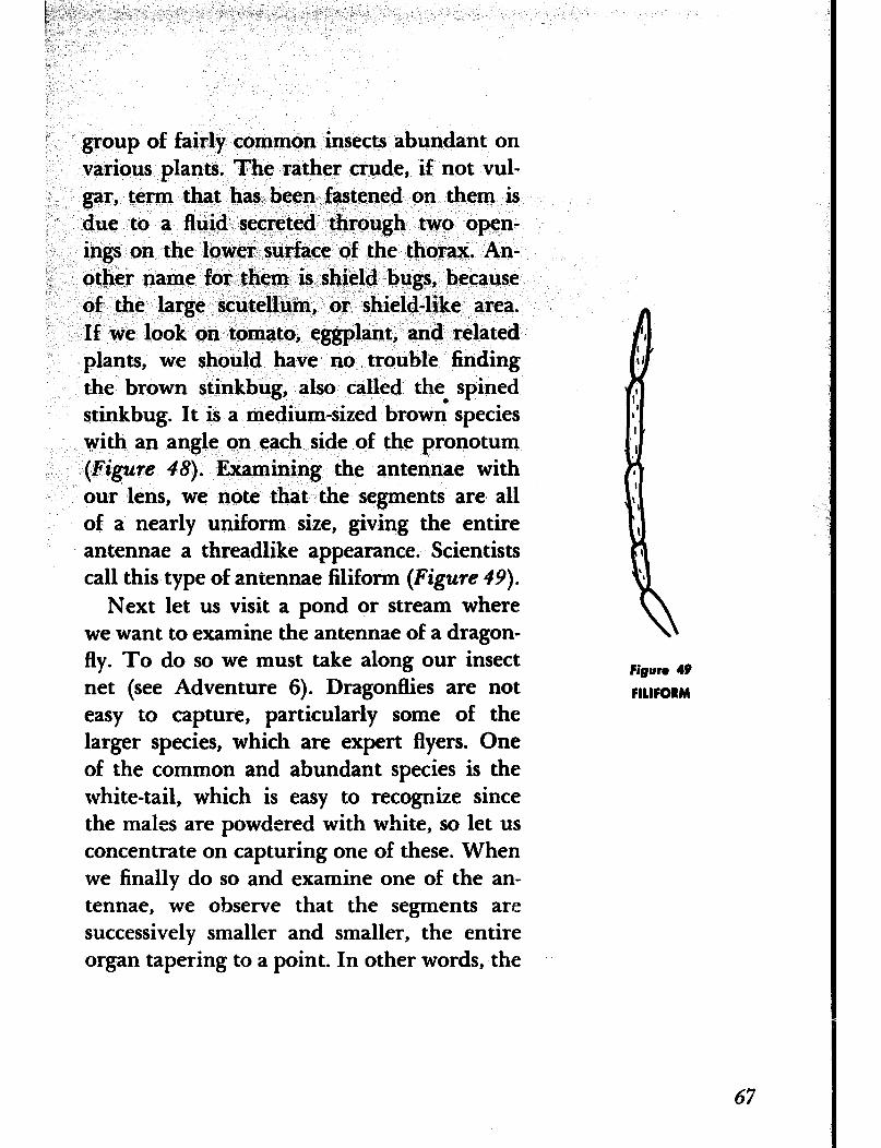
i” _
!I(
group of fairly common insects abundant on various plants. The rather crude, if not vul- gar, term that has been fastened on them is due to a fluid secreted through two open- ings on the lower surface of the thorax. An- other name for them is shield bugs, because of the large scutellum, or shield-like area. If we look on tomato, eggplant, and related plants, we should have no trouble finding the brown stinkbug, also called the. spined stinkbug. It is a medium-sized brown species with an angle on each side of the pronotum (Figure 48). Examining the antennae with our lens, we note that the segments are all of a nearly uniform size, giving the entire antennae a threadlike appearance. Scientists call this type of antennae filiform (Figure 49).
Next let us visit a pond or stream where we want to examine the antennae of a dragon- fly. To do so we must take along our insect fiRun 49
net (see Adventure 6). Dragonflies are not FILlFORM
easy to capture, particularly some of the larger species, which are expert flyers. One of the common and abundant species is the white-tail, which is easy to recognize since the males are powdered with white, so let us concentrate on capturing one of these. When we finally do so and examine one of the an- tennae, we observe that the segments are successively smaller and smaller, the entire organ tapering to a point. In other words, the
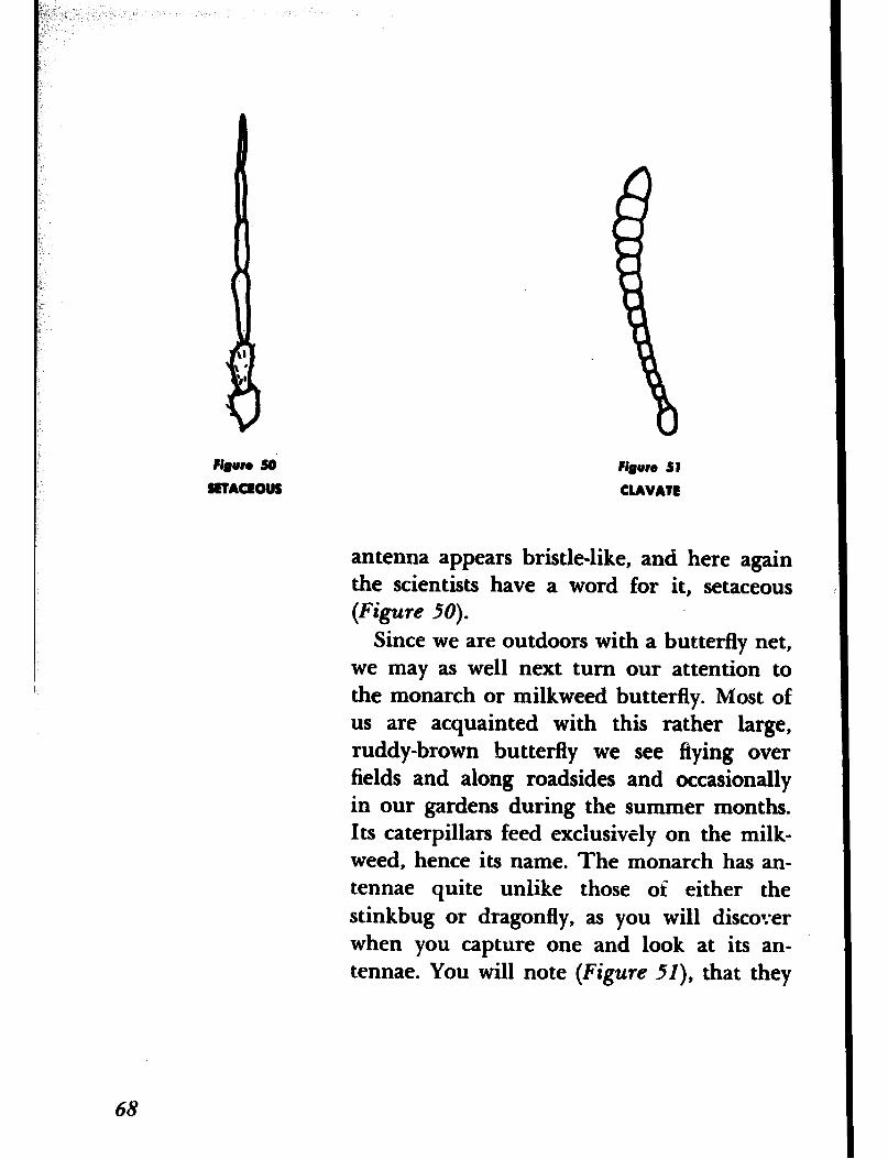
?i#un so SETACEOUS
68
figure 51 CLAVATE
antenna appears bristle-like, and here again the scientists have a word for it, setaceous (Figure 50).
Since we are outdoors with a butterfly net, we may as well next turn our attention to the monarch or milkweed butterfly. Most of us are acquainted with this rather large, ruddy-brown butterfly we see flying over fields and along roadsides and occasionally in our gardens during the summer months. Its caterpillars feed exclusively on the milk- weed, hence its name. The monarch has an- tennae quite unlike those oi either the stinkbug or dragonfly, as you will discover when you capture one and look at its an- tennae. You will note (Figure iI), that they
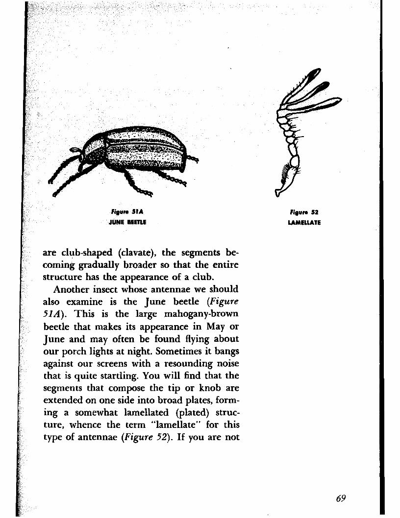
figure SIA
JUNE BEETLE
are club-shaped (clavate), the segments be- coming gradually broader so that the entire structure has the appearance of a club.
Another insect whose antennae we should also examine is the June beetle (Figure
HA). This is the large mahogany-brown
beetle that makes its appearance in May or June and may often be found flying about our porch lights at night. Sometimes it bangs against our screens with a resounding noise that is quite startling. You will find that the segments that compose the tip or knob are extended on one side into broad plates, form- ing a somewhat lamellated (plated) struc- ture, whence the term “lamellate” for this type of antennae (Figure 52). If you are not
#gun 52
LAMELUTE
69
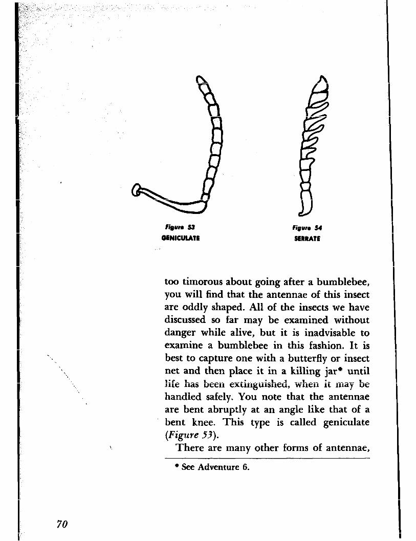
fieun S3 fiuum 54
GENICULATE SERRATE
too timorous about going after a bumblebee, you will find that the antennae of this insect are oddly shaped. All of the insects we have discussed so far may be examined without danger while alive, but it is inadvisable to examine a bumblebee in this fashion. It is best to capture one with a butterfly or insect net and then place it in a killing jar* until life has been extinguished, when it may be handled safely. You note that the antennae are bent abruptly at an angle like that of a bent knee. This type is called geniculate (Figure 53).
There are many other forms of antennae,
+ See Adventure 6.
70
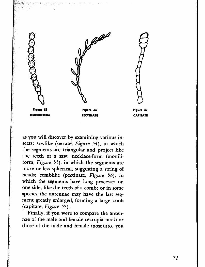
fisun 55
MONILIFORM
figure 56
PECTINATE
as you will discover by examining various in- sects: sawlike (serrate, Figure 54), in which the segments are triangular and project like the teeth of a saw; necklace-form (monili- form, Figure 55), in which the segments are more or less spherical, suggesting a string of beads; comblike (pectinate, Figure 56), in which the segments have long processes on one side, like the teeth of a comb; or in some species the antennae may have the last seg- ment greatly enlarged, forming a large knob (capitate, Figure 57).
Finally, if you were to compare the anten- nae of the male and female cecropia moth or those of the male and female mosquito, you
figure 57
CAPITATR
71
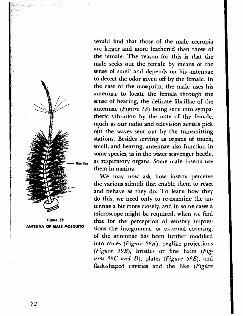
would find that those of the male cecropia are larger and more feathered than those of the female. The reason for this is that the male seeks out the female by means of the sense of smell and depends on his antennae to detect the odor given off by the female. In the case of the mosquito, the male uses his antennae to locate the female through the sense of hearing, the ‘delicate fibrillae of the antennae (Figure 58) being sent into sympa- thetic vibration by the note of the female, much as our radio and television aerials pick obt the waves sent out by the transmitting stations. Besides serving as organs of touch, smell, and hearing, antennae also function in some species, as in the water scavenger beetle, as respiratory organs. Some male insects use them in mating.
We may now ask how insects perceive the various stimuli that enable them to react and behave as they do. To learn how they do this, we need only to re-examine the an- tennae a bit more closely, and in some cases a microscope might be required, when we find
figure 58 that for the perception of sensory impres- ANTENNA OF MALE MOSQUITO sions the integument, or external covering,
of the antennae has been further modified into cones (Figure 59A), peglike projections (Figure 594, bristles or fine hairs (Fig- ures 59C and D), plates (Figure 59E), and flask-shaped cavities and the like (Figure
72
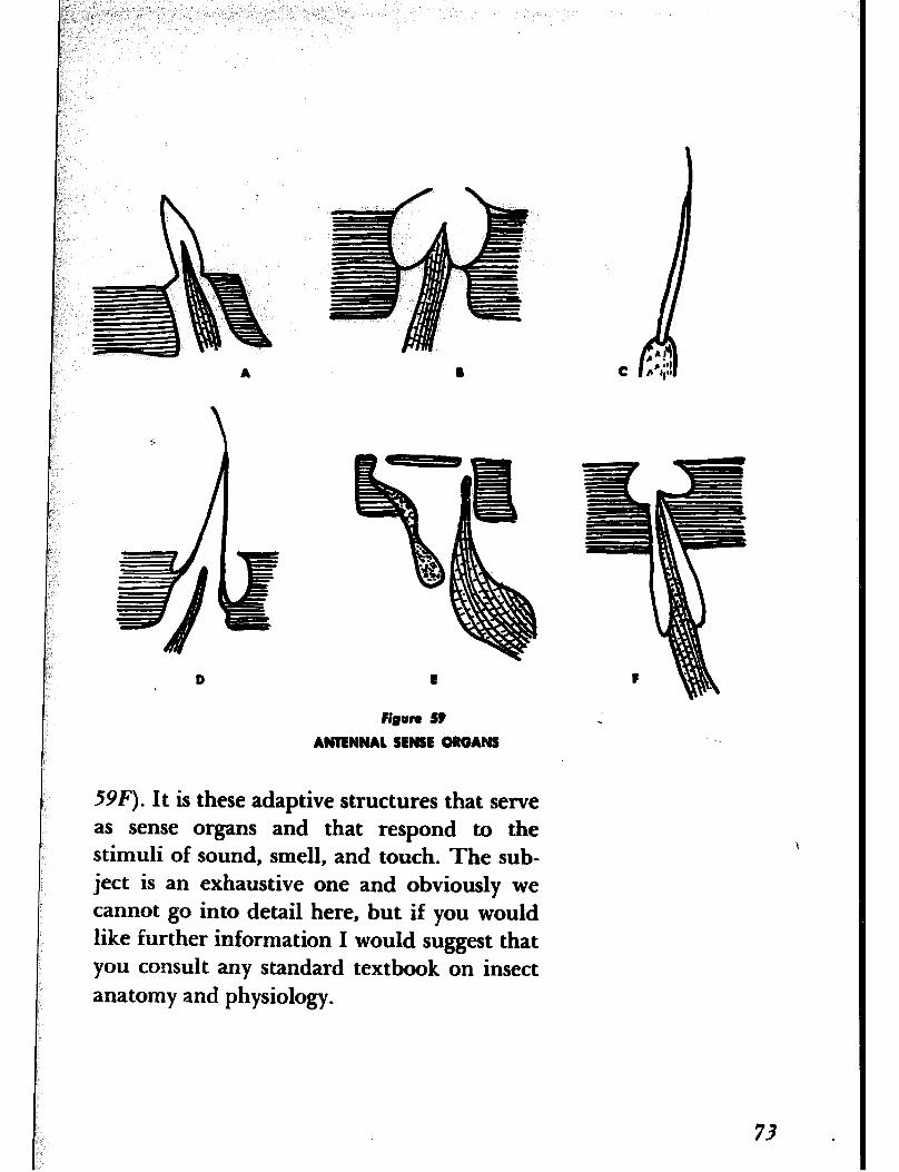
D
figum 59 5 ANTENNAI SENSE ORGANS
59F). It is these adaptive structures that serve
as sense organs and that respond to the stimuli of sound, smell, and touch. The sub- ject is an exhaustive one and obviously we cannot go into detail here, but if you would like further information I would suggest that you consult any standard textbook on insect
anatomy and physiology.

ADVENTURE 14
We See How Ants Keep Clean
Methmls \
figure 60
ANTENNA CLEANER OF ANT
IN OUR LAST AUVENTURE we learned that in- sects use their antennae to detect sound and odors, and for various other purposes. In order to function successfully it seems rather obvious that these organs must be kept clean. Ants, then, have a utilitarian reason for per- sonal grooming, and as we have combs and brushes, so, too, do they have means of re- moving dirt and other debris from their bodies. Let us see what they are.
If you have not already observed ants in the act of cleaning their antennae, I would suggest that you get three or four of them and place them in some kind of a container that would serve as a sort of observation chamber. A large glass dish filled with earth and provided with a cover so that the ants cannot escape would do nicely. It might be well before watching them at their ablutions to examine first the front leg with your lens. You will note a curved, movable, comblike spur on the distal end of the tibia, and oppo- site it, on the base of the metatarsus, a con- cavity tipped with hairs (Figure 60). This is the antenna cleaner. In cleaning its antenna the ant, as you will observe, lifts its leg over the antenna and then draws the antenna through the space between the spur and hairs, which effectively act as a brush. A dirty brush is of little value, so after cleaning the anten- nae the ant proceeds to do the same with the
74
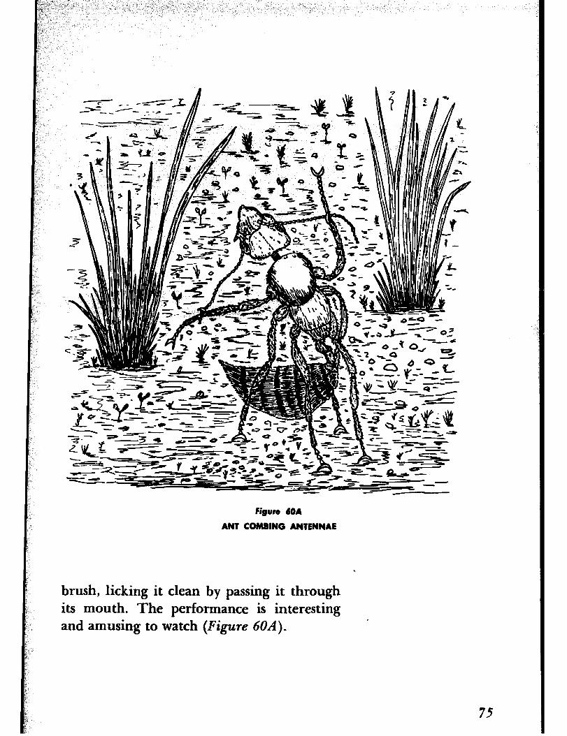
figure MA
ANT COMBING ANTENNAE
brush, licking it clean by passing it through its mouth. The performance is interesting and amusing to watch (Figure 6OA).
75
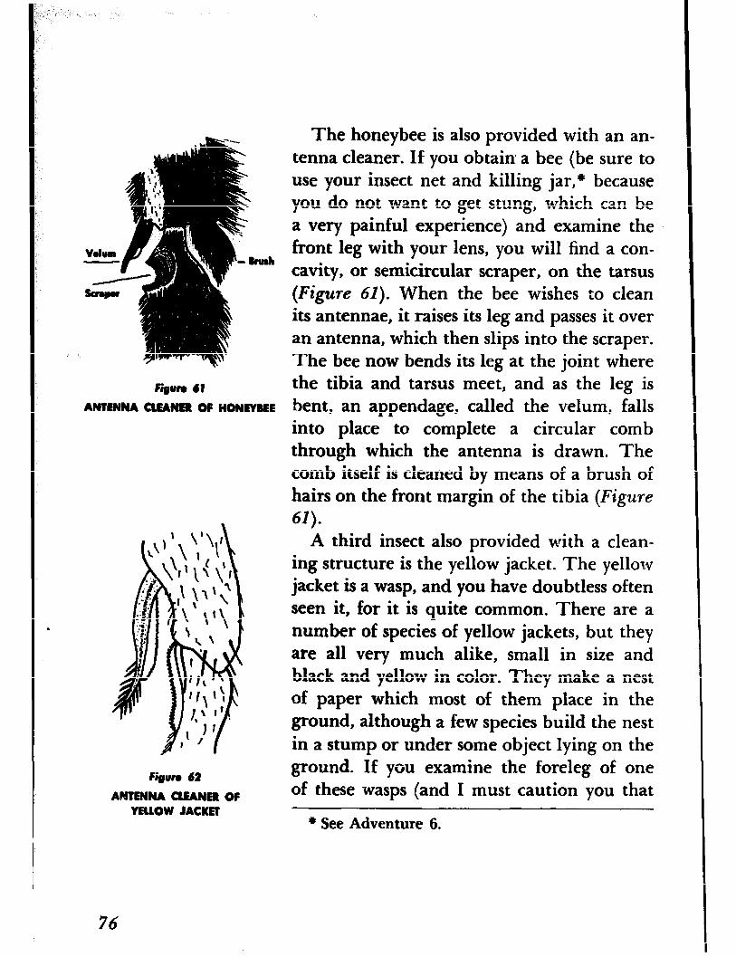
.
fiiun 41 ANTENNA CLEANER OF HONEYBEE
finurr 62
ANTENNA CLEANER OF YELLOW JACKET
The honeybee is also provided with an an- tenna cleaner. If you obtain a bee (be sure to use your insect net and killing jar,* because you do not want to get stung, which can be a very painful experience) and examine the front leg with your lens, you will find a con- cavity, or semicircular scraper, on the tarsus (Figure 61). When the bee wishes to clean its antennae, it raises its leg and passes it over an antenna, which then slips into the scraper. The bee now bends its leg at the joint where the tibia and tarsus meet, and as the leg is bent, an appendage, called the velum, falls into place to complete a circular comb through which the antenna is drawn. The comb itself is cleaned by means of a brush of hairs on the front margin of the tibia (Figure 61).
A third insect also provided with a clean- ing structure is the yellow jacket. The yellow jacket is a wasp, and you have doubtless often seen it, for it is quite common. There are a number of species of yellow jackets, but they are all very much alike, small in size and black and yellow in color. They make a nest of paper which most of them place in the ground, although a few species build the nest in a stump or under some object lying on the ground. If you examine the foreleg of one of these wasps (and I must caution you that
l See Adventure 6.
76
wasps can inflict a severe sting, so use your net and killing jar), you will find that its an- tenna cleaner is really a comb in every sense of the word and quite ornate at that (Figure
MANY YEARS AGO Charles Darwin, whose ADVENTURE 15 book Ora the Origin of Species is a milestone in the history of biology, wrote another book, We Find out how The Formation of Vegetable Mold through the Earthworm Moves
the Action of Worms, in which he shows how valuable the earthworm is to the farmer be- cause of its underground activities. To most of us the earthworm seems of no possible use except as bait for catching fish, but actually it is among our best friends, for, unseen, it works day and night, harrowing and fertiliz- ing the soil for our benefit. it burrows into the ground from twelve to eighteen inches and brings the subsoil to the surface. It also grinds the soil in its gizzard and turns it into
a finer texture than we are able to do; it even fertilizes the soil by secreting lime that neu- tralizes the acids in it.
The earthworm is not only a mere tiller of the soil, it is also an agriculturist, for it plants fallen seeds by covering them with soil and cares for the growing plants by cultivating the soil around the roots. Furthermore, it en- riches the soil by burying the bones of dead
77
animals, shells, leaves, twigs, and other or- ganic matter that, upon decaying, furnishes the necessary minerals to the plants. It even provides drainage by boring holes to carry off the surplus water, and by so doing also promotes aeration.
As you can see, the earthworm is not so useless after all. The chapging character of the landscape and much of the beauty of our
* fields and forests can be attributed to the labors of this diminutive workman. The fa- miliar mounds of black earth or castings that can often be seen on the ground are particles of soil swallowed in their burrows and brought to the surface. Since there may be as many as fifty thousand worms in an acre of ground, Darwin estimated that more than eighteen tons of earthy castings may be car- ried to the surface in a single year on one acre of ground, and in twenty years a layer three inches thick would be transferred from the subsoil to the surface. Then he goes on to say that “it may be doubted if there are any other animals which have played such an im- portant part in the history of the world as these lowly organized creatures.” That their work is of tremendous agricultural impor- tance can hardly be disputed. Remember this the next time you impale one on a hook.
We are not particularly interested here in the agricultural activities of the earthworm,
78

Figure 63
EARTHWORM IN ITS BURROW
but rather in the way it gets around. No doubt you have seen an earthworm wriggling over the ground, but have you ever wondered how it does so with what appears to be a rela- tively smooth body? It would seem that all the earthworm could accomplish by means of its wriggling movements would be merely to thrash about in one spot. Again, have you ever seen a robin tugging away at a protest-
i ing worm, or tried yourself to pull one out of its burrow and to your surprise found it was not an easy thing to do? You found it anchored there but did you ever wonder why it was so difficult to dislodge (Figure 63)? The
explanation is a simple one. If you take hold of an earthworm and run your fingers along its sides or lower surface, you will find it rough to your touch. The effect is much the same as if you ran your Sger over a stiff toothbrush.
In both instances the roughness is due to stiff bristles. Examine the earthworm with your lens and you wilI see these stiff bristles projecting from hhe body. There are four pairs of these bristles, or Tetae, as they are called, on every segmer. : :r ;he body except the first three and the la-,:. Wou will have noticed by this time that :iie body of the worm is composed of a linear series of seg- ments, hence the animal is known as a seg- mented worm). The bristles protrude from small sacs in the body wall and can be ex- tended or retracted by special muscles. If the earthworm wants to remain fixed in its bur- row, all it has to do is to extend the bristles out beyond the body surface and into the sides of its burrow and it will be securely anchored, much as an anchor holds a ship in place. Of course you can dislodge an earth- worm from its burrow by the use of superior force, but you will very likely injure it or even kill it.
The presence of these bristles also explains how the earthworm can move over the sur- face of the ground or in and out of its bur-
80
“:’ -. ,: , .: p,z I _ i ,, : .,i ” : (‘ ( ( _ ,- <,.x: ,.( i,:.- p; t; j:
(,
I.
L
row, although as a rule it rarely leaves its burrow, because the wbrm, for some reason,
- cannot seem to find its way back once it has ,. left. Watch the- worm crawl over the ground -. ’ i and you wil1 observe that it makes its way
along by first -extending the anterior part of its body, anchoring this part of the body by means of its bristles, and lastly drawing up the posterior part. The bristles on this part are then extended into the ground, thoae of the anterior part are retracted and the an- terior part extended lengthwise over the ground. All this is accomplished by two sets of powerful muscles, one set running circu- larly around the body, the other lengthwise. You can see the movements of these muscles if you look closely. While you have the earth- worm in your hand, look at one of the seg- ments with your lens about part way between the pair of bristles on the side and the pair on the lower surface. You should find a small pore, or opening. This is the external open- ing of the excretory system and is called the nephridiopore. There is a pair of them on every segment except the first three and the last. Now look on the upper surface of the animal and you will observe a reddish-purple
line. This is the main blood vessel, and you can see the pulsations as the blood flows through it. You will also note at the anterior end a fleshy lip called the prostomium. The
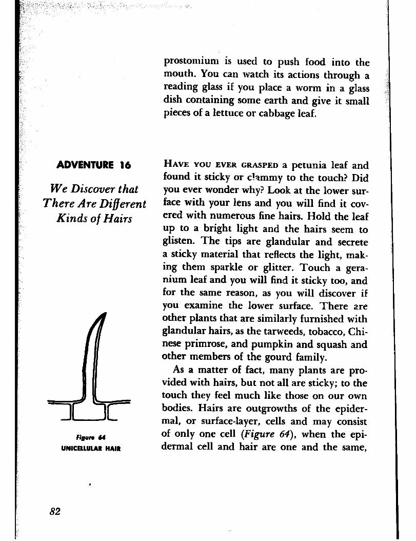
ADVENTURE 16
We Discover that There Are Different
Kinds of Hairs
Figure 64
UNICELLULAR HAIR
prostomium is used to push food into the mouth. You can watch its actions through a reading glass if you place a worm in a glass d&h containing some earth and give it small pieces of a lettuce or cabbage leaf.
HAVE YOU EVER GRASPED a petunia leaf and found it sticky or clammy to the touch? Did you ever wonder why? Look at the lower sur- face with your lens and you will find it cov- ered with numerous fine hairs. Hold the leaf up to a bright light and the hairs seem to glisten. The tips are glandular and secrete a sticky material that reflects the light, mak- ing them sparkle or glitter. Touch a gera- nium leaf and you will fin-d it sticky too, and for the same reason, as you will discover if you examine the lower surface. There are other plants that are similarly furnished with glandular hairs, as the tarweeds, tobacco, Chi- nese primrose, and pumpkin and squash and other members of the gourd family.
As a matter of fact, many plants are pro- vided with hairs, but not all are sticky; to the touch they feel much like those on our own bodies. Hairs are outgrowths of the epider- mal, or surface-layer, cells and may consist of only one cell (Figure 64), when the epi- dermal cell and hair are one and the same,
82
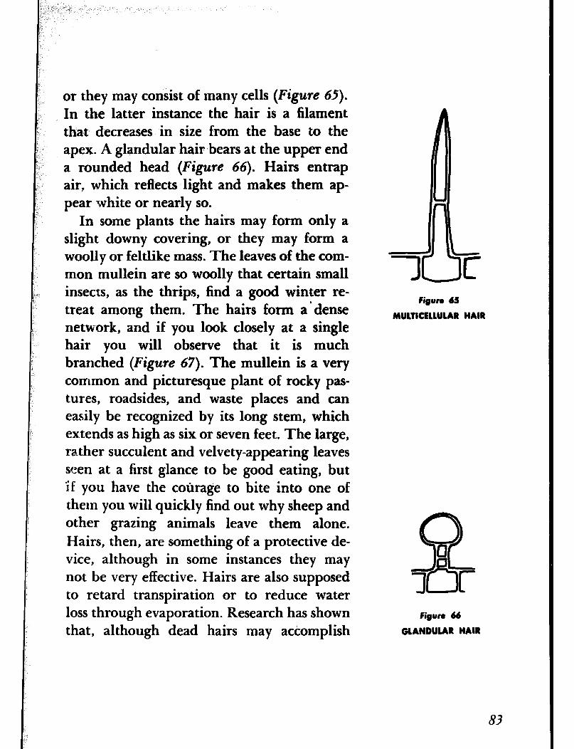
or they may consist of many cells (Figure 65).
In the latter instance the hair is a filament that decreases in size from the base to the apex. A glandular hair bears at the upper end a rounded head (Figure 66). Hairs entrap air, which reflects light and makes them ap- pear white or nearly so.
In some plants the hairs may form only a slight downy covering, or they may form a woolly or feltlike mass. The leaves of the com- mon mullein are so woolly that certain small insects, as the thrips, find a good winter re- treat among them. The hairs form a’ dense network, and if you look closely at a single hair you will observe that it is much branched (Figure 67). The mullein is a very common and picturesque plant of rocky pas- tures, roadsides, and waste places and can easily be recognized by its long stem, which ex.tends as high as six or seven feet. The large, rather succulent and velvety-appearing leaves seen at a first glance to be good eating, but ‘if you have the corirage to bite into one of them you will quickly find out why sheep and other grazing animals leave them alone. Hairs, then, are something of a protective de- vice, although in some instances they may not be very effective. Hairs are also supposed to retard transpiration or to reduce water loss through evaporation. Research has shown that, although dead hairs may actomplish
J
Figure 6S
MULTICEllUlAR HAIR
Figure 66
GLANDULAR HAIR
83
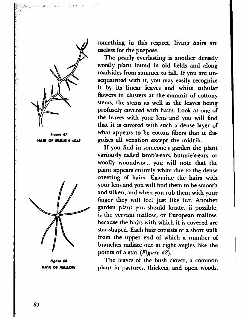
Figun 67
HAIR OF MUUEIN LEAF
Figure &R
HAIR OF MALLOW
something in this respect, living hairs are useless for the purpose.
The pearly everlasting is another densely woolly plant found in old fields and along roadsides from summer to fall. If you are un- acquainted with it, you may easily recognize it by its linear leaves and white tubular flowers in clusters at the summit of cottony stems, the stems as well as the leaves being profusely covered with hairs. Look at one of the leaves with your lens and you will find that it is covered with such a dense layer of what appears to be cotton fibers that it dis- guises all venation except the midrib.
If you find in someone’s garden the plant variously called lamb’s-ears, bunnie’s-ears, or woolly woundwort, you will note that the plant appears entirely white due to the dense covering of hairs. Examine the hairs with your lens and you will find them to be smooth and silken, and when you rub them with your finger they will feel just like fur. Another garden plant you should locate, if possible, is the vervain mallow, or European mallow, because the hairs with which it is covered are star-shaped. Each hair consists of a short stalk from the upper end of which a number of branches radiate out at right angles like the points of a star (Figure 68).
The leaves of the bush clover, a common plant in pastures, thickets, and open woods,
84
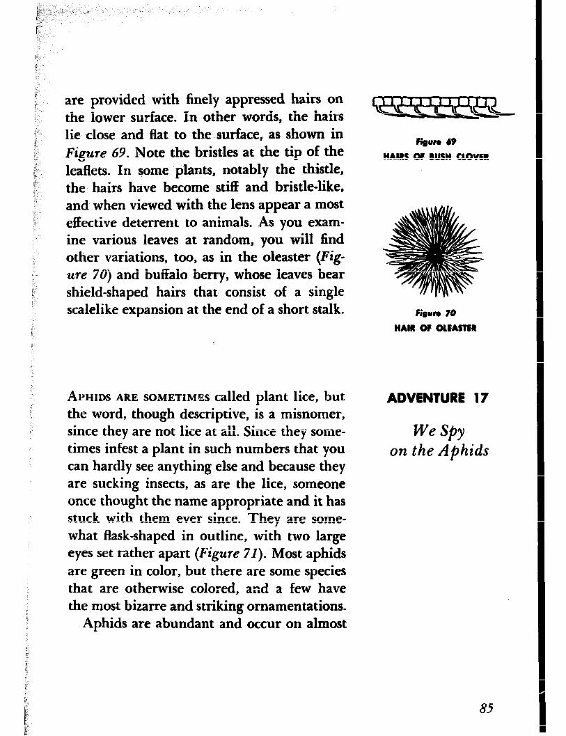
are provided with finely appressed hairs on the lower surface. In other words, the hairs lie close and flat to the surface, as shown in
Figure 69. Note the bristles at the tip of the leaflets. In some plants, notably the thistle, the hairs have become stiff and bristle-like, and when viewed with the lens appear a most effective deterrent to animals. As you exam- ine various leaves at random, you will find other variations, too, as in the oleaster (Fig-
ure 70) and buffalo berry, whose leaves bear shield-shaped hairs that consist of a single scalelike expansion at the end of a short stalk.
APHIDS ARE SOMETMES called plant lice, but the word, though descriptive, is a misnomer, since they are not lice at all. Since they some- times infest a plant in such numbers that you can hardly see anything else and because they are sucking insects, as are the lice, someone once thought the name appropriate and it has stuck with them ever since. They are some- what flask-shaped in outline, with two large eyes set rather apart (Figure 71). Most aphids are green in color, but there are some species that are otherwise colored, and a few have the most bizarre and striking ornamentations.
Aphids are abundant and occur on almost
Fi~un 69
HAIRS OF BUSH CLOVER
FiBurn 70
HAIR O? OlEASTER
ADVENTURE 17
we SPY on the Aphids
85
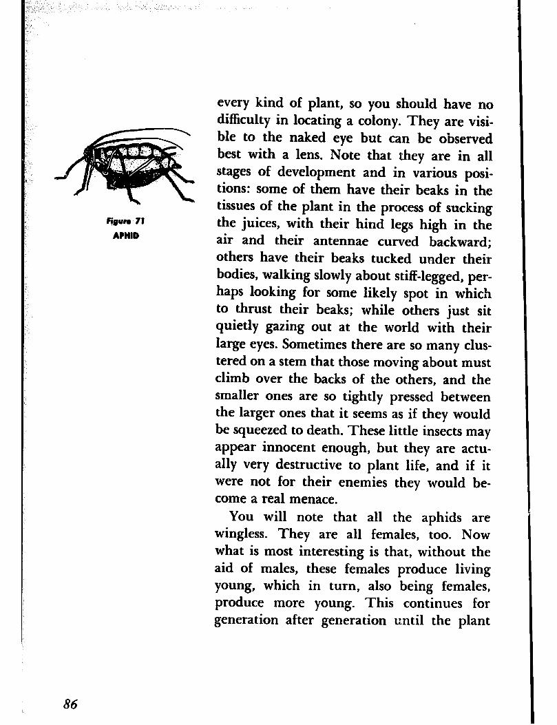
@gun, 71
APHID
every kind of plant, so you should have no difficulty in locating a colony. They are visi- ble to the naked eye but can be observed best with a lens. Note that they are in all stages of development and in various posi- tions: some of them have their beaks in the tissues of the plant in the process of sucking the juices, with their hind legs high in the air and their antennae curved backward; others have their beaks tucked under their bodies, walking slowly about stiff-legged, per- haps looking for some likely spot in which to thrust their beaks; while others just sit quietly gazing out at the world with their large eyes. Sometimes there are so many clus- tered on a stem that those moving about must climb over the backs of the others, and the smaller ones are so tightly pressed between the larger ones that it seems as if they would be squeezed to death. These little insects may appear innocent enough, but they are actu- ally very destructive to plant life, and if it were not for their enemies they would be- come a real menace.
You will note that all the aphids are wingless. They are all females, too. Now what is most interesting is that, without the aid of males, these females produce living young, which in turn, also being females, produce more young. This continues for generation after generation until the plant
86
d d
I. I.
1: 1:
becomes so crowded that it cannot possibly becomes so crowded that it cannot possibly .
support any more. What happens then? When support any more. What happens then? When the colony has reached such a size, winged fe- the colony has reached such a size, winged fe- males are produced that fly to another plant. males are produced that fly to another plant. Usually it is the same kind of plant, but in Usually it is the same kind of plant, but in some species of aphids it may be a different some species of aphids it may be a different kind of plant. In either case these winged fe- kind of plant. In either case these winged fe- males start another series of wingless genera- males start another series of wingless genera- tions. You can readily see why there are so tions. You can readily see why there are so many aphids. Even through a single female doesn’t produce many young, the young are all females that in turn produce more fe- males.
This sort of thing cannot continue forever, of course, especially when cold weather comes. As adults the aphids cannot survive the winter, but as they found out how to cope with overcrowding they also discovered how to solve the problem of low temperatures. When the first cold days of autumn herald the approach of winter, perfect winged males and females are produced. The males and fe- males mate and the females lay their eggs in the crevices of bark or at the base of buds and branches. The eggs remain unhatched through the winter and in the following spring, or in midsummer, according to the species, hatch into young. These young estab- lish themselves on a favorable plant and within a few days produce young that, of course, are females, and the cycle starts all
87
over again. The young females that hatched from the eggs are known as “stem mothers” because they initiate the new colonies.
Another odd and interesting facet in the life history of the aphids is that during the successive generations a number of different forms may be produced in each species. In one species as many as twenty-one different forms have been noted. This makes the study of aphids rather confusing. Hence it is very likely that the four hundred or so aphids that have been described from the United States as distinct species may actually be forms of a smaller number of real species.
Aphids appear to be utterly defenseless and they are for the most part, but some of them can secrete a waxy substance in the face of an enemy and thus effect their escape. Most aphids also secrete a sweetish substance called honeydew, which has a particular appeal to bees, wasps, and ants. The bees and wasps take it where they can find it but some species of ants go to considerable lengths to care for the aphids, herding them as “cows” and pro- tecting them against enemies, so that they always have a supply at hand. As you watch the aphids through your lens, you may see some ants stroking the aphids with their an- tennae until they respond to such caresses with a glistening drop of the fluid, which is immediately snatched up by the ants.
88
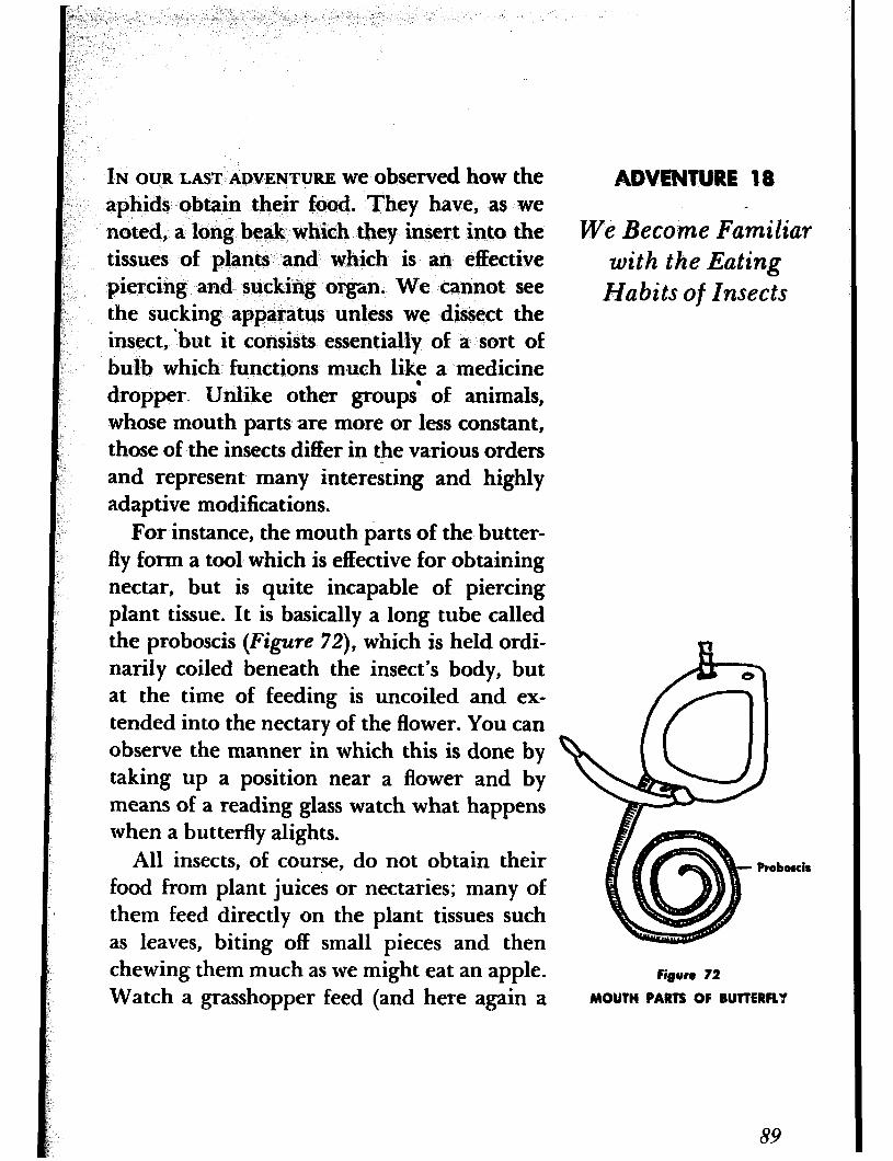
IN OURLASTADVENTURE weobservedhowthe aphids obtain their food. They have, as we noted, a long beak which they insert into the tissues of plants and which is an effective piercing and sucking organ. We cannot see the ‘sucking apparatus unless we dissect the insect, ‘but it consists essentially of a sort of bulb which functions much like a medicine dropper- Unlike other groups’ of animals, whose mouth parts are more or less constant, those of the insects differ in the various orders and represent many interesting and highly adaptive modifications.
For instance, the mouth parts of the butter- fly form a tool which is effective for obtaining nectar, but is quite incapable of piercing plant tissue. It is basically a long tube called the proboscis (Figure 72), which is held ordi- narily coiled beneath the insect’s body, but at the time of feeding is uncoiled and ex- tended into the nectary of the flower. You can observe the manner in which this is done by taking up a position near a flower and by means of a reading glass watch what happens when a butterfly alights.
All insects, of course, do not obtain their food from plant juices or nectaries; many of them feed directly on the plant tissues such as leaves, biting off small pieces and then chewing them much as we might eat an apple. Watch a grasshopper feed (and here again a
Figure 72
MOUTH PARTE OF BUTTERFLY
We Beco;me Familiar with the Eating
Habits of Insects
89
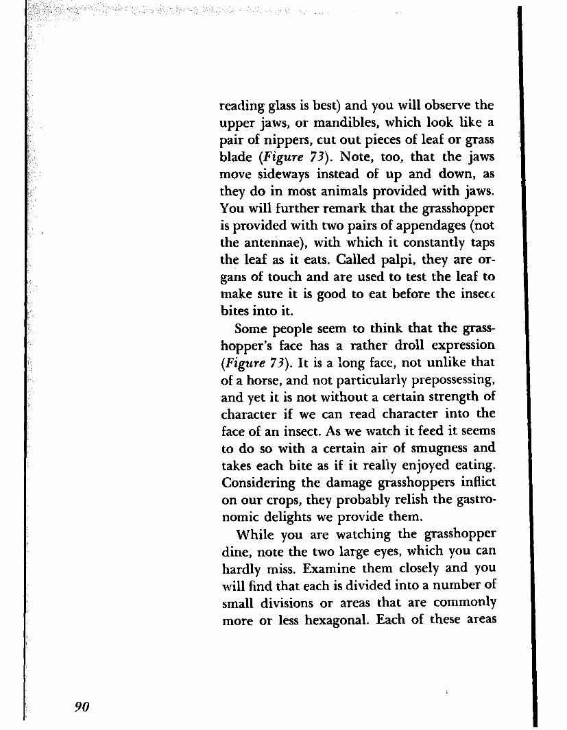
reading glass is best) and you will observe the upper jaws, or mandibles, which look like a pair of nippers, cut out pieces of leaf or grass blade (Figure 73). Note, too, that the jaws move sideways instead of up and down, as they do in most animals provided with jaws. You will further remark that the grasshopper is provided with two pairs of appendages (not the antennae), with which it constantly taps the leaf as it eats. Called palpi, they are or- gans of touch and are used to test the leaf to make sure it is good to eat before the insect bites into it.
Some people seem to think that the grass- hopper’s face has a rather droll expression (Figure 73). It is a long face, not unlike that of a horse, and not particularly prepossessing, and yet it is not without a certain strength of character if we can read character into the face of an insect. As we watch it feed it seems to do so with a certain air of smugness and takes each bite as if it really enjoyed eating. Considering the damage grasshoppers inflict on our crops, they probably relish the gastro- nomic delights we provide them.
While you are watching the grasshopper dine, note the two large eyes, which you can hardly miss. Examine them closely and YOU
will find that each is divided into a number of small divisions or areas that are commonly more or less hexagonal. Each of these areas
90
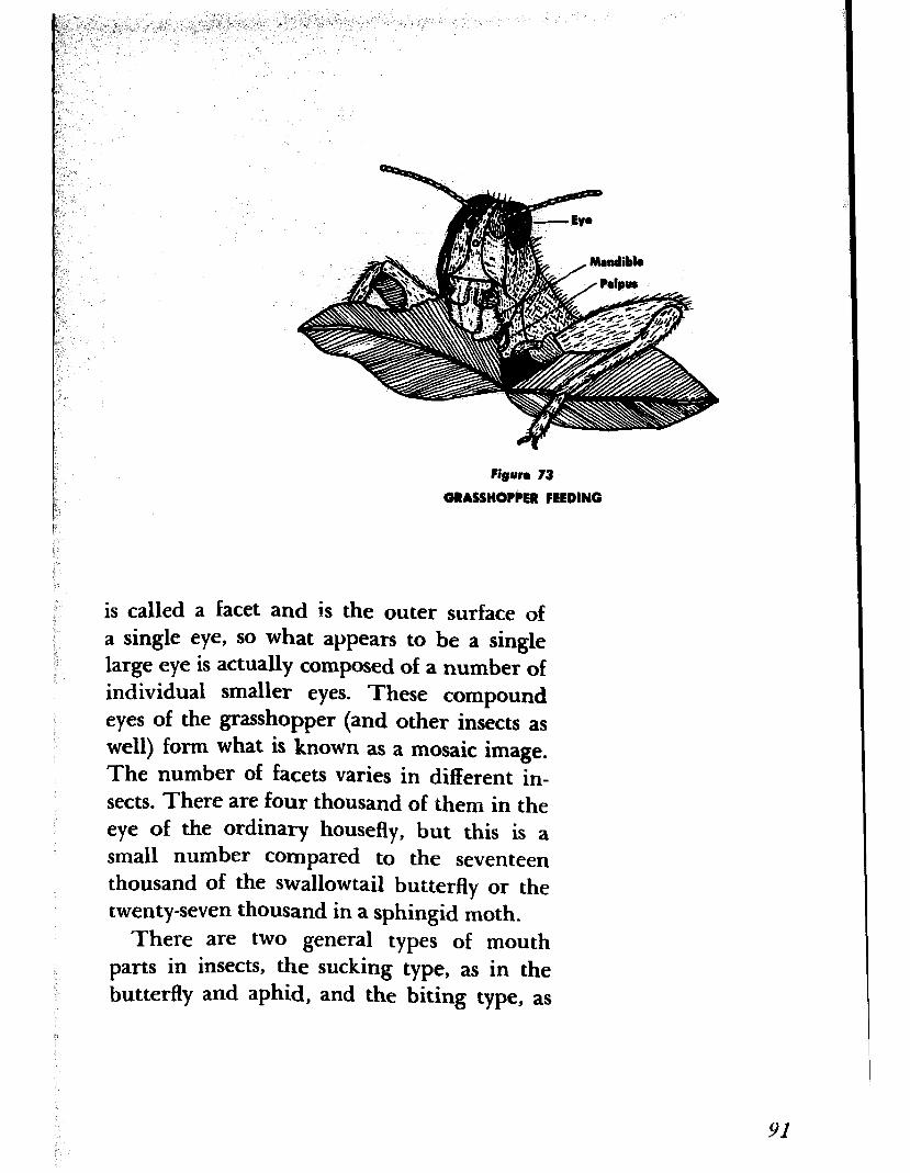
Figvra 73
GRASSHOPPER FEEDING
is called a facet and is the outer surface of a single eye, SO what appears to be a single large eye is actually composed of a number of individual smaller eyes. These compound eyes of the grasshopper (and other insects as well) form what is known as a mosaic image. The number of facets varies in different in- sects. There are four thousand of them in the eye of the ordinary housefly, but this is a small number compared to the seventeen thousand of the swallowtail butterfly or the twenty-seven thousand in a sphingid moth.
There are two general types of mouth parts in insects, the sucking type, as in the butterfly and aphid, and the biting type, as
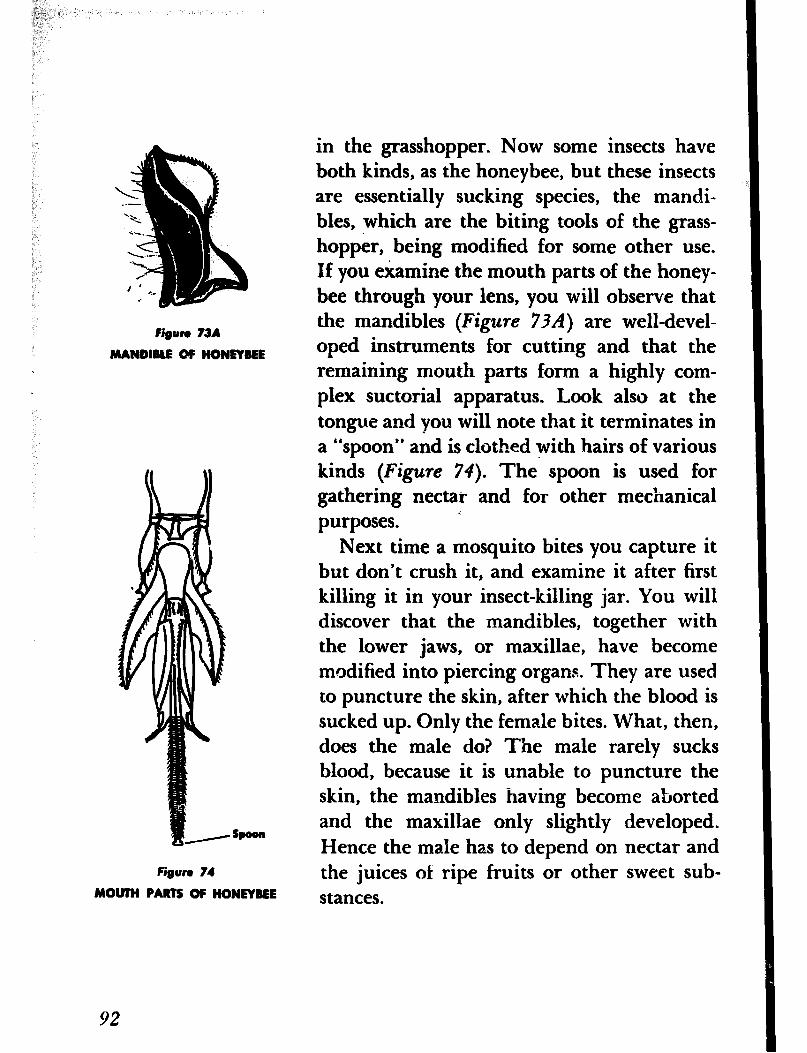
@un 73A
MANDIRLE OF HONEYREE
in the grasshopper. Now some insects have both kinds, as the honeybee, but these insects are essentially sucking species, the mandi- bles, which are the biting tools of the grass- hopper, being modified for some other use. If you examine the mouth parts of the honey- bee through your lens, you will observe that the mandibles (Figure 734 are well-devel- oped instruments for cutting and that the remaining mouth parts form a highly com- plex suctorial apparatus. Look also at the tongue and you will note that it terminates in a “spoon” and is clothed with hairs of various kinds (Figure 74). The spoon is used for gathering nectar and for other mechanical purposes. c
Spoon
Figure 74
MOWH PARTS OF HONEYREE
Next time a mosquito bites you capture it but don’t crush it, and examine it after first killing it in your insect-killing jar. You will discover that the mandibles, together with the lower jaws, or maxillae, have become modified into piercing organs. They are used to puncture the skin, after which the blood is sucked up. Only the female bites. What, then, does the male do? The male rarely sucks blood, because it is unable to puncture the skin, the mandibles having become aborted and the maxillae only slightly developed. Hence the male has to depend on nectar and the juices of ripe fruits or other sweet sub- stances.
92
2,. “. ‘5
HAVE YOU EVER LOOKED a spider in the eye? ADVENTURE 19 Probably not, for few of us ever have the occasion to do so. But let us do it and see We Look a Spider what we find. in the Eye
One of the jumping spiders will do nicely, as they are quite common and easy to find. We need not be afraid to handle spiders, for they are quite harmless, contrary to what most people think. True, they are all poison- ous, but the amount of poison they can in- ject into our skin is so small that we would hardly notice it unless, of course, we are al- lergic to spider venom. However, it might be best, after capturing a spider, to put it in the killing jar before examining it. There is one spider, the black widow, which has spread over most of our country and can inject enough venom to prove dangerous and in some cases to produce fatal results. So beware of any coal-black spider that is marked with red or yellow or both.
To return to our jumping spiders. They are common on plants, logs, fences, and the sides of buildings, and we need only to step outdoors to find them. They are small or of medium size, measuring about a fourth of an inch or less in length, with short, stout legs, and usually of bright color. They regd- ily attract our attention by their quick jump-
ing movements. Watch one of them for a few moments and you will find that it can move
93
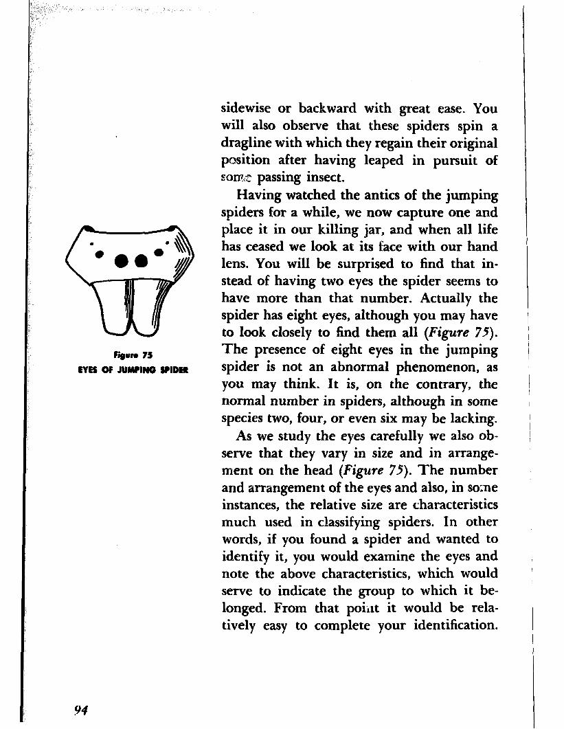
Figam 75
EVEE OF JUMPING SPIDER
94
sidewise or backward with great ease. You will also observe that these spiders spin a dragline with which they regain their original position after having leaped in pursuit of SOXFZ~ passing insect.
Having watched the antics of the jumping spiders for a while, we now capture one and place it in our killing jar, and when all life has ceased we look at its face with our hand lens. You will be surprised to find that in- stead of having two eyes the spider seems to have more than that number. Actually the spider has eight eyes, although you may have to look closely to find them all (Figure 75).
The presence of eight eyes in the jumping spider is not an abnormal phenomenon, as you may think. It is, on the contrary, the normal number in spiders, although in some species two, four, or even six may be lacking.
As we study the eyes carefully we also ob- serve that they vary in size and in arrange- ment on the head (Figure 75). The number and arrangement of the eyes and also, in some instances, the relative size are characteristics much used in classifying spiders. In other words, if you found a spider and wanted to identify it, you would examine the eyes and note the above characteristics, which would serve to indicate the group to which it be- longed. From that poiilt it would be rela- tively easy to complete your identification.

A spider’s-eyes have been given different names, according to their position. The nor- mal position of the eyes is in two transverse rows each containing four eyes. The two in- termediate eyes of the first row are called the corhrior M~i.n Eve anterior median; the two intermediate eyes of the second row are called posterior median;
Posterior latarml Eye
and the one at each side of the first row an- terior lateral and the one at each end of the second row posterior lateral (Figure 76).
In the jumping spiders we have a depar- ture from the normal arrangement and also a marked difference in the relative size of Antodor WarmI Eye
the different pairs, characters that enable us AMWIW MWUU, EVO
to recognize this group at a glance. Instead of two rows there are three. The first row
Figure 76
EM OF A SPIDER
is somewhat curved and consists of four eyes: the anterior median, which are very large, and the two anterior lateral, which are smaller. Behind this first row is a second row, of two eyes: the posterior median, which are very small, and you may have to look very closely to locate them. Behind these two small eyes is still a third row, of two eyes: the pos- terior lateral.
One more word about the eyes of spiders. Two types of eyes are usually recopized: the so-called nocturnal eyes and the diwrnal eyes. Spiders that live in the dark or frequent shady places have eyes that are pearly white in color-they are nocturnal eyes. Diurnal
95
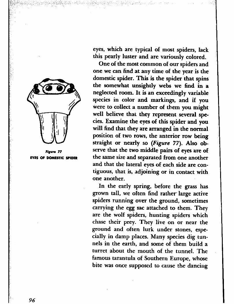
Figuro 77
EYES OF DOMESTIC SPIDER
eyes, which are typical of most spiders, lack this pearly luster and are variously colored.
One of the most common of our spiders and one we can find at any time of the year is the domestic spider. This is the spider that spins the somewhat unsightly webs we find in a neglected room. It is an exceedingly variable species in color and markings, and if you ivere to collect a number of them you might well believe that they represent several spe- cies. Examine the eyes of this spider and you will find that they are arranged in the normal position of two rows, the anterior row being straight or nearly so (Figure 77). Also ob- serve that the two middle pairs of eyes are of the same size and separated from one another and that the lateral eyes of each side are con- tiguous, that is, adjoining or in contact with one another.
In the early spring, before the grass has grown tall, we often find rather large active spiders running over the ground, sometimes carrying the egg sac attached to them. They are the wolf spiders, hunting spiders which chase their prey. They live on or near the ground and often lurk under stones, espe- cially in damp places. Many species dig tun- nels in the earth, and some of them build a turret about the mouth of the tunnel. The famous tarantula of Southern Europe, whose bite was once supposed to cause the dancing
96
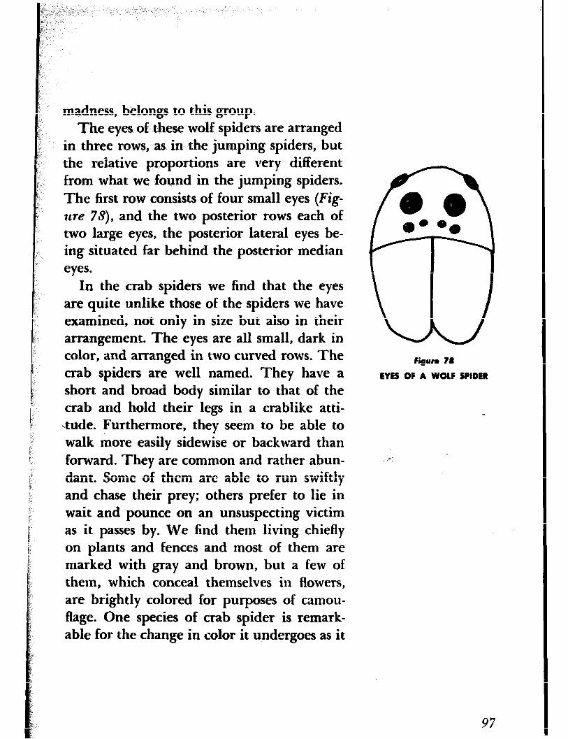
madness, belongs to this group. The eyes of these wolf spiders are arranged
in three rows, as in the jumping spiders, but the relative proportions are very different from what we found in the jumping spiders. The first row consists of four small eyes (Fig- t&re 75), and the two posterior rows each of two large eyes, the posterior lateral eyes be- ing situated far behind the posterior median eyes.
In the crab spiders we find that the eyes are quite unlike those of the spiders we have examined, not only in size but also in their arrangement. The eyes are all small, dark in color, and arranged in two curved rows. The crab spiders are well named. They have a short and broad body similar to that of the crab and hold their legs in a crablike atti- tude. Furthermore, they seem to be able to walk more easily sidewise or backward than forward. They are common and rather abun- dant. Some of them are able to run swiftly and chase their prey; others prefer to lie in wait and pounce on an unsuspecting victim as it passes by. We find them living chiefly on plants and fences and most of them are marked with gray and brown, but a few of them, which conceal themselves in flowers, are brightly colored for purposes of camou- flage. One species of crab spider is remark- able for the change in color it undergoes as it
figure 78
EYES OF A WOLF SPIDER
:-:
97
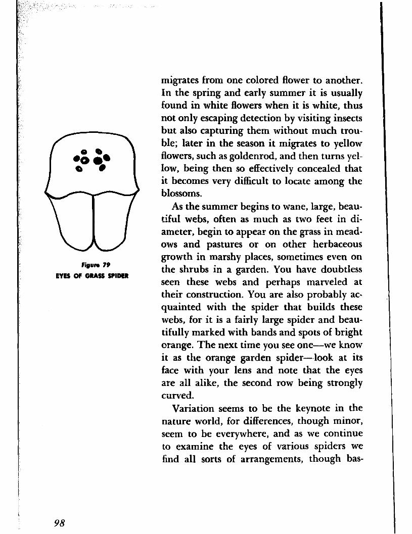
figure 79
EYEE OF GRASS SPIDER
migrates from one colored flower to another. In the spring and early summer it is usually found in white flowers when it is white, thus not only escaping detection by visiting insects but also capturing them without much trou- ble; later in the season it migrates to yellow flowers, such as goldenrod, and then turns yel- low, being then so effectively concealed that it becomes very difficult to locate among the blossoms.
As the summer begins to wane, large, beau- tiful webs, often as much as two feet in di- ameter, begin to appear on the grass in mead- ows and pastures or on other herbaceous growth in marshy places, sometimes even on the shrubs in a garden. You have doubtless seen these webs and perhaps marveled at their construction. You are also probably ac- quainted with the spider that builds these webs, for it is a fairly large spider and beau- tifully marked with bands and spots of bright orange. The next time you see one-we know it as the orange garden spider--look at its face with your lens and note that the eyes are all alike, the second row being strongly curved.
Variation seems to be the keynote in the nature world, for differences, though minor, seem to be everywhere, and as we continue to examine the eyes of various spiders we find all sorts of arrangements, though bas-
98

ically they are much the same. In the grass spider, for instance, the two rows of eyes are so strongly curved backward that the anterior median and the posterior lateial eyes form nearly a straight line (Figure 79). Speaking of variation, there is a wide range in the size of this spider as well as in the general color. There is also considerable variation. in cer- tain structures. All of this makes for consid- erable confusion, not alone for the amateur student of spiders, but also for the specialist The grass spider is well named, because its webs are commonly found on .grass. Indeed, we seldom realize the immense numbers of webs spun upon the grass by this spider, ex- cept when, in early morning, they are made visible by the dew that has condensed upon them. L4t such times the grass appears to be covered with an almost continuous carpet of silk.
THE GRASSES are a basic form of plant life, performing a dual function as soil binders and as a source of food for many different kinds of animals, including man, since corn, wheat, rye, oats, sugar, and rice are species of grass. In some parts of the world grasses also provide shelter and clothing in the form of bamboo. From earliest spring until late
ADVENTURE 20
We Inimtigate the Structure
of Grass Flowers
99
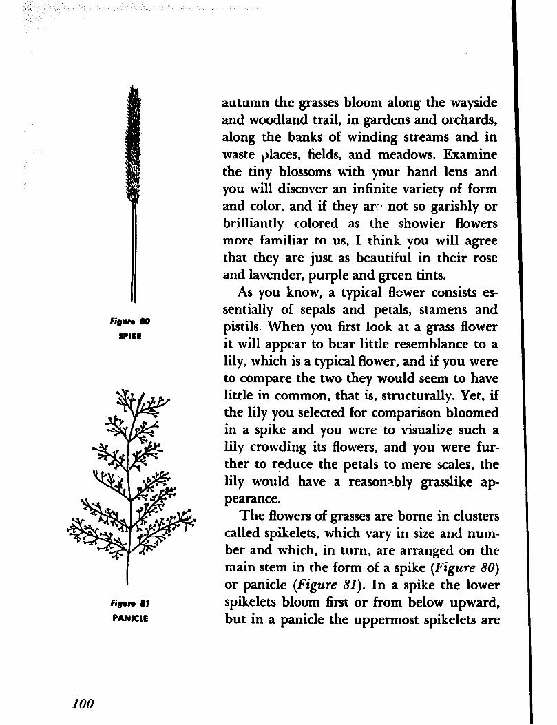
Figwa a0
SPIKE
Figure 61
PANICLE
100
autumn the grasses bloom along the wayside and woodland trail, in gardens and orchards, along the banks of winding streams and in waste places, fields, and meadows. Examine the tiny blossoms with your hand lens and you will discover an infinite variety of form and color, and if they arc\ not so garishly or brilliantly colored as the showier flowers more familiar to us, I think you will agree that they are just as beautiful in their rose and lavender, purple and green tints.
As you know, a typical flower consists es- sentially of sepals and petals, stamens and pistils. When you first look at a grass flower it will appear to bear little resemblance to a lily, which is a typical flower, and if you were to compare the two they would seem to have little in common, that is, structurally. Yet, if the lily you selected for comparison bloomed in a spike and you were to visualize such a lily crowding its flowers, and you were fut- ther to reduce the petals to mere scales, the lily would have a reasonF.bly grasslike ap- pearance.
The flowers of grasses are borne in clusters called spikelets, which vary in size and num- ber and which, in turn, are arranged on the main stem in the form of a spike (Figure 80)
or panicle (Figure 81). In a spike the lower spikelets bloom first or from below upward, but in a panicle the uppermost spikelets are
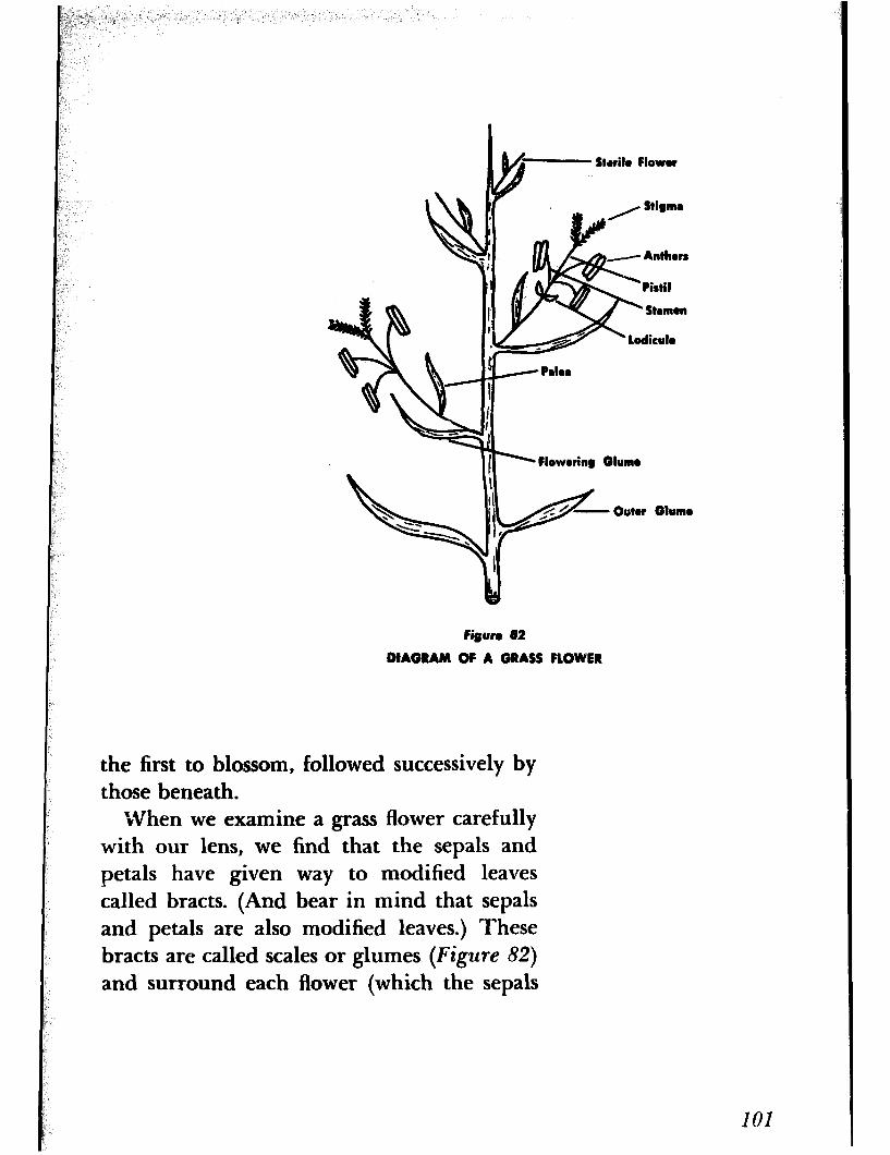
Strilo Flowor
i /e- Anthers
DIAGRAM OF A GRASS FLOWER
the first to blossom, followed successively bY
Stigma
those beneath. M/hen we examine a grass flower carefully
with our lens, we find that the sepals and petals have given way to modified leaves called bracts. (And bear in mind that sepals and petals are also modified leaves.) These bracts are called scales or glumes (Figure 82) and surround each flower (which the sepals
101
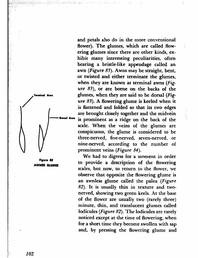
and petals also do in the more conventional flower). The glumes, which are called flow- ering glumes since there are other kinds, ex- hibit many interesting peculiarities, often bearing a bristle-like appendage called an awn (Figure 83). Awns may be straight, bent, ot twisted and either terminate the glumes, when they are known as terminal awns (Fig-
ure 83), or are borne on the backs of the
/
Tomind Awn glumes, when they are said to be dorsal (Fig- we 83). A flowering glume is keeled when it is flattened and folded so that its two edges are brought closely together and the midvein
-Dorsal Awn l is prominent as a ridge on the back of the scale. When the veins of the glumes are conspicuous, the glume is considered to be three-nerved, five-nerved, seven-nerved, or nine-nerved, according to the number of prominent veins (Figure 84).
102
We had to digress for a moment in order figun 83
AWNED OIUMES to provide a description of the flowering scales, but now, to return to the flower, we observe that opposite the flowering glume is an awnless glume called the palea (Figure
82). It is usually thin in texture and two- nerved, showing two green keels. At the base of the flower are usually two (rarely three) minute, thin, and translucent glumes called lodicules (Figure 82). The lodicules are rarely noticed except at the time of flowering, when for a short time they become swollen with sap
by pressing the flowering glume and
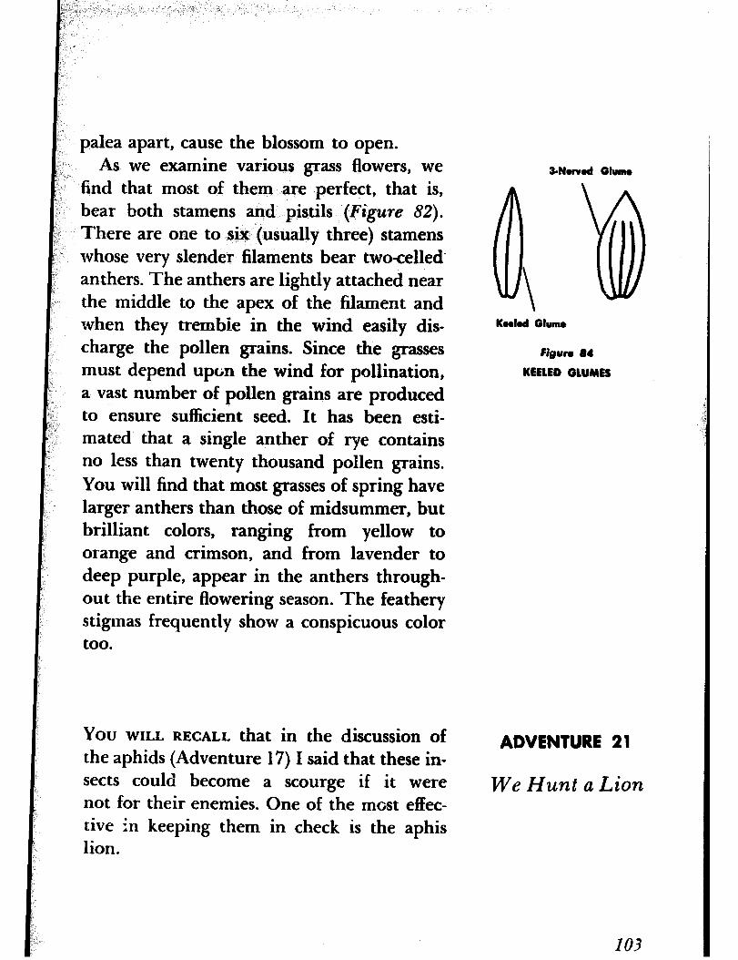
palea apart, cause the blossom to open. As we examine various grass flowers, we
find that most of them are perfect, that is, bear both stamens and pistils (Figure 82).
There are one to six (usually three) stamens whose very slender filaments bear two-celled anthers. The anthers are lightly attached near the middle to the apex of the filament and when they tremble in the wind easily dis- charge the pollen grains. Since the grasses must depend upon the wind for pollination, a vast number of pollen grains are produced to ensure sufficient seed. It has been esti- mated that a single anther of rye contains no less than twenty thousand pollen grains. You will find that most grasses of spring have larger anthers than those of midsummer, but brilliant colors, ranging from yellow to orange and crimson, and from lavender to deep purple, appear in the anthers through- out the entire flowering season. The feathery stigmas frequently show a conspicuous color too.
You WILL RECALL that in the discussion of the aphids (Adventure 17) I said that these in- sects could become a scourge if it were not for their enemies. One of the most effec- tive in keeping them in check is the aphis lion.
3-Now04 Glumo
Kook4 Glumo
Figun 84
KEELED GLUMES
ADVENTURE 21
We Hunt a Lion
103
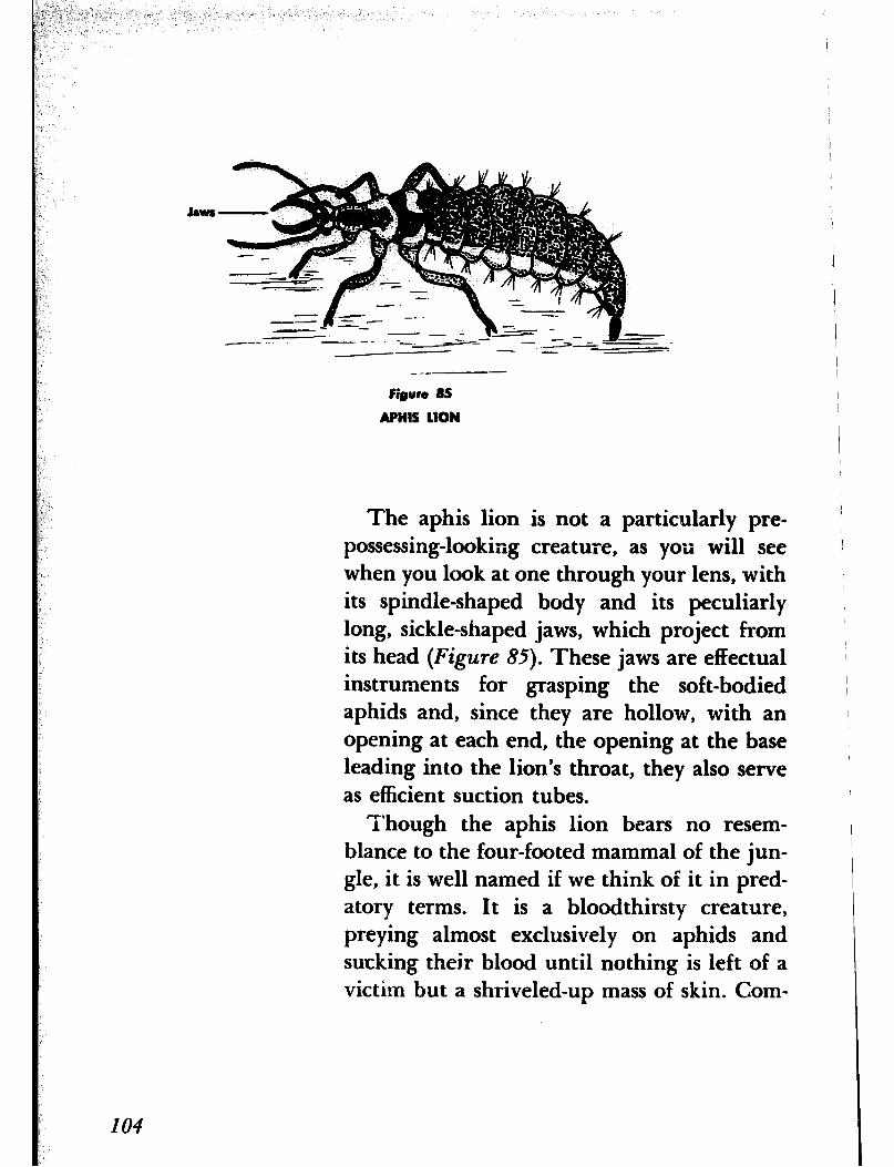
Jaws -
Figure 85
APNIS LION
The aphis lion is not a particularly pre- possessing-looking creature, as you will see when you look at one through your lens, with its spindle-shaped body and its peculiarly long, sickle-shaped jaws, which project from its head (Figure 85). These jaws are effectual instruments for grasping the soft-bodied aphids and, since they are hollow, with an opening at each end, the opening at the base leading into the lion’s throat, they also serve as efficient suction tubes.
Though the aphis lion bears no resem- blance to the four-footed mammal of the jun- gle, it is well named if we think of it in pred- atory terms. It is a bloodthirsty creature, preying almost exclusively on aphids and sucking their blood until nothing is left of a victim but a shriveled-up mass of skin. Com-
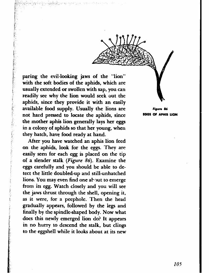
paring the evil-looking jaws of the “lion” with the soft bodies of the aphids, which are usually extended or swollen with sap, you can readily see why the lion would seek out the aphids, since they provide it with an easily available food supply. Usually the lions are Figure 16
not hard pressed to locate the achids, since EOOS OF APHIS 1lON
the mother aphis lion generally lays her eggs in a colony of aphids so that her young, when they hatch, have food ready at hand.
After you have watched an aphis lion feed on the aphids, look for the eggs. They are easily seen for each egg is placed on the tip of a slender stalk (Figure 86). Examine the eggs carefully and you should be able to de- tect the little doubled-up and still-unhatched lions. You may even find one aPmt to emerge from its egg. Watch closely and you will see the jaws thrust through the shell, opening it, as it were, for a peephole. Then the head gradually appears, followed by the legs and finally by the spindle-shaped body. Now what does this newly emerged lion do? It appears in no hurry to descend the stalk, but clings to the eggshell while it looks about at its new
IO6
surroundings. Eventually, as if it were satis- fied with what it sees, it begins to climb about the eggshell until it discovers the stalk. Since there is now no place to go but down the stalk, it grasps the stalk with its first pair of legs and with the help of the other two pairs of legs begins a careful descent, a feat for a creature only a few minutes old and with no previous experience at such gymnastics. At last it arrives safely on the surface of the leaf and pauses for a moment to look around. Spying the nearby aphids and without fur- ther ado, it proceeds to satisfy its hunger.
You may ask why the eggs were placed separately on the tips of stalks instead of be- ing laid in a mass on the surface of the leaf. The explanation is a simple one and at the same time illustrates an interesting provision of nature. The carnivorous lions that hatch from the eggs would just as soon eat one another or the unhatched eggs as search for aphids. But as the eggs hatch one at a time and since there is nothing to eat but an inedi- ble stalk, the newly hatched “lion” must per- force descend the stalk in order to find some- thing to eat. Of course, it could ascend the neighboring stalks and feed on its unhatched brothers and sisters, but how much better merely to seize the helpless and defenseless aphids that are at hand for the taking.
The mother aphis lion appears so entirely
unlike,her offspring that it would be difficult to believe they are related. Have you ever observed a small green insect with. golden eyes and delicate, lacelike wings flying about? That is the mother, commonly known as the lacewing but sometimes as the golden-eyed lacewing.
If you can find a mother lacewing in the act of laying her eggs, watch how she does it. You will need -your lens, of course, or, better still, youl’ reading glass. You will observe that first she deposits a drop of sticky fluid that she ejects from the tip of her body on the sur- face of the leaf. Then she lifts up her slender abdomen and spins the drop into a thread, a half inch long or more, that hardens almost immediately upon exposure to the air. She next lays an oblong egg about the size of a pin point on the tip of the thread or stalk and, with this accomplished, proceeds to spin another stalk and lay another egg. She re- peats this performance until she has laid her full complement of eggs.
THE STUDY OF NATURE or the outdoors is the study of life itself. No one has yet been able to define life to anyone’s complete satisfac- tion, and, of course, no one has been able to create life, although we seem to be on the
We Trace a Tadpole’s
Development
107
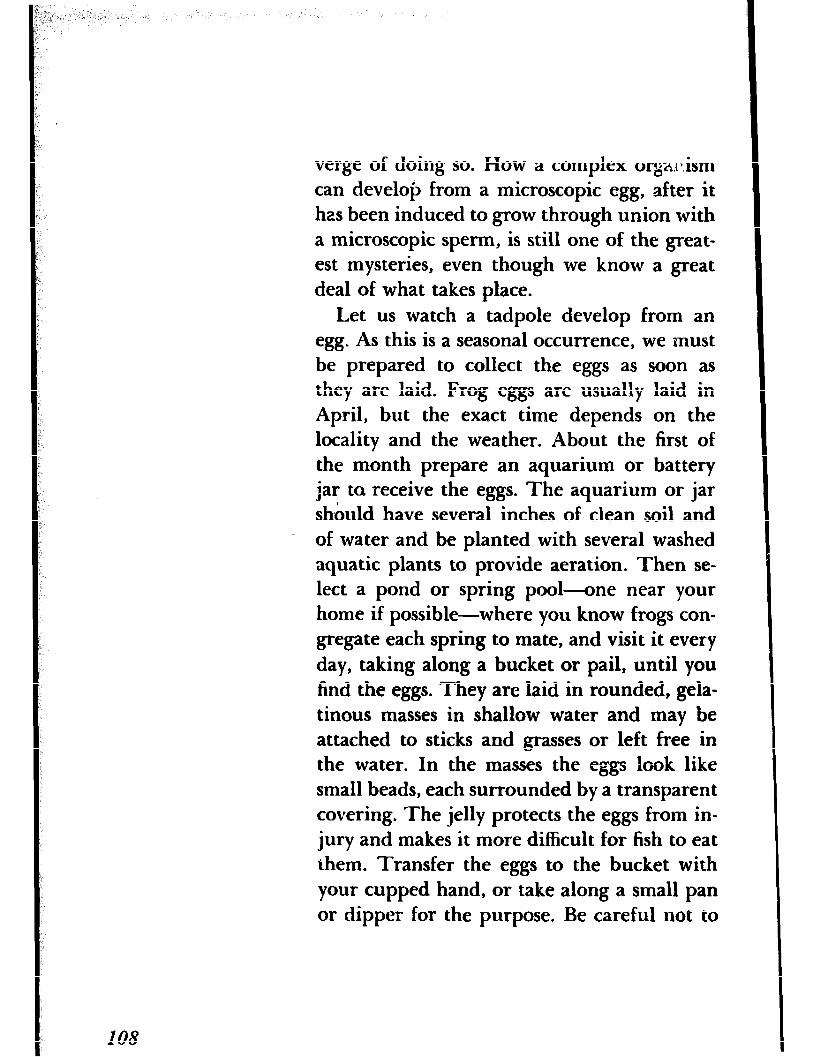
verge of doing so. How a complex ore.ix.ism can develop from a microscopic egg, after it has been induced to grow through union with a microscopic sperm, is still one of the great- est mysteries, even though we know a great deal of what takes place.
Let us watch a tadpole develop from an egg. As this is a seasonal occurrence, we must be prepared to collect the eggs as soon as they are laid. Frog eggs are usually laid in April, but the exact time depends on the locality and the weather. About the first of the month prepare an aquarium or battery jar ta receive the eggs. The aquarium or jar should have several inches of clean soil and
- of water and be planted with several washed aquatic plants to provide aeration. Then se- lect a pond or spring pool-one near your home if possible-where you know frogs con- gregate each spring to mate, and visit it every day, taking along a bucket or pail, until you find the eggs. They are laid in rounded, gela- tinous masses in shallow water and may be attached to sticks and grasses or left free in the water. In the masses the eggs look like small beads, each surrounded by a transparent covering. The jelly protects the eggs from in- jury and makes it more difficult for fish to eat them. Transfer the eggs to the bucket with your cupped hand, or take along a small pan or dipper for the purpose. Be careful not to
IO8
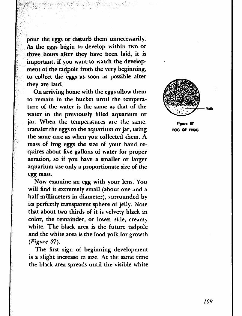
pour the eggs or disturb them unnecessarily. As the eggs begin to develop within two or three hours after they have been laid, it is important, if you want to watch the develop- ment of the tadpole from the very beginning, to collect the eggs as soon as possible after they are laid.
On arriving home with the eggs allow them to remain in the bucket until the tempera- ture of the water is the same as that of the water in the previously filled aquarium or jar. When the temperatures are the same, transfer the eggs to the aquarium or jar, using the same care as when you collected them. A mass of frog eggs the size of your hand re- quires about five gallons of water for proper aeration, so if you have a smaller or larger aquarium use only a proportionate size of the egg mass.
Now examine an egg with your lens. You will find it extremely small (about one and a half millimeters in diameter), surrounded by its perfectly transparent sphere of jelly. Note that about two thirds of it is velvety black in color, the remainder, or lower side, creamy white. The black area is the future tadpole and the white area is the food yolk for growth (Figure 87).
The first sign of beginning development is a slight increase in size. At the same time the black area spreads until the visible white
Yolk
Figure 87
EGG OF FROG
109
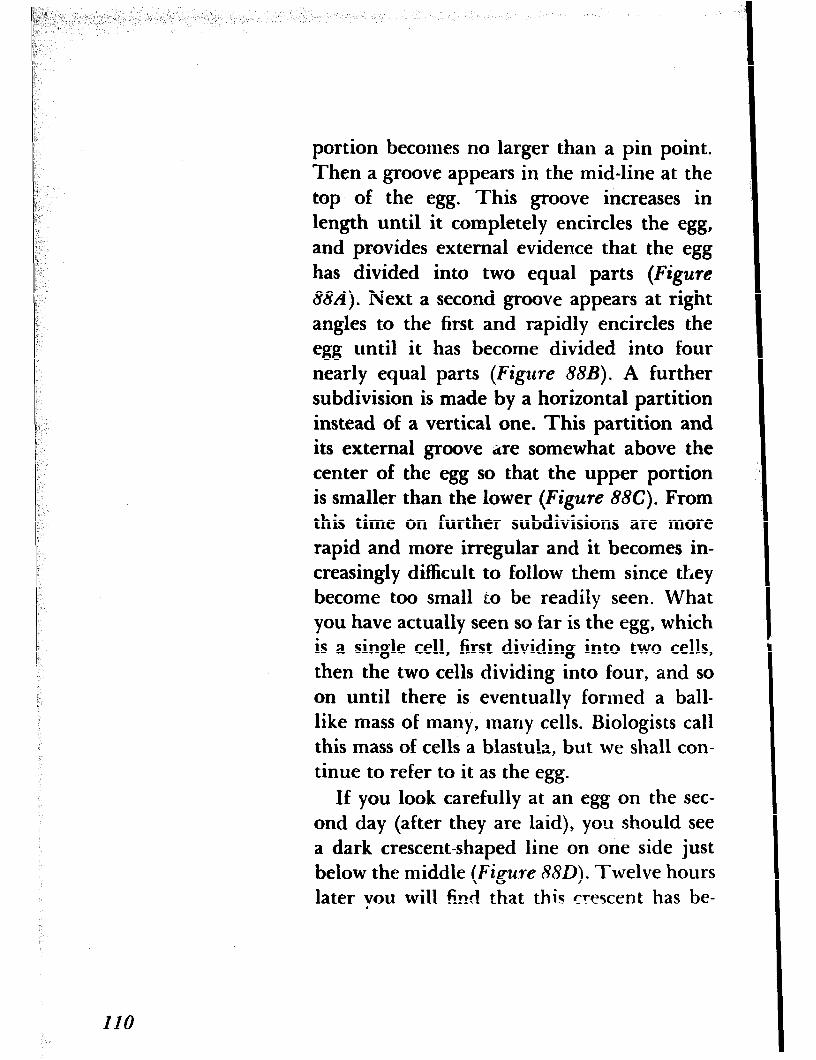
portion becomes no larger than a pin point. Then a groove appears in the mid-line at the top of the egg. This groove increases in length until it completely encircles the egg, and provides external evidence that the egg has divided into two equal parts (Figure
884. Next a second groove appears at right angles to the first and rapidly encircles the egg until it has become divided into four nearly equal parts (Figure 88B). A further subdivision is made by a horizontal partition instead of a vertical one. This partition and its external groove ;ire somewhat above the center of the egg so that the upper portion is smaller than the lower (Figure 88C). From this time on further subdivisions are more rapid and more irregular and it becomes in- creasingly difficult to follow them since they become too small to be readily seen. What you have actually seen so far is the egg, which is a single cell, first dividing into two cells, then the two cells dividing into four, and so on until there is eventually formed a ball- like mass of many, many cells. Biologists call this mass of cells a blastu!a, but we shall con- tinue to refer to it as the egg.
If you look carefully at an egg on the sec- ond day (after they are laid), you should see a dark crescent-shaped line on one side just below the middle (Figure 880). Twelve hours later you will find that this crescent has be-
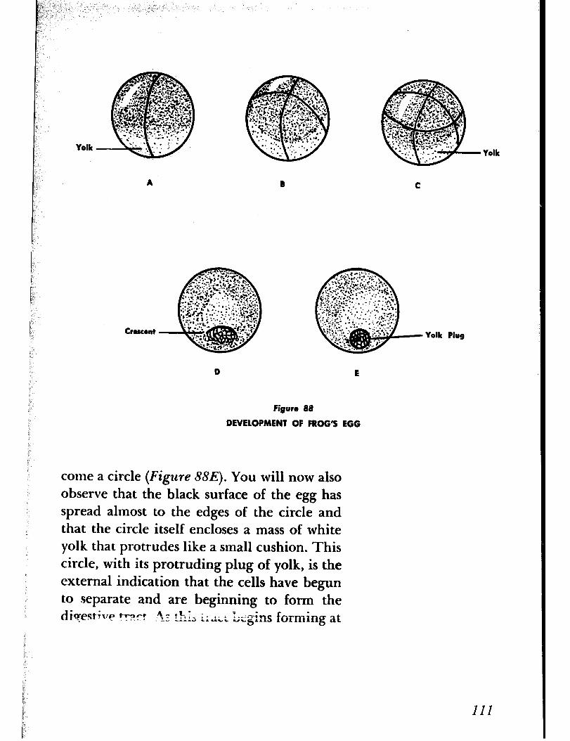
Yolk Yolk
Crescent Yolk Plug
D E
Figure 88
DEVELOPMENT OF FROG’S EGG
come a circle (Figure 88E). You will now also observe that the black surface of the egg has spread almost to the edges of the circle and that the circle itself encloses a mass of white yolk that protrudes like a small cushion. This circle, with its protruding plug of yolk, is the external indication that the cells have begun to separate and are beginning to form the djcrestlrry tr~rt +: thy ; : _ -L c-a- 2 c.&.L UN. !--&s forming at
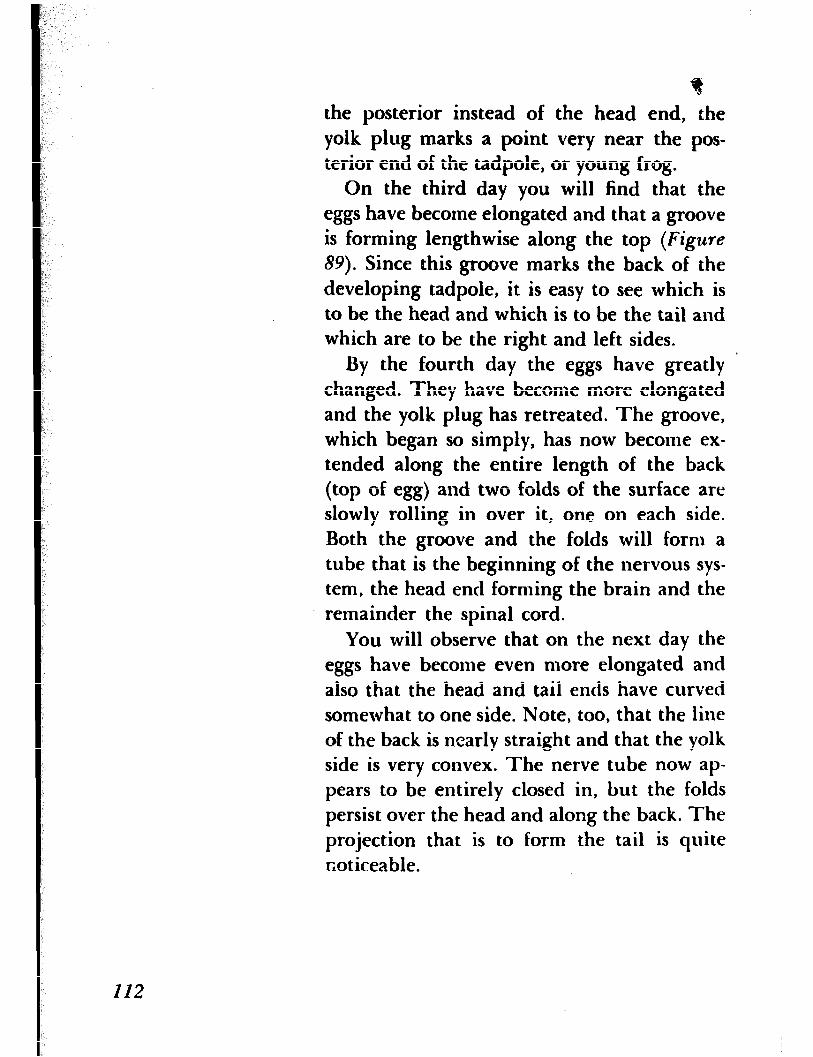
f the posterior instead of the head end, the yolk plug marks a point very near the pos- terior end of the tadpole, oT young frog.
On the third day you will find that the eggs have become elongated and that a groove is forming lengthwise along the top (Figure
89). Since this groove marks the back of the developing tadpole, it is easy to see which is to be the head and which is to be the tail and which are to be the right and left sides.
By the fourth day the eggs have greatly ’ changed. They have become more elongated and the yolk plug has retreated. The groove, which began so simply, has now become ex- tended along the entire length of the back (top of egg) and two folds of the surface are slowly rolling in over it, one on each side. Both the groove and the folds will form a tube that is the beginning of the nervous sys- tem, the head end forming the brain and the remainder the spinal cord.
You will observe that on the next day the eggs have become even more elongated and also that the head and tail ends have curved somewhat to one side. Note, too, that the line of the back is n.early straight and that the yolk side is very convex. The nerve tube now ap- pears to be entirely closed in, but the folds persist over the head and along the back. The projection that is to form the tail is quite r,oticeable.
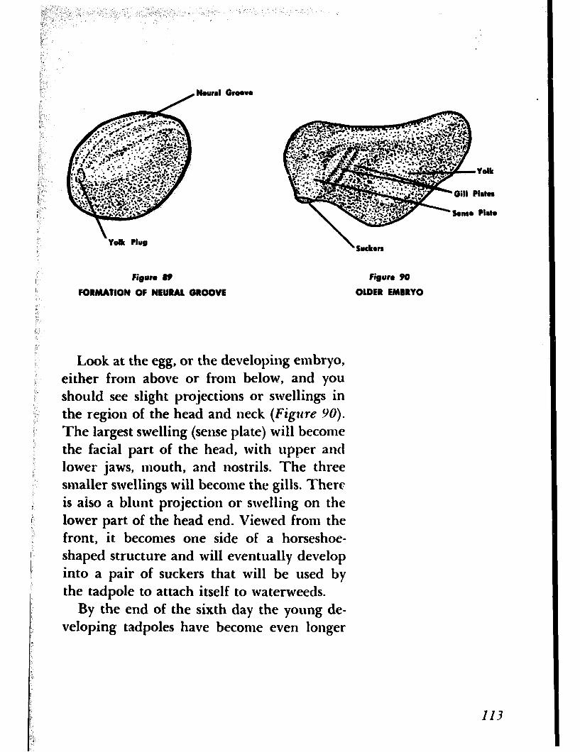
Nourel Groove
Yolk Plug
Figwo 89 Figure 90
FORMATION OF NEURAL GROOVE OLDER EMBRYO
Look at the egg, or the developing embryo, either from above or from below, and you should see slight projections or swellings in the region of the head and neck (Figure YO).
The largest swelling (sense plate) will become the facial part of the head, with upper and lower jaws, mouth, and nostrils. The three smaller swellings will become the gills. There is also a blunt projection or swelling on the lower part of the head end. Viewed from the front, it becomes one side of a horseshoe- shaped structure and will eventually develop into a pair of suckers that will be used by the tadpole to attach itself to waterweeds.
By the end of the sixth day the young de- veloping tadpoles have become even longer
and more curved. The body is thinner, the head and tail ends are unmistakable, and the swellings on the sides of the neck more dis- tinct. On the following day the head is more delineated and the tail is longer and clearly finned at its edges. You may observe the tad- poles move occasionally.
Nine days after the eggs are laid the tad- poles are out of the jelly, clinging, by means of their suckers, to the deserted jelly mass or water plants. The suckers do not act in the way their names would seem to imply but function by reason of a sticky substance or cement they secrete. If all the tadpoles are not out of the jelly, you should be able to see some of them hatch from the egg: with a vigorous wriggle a tadpole escapes from the _ egg, the jelly ruptures, and the tadpole is free. Note that the tadpoles are very slender and black and that the transparent tail is quite conspicuous. Observe, too, that the swellings at the sides of the neck are branched, fingerlike gills. With your lens examine the tadpole closely and-you will see the swellings that are to become eyes, and an opening just forward of the suckers that is to be the mouth.
On the following day all the tadpoles should be out of their eggs. They hang quietly from the water plants or circle about with vigorous wrigglings of the tail when-
II4

ever disturbed. ‘Through the next few days further changes take place. The tadpoles be- come longer, although the increase in length is mainly in the tail, and they also become wider, due to the further development of internal organs. Then, on the sixteenth day, they are swimming about rapidly, with mouths open, and nibbling at the water plants. They are no longer jet black but rather brownish, with a fine mottling of gold spots on the back.
MANY OF THE WAYS in which nature has con- trived to effect seed dispersal are most in- genious. The seeds of some plants, as those of the dandelion, are provided with silken hairs that catch in the wind; others are able to float on water and in that manner are transported to distant places. Then there are those plants that shoot or propel their seeds considerable distances into the air, as the witch hazel. Finally, we have the plants whose seeds are furnished with structures that catch in the fur of animals or in our clothing when we come in contact with them.
Most familiar of these so-called “hitch- hikers” are the burs that occur on a familiar and rather rank-odored weed called the bur- dock, which is common along roadsides and
ADVENTURE 23
We Encounter Some Hitchhikers
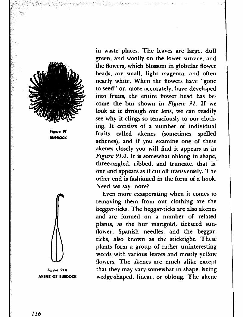
Figure 91
BURDOCK
Figure 91A
AKENE OF BURDOCK
in waste places. The leaves are large, dull green, and woolly on the lower suriace, and the flowers, which blossom in globular flower heads, are small, light magenta, and often nearly white. When the flowers have “gone to seed” or, more accurately, have developed into fruits, the entire flower head has be- come the bur shown in Figure 91. If we look at it through our lens, we can readily see why it clings so tenaciously to our cloth- ing. It consists of a number of individual fruits called akenes (sometimes spelled achenes), and if you examine one of these akenes closely you will find it appears as in Figure 91A. It is somewhat oblong in shape, three-angled, ribbed, and truncate, that is,’ one end appears as if cut off transversely. The other end is fashioned in the form of a hook. Need we say more?
Even more exasperating when it comes to removing them from our clothing are the beggar-ticks. The beggar-ticks are also akenes and are formed on a number of related plants, as the bur marigold, tickseed sun- flower, Spanish needles, and the beggar- ticks, also known as the sticktight. These plants form a group of rather uninteresting weeds with various leaves and mostly yellow flowers. The akenes are much alike except that they may vary somewhat in shape, being wedge-shaped, linear, or oblong. The akene

shown in Figure 92 is from the beggar-ticks, which can usually be found growing in damp situations. Sometimes my trousers have been so completely covered with these alcenes that it has taken me hours to get them off. Look tit one with your lens and see why. Figure 93 shows an akene from Spanish ineedles. It re- sembles the akene of beggar-ticks except that it is four-awned, whereas the former is two- awned.
If we should walk in the woody during July or August, we would likely come upon a plant having a generally leaflevs flower sttm with a scattered cluster of very small magenta- pink or lilac flowers and known as the tick trefoil- A few weeks later, were we to return to this same plant, we wduld find that the flowers had developed into seed pods shaped somewhat ,like that in Figure 94. To our naked eye there would be no apparent reason why these seed pods should become attached to our clothing, as they do should we brush against them. But we need only to view them through our lens to see that they are provided with minute hooked hairs that are as effec- tive in hitchhiking a ride as the barbed akenes of the beggar-ticks or the hooked akenes of the burdock. There are many species of tick trefoils, each with its own characteristically shaped seed pods, so that if you find pods slightly different in shape from that illus-
I
Figvm 93
SPANISH NEEDLES
117
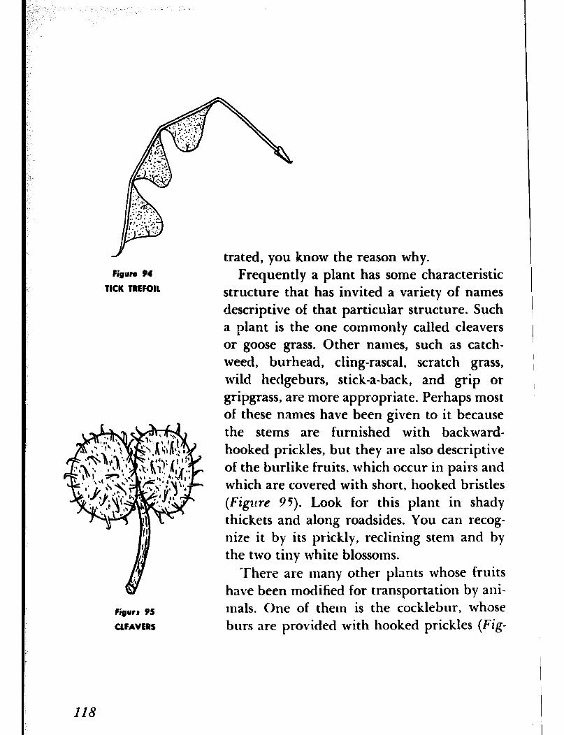
Figurn 94
TICK TREFOIB
Figure 95
CLEAVERS
trated, you know the reason why. Frequently a plant has some characteristic
structure that has invited a variety of names descriptive of that particular structure. Such a plant is the one commonly called cleavers or goose grass. Other names, such as catch- weed, burhead, cling-rascal, scratch grass, wild hedgeburs, stick-a-back, and grip or gripgrass, are more appropriate. Perhaps most of these names have been given to it because the stems are furnished with backward- hooked prickles, but they are also descriptive of the burlike fruits, which occur in pairs and which are covered with short, hooked bristles (Figzlre 95). Look for this plant in shady thickets and along roadsides. You can recog- nize it by its prickly, reclining stem and by the two tiny white blossoms.
There are many other plants whose fruits have been modified for transportation by ani- mals. One of them is the cocklebur, whose but-s are provided with hooked prickles (Fig-
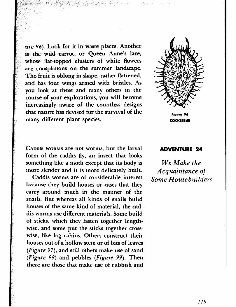
we Y6). Look for it in waste places. Another is the wild carrot, or Queen Anne’s lace, whose flat-topped clusters of white flowers are conspicuous on the summer landscape. The fruit is oblong in shape, rather flattened, and has four wings armed with bristles. As you look at these and many others in the course of your explorations, you will become increasingly aware of the countless designs that nature has devised for the survival of the
I many different plant species. Figure 96
COCKLEBUR
CADDIS WORMS are not worms, but the larval form of the caddis Hy, an insect that looks something like a moth except that its body is more slender and it is more delicately built.
ADVENTURE 24
We Make the
Caddis worms are of considerable interest Acquaintance of
Some Housebuilders because they build houses or cases that they carry around much in the manner of the snails. But whereas all kinds of snails build houses of the same kind of material, the cad- dis worms use different materials. Some build of sticks, which they fasten together length- wise, and some put the sticks together cross- wise, like log cabins. Others construct their houses out of a hollow stem or of bits of leaves (Figure 97), and still others make use of sand (Figure 98) and pebbles (Figure 99). Then there are those that make use qf rubbish and
I
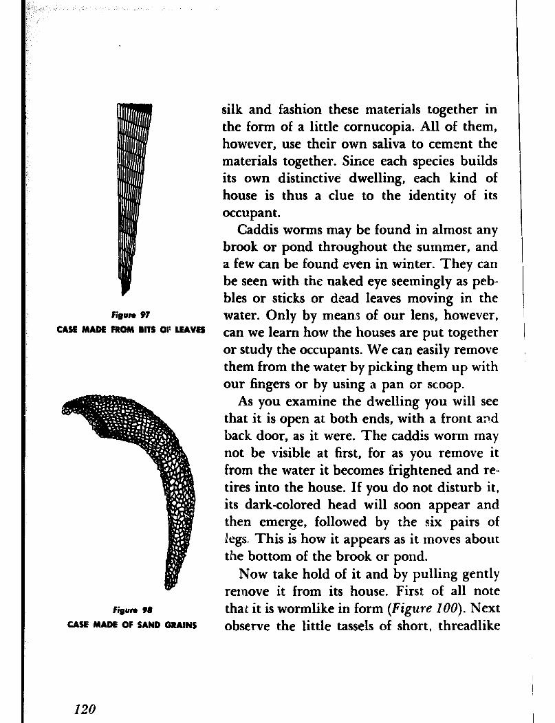
Figure 97
CASE MADE FROM BITS OF LEAVES
Figure 98
CASE MADE OF SAND GRAINS
silk and fashion these materials together in the form of a little cornucopia. All of them, however, use their own saliva to cement the materials together. Since each species builds its own distinctive dwelling, each kind of house is thus a clue to the identity of its occupant.
Caddis worms may be found in almost any brook or pond throughout the summer, and a few can be found even in winter. They can be seen with the naked eye seemingly as peb- bles or sticks or drad leaves moving in the water. Only by means of our lens, however, can we learn how the houses are put together or study the occupants. We can easily remove them from the water by picking them up with our fingers or by using a pan or scoop.
As you examine the dwelling you will see that it is open at both ends, with a front apd back door, as it were. The caddis worm may not be visible at first, for as you remove it from the water it becomes frightened and re- tires into the house. If you do not disturb it, its dark-colored head will soon appear and then emerge, followed by the six pairs of legs. This is how it appears as it moves about the bottom of the brook or pond.
Now take hold of it and by pulling gently remove it from its house. First of all note that it is wormlike in form (Figure IOO). Next observe the little tassels of short, threadlike
120
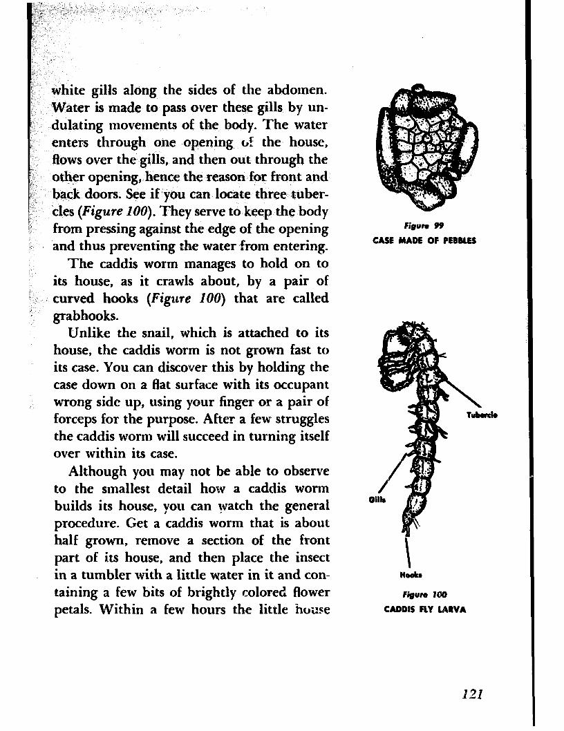
white gills along the sides of the abdomen. Water is made to pass over these gills by un- dulating movements of the body. The water enters through one opening ~2 the house, flows over the gills, and then out through the other opening, hence the reason for front and back doors. See if you can locate three tuber- cles (Figure 100). They serve to keep the body from pressing against the edge of the opening and thus preventing the water from entering.
The caddis worm manages to hold on to its house, as it crawls about, by a pair of curved hooks (Figzlre 100) that are called grabhooks.
Unlike the snail, which is attached to its house, the caddis worm is not grown fast to its case. You can discover this by holding the case down on a flat surface with its occupant wrong side up, using your finger or a pair of forceps for the purpose. After a few struggles the caddis worm will succeed in turning itself over within its case.
Although you may not be able to observe to the smallest detail how a caddis worm builds its house, you can watch the general procedure. Get a caddis worm that is about half grown, remove a section of the front part of its house, and then place the insect in a tumbler with a little water in it and con- taining a few bits of brightly colored flower petals. Within a few hours the little house
Figure 99
CASE MADE OF PEBBLES
Figure 100
CADDIS FLY LARVA
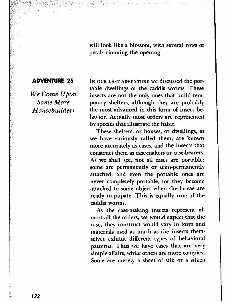
will look like a blossom, with several rows of petals rimming the opening.
ADVENTURE 25
We Come Upon Some More
Housebuildexs
IN OURLASTADVENTURE wediscussedthepor- table dwellings of the caddis worms. These insects are not the only ones that build tem- porary shelters, although they are probably the most advanced in this form of insect be- havior. Actually most orders are represented by species that illustrate the habit.
These shelters, or houses, or dwellings, as we have variously called them, are known more accurately as cases, and the insects that construct them as case-makers or case-bearers. As we shall see, not all cases are portable; some are permanently or semi-permanently attached, and even the portable ones are never completely portable, for they become attached to.some object when the larvae are ready to pupate. This is equally true of the caddis worms.
As the case-making insects represent al- most all the orders, we would expect that the cases they construct would vary in form and materials used as much as the insects them- selves exhibit different types of behavioral patterns. Thus we have cases that are very simple affairs, while others are more complex. Some are merely a sheet of silk or a silken
122

tube; others consist of various materials added or blended with the silk, but all have i.n common the use of silk as the primary material.
Much can be written about the origin and development of the case-making habit, but what we are particularly interested in at pres- ent is to find and examine a few typical cases and to observe how they are put together. Since many of them serve as winter retreats for the hibernating larvae, we can engage in Figure 101
this study almost any time of the year. How- SILKEN CASE OF CLO?HES MOTH
ever, if we wish to observe the habits of the larvae, we can do so only when they are active and feeding, during the warmer months. An exception would be the clothes moths, which may be found throughout the year. It may be somewhat difficult to find these insects, for no one will admit their presence, since to do so would be a confession of neglect. Yet you may come across them sometimes. Should you do so, transfer a few of’the cases with their occupants to a bottle or jar. You can then observe with your lens how the larvae have to enlarge their cases from time to time to accommodate an increase in size The cases are simply silken tubes in which bits of ma- terial on which the insects feed are sometimes incorporated (Figure 101). Figure 102
Far more available are the curious baglike CASE OF BAGWORM
shelters of the bagworm (Figure 102). This
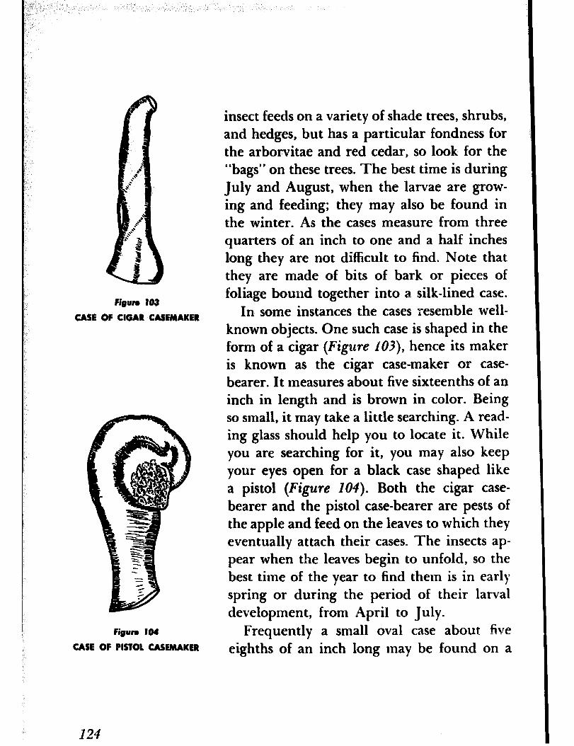
Figure 103
CASE OF CIGAR CASEMAKER
Figun 104
CASE OF PISTOL CASEMAKER
124
insect feeds on a variety of shade trees, shrubs, and hedges, but has a particular fondness for the arborvitae and red cedar, so look for the “bags” on these trees. The best time is during July and August, when the larvae are grow- ing and feeding; they may also be found in the winter. As the cases measure from three quarters of an inch to one and a half inches long they are not difficult to find. Note that they are made of bits of bark or pieces of foliage bound together into a silk-lined case.
In some instances the cases resemble well- known objects. One such case is shaped in the form of a cigar (Figure 103), hence its maker is known as the cigar case-maker or case- bearer. It measures about five sixteenths of an inch in length and is brown in color. Being so small, it may take a little searching. A read- ing glass should help you to locate it. While you are searching for it, you may also keep your eyes open for a black case shaped like a pistol (Figure 104). Both the cigar case- bearer and the pistol case-bearer are pests of the apple and feed on the leaves to which they eventually attach their cases. The insects ap- pear when the leaves begin to unfold, so the best time of the year to find them is in early spring or during the period of their larval development, from April to July.
Frequently a small oval case about five eighths of an inch long may be found on a
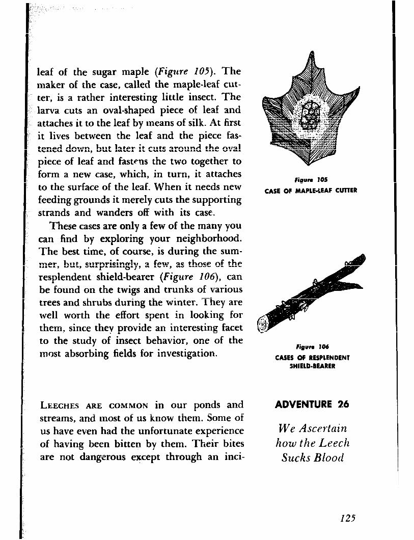
leaf of the sugar maple (Figure 105). The maker of the case, called the maple-leaf cut- ter, is a rather interesting little insect. The larva cuts an oval-shaped piece of leaf and attaches it to the leaf by means of silk. At first it lives between the leaf and the piece fas- tened down, but later it cuts around the oval piece of leaf and fastens the two together to form a new case, which, in turn, it attaches to the surface of the leaf. When it needs new feeding grounds it merely cuts the supporting strands and wanders off with its case.
Fimum 105
CASE OF MAPLE-LEAF CUTTER
These cases are only a few of the many you can find by exploring your neighborhood. The best time, of course, is during the sum- mer, but, surprisingly, a few, as those of the resplendent shield-bearer (Figure 106)) can be found on the twigs and trunks of various trees and shrubs during the winter. They are well worth the effort spent in looking for them, since they provide an interesting facet to the study of insect behavior, one of the most absorbing fields for investigation.
Figun 106
CASES OF RESPLENDENT SlIELD-BEARER
LEECHES ARE COMMON in our ponds and streams, and most of us know them. Some of us have even had the unfortunate experience of having been bitten by them. Their bites are not dangerous except through an inci-
ADVENTURE 26
We Ascertain
how the Leech Sucks Blood
I25
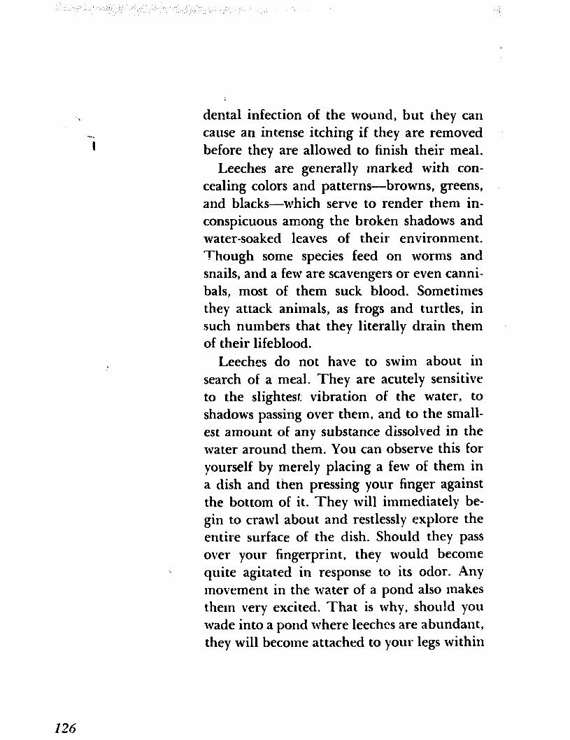
dental infection of the wound, but they can cause an intense itching if they are removed before they are allowed to finish their meal.
Leeches are generally marked with con- cealing colors and patterns-browns, greens, and blacks-which serve to render them in- conspicuous among the broken shadows and water-soaked leaves of their environment. Though some species feed on worms and snails, and a few are scavengers or even canni- bals, most of them suck blood. Sometimes they attack animals, as frogs and turtles, in such numbers that they literally drain them of their lifeblood.
Leeches do not have to swim about in search of a meal. They are acutely sensitive to the slightest. vibration of the water, to shadows passing over them, and to the small- est amount of any substance dissolved in the water around them. You can observe this for yourself by merely placing a few of them in a dish and then pressing your finger against the bottom of it. They will immediately be- gin to crawl about and restlessly explore the entire surface of the dish. Should they pass over your fingerprint, they would become quite agitated in response to its odor. Any movement in the water of a pond also makes them very excited. That is why, should you wade into a pond where leeches are abundant, they will become attached to your legs within
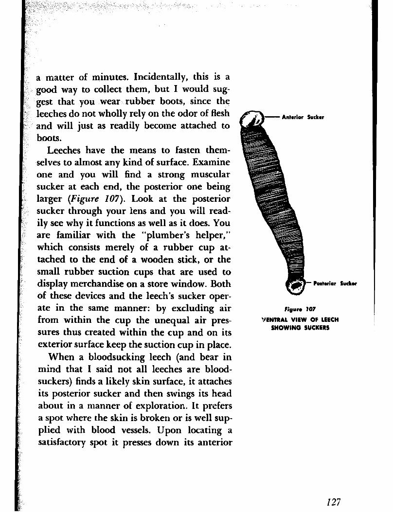
a matter of minutes. Incidentally, this is a good way to collect them, but I would sug- gest that you wear rubber boots, since the leeches do not wholly rely on the odor of flesh and will just as readily become attached to boots.
Leeches have the means to fasten them- selves to almost any kind of surface. Examine one and you will find a strong muscular sucker at each end, the posterior one being larger (Figzcre 107). Look at the posterior sucker through your lens and you will read- ily see why it functions as well as it does. You are familiar with the “plumber’s helper,” which consists merely of a rubber cup at- tached to the end of a wooden stick, or the small rubber suction cups that are used to display merchandise on a store window. Both of these devices and the leech’s sucker oper- ate in the same manner: by excluding air from within the cup the unequal air pres- sures thus created within the cup and on its exterior surface keep the suction cup in place.
When a bloodsucking leech (and bear in mind that I said not all leeches are blood- suckers) finds a likely skin surface, it attaches its posterior sucker and then swings its head about in a manner of exploration. It prefers a spot where the skin is broken or is well sup- plied with blood vessels. Upon locating a satisfactory spot it presses down its anterior
SUCIW
I- Ibtr rior such
Figure 107
VENTRAL VIEW OF 1EECH SHOWING SUCKERS
127
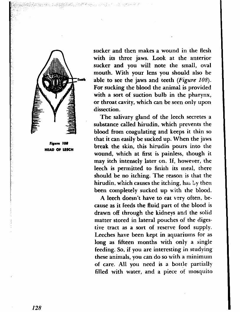
n
Figun 108
HEAD OF UKH
sucker and then makes a wound in the flesh with its three jaws. Look at the anterior sucker and you will note the small, oval mouth. With your lens you should also be able to see the jaws and teeth (Figure 108).
For sucking the blood the animal is provided with a sort of suction bulb in the pharynx, or throat cavity, which can be seen only upon dissection.
The salivary gland of the leech secretes a substance called hirudin, which prevents the blood from coagulating and keeps it thin so that it can easily be sucked up. When the jaws break the skin, this hirudin pours into the wound, which at first is painless, though it may itch intensely later on. If, however, the leech is permitted to finish its meal, there should be no itching. The reason is that the hirudin, which causes the itching, haz Ly then been completely sucked up with the blood.
A leech doesn’t have to eat very often, be- cause as it feeds the fluid part of the blood is drawn off through the kidneys and the solid matter stored in lateral pouches of the diges- tive tract as a sort of reserve food supply. Leeches have been kept in aquariums for as long as fifteen months with only a single feeding. So, if you are interesting in studying these animals, you can do so with a minimum of care. All you need is a bottle partially filled with water, and a piece df mosquito
I28

netting or similar material tied over the mouth to prevent the animals from crawling out. A meal of ground meat, beef liver, or earthworms will do them for several weeks. Be sure to change the water each day to pro- vide them with a fresh supply of oxygen and to prevent the water from becoming polluted. In observing their habits I would suggest a reading glass, although for close observa- tion a hand lens should be used. In olden times leeches were used in bloodletting, and I read lately that the practice is being re- vived; some drugstores again carry them.
FERNS ARE NOT a particularly important form of plant life. However, it was not always so. At one time they and their allies were the dominant plants of the earth. They are gen- erally supposed to be the oldest form of ter- restrial vegetation now in existence, and the coal we now mine is said to h.ave been formed from their remains many millions of years ago. At that time they grew to an enormous size, often attaining a height of fifty feet or more. Some of the ferns found in the tropical rain forests today grow to considerable heights, but the ferns with which most of ‘~1s are familiar are low-growing species.
ADVENTURE 27
We Identify Some Ferns
There are many kinds of ferns, and though,
129

Figun IOU
COMMON FOLYPODY
at a glance, they may appear to be much alike, they all differ from one another if only slightly. Sometimes only an expert can rec- ognize certain species. Many of them can readily be recognized by their habits of growth, by the shapes of their fronds, or by the manner in which the fronds have been cut, so we need not be an expert to know at least a few of them. Once we have become familiar with such easily recognizable species as the bracken, the sensitive fern, and the common polypody, for example, we can go a little further and learn how to identify others by their fruit dots. These fruit dots appear as little brown or gray specks or dots on the lower surface of the frond; perhaps you have noticed them.
Before we continue, let me point out that a frond is merely a leaf. In most ferns the fronds have been cut into divisions or leaflets called pinnae, and in some species these divi- sions have been further subdivided i’nto still smaller leaflets, or pinnules. Ferns reproduce bv means of spores, which are produced in small structures called sporangia (singular sporangium). These sporangia are usually clustered in groups called sori (singular sorus), which occur in various shapes in the different species. Thus they may be linear, oblong, kidney-shaped, or curved. Their shape and position on the frond, or more ac-
130
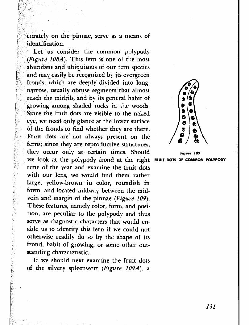
curately on the pinnae, serve as a means of identification.
Let us consider the common polypody (Figzdre IO8A). This fern is one of the most abundant and ubiquitous of our fern species and may easily be recognized by its evergreen fronds, which are deeply divided into long, narrow, usually obtuse segments that almost reach the midrib, and by its general habit of growing among shaded rocks in t!ae woods. Since the fruit dots are visible to the naked eye, we need only glance at the lower surface of the fronds to find whether they are there. Fruit dots are not always present on the ferns; since they are reproductive structures, they occur only at certain times. Should Figure 109
we look at the polypody frond at the right FM DOTS OF COMM POWODY time of the year and examine the fruit dots with our lens, we would find them rather large, yellow-brown in color, roundish in form, and located midway between the mid- vein and margin of the pinnae (Figure IO9).
These features, namely color, form, and posi- tion, are peculiar to the polypody and thus serve as diagnostic characters that would en- able us to identify this fern if we could not otherwise readily do so by the shape of it.s frond, habit of growing, or some other out- standing characteristic.
If we should next examine the fruit dots of the silvery spleenwort (Figure 109A), a
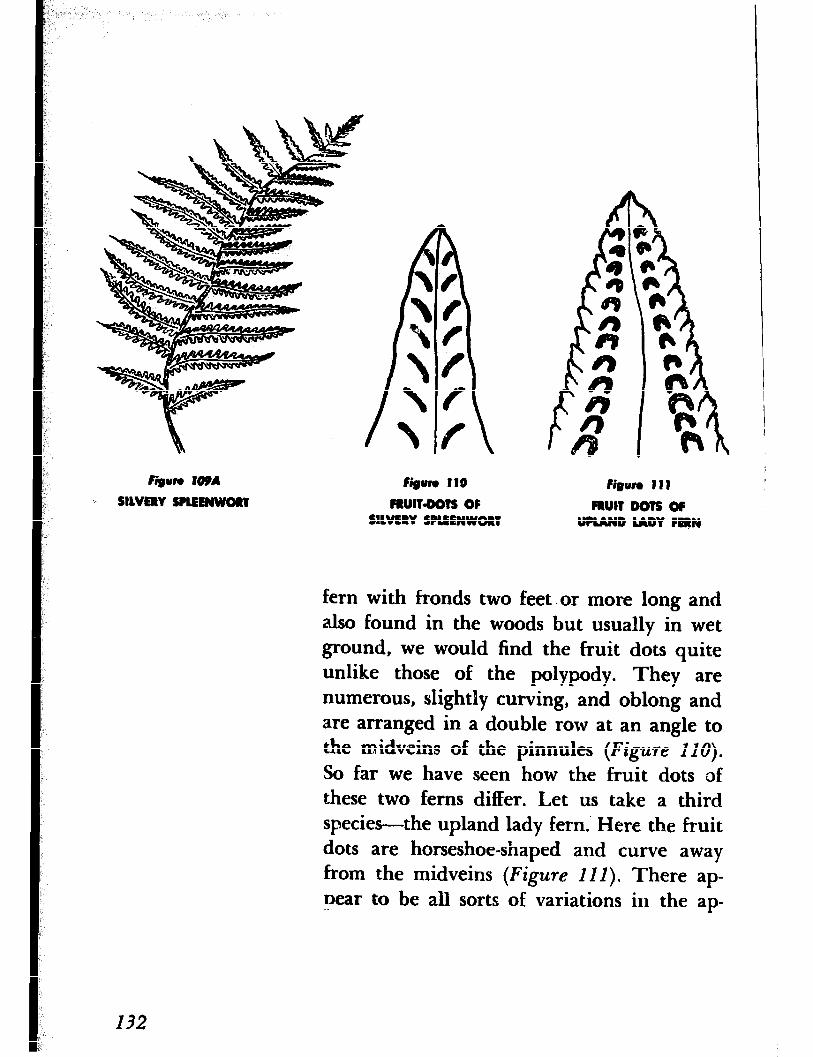
Figun f10 Firnun 111
FRUlT=DOTS OF FRUIT DOTE OP SILVERY SPLEENWORT UPLAND LADY FRRN
fern with fronds two feet. or more long and also found in the woods but usually in wet ground, we would find the fruit dots quite unlike those of the polypody. They are numerous, slightly curving, and oblong and are arranged in a double row at an angle to the midveins of the pinnules (Figure I IO).
So far we have seen how the fruit dots of these two ferns differ. Let us take a third species-the upland lady fern.’ Here the fruit dots are horseshoe-shaped and curve away from the midveins (Figure 111). There ap- pear to be all sorts of variations in the ap-
I32
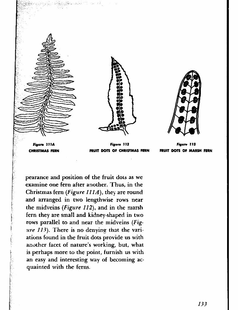
Figun IIIA
CHRISTMAS FERN
Figure 112 Figure 113
FRUIT DOTS OF CHRlETMAS FERN FRUIT DOTS 0F MARSH FERN
pearance and position of the fruit dots as we examine one fern after another. Thus, in the Christmas fern (Figure II I A), they are round and arranged in two lengthwise rows near the midveins (Figure 112), and in the marsh fern they are small and kidney-shaped in two rows parallel to and near the midveins (Fig-
zm II+ There is no denying that the vari- ations found in the fruit dots provide us with another facet of nature’s working, but, what is perhaps more to the point, furnish us with an easy and interesting way of becoming ac- quainted with the ferns.
133
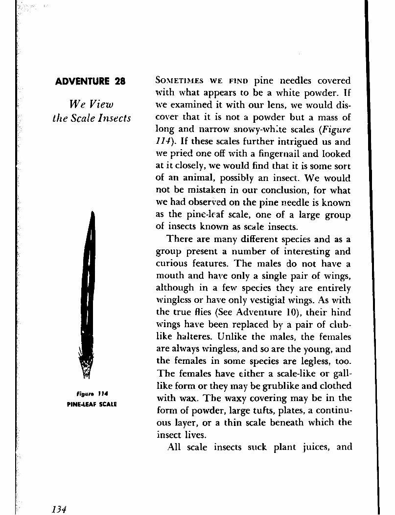
figure 114
PlNEdEAF SCALE
134
ADVENTURE 28
We View the Scale Insects
SOMETIMES WE FIND pine needles covered with what appears to be a white powder. If we examined it with our lens, we would dis- cover that it is not a powder but a mass of long and narrow snowy-white scales (Figure
IN). If these scales further intrigued us and we pried one off with a fingernail and looked at it closely, we would find that it is some sort of an animal, possibly an insect. We would not be mistaken in our conclusion, for what we had observed on the pine needle is known as the pine-leaf scale, one of a large group of insects known as scale insects.
There are many different species and as a group present a number of interesting and curious features. The males do not have a mouth and have only a single pair of wings, although in a few species they are entirely wingless or have only vestigial wings. As with the true flies (See Adventure lo), their hind wings have been replaced by a pair of club- like halteres. Unlike the males, the females are altvays wingless, and so are the young, and the females in some species are legless, too. The females have either a scale-like or gall- like form or they may be grublike and clothed with wax. The waxy covering may be in the form of powder, large tufts, plates, a continu- ous layer, or a thin scale beneath which the insect lives.
All scale insects suck plant juices, and
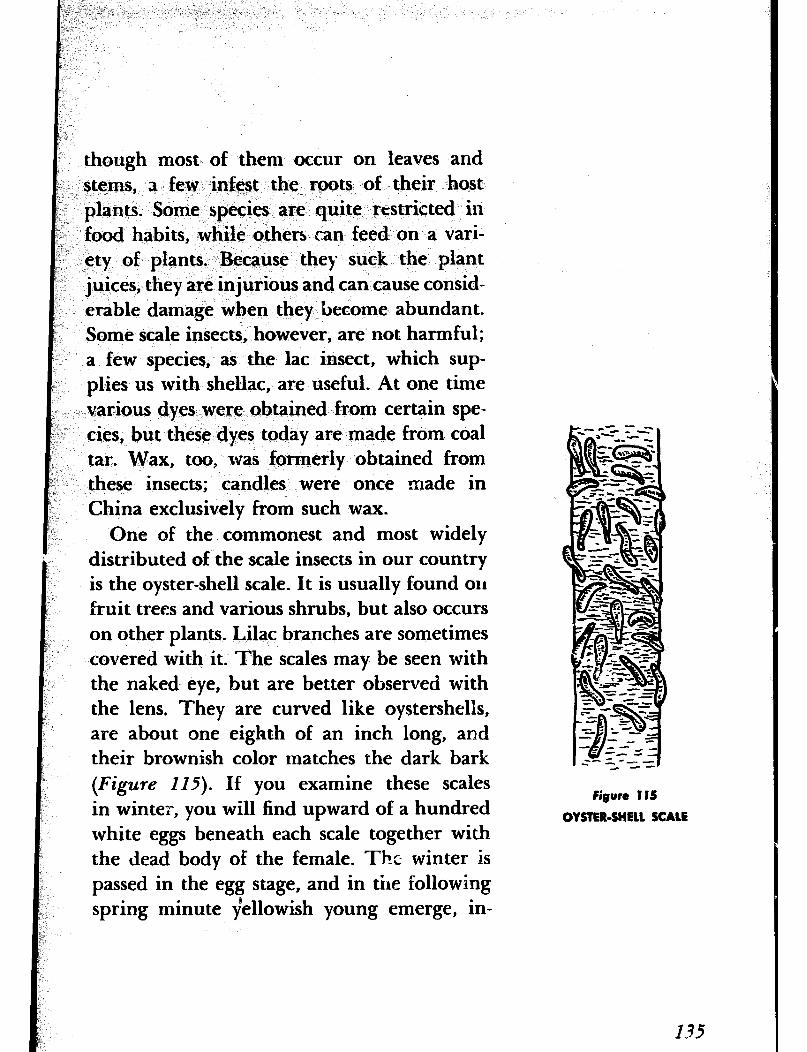
though most of them occur on leaves and stems, 3 few infest the roots of their host
F plants. Some species are quite restricted in is food habits, while others can feed on a vari- jk ety of plants. Because they suck the plant
juices, they are injurious and can cause consid- erable damage when they become abundant. Some scale insects, however, are not harmful; a few species, as the lac insect, which sup- plies us with shellac, are useful. At one time
;. various dyes were obtained from certain spe- cies, but these dyes today are made from coal tar. Wax, too, was formerly obtained from these insects; candles were once made in China exclusively from such wax.
One of the commonest and most widely distributed of the scale insects in our country is the oyster-shell scale. It is usually found on fruit trees and various shrubs, but also occurs on other plants. Lilac branches are sometimes covered with it. The scales may be seen with the naked eye, but are better observed with the lens, They are curved like oystershells, are about one eighth of an inch long, and their brownish color matches the dark bark (Figure 115). If you examine these scales in winter, you will find upward of a hundred white eggs beneath each scale together with the dead body of the female. The winter is passed in the egg stage, and in the following spring minute fellowish young emerge, in-
Figure 115
OYSTER-SHELL SCALE
63.5
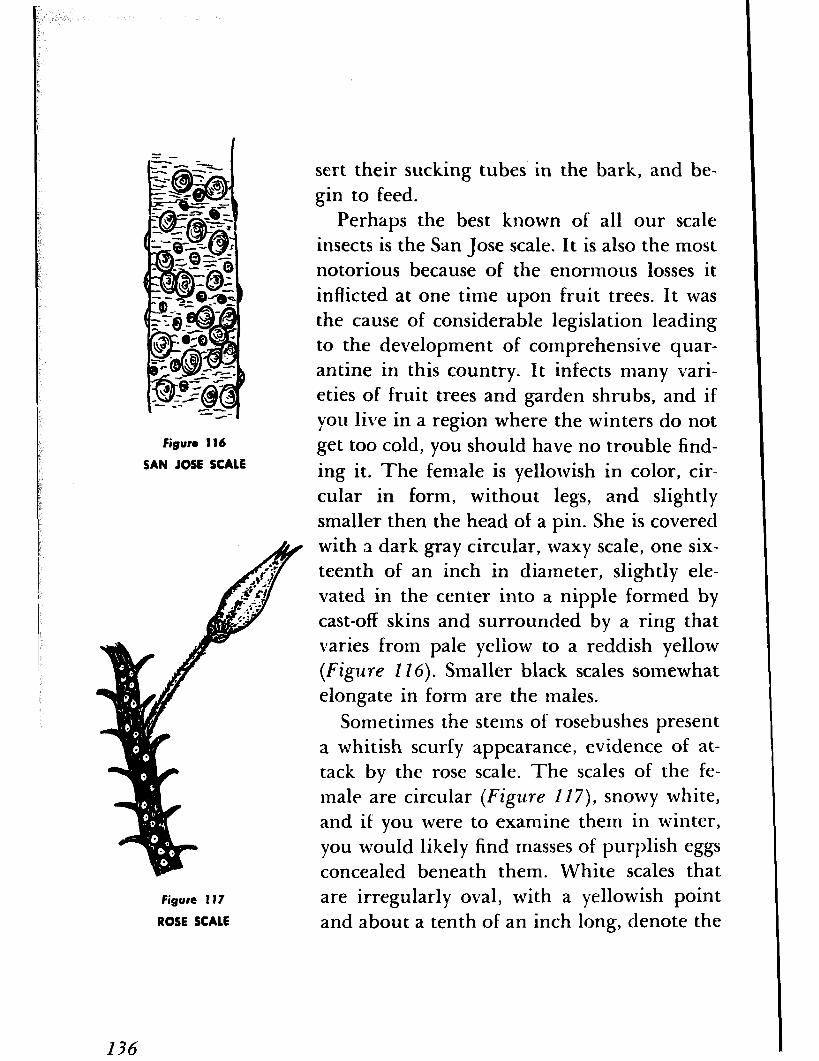
Figure 116
SAN JOSE SCALE
Figure 1 I7
ROSE SCALE
sert their sucking tubes in the bark, and be- gin to feed.
Perhaps the best known of all our scale insects is the San Jose scale. It is also the most notorious because of the enormous losses it inflicted at one time upon fruit trees. It was the cause of considerable legislation leading to the development of comprehensive quar- antine in this country. It infects many vari- eties of fruit trees and garden shrubs, and if you live in a region where the winters do not get too cold, you should have no trouble find- ing it. The female is yellowish in color, cir- cular in form, without legs, and slightly smaller then the head of a pin. She is covered with a dark gray circular, tvaxy scale, one six- teenth of an inch in diameter, slightly ele- vated in the center into a nipple formed by cast-off skins and surrounded by a ring that varies from pale yellow to a reddish yellow (Figure 116). Smaller black scales somewhat elongate in form are the males.
Sometimes the stems of rosebushes present a whitish scurfy appearance, evidence of at- tack by the rose scale. The scales of the fe- male are circular (Figure 2 17), snowy white, and if you were to examine them in winter, you would likely find masses of purplish eggs concealed beneath them. White scales that are irregularly oval, with a yellowish point and about a tenth of an inch long, denote the
I36
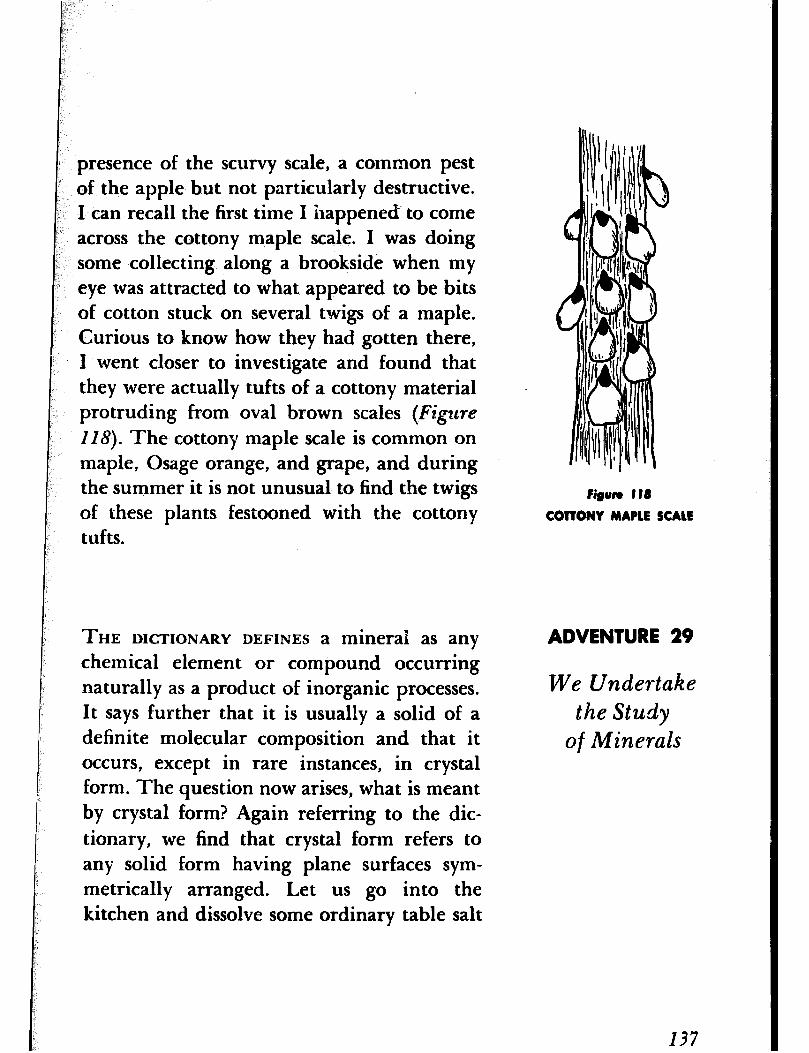
presence of the scurvy scale, a common pest of the apple but not particularly destructive. I can recall the first time I happened to come across the cottony maple scale. I was doing some collecting along a brookside when my eye was attracted to what appeared to be bits of cotton stuck on several twigs of a maple. Curious to know how they had gotten there, I went closer to investigate and found that they were actually tufts of a cottony material protruding from oval brown scales (Figure
1 I8). The cottony maple scale is common on maple, Osage orange, and grape, and during the summer it is not unusual to find the twigs of these plants festooned with the cottony tufts.
Bigurn t 18
COTTONY MAPLE SCALE
THE DICTIONARY ,DEFINES a mineral as any chemical element or compound occurring naturally as a product of inorganic processes. It says further that it is usually a solid of a definite molecular composition and that it occurs, except in rare instances, in crystal form. The question now arises, what is meant by crystal form? Again referring to the dic- tionary, we find that crystal form refers to any solid form having plane surfaces sym- metrically arranged. Let us go into the kitchen and dissolve some ordinary table salt
ADVENTURE 29
We Undertake
the Study of Minerals
137
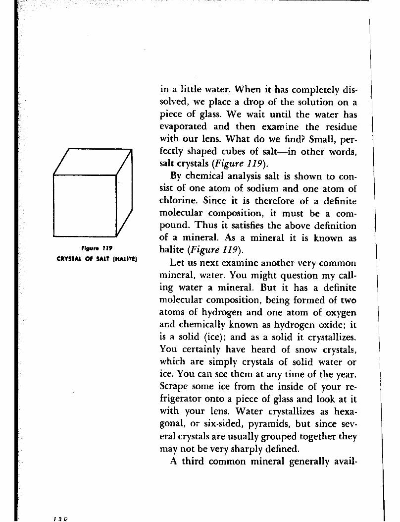
Figure 119
CRYSTAL OF SALT (HALVE)
72Q
in a little water. When it has completely dis- solved, we place a drop of the solution on a piece of glass. We wait until the water has evaporated and then examine the residue with our lens. What do we find? Small, per- fectly shaped cubes of salt-in other words, salt crystals (Figure 119).
By chemical analysis salt is shown to con- sist of one atom of sodium and one atom of chlorine. Since it is therefore of a definite molecular composition, it must be a com- pound. Thus it satisfies the above definition of a mineral. As a mineral it is known as halite (Figure 119).
Let us next examine another very common mineral, water. You might question my call- ing water a mineral. But it has a definite molecular composition, being formed of two atoms of hydrogen and one atom of oxygen ar,d chemically known as hydrogen oxide; it is a solid (ice); and as a solid it crystallizes. You certainly have heard of snow crystals, which are simply crystals of solid water or ice. You can see them at any time of the year. Scrape some ice from the inside of your re- frigerator onto a piece of glass and look at it with your lens. Water crystallizes as hexa- gonal, or six-sided, pyramids, but since sev- eral crystals are usually grouped together they may not be very sharply defined.
A third common mineral generally avail-
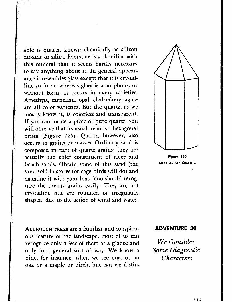
able is quartz, known chemically as silicon dioxide or silica. Everyone is so familiar with this mineral that it seems hardly necessary to say anything about it. In general appear- ance it resembles glass except that it is crystal- line in form, whereas glass is amorphous, or without form. It occurs in many varieties. Amethyst, carnelian, opal, chalcedonv, agate are all color varieties. But the quartz, as we mostly know it, is colorless and transparent. If you can locate a piece of pure quartz, you will observe that its usual form is a hexagonal prism (Figure 120). Quartz, however, also occurs in grains or masses. Ordinary sand is composed in part of quartz grains; they are actually the chief constituent of river and beach sands. Obtain some of this sand (the sand sold in stores for cage birds will do) and examine it with your lens. You should recog- nize the quartz grains easily. They are not crystalline but are rounded or irregularly shaped, due to the action of wind and water.
ALTHOUGH TREES are a familiar and conspicu- ous feature of the landscape, most of us can recognize only a few of them at a glance and only in a general sort of way. We know a pine, for instance, when we see one, or an oak or a maple or birch, but can we distin-
Figure 120
CRYSTAL OF QUARTZ
ADVENTURE 30
We Conside?* Some Diagnostic
Characters
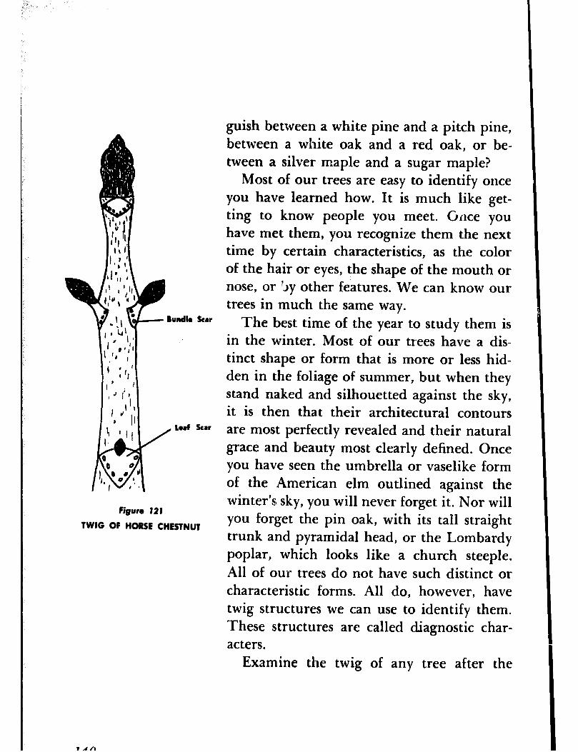
Scar
scar
Figure 121
TWIG OF HORSE CHRStNllT
guish between a white pine and a pitch pine, between a white oak and a red oak, or be- tween a silver maple and a sugar maple?
Most of our trees are easy to identify once you have learned how. It is much like get- ting to know people you meet. Grace you have met them, you recognize them the next time by certain characteristics, as the color of the hair or eyes, the shape of the mouth or nose, or 5y other features. We can know our trees in much the same way.
The best time of the year to study them is in the winter. Most of our trees have a dis- tinct shape or form that is more or less hid- den in the foliage of summer, but when they stand naked and silhouetted against the sky, it is then that their architectural contours are most perfectly revealed and their natural grace and beauty most clearly defined. Once you have seen the umbrella or vaselike form of the American elm outlined against the winter’s sky, you will never forget it. Nor will you forget the pin oak, with its tall straight trunk and pyramidal head, or the Lombardy poplar, which looks like a church steeple. All of our trees do not have such distinct or characteristic forms. All do, however, have twig structures we can use to identify them. These structures are called diagnostic char- acters.
Examine the twig of any tree after the
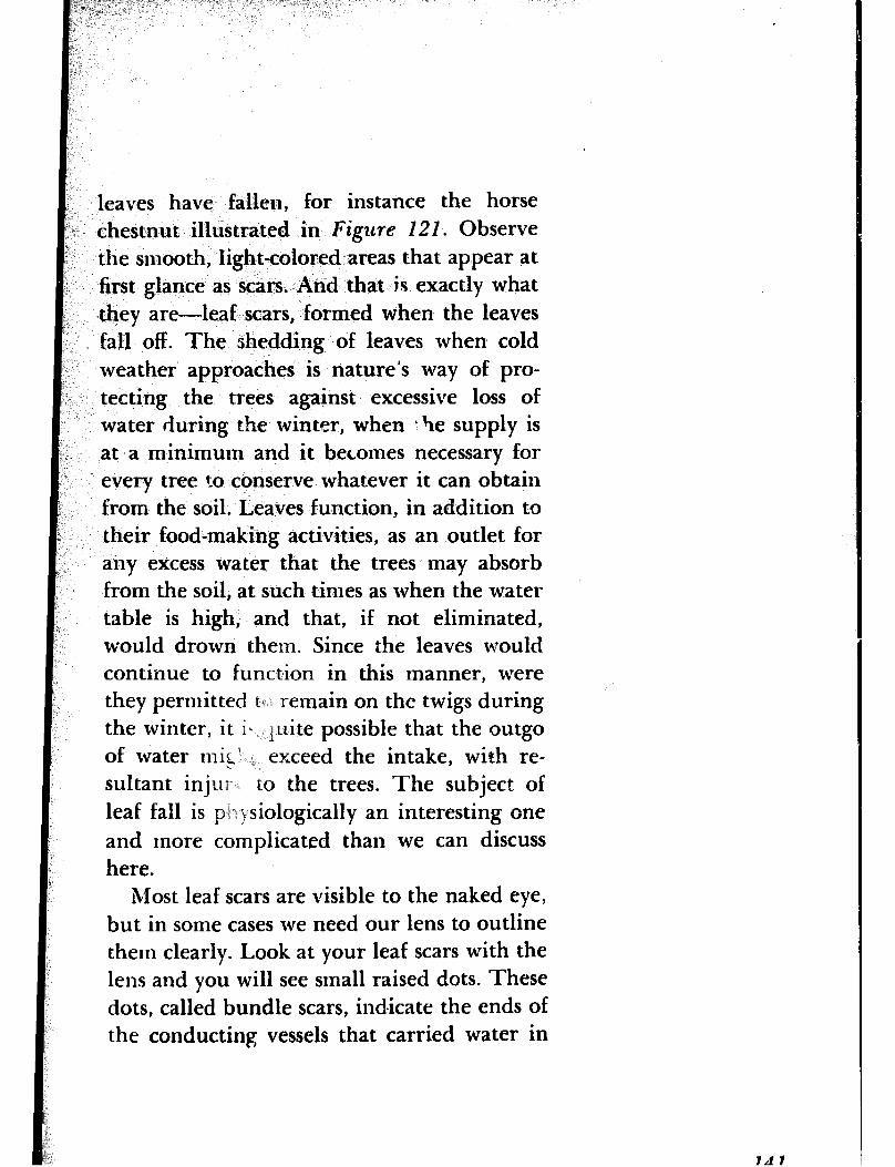
leaves have fallen, for instance the horse chestnut illustrated in Figzlre 121. Observe the smooth, light-colored areas that appear at first glance as scars. And that is exactly what they are-leaf scars, formed when the leaves fall off. The shedding of leaves when cold weather approaches is nature’s way of pro- tecting the trees against excessive loss of water during the winter, when t he supply is at a minimum and it becomes necessary for every tree to conserve whatever it can obtain from the soil. Leaves function, in addition to their food-making activities, as an outlet for any excess water that the trees may absorb from the soil, at such times as when the water table is high, and that, if not eliminated, would drown them. Since the leaves would continue to function in this manner, were they permitted t* remain on the twigs during the winter, it ii quite possible that the outgo of water mis1 _ exceed the intake, with re- sultant injur to the trees. The subject of leaf fall is pi .J siologically an interesting one and more complicated than we can discuss here.
Most leaf scars are visible to the naked eye, but in some cases we need our lens to outline them clearly. Look at your leaf scars with the lens and you will see small raised dots. These dots, called bundle scars, indicate the ends of the conducting vessels that carried water in
Id 7
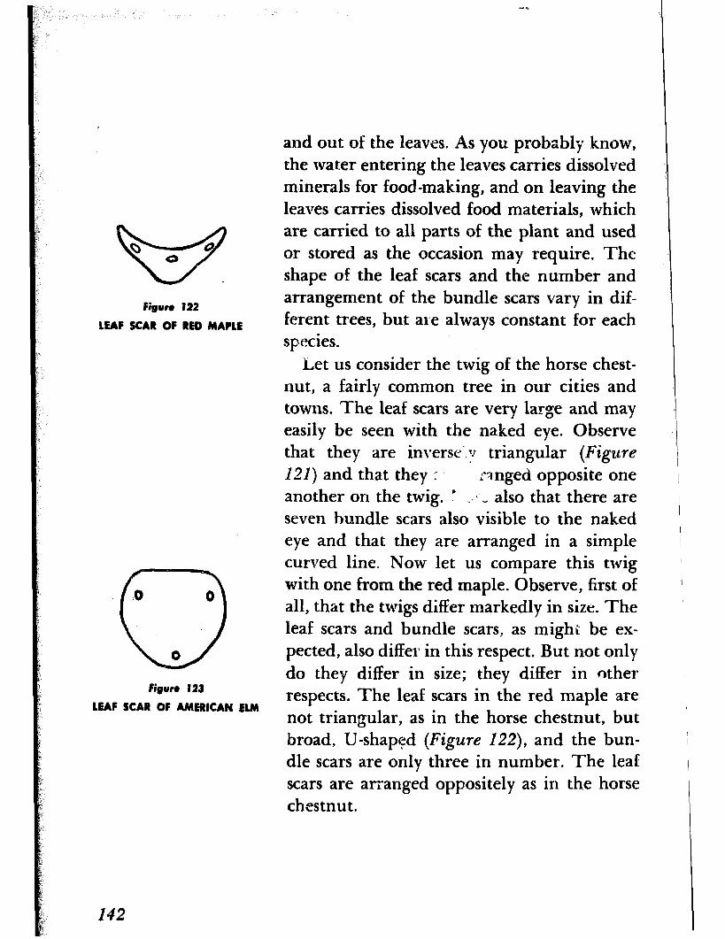
and out of the leaves. As you probably know, the water entering the leaves carries dissolved minerals for food-making, and on leaving the leaves carries dissolved food materials, which
w
are carried to all parts of the plant and used
Q Q @ or stored as the occasion may require. The shape of the leaf scars and the number and
Figure 122
LEAF SCAR OF RED MAPLE
arrangement of the bundle scars vary in dif- ferent trees, but are always constant for each species.
Let us consider the twig of the horse chest- nut, a fairly common tree in our cities and towns. The leaf scars are very large and may easily be seen with the naked eye. Observe that they are inverse‘.;1 triangular (Figure
121) and that they r’ 3 nged opposite one another on the twig. ’ _#I- _ also that there are seven bundle scars also visible to the naked eye and that they are arranged in a simple
.o 0
0
curved line. Now let us compare this twig with one from the red maple. Observe, first of all, that the twigs differ markedly in size. The leaf scars and bundle scars, as mighi be ex-
0 petted, also differ in this respect. But not only
Figure 123 do they differ in size; they differ in other
LEAF SCAR OF AMERICAN ELM respects. The leaf scars in the red maple are not triangular, as in the horse chestnut, but broad, U-shaped (Figure 122), and the bun- dle scars are only three in number. The leaf scars are arranged oppositely as in the horse chestnut.
142
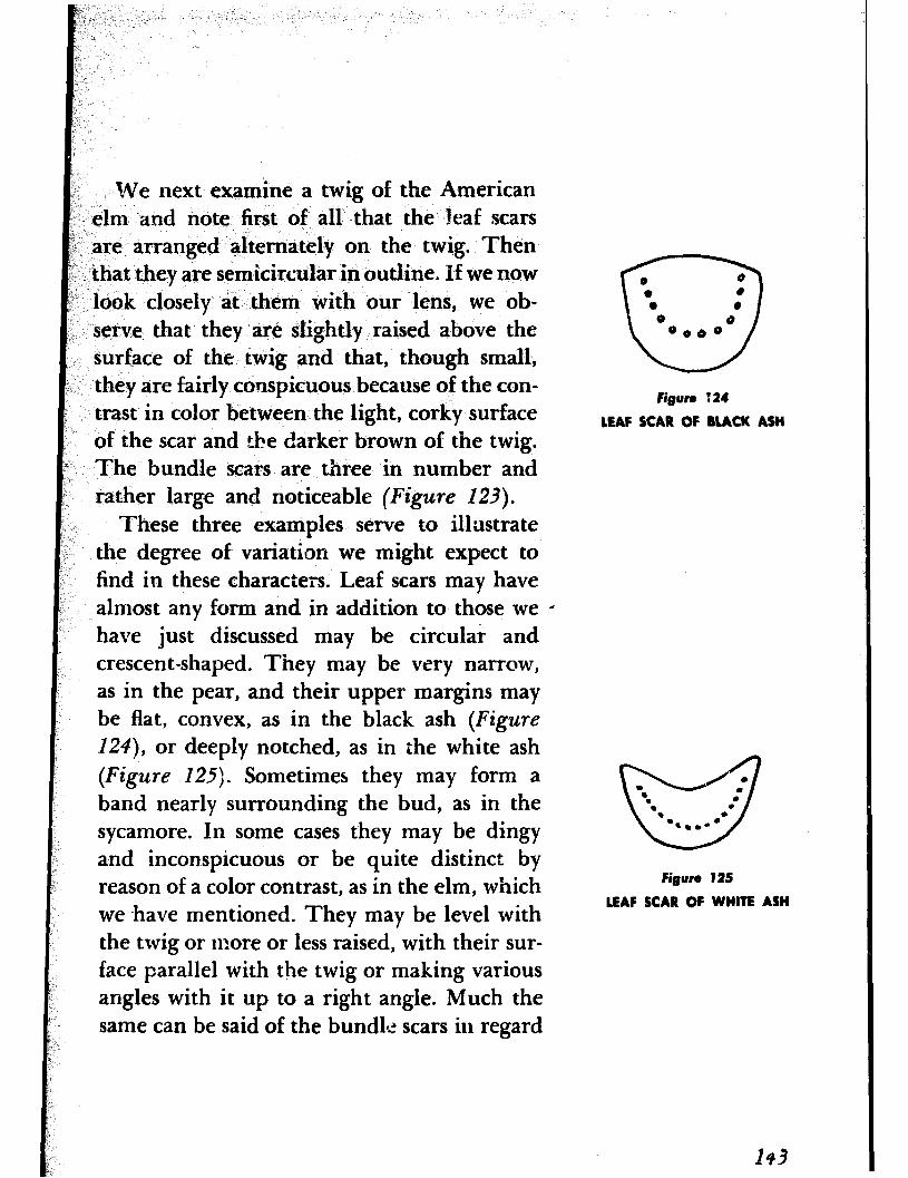
We next examine a twig of the American elm -and note first of all that the leaf scars are arranged alternately on the twig. Then that they are semicircular in outline, If we now look closely at them with our lens, we ob- serve that they are slightly raised above the surface of the twig and that, though small, they are fairly conspicuous because of the con- trast in color between the light, corky surface of the scar and the darker brown of the twig. The bundle scars are three in number and rather large and noticeable (Figure 123).
These three examples serve to illustrate the degree of variation we might expect to find in these characters. Leaf scars may have almost any form and in addition to those we 0 have just discussed may be circular and crescent-shaped. They may be very narrow, as in the pear, and their upper margins may be flat, convex, as in the black ash (Figure
124), or deeply notched, as in the white ash (Figure 125). Sometimes they may form a band nearly surrounding the bud, as in the sycamore. In some cases they may be dingy and inconspicuous or be quite distinct by reason of a color contrast, as in the elm, which we -have mentioned. They may be level with the twig or more or less raised, with their sur- face parallel with the twig or making various angles with it up to a right angle. Much the same can be said of the bundle scars in regard
0 0 0 0 a (I
0
0 8 0080
Figure 124
LEAF SCAR OF BUCK ASH
0 .
w
‘. .
. : . . l *..- .
Figure 125
LEAF SCAR OF WHITE ASH
143
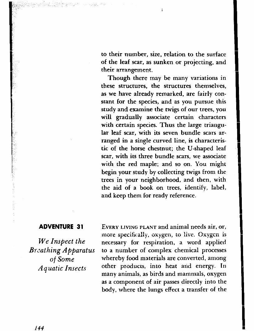
to their number, size, relation to the surface of the leaf scar, as sunken or projecting, and their arrangement.
Though there may be many variations in th.ese structures, the structures themselves, as we have already remarked, are fairly con- stant for the species, and as you pursue this study and examine the twigs of our trees, you will gradually associate certain characters with certain species. Thus the large triangu- lar leaf scar, with its seven bundle scars ar- ranged in a single curved line, is characteris- tic of the horse chestnut; the U-shaped leaf scar, with its three bundle scars, we associate with the red maple; and so on. You might begin your study by collecting twigs from the trees in your neighborhood, and then, with the aid of a book on trees, identify, label, and keep them for ready reference.
ADVENTURE 3 1 EVERY LIVING PLANT and animal needs air, or,
We Inspect the more specifically, oxygen, to live. Oxygen is necessary for respiration, a word applied
Bwathing Apparatus to a number of complex chemical processes
of Some whereby food materials are converted, among
Aquatic Insects other products, into heat arid energy. III
many animals, as birds and mammals, oxygen as a component of air passes directly into the body, where the lungs effect a transfer of the
I44
oxygen from the air into the blood stream; in other animals, as fishes, the gills effect a transfer of oxygen from water into the blood stream.
Insects have neither lungs nor gills. Instead they have a network of tubes called tracheae and smaller tubes called tracheoles, which convey air to the remotest tissues of the body. The tracheae are provided with opeCngs to the exterior through which air enters, as in our nostrils. These openings are called spira- cles and, with a hand lens, are easily visible on the abdominal segments (of the grasshop- per, for instance).
This kind of respiratory system does very well for terrestrial or land insects, but how about those that live in water? Would not the water enter through the spiracles and then course throughout the tracheal system and drown them, much as water would en- ter our nostrils and into our lungs if we sub- merged and remained ben.eath the surface for any length of time? Aquatic insects, how- ever, have developed modifications of the tracheal system that enable them to live in water.
Between the terrestrial insects and those which are truly aquatic is a group of insects
k : f
that can be referred to as semi-aquatic. They
1 dwell, for the most part, on or near the sur-
i.~ 1
face of the water and, being arr breathers,
F p B
646
submerge for only r-ather short periods. All have tracheal systems with open spir- acles. In this group belong such insects as the water striders, which we often see skimming over the surface of a pond, and the whirligig beetles, the little blue, oval-shaped, metallic- appearing beetles that swim about in circles in the quiet parts of a pond or stream. Also in the group are such insects as the diving beetles, watch boatmen, back swimmers, Iva ter scorpions, rat-tailed maggots, and mosquito larvae and pupae, which are found usdi.y beneath the surface. But they are not truly aquatic forms, since they are air breathers. How, then, do they obtain air?
Some of them, as the diving beetles, water boatmen, and back swimmers, periodically rise to the surface to breathe in the normal manner, and when they dive carry down bub- bles of air, which permit them to remain sub- merged for varying periods. When the air supply is exhausted they rise to the surface. The others, as the water scorpions, rat-tailed maggots, and the larvae and pupa” of mos-
quitoes, are provided with special breathing tubes, which they exrend above the surface of the v:ater. The various adaptations ex- hibited b-y these and other semi-aquatic in- sects, nst alone for respiration, but for living in an aquatic habitat, makes for the most in- teresting reading. We are not concerned with
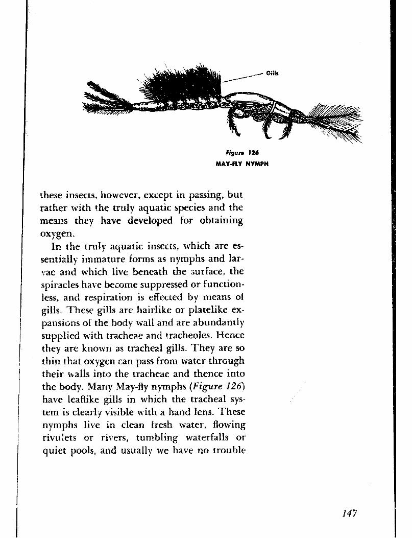
these insects, however, except in passing, but rather Gth the truly aquatic species and the means they have developed for obtaining oxygen.
In the truly aquatic insects, which are es- sentially immature forms as nymphs and lar- a:ae and which live beneath the surface, the spiracies have become suppressed or function- less, and respiration is effected by means of gills. These gilIs are hairlike or platelike ex- pznsions of the body wall and are abundantI) supplied with tracheae and tracheoles. Hence they are knowrl as tracheai gills. They are so thin that oxygen can pass from water through their falls into the tracheae and thence into the body. Many May-fly nymphs (Figure 126)
have leaflike gills in which the tracheal sys- tem is clearly visible with a hand lens. These nymphs live in clean fresh water, flowing rivuhzts or rivers, tumbling waterfalls or quiet pools, and usually we have no trouble
147
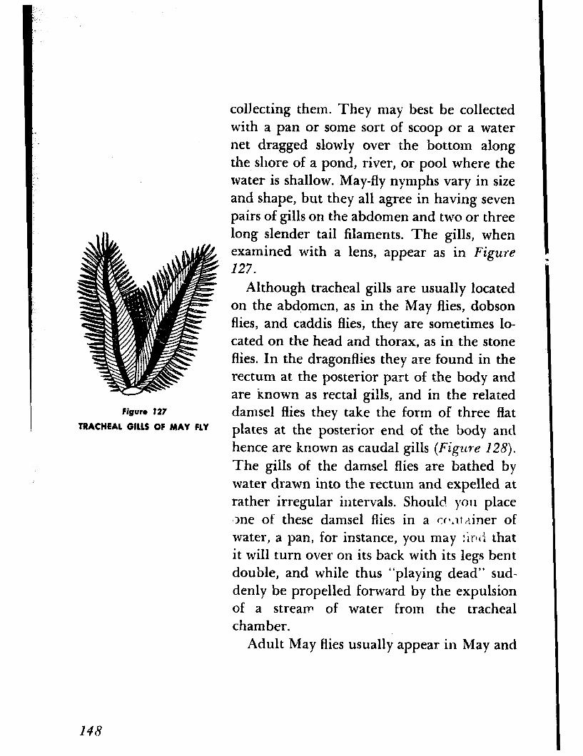
Figure 127
TRACHEAL 0U.S OF MAY FLY
148
collecting them. They may best be collected with a pan or some sort of scoop or a water net dragged slowly over the bottom along the shore of a pond, river, or pool where the water is shallow. May-fly nymphs vary in size and shape, but they all agree in having seven pairs of gills on the abdomen and two or three long slender tail filaments. The gills, when examined with a lens, appear as in Figure 127.
Although tracheal gills are usually located on the abdomen, as in the May ffies, dobson flies, and caddis flies, they are sometimes lo- cated on the head and thorax, as in the stone flies. In the dragonflies they are found in the rectum at the posterior part of th.e body and are known as rectal gills, and in the related damsel flies they take the form of three flat plates at the posterior end of the body and hence are known as caudal gills (Figure 128).
The gills of the damsel flies are bathed by water drawn into the rectum and expelled at rather irregular intervals. Should you place +ne of these damsel flies in a cc~lt {liner of water, a pan, for instance, you may :irltI that it will turn over on its back with its legs bent double, and while thus “playing dead” sud- denly be propelled forward by the expulsion of a stream of water from the tracheal chamber.
Adult May flies usually appear in May and
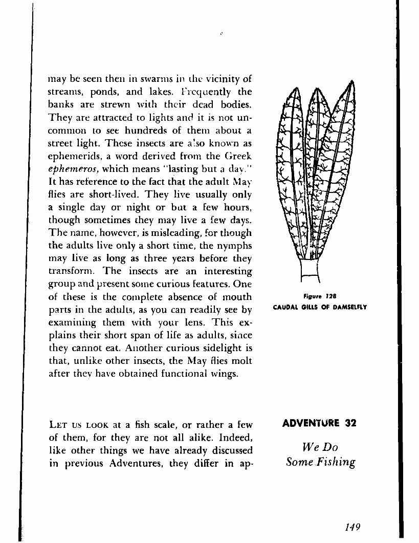
may be seen then in swarms in rhe vicinity of streams, ponds, and lakes. l’rcquently the banks are strewn with their dead bodies. They are attracted to lights and it is not un- common to see hundreds of them about a street light. These insects are also known as ephemerids, a word derived from the Greek ephetneros, which means “lasting but a day.” It has reference to the fact that the adult Ma) flies are short-lived. They live usually only a single day or night or but a few hours, though sometimes they may live a few days. The name, however, is misleading, for though the adults live only a short time, the nymphs may live as long as three years before they transform. The insects are an interesting group and present some curious features. One of these is the complete absence of mouth parts in the adults, as you can readily see by examining them with your lens. This ex- plains their short span of life as adults, since they cannot eat. Another curious sidelight is that, unlike other insects, the May flies molt after they have obtained functional wings.
LET us LOOK at a fish scale, or rather a few of them, for they are not all alike. Indeed, like other things we have already discussed in previous Adventures, they differ in ap-
n Figure 128
CAUOAL GILLS OF DAMSELFLY
ADVENWPRE 32
We Do Some.Fishing
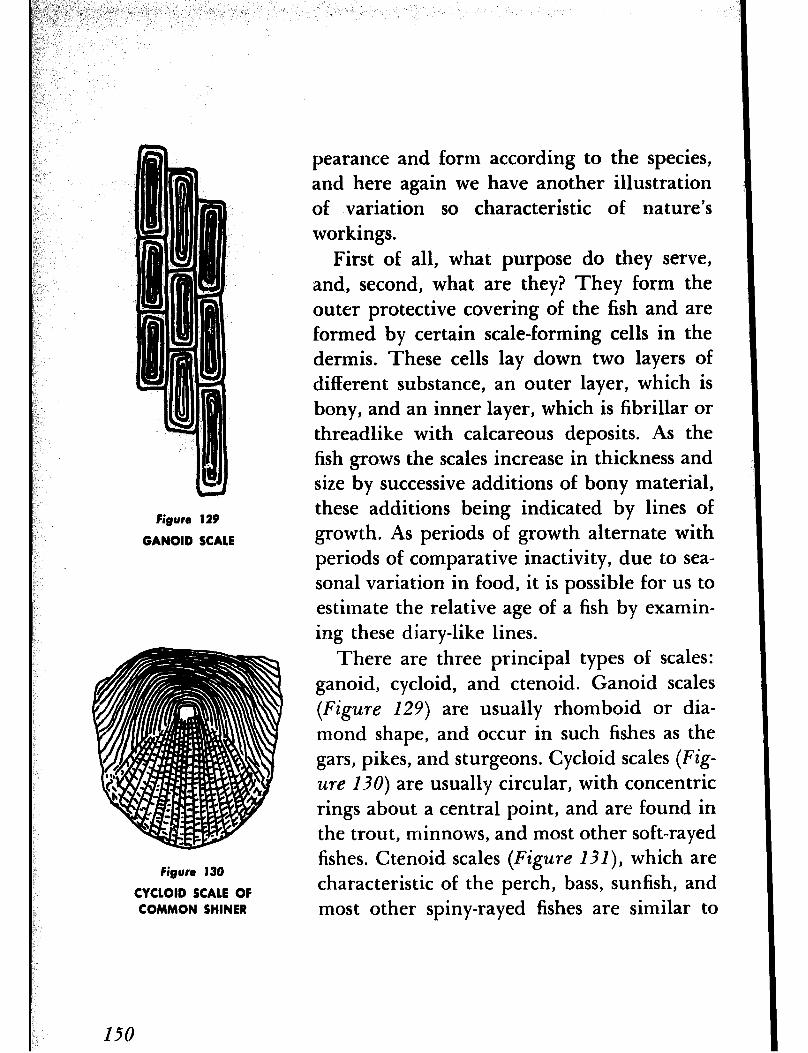
Figure 129
GANOID SCALE
Figure 130
CYCLOID SCALE OF COMMON SHINER
I50
pearance and form according to the species, and here again we have another illustration of variation so characteristic of nature’s workings.
First of all, what purpose do they serve, and, second, what are they? They form the outer protective covering of the fish and are formed by certain scale-forming cells in the dermis. These cells lay down two layers of different substance, an outer layer, which is bony, and an inner layer, which is fibrillar or threadlike with calcareous deposits. As the fish grows the scales increase in thickness and size by successive additions of bony material, these additions being indicated by lines of growth. As periods of growth alternate with periods of comparative inactivity, due to sea- sonal variation in food, it is possible for us to estimate the relative age of a fish by examin- ing these diary-like lines.
There are three principal types of scales: ganoid, cycloid, and ctenoid. Ganoid scales (Figure 129) are usually rhomboid or dia- mond shape, and occur in such fishes as the gars, pikes, and sturgeons. Cycloid scales (Fig- ure 130) are usually circular, with concentric rings about a central point, and are found in the trout, minnows, and most other soft-rayed fishes. Ctenoid scales (Figure I3I), which are characteristic of the perch, bass, sunfish, and most other spiny-rayed fishes are similar to
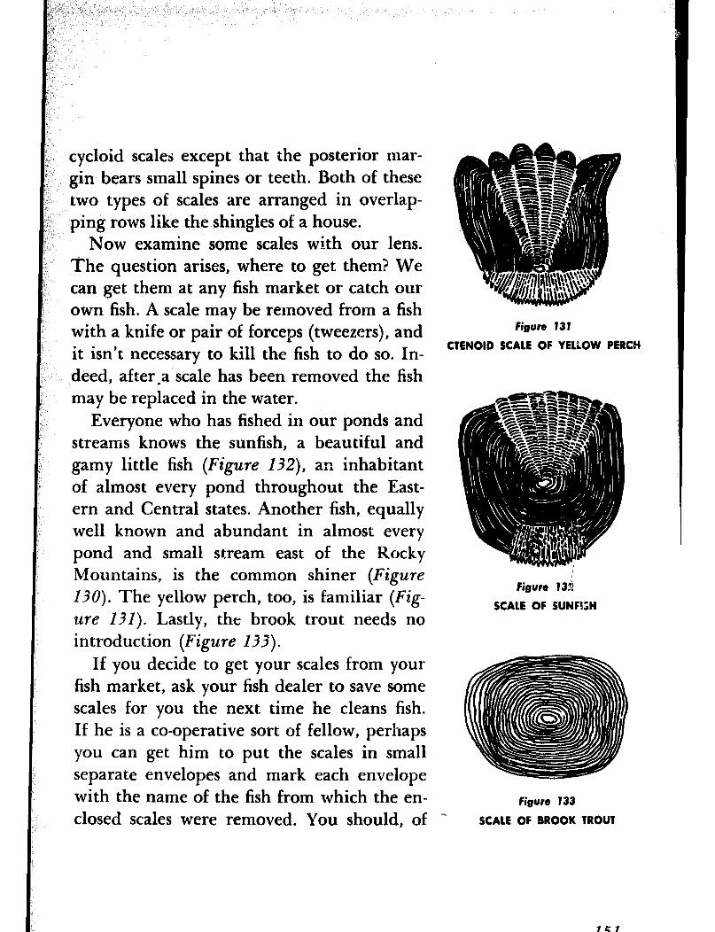
cycloid scales except that the posterior mar- gin bears small spines or teeth. Both of these two types of scales are arranged in overlap- ping rows like the shingles of a house.
Now examine some scales with our lens. The question arises, where to get them? We can get them at any fish market or catch our own fish. A scale may be removed from a fish with a knife or pair of forceps (tweezers), and it isn’t necessary to kill the fish to do so. In- deed, after,a scale has been removed the fish may be replaced in the water.
Everyone who has fished in our ponds and streams knows the sunfish, a beautiful and gamy little fish (Figure I32), an inhabitant of almost every pond throughout the East- ern and Central states. Another fish, equally well known and abundant in almost every pond and small stream east of the Rocky Mountains, is the common shiner (Figure
130). The yellow perch, too, is familiar (Fig- 2lre 131). Lastly, the brook trout needs no introduction (Figure 133).
If you decide to get your scales from your fish market, ask your fish dealer to save some scales for you the next time he cleans fish. If he is a co-operative sort of fellow, perhaps you can get him to put the scales in small separate envelopes and mark each envelope with the name of the fish from which the en- closed scales were removed. You should, of
Figure 131
CTENOID SCALE OF YELLOW PERCH
Figure 13s
SCALE OF SUNFISH
Figure 133
SCALE OF BROOK TROUT
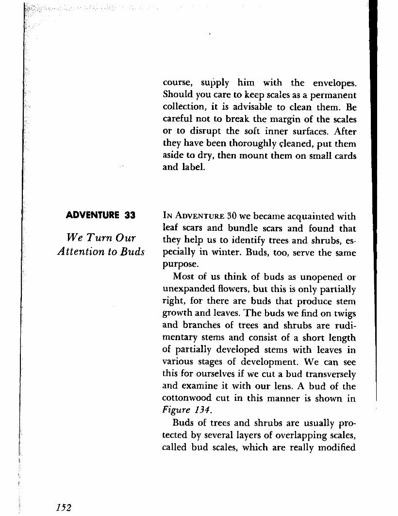
course, subply him with the envelopes. Should you care to keep scales as a permanent collection, it is advisable to clean them. Be careful not to break the margin of the scales or to disrupt the soft inner surfaces. After they have been thoroughly cleaned, put them aside to dry, then mount them on small cards and label.
ADVENTURE 33 IN ADVENTURE 30 we became acquainted with leaf scars and bundle scars and found that
We Turn Our they help us to identify trees and shrubs, es- Attention to Buds pecially in winter. Buds, too, serve the same
purpose. Most of us think of buds as unopened or
unexpanded flowers, but this is only partially right, for there are buds that produce stem growth and leaves. The buds we find on twigs and branches of trees and shrubs are rudi- mentary stems and consist of a short length of partially developed stems with leaves in various stages of development. We can see this for ourselves if we cut a bud transversely and examine it with our lens. A bud of the cottonwood cut in this manner is shown in Figure 134.
Buds of trees and shrubs are usually pro- tected by several layers of overlapping scales, called bud scales, which are really modified
152
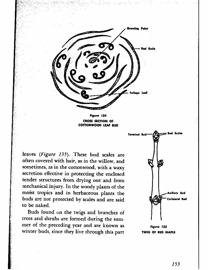
8ud kale
Led
Figun 1’34
CROSS SECTION OF COTTONWOOD LEAF BUD
Tormincll Bud- Bud krbs
leaves (Figure 135). These bud scales are often covered with hair, as in the willow, and sometimes, as in the cottonwood, with a waxy secretion effective in protecting the enclosed tender structures from drying out and from mechanical injury. In the woody plants of the moist tropics and in herbaceous plants the buds are not protected by scales and are said to be naked.
Axillry Bud
collatorrl Bud
Buds found on the twigs and branches of trees and shrubs are formed during the sum- mer of the preceding year and are known as winter buds, since they live through this part
Figure 135
TWIG OF RED MAPLE
153
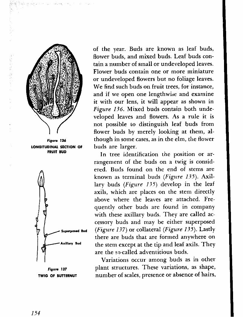
Figure 136
LONGITUDINAL SECTION OF FRUIT BUD
Superposed Bud
Axillrry Bud
Figure 137
TWIG OF BUTTERNUT
of the year. Buds are known as leaf buds, flower buds, and mixed buds. Leaf buds con- tain a number of small or undeveloped leaves. Flower buds contain one or more miniature or undeveloped flowers but no foliage leaves. We find such buds on fruit trees, for instance, and if we open one lengthwide and examine it with our lens, it will appear as shown in Figure I3 6. Mixed buds contain both unde- veloped leaves and flowers. As a rule it is not possible to distinguish leaf buds from flower buds by merely looking at them, al- though in some cases, as in the elm, the flower buds are larger.
In tree identification the position or ar- rangement of the buds on a twig is consid- ered. Buds found on the end of stems are known as terminal buds (Figztre 135). Axil- lary buds (Figure 135) develop in the leaf axils, which are places on the stem directly above where the leaves are attached. Fre- quently other buds are found in company with these axillary buds. They are called ac- cessory buds and may be either superposed (Fig24re 137) or collateral (Figure 135). Lastly there are buds that are formed anywhere on the stem except at the tip and leaf axils. They are the zo-called adventitious buds.
Variations occur among buds as in other plant structures. These variations, as shape, number of scales, presence or absence of hairs,
154

etc., may seem insignificant, yet they are con- stant for the species. After a while you will associate certain kinds of buds with specific trees and shrubs, and tree identification will be almost a perfunctory habit.
We have discussed buds-now let us look at a few of them as a sort of practice exercise. We shall select first the Norway maple, which has been planted extensively as a shade tree and thus should be available. The buds, which are rather large for a maple, are com- monly red or yellowish green toward the base, and sometimes the entire bud is strongly tinged with green. The terminal bud is larger than the lateral buds, oval or ovate in shape. In comparison the lateral buds are small and appressed, that is, they lie close against the twig (Figure 138). We can see all this with the naked eye. With our lens we now examine the bud scales and observe that they are more or less keeled, or with a ridge like the keel of a boat, and that the margins are cov- ered with fine hairs. We next count the scales of the terminal bud. To find them all, we must lift up some of the scales with a pin. There are five pairs. The enclosed scales are covered with dark, rusty-brown hairs.
For our second choice we find ourselves in something of a dilemma. Since some of you live in the city and others in the country, the same trees may not be available to everyone,
Figure 138
TWIG OF NORWAY MAPLE
Figure 139
TWIG OF SUGAR MAPLE
I55
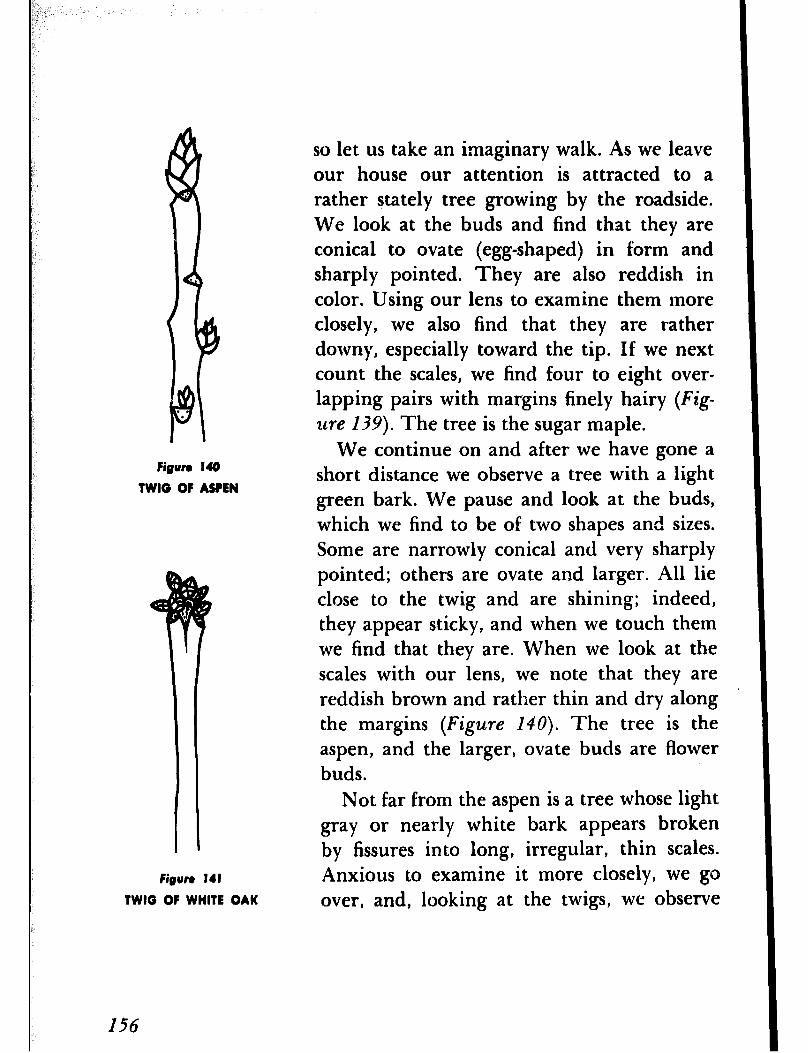
Figure 148
TWIG OF ASPEN
so let us take an imaginary walk. As we leave our house our attention is attracted to a rather stately tree growing by the roadside. We look at the buds and find that they are conical to ovate (egg-shaped) in form and sharply pointed. They are also reddish in color. Using our lens to examine them more closely, we also find that they are rather downy, especially toward the tip. If we next count the scales, we find four to eight over- lapping pairs with margins finely hairy (Fig- 2lre 139). The tree is the sugar maple.
We continue on and after we have gone a short distance we observe a tree with a light green bark. We pause and look at the buds, which we find to be of two shapes and sizes. Some are narrowly conical and very sharply pointed; others are ovate and larger. All lie close to the twig and are shining; indeed, they appear sticky, and when we touch them we find that they are. When we look at the scales with our lens, we note that they are reddish brown and rather thin and dry along the margins (Figure 140). The tree is the aspen, and the larger, ovate buds are flower buds.
Not far from the aspen is a tree whose light gray or nearly white bark appears broken by fissures into long, irregular, thin scales.
Figun 141 Anxious to examine it more closely, we go TWIG OF WHITE OAK over, and, looking at the twigs, we observe
I56
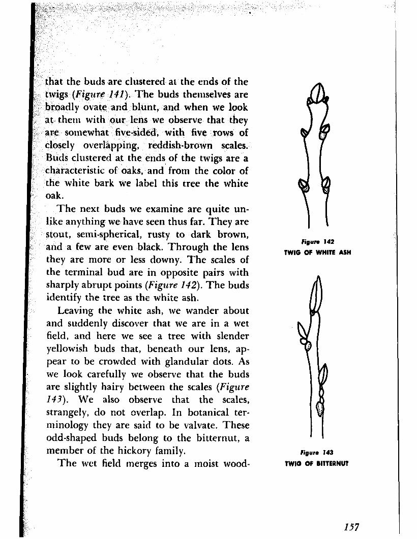
that the buds are clustered at’ the ends of the twigs (Figlcre I#]). The buds themselves are broadly ovate and blunt, and when we look at them with our lens we observe that they are somewhat five-sided, with five rows of closely overlapping, reddish-brown scales. Buds clustered at the ends. of the twigs are a characteristic of oaks, and from the color of the white bark we label this tree the white oak.
The next buds we examine are quite un- like anything we have seen thus far. They are stout, semi-spherical, rusty to dark brown, and a few are even black. Through the lens they are more or less downy. The scales of the terminal bud are in opposite pairs with sharply abrupt points (Figure 142). The buds identify the tree as the white ash.
Leaving the white ash, we wander about
Figure 142
TWIG OF WHITE ASH
! and suddenly discover that we are in a wet field, and here we see a tree with slender yellowish buds that, beneath our lens, ap- pear to be crowded with glandular dots. As we look carefully we observe that the buds are slightly hairy between the scales (Figure
143). We also observe that the scales, strangely, do not overlap. In botanical ter- minology they are said to be valvate. These odd-shaped buds belong to the bitternut, a member of the hickory family.
The wet field merges into a moist wood-
I Ngura 143
TWIG OF BITTERNUT
I57
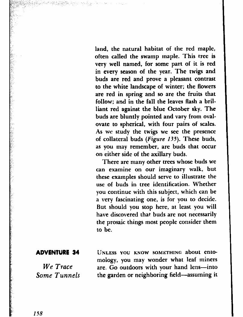
land, the natural habitat of the red maple, often called the swamp maple. This tree is very well named, for some part of it is red in every season of the year, The twigs and buds are red and prove a pleasant contrast to the white landscape of winter; the flowers are red in spring and so are the fruits that follow; and in the fall the leaves flash a bril- liant red against the blue October sky. The buds are bluntly pointed and vary from oval- ovate to spherical, with four pairs of scales. As we study the twigs we see the presence of collateral buds (Figure 135). These buds, as you may remember, are buds that occur on either side of the axillary buds.
There are many other trees whose buds we can examine on our imaginary walk, but these exatnples should serve to illustrate the use of buds in tree identification. Whether you continue with this subject, which can be a very fascinating one, is for you to decide. But should you stop here, at least you will have discovered that buds are not necessarily the prosaic things most people consider them to be.
ADVENTURE 34
We Trace Some Tunnels
UNLESS YOU KNOW SOMETHING about ento- mology, you may wonder what leaf miners are. Go outdoors with your hand lens--into the garden or neighboring field-assuming it
158
is summer, and look for a leaf with blotches (Figure 244) or twisting lines (Figure 145)- leaves with these disfigurements are common.
; Remove the leaf from its stem and hoid it ; up so the light will pass through it. You ( should find the blotch or twisted line occu- 1 pied by a small, wormlike creature, unless I the little animal has reached maturity and
been transformed into an adult insect, when it would have left its temporary home and taken up a different kind of existence.
The twisting lines and blotches are pas- sages or tunnels that the larvae of certain in- sects excavate in the tissues of the leaves. The insects may be beetles, flies, moths, or saw- flies, but despite their diversity as insects they all have one thing in common-small size. It seems incredible that some insects are so small that they are able to live between the upper and lower surfaces of a leaf that is al- most of paper thinness. Not only can they live there, but they manage to grow and in- crease in size. This is indicated by the increas- ing width of the passageways. Follow one of these twisting passageways from the begin- ning. Note that it begins almost as a pinpoint, and here is where the egg hatched. You will see that it becomes progressively wider and wider.
There are two general types of mines, as the blotches and tunnels are known: the
figure 144
MINE OF ELM-LEAF MINER
figura 14s
MINE OF COLUMBINE-LEAF MINER
159
160
Figrn 144
MINE OF NASTURTlUM-lEAF MINER
twisting, or linear, mine made by the insect as it moved forward, and the round, 01 blotch, mine made by the insect as it moved around and around within the leaf. There are many modifications of these two types as the linear-blotch, the trumpet, the digitate, and the tentiform. Indeed, the patterns de- scribed by the insects as they eat their way through the plant tissues may be as varied as the insects themselves. But each species makes its own characteristic design, so that anyone familiar with these insects can usually iden- tify the species merely by looking at the form of the mine. As one writer has put it, “they write their signatures on the leaves.”
Even though the leaf miners are provided with shelter and plenty of food, they do not live an altogether carefree existence. Like anyone else, they too have their problems. Sometimes they find the veins of the leaves a barrier to further progress, and they must either eat through them or confine their operations to a limited area. They must be careful, too, not to cut the latex cells lest the secretions from these cells pour into their mines and drown them. But theirs gravest problem is waste disposal. So;ile miners dis- tribute the wastes over the floor of the mine, as you can see wilin your lens, while others go to the trouble of excavating side chambers in which to dispose of their refuse. Still others
have developed the habit of cutting holes in the surface of the leaf through which they push out their fecula, which is the accepted term for the waste material or excrement voided by insects.
A common leaf miner is a small fly whose larva makes a kind of twisting tunnel, called a serpentine mine, in the leaf of the colum- bine ‘(Figure 145). The columbine is a fa- vorite perennial of most gardeners, and you should have no difficulty finding it. Observe that the mine is a whitish tortuous or twist- ing trail that frequently crosses itself and finally ends in a spot about an eighth of an inch wide. Here is where the maggot pupated and transformed into the adult. The nastur- tium is another garden plant frequently in- habited by a leaf miner (Figure 146). Even a pine needle may have its little inhabitant- the pine-leaf miner. If you can find a pine needle with its occupant and hold it up to the light, you may observe the little creature run- ning up and down in its tunnel as if it were dismayed at being disturbed (Figure 147).
Note a small hole near the lower end of the tunnel. This hole is where the larva entered the leaf and through which, after it has en- larged it slightly, it will emerge as an adult.
Leaf miners attack nearly all families of plants and some of them are quite destruc- tive. Frequently an entire family may occupy
figure 147
MINE OF PINE-LEAF MINER
161
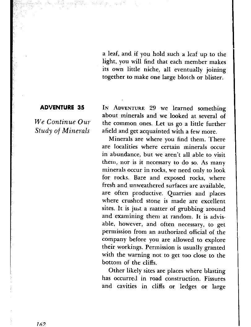
a leaf, and if you hold such a leaf up to the light, you will find that each member makes its own little niche, all eventually joining together to make one large blotch or blister.
ADVENTURE 35 IN ADVENTURE 29 we learned someth.ing about minerals and we looked at several of
We Continue Ow the common ones. Let us go a little further Study of Minerals afield and get acquainted with a few more.
Minerals are where you find them. There are localities where certain minerals occur in abundance, but we aren’t all able to visit them, nor is it necessary to do so. As many minerals occur in rocks, we need only to look for rocks. Bare and exposed rocks, where fresh and unweathered surfaces are available, are often productive. Quarries and places where crushed stone is made are excellent sites. It is just a matter of grubbing around and examining them at random. It is advis- able, however, and often necessary, to get permission from an authorized official of the company before you are allowed to explore their workings. Permission is usually granted with the warning not to get too close to the bottom of the cliffs.
Other likely sites are places where blasting has occurred in road construction. Fissures and cavities in cliffs or ledges or large
762

boulders should be explored, for they fre- quently contain fine crystals. All you will need for equipment is your lens ant; a ham- mer, preferably a stonemason’s hammer, for breaking,-up the rocks. A pair of thick-soled shoes will make walking among the rocks somewhat more comfortable, and unless you want to stuff your pockets, take a bag or box in which to carry home your specimens. I might add that if you are really interested in looking for minerals it would pay you to lo- cate someone who is either an amateur or professional mineralogist and write for a list of localities in your state where specific min- erals occur. Colleges and universities that have a mineralogy department have such lists and would be glad to give one to you.
You have doubtless heard of fool’s gold. This is one of our commonest minerals and one you should have no trouble finding. It occurs in all kinds of rocks in the form of cubes, octahedrons, and pyritohedrons (Fig-
zlre 1429, terms that need not concern you too much. You will readily recognize it by its brassy color and metallic luster. As a mineral, figuru 148 it is known as pyrite. It is a compound of iron PYRITOMEDRON CRYSTAL OF PYRITE
and sulphur and though very abundant is rarely used as a source of iron because the sulphur is difficult to remove and to leave it in the iron would make the latter brittle and useless for most purposes.
163
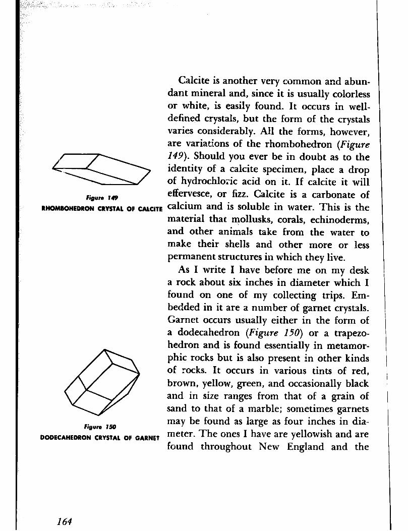
Calcite is another very common and abun- dant mineral and, since it is usually colorless or white, is easily found. It occurs in well- defined crystals, but the form of the crystals varies considerably. All the forms, however, are variations of the rhombohedron (Figure
149). Should you ever be in doubt as to the identity of a calcite specimen, place a drop of hydrochloric acid on it. If calcite it will
Figure 149 effervesce, or fizz. Calcite is a carbonate of
RHOMBOHEDRON CRYSTAL OF CALCITE calcium and is soluble in water. This is the material that mollusks, corals, echinoderms, and other animals take from the water to make their shells and other more or less permanent structures in which they live.
As I write I have before me on my desk a rock about six inches in diameter which I found on one of my collecting trips. Em- bedded in it are a number of garnet crystals. Garnet occurs usually either in the form of a dodecahedron (Figure 150) or a trapezo- hedron and is found essentially in metamor- phic rocks but is also present in other kinds of rocks. It occurs in various tints of red, brown, yellow, green, and occasionally black and in size ranges from that of a grain of sand to that of a marble; sometimes garnets
Figure 150 may be found as large as four inches in dia-
DODECAHEDRON CRYSTAL OF GARNET meter. The ones I have are yellowish and are found throughout New England and the
164

Piedmont Plateau. Garnet is usually crushed and used as an abrasive, especially for finish- ing wood and leather, but pure and clear specimens are frequently used as gems. I also have a specimen of tourmaline on my desk. This is not a common mineral and may be found only in certain localities; hence the need for a list of localities. This mineral may easily be recognized by its black cr brown color and its three-sided prismatic crystals (Figure 151). @
Galena, which is a compound of lead, is a Figure IS1 I.
PRISMATIC CRYSTAL OF TGURMALINE
mineral found in every state in the Union and is usually associated with pyrite and cal- cite. Be on the watch for it when you find specimens of these two minerals. It is lead gray in color, with a metallic luster, and oc- curs in well-formed cubic crystals. Beryl is another mineral that is fairly common in cer- tain rocks. It i? usually some tint of green and occurs in hexagonal crystals (Figure 152). A specimen free from cracks and inclosures and of gem quality is so rare that its value is greater than that of the diamond. We know it as the emerald.
No matter where you look you are sure to find the mineral limonite, for it is nothing more than iron rust. It does not occur in the crystal form, but in masses and incrustations. A brown stain on a rock is sure to be limonite.
Figure 152
HEXAGONAL CRYSTAL OF BERYL
165
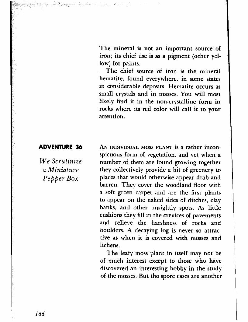
The mineral is not an important source of iron; its chief use is as a pigment (ocher yel- low) for paints.
The chief source of iron is the mineral hematite, found everywhere, in some states in considerable deposits. Hematite occurs as small crystals and in masses. You will most likely find it in the non-crystalline form in rocks where its red color will call it to your attention.
ADVENTURE 36 AN INDIVIDUAL MOSS PLANT is a rather incon-
We Scwtinize a Miniature Pepper Box
spicuous form of vegetation, and yet when a number of them are found growing together they collectively provide a bit of greenery to places that would otherwise appear drab and barren. They cover the woodland floor with a soft green carpet and are the first plants to appear on the naked sides of ditches, clay banks, and other unsightly spots. As little cushions they fill in the crevices of pavements and relieve the harshness of rocks and boulders. -4 decaying log is never so attrac- tive as when it is covered with mosses and lichens.
The leafy moss plant in itself may not be of much interest except to those who have discovered an interesting hobby in the study of the mosses. But the spore cases are another
166
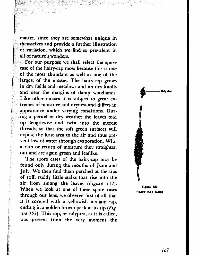
matter, since they are somewhat unique in themselves and provide a further illustration of variation, which we find so prevalent in all of nature’s wonders.
For our purpose we shall select the spore case of the hairy-cap moss because this is one of the most abundant as well as one of the largest of the mosses. The hairy-cap grows in dry fields and meadows and on dry knolls and near the margins of damp woodlands. Like other -mosses it is subject to great ex- tremes of moisture and dryness and differs in appearance under varying conditions. Dur- ing a period of dry weather the leaves fold up lengthwise and twist into the merest threads, so that the soft green surfaces will expose the least area to the air and thus pre- vent loss of water through evaporation. WiLlI a rain or return of moisture they straighten out and are again green and leaflike.
The spore cases of the hairy-cap may be found only during the months of June and July. We then find them perched at the tips of stiff, ruddy little stalks that rise into the air from among the leaves (Figure 153).
When we look at one of these spore cases through out lens, we observe first of all that it is covered with a yellowish mohair cap, ending in a golden-brown peak at its tip (Fig- ure 153). This cap, or calyptra, as it is called, was present from the very moment the
Figure 153 HAIRY CAP MOSS
167
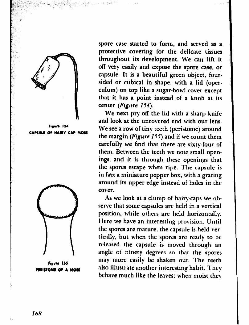
Fieun IS4
CAPSluLE OF HAIRY CAP MOSS
Cipn tSS
PCllSTOMt OF A MOSS
spore case started to form, and served as a protective covering for the delicate tissues throughout its development. We can lift it off very easily and expose the spore case, or capsule. It is a beautiful green object, four- sided or cubical in shape, with a lid (oper- culum) on top like a sugar-bowl cover except that it has a point instead of a knob at its center (Figure 154).
We next pry off the lid with a sharp knife and look at the uncovered end with our lens. We see a row of tiny teeth (peristome) around the margin (Figure 155) and if we count them carefully we find that there are sixty-four of them. Between the teeth we note small open- ings, and it is through these openings that the spores escape when ripe. The capsule is in fact a miniature pepper box, with a grating around its upper edge instead of holes in the cover.
As we look at a clump of hairy-caps we ob- serve that some capsules are held in a vertical position, while others are held horizontally. Here we have an interesting provision. Until the spores are mature, the capsule is held ver- tically, but when the spores are ready to bc released the capsule is moved through an angle of ninety degrees so that the spores may more easily be shaken out. The teeth also illustrate another interesting habit. They behave much like the leaves; when moist they

,
swell and close up the openings between them. The reason for this is to prevent the spores from falling out in wet weather, when they would fall to the ground among the parent plants, where there is little room for them to develop into new plants. In dry weather the wind scatters them to places where they would likely find more favorable conditions.
Capsules of mosses may be found at almost any time of the year, since the many species do not all mature their spores at the same time. Examine them whenever you find them and you will discover that some are round, others are cylindrical, while still others are curved. You will also find that the number of teeth vary and that the teeth may be entire or split or irregular, and in some instances there is a double row of them. There are countless variations, and for those who are interested in these plants they help in iden- tification.
GALLS ARE the strange and eye-catching out- growths or excrescences so common on plants. These odd structures, formed mostly by in- sects, occur in a variety of shapes and sizes. Some are only blister-like swellings on the leaves, while others are hairy or pile-like
ADVENTURE 37
We Invade the Privacy of Gall Makers
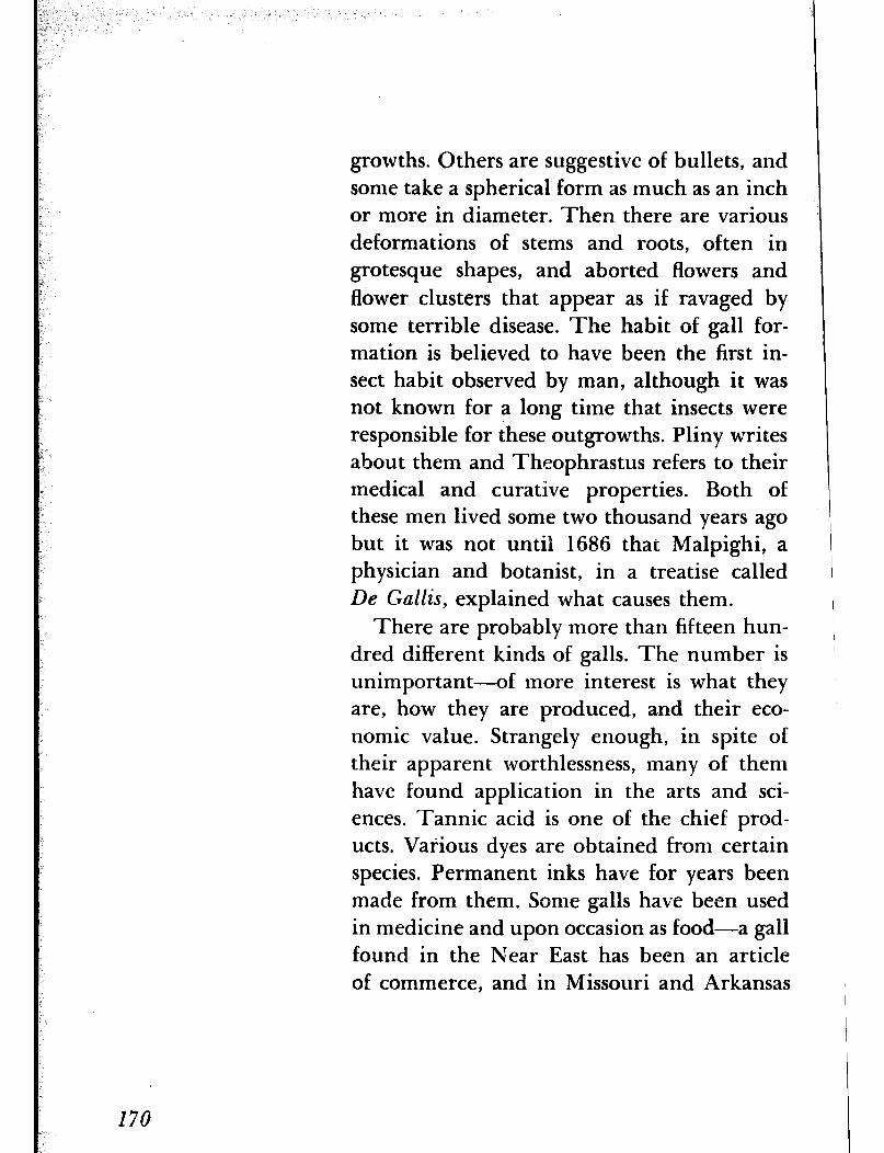
growths. Others are suggestive of bullets, and some take a spherical form as much as an inch or more in diameter. Then there are various deformations of stems and roots, often in grotesque shapes, and aborted flowers and flower clusters that appear as if ravaged by some terrible disease. The habit of gall for- mation is believed to have been the first in- sect habit observed by man, although it was not known for a long time that insects were responsible for these outgrowths. Pliny writes about them and Theophrastus refers to their medical and curative properties. Both of these men lived some two thousand years ago but it was not until 1686 that Malpighi, a physician and botanist, in a treatise called De Gallis, explained what causes them.
There are probably more than fifteen hun- dred different kinds of galls. The number is unimportant-of more interest is what they are, how they are produced, and their eco- nomic value. Strangely enough, in spite of their apparent worthlessness, many of them have found application in the arts and sci- ences. Tannic acid is one of the chief prod- ucts. Various dyes are obtained from certain species. Permanent inks have for years been made from them, Some galls have been used in medicine and upon occasion as food-a gall found in the Near East has been an article of commerce, and in Missouri and Arkansas
;’ ‘tin’ oak, gall is fed, when abundant, to cattle, )’ hogs, and sheep, :asi well as to chickens and i lx ;; turkeys, with excellent results. r;, <-‘, Galls occur, on almost .every form of plant ;: life and may be found on any part of a plant ; 8, Ij, ~4 +-root, branch, leaf,‘blossom, fruit, and even b” ‘.-’ seed-in fact, on, any. part that furnishes food “. ‘l: to the gall :maker. Some galls are very simple ) ,’ z in structure; others are most complicated. : Many are highly attractive and striking in ; form and coloration. There is no evidence L that the form is of any adaptive importance, : ” ,-arid the answer may be that the formation of I. any specific form is purely mechanical. But ,- the remarkable feature about galls is that
those made by the same species of insect are all of the same form, are all formed on the same species of plant, and are always on the same part of. the plant, so that those versed in g&l lore may know the identity of the gall ‘maker by merely looking at the gall.
Although we know that galls are produced by insects-and in some instances also by mites and nematode worms-we still do not know what causes them or just how they are produced. It is generally believed that the formation of a gall is initiated, or that the plant tissues are stimulated in growing ab- normally, by a secretion from the larva. Yet the fact remains that the physiology of gall
i / formation is still obscure. The study of galls :, !
I ,-, i”, 171
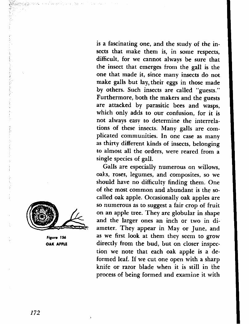
Figure 156
QAK APPLE
is a fascinating one, and the study of the in- sects that make them is, in some respects, difficult, for we cannot always be sure that the insect that emerges from the gall is the one that made it, since many insects do not make galls but lay,their eggs in those made by others. Such insects are called “guests.” Furthermore, both the makers and the guests are attacked by parasitic bees and wasps, which only adds to our confusion, for it is not always easy to determine the interrela- tions of these insects. Many galls are com- plicated communities. In one case as many as thirty different kinds of insects, belonging to almost all the orders, were reared from a single species of gall.
Galls are especially numerous on willows, oaks, roses, legumes, and composites, so we should have no difficulty finding them. One of the most common and abundant is the so- called oak apple. Occasionally oak apples are so numerous as to suggest a fair crop of fruit on an apple tree. They are globular in shape and the larger ones an inch or two in di- ameter. They appear in May or June, and as we first look at them they seem to grow directly from the bud, but on closer inspec- tion we note that each oak apple is a de- formed leaf. If we cut one open with a sharp knife or razor blade when it is still in ihe process of being formed and examine it with
I72
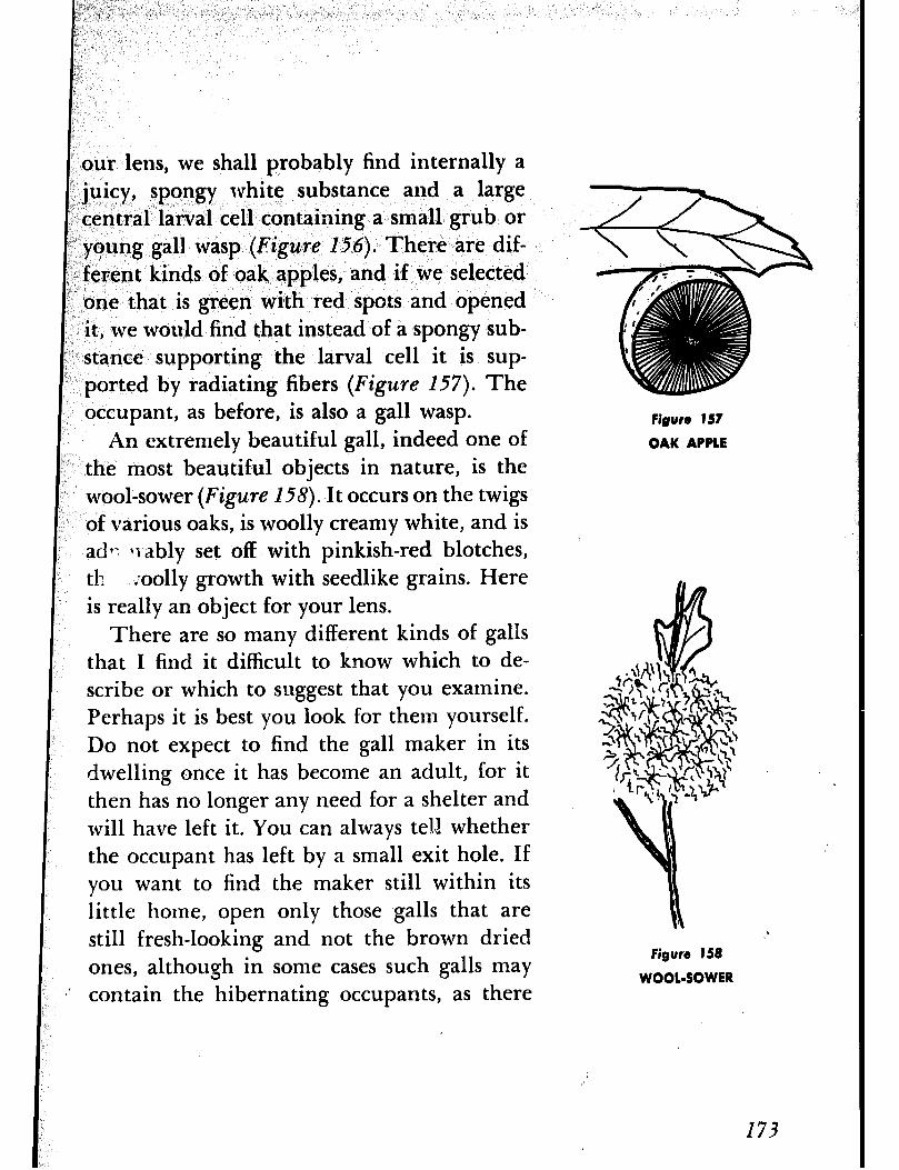
our lens, we shall probably find internally a juicy, spongy white substance and a large central larval cell containing a small grub or young gall wasp (Figme 156). There are dif- ferent kinds of oak apples, and if we selected one that is green with red spots and opened it, we would find that instead of a spongy sub- stance supporting the larval cell it is sup- ported by radiating fibers (Figure 157). The occupant, as before, is also a gall wasp.
An extremely beautiful gall, indeed one of the most beautiful objects in nature, is the wool-sower (Figure l-58). It occurs on the twigs of various oaks, is woolly creamy white, and is ad? Ir.ably set off with pinkish-red blotches, tl! ;oolly growth with seedlike grains. Here is really an object for your lens.
There are so many different kinds of galls that I find it difficult to know which to de- scribe or which to suggest that you examine. Perhaps it is best you look for them yourself. Do not expect to find the gall maker in its dwelling once it has become an adult, for it then has no longer any need for a shelter and will have left it. You can always tell whether the occupant has left by a small exit hole. If you want to find the maker still within its little home, open only those galls that are still fresh-looking and not the brown dried ones, although in some cases such galls may contain the hibernating occupants, as there
Figure 157
OAK APPLE
,’
Figure 158
WOOL-SOWER
173

Figuro 159
GOLDENROD GA11
Figure 160
GOLDENROD GAL& SHOWING HIRERNATING 1kRYA
ADVENTURE 38
We Are introduced to Some @eel-
Plants
are some gall makers that use their shelters for winter retreats. Three such galls come to mind, two of which are found on golden- rod stems. One is oval (Figure 159) and pro- vides a winter home for the larva of a moth, and the other is spherical (Figure 160) and serves as the hibernaculum for the larva of a fly. Both may be found in midwinter. The third is the pine cone willow gall (Figure
IdI), shaped like a pine cone and formed on the terminal buds of willow stems by the gall gnat. It is very common and there are often dozens of them on one willow. An int.erest- ing facet of this gall is that it often shelters a number of insect guests. As many as thirty- one dwellers have been listed as occupying one of these galls at one time, in addition to its maker. Open one and see how many in- sects you can find and how many different species they represent.
IN EARLY SPRING web often notice queer, pale- colored plants that shoot up above the groutid in sandy’ and gravelly places, as along the roa;dside, in a waste place., and along a rail- way embankment. These plants are called horsetails, possibly because of the fancied resemblance to a horse’s tail of the green
174
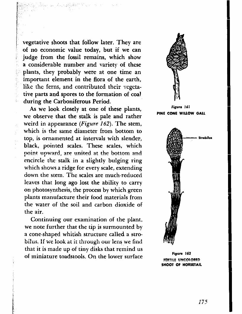
vegetative shoots that follow later. They are of no economic value today, but if we can
l judge from the fossil remains, which show a considerable number and variety of these plants, they probably were at one time an important element in the flora of the earth, like the ferns, and contributed their vegeta- tive parts and spores to the formation of coal during the Carboniferous Period,
As we look closely at one of these plants, we observe that the stalk is pale and rather weird in appearance (Figure 162). The stem, which is the same diameter from bottom to top, is ornamented at intervals with slender, black, pointed scales. These scales, which point upward, are united at the bottom and encircle the stalk in a slightly bulging ring which shows a ridge for every scale, extending down the stem. The scales are much-reduced leaves that long ago lost the ability to carry on photosynthesis, the process by which green plants manufacture their food materials from the water of the soil and carbon dioxide of the air.
Figure 161
PINE CONE WILLOW GALL
Strobilut
Continuing our examination of the plant, we note further that the tip is surmounted by a cone-shaped whitish structure called a stro- bilus. If we look at it through our lerqwe find that it is made up of tiny disks that remind us of miniature toadstools. On the lower surface
Figure 162
FERTILE UNCOLORED SHOOT OF HORSETAIL

Figure 143
SPORANGIUM OF HORSETAIL
Figure 164
STERILE GREEN SHOOT OF HORSnAll
of each disk can be seen from five to ten
sacs that are spore-producing structures, or
sporangia (Figure 163). After the spores have
been released, these sporangia hang around
the disk in torn scallops.
The spore-producing shoot is only part of
the horsetail plant. As soon as the spores have
been released, the shoot dies clown and is fol-
lowed by a slender green shoot with numer-
ous branches set in whorls at intervals, or
nodes (Figure 164). At one time the plant
probably had a whorl of leaves at each node,
but since there are now so many green
branches, the leaves have been reduced to
mere points and appear to be merely a sort
of trimming. Each little cup or socket of the
joint or node, in branch or stem, has a row
of points around its margin. If a branch is
triangular in cross-section, it will have three
points, if quadrangular, four points, and so
on. Both the stem and branches are made LIP entirely of segments, each set at its lower end
in the socket of the segment below it and each
easily pulled out.
As you handle the horsetails you will find
that they are rough and of harsh texture. The
reason for this is that they are impregnated
with silica, and on this account the plant was
formerly used for cleaning and polishing
metal utensils and was given the name “scour-
ing rush.”
176
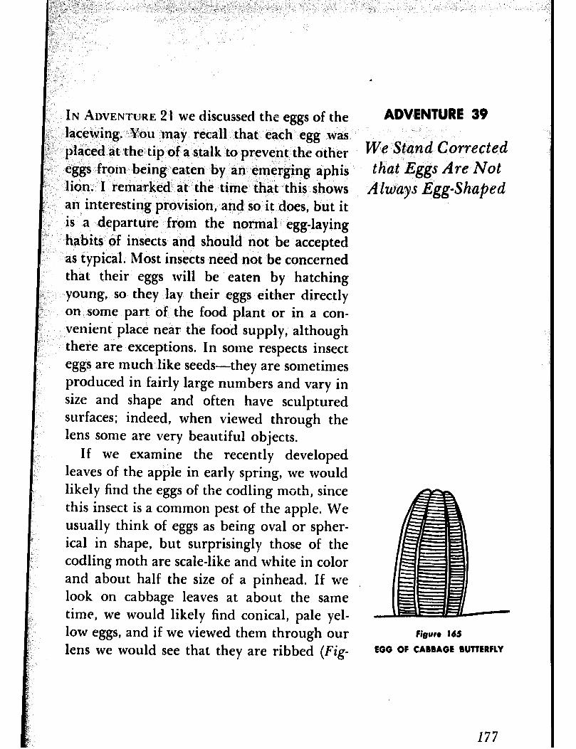
IN ihm~~u~~: 21 we discussed the eggs of the lacewing. You may recall that each egg was placed at the tip of a stalk to prevent the other eggs from being eaten by an emerging aphis lion. I remarked at the time that this shows an interesting provision, and so it does, but it is a departure from the normal egg-laying habits of insects and should not be accepted as typical. Most insects need not be concerned that their eggs will be eaten by hatching young, so they lay their eggs either directly on some part of the food plant or in a con- venient place near the food supply, although there are exceptions. In some respects insect eggs are much like seeds-they are sometimes produced in fairly large numbers and vary in size and shape and often have sculptured surfaces; indeed, when viewed through the lens some are very beautiful objects.
If we examine the recently developed leaves of the apple in early spring, we would likely find the eggs of the codling moth, since this insect is a common pest of the apple. We usually think of eggs as being oval or spher- ical in shape, but surprisingly those of the codling moth are scale-like and white in color and about half the size of a pinhead. If we look on cabbage leaves at about the same time, we would likely find conical, pale yel- low eggs, and if we viewed them through our lens we would see that they are ribbed (Fig-
.
ADVENTURE 39
We Stand Corrected that Eggs Are Not
Always Egg-Shaped
Figure 165
EGG OF CABBAGE BUTTERFLY
177
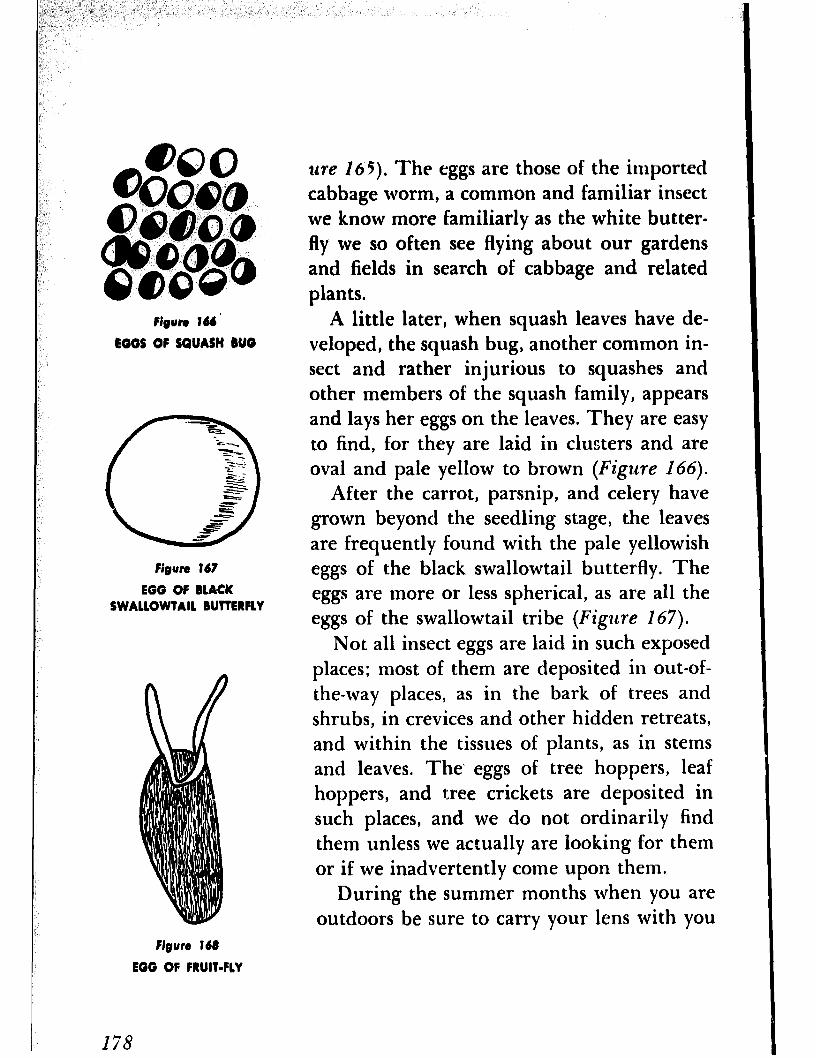
Nguro 166. EGGS OF SQUASH BUG
Figure 167
EGG OF BLACK SWALLOWTAIL BUTTERFLY
llre 165). The eggs are those of the imported cabbage worm, a common and familiar insect we know more familiarly as the white butter- fly we so often see flying about our gardens and fields in search of cabbage and related plants.
A little later, when squash leaves have de- veloped, the squash bug, another common in- sect and rather injurious to squashes and other members oE the squash family, appears and lays her eggs on the leaves. They are easy to find, for they are laid in clusters and are oval and pale yellow to brown (Figure 166).
After the carrot, parsnip, and celery have grown beyond the seedling stage, the leaves are frequently found with the pale yellowish eggs of the black swallowtail butterfly. The eggs are more or less spherical, as are all the eggs of the swallowtail tribe (Figzlre 267).
Not all insect eggs are laid in such exposed places; most of them are deposited in out-of- the-way places, as in the bark of trees and shrubs, in crevices and other hidden retreats, and within the tissues of plants, as in stems and leaves. The’ eggs of tree hoppers, leaf hoppers, and tree crickets are deposited in such places, and we do not ordinarily find them unless we actually are looking for them or if we inadvertently come upon them.
During the summer months when you are outdoors be sure to carry your lens with you
Figure 168
EGG OF FRUIT-FLY
178
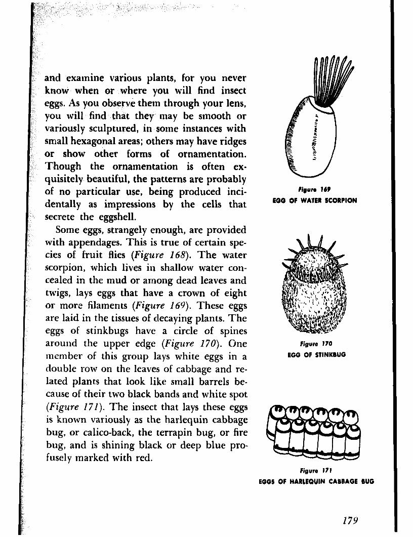
and examine various plants, for you never know when or where you will find insect eggs. As you observe them through your lens, you will find that they may be smooth or variously sculptured, in some instances with small hexagonal areas; others may have ridges or show other forms of ornamentation. Though the ornamentation is often ex- quisitely beautiful, the patterns are probably of no particular use, being produced inci- dentally as impressions by the cells that secrete the eggshell.
Some eggs, strangely enough, are provided with appendages. This is true of certain spe- cies of fruit flies (Figure 168). The water scorpion, which lives in shallow water con- cealed in the mud or among dead leaves and twigs, lays eggs that have a crown of eight or more filaments (Figure 169). These eggs are laid in the tissues of decaying plants. The eggs of stinkbugs have a circle of spines around the upper edge (Figzlre 170). Gne member of this group lays white eggs in a double row on the leaves of cabbage and re- lated plants that look like small barrels be- cause of their two black baxlds and white spot (Figure 171). The insect that lays these eggs is known variously as the harlequin cabbage bug, or calico-back, the terrapin bug, or fire bug, and is shining black or deep blue pro- fusely marked with red.
Figun 169
EGG OF WATER SCORPION
Figurt 170
EGG OF STINKBUG
Figurt 171
EGGS OF HARCEQUIN CABBAGE BUG
179

0 6 Figure 172
EGG OF WHEEL RUG
Figun 173
EGG OF POULTRY LOUSE
ADVENTURE 40
We Play Amateur Petrologists
180
The assassin bugs include a number of dif- ferent species, as the wheel bug, and are rather striking insects. They are all fairly large and some of them are gaily colored. Their eggs take various forms and may be cylindrical or elongate-oval (Figure 172). But it is not their shape that is of interest, rather the presence of a cap at one end which is pushed off in the hatching process and which is often decorated with raylike extensions. Of all the ornamental structures with which insect eggs may be furnished the most strik- ing are undoubtedly those found on the eggs of the poultry louse. The eggs are white, elliptical, and are provided with white, glass- like spines (Figure 173). They are worth looking for and viewing through your lens if you are not averse to examining the feathers of chickens to which they are at- tached.
IN TERMS 0~ GEOLOGY a rock is a mineral found in large quantities in the earth or is a mixture, or aggregate, of minerals. The familiar granite is chiefly a mixture of three minerals, quartz, feldspar, and mica, whereas the less familiar sandstone is composed usually of a single mineral, quartz.
According to their origin, position in the
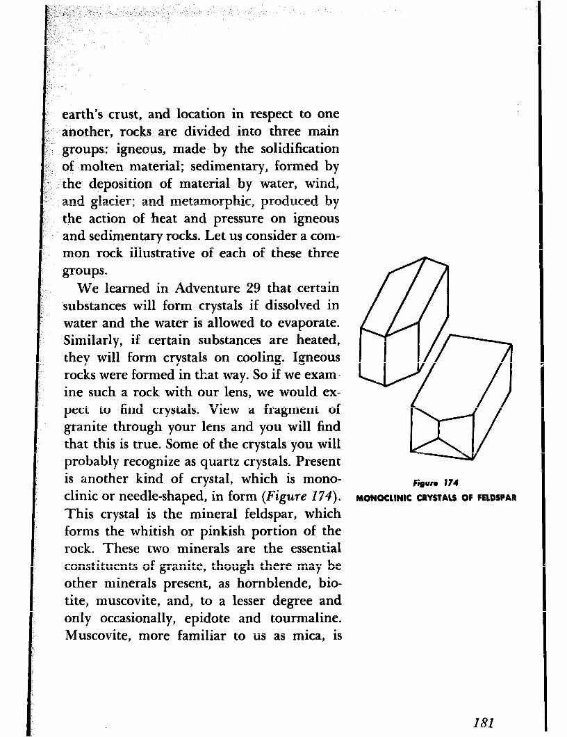
earth’s crust, and location in respect to one another, rocks are divided into three main groups: igneous, made by the solidification of molten material; sedimentary, formed by the deposition of material by water, wind, and glacier; and metamorphic, produced by the action of *heat and pressure on igneous and sedimentary rocks. Let us consider a com- mon rock illustrative of each of these three groups.
We learned in Adventure 29 that certain substances will form crystals if dissolved in water and the water is allowed to evaporate. Similarly, if certain substances are heated, they will form crystals on cooling. Igneous rocks were formed in that way. So if we exam- ine such a rock with our lens, we would ex- pect to find crystals. View a fragment of granite through your lens and you will find that this is true. Some of the crystals you will probably recognize as quartz crystals. Present is another kind of crystal, which is mono- clinic or needle-shaped, in form (Figure 174).
This crystal is the mineral feldspar, which forms the whitish or pinkish portion of the rock. These two minerals are the essential constituents of granite, though there may be other minerals present, as hornblende, bio- tite, muscovite, and, to a lesser degree and only occasionally, epidote and tourmaline. Muscovite, more familiar to us as mica, is
Figurt 174
MONOCLINIC CRYSTALS OF FROSPAR
extensively used in electrical insulations. It occurs as pearly scales that you can easily lift off with a knife. If black particles that are not easily separated into thin layers are present, they probably consist of the mineral horn- blende.
Obviously sandstone is a rock formed from sand, but actually the word is used to describe any sedimentary rock whose particles are about the size of sand grains. When we view a fragment of sandstone through our lens, we find it ccmposed of subangular to rounded grains we recognize as quartz. We may also observe that there are small spaces or pores between the grains ,+ although there are some sandstones without such pores. Some sand- stones are composed entirely of grains held together by the compactness of the rock, but in others the grains are held together by a cementing material that then fills all the pores. Essentially a sandstone consists of quartz grains, but frequently other minerals may be present, such as feldspar, muscovite, and calcite. I said at the beginning of this paragraph that in a sandstone the particles are about the size of sand grains. If the par- ticles are smaller, less than one five hun- dredth of an inch, the sandstone grades into
+ In general there is a considerable amount of space between the grains of sand so that a sandstone will absorb large amounts of water-up to 25% of its bulk.
182
shale; when they are larger than that of a pea, it grades into conglomerate. If a sandstone has been subjected to heat and pressure, it is then known as quartzite, a metamorphic rock. Frequently one grades into another. Quartzite may be distinguished from sand- stone by ihe almost complete lack of pore spaces, its greater hardness, and by its crystal- line structure. It may be further distinguished by the fact that a sandstone, in breaking, sepa- rates between the grains of sand, while a quartzite breaks through the grains.
In Adventure 35 we said that the mineral calcite is removed from water by various ani- mals that use it in building shells or other structures in which to live. When the animals die the shells sink to the bottom, where they are pulverized a,ld accumulate in large de- posits of mudlike consistency which eventu- ally are compressed to form a sedimentary rock. We know this rock as limestone. Through the lens a fragment of limestone ap- pears much like sandstone. Pure limestone is white, but it may be variously colored by impurities. As in sandstone, other minerals may also be present. As you will recall that we can determine the identity of calcite by adding a drop of hydrochloric acid to it. Since limestone is calcite, we can apply the same test to a rock we suspect to be limestone.
If limestone has been subjected to heat and
183
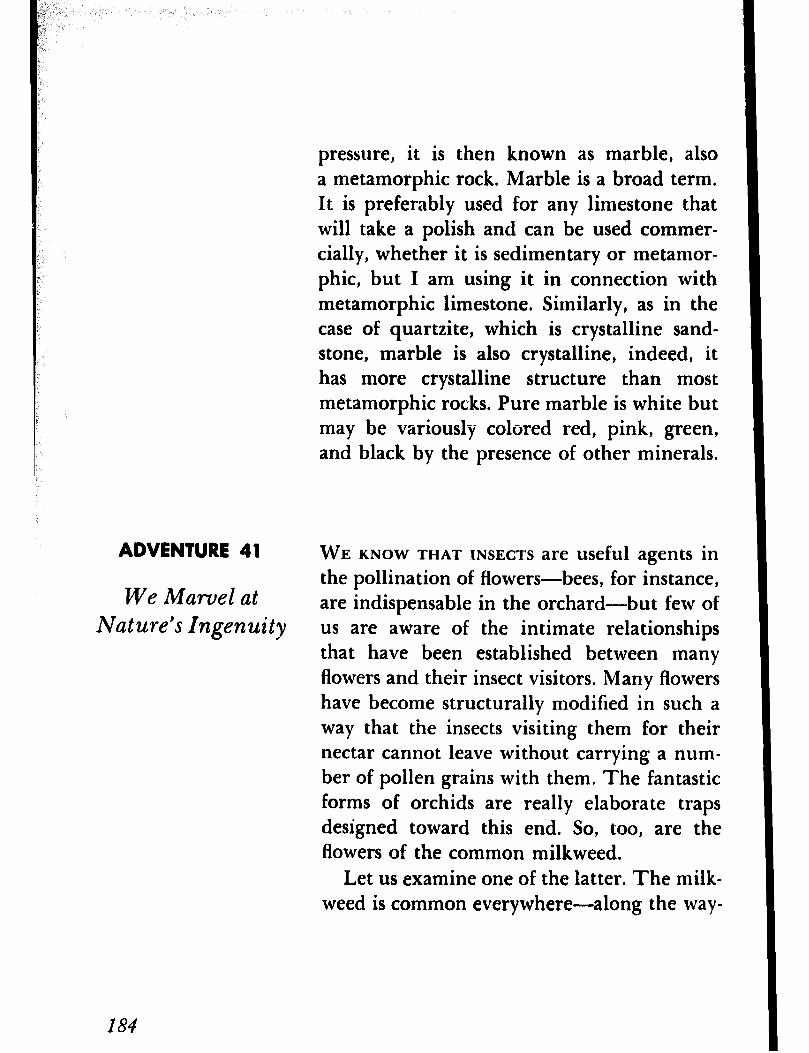
pressure, it is then known as marble, also a metamorphic rock. Marble is a broad term, It is preferably used for any limestone that will take a polish and can be used commer- cially, whether it is sedimentary or metamor- phic, but I am using it in connection with metamorphic limestone. Similarly, as in the case of quartzite, which is crystalline sand- stone, marble is also crystalline, indeed, it has more crystalline structure than most metamorphic rocks. Pure marble is white but may be variously colored red, pink, green, and black by the presence of other minerals.
ADVENTURE 41 WE KNOW THAT INSECTS are useful agents in
We Marvel at the pollination of flowers-bees, for instance, are indispensable in the orchard-but few of
Nature’s Ingenuity us are aware of the intimate relationships that have been established between many flowers and their insect visitors. Many flowers have become structurally modified in such a way that the insects visiting them for their nectar cannot leave without carrying a num- ber of pollen grains with them. The fantastic forms of orchids are really elaborate traps designed toward this end. So, too, are the flowers of the common milkweed.
Let us examine one of the latter. The milk- weed is common everywhere-along the way-
184
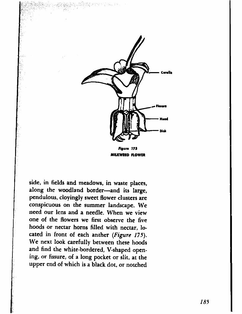
side, in fields and meadows, in waste places, along the woodland border-and its large, pendulous, cloyingly sweet flower clusters are conspicuous on the summer landscape. We need our lens and a needle. When we view one of the flowers we first observe the five hoods or nectar horns filled with nectar, lo- cated in front of each anther (Figure 175). We next look carefully between these hoods and find the white-bordered, V-shaped open- ing, or fissure, of a long pocket or slit, at the upper end of which is a black dot, or notched
182
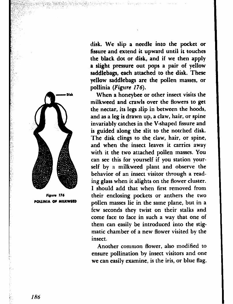
Figun I74
POLLINIA OF MILKWEED
disk. We slip a needle into the pocket or fissure and extend it upward until it touches the black dot or disk, and if we then apply a slight pressure out pops a pair of yellow saddlebags, each attached to the disk. These yellow saddlebags are the pollen masses, or pollinia (Figure 176).
When a honeybee or other insect visits the milkweed and crawls over the flowers to get the nectar, its legs slip in between the hoods, and as a leg is drawn up, a claw, hair, or spine invariably catches in the V-shaped fissure and is guided along the slit to the notched disk. The disk clings to the claw, hair, or spine, and when the insect leaves it carries away with it the two attached pollen masses. You can see this for yourself if you station your- self by a milkweed plant and observe the behavior of an insect visitor through a read- ing glass when it alights on the flower cluster. I should add that when first removed from their enclosing pockets or anthers the two pollen masses lie in the same plane, but in a few seconds they twist on their stalks and come face to face in such a way that one of them can easily be introduced into the stig matic chamber of a new flower visited by the insect.
Another common flower, also modified to ensure pollination by insect visitors and one we can easily examine, is the iris, or blue flag.
186
tmry
Pigor. 177
SECTION OF IRIS FLOWER
The iris is one of the more familiar spring flowers of swamps and marshes, and related members of the group are extensively cul- tivated in gardens. The blue flag, with its large showy blossoms, seems especially de- signed for bees, since blue is their favorite color, but other insects also visit it. Each of the three drooping sepals forms the floor of an arched passageway that leads to the nec- tary, the roof being formed by three strap- like divisions of the style (the middle portion of the pistil). Over the entrance to the pas- sageway and pointing outward is a movable lip, the stigma (Figure 177). In back of the stigma and beneath the roof is the stamen with its anther. When a bee visits the blos- som, carrying pollen from another flower, it alights on the tip of the sebal, and then as it presses forward it hits and bends down the
187
stigma, which scrapes the pollen from the back of the bee and then springs back into place. As the bee continues on its way toward the nectary, its hairy back rubs against the overhanging anther and becomes powdered with pollen grains. When it backs out of the passageway, after getting its fill of nectar, it again encounters the stigma, but this side of the stigma cannot receive pollen and so im- mediate close pollination is averted. Just how all this is accomplished may easily be ob- served through the reading glass by waiting for a bee to enter a blossom and then watch- ing its actions.
The milkweed and iris are but two exam- ples of structural modifications for insect pol- lination. There are countless others. The sage, nasturtium, and larkspur of our gardens come to mind. The butter-and-eggs, with its delightful two-lipped, spurred yellow and orange flowers and so common from July to October along roadsides and in waste places, and the jewelweed, blooming by the brook- side, its bright flowers hanging like jewels from a lady’s ears, also come to mind. Why not examine these and others, too, with your lens and discover, if you can, the various and often ingenious mechanisms that plants have developed to compel their insect visitors to carry their pollen to other flowers and thus ensure their survival as species.
188
;p,. IL,‘; pi ,‘.$, ,,,‘,
\6 ,._’
I;:‘
_ _-
HAVE YOU EVERNOTICED small flies about the ADVENTURE 42 :s: fruit bowl or fruit basket especially in sum-
mer? Have you ever wondered what they were We Breed y, doing there? The larvae feed on ripe or over- Some Flies “.
ripe bananas and other. fruit, and the flies we I 1. see about the fruit are either females prepar-
ing to lay their eggs in the fruit ,or newly de- veloped adults recently emerged from their pupal state. The larvae also feed on vinegar, stale beer, and the like, hence the insects are variously called fruit flies, sour flies, vinegar
“. flies, and pomace flies. They are, however, best known as fruit flies.
These flies are insects of distinction. Some years ago Thomas Hunt Morgan used them in studying heredity and sex, and because they lend themselves so readily to investiga- tive work on such studies, they have been extensively used since then on both simple and complex cases of Mendelian inheritance and the relations between body characteris- tics and genes. There are several reasons why they are ideal experimental animals. One is that they reproduce quickly. The average duration of the egg period at ordinary room temperature is about two days; of the larval
j ; period about six days; and of the pupal pe-
riod about five days. Another is that a bit of banana in a bottle is about all that is neces- sary to breed the insects.
We are not interested at present in inheri-
I89
tance or inherited characters, but in breeding a few generations. To do so, we need first of all a few flies, and the best time to get them is in the summertime. We can place some fruit in a dish and trust to luck that the flies will appear, or we can dip a piece of ripe banana in a suspension of yeast (which can easily be prepared by dissolving a quarter of a yeast cake in some water) and inserting the piece of banana along with a strip of paper towel- ing into a clean bottle. We can put the bottle aside or, better still, we can take it to our neighborhood fruit store and ask the owner if we can leave it there overnight. When we return to the store the following morning, there will be some flies in the bottle. We plug the bottle with some cotton wrapped in cheesecloth, take it home, and set it aside. We call this our culture bottle. Within two weeks we should have an ample supply of flies.
To breed the flies, we select a male and a female from our culture bottle and place them in another bottle, similar to the first, which we have meanwhile prepared. As the flies are small and active it will be almost im- possible to transfer them unless we first ren- der them unconscious. To do this, we must have an etherizer. We get a bottle having a mouth the same size as the mouth of the cul- ture bottle. We insert a nail one inch long into a cork stopper that fits neatly into the
I90
_ ‘, ‘. .“. /’ (, .,:,, ‘,
‘,
mouth of the. bottle and cover the nail with yers, of cotton,, tying the cotton to
: the nail with thread’#‘or string. We then put j a few drops of ether on the cotton, taking care
not to use it near an open ,flame, since ether _ ., is highly inflammable.
We are now ready to transfer the flies from the culture bottle to the etherizer. We tap the ’ culture bottle on the table so that the flies will drop to the bottom, quickly remove the cotton plug, and place the mouth of the etherizer closely against the mouth of the cul- ture bottle. We next invert the two bottles and again tap the culture bottle so the flies will drop into the etherizer. To hasten the flies into the latter, we place an electric light near the etherizer because the flies are helio- tropic and are attracted to light. When the flies have moved into the etherizer, we quickly separate the two bottles, replacing the cotton plug in the culture bottle and stopping the etherizer with the cork stopper containing the nail and cotton soaked with ether. The flies will be anesthetized in a few
When the flies are completely unconscious, we remove the stopper and spill them out on a piece of white paper. Since the flies will re- main unconscious only a few minutes, we have to examine them quickly for our male and female. Frequently this may take a little
191
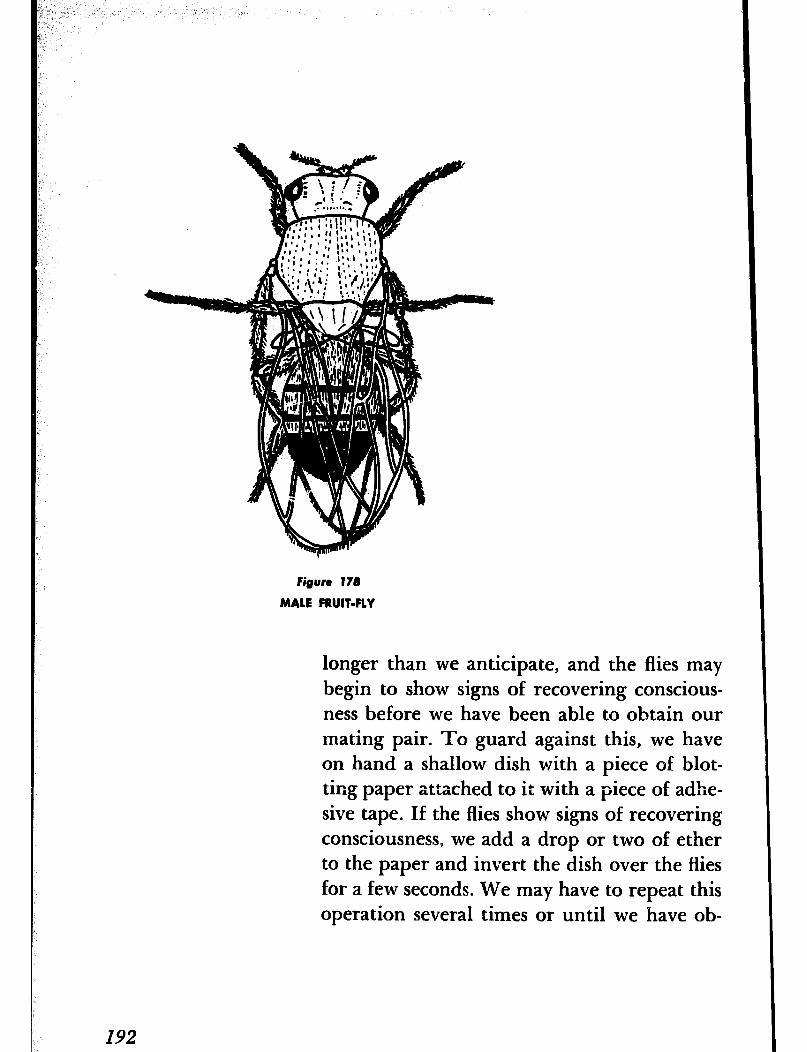
Figure 178
MALE FRUIT-FLY
longer than we anticipate, and the flies may begin to show signs of recovering conscious- ness before we have been able to obtain our mating pair. To guard against this, we have on hand a shallow dish with a piece of blot- ting paper attached to it with a piece of adhe- sive tape. If the flies show signs of recovering consciousness, we add a drop or two of ether to the paper and invert the dish over the flies for a few seconds. We may have to repeat this operation several times or until we have ob-
192
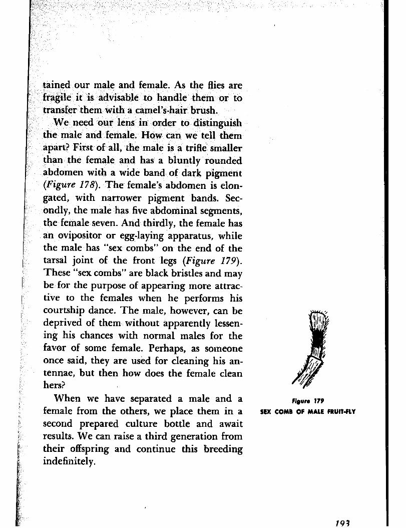
I, /‘) ‘,
tained our male and female. As the flies are fragile it is advisable to handle them or to transfer them with a camel’s-hair brush.
We need our lens in order to distinguish the male and female. How can we tell them apart? First of all, the male is a trifle smaller than the female and has a bluntly rounded abdomen with a wide band of dark pigment (Figure 178). The. female’s abdomen is elon- gated, with narrower pigment bands. Sec- ondly, the male has five abdominal segments, the female seven. And thirdly, the female has an ovipositor or egg-laying apparatus, while the male has “sex combs” on the end of the tarsal joint of the front legs (Figure I79).
These “sex combs” are black bristles and may be for the purpose of appearing more attrac- tive to the females when he performs his courtship dance. The male, however, can be deprived of them without apparently lessen- ing his chances with normal males for the favor of some female. Perhaps, as someone once said, they are used for cleaning his an- tennae, but then how does the female clean hers?
When we have separated a male and a female from the others, we place them in a second prepared culture bottle and await results. We can raise a third generation from their offspring and continue this breeding indefinitely.
Figure 179
SEX COMB OF MALE FRUIT-FLY
19?
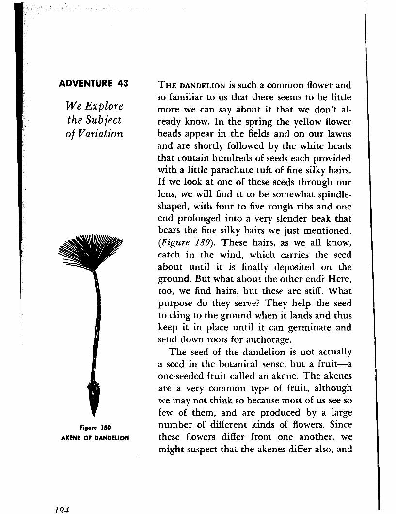
ADVENTURE 43
We Explore the Subject of Variation
Figure 180
AKENE OF DANDELION
THE DANDELION is such a common flower and so familiar to us that there seems to be little more we can say about it that we don’t al- ready know. In the spring the yellow flower heads appear in the fields and on our lawns and are shortly followed by the white heads that contain hundreds of seeds each provided with a little parachute tuft of fine silky hairs. If .we look at one of these seeds through our lens, we will find it to be somewhat spindle- shaped, with four to five rough ribs and one end prolonged into a very slender beak that bears the fine silky hairs we just mentioned. (Figure 180). These hairs, as we all know, catch in the wind, which carries the seed about until it is finally deposited on the ground. But what about the other end? Here, too, we find hairs, but these are stiff. What purpose do they serve? They help the seed to cling to the ground when it lands and thus keep it in place until it can germinate and send down roots for anchorage.
The seed of the dandelion is not actually a seed in the botanical sense, but a fruit-a one-seeded fruit called an akene. The akenes are a very common type of fruit, although we may not think so because most of us see so few of them, and are produced by a large number of different kinds of flowers. Since these flowers differ from one another, we might suspect that the akenes differ also, and
194
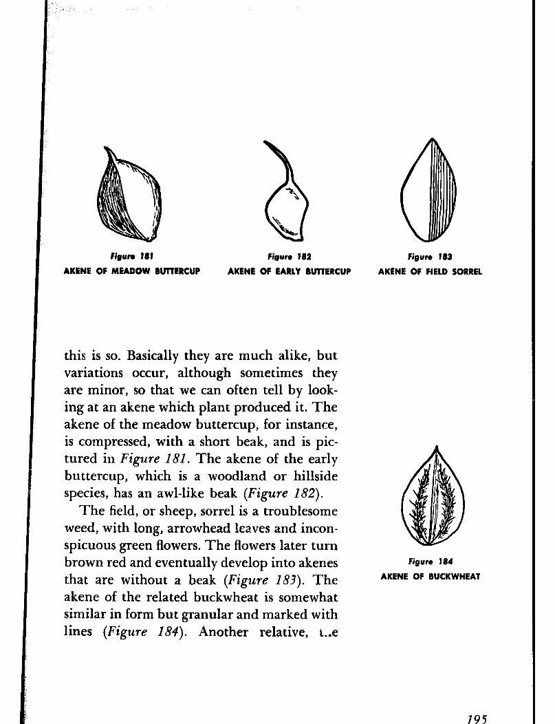
FiRun ?8t Figure 182
AKENE OF MEADOW BUTTERCUP AKENE OF EARLY BUTTERCUP
this is so. Basically they are much alike, but variations occur, although sometimes they are minor, so that we can often tell by look- ing at an akene which plant produced it. The akene of the meadow buttercup, for instance, is compressed, with a short beak, and is pic- tured in Figure 181. The akene of the early buttercup, which is a woodland or hillside species, has an awl-like beak (Figure 182).
The field, or sheep, sorrel is a troublesome weed, with long, arrowhead leaves and incon- spicuous green flowers. The flowers later turn brown red and eventually develop into akenes that are without a beak (Figure 183). The akene of the related buckwheat is somewhat similar in form but granular and marked with lines (Figure 184). Another relative, t-*e
Figure 183
AKENE OF FIELD SORREL
Figure 184
AKENE OF BUCKWHEAT
195
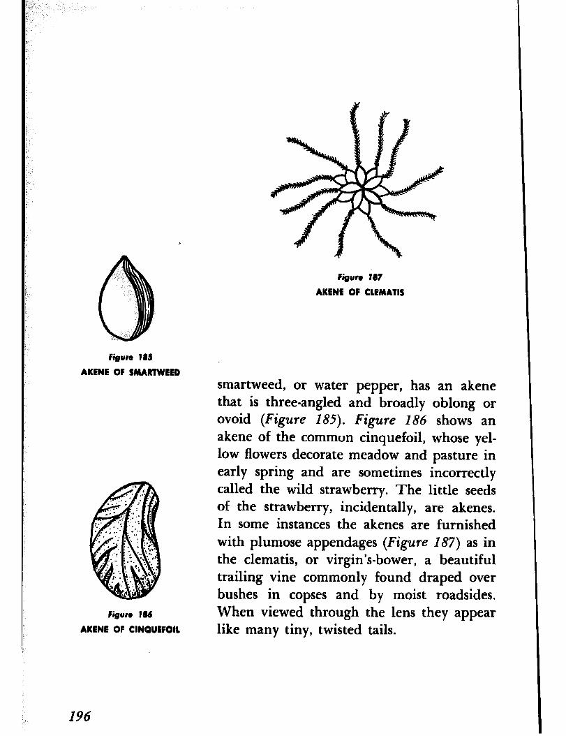
Figure 187
AKENE OF CLEMATIS
Figure 18s
AKENE OF SMARTWEED
Figure 186
AKENE OF CINQUEFOIL
smartweed, or water pepper, has an akene that is three-angled and broadly oblong or ovoid (Figure I85). Figure 186 shows an akene of the common cinquefoil, whose yel- low flowers decorate meadow and pasture in early spring and are sometimes incorrectly called the wild strawberry. The little seeds of the strawberry, incidentally, are akenes. In some instances the akenes are furnished with plumose appendages (Figure 187) as in the clematis, or virgin’s-bower, a beautiful trailing vine commonly found draped over bushes in copses and by moist roadsides. When viewed through the lens they appear like many tiny, twisted tails.
196
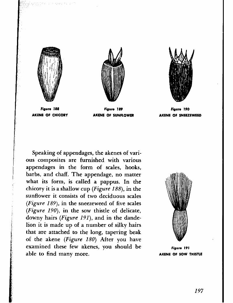
Figure 188
AKENE OF CHICORY
Figure 189 Figure 190
AKRNE OF SUNFLOWER AKENE OF SNEEZEWEED
Speaking of appendages, the akenes of vari- ous composites are furnished with various appendages in the form of scales, hooks, barbs, and chaff. The appendage, no matter what its form, is called a pappus. In the chicory it is a shallow cup (Figure 188), in the sunflower it consists of two deciduous scales (Figure ISS), in the sneezeweed of five scales (Figure ISO), in the sow thistle of delicate, downy hairs (Figure I Sl), and in the dande- lion it is made up of a number of silky hairs that are attached to the long, tapering beak of the akene (Figure 180) After you have examined these few akenes, you should be able to find many more.
Figure 191
AKENE OF SOW THISTLE
I97
ADVENTURE 44
We Get to Know the Barnacle
THOSE OF us WHO LIVE near the coast can find many things of interest along the sea beach, not the least of which are the bar- nacles. Barnacles are shrimplike animals that live within a calcareous shell. They are fa- miliar objects along the rocky shore. Because of their shell they were at one time believed to be mollusks, but since they have jointed appendages they are now placed among the arthropods. The appendages are fringed like feathers and are drawn into or protruded from the shell at will. When the animal is covered with water the appendages are ex- tended out of the shell and sweep through the water like a casting net, capturing small swimming creatures and organic fragments that serve as food.
The life history of the barnacle is most in- teresting. The young barnacle, which hatches from the egg, is free-swimming and in no way resembles the adult. After swimming about for a while it undergoes changes and then set- tles on some solid object, such as a rock, wharf pile, drifting timber, the bottom of a ship, and even on a whale, fastening itself, head foremost, by means of a cementlike secretion. (If you think barnacles are not securely attached just try to pry one off). After it has found a permanent location it undergoes further changes until it has at- tained its adult form.
I98
Figure 192
BARNACLE
There are several different kinds of bar- nacles, but the one familiar to most of us is the rock barnacle, which we find attached to rocks along the shore. Sometimes the rocks are so thickly covered with the barnacles that they appear white from a distance. If we look at the shell through our lens, we find it com- posed of a number of thick calcareous plates that fit together in a tentlike form (Figure
192). These plates, which are usually six in number, are rigidly attached to each other and to a fold of skin surrounding the body of the animal, but there are two pairs of hinged valves at the top which may open and close like double doors. It is said that if you tap a rock encrusted with barnacles and hold
199

your ear near you can hear the closing of many doors.
If you visit the seashore when the rocks are covered with water and if the barnacles are unmolested, you will see thousands of tiny fringed feet waving to and fro. They are extended out through the open doors and serve as a sort of casting net. Their move- ments are perfectly regular and rapid and may beat back and forth as many as a hundred times a minute. As someone once remarked, a barnacle is a little shrimplike animal stand- ing on its head within a limestone house and kicking its food into its mouth with its feet.
ADVENTURE 45
We Seek the Liverwort
IF YOU LOOK along the mossy bank of a brook or stream or among the mosses in damp woods, you should find flat, ribbonlike plants growing close to the ground. They are papery thin and may be either long and slender or repeatedly lobed and forked. In a way they resemble the lichens except that they are distinctly green in color. Called liverworts ,* they are very simple plants with- out stem or leaves. Of no economic impor- tance, they are of interest, however, because
* The name “liverwort” is derived from the somewhat liver-shaped thallus and the belief at one time that they could cure diseases of the liver.
200
they represent the transition stage from a water living habit to a land living habit, in other words, they bridge the gap between the algae, which are fundamentally aquatic, and the higher flowering land plants.
There are some four thousand species of liverworts, but one of the most common and the one you are most apt to find is Marchan- tia, a name derived from a French botanist who lived about three hundred years ago. It has a peculiar dull-green color with a broad, ribbon-shaped thallus, which is simply a plant without true roots, stems or leaves, and gen- erally forked once or twice. The upper sur- face is divided into angular areas in the cen- ter of which we can distinguish an air pore. If you will examine the lower surface with your lens, you will find numerous hairs, called rhizoids, that anchor it to the ground and that serve to some extent the same purpose as the roots of higher plants.
Note if umbrellalike structures extend into the air from the surface of the thallus. These upright growths are the reproductive organs. The umbrella part may be either flat, shield-shaped, radially lobed, or it may be a disk with deep, fingerlike lobes that usually curve downward. The former is the male organ, or antheridial disk (Figure 193,
the latter the female, or archegonial disk (Figure 194), and you will not find them on
Figure 193
ANTHEREDIAL DISK OF MARCHANTIA
Figure 194
ARCHEGONIAL DISK OF MARCHANTIA
the same plant. Sperms are produced in the male organ and eggs in the female, and since these organs occur on separaLe plants, it is necessary for the sperms to swim to the eggs so that the latter may be fertilized. This ex- plains why liverworts always grow in wet or damp places. At first there seems to be a con- siderable element of chance involved in this method of reproduction, for the question arises, how do the sperms locate the eggs? The probability of a sperm’s locating an egg
would appear to be one in a million, if the odds are not even greater. Actually the
202
Figure 195 Figure 196
GEMMAE CUPS OF MARCHANTIA GEMMA OF MARCHANTIA
sperms have no difficulty finding the eggs because the female -organ discharges sub- stances that help to orient the sperms in the right direction. The phenomenon is called positive chemotaxis.
Instead of the umbrella-like structures you may find little cup-shaped or saucer-shaped structures with toothed margins on the upper surface of the thallus (Figure 19.5). These structures produce green disks called gem- mae, or brood bodies (Figure 196). When washed out or blown to some distant place they grow into new plants.
203
-1
ADVENTURE 46 THE INCTIONARY TELLS us that a trap is a de-
We Set Some Traps vice that shuts suddenly with a springlike action for taking game and other animals. Man has devised many ingenious traps, but I doubt if he has improved upon those de- signed by nature or will ever do so. I am thinking specifically of the traps developed by certain plants for catching insects. Best known are the Venus-flytrap, the sundew, and the pitcher plant, Perhaps you are fa- miliar with them.
The Bladderwort (Figure 197) is perhaps not quite so generally known, but it has de- signed a trap that, I think, is more ingenious than those developed by the plants men- tioned above. It is a plant of ponds and slug- gish streams, ‘delicate and vinelike in appear- ance, and floats beneath the surface among the stems of water lilies and pondweeds. Without roots it has instead twiglike rhizoids that function as roots. The stem is slender and bears finely branched leaves arranged alternately but divided so closely to the base that each leaf appears to be two leaves grow- ing opposite one another. The leaves are furnished with small bladders that at one time were believed to function entirely as floats but which also serve as traps for cap- turing small aquatic creatures. They deserve our attention, so let us examine them.
To do so, we need to lift the plant out of
204
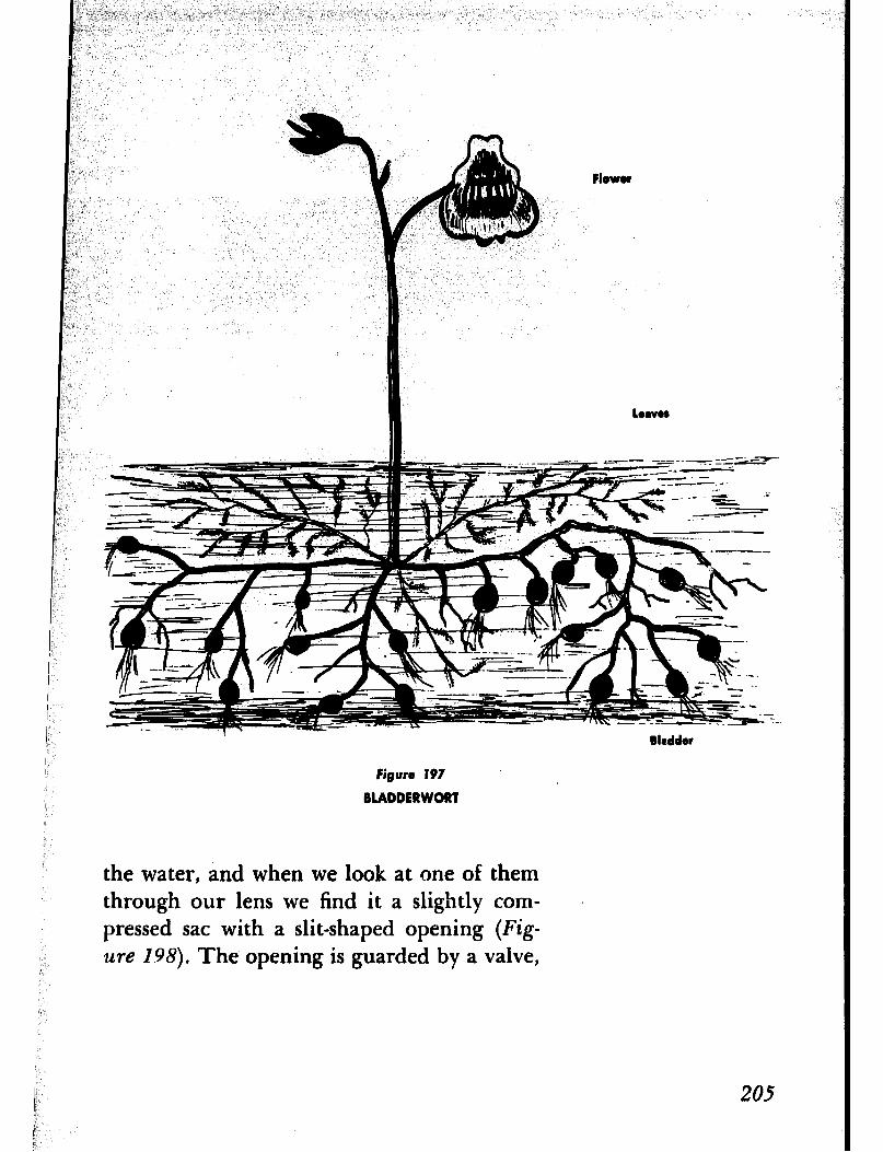
bevr
Bladder
Figure I97
BLADDERWORT
the water, and when we look at one of them through our lens we find it a slightly corn- pressed sac with a slit-shaped opening (Fig- ure 1.98). The opening is guarded by a valve,
205
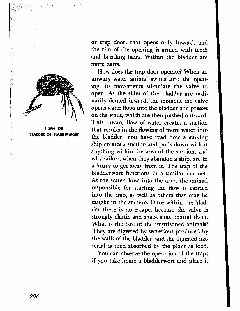
Figure 198
MADDER OF BLADDERWORT
or trap door, that opens only inward, and the rim of the opening is armed with teeth and bristling hairs. Within the bladder are more hairs.
How does the trap door operate? When an unwary water animal swims into the open- ing, its movements stimulate the valve to open. As the sides of the bladder are ordi- narily dented inward, the moment the valve opens water flows into the bladder and presses on the walls, which are then pushed outward. This inward flow of water creates a suction that results in the flowing of more water into the bladder. You have read how a sinking ship creates a suction and pulls down with it anything within the area of the suction, and why sailors, when they abandon a ship, are in a hurry to get away from it. The trap of the bladderwort functions in a similar manner. As the water flows into the trap, the animal responsible for starting the flow is carried into the trap, as well as others that may be caught in the suction. Once within the blad- der there is no escape, because the valve is strongly elastic and snaps shut behind them. What is the fate of the imprisoned animals? They are digested by secretions produced by the walls of the bladder, and the digested ma- terial is then absorbed by the plant as food.
You can observe the operation of the traps if you take home a bladderwort and place it
206
in an aquarium filled with clear water and well supplied with water fleas and other small aquatic creatures. For best results take a plant whose bladders are empty. The water fleas and other animals can be obtained from the same pond or stream where you found the plant by merely scooping out a pan full of water and transferring it to a bottle. Or you can submerge a large, widemouth bottle and allow the water to run into it. When observ- ing the traps function it is advisable to use a reading glass rather than a small hand lens or pocket magnifier, since you probably can’t get close enough with the latter to see what takes place.
I should add, as a final word, that the blad- ders, which at one time were believed to act as floats, actually function as such at the time of the flowering season. At that time they become filled with air and serve as pontoons to buoy up the plant so that the yellow flowers may be kept out of the water where insects can visit and pollinate them (Figure
I97).
AT ONE TIME herbs were used extensively in cooking to improve the taste and flavor of various foods, and in recent years they have again become very popular. Almost everyone
ADVENTURE 47
Wz Quest for Oil
287
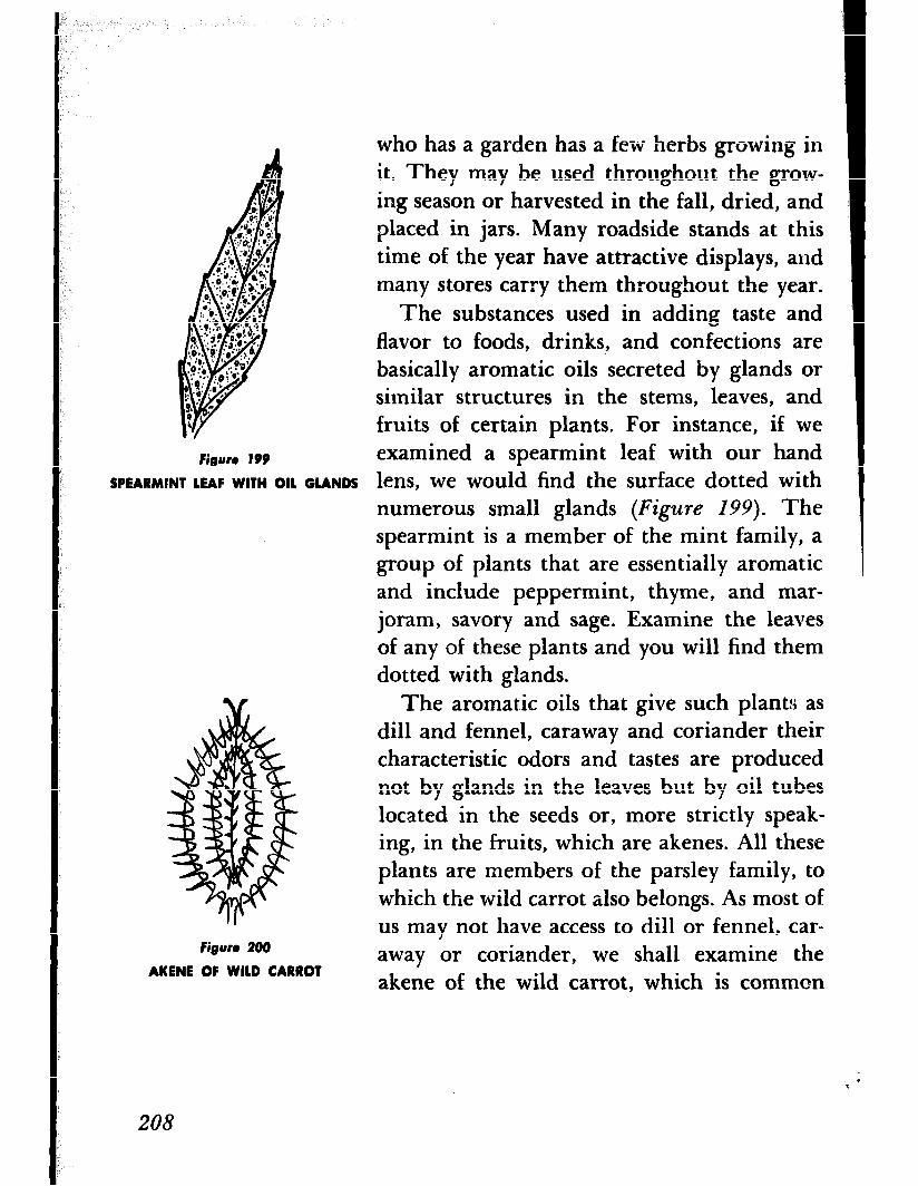
who has a garden has a few herbs growing in it. They may be used throughout the grow- ing season or harvested in the fall, dried, and placed in jars. Many roadside stands at this time of the year have attractive displays, and many stores carry them throughout the year.
The substances used in adding taste and flavor to foods, drinks,, and confections are basically aromatic oils secreted by glands or similar structures in the stems, leaves, and fruits of certain plants. For instance, if we
figure 199 examined a spearmint leaf with our hand SPEARMINT LEAF WITH OIL GLANDS lens, we would find the surface dotted with
numerous small glands (Figure 199). The spearmint is a member of the mint family, a group of plants that are essentially aromatic and include peppermint, thyme, and mar- joram, savory and sage. Examine the leaves of any of these plants and you will find them dotted with glands.
Figure 200
AKENE OF WILD CARROT
The aromatic oils that give such plant:; as dill and fennel, caraway and coriander their characteristic odors and tastes are produced not by glands in the leaves but by oil tubes located in the seeds or, more strictly speak- ing, in the fruits, which are akenes. All these plants are members of the parsley family, to which the wild carrot also belongs. As most of us may not have access to dill or fennel, car- away or coriander, we shall examine the akene of the wild carrot, which is common
208
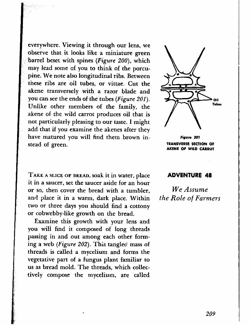
everywhere. Viewing it through our lens, we observe that it looks like a miniature green barrel beset with spines (Figure ZOO), which may lead some of you to think of the porcu- pine. We note also longitudinal ribs. Between these ribs are oil tubes, or vittae. Cut the akene transversely with a razor blade and you can see the ends of the tubes (Figure 201).
Unlike other members of the family, the akene of the wild carrot produces oil that is not particularly pleasing to our taste. I might add that if you examine the akenes after they have matured you will find them brown in- stead of green.
TAKE A SLICE OF BREAD, soakitin water, place it in a saucer, set the saucer aside for an hour or so, then cover the bread with a tumbler, and place it in a warm, dark place. Within two or three days you should find a cottony or cobwebby-like growth on the bread.
Examine this growth with your lens and you wili find it composed of long threads passing in and out among each other form- ing a web (Figure 202). This tangled mass of threads is called a mycelium and forms the vegetative part of a fungus plant familiar to us as bread mold. The threads, which collec- tively compose the mycelium, are called
Oil tuber
Figure 201
TRANSVERSE SECTION OF AKENE OF WILD CARRO?
ADVENTURE 48
We Assume the Role of Farmers
209
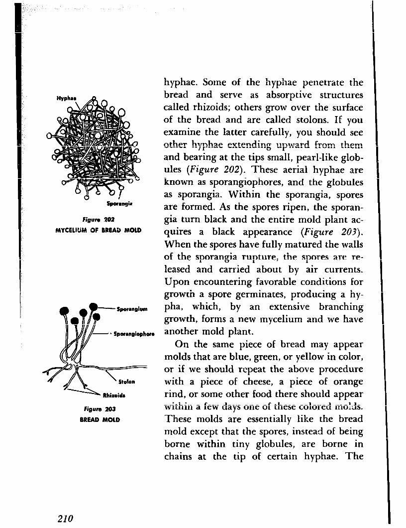
Spormgie
Figure 202
MYCEWM OF BREAD MOLD
Figure 203
BREAD MOLD
hyphae. Some of the hyphae penetrate the bread and serve as absorptive structures called rhizoids; others grow over the surface of the bread and are called stolons. If you examine the latter carefully, you should see other hyphae extending upward from them and bearing at the tips small, pearl-like glob- ules (Figure 202). These aerial hyphae are known as sporangiophores, and the globules as sporangia. Within the sporangia, spores are formed. As the spores ripen, the sporan- gia turn black and the entire mold plant ac- quires a black appearance (Figure 203).
When the spores have fully matured the walls of the sporangia rupture, the spores are re- leased and carried about by air currents. Upon encountering favorable conditions for growth a spore germinates, producing a hy-
pha, which, by an extensive branching growth, forms a new mycelium and we have another mold plant.
On the same piece of bread may appear molds that are blue, green, or yellow in color, or if we should repeat the above procedure with a piece of cheese, a piece of orange rind, or some other food there should appear within a few days one of these colored molds. These molds are essentially like the bread mold except that the spores, instead of being borne within tiny globules, are borne in chains at the tip of certain hyphae. The
210

Figure 204
COMMON BLUE MOLD
spores are called conidiospores and the hy- phae that bear them are called conidiophores and resemble tiny brooms (Figure 204). The conidiospores are produced in great numbers and are very small and light in weight. When mature they become detached and are carried about by air currents, and when they find favorable conditions quickly germinate and develop into new mold plants. Since the spores are variously colored, the character- istic color of the mold is due to the color of their conidial masses.
3ince our experience with molds may be limited to those that appear on our foods, leather goods, fabrics, tobacco, and other or-
211
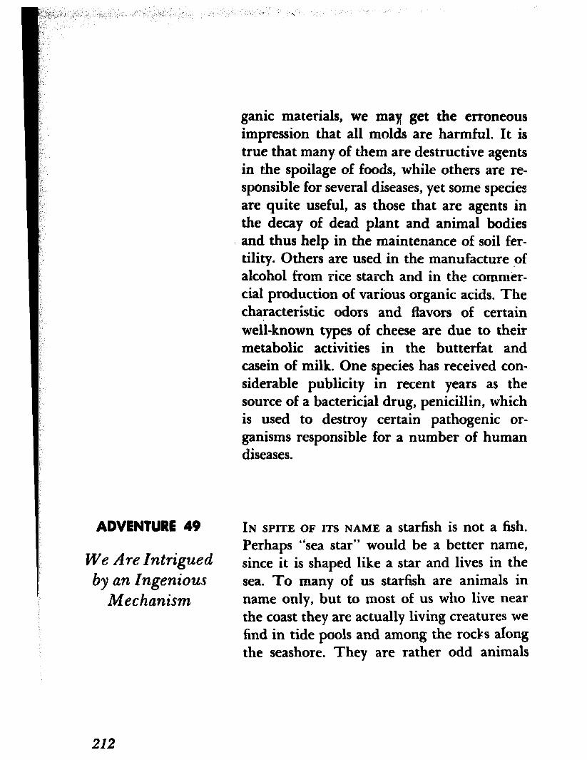
ganic materials, we may get the erroneous impression that all molds are harmful. It is true that many of them are destructive agents in the spoilage of foods, while others are re- sponsible for several diseases, yet some species are quite useful, as those that are agents in the decay of dead plant and animal bodies and thus help in the maintenance of soil fer- tility. Others are used in the manufacture of alcohol from rice starch and in the commer- cial production of various organic acids. The characteristic odors and flavors of certain well-known types of cheese are due to their metabolic activities in the butterfat and casein of milk. One species has received con- siderable publicity in recent years as the source of a bactericial drug, penicillin, which is used to destroy certain pathogenic or- ganisms responsible for a number of human diseases.
ADVENTURE 49
We Are Intrigued by an Ingeni0u.s
Mechanism
IN SPITE OF ITS NAME a starfish is not a fish. Perhaps “sea star” would be a better name, since it is shaped like a star and lives in the sea. To many of us starfish are animals in name only, but to most of us who live near the coast they are actually living creatures we find in tide pools and among the rocks along the seashore. They are rather odd animals
212
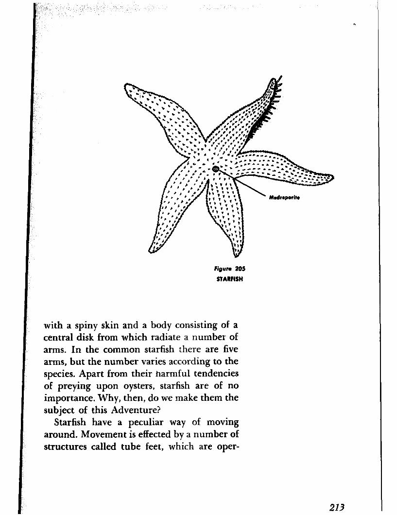
FiRun 205
STARFISH
with a spiny skin and a body consisting of a central disk from which radiate a number of arms. In the common starfish there are five arms, but the number varies according to the species. Apart from their harmful tendencies of preying upon oysters, starfish are of no importance. Why, then, do we make them the subject of this Adventure?
Starfish have a peculiar way of moving around. Movement is effected by a number of structures called tube feet, which are oper-
213
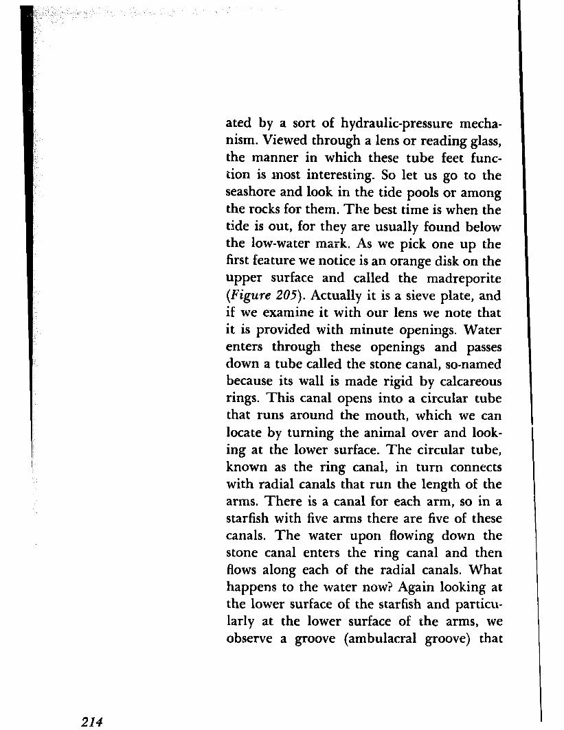
ated by a sort of hydraulic-pressure mecha- nism. Viewed through a lens or reading glass, the manner in which these tube feet func- tion is most interesting. So let us go to the seashore and look in the tide pools or among the rocks for them. The best time is when the tide is out, for they are usually found below the low-water mark. As we pick one up the first feature we notice is an orange disk on the upper surface and called the madreporite (Figure 205). Actually it is a sieve plate, and if we examine it with our lens we note that it is provided with minute openings. Water enters through these openings and passes down a tube called the stone canal, so-named because its wall is made rigid by calcareous rings. This canal opens into a circular tube that runs around the mouth, which we can locate by turning the animal over and look- ing at the lower surface. The circular tube, known as the ring canal, in turn connects with radial canals that run the length of the arms. There is a canal for each arm, so in a starfish with five arms there are five of these canals. The water upon flowing down the stone canal enters the ring canal and then flows along each of the radial canals. What happens to the water now? Again looking at the lower surface of the starfish and particu- larly at the lower surface of the arms, we observe a groove (ambulacral groove) that
214
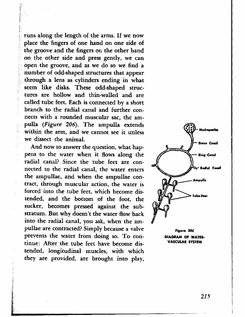
runs along the length of the. arms. If we now ( place the fingers of one hand on one side of : the groove and the fingers on the other hand
on the other side and press gently, we can open the groove, and as we do so we find a
’ number of odd-shaped structures that appear through a lens as cylinders ending in what seem like disks. These odd-shaped struc- tures are hollow and thin-walled and are called tube feet. Each is connected by a short branch to the radial canal and further con- nects with a rounded muscular sac, the am- pulla (Figure 206). The amp&la extends within the arm, and we cannot see it unless we dissect the animal.
And now to answer the question, what hap- pens to the water when it flows along the radial canal? Since the tube feet are con- nected to the radial canal, the water enters the ampullae, and when the ampullae con- tract, through muscular action, the water is forced into the tube feet, which become dis- tended, and the bottom of the foot, the sucker, becomes pressed against the sub- stratum. But why doesn’t the water flow back into the radial canal, you ask, when the am- pullae are contracted? Simply because a valve prevents the water from doing so. To con- tinue: After the tube feet have become dis- tended, longitudinal muscles, with which they are provided, are brought into play,
Figure 206
DIAGRAM OF WATER- VASCULAR SYSTEM
215
shortening the tube feet, forcing the water back into the ampullae, and drawing the ani- mal forward. One tube foot, of course, is not particularly effective, but the combined ef- forts of hundreds of tube feet are capable of moving the animal.
If you are skeptical that the tube feet are able to exert enough force to move a starfish in this manner, try to open a live clam or oyster barehanded. You will find it almost impossible. But a starfish, which is not much larger then either of these animals, does it easily. The starfish mounts the clam or oyster in a humped-up position, attaches its tube feet to the two shells, and begins to pull. The clam or oyster reacts to the attack of the star- fish by closing its two shells tightly. But the ) starfish continues to pull and by using its ) tube feet in relays is able to outlast the clam , or oyster, whose muscles finally become fatigued and relax. When the shells gape the starfish turns the lower part of its stomach inside out, extends it through its mouth in between the two shells, and proceeds to digest the soft parts of the clam or oyster.
As you watch the movement of the tube feet through your reading glass, note the movable spines on either side of the ambu- lacral groove. These spines can be brought close together and thus protect the soft tube feet when the starfish is attacked.
216
IN SPRING, about the time apple blossoms be- ADVENTURE 50
gin to show a touch of pink, we sometimes find on cedar trees brown swellings from We Probe
which extend numerous long, thin, bright into a Complicatel orange tendrils, or horns, that twist about Life History like petals of a flower (Figure 207). Known as cedar apples, they excite our interest by their curious appearace. Perhaps you have seen them and wondered what they were.
Actually they are galls, but, unlike those we have already discussed (see Adventure 37), are produced by a fungus plant. There is more to it than this, however, for these galls produce spores that are one of several kinds involved in a rather complex life history. The plant is known as the apple rust and is in reality a disease of the apple. Why it is called a rust we shall see presently.
The tendrils, or horns, which are small at first but which grow longer with every spring shower, produce a certain kind of spores called teliospores. These teliospores develop within the tendrils, or horns, into a second kind, known as basidiospores. When fully ripe the basidiospores are released and carried by the wind. If they fall on apple or crab- apple leaves and the conditions are favorable, they germinate quickly, penetrate the leaves, and produce a mycelium that branches throughout the leaf tissues. Their presence in the leaves may be detected by the appear-
217

i
Figure 207
CEDAR APPLE
ante of yellow spots. The yellow spots resem- ble rust, whence the name, and if we exam- ine them with our lens we find amber blisters. These blisters contain flask-shaped struc- tures, pycnia (singular pycnium), which in- crease in size and exude a sticky substance. This exudation contains a third kind of spores called pycniospores.
The pycniospores are one of two sexes and a pycniospore of one sex must reach one of the other sex for fertilization to take place. This is effected by insects that are attracted to the exudation. After fertilization t.he
218
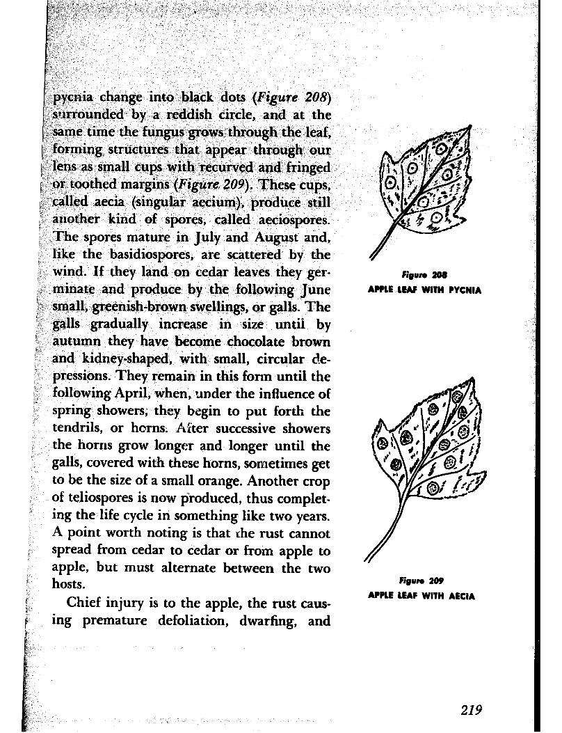
.i ;,#--I ,“: ._ ~~~~~~~_i~‘i’,,, (, ‘J * iI
,,“. ,;’ “.
&?, ‘I.. < i
$2’. i i, ;
,< ,’ i-7 _’ ’ ‘.’
f,F j
I:; ,
:., :,’
,,:.:
I, f:
PI; .,
i., I_
i: pycnia change into black dots (Figure 208) -surrounded by a reddish circle, and at the ,y<y ’ \~i,-same time the fungus grows through the leaf, r, !,: forming structures that appear through our “: glens as small cups with recurved and fringed 4 _ i v-or toothed margins (Figure 209). These cups, I.,.
/ ,called aecia (singular aecium), produce still F another kind of spores, called aeciospores. ; The spores mature in July and August and, i like the basidiospores, are scattered by the t wind. If they land on cedar leaves they ger- 1 minate and produce by the following June 1~. small, greenish-brown swellings, or galls. The )“ -1 galls gradually increase in size until by
autumn they have become chocolate brown and kidney-shaped, with small, circular de- pressions. They remain in this form until the
; following April, when, under the influence of spring showers, they begin to put forth the tendrils, or horns. After successive showers the horns grow longer and longer until the galls, covered with these horns, sometimes get to be the size of a small orange. Another crop of teliospores is now $roduced, thus complet- ing the life cycle in something like two years.
F;oun 208
APPLE LEAF WITH PYCNIA
A point worth noting is that the rust cannot spread from cedar to cedar or from apple to apple, but must alternate between the two hosts. Fi~un 209
f @ Chief injury is to the apple, the rust caus-
APPLE LEAF WITH AECIA
ing premature defoliation, dwarfing, and
219

fruit of poor quality. Control methods, how- ever, have reduced infection on orchard trees. Most damage, today, is to crab apples planted as ornamentals.
220


![5025 Game List - highscoresave.com · 76. Adventures of Lolo 2 (USA) 77. Adventures of Lolo 3 (USA) 78. Adventures of Lolo (USA) 79. Adventures of Mighty Max, The (U) [!] 80. Adventures](https://static.fdocuments.in/doc/165x107/601b936f675dc07ea30da387/5025-game-list-76-adventures-of-lolo-2-usa-77-adventures-of-lolo-3-usa-78.jpg)

