
Advantages and disadvantages of 3-dimensional …...Advantages and disadvantages of 3-dimensional...
16
Innovation Advantages and disadvantages of 3-dimensional printing in surgery: A systematic review Nicolas Martelli, PharmD, PhD, a,b Carole Serrano, PharmD, a H el ene van den Brink, PharmD, PhD, b Judith Pineau, PharmD, a Patrice Prognon, PharmD, PhD, a Isabelle Borget, PharmD, PhD, b,c and Salma El Batti, MD, d,e Paris, Ch^ atenay-Malabry, and Villejuif, France Background. Three-dimensional (3D) printing is becoming increasingly important in medicine and especially in surgery. The aim of the present work was to identify the advantages and disadvantages of 3D printing applied in surgery. Methods. We conducted a systematic review of articles on 3D printing applications in surgery published between 2005 and 2015 and identified using a PubMed and EMBASE search. Studies dealing with bioprinting, dentistry, and limb prosthesis or those not conducted in a hospital setting were excluded. Results. A total of 158 studies met the inclusion criteria. Three-dimensional printing was used to produce anatomic models (n = 113, 71.5%), surgical guides and templates (n = 40, 25.3%), implants (n = 15, 9.5%) and molds (n = 10, 6.3%), and primarily in maxillofacial (n = 79, 50.0%) and orthopedic (n = 39, 24.7%) operations. The main advantages reported were the possibilities for preoperative planning (n = 77, 48.7%), the accuracy of the process used (n = 53, 33.5%), and the time saved in the operating room (n = 52, 32.9%); 34 studies (21.5%) stressed that the accuracy was not satisfactory. The time needed to prepare the object (n = 31, 19.6%) and the additional costs (n = 30, 19.0%) were also seen as important limitations for routine use of 3D printing. Conclusion. The additional cost and the time needed to produce devices by current 3D technology still limit its widespread use in hospitals. The development of guidelines to improve the reporting of experience with 3D printing in surgery is highly desirable. (Surgery 2016;159:1485-500.) From the Pharmacy Department, a Georges Pompidou European Hospital, Paris; University Paris-Sud, b GRADES, Faculty of Pharmacy, Ch^ atenay-Malabry; Department of Health Economics, c Gustave Roussy Institute, Villejuif; Department of Cardiac and Vascular Surgery, d Georges Pompidou European Hospital; and URDIA - Unit e de Recherche en D eveloppement, e Imagerie et Anatomie - EA 4465, Universit e Paris Descartes, Paris, France THREE-DIMENSIONAL (3D) PRINTING, also known as ad- ditive manufacturing or rapid prototyping, is described frequently as a technical and industrial revolution that might significantly change the way we live. This manufacturing method is based on 3D computer models for the reconstruction of a 3D object by the addition of material layers, such as plaster, metal, plastic, and so on. 1 The concept of 3D printing actually began as “stereoli- thography” in the early 1980s by Charles W. Hull, who started selling the first 3D printers for com- mercial applications in 1988. 2 Stereolithography enables the creation of an object, usually by curing a photoreactive resin with a ultraviolet laser in a layer-by-layer fashion. Since then, different 3D printing processes have been developed for many applications; in the field of medicine, the numbers of such applications have increased dramatically since the early 2000s. 3 In health care, three 3D printing processes--- selective laser sintering (SLS), fused deposition modeling, and inkjet printing 1 ---have emerged and almost overtaken stereolithography in terms of fre- quency of use. The first of these, SLS, uses a laser to selectively fuse together particles of powdered material within a powder bed. Lowering of the Funding: None. Accepted for publication December 11, 2015. Reprint requests: Nicolas Martelli, PharmD, PhD, Pharmacy Department, Georges Pompidou European Hospital, AP-HP, 20, rue Leblanc, 75015 Paris, France. E-mail: nicolas.martelli@ aphp.fr . 0039-6060/$ - see front matter Ó 2016 Elsevier Inc. All rights reserved. http://dx.doi.org/10.1016/j.surg.2015.12.017 SURGERY 1485
Transcript of Advantages and disadvantages of 3-dimensional …...Advantages and disadvantages of 3-dimensional...

Innovation
Funding
Accepte
ReprintDepartm20, rueaphp.fr.
0039-60
� 2016
http://d
Advantages and disadvantages of3-dimensional printing insurgery: A systematic review
Nicolas Martelli, PharmD, PhD,a,b Carole Serrano, PharmD,a H�el�ene van den Brink, PharmD, PhD,bJudith Pineau, PharmD,a Patrice Prognon, PharmD, PhD,a Isabelle Borget, PharmD, PhD,b,c andSalma El Batti, MD,d,e Paris, Chatenay-Malabry, and Villejuif, France
Background. Three-dimensional (3D) printing is becoming increasingly important in medicine andespecially in surgery. The aim of the present work was to identify the advantages and disadvantages of3D printing applied in surgery.Methods. We conducted a systematic review of articles on 3D printing applications in surgery publishedbetween 2005 and 2015 and identified using a PubMed and EMBASE search. Studies dealing withbioprinting, dentistry, and limb prosthesis or those not conducted in a hospital setting were excluded.Results. A total of 158 studies met the inclusion criteria. Three-dimensional printing was used toproduce anatomic models (n = 113, 71.5%), surgical guides and templates (n = 40, 25.3%), implants(n = 15, 9.5%) and molds (n = 10, 6.3%), and primarily in maxillofacial (n = 79, 50.0%) andorthopedic (n = 39, 24.7%) operations. The main advantages reported were the possibilities forpreoperative planning (n = 77, 48.7%), the accuracy of the process used (n = 53, 33.5%), and the timesaved in the operating room (n = 52, 32.9%); 34 studies (21.5%) stressed that the accuracy was notsatisfactory. The time needed to prepare the object (n = 31, 19.6%) and the additional costs (n = 30,19.0%) were also seen as important limitations for routine use of 3D printing.Conclusion. The additional cost and the time needed to produce devices by current 3D technology stilllimit its widespread use in hospitals. The development of guidelines to improve the reporting of experiencewith 3D printing in surgery is highly desirable. (Surgery 2016;159:1485-500.)
From the Pharmacy Department,a Georges Pompidou European Hospital, Paris; University Paris-Sud,b
GRADES, Faculty of Pharmacy, Chatenay-Malabry; Department of Health Economics,c Gustave RoussyInstitute, Villejuif; Department of Cardiac and Vascular Surgery,d Georges Pompidou European Hospital;and URDIA - Unit�e de Recherche en D�eveloppement,e Imagerie et Anatomie - EA 4465, Universit�e ParisDescartes, Paris, France
THREE-DIMENSIONAL (3D) PRINTING, also known as ad-ditive manufacturing or rapid prototyping, isdescribed frequently as a technical and industrialrevolution that might significantly change theway we live. This manufacturing method is basedon 3D computer models for the reconstructionof a 3D object by the addition of material layers,such as plaster, metal, plastic, and so on.1 The
: None.
d for publication December 11, 2015.
requests: Nicolas Martelli, PharmD, PhD, Pharmacyent, Georges Pompidou European Hospital, AP-HP,Leblanc, 75015 Paris, France. E-mail: nicolas.martelli@
60/$ - see front matter
Elsevier Inc. All rights reserved.
x.doi.org/10.1016/j.surg.2015.12.017
concept of 3D printing actually began as “stereoli-thography” in the early 1980s by Charles W. Hull,who started selling the first 3D printers for com-mercial applications in 1988.2 Stereolithographyenables the creation of an object, usually by curinga photoreactive resin with a ultraviolet laser in alayer-by-layer fashion. Since then, different 3Dprinting processes have been developed for manyapplications; in the field of medicine, the numbersof such applications have increased dramaticallysince the early 2000s.3
In health care, three 3D printing processes---selective laser sintering (SLS), fused depositionmodeling, and inkjet printing1---have emerged andalmost overtaken stereolithography in terms of fre-quency of use. The first of these, SLS, uses a laserto selectively fuse together particles of powderedmaterial within a powder bed. Lowering of the
SURGERY 1485

SurgeryJune 2016
1486 Martelli et al
powder bed by 1 layer thickness allows the sequen-tial building of an object layer by layer. The secondof these processes, fused deposition modeling, isbased on the same principle as classic2-dimensional printing and deposits small beadsof thermoplastic material in layers to eventuallybuild up a 3D object. Finally, inkjet printing alsouses a printhead that deposits thermally or me-chanically droplets of “material ink” layer by layerto form the object. By virtue of its high resolution,inkjet printing is considered currently the mostsuitable technique for “bioprinting,” which isapplied in regenerative medicine to produce tis-sues and organs.4 Bioprinting represents a hugestep forward in tissue engineering and is likely toenable the repair of anatomic defects and thereconstruction of complex organs in the foresee-able future.5 In the present review, we focus onnonbiologic printing techniques for which thedata are most extensive currently.
Many reviews have reported advantages anddisadvantages of 3D printing in medicine.1,6-13
Among the advantages, 3D printing techniquescan be used in indications, such as preoperativeplanning, implant designing, training, and/or ed-ucation. Specifically in surgical applications, thesetechniques have been described to provide a betterunderstanding of complex anatomy/morphologyor the possibility to create customized implantsor surgical guides. Among the disadvantages iden-tified, the required time and cost of the techniquesare seen as the most important limitations.Although techniques of 3D printing are usedincreasingly in surgery, the advantages and disad-vantages of their use remain to be investigated.Indeed, in view of the remarkable developmentof 3D printing over the last decade, close attentionmust now be devoted to detailing and ranking thisdistribution, especially in surgery. Such informa-tion would help surgical teams intending todevelop 3D printing for in-house manufacturing.
The aim of the present systematic review was toidentify the advantages and disadvantages reportedover the last 10 years, on the use of 3D printingtechniques in surgery. We then analyzed theirdistribution and discussed their relative occur-rence in the literature. We focused on surgeryand excluded dental surgery from the search.
MATERIALS AND METHODS
Study selection. The Preferred Reporting Itemsfor Systematic Reviews andMeta-Analyses (PRISMA)guidelines were followed to perform the systematicreview. The PRISMA checklist is available as
supporting information (Supplementary Table I).In addition, a studyprotocolwasestablished to clarifythe review questions and eligibility criteria(Supplementary Table II). The systematic searchwas performed on PubMed and EMBASE to collectclinical studies on 3D printing applications in sur-gery in a hospital context. The search terms usedare presented in the study protocol(Supplementary Table II). Limits were defined onlanguage and publication date; only reports inFrench or English published between February2005 and February 2015 were considered. Titlesand abstracts were screened independently by 2 re-viewers to exclude irrelevant or duplicate abstracts.Exclusion criteria were study reviews, fundamentalresearch studies or those with no hospital applica-tion, and those involving bioprinting, dentistry, andlimb prosthesis. Then, included articles underwenta full-text review. Exclusion criteria were the sameas in the first step. When the full text of the publica-tion was not available online, the article was re-quested directly from the corresponding author.
Data analysis. A data extraction form wasdeveloped in Microsoft Office Excel 2010 tostandardize extraction and analysis of data.Collected information included first author anddate of publication, country, study design andnumber of patients, medical field, application(ie, anatomic 3D printed models, surgical guideand templates, etc), printing technique, and ma-terial used. After a first reading of every articleretrieved, 2 researchers (N.M. and C.S.) drew up alist of categories able to regroup the variousadvantages and disadvantages identified in thestudies (Table I). To prevent problems relating tothe variability of terms denoting similar items,the categories and their definition were given inthe study protocol. The 2 researchers referred tothis protocol and grouped independently and un-der 1 single category all identified advantagesand disadvantages concerning 3D printing applica-tions. Data were reported in a spreadsheet (Micro-soft Office Excel 2010).
RESULTS
Study selection. After excluding duplicates,1,067 studies were identified; 794 were excludedbased on the content of their titles and abstracts.The remaining 273 studies were considered intheir entirety, after which a further 115 wereexcluded. Thus, a total of 158 studies met theselection criteria and were suitable for completeanalysis (Fig 1). The number of published studiesshowed a regular increase year by year between
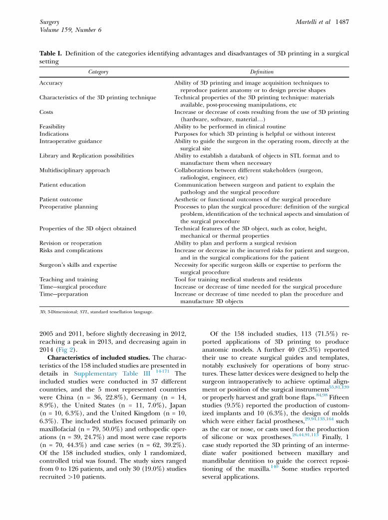
Table I. Definition of the categories identifying advantages and disadvantages of 3D printing in a surgicalsetting
Category Definition
Accuracy Ability of 3D printing and image acquisition techniques toreproduce patient anatomy or to design precise shapes
Characteristics of the 3D printing technique Technical properties of the 3D printing technique: materialsavailable, post-processing manipulations, etc
Costs Increase or decrease of costs resulting from the use of 3D printing(hardware, software, material.)
Feasibility Ability to be performed in clinical routineIndications Purposes for which 3D printing is helpful or without interestIntraoperative guidance Ability to guide the surgeon in the operating room, directly at the
surgical siteLibrary and Replication possibilities Ability to establish a databank of objects in STL format and to
manufacture them when necessaryMultidisciplinary approach Collaborations between different stakeholders (surgeon,
radiologist, engineer, etc)Patient education Communication between surgeon and patient to explain the
pathology and the surgical procedurePatient outcome Aesthetic or functional outcomes of the surgical procedurePreoperative planning Processes to plan the surgical procedure: definition of the surgical
problem, identification of the technical aspects and simulation ofthe surgical procedure
Properties of the 3D object obtained Technical features of the 3D object, such as color, height,mechanical or thermal properties
Revision or reoperation Ability to plan and perform a surgical revisionRisks and complications Increase or decrease in the incurred risks for patient and surgeon,
and in the surgical complications for the patientSurgeon’s skills and expertise Necessity for specific surgeon skills or expertise to perform the
surgical procedureTeaching and training Tool for training medical students and residentsTime---surgical procedure Increase or decrease of time needed for the surgical procedureTime---preparation Increase or decrease of time needed to plan the procedure and
manufacture 3D objects
3D, 3-Dimensional; STL, standard tessellation language.
SurgeryVolume 159, Number 6
Martelli et al 1487
2005 and 2011, before slightly decreasing in 2012,reaching a peak in 2013, and decreasing again in2014 (Fig 2).
Characteristics of included studies. The charac-teristics of the 158 included studies are presented indetails in Supplementary Table III 14-171 Theincluded studies were conducted in 37 differentcountries, and the 5 most represented countrieswere China (n = 36, 22.8%), Germany (n = 14,8.9%), the United States (n = 11, 7.0%), Japan(n = 10, 6.3%), and the United Kingdom (n = 10,6.3%). The included studies focused primarily onmaxillofacial (n = 79, 50.0%) and orthopedic oper-ations (n = 39, 24.7%) and most were case reports(n = 70, 44.3%) and case series (n = 62, 39.2%).Of the 158 included studies, only 1 randomized,controlled trial was found. The study sizes rangedfrom 0 to 126 patients, and only 30 (19.0%) studiesrecruited >10 patients.
Of the 158 included studies, 113 (71.5%) re-ported applications of 3D printing to produceanatomic models. A further 40 (25.3%) reportedtheir use to create surgical guides and templates,notably exclusively for operations of bony struc-tures. These latter devices were designed to help thesurgeon intraoperatively to achieve optimal align-ment or position of the surgical instruments55,81,139
or properly harvest and graft bone flaps.84,98 Fifteenstudies (9.5%) reported the production of custom-ized implants and 10 (6.3%), the design of moldswhich were either facial prostheses,29,94,133,164 suchas the ear or nose, or casts used for the productionof silicone or wax prostheses.26,44,91,113 Finally, 1case study reported the 3D printing of an interme-diate wafer positioned between maxillary andmandibular dentition to guide the correct reposi-tioning of the maxilla.140 Some studies reportedseveral applications.

Fig 1. Preferred Reporting Items for Systematic Reviews and Meta-Analyses flow chart of included studies. 3D, 3-Dimensional.
SurgeryJune 2016
1488 Martelli et al
Concerning techniques used, stereolithographywas used in 41 of the included studies (25.9%),SLS in 32 (20.3%), inkjet printing in 28 (17.7%),fused deposition modeling in 22 (13.9%), directmetal laser sintering in 2 (1.3%), and powderdepositional modeling in 2 (1.3%). The techniquewas not stated clearly in 42 studies (26.6%). Some
studies reported the use of several techniques.Among these studies, 2 specified only 1 of thetechniques used.90,135 The distribution of materialsrequired according to the 3D-printing techniqueused is presented in Table II. Of the 158 studies,24 (15.2%) provided no information on eitherthe material or the technique used.

Fig 2. Timeline of studies included.
SurgeryVolume 159, Number 6
Martelli et al 1489
Data synthesis. Global distribution of advantagesand disadvantages is presented in Tables III and IV,respectively. Their distribution according to thetechnique used and the application reported ispresented in details in Supplementary Table IV.Illustrative examples are highlighted below. Formore exhaustive references, the reader can referto Supplementary Table IV. The most reportedadvantage of 3D printing in the included studiesinvolved the new possibilities for planning the surgi-cal procedure (n = 77, 48.7%). Several surgeonsstated that the printed model gave a better impres-sion of the anatomic characteristics48,66,67,85,112,114
and then facilitated the preoperative planningby visualization of potential difficulties and/oranatomic variations.17,36,67,79,103,142,144,152,154,159,162
Whatever the 3D printing technique used, manystudies underlined that the technique was veryhelpful for the preoperative planning in complexanatomies.23,67,68,112,146 This model allowed the sur-gical team to select the most suitable implants and/or devices for the procedure.30,45,47,99,100,103,123,159
Surgeons could also anticipate difficulties thatmay arise by simulating the operative proce-dure.27,106,123,124,146,150,152 Many surgeons appre-ciated the “hands-on” aspect provided by thephysical model.51,66,67,138,145 In addition, 1 studyspecified that the preoperative simulation with aphysical model printed with SLS was easier thanthat allowed with surgical navigation systems.90
The accuracy of 3D printing techniques wasseen as a major advantage in many studies (n = 53,33.5%). Only one of the included studiescompared the accuracy of 3 different 3D printingtechniques: inkjet, SLS, and a third technique notstated clearly, but possibly the powder bed tech-nique.135 The inkjet technique was found to pre-sent greater accuracy compared with the othertwo. Elsewhere, whatever technique was used, 3Dprinting allowed the generation of precise implantshapes that fitted perfectly to the anatomic site143
or copied exactly the shape of the defect they cor-rected.70,75 The consequential lack of implantcorrection required before its implantation inturn facilitated the surgical procedure andincreased its accuracy.37,49 A similarly increased ac-curacy of surgical guides and templates obtainedwith 3D printing techniques improved the preci-sion and positioning of incisions.31,84 This advan-tage was highlighted particularly in bonyreconstruction with osteotomy.58,132 The use ofan accurate anatomic model also enabled betterpreforming of implants and to better assess screwtrajectories.76 An excellent level of symmetryachieved in bony reconstruction was alsoreported.34,47,170
Many studies (n = 52, 32.9%) underlined adecrease in operating time by using 3D printing.Thirty-eight studies (24.1%) reported that use of3D-printed anatomic models improved preopera-tive planning, thereby contributing to a decreasein operating time. Indeed, the anatomic modelsenabled preforming implants such as orthopedicplates and also to anticipate the anatomic diffi-culties.61,70,73,86,95 In addition, several studies re-ported that the use of surgical guides andtemplates (n = 17, 10.8%) permitted a moretime-efficient surgical procedure. The use of a3D-printed surgical guide was also considered afaster approach than use of an image-guided tech-nique.99,100 Of the 52 studies reporting a decreasein operating time, only a few actually quantifiedthe time saved.40,67,86,162,168,171 In a comparativestudy reporting the use of surgical guides in com-parison with conventional surgery, the mean timesaved was estimated at 5.7 minutes per procedurefrom a cohort of 22 patients.168 One case series re-ported an average saving of 25.2 minutes per pro-cedure with the use of an anatomic model,86 anda second estimated the time saved to be 63 minutesper case.67
Forty-eight studies (30.4%) pointed out thedecreased level of risk and number of postopera-tive complications that resulted from the use of 3Dprinting. Whatever the application, use of 3Dprinting technology contributed to improving sur-gical patient safety by decreasing morbid-ities.14,21,40,44,51,61,67,84,89,96,136,155,161 Many studiesalso reported a decrease in blood loss and transfu-sion requirements.37,39,55,163 Also highlighted was adecrease in the radiologic exposure of patients andthe surgical team allowed with the use of a surgicalguide, which facilitated not only the placement ofa device, but also decreases the need for intraoper-ative radiographic imaging to guide the sur-geon.43,99 In osseous surgeries, the use of
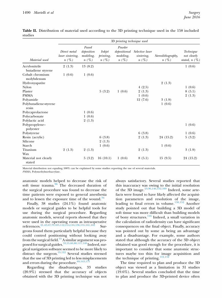
Table II. Distribution of material used according to the 3D printing technique used in the 158 includedstudies
Material used
3D printing technique used
Direct metallaser sintering,
n (%)
Fuseddepositionmodeling,n (%)
Inkjetprinting,n (%)
Powderdepositionalmodeling,n (%)
Selective lasersintering,n (%)
Stereolithography,n (%)
Techniquenot clearly
stated, n (%)
Acrylonitrilebutadiene styrene
2 (1.3) 13 (8.2) 1 (0.6)
Cobalt chromiummolybdenum
1 (0.6) 1 (0.6)
Hydroxyapatite 2 (1.3)Nylon 4 (2.5) 1 (0.6)Plaster 5 (3.2) 1 (0.6) 2 (1.3) 8 (5.1)PMMA 1 (0.6) 2 (1.3)Polyamide 12 (7.6) 3 (1.9)Polybutadiene-styrene
resin1 (0.6)
Polycaprolactone 1 (0.6)Polycarbonate 1 (0.6)Polylactic acid 2 (1.3)Polypropylene–
polyester1 (0.6)
Polystyrene 6 (3.8) 1 (0.6)Resin (acrylic) 6 (3.8) 2 (1.3) 24 (15.2) 5 (3.2)Silicone 2 (1.3)Starch 1 (0.6) 1 (0.6)Titanium 2 (1.3) 2 (1.3) 2 (1.3) 3 (1.9)WaxMaterial not clearly
stated5 (3.2) 16 (10.1) 1 (0.6) 8 (5.1) 15 (9.5) 24 (15.2)
Material distribution not equaling 100% can be explained by some studies reporting the use of several materials.PMMA, Polymethylmethacrylate.
SurgeryJune 2016
1490 Martelli et al
anatomic models helped to decrease the risk ofsoft tissue trauma.96 The decreased duration ofthe surgical procedure was found to decrease thetime patients were exposed to general anesthesiaand to lessen the exposure time of the wound.34
Finally, 38 studies (24.1%) found anatomicmodels or surgical guides to be helpful tools foruse during the surgical procedure. Regardinganatomic models, several reports showed that theywere used in the operating room as intraoperativereferences.21,43,67,79,105,112,114,116,136,155,161,162 Sur-geons found them particularly helpful because theycould control positioning without looking awayfrom the surgical field.55 A similar argument was pro-posed for surgical guides.33,55,60,80,111,148 Indeed, sur-gical navigation software seemed to bemore likely todistract the surgeon.55,112 Several studies stressedthat the use of 3D printing led to less misplacementsand errors during the procedure.89,143,171
Regarding the disadvantages, 33 studies(20.9%) stressed that the accuracy of objectsobtained with the 3D printing technique was not
always satisfactory. Several studies reported thatthis inaccuracy was owing to the initial resolutionof the 3D image.23,96,146,152,169 Indeed, some arte-facts were found to have likely affected the acquisi-tion parameters and resolution of the image,leading to final errors in volume.138,152 Anotherstudy pointed out that building a 3D model ofsoft tissue was more difficult than building modelsof bony structures.116 Indeed, a small variation inthe calculation of radiodensity can have significantconsequences on the final object. Finally, accuracywas pointed out by some as being an advantageand a disadvantage. For example, some authorsstated that although the accuracy of the 3D objectobtained was good enough for the procedure, it isimportant to consider that some anatomic struc-tures maybe too thin for image acquisition andthe technique of printing.112,152
The time required to plan and produce the 3Dobject was viewed as a limitation in 31 studies(19.6%). Several studies concluded that the timeto plan and produce the 3D-printed device often

Table III. Global distribution of advantages reported in the 158 included studies
Category
Advantages
n (%) Examples
Preoperative planning 77 (48.7) Direct visualization of malformationsBetter anticipation of anatomic difficulties
Accuracy 53 (33.5) Precise implant shapesAccurate guides and templatesNo need for correction or manipulation of the model
Time---surgical procedure 52 (32.9) Decreased operating timeIncreased time-efficiency of the surgical procedure
Risks and complications 48 (30.4) Decreased incidence of postoperative complications suchas blood loss, infection, etc
Decreased radiologic exposure of patients during thesurgical procedure
Intraoperative guidance 38 (24.1) Positioning improvementPatient outcome 25 (15.2) Better surgical results
Minimal posttreatment discomfortBetter aesthetic results
Costs 24 (15.2) Less cost per patientLess cost per implant/guide/model
Teaching and training 19 (12.0) Teaching and training toolsFeasibility 16 (10.1) No equipment required (external manufacturer)
Easy to integrate into the workflowProperties of the object obtained 15 (9.5) Good mechanical and thermal properties
Easy to work withTime---preparation 14 (8.9) Faster than conventional techniques for producing
implantsPatient education 13 (8.2) Improved transfer of information to patients
Improved communication with patientsCharacteristics of the 3D printing technique 10 (6.3) Alternative to 3D imaging techniques
Automated fabricationLibrary and Replication possibilities 7 (4.4) Creation of model library for replicationIndications 5 (3.2) Many indicationsSurgeon skills and expertise 4 (2.5) Less requirement for a surgeon expertiseMultidisciplinary approach 3 (1.9) Better coordination with other specialists allowedRevision or reoperation 2 (1.5) Ease of access for surgical revision or reoperation
3D, 3-Dimensional.
SurgeryVolume 159, Number 6
Martelli et al 1491
delayed the procedure and argued that this tech-nique may be unsuitable for use in emergencycases.28,54,70,143,171 Even when applied on a routinebasis, surgeons were required to anticipate well inadvance their preoperative planning and print-ing.68 Estimations of the time required for both vir-tual plan design and printing of an anatomicmodel varied with a range from 10 hours to 2weeks.68,77,98,114,124 Some studies only reportedthe time needed to print the model, which variedfrom 3 to 7 hours.69,124 One time-consumingelement even for trained operators seemed to bethe computer-aided design, which also requiredconsiderable involvement of the surgeon duringthe preoperative planning.73,98,114,124,143 A studyin orthopedic surgery claimed that the time tobend a plate in conventional surgery was
considerably shorter than the time needed to pre-pare a model.73
Numerous studies (n = 30, 19.0%) reported theadditional costs of 3D printing techniques as amajor disadvantage over conventional methods.The costs of the required equipment, such ascomputer-aided design software, camera, or the3D printing machine, were often viewed asa barrier to the use of the tech-nique.31,36,70,73,85,94,113,123,133 Only a few studies,however, actually estimated or discussed the addi-tional costs in different ways: additional cost perimplant, additional cost per patient, and soon.28,67,94,96,101,113,139,153,156 The additional costsper patient varied widely from 150 to 700V de-pending on the reported application.113,139,153
Some authors stressed that this cost was often
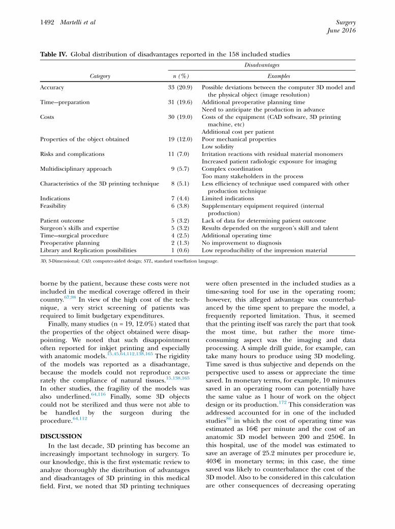
Table IV. Global distribution of disadvantages reported in the 158 included studies
Category
Disadvantages
n (%) Examples
Accuracy 33 (20.9) Possible deviations between the computer 3D model andthe physical object (image resolution)
Time---preparation 31 (19.6) Additional preoperative planning timeNeed to anticipate the production in advance
Costs 30 (19.0) Costs of the equipment (CAD software, 3D printingmachine, etc)
Additional cost per patientProperties of the object obtained 19 (12.0) Poor mechanical properties
Low solidityRisks and complications 11 (7.0) Irritation reactions with residual material monomers
Increased patient radiologic exposure for imagingMultidisciplinary approach 9 (5.7) Complex coordination
Too many stakeholders in the processCharacteristics of the 3D printing technique 8 (5.1) Less efficiency of technique used compared with other
production techniqueIndications 7 (4.4) Limited indicationsFeasibility 6 (3.8) Supplementary equipment required (internal
production)Patient outcome 5 (3.2) Lack of data for determining patient outcomeSurgeon’s skills and expertise 5 (3.2) Results depended on the surgeon’s skill and talentTime---surgical procedure 4 (2.5) Additional operating timePreoperative planning 2 (1.3) No improvement to diagnosisLibrary and Replication possibilities 1 (0.6) Low reproducibility of the impression material
3D, 3-Dimensional; CAD, computer-aided design; STL, standard tessellation language.
SurgeryJune 2016
1492 Martelli et al
borne by the patient, because these costs were notincluded in the medical coverage offered in theircountry.67,98 In view of the high cost of the tech-nique, a very strict screening of patients wasrequired to limit budgetary expenditures.
Finally, many studies (n = 19, 12.0%) stated thatthe properties of the object obtained were disap-pointing. We noted that such disappointmentoften reported for inkjet printing and especiallywith anatomic models.15,45,64,112,138,165 The rigidityof the models was reported as a disadvantage,because the models could not reproduce accu-rately the compliance of natural tissues.15,138,165
In other studies, the fragility of the models wasalso underlined.64,116 Finally, some 3D objectscould not be sterilized and thus were not able tobe handled by the surgeon during theprocedure.64,112
DISCUSSION
In the last decade, 3D printing has become anincreasingly important technology in surgery. Toour knowledge, this is the first systematic review toanalyze thoroughly the distribution of advantagesand disadvantages of 3D printing in this medicalfield. First, we noted that 3D printing techniques
were often presented in the included studies as atime-saving tool for use in the operating room;however, this alleged advantage was counterbal-anced by the time spent to prepare the model, afrequently reported limitation. Thus, it seemedthat the printing itself was rarely the part that tookthe most time, but rather the more time-consuming aspect was the imaging and dataprocessing. A simple drill guide, for example, cantake many hours to produce using 3D modeling.Time saved is thus subjective and depends on theperspective used to assess or appreciate the timesaved. In monetary terms, for example, 10 minutessaved in an operating room can potentially havethe same value as 1 hour of work on the objectdesign or its production.172 This consideration wasaddressed accounted for in one of the includedstudies86 in which the cost of operating time wasestimated as 16V per minute and the cost of ananatomic 3D model between 200 and 250V. Inthis hospital, use of the model was estimated tosave an average of 25.2 minutes per procedure ie,403V in monetary terms; in this case, the timesaved was likely to counterbalance the cost of the3D model. Also to be considered in this calculationare other consequences of decreasing operating
SurgeryVolume 159, Number 6
Martelli et al 1493
time, including a shortened anesthesia time, whichwould be expected generally to decrease therequirement for analgesics, lessen the risk of infec-tion, and thus possibly even decrease the need forthe use of antibiotics. It is very difficult to gener-alize such calculations across hospitals, however,because many other factors must also be consid-ered, such as the level of urgency, the type of surgi-cal procedure, the number of cases per year, thecountry, and so on.
The cost of the technique is also a majorlimitation reported in the included studies, whichis not specific to 3D printing techniques; indeed,the issue of cost is very often a source of concernwhen new and costly technologies are introducedinto medical practice.173 This point, however, islikely to evolve quite rapidly over the coming yearswith the decreasing cost of 3D printing.
This cost is not the only barrier to the expandedimplementation of the technique in hospitals.Indeed, we noted that the organizational impactwas an issue for several surgical teams who stressedthat the cooperation between many stakeholderswas complex and was a hurdle to the use of thetechnique. Indeed, 3D software requires specificskills that most surgeons do not have. Consideringthe huge responsibility played by surgeons at thecritical stage of preoperative planning174 to ensurethe outcome of their patients, some surgeons mayexperience a fear of losing control over the deci-sions that affect their patients. In contrast,perceived benefits of 3D printing to preoperativeplanning were reported in nearly one-half of theincluded studies. The improved understanding ofpatient-specific, 3D anatomy offered by 3D print-ing was found to allow surgeons to anticipatepossible problems that might arise during theoperative procedure and thus potentially improvepatient outcome. It seems to be important in thiscontinually evolving and highly specialized fieldthat surgeons accept the support of external tech-nicians without fearing loss over their leader-ship.175 Changes in this direction could occurrapidly with a substantial improvement in theaccessibility of 3D modeling software.176
The high accuracy of the object obtained wasvalued in one-third of the included studies, what-ever 3D printing technique was used. We notedthat “accuracy” was difficult to estimate objectivelyin most studies, which led some authors toconsider the resultant accuracy of the deviceboth as an advantage and a disadvantage of 3Dprinting. Because only 1 study provided a directcomparison of different 3D printing techniques, itis difficult to say conclusively whether inkjet
printing is, as they found, more accurate thanany other techniques for surgical purposes.135 Thehigh accuracy of inkjet printing was stated else-where, making it the preferred technique for bio-printing3,177; however, we also observed thatmany users found the mechanical properties ofmodels obtained with inkjet printing disap-pointing, because they did not allow easy handlingor a correct simulation of the procedure. The qual-ity of the 3D-printed physical model seems to notonly depend on the accuracy of the 3D printingtechnique, but also and sometimes more impor-tantly on the 3D image resolution, the errors ofwhich likely influence the accuracy of the ob-ject.114 The resolution can also be affected by the3D slicing software during the segmentation step.Another limitation raised was that most techniquesdo not enable a precise reproduction of both hardand soft tissues. In procedures involving bonystructures, the process of 3D printing can lead toloss of information on soft tissue disease or on vitalsurrounding tissues, such as arteries or nerves,associated with the bony structures.101 Neverthe-less, this issue may be overcome successfully usingmultimaterial 3D printers or color models to simu-late the different tissues.156 Finally, postprintingsteps, such as cleaning, finishing, and sterilization,are essential to provide suitable and flawless 3D-printed physical objects to surgical teams. The ster-ilization process chosen depends on the materialused to build the object; for example, polylacticacid is not resistant to high temperatures andcannot be autoclaved, thus presenting a criticalissue for intraoperative purposes. According tothe shape and/or size of the printed object, finish-ing can be indispensable to remove extra materialor support material around the object achievedeither manually or chemically, depending on the3D printing technique or the material used.
Surprisingly, of the 158 studies included, fewstudies reported the use of 3D printing techniquesin the fabrication of customized implants. In ourview, this observation underlines that fact 3Dprinting is still in its infancy for this surgicalpurpose. Indeed, designing and producingimplantable devices is much more challengingthan designing and producing anatomic modelsor surgical guides, which are only used beforeand/or during the surgical procedure. This isparticularly true for hospitals deciding to producecustomized implants in house, not only in terms ofthe equipment required but also the technicalexpertise not necessarily available within theirinstitution. As a result, 3D printing seems to beoutsourced frequently to an external company for
SurgeryJune 2016
1494 Martelli et al
this application, using often expensive and com-plex techniques that hospitals may not be able toafford in the current financial environment.177 Aspart of our review, we noted for example that anexternal company manufactured customized tita-nium meshes using direct metal laser sintering,which is a very costly process.30,178 In addition,some regulatory considerations in the design andmanufacturing of implantable 3D-printed devicesremain very challenging for hospitals.179 Indeed,the quality controls likely to be imposed by thehealth authorities to ensure safety and sustainabil-ity of the 3D printed products will put added pres-sure on hospitals aiming to manufacture in-houseimplants that meet the regulatory standards. TheFood and Drug Administration is exploringcurrently ways of developing new standards thatwould take into account differences between tradi-tional and additive (3D printing) manufacturing,and also the issue of the in-house manufacturingin hospitals.180 Finally, our findings suggest thatto date all 3D-printed implants have been usedonly in osseous operations. More reports on appli-cations for soft tissue surgeries might have been ex-pected, because custom-made implants inendovascular surgery, for example, tend to providebetter functional outcomes than standard implantsin patients with complex anatomy.181 Modelingmechanical constraints with devices such as endo-vascular stent grafts, however, remains complicatedand requires consistently the expertise of biome-chanical engineers. The current technical limitsof 3D printing could explain the still rare applica-tion of this technology in soft tissue operations.This will very likely may change with the develop-ment of bioprinting, which will provide additionalpossibilities for such operations.
We observed a majority of case reports and caseseries, and only 1 randomized, controlled trialamong the included studies, which is not verysurprising to date. These studies are important forhypothesis generation and enable the collection ofimportant data on a new subject182; however, case re-ports and case series are not enough to demonstratethe benefits to surgical procedures of using one 3Dprinting technique in comparison with conventionalsurgery alone or to that procedure performed usinganother 3D printing technique. Although it isimportant to not consider randomized, controlledtrials as the only valid source of evidence, the levelof evidence could be improved with well-designedand conducted cohort or case-control studies.183
In addition, we urge authors to expand on the infor-mation provided concerning the technique and/or
on the material used in all case reports and futurecase series. The inadequate level of informationon the 3D printing technique found in many ofthe case report and series included in our systematicreview was owing probably to most authors beingsurgeons who are more focused on the results ob-tained and the surgical procedure than on the tech-nical details of 3D printing. Guidelines to improvethe reporting of experience with 3D printing in sur-gery could be developed that specify compulsoryitems, such as the technique used, the 3D printermodel, the material, the resolution and so on. Theresulting standardized reporting would help to in-crease knowledge on the applications of 3D printingtechnique in surgery and improve the quality ofpublished studies.
This systematic review has limitations that needmentioning. First, we did not retrieve in full-text allthe articles we identified. Although we made everyeffort to collect the articles and contacted directlythe corresponding authors where necessary, somearticles are still missing. In addition, we did notinclude unpublished studies, which may haveintroduced some bias, although the quality andlevel of information provided within such studiesmay be questionable. Our potential overestimationor underestimation of the occurrence of someadvantages/disadvantages cannot be excluded.Indeed, the collection of every item was a time-consuming and laborious exercise. Nevertheless,the 2 investigators attempted rigorously to recordall available information and used exactly the samemethod according to a study protocol. Finally, wenoted that the number of advantages reported on3D printing was twice that of reported disadvan-tages; however, this could be owing to a publicationbias with some authors or journal editors beingreluctant to publish negative findings.
In conclusion, until recently, only specializedand private manufacturers were able to produce3D printed medical devices in optimal conditionsand at a reasonable cost. This already has changedwith the availability of more affordable 3D printersand user-friendly 3D software that allow anincreasing number of health facilities to produce3D objects in house. The sharing between surgicalteams of the cost of hardware, software, andmaterial is, in our opinion, the best way to pro-mote the dissemination of this technology withinhospitals.
SUPPLEMENTARY DATA
Supplementary data related to this article can be foundat http://dx.doi.org/10.1016/j.surg.2015.12.017.

SurgeryVolume 159, Number 6
Martelli et al 1495
REFERENCES
1. Rengier F, Mehndiratta A, von Tengg-Kobligk H,Zechmann CM, Unterhinninghofen R, Kauczor H-U,et al. 3D printing based on imaging data: review ofmedical applications. Int J Comput Assist Radiol Surg2010;5:335-41.
2. Gross BC, Erkal JL, Lockwood SY, Chen C, Spence DM.Evaluation of 3D printing and its potential impact onbiotechnology and the chemical sciences. Anal Chem2014;86:3240-53.
3. Ventola CL. Medical applications for 3D printing: currentand projected uses. P T 2014;39:704-11.
4. Cui X, Boland T, D’Lima DD, Lotz MK. Thermal inkjetprinting in tissue engineering and regenerative medicine.Recent Pat Drug Deliv Formul 2012;6:149.
5. Chia HN, Wu BM. Recent advances in 3D printing of bio-materials. J Biol Eng 2015;9:4.
6. Kim MS, Hansgen AR, Carroll JD. Use of rapid prototypingin the care of patients with structural heart disease. TrendsCardiovasc Med 2008;18:210-6.
7. Webb PA. A review of rapid prototyping (RP) techniquesin the medical and biomedical sector. J Med Eng Technol2000;24:149-53.
8. McGurk M, Amis AA, Potamianos P, Goodger NM. Rapidprototyping techniques for anatomical modelling in med-icine. Ann R Coll Surg Engl 1997;79:169-74.
9. Torabi K, Farjood E, Hamedani S. Rapid prototyping tech-nologies and their applications in prosthodontics, a reviewof literature. J Dent (Shiraz) 2015;16:1-9.
10. Hoarau R, Zweifel D, Lanthemann E, Zrounba H,Broome M. [3D planning in maxillofacial surgery]. RevMed Suisse 2014;10:1829-30;1832–3.
11. Horn TJ, Harrysson OLA. Overview of current additivemanufacturing technologies and selected applications.Sci Prog 2012;95(Pt 3):255-82.
12. Malik HH, Darwood ARJ, Shaunak S, Kulatilake P, El-Hilly AA, Mulki O, et al. Three-dimensional printing insurgery: a review of current surgical applications. J SurgRes 2015;199:512-22.
13. Farr�e-Guasch E, Wolff J, Helder MN, Schulten EAJM,Forouzanfar T, Klein-Nulend J. Application of additivemanufacturing in oral and maxillofacial surgery. J OralMaxillofac Surg 2015;73:2408-18.
14. Abdel-Moniem Barakat A, Abou-ElFetouh A, Hakam MM,El-Hawary H, Abdel-Ghany KM. Clinical and radiographicevaluation of a computer-generated guiding device inbilateral sagittal split osteotomies. J CraniomaxillofacSurg 2014;42:e195-203.
15. Abdel-Sayed P, Kalejs M, von Segesser LK. A new trainingset-up for trans-apical aortic valve replacement. InteractCardiovasc Thorac Surg 2009;8:599-601.
16. Al-Ahmad HT, M Saleh MW, Hussein AM. Evaluation ofan innovative computer-assisted sagittal split ramus os-teotomy to reduce neurosensory alterations following or-thognathic surgery: a pilot study. Int J Med Robot 2013;9:134-41.
17. Arora A, Datarkar AN, Borle RM, Rai A, Adwani DG.Custom-made implant for maxillofacial defects usingrapid prototype models. J Oral Maxillofac Surg 2013;71:e104-10.
18. Ayoub N, Ghassemi A, Rana M, Gerressen M, Riediger D,H€olzle F, et al. Evaluation of computer-assisted mandibularreconstruction with vascularized iliac crest bone graftcompared to conventional surgery: a randomized prospec-tive clinical trial. Trials 2014;15:114.
19. Azuma M, Yanagawa T, Ishibashi-Kanno N, Uchida F, Ito T,Yamagata K, et al. Mandibular reconstruction using platesprebent to fit rapid prototyping 3-dimensional printingmodels ameliorates contour deformity. Head Face Med2014;10:45.
20. Bai S, Bo B, Bi Y, Wang B, Zhao J, Liu Y, et al. CAD/CAMsurface templates as an alternative to the intermediatewafer in orthognathic surgery. Oral Surg Oral Med OralPathol Oral Radiol Endod 2010;110:e1-7.
21. Bellanova L, Paul L, Docquier P-L. Surgical guides (pa-tient-specific instruments) for pediatric tibial bone sar-coma resection and allograft reconstruction. Sarcoma2013;2013:787653.
22. Brie J, Chartier T, Chaput C, Delage C, Pradeau B, Caire F,et al. A new custom made bioceramic implant for therepair of large and complex craniofacial bone defects.J Craniomaxillofac Surg 2013;41:403-7.
23. Bruy�ere F, Leroux C, Brunereau L, Lermusiaux P. Rapidprototyping model for percutaneous nephrolithotomytraining. J Endourol 2008;22:91-6.
24. Bullock P, Dunaway D, McGurk L, Richards R. Integrationof image guidance and rapid prototyping technology incraniofacial surgery. Int J Oral Maxillofac Surg 2013;42:970-3.
25. Byrne PJ, Garcia JR. Autogenous nasal tip reconstruc-tion of complex defects: a structural approach employ-ing rapid prototyping. Arch Facial Plast Surg 2007;9:358-64.
26. Cheung CL, Looi T, Lendvay TS, Drake JM, Farhat WA.Use of 3-dimensional printing technology and siliconemodeling in surgical simulation: development and facevalidation in pediatric laparoscopic pyeloplasty. J SurgEduc 2014;71:762-7.
27. Ching W-C, Goh RCW, Lin C-L, Lo L-J, Chen Y-R.Aesthetic restoration of fronto-orbital deformity with pre-fabricated implant utilizing modeling clay and rapid-prototyping technology. Aesthetic Plast Surg 2011;35:1176-9.
28. Chow LK, Cheung LK. The usefulness of stereomodels inmaxillofacial surgical management. J Oral Maxillofac Surg2007;65:2260-8.
29. Ciocca L, De Crescenzio F, Fantini M, Scotti R. Rehabilita-tion of the nose using CAD/CAM and rapid prototypingtechnology after ablative surgery of squamous cell carci-noma: a pilot clinical report. Int J Oral Maxillofac Im-plants 2010;25:808-12.
30. Ciocca L, Fantini M, De Crescenzio F, Corinaldesi G,Scotti R. Direct metal laser sintering (DMLS) of a custom-ized titanium mesh for prosthetically guided bone regen-eration of atrophic maxillary arches. Med Biol EngComput 2011;49:1347-52.
31. Ciocca L, Fantini M, De Crescenzio F, Persiani F, Scotti R.Computer-aided design and manufacturing constructionof a surgical template for craniofacial implant positioningto support a definitive nasal prosthesis. Clin Oral ImplantsRes 2011;22:850-6.
32. Ciocca L, Mazzoni S, Fantini M, Persiani F, Marchetti C,Scotti R. CAD/CAM guided secondary mandibular recon-struction of a discontinuity defect after ablative cancer sur-gery. J Craniomaxillofac Surg 2012;40:e511-5.
33. Ciocca L, Marchetti C, Mazzoni S, Baldissara P, Gatto MRA,Cipriani R, et al. Accuracy of fibular sectioning and insertioninto a rapid-prototyped bone plate, for mandibular recon-struction using CAD-CAM technology. J CraniomaxillofacSurg 2015;43:28-33.
SurgeryJune 2016
1496 Martelli et al
34. Cohen A, Laviv A, Berman P, Nashef R, Abu-Tair J. Mandib-ular reconstruction using stereolithographic 3-dimensionalprinting modeling technology. Oral Surg Oral Med OralPathol Oral Radiol Endod 2009;108:661-6.
35. Cui J, Chen L, Guan X, Ye L, Wang H, Liu L. Surgical plan-ning, three-dimensional model surgery and preshapedimplants in treatment of bilateral craniomaxillofacialpost-traumatic deformities. J Oral Maxillofac Surg 2014;72:1138.e1-14.
36. da Rosa ELS, Oleskovicz CF, Arag~ao BN. Rapid prototyp-ing in maxillofacial surgery and traumatology: case report.Braz Dent J 2004;15:243-7.
37. Dai K-R, Yan M-N, Zhu Z-A, Sun Y-H. Computer-aidedcustom-made hemipelvic prosthesis used in extensive pel-vic lesions. J Arthroplasty 2007;22:981-6.
38. Day RE, Guy DT, Kop AM, Morrison DA. The Royal PerthHospital method for the design and manufacture of tita-nium cranioplasty plates. Br J Oral Maxillofac Surg 2012;50:376-7.
39. Deshmukh TR, Kuthe AM, Vaibhav B. Preplanning andsimulation of surgery using rapid modelling. J Med EngTechnol 2010;34:291-4.
40. Dhakshyani R, Nukman Y, Osman NAA, Vijay C. Prelimi-nary report: rapid prototyping models for Dysplastic hipsurgery. Cent Eur J Med 2011;6:266-70.
41. Dobbe JGG, Vroemen JC, Strackee SD, Streekstra GJ. Pa-tient-tailored plate for bone fixation and accurate 3D posi-tioning in corrective osteotomy. Med Biol Eng Comput2013;51:19-27.
42. Du H, Tian X, Li T, Yang J, Li K, Pei G, et al. Use of patient-specific templates in hip resurfacing arthroplasty: experi-ence from sixteen cases. Int Orthop 2013;37:777-82.
43. D’Urso PS, Williamson OD, Thompson RG. Biomodelingas an aid to spinal instrumentation. Spine 2005;30:2841-5.
44. Eggbeer D, Evans P. Computer-aided methods in bespokebreast prosthesis design and fabrication. Proc Inst MechEng H 2011;225:94-9.
45. Erbano BO, Opolski AC, Olandoski M, Foggiatto JA,Kubrusly LF, Dietz UA, et al. Rapid prototyping of three-dimensional biomodels as an adjuvant in the surgicalplanning for intracranial aneurysms. Acta Cir Bras 2013;28:756-61.
46. Faur C, Crainic N, Sticlaru C, Oancea C. Rapid prototyp-ing technique in the preoperative planning for total hiparthroplasty with custom femoral components. Wien KlinWochenschr 2013;125:144-9.
47. Feng F, Wang H, Guan X, Tian W, Jing W, Long J, et al.Mirror imaging and preshaped titanium plates in the treat-ment of unilateral malar and zygomatic arch fractures.Oral Surg Oral Med Oral Pathol Oral Radiol Endod2011;112:188-94.
48. Frame M, Huntley JS. Rapid prototyping in orthopaedicsurgery: a user’s guide. ScientificWorldJournal 2012;2012:838575.
49. Gerber N, Stieglitz L, Peterhans M, Nolte LP, Raabe A,Weber S. Using rapid prototyping molds to create pa-tient specific polymethylmethacrylate implants in cranio-plasty. Conf Proc IEEE Eng Med Biol Soc 2010;2010:3357-60.
50. Gong X, Yu Q. Correction of maxillary deformity in infantswith bilateral cleft lip and palate using computer-assisteddesign. Oral Surg Oral Med Oral Pathol Oral Radiol2012;114(5 Suppl):S74-8.
51. Guarino J, Tennyson S, McCain G, Bond L, Shea K,King H. Rapid prototyping technology for surgeries of
the pediatric spine and pelvis: benefits analysis. J PediatrOrthop 2007;27:955-60.
52. Guevara-Rojas G, Figl M, Schicho K, Seemann R,Traxler H, Vacariu A, et al. Patient-specific polyetherether-ketone facial implants in a computer-aided planning work-flow. J Oral Maxillofac Surg 2014;72:1801-12.
53. H�akansson A, Rantatalo M, Hansen T, Wanhainen A. Pa-tient specific biomodel of the whole aorta - the importanceof calcified plaque removal. VASA 2011;40:453-9.
54. Hammer B, Zizelmann C, Scheufler K. Solid modeling insurgery of the anterior skull base. J Oral Maxillofac Surg2010;21:96-9.
55. Hananouchi T, Saito M, Koyama T, Hagio K, Murase T,Sugano N, et al. Tailor-made surgical guide based on rapidprototyping technique for cup insertion in total hip ar-throplasty. Int J Med Robot 2009;5:164-9.
56. Hatamleh MM, Watson J. Construction of an implant-retained auricular prosthesis with the aid of contemporarydigital technologies: a clinical report. J Prosthodont 2013;22:132-6.
57. He Y, Zhu HG, Zhang ZY, He J, Sader R. Three-dimen-sional model simulation and reconstruction of compos-ite total maxillectomy defects with fibulaosteomyocutaneous flap flow-through from radial fore-arm flap. Oral Surg Oral Med Oral Pathol Oral RadiolEndod 2009;108:e6-12.
58. Herlin C, Koppe M, B�eziat J-L, Gleizal A. Rapid prototyp-ing in craniofacial surgery: using a positioning guide afterzygomatic osteotomy - A case report. J CraniomaxillofacSurg 2011;39:376-9.
59. Hierl T, Arnold S, Kruber D, Schulze F-P, H€umpfner-Hierl H. CAD-CAM-assisted esthetic facial surgery. J OralMaxillofac Surg 2013;71:e15-23.
60. Hirao M, Ikemoto S, Tsuboi H, Akita S, Ohshima S,Saeki Y, et al. Computer assisted planning and custom-made surgical guide for malunited pronation deformityafter first metatarsophalangeal joint arthrodesis in rheu-matoid arthritis: a case report. Comput Aided Surg 2014;19:13-9.
61. Hou J-S, Chen M, Pan C-B, Tao Q, Wang J-G, Wang C,et al. Immediate reconstruction of bilateral mandible de-fects: management based on computer-aided design/computer-aided manufacturing rapid prototyping tech-nology in combination with vascularized fibular osteo-myocutaneous flap. J Oral Maxillofac Surg 2011;69:1792-7.
62. Hsieh M-K, Chen AC-Y, Cheng C-Y, Chou Y-C, Chan Y-S,Hsu K-Y. Repositioning osteotomy for intra-articular mal-union of distal radius with radiocarpal and/or distal radio-ulnar joint subluxation. J Trauma 2010;69:418-22.
63. Hu YJ, Hardianto A, Li SY, Zhang ZY, Zhang CP. Recon-struction of a palatomaxillary defect with vascularized iliacbone combined with a superficial inferior epigastric arteryflap and zygomatic implants as anchorage. Int J Oral Max-illofac Surg 2007;36:854-7.
64. Hung S-S, Lee Z-L, Lee Z-L. Clinical application of rapidprototype model in pediatric proximal femoral correctiveosteotomy. Orthopedics 2008;31:72.
65. Huotilainen E, Jaanimets R, Val�a�sek J, Marci�an P,Salmi M, Tuomi J, et al. Inaccuracies in additive manufac-tured medical skull models caused by the DICOM to STLconversion process. J Craniomaxillofac Surg 2014;42:e259-65.
66. Hurson C, Tansey A, O’Donnchadha B, Nicholson P,Rice J, McElwain J. Rapid prototyping in the assessment,
SurgeryVolume 159, Number 6
Martelli et al 1497
classification and preoperative planning of acetabular frac-tures. Injury 2007;38:1158-62.
67. Izatt MT, Thorpe PLPJ, Thompson RG, D’Urso PS,Adam CJ, Earwaker JWS, et al. The use of physical bio-modelling in complex spinal surgery. Eur Spine J 2007;16:1507-18.
68. Jacobs S, Grunert R, Mohr FW, Falk V. 3D-Imaging of car-diac structures using 3D heart models for planning inheart surgery: a preliminary study. Interact CardiovascThorac Surg 2008;7:6-9.
69. Jeong H-S, Park K-J, Kil K-M, Chong S, Eun H-J, Lee T-S,et al. Minimally invasive plate osteosynthesis using 3Dprinting for shaft fractures of clavicles: technical note.Arch Orthop Trauma Surg 2014;134:1551-5.
70. Jirman R, Hor�ak Z, Maz�anek J, Rezn�ıcek J. Individualreplacement of the frontal bone defect: case report.Prague Med Rep 2009;110:79-84.
71. Juergens P, Krol Z, Zeilhofer H-F, Beinemann J, Schicho K,Ewers R, et al. Computer simulation and rapid prototypingfor the reconstruction of the mandible. J Oral MaxillofacSurg 2009;67:2167-70.
72. Kasprzak P, Tomaszewski G, Wr�obel-Wi�sniewska G,Zawirski M. Polypropylene-polyester cranial prosthesesprepared with CAD/CAM technology. Report of first 15cases. Clin Neurol Neurosurg 2011;113:311-5.
73. Kataoka T, Oka K, Miyake J, Omori S, Tanaka H,Murase T. 3-Dimensional prebent plate fixation incorrective osteotomy of malunited upper extremity frac-tures using a real-sized plastic bone model prepared bypreoperative computer simulation. J Hand Surg Am2013;38:909-19.
74. Kawaguchi Y, Nakano M, Yasuda T, Seki S, Hori T,Kimura T. Development of a new technique for pediclescrew and Magerl screw insertion using a 3-dimensionalimage guide. Spine 2012;37:1983-8.
75. Key SJ, Evans PL, Bocca A, Whittet H, Silvester KC. Pro-duction of custom-made bungs using computed tomog-raphy and rapid prototyping: a novel method tocorrect nasoseptal defects. Br J Oral Maxillofac Surg2008;46:507-8.
76. Klammert U, B€ohm H, Schweitzer T, W€urzler K,Gbureck U, Reuther J, et al. Multi-directional Le Fort IIImidfacial distraction using an individual prefabricated de-vice. J Craniomaxillofac Surg 2009;37:210-5.
77. Kono K, Shintani A, Okada H, Terada T. Preoperative sim-ulations of endovascular treatment for a cerebral aneu-rysm using a patient-specific vascular silicone model.Neurol Med Chir (Tokyo) 2013;53:347-51.
78. Konstantinovi�c VS, Todorovi�c VS, Lazi�c VM. Possibilitiesof reconstruction and implant-prosthetic rehabilitationfollowing mandible resection. Vojnosanit Pregl 2013;70:80-5.
79. Kozakiewicz M, Elgalal M, Loba P, Komu�nski P,Arkuszewski P, Broniarczyk-Loba A, et al. Clinical applica-tion of 3D pre-bent titanium implants for orbital floor frac-tures. J Craniomaxillofac Surg 2009;37:229-34.
80. Kozakiewicz M, Elgalal M, Piotr L, Broniarczyk-Loba A,Stefanczyk L. Treatment with individual orbital wall im-plants in humans - 1-year ophthalmologic evaluation.J Craniomaxillofac Surg 2011;39:30-6.
81. Kunz M, Rudan JF, Xenoyannis GL, Ellis RE. Computer-as-sisted hip resurfacing using individualized drill templates.J Arthroplasty 2010;25:600-6.
82. Kunz M, Rudan JF, Wood GCA, Ellis RE. Registration sta-bility of physical templates in hip surgery. Stud HealthTechnol Inform 2011;163:283-9.
83. Kunz M, Ma B, Rudan JF, Ellis RE, Pichora DR. Image-guided distal radius osteotomy using patient-specific in-strument guides. J Hand Surg Am 2013;38:1618-24.
84. Leiggener C, Messo E, Thor A, Zeilhofer H-F, Hirsch J-M.A selective laser sintering guide for transferring a virtualplan to real time surgery in composite mandibular recon-struction with free fibula osseous flaps. Int J Oral Maxillo-fac Surg 2009;38:187-92.
85. Lethaus B, Kessler P, Boeckman R, Poort LJ, Tolba R.Reconstruction of a maxillary defect with a fibula graftand titanium mesh using CAD/CAM techniques. HeadFace Med 2010;6:16.
86. Lethaus B, Poort L, B€ockmann R, Smeets R, Tolba R,Kessler P. Additive manufacturing for microvascular recon-struction of the mandible in 20 patients. J CraniomaxillofacSurg 2012;40:43-6.
87. Levine JP, Bae JS, Soares M, Brecht LE, Saadeh PB,Ceradini DJ, et al. Jaw in a day: total maxillofacial recon-struction using digital technology. Plast Reconstr Surg2013;131:1386-91.
88. Li H, Wang L, Mao Y, Wang Y, Dai K, Zhu Z. Revision ofcomplex acetabular defects using cages with the aid ofrapid prototyping. J Arthroplasty 2013;28:1770-5.
89. Li J, Hsu Y, Luo E, Khadka A, Hu J. Computer-aided designand manufacturing and rapid prototyped nanoscale hy-droxyapatite/polyamide (n-HA/PA) construction forcondylar defect caused by mandibular angle ostectomy.Aesthetic Plast Surg 2011;35:636-40.
90. Li J, Li P, Lu H, Shen L, Tian W, Long J, et al. Digitaldesign and individually fabricated titanium implants forthe reconstruction of traumatic zygomatico-orbital defects.J Craniofac Surg 2013;24:363-8.
91. Li M, Lin X, Xu Y. The application of rapid prototypingtechnique in chin augmentation. Aesthetic Plast Surg2010;34:172-8.
92. Li P, Tang W, Li J, Tian DW. Preliminary application of vir-tual simulation and reposition template for zygomatico-orbitomaxillary complex fracture. J Craniofac Surg 2012;23:1436-9.
93. Li WZ, Zhang MC, Li SP, Zhang LT, Huang Y. Applicationof computer-aided three-dimensional skull model withrapid prototyping technique in repair of zygomatico-orbito-maxillary complex fracture. Int J Med Robot 2009;5:158-63.
94. Liacouras P, Garnes J, Roman N, Petrich A, Grant GT.Designing and manufacturing an auricular prosthesis us-ing computed tomography, 3-dimensional photographicimaging, and additive manufacturing: a clinical report.J Prosthet Dent 2011;105:78-82.
95. Lieger O, Richards R, Liu M, Lloyd T. Computer-assisteddesign and manufacture of implants in the late reconstruc-tion of extensive orbital fractures. Arch Facial Plast Surg2010;12:186-91.
96. Lim CGT, Campbell DI, Clucas DM. Rapid prototypingtechnology in orbital floor reconstruction: application inthree patients. Craniomaxillofac Trauma Reconstr 2014;7:143-6.
97. Liu X, Gui L, Mao C, Peng X, Yu G. Applying computertechniques in maxillofacial reconstruction using a fibulaflap: a messenger and an evaluation method. J CraniofacSurg 2009;20:372-7.
98. Liu Y, Xu L, Zhu H, Liu SS-Y. Technical procedures fortemplate-guided surgery for mandibular reconstructionbased on digital design and manufacturing. Biomed EngOnline 2014;13:63.
SurgeryJune 2016
1498 Martelli et al
99. Lu S, Xu YQ, Lu WW, Ni GX, Li YB, Shi JH, et al. A novelpatient-specific navigational template for cervical pediclescrew placement. Spine 2009;34:E959-66.
100. Lu S, Xu YQ, Zhang YZ, Xie L, Guo H, Li DP. A novelcomputer-assisted drill guide template for placement ofC2 laminar screws. Eur Spine J 2009;18:1379-85.
101. Madrazo I, Zamorano C, Magall�on E, Valenzuela T,Ibarra A, Salgado-Ceballos H, et al. Stereolithography inspine pathology: a 2-case report. Surg Neurol 2009;72:272-5; discussion 275.
102. Mahmood F, Owais K, Montealegre-Gallegos M, Matyal R,Panzica P, Maslow A, et al. Echocardiography derivedthree-dimensional printing of normal and abnormalmitral annuli. Ann Card Anaesth 2014;17:279-83.
103. Mao K, Wang Y, Xiao S, Liu Z, Zhang Y, Zhang X, et al.Clinical application of computer-designed polystyrenemodels in complex severe spinal deformities: a pilot study.Eur Spine J 2010;19:797-802.
104. Maravelakis E, David K, Antoniadis A, Manios A, Bilalis N,Papaharilaou Y. Reverse engineering techniques for cra-nioplasty: a case study. J Med Eng Technol 2008;32:115-21.
105. Markert M, Weber S, Lueth TC. A beating heart model 3Dprinted from specific patient data. Conf Proc IEEE EngMed Biol Soc 2007;2007:4472-5.
106. Mavili ME, Canter HI, Saglam-Aydinatay B, Kamaci S,Kocadereli I. Use of three-dimensional medical modelingmethods for precise planning of orthognathic surgery.J Craniofac Surg 2007;18:740-7.
107. Merc M, Drstvensek I, Vogrin M, Brajlih T, Recnik G.A multi-level rapid prototyping drill guide template re-duces the perforation risk of pedicle screw placement inthe lumbar and sacral spine. Arch Orthop Trauma Surg2013;133:893-9.
108. Mizutani J, Matsubara T, Fukuoka M, Tanaka N, Iguchi H,Furuya A, et al. Application of full-scale three-dimensionalmodels in patients with rheumatoid cervical spine. EurSpine J 2008;17:644-9.
109. Modabber A, Gerressen M, Stiller MB, Noroozi N,F€uglein A, H€olzle F, et al. Computer-assisted mandibularreconstruction with vascularized iliac crest bone graft.Aesthetic Plast Surg 2012;36:653-9.
110. Modabber A, Legros C, Rana M, Gerressen M, Riediger D,Ghassemi A. Evaluation of computer-assisted jaw recon-struction with free vascularized fibular flap compared toconventional surgery: a clinical pilot study. Int J MedRobot 2012;8:215-20.
111. Modabber A, Ayoub N, M€ohlhenrich SC, Goloborodko E,S€onmez TT, Ghassemi M, et al. The accuracy of computer-assisted primary mandibular reconstruction withvascularized boneflaps: iliac crest boneflap versus osteomyo-cutaneous fibula flap. Med Devices (Auckl) 2014;7:211-7.
112. Mottl-Link S, H€ubler M, K€uhne T, Rietdorf U, Krueger JJ,Schnackenburg B, et al. Physical models aiding in complexcongenital heart surgery. Ann Thorac Surg 2008;86:273-7.
113. Murray DJ, Edwards G, Mainprize JG, Antonyshyn O. Opti-mizing craniofacial osteotomies: applications of haptic andrapid prototyping technology. J Oral Maxillofac Surg 2008;66:1766-72.
114. Murray DJ, Edwards G, Mainprize JG, Antonyshyn O.Advanced technology in the management of fibrousdysplasia. J Plast Reconstr Aesthet Surg 2008;61:906-16.
115. Mustafa SF, Evans PL, Bocca A, Patton DW, Sugar AW,Baxter PW. Customized titanium reconstruction of post-traumatic orbital wall defects: a review of 22 cases. Int JOral Maxillofac Surg 2011;40:1357-62.
116. Ngan EM, Rebeyka IM, Ross DB, Hirji M, Wolfaardt JF,Seelaus R, et al. The rapid prototyping of anatomic modelsin pulmonary atresia. J Thorac Cardiovasc Surg 2006;132:264-9.
117. Ngo RYS, Lee HP. Fabrication of patient specific fascia-form molds for formaldehyde fasciaform grafting tympa-noplasty. Technol Health Care 2009;17:411-8.
118. Olivieri L, Krieger A, Chen MY, Kim P, Kanter JP. 3D heartmodel guides complex stent angioplasty of pulmonaryvenous baffle obstruction in a Mustard repair of D-TGA.Int J Cardiol 2014;172:e297-8.
119. Olszewski R, Tranduy K, Reychler H. Innovative procedurefor computer-assisted genioplasty: three-dimensional ceph-alometry, rapid-prototyping model and surgical splint. IntJ Oral Maxillofac Surg 2010;39:721-4.
120. Olszewski R, Reychler H. Three-dimensional surgicalguide for frontal-nasal-ethmoid-vomer disjunction in LeFort III osteotomy. J Craniofac Surg 2011;22:1791-2.
121. Otsuki B, Takemoto M, Kawanabe K, Awa Y, Akiyama H,Fujibayashi S, et al. Developing a novel custom cuttingguide for curved peri-acetabular osteotomy. Int Orthop2013;37:1033-8.
122. Paeng J-Y, Lee J-H, Lee J-H, Kim M-J. Condyle as the pointof rotation for 3-D planning of distraction osteogenesis forhemifacial microsomia. J Craniomaxillofac Surg 2007;35:91-102.
123. Paiva WS, Amorim R, Bezerra DAF, Masini M. Applicationof the stereolithography technique in complex spine sur-gery. Arq Neuropsiquiatr 2007;65(2B):443-5.
124. Parchi PD, Ferrari V, Piolanti N, Andreani L, Condino S,Evangelisti G, et al. Computer tomography prototypingand virtual procedure simulation in difficult cases ofhip replacement surgery. Surg Technol Int 2013;23:228-34.
125. Patel A, Otterburn D, Saadeh P, Levine J, Hirsch DL. 3Dvolume assessment techniques and computer-aided designand manufacturing for preoperative fabrication of im-plants in head and neck reconstruction. Facial Plast SurgClin North Am 2011;19:683-709;ix.
126. Peltola MJ, Vallittu PK, Vuorinen V, Aho AAJ, Puntala A,Aitasalo KMJ. Novel composite implant in craniofacialbone reconstruction. Eur Arch Otorhinolaryngol 2012;269:623-8.
127. Pilley MJ, Hitchens C, Rose G, Alexander S,Wimpenny DI. The use of non-contact structured lightscanning in burns pressure splint construction. Burns2011;37:1168-73.
128. Prisman E, Haerle SK, Irish JC, Daly M, Miles B, Chan H.Value of preoperative mandibular plating in reconstruc-tion of the mandible. Head Neck 2014;36:828-33.
129. Rohner D, Guijarro-Mart�ınez R, Bucher P, Hammer B.Importance of patient-specific intraoperative guides incomplex maxillofacial reconstruction. J CraniomaxillofacSurg 2013;41:382-90.
130. Rotaru H, Baciut M, Stan H, Bran S, Chezan H, Iosif A,et al. Silicone rubber mould cast polyethylmethacrylate-hydroxyapatite plate used for repairing a large skull defect.J Craniomaxillofac Surg 2006;34:242-6.
131. Rotaru H, Stan H, Florian IS, Schumacher R, Park Y-T,Kim S-G, et al. Cranioplasty with custom-made implants:analyzing the cases of 10 patients. J Oral Maxillofac Surg2012;70:e169-76.
132. Rude K, Thygesen TH, Sørensen JA. Reconstruction of themaxilla using a fibula graft and virtual planning tech-niques. BMJ Case Rep 2014;2014: bcr2014203601.

SurgeryVolume 159, Number 6
Martelli et al 1499
133. Sabol JV, Grant GT, Liacouras P, Rouse S. Digital imagecapture and rapid prototyping of the maxillofacial defect.J Prosthodont 2011;20:310-4.
134. Salles F, Anchieta M, Costa Bezerra P, Torres MLGM,Queiroz E, Faber J. Complete and isolated congenitalaglossia: case report and treatment of sequelae using rapidprototyping models. Oral Surg Oral Med Oral Pathol OralRadiol Endod 2008;105:e41-7.
135. Salmi M, Paloheimo K-S, Tuomi J, Wolff J, M€akitie A. Accu-racy of medical models made by additive manufacturing(rapid manufacturing). J Craniomaxillofac Surg 2013;41:603-9.
136. Sannomiya EK, Silva JVL, Brito AA, Saez DM, Angelieri F,Dalben G da S. Surgical planning for resection of an ame-loblastoma and reconstruction of the mandible using a se-lective laser sintering 3D biomodel. Oral Surg Oral MedOral Pathol Oral Radiol Endod 2008;106:e36-40.
137. Schantz J-T, Lim T-C, Ning C, Teoh SH, Tan KC, Wang SC,et al. Cranioplasty after trephination using a novel biode-gradable burr hole cover: technical case report. Neurosur-gery 2006;58(1). ONS – E176; discussion ONS – E176.
138. Schievano S, Migliavacca F, Coats L, Khambadkone S,Carminati M, Wilson N, et al. Percutaneous pulmonaryvalve implantation based on rapid prototyping of rightventricular outflow tract and pulmonary trunk from MRdata. Radiology 2007;242:490-7.
139. Schweizer A, F€urnstahl P, Nagy L. Three-dimensionalcorrection of distal radius intra-articular malunions usingpatient-specific drill guides. J Hand Surg Am 2013;38:2339-47.
140. Seres L, Varga E, Kocsis A, Rasko Z, Bago B, Varga E, et al.Correction of a severe facial asymmetry with computerizedplanning and with the use of a rapid prototyped surgicaltemplate: a case report/technique article. Head FaceMed 2014;10:27.
141. Shu D, Liu X, Guo B, Ran W, Liao X, Zhang Y. Accuracy ofusing computer-aided rapid prototyping templates formandible reconstruction with an iliac crest graft. World JSurg Oncol 2014;12:190.
142. Silberstein JL, Maddox MM, Dorsey P, Feibus A, Thomas R,Lee BR. Physical models of renal malignancies using stan-dard cross-sectional imaging and 3-dimensional printers: apilot study. Urology 2014;84:268-72.
143. Singare S, Liu Y, Li D, Lu B, Wang J, He S. Individually pre-fabricated prosthesis for maxilla reconstruction.J Prosthodont 2008;17:135-40.
144. Sodian R, Weber S, Markert M, Rassoulian D, Kaczmarek I,Lueth TC, et al. Stereolithographic models for surgicalplanning in congenital heart surgery. Ann Thorac Surg2007;83:1854-7.
145. Sodian R, Schmauss D, Markert M, Weber S, Nikolaou K,Haeberle S, et al. Three-dimensional printing createsmodels for surgical planning of aortic valve replacementafter previous coronary bypass grafting. Ann Thorac Surg2008;85:2105-8.
146. Sodian R, Schmauss D, Schmitz C, Bigdeli A, Haeberle S,Schmoeckel M, et al. 3-dimensional printing of modelsto create custom-made devices for coil embolization ofan anastomotic leak after aortic arch replacement. AnnThorac Surg 2009;88:974-8.
147. Stoetzer M, Rana M, von See C, Eckardt AM, Gellrich N-C.Reconstruction of defects of maxillary sinus wall afterremoval of a huge odontogenic lesion using prebended3D titanium-mesh and CAD/CAM technique. Head FaceMed 2011;7:21.
148. Sun H, Li B, Zhao Z, Zhang L, Shen SGF, Wang X. Erroranalysis of a CAD/CAM method for unidirectionalmandibular distraction osteogenesis in the treatment ofhemifacial microsomia. Br J Oral Maxillofac Surg 2013;51:892-7.
149. Tam MD, Laycock SD, Bell D, Chojnowski A. 3-D printoutof a DICOM file to aid surgical planning in a 6 year oldpatient with a large scapular osteochondromacomplicating congenital diaphyseal aclasia. J Radiol CaseRep 2012;6:31-7.
150. Tam MDBS, Laycock SD, Brown JRI, Jakeways M. 3Dprinting of an aortic aneurysm to facilitate decisionmaking and device selection for endovascular aneurysmrepair in complex neck anatomy. J Endovasc Ther2013;20:863-7.
151. Tang W, Guo L, Long J, Wang H, Lin Y, Liu L, et al. Indi-vidual design and rapid prototyping in reconstruction oforbital wall defects. J Oral Maxillofac Surg 2010;68:562-70.
152. Toso F, Zuiani C, Vergendo M, Salvo I, Robiony M,Politi M, et al. Usefulness of computed tomography inpre-surgical evaluation of maxillo-facial pathology withrapid prototyping and surgical pre-planning by virtual real-ity. Radiol Med 2005;110:665-75.
153. Tricot M, Duy KT, Docquier P-L. 3D-corrective osteotomyusing surgical guides for posttraumatic distal humeraldeformity. Acta Orthop Belg 2012;78:538-42.
154. Wang G, Li J, Khadka A, Hsu Y, Li W, Hu J. CAD/CAM andrapid prototyped titanium for reconstruction of ramusdefect and condylar fracture caused by mandibular reduc-tion. Oral Surg Oral Med Oral Pathol Oral Radiol 2012;113:356-61.
155. Wang WH, Zhu J, Deng JY, Xia B, Xu B. Three-dimensionalvirtual technology in reconstruction of mandibular defectincluding condyle using double-barrel vascularized fibulaflap. J Craniomaxillofac Surg 2013;41:417-22.
156. Waran V, Narayanan V, Karuppiah R, Owen SLF,Aziz T. Utility of multimaterial 3D printers in creatingmodels with pathological entities to enhance thetraining experience of neurosurgeons. J Neurosurg2014;120:489-92.
157. Waran V, Narayanan V, Karuppiah R, Pancharatnam D,Chandran H, Raman R, et al. Injecting realism in surgicaltraining-initial simulation experience with custom 3Dmodels. J Surg Educ 2014;71:193-7.
158. Watson RA. A low-cost surgical application of additivefabrication. J Surg Educ 2014;71:14-7.
159. Westendorff C, Kaminsky J, Ernemann U, Reinert S,Hoffmann J. Image-guided sphenoid wing meningiomaresection and simultaneous computer-assisted cranio-orbital reconstruction: technical case report. Neurosur-gery 2007;60(2):ONSE173–4; discussion ONSE174.
160. Williams JV, Revington PJ. Novel use of an aerospace selec-tive laser sintering machine for rapid prototyping of anorbital blowout fracture. Int J Oral Maxillofac Surg 2010;39:182-4.
161. Yamazaki M, Akazawa T, Okawa A, Koda M. Usefulness ofthree-dimensional full-scale modeling of surgery for a gi-ant cell tumor of the cervical spine. Spinal Cord 2007;45:250-3.
162. Yang JC, Ma XY, Lin J, Wu ZH, Zhang K, Yin QS. Personal-ised modified osteotomy using computer-aided design-rapid prototyping to correct thoracic deformities. IntOrthop 2011;35:1827-32.
163. Yang M, Li C, Li Y, Zhao Y, Wei X, Zhang G, et al. Applica-tion of 3D rapid prototyping technology in posterior

SurgeryJune 2016
1500 Martelli et al
corrective surgery for Lenke 1 adolescent idiopathic scoli-osis patients. Medicine (Baltimore) 2015;94:e582.
164. Yoshioka F, Ozawa S, Okazaki S, Tanaka Y. Fabrication ofan orbital prosthesis using a noncontact three-dimensional digitizer and rapid-prototyping system.J Prosthodont 2010;19:598-600.
165. Zein NN, Hanouneh IA, Bishop PD, Samaan M,Eghtesad B, Quintini C, et al. Three-dimensional print ofa liver for preoperative planning in living donor livertransplantation. Liver Transpl 2013;19:1304-10.
166. Zhang S, Liu X, Xu Y, Yang C, Undt G, Chen M, et al.Application of rapid prototyping for temporomandibularjoint reconstruction. J Oral Maxillofac Surg 2011;69:432-8.
167. Zhang Y, Lu S, Xu Y, Shi J, Li Y, Feng Z. Application ofnavigation template to fixation of sacral fracture usingthree-dimensional reconstruction and reverse engineeringtechnique. Chin J Traumatol 2009;12:214-7.
168. Zhang YZ, Chen B, Lu S, Yang Y, Zhao JM, Liu R, et al. Pre-liminary application of computer-assisted patient-specificacetabular navigational template for total hip arthroplastyin adult single development dysplasia of the hip. Int J MedRobot 2011;7:469-74.
169. Zhang YZ, Lu S, Chen B, Zhao JM, Liu R, Pei GX. Applica-tion of computer-aided design osteotomy template fortreatment of cubitus varus deformity in teenagers: a pilotstudy. J Shoulder Elbow Surg 2011;20:51-6.
170. Zhou L, He L, Shang H, Liu G, Zhao J, Liu Y. Correction ofhemifacial microsomia with the help of mirror imagingand a rapid prototyping technique: case report. Br J OralMaxillofac Surg 2009;47:486-8.
171. Zhou L, Shang H, He L, Bo B, Liu G, Liu Y, et al. Accuratereconstruction of discontinuous mandible using a reverseengineering/computer-aided design/rapid prototypingtechnique: a preliminary clinical study. J Oral MaxillofacSurg 2010;68:2115-21.
172. Macario A. What does one minute of operating room timecost? J Clin Anesth 2010;22:233-6.
173. Sorenson C, Drummond M, Bhuiyan-Khan B. Medicaltechnology as a key driver of rising health expenditure:
disentangling the relationship. Clinicoecon OutcomesRes 2013;5:223-34.
174. Morineau T, Morandi X, Mo€ellic NL, Diabira S, Riffaud L,Haegelen C, et al. Decision making during preoperativesurgical planning. Hum Factors 2009;51:67-77.
175. Parker SH, Yule S, Flin R, McKinley A. Towards a model ofsurgeons’ leadership in the operating room. BMJ Qual Saf2011;20:570-9.
176. Fullerton JN, Frodsham GCM, Day RM. 3D printingfor the many, not the few. Nat Biotechnol 2014;32:1086-7.
177. Chae MP, Rozen WM, McMenamin PG, Findlay MW,Spychal RT, Hunter-Smith DJ. Emerging Applicationsof Bedside 3D Printing in Plastic Surgery. Front Surg2015;2:25.
178. Bertol LS, J�unior WK, da Silva FP, Aumund-Kopp C. Med-ical design: direct metal laser sintering of Ti–6Al–4V.Mater Des 2010;31:3982-8.
179. Morrison RJ, Kashlan KN, Flanangan CL, Wright JK,Green GE, Hollister SJ, et al. Regulatory consider-ations in the design and manufacturing ofimplantable 3D-printed medical devices. Clin TranslSci 2015;8:594-600.
180. Food and Drug Administration. Public Workshop - Addi-tive Manufacturing of Medical Devices: An Interactive Dis-cussion on the Technical Considerations of 3D Printing,October 8-9, 2014 [Internet]. [cited 2015 Aug 12]. Avail-able from: www.fda.gov/MedicalDevices/NewsEvents/WorkshopsConferences/ucm397324.htm
181. Ketelsen D, Thomas C, Schmehl J, K€onig CW, Syha R,Rittig K, et al. Endovascular aneurysm repair of abdominalaortic aneurysms: standards, technical options andadvanced indications. Rofo 2014;186:337-47.
182. Burns PB, Rohrich RJ, Chung KC. The Levels of Evidenceand their role in Evidence-Based Medicine. Plast ReconstrSurg 2011;128:305-10.
183. Merkow RP, Ko CY. Evidence-based medicine in surgery:the importance of both experimental and observationalstudy designs. JAMA 2011;306:436-7.