Advances in Atomic Force Microscopy for Probing …Advances in Atomic Force Microscopy for Probing...
22
Advances in Atomic Force Microscopy for Probing Polymer Structure and Properties Dong Wang* ,† and Thomas P. Russell ‡,§,∥ † State Key Laboratory of Organic−Inorganic Composites, College of Materials Science and Engineering, and ‡ Beijing Advanced Innovation Center for Soft Matter Science and Engineering, Beijing University of Chemical Technology, Beijing 100029, China § Polymer Science and Engineering Department, University of Massachusetts Amherst, Amherst, Massachusetts 01003, United States ∥ Materials Sciences Division, Lawrence Berkeley National Laboratory, 1 Cyclotron Road, Berkeley, California 94720, United States ABSTRACT: Over the past 30 years, atomic force micros- copy (AFM) has played an important role in elucidating the structure and properties of polymer surfaces. AFM-based techniques have enabled the quantitative determination of the physicochemical properties of polymer surfaces with high spatial resolution and under a wide variety of conditions. Coupled with the improvements in spatial and temporal resolution, multiparametric and multifunctional character- ization has revealed the delicate interplay between structure, dynamics, and properties at the surfaces of complex systems. Here we summarize some of the significant advances that have been made in synthetic polymeric materials, most in the past 10 years, where AFM has been crucial, and we provide our perspective on where AFM will be insightful in future and instrumental in advancing emerging areas. ■ INTRODUCTION Atomic force microscopy (AFM), since its invention in 1986, 1 has emerged as the dominant tool for imaging the surface topography and quantitatively measuring/mapping the phys- icochemical properties of a wide range of materials. 2−4 Unlike most imaging techniques that rely on the interaction of photons or electrons with matter, AFM scans a sharp tip attached to the end of a force-sensing cantilever over the surface, measuring the spatial variations of the interactions between the tip and the surface, providing a nanometer-scale (and in some cases atomic-scale) 2D mapping of the mechanical, electrical, magnetic, or topographical properties of the surface. 1−5 The operation conditions of the AFM are flexible, from vacuum to air to liquid media at reduced or elevated temperatures. The AFM tip can also be used for nanomanipulation and nanofabrication. 6 The multiparametric and multifunctional characterization, high spatial resolution, and the wide range of operational conditions have made AFM an exceptionally versatile tool that has given rise to numerous discoveries and technologies and opened new opportunities in physics, chemistry, materials, and biology. 7,8 Polymeric materials exhibit spatial and temporal hetero- geneities in their properties and, for multicomponent systems, chemical composition that fluctuate about an average value. With the rapid development of advanced polymerization techniques, 9−11 polymer morphologies are becoming increas- ingly more complex. Both the applications and developments pose challenges regarding their micro- and nanoscale structure and properties and how they are coupled and finally lead to the emergent bulk properties that determine their ultimate applications. Consequently, imaging polymeric materials with nanoscale resolution and characterizing the surface morphology and topography, while simultaneously measuring and mapping properties, like the storage and loss modulus, provides a unique means of linking structure to properties, deciphering their relationship, and opening pathways for the development of more advanced materials. Various microscopy techniques, including AFM, optical microscopy (OM), scanning electron microscopy (SEM), transmission electron microscopy (TEM), transmission elec- tron microtomography (TEMT), laser scanning confocal microscopy (LSCM), and X-ray tomography, have been widely used to study the morphologies of polymeric materials. Among these, generally speaking, only AFM can measure a range of properties of polymeric materials, in addition to providing an image. New imaging modes, such as high-speed (HS) scanning, 12 infrared spectroscopy, 13,14 and multifrequency imaging, 5 have emerged, significantly expanding the spatial and temporal resolution and capabilities of AFM. High spatial and temporal resolution over large areas allows two- or three- dimensional mapping of the surface topography and the variation in properties with sub-nanometer lateral and sub-tenth nanometer height resolution. HS-AFM can record images at a rate of ∼33 frames/s 15 over areas having dimensions from tens of nm to ∼100 μm. Operating conditions can be varied from vacuum to air to liquid media over temperature ranges from Received: July 10, 2017 Revised: September 21, 2017 Published: October 19, 2017 Perspective Cite This: Macromolecules 2018, 51, 3-24 © 2017 American Chemical Society 3 DOI: 10.1021/acs.macromol.7b01459 Macromolecules 2018, 51, 3−24
Transcript of Advances in Atomic Force Microscopy for Probing …Advances in Atomic Force Microscopy for Probing...

Advances in Atomic Force Microscopy for Probing Polymer Structureand PropertiesDong Wang*,† and Thomas P. Russell‡,§,∥
†State Key Laboratory of Organic−Inorganic Composites, College of Materials Science and Engineering, and ‡Beijing AdvancedInnovation Center for Soft Matter Science and Engineering, Beijing University of Chemical Technology, Beijing 100029, China§Polymer Science and Engineering Department, University of Massachusetts Amherst, Amherst, Massachusetts 01003, United States∥Materials Sciences Division, Lawrence Berkeley National Laboratory, 1 Cyclotron Road, Berkeley, California 94720, United States
ABSTRACT: Over the past 30 years, atomic force micros-copy (AFM) has played an important role in elucidating thestructure and properties of polymer surfaces. AFM-basedtechniques have enabled the quantitative determination of thephysicochemical properties of polymer surfaces with highspatial resolution and under a wide variety of conditions.Coupled with the improvements in spatial and temporalresolution, multiparametric and multifunctional character-ization has revealed the delicate interplay between structure,dynamics, and properties at the surfaces of complex systems.Here we summarize some of the significant advances that havebeen made in synthetic polymeric materials, most in the past 10 years, where AFM has been crucial, and we provide ourperspective on where AFM will be insightful in future and instrumental in advancing emerging areas.
■ INTRODUCTION
Atomic force microscopy (AFM), since its invention in 1986,1
has emerged as the dominant tool for imaging the surfacetopography and quantitatively measuring/mapping the phys-icochemical properties of a wide range of materials.2−4 Unlikemost imaging techniques that rely on the interaction of photonsor electrons with matter, AFM scans a sharp tip attached to theend of a force-sensing cantilever over the surface, measuring thespatial variations of the interactions between the tip and thesurface, providing a nanometer-scale (and in some casesatomic-scale) 2D mapping of the mechanical, electrical,magnetic, or topographical properties of the surface.1−5 Theoperation conditions of the AFM are flexible, from vacuum toair to liquid media at reduced or elevated temperatures. TheAFM tip can also be used for nanomanipulation andnanofabrication.6 The multiparametric and multifunctionalcharacterization, high spatial resolution, and the wide range ofoperational conditions have made AFM an exceptionallyversatile tool that has given rise to numerous discoveries andtechnologies and opened new opportunities in physics,chemistry, materials, and biology.7,8
Polymeric materials exhibit spatial and temporal hetero-geneities in their properties and, for multicomponent systems,chemical composition that fluctuate about an average value.With the rapid development of advanced polymerizationtechniques,9−11 polymer morphologies are becoming increas-ingly more complex. Both the applications and developmentspose challenges regarding their micro- and nanoscale structureand properties and how they are coupled and finally lead to theemergent bulk properties that determine their ultimate
applications. Consequently, imaging polymeric materials withnanoscale resolution and characterizing the surface morphologyand topography, while simultaneously measuring and mappingproperties, like the storage and loss modulus, provides a uniquemeans of linking structure to properties, deciphering theirrelationship, and opening pathways for the development ofmore advanced materials.Various microscopy techniques, including AFM, optical
microscopy (OM), scanning electron microscopy (SEM),transmission electron microscopy (TEM), transmission elec-tron microtomography (TEMT), laser scanning confocalmicroscopy (LSCM), and X-ray tomography, have been widelyused to study the morphologies of polymeric materials. Amongthese, generally speaking, only AFM can measure a range ofproperties of polymeric materials, in addition to providing animage. New imaging modes, such as high-speed (HS)scanning,12 infrared spectroscopy,13,14 and multifrequencyimaging,5 have emerged, significantly expanding the spatialand temporal resolution and capabilities of AFM. High spatialand temporal resolution over large areas allows two- or three-dimensional mapping of the surface topography and thevariation in properties with sub-nanometer lateral and sub-tenthnanometer height resolution. HS-AFM can record images at arate of ∼33 frames/s15 over areas having dimensions from tensof nm to ∼100 μm. Operating conditions can be varied fromvacuum to air to liquid media over temperature ranges from
Received: July 10, 2017Revised: September 21, 2017Published: October 19, 2017
Perspective
Cite This: Macromolecules 2018, 51, 3−24
© 2017 American Chemical Society 3 DOI: 10.1021/acs.macromol.7b01459Macromolecules 2018, 51, 3−24
subambient to elevated temperatures. Multiparametric imagesof the structure as well as mechanical (such as adhesion,elasticity, and dissipation), electrical, magnetic, and thermalproperties can be measured at the speed of conventionaltopographic imaging. Nondestructive, time-resolved real-spaceimaging of dynamics and dynamic processes over length scalesfrom the nanometer to many hundreds of micrometers arereadily accessible.Among a wide variety of AFM modes, amplitude-modulation
AFM (usually known as tapping mode AFM), force modes(e.g., single-molecule force spectroscopy, nanoindentation, andAFM nanomechanical mapping), electrical modes (e.g.,conductive AFM, photoconductive AFM, and Kelvin probeforce microscopy) are commonly used to probe polymericmaterials. Moreover, the never-ending developments in themodes of operation strengthen AFM continuously, making it animportant, if not indispensible, tool for the characterization ofpolymeric materials. There have been many reviews andtreatises on these different methods2−4,16−20 and many reviewsand books that describe the use of AFM to characterize softmaterials;3,4,21 most though focus on biological systems.8,22,23
For all AFM modes, a thorough and fundamental under-standing of the tip−sample interaction is needed to obtainhigh-precision images and reliable data. Even for topographyimaging, one must understand that AFM is not a camera or asimple profilometer. For example, the most commonly usedtapping mode has two tip−sample interaction regimes:attractive and repulsive.24,25 The attractive interaction regimeallows imaging of the basic morphologies of the sample. Moreimportantly, this regime is able to resolve the detailed structurewith high resolution. Imaging in the repulsive interactionregime is associated with the irreversible deformation of acompliant sample, which will lead to a significant loss inresolution and contrast. The crossover between attractive andrepulsive regimes will cause changes in height and phasecontrast, sometimes appearing as artifacts, and can be related tothe particular tip−sample interactions dominating in differentset point regimes.26 Here, it also indicates the “height” imagesobtained in both regimes, particularly in repulsive regimes, donot necessarily reflect the “real” surface topography of thecompliant sample because of sample deformation.26,27 On theother hand, quantitative nanomechanical mapping has beenwidely used to measure the nanoscale mechanical properties ofpolymer materials, but we are still challenged to obtain morethan relative values. The factors that affect the accuratemeasurements of the mechanical properties include not onlythe commonly stated caveats about unknown tip geometry,errors in instrument calibration (e.g., the cantilever springconstant), or the presence of extrinsic mechanical hetero-geneities (e.g., the rigid substrate effect) but also, moreimportantly, the physical justifications of contact mechanicsmodels on complex polymeric materials.28−32 Therefore,developing an understanding of the tip−sample interactionand modeling are always essential to achieve and interpret high-quality AFM data (e.g., images, force−distance curves, etc.) andto extract mechanical, electrical, and magnetic propertiesaccurately.With the rapid development of various AFM techniques, it
has now become a routine and, in some cases, an indispensabletool for unravelling the structural and properties of polymers.This is a very broad topic, and as such, we refrain fromdiscussing the wealth of AFM instruments and developmentsand the basic principles of imaging and measurements in detail
here, as they are described in detail in the references cited (e.g.,refs 3, 4, 8, and 20). Here, we summarize some of thesignificant advances that have been made in synthetic polymers,most in the past 10 years, where the advances in AFM played acritical role, and provide some glimpse into future andemerging areas.
■ STRUCTURE BY AFM: FROM SURFACEMORPHOLOGY TO DYNAMICS
Tapping mode is currently the most commonly used AFMmode for probing polymeric structures. In this mode, the heightimage depicts the topography, while the phase image, a measureof the energy dissipation during the tip−sample interac-tions,33−36 contains information on the elastic, viscoelastic,and adhesive properties of the sample and topography. Tappingmode AFM has been the mainstay for imaging surfacetopography, characterizing lateral variation in composition,and lateral heterogeneities in dynamics for a wide range ofpolymers, from glasses to semicrystalline to block copolymers,rubbers, gels, polymer fibers, polymer blends, and polymercomposites.3,4,37
Surface Mobility and Heterogeneity in Glassy Poly-mer Thin Films. Thin polymer films have striking dynamicproperties that differ from their bulk counterparts and,therefore, have practical implications for thin film coatings,lubrication, adhesion, and friction.38 There have beensubstantial efforts to determine the glass transition anddynamics in films, less than ∼100 nm in thickness, using, forexample, ellipsometry,39 dielectric spectroscopy,40 and X-rayphoton correlation spectroscopy (XPCS).41 Advances in AFMhave afforded unprecedented spatial resolution, both parallel toand normal to the film surface, in characterizing the surfacetopography and viscoelastic properties, providing a directmeasure of surface mobility.42−49
A fundamental question with thin polymer films is whetherthere is a reduction in glass transition temperature Tg at thesurface and/or if there is a liquid-like layer at the surface. InitialAFM force modulation and lateral force microscopy measure-ments indicated that the surface mobility of polystyrene (PS)films (200 nm thick) supported on a substrate was higher thanthat in the bulk, suggesting a reduced Tg at the surface.42,50,51
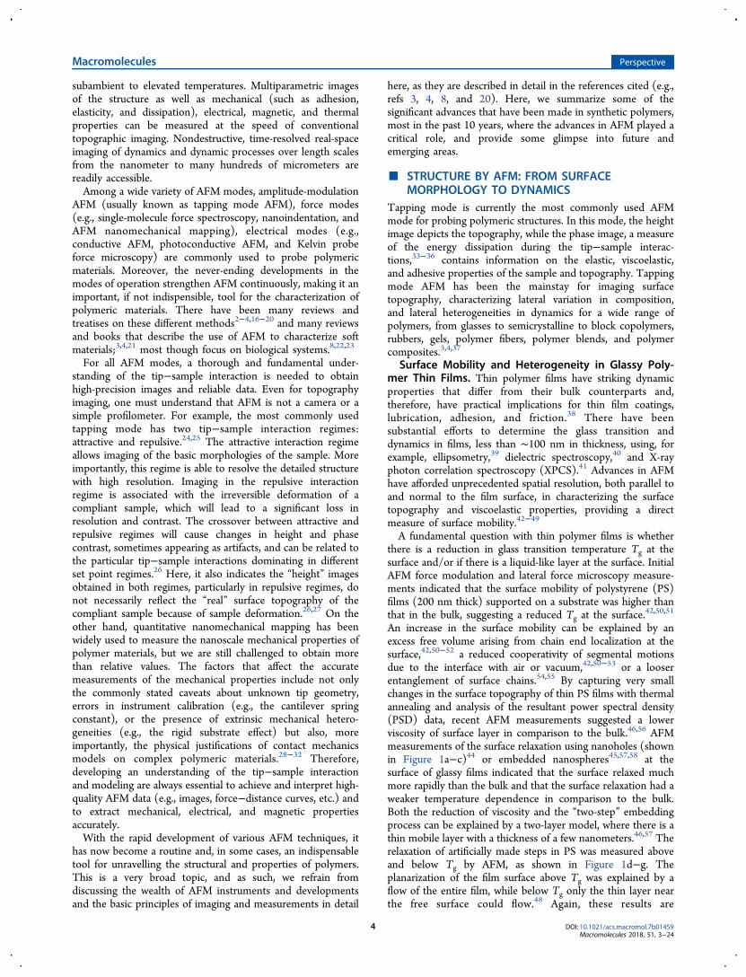
An increase in the surface mobility can be explained by anexcess free volume arising from chain end localization at thesurface,42,50−52 a reduced cooperativity of segmental motionsdue to the interface with air or vacuum,42,50−53 or a looserentanglement of surface chains.54,55 By capturing very smallchanges in the surface topography of thin PS films with thermalannealing and analysis of the resultant power spectral density(PSD) data, recent AFM measurements suggested a lowerviscosity of surface layer in comparison to the bulk.46,56 AFMmeasurements of the surface relaxation using nanoholes (shownin Figure 1a−c)44 or embedded nanospheres45,57,58 at thesurface of glassy films indicated that the surface relaxed muchmore rapidly than the bulk and that the surface relaxation had aweaker temperature dependence in comparison to the bulk.Both the reduction of viscosity and the “two-step” embeddingprocess can be explained by a two-layer model, where there is athin mobile layer with a thickness of a few nanometers.46,57 Therelaxation of artificially made steps in PS was measured aboveand below Tg by AFM, as shown in Figure 1d−g. Theplanarization of the film surface above Tg was explained by aflow of the entire film, while below Tg only the thin layer nearthe free surface could flow.48 Again, these results are
Macromolecules Perspective
DOI: 10.1021/acs.macromol.7b01459Macromolecules 2018, 51, 3−24
4
qualitatively consistent with enhanced mobility at the freesurface of polymer thin films. AFM measurements on therelaxation of thin films showed that substrate interactions canalso influence the surface mobility.58,59 Contrary to theseobservations, however, shear-modulated AFM measurements ofsurface mobility (at 1400 Hz) of PS thin films were found to beindependent of film thickness (17−500 nm), strength ofsubstrate interactions, or even the presence of substrate.60 Thisobservation may be related to the temperature dependence ofsurface mobility, where enhancements may be apparent nearand below the bulk Tg and, thus, are observable only on verylong time scales or at very low frequencies. Measurementsperformed at higher frequencies, such as those described above,may not be able to discern distinct surface and bulk relaxationprocesses.38
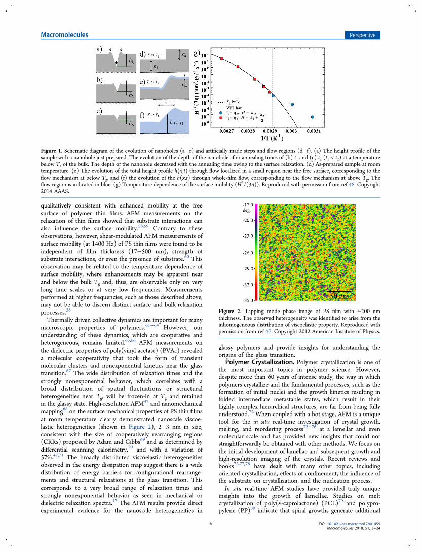
Thermally driven collective dynamics are important for manymacroscopic properties of polymers.61−64 However, ourunderstanding of these dynamics, which are cooperative andheterogeneous, remains limited.65,66 AFM measurements onthe dielectric properties of poly(vinyl acetate) (PVAc) revealeda molecular cooperativity that took the form of transientmolecular clusters and nonexponential kinetics near the glasstransition.67 The wide distribution of relaxation times and thestrongly nonexponential behavior, which correlates with abroad distribution of spatial fluctuations or structuralheterogeneities near Tg, will be frozen-in at Tg and retainedin the glassy state. High-resolution AFM47 and nanomechanicalmapping68 on the surface mechanical properties of PS thin filmsat room temperature clearly demonstrated nanoscale viscoe-lastic heterogeneities (shown in Figure 2), 2−3 nm in size,consistent with the size of cooperatively rearranging regions(CRRs) proposed by Adam and Gibbs69 and as determined bydifferential scanning calorimetry,70 and with a variation of57%.47,71 The broadly distributed viscoelastic heterogeneitiesobserved in the energy dissipation map suggest there is a widedistribution of energy barriers for configurational rearrange-ments and structural relaxations at the glass transition. Thiscorresponds to a very broad range of relaxation times andstrongly nonexponential behavior as seen in mechanical ordielectric relaxation spectra.47 The AFM results provide directexperimental evidence for the nanoscale heterogeneities in
glassy polymers and provide insights for understanding theorigins of the glass transition.
Polymer Crystallization. Polymer crystallization is one ofthe most important topics in polymer science. However,despite more than 60 years of intense study, the way in whichpolymers crystallize and the fundamental processes, such as theformation of initial nuclei and the growth kinetics resulting infolded intermediate metastable states, which result in theirhighly complex hierarchical structures, are far from being fullyunderstood.72 When coupled with a hot stage, AFM is a uniquetool for the in situ real-time investigation of crystal growth,melting, and reordering process73−76 at a lamellar and evenmolecular scale and has provided new insights that could notstraightforwardly be obtained with other methods. We focus onthe initial development of lamellae and subsequent growth andhigh-resolution imaging of the crystals. Recent reviews andbooks72,77,78 have dealt with many other topics, includingoriented crystallization, effects of confinement, the influence ofthe substrate on crystallization, and the nucleation process.In situ real-time AFM studies have provided truly unique
insights into the growth of lamellae. Studies on meltcrystallization of poly(ε-caprolactone) (PCL)79 and polypro-pylene (PP)80 indicate that spiral growths generate additional
Figure 1. Schematic diagram of the evolution of nanoholes (a−c) and artificially made steps and flow regions (d−f). (a) The height profile of thesample with a nanohole just prepared. The evolution of the depth of the nanohole after annealing times of (b) t1 and (c) t2 (t1 < t2) at a temperaturebelow Tg of the bulk. The depth of the nanohole decreased with the annealing time owing to the surface relaxation. (d) As-prepared sample at roomtemperature. (e) The evolution of the total height profile h(x,t) through flow localized in a small region near the free surface, corresponding to theflow mechanism at below Tg, and (f) the evolution of the h(x,t) through whole-film flow, corresponding to the flow mechanism at above Tg. Theflow region is indicated in blue. (g) Temperature dependence of the surface mobility (H3/(3η)). Reproduced with permission from ref 48. Copyright2014 AAAS.
Figure 2. Tapping mode phase image of PS film with ∼200 nmthickness. The observed heterogeneity was identified to arise from theinhomogeneous distribution of viscoelastic property. Reproduced withpermission from ref 47. Copyright 2012 American Institute of Physics.
Macromolecules Perspective
DOI: 10.1021/acs.macromol.7b01459Macromolecules 2018, 51, 3−24
5

lamellar layers at low undercoolings. For poly(bisphenol Aoctane ether) (PBA-C8) an induced nucleation and subsequentbranching was found.81 The growth tip of PBA-C8 wasobserved to be softer than the rest of the crystal,82 indicatingthere may exist intermediate degrees of order that aremetastable. Lamellae crystallized from the melt showed anodular texture,83,84 suggesting a substructure forms at thegrowth front of the lamellae, in keeping with the argumentsmade by Strobl.85−87 The crystallization rate, as evidenced by
the work on individual lamellae and lamellar aggregates, variesfrom crystal to crystal and for each crystal with time.72,74
For spherulite growth, a depletion zone at the front of agrowing spherulite was observed.74,75 Initial crystal growth isfollowed by a protracted period of a backfilling growth andpossibly crystal reorganization.72 In situ observations showedlamellae twisting to produce banded spherulites where thetwisting was a continuous over a substantial fraction of therotation.88 This observation is consistent with X-ray studies89
and provides evidence for models on the long distance self-organization of the lamellae.90,91 Screw dislocation branches76
and other branching mechanisms82 were directly visualized.Based on these studies, the formation of a screw dislocationfrom a defect or fluctuation at the edge of a growing lamella wasargued to be the main source of branching in polymers.72 Exsitu AFM observations of polyethylene (PE) crystal growthsuggested an instability-driven branching mechanisms, arisingfrom a self-induced pressure gradient due to the difference indensities of the crystalline and amorphous phases.92,93
While there are numerous models to describe crystallization,crystallization was not understood on a molecular level.Recently, direct imaging with submolecular resolution, i.e.,high-resolution imaging of a two-dimensional (2D) crystalprepared by very slow compression of an isotactic poly(methylmethacrylate) (i-PMMA) Langmuir film has been available.94
The AFM images clearly showed the folding and tie-chains andprovided a remarkable snapshot of the arrangement of chainswithin a crystal.94 The crystalline nuclei preferentially form atthe end of the chains, and the size of the nuclei was found to beindependent of the chain length (molecular weight). Atextremely slow compression rates, crystallization was promoted,leading to the crystallization of the entire chain.95 The meltingbehavior of the 2D i-PMMA crystals was also observed in situ athigh temperatures with molecular resolution.96 The Tm of the2D crystals was depressed significantly, by up to 90 °C, andshowed a strong dependence on the molecular weight andnature of the substrate. The large depressions in Tm of the 2Dcrystals could not be explained by a simple modifiedThompson−Gibbs equation, creating a theoretical challengeof the age-old problem. The molecular scale observation of 2D
Figure 3. Torsional-tapping AFM images of the oriented “shish-kebab”crystallization of polyethylene. (a) The amplitude image (grayscale 17mV) clearly visualized molecular steps from which the stems’orientation could be inferred. (Inset) Distribution of widths oflamellae measured along the stem direction. (b) Details from phaseimages showing a first-neighbor fold, marked by an arrow, and (c) asecond-neighbor fold, with arrows marking the two stems. Thegrayscales represent a phase lag of (b) 7.7° and (c) 9.2°. Reproducedwith permission from ref 98.
Figure 4. (a, b) Phase images of thin SBS films after annealing in chloroform vapor. Bright (dark) corresponds to PS (PB) microdomains. Contourlines from the corresponding height images are superimposed. (c) Schematic height profile of (a) and (b). (d) Simulated morphology of a BCP filmin one large simulation box with increasing film thickness (from left to right). Reproduced with permission from ref 105. Copyright 2002 AmericanPhysical Society.
Macromolecules Perspective
DOI: 10.1021/acs.macromol.7b01459Macromolecules 2018, 51, 3−24
6

crystals has provided new insights into the polymercrystallization and has presented new challenges.Direct imaging of chains within a three-dimensional
semicrystalline material has been done (Figure 3).97,98
Torsional tapping mode AFM imaging of PE, with a 0.37 nmresolution, showed individual chains in the crystalline lattice inair under ambient conditions.97 Loose molecular loops at thecrystal−amorphous interface and the existence of a tightadjacent fold within this interfacial region were observed.97
Images of this nature have put to rest the very long-standingdebate of the adjacent re-entry or random switchboardmodels.99,100 Results from such studies can be directlycompared to polymer crystallization theories and molecularsimulations, addressing very detailed issues, like the length ofstem-to-stem overhang, as shown in Figure 3a.98
High-resolution AFM imaging has provided insight intoalmost every aspect of polymer science and has mappednanoscale mechanical heterogeneities in glassy polymers47 andhas been used to characterize phase separation in polymer-based bulk heterojunction (BHJ) organic photovoltaics(OPV).101 Because of structural similarities and low imagingcontrast between polymer donors and fullerene acceptors,visualizing structure can be difficult.102 High-resolution AFMimaging of PTB7/PCBM active layers showed the free surfacewas enriched with polymer crystals having a “face-on”orientation and an average spacing of ∼1.9 nm, and themorphology at the anode interface was markedly different.101
Block Copolymer Assembly. AFM has been extensivelyused to study block copolymer (BCP) self-assembly,103−105
which has recently attracted significant interest from bothindustry and academia, since the resultant bulk and thin filmmorphologies offer ideal platforms for the generation ofnanoscopically ordered patterns for a range of promisingapplications.106−109 Therefore, an in-depth understanding ofthese ordered nanostructures with material characteristics andfilm preparation conditions is essential to further tailor thesynthesis of BCPs and for the fabrication of nanopatterns orarrays (Figure 4). In most cases, AFM is combined withelectron microscopies or X-ray-based scattering methods toreveal the effects of composition, molecular weight, interactionparameter, interfacial interactions, film thickness, annealing,crystallization, additives, and chain rigidity on the assemblyprocess and the resultant size, shape, and orientation of thesenanoscopic structures.107,109 Correct assignment of theobserved surface pattern to a particular morphology isimportant for the interpretation of the morphological behaviorin thin films. Beyond routine applications, however, advances inAFM make it ideal for studying structural defects and dynamics,self-assembly process, where the aggregates are swollen or filledwith solvent (such as micelles or vesicles), and changes of theshape due to deposition on the solid surface and drying.Structural defects in BCP thin films are common and are
known to compromise the long-range lateral order that limitthe technological performance of BCPs.107,110−113 AFM studieson BCP thin films have identified classical defects (i.e.,disclinations and dislocations) as well as grain boundary defectconfigurations.110,112−115 It was found that the motion of lateraldefects in cylinder-forming BCPs was diffusion controlled.114
The minimum feature spacing accessible in thin films is limitedby thermal defect generation, not by the bulk order−disordertemperature (ODT).116 Such defect densities and the ODT arehighly sensitive to variations as small as 2 nm in themicrodomain spacing.116 The large shift of monolayer and
bilayer ODT caused by variations of the monolayer domainspacing can be explained by the energetic cost of defectproduction in terms of the domain spacing, interactionparameter, and BCP composition.116 Early works using time-resolved AFM identified relinking, joining, and clustering asbasic processes of structural rearrangements.112 When applyingan electric field to ABC BCP thin films, AFM showed twodistinct defect types that govern the orientation mechanism.117
Moreover, in situ imaging of the dynamics and defectannihilation of polystyrene-b-polybutadiene-b-polystyrene(SBS) during solvent annealing showed a low interfacial energydifference between the cylinder and perforated lamella phasesthat may account for the energetically favorable path way ofstructural rearrangements by temporal phase transitions,118,119
which was also observed by in situ AFM imaging of SB diblockcopolymers under thermal annealing.111 This structuralrearrangement consists of several elementary dynamicprocesses, such as short-term interfacial undulations, fastrepetitive transitions between distinct defect configurations,120
and collective/coordinated movement of annihilating de-fects,114,120 as revealed by high-speed AFM scanning. The fastdynamics of individual defects and their annihilation in thinfilms of a cylinder-forming BCPs121 are illustrated in Figure 5.BCP micelles, vesicles, and other aggregate structures have
emerged as versatile drug delivery systems, nanoreactors, or astemplates for nanoparticle synthesis.122,123 Besides scattering-based methods124 and wet scanning and cryogenic TEM,125
AFM has been used to provide in situ information about thegrowth of these aggregate structures in solution by tracking theshape of self-assembled aggregates.126−128 Varying thearchitecture of the BCPs, the nature of the solution,126,127
and the underlying surface126,128 have opened numerous routesto control surface topology, domain size, and wall thickness ofthe aggregate structures. For example, the poly(2-(dimethyl-amino)ethyl methacrylate)-b-poly(methyl methacrylate)(DMA-MMA) adsorbed on mica showed surface micelles atlow pH and regions of close-packed structure at higher pH,indicating the importance of pH in the resultant morphology of
Figure 5. (a) AFM phase images visualized climb motion of twodislocations of opposite Burgers vectors with annealing time. The twodislocation cores were indicated by the arrows. (b) The plot of squareof the distance between the two dislocation cores versuscorresponding time. Reproduced with permission from ref 121.
Macromolecules Perspective
DOI: 10.1021/acs.macromol.7b01459Macromolecules 2018, 51, 3−24
7

the polyelectrolyte diblock.126,128 For a poly(oyxethylene)-b-poly(oxypropylene) diblock, a more highly ordered structurewas found on a hydrophobic silica substrate,128 demonstratingthe importance of the chemical nature of the surface on theresultant morphology of the amphiphilic diblock.Polymer Subsurface Structure. The surface and bulk
morphologies of polymeric systems are often different.Reconstruction of three-dimensional (3D) structures isbecoming increasingly important.129 TEMT has been widelyused to this end.130 AFM is a surface technique, but ifsectioning or etching is used in tandem with AFM, subsurfacestructures can be determined. This was demonstrated in theelucidation of the bicontinuous interpenetrating network inPTB7-based bulk heterojunction active layers.101,131 Nano-tomography132 has also been developed for the in situobservation of crystal growth of elastomeric polypropylene,where the origins of a lamellar branch was found to originate in
a screw dislocation.133 This layer-by-layer imaging techniquewas further developed to operate under various conditions andAFM modes134−136 and found use in the solvent vaporannealing of BCPs under electric fields137 and visualization ofconductive 3D networks of polymer/MWCNT nanocompo-sites using c-AFM.135
The methods described above involve destructive sectioningor etching procedures. Nondestructive subsurface structureimaging by AFM is under very active development with someprogress being made using ultrasonic wave or energydissipation. These have been demonstrated in several polymericand cellular systems.138−141 For example, the reconstruction ofthe subsurface structure of supramolecular aggregates ofoligothiophenes yielded a 3D picture consisting of 15 nmwide fibrils with a rigid core and a soft shell.140 In the P3HTsystem, the crystalline regions and crystalline fibers were foundto be covered by an ∼7 nm amorphous layer after solvent
Figure 6. (a) AFM height image of polystyrene-b-poly(methyl methacrylate) (PS-b-PMMA). The width scale of (a) is 500 nm. Reproduced withpermission from ref 169. (b) AFM image of poly(n-butyl acrylate) (PBA) brushes made from poly(alkyl acrylate) and poly(alkyl methacrylate)backbones (white arrow pointing to branch junctions). Reproduced with permission from ref 174. (c) AFM height images of the PBA bottlebrusheswith the same backbone but different of the degree of polymerization of side chains on a mica substrate. Large images: LB monolayers. Insets: singlemolecules prepared by spin-casting. Reproduced with permission from ref 170. Copyright 2016 the Nature Publishing Group.
Macromolecules Perspective
DOI: 10.1021/acs.macromol.7b01459Macromolecules 2018, 51, 3−24
8

casting that decreased to 5 nm after thermal annealing.139 Thepresence of the amorphous surface layer has importantconsequences in the charge-transfer process. For polymernanocomposites, related techniques were used to probe 50 nmgold particles buried in polymer matrix under ∼1 μm.142 Thenondestructive subsurface imaging was also operated under anapplied electric field, where the dispersion and orientation ofCNTs in polymers were determined with nanometer-scaleresolution.143,144 These results provided in-depth morpholog-ical information and, hence, are expected to facilitate theanalysis and preparation of polymer nanocomposites in thefuture.Polymer Molecules Engineered Surface. AFM has
played a key role in understanding surface modification bytethering various polymer molecules using grafting, self-assembly, or adsorption onto substrates.145 The morphologyof the engineered surfaces is critical in defining the properties.AFM topography imaging of such surface has quantified themorphologies and morphological transitions as functions ofgrafting density146 and thickness of grafted layers.147,148 Thethickness of the engineered layers especially for polymer brushmodified surface is often used to quantify structural changes asa function of solvents, pH, temperature, and ionic strength.AFM has subangstrom height resolution and, therefore, canmeasure the layer thickness fast and accurately by cross-sectionanalysis of the polymer covered and uncovered regions.149
Recently, AFM force−distance curve measurements were usedto determine the layer thickness.150−152 The applied loadingforce should must be considered when using AFM to measurethe layer thickness, especially with compliant polymer layers,since the deformation of the polymers will cause errors in themeasured height values.153,154 For polymer brush modifiedsurfaces, average molecular weights of the brush weredetermined by measuring the heights of the brushes in agood solvent in comparison to the known monomer length.155
Stimuli-Responsive Behavior. AFM can be used tomonitor in situ the changes in properties (such as adhesion,
wetting, mechanical performance, and friction) of stimuli-responsive polymers in response to external stimuli, such aspH,156,157 temperature,155,158 ionic strength,159 light,160 etc.Here, well-known responsive poly(N-isopropylacrylamide)(PNIPAM)-based polymers161 show a reversible coil to globuletransition at the so-called lower critical solution temperature(LCST). Above the LCST, in the collapsed state, hydrophobicpolymeric aggregates are seen at the polymer surface, causingan abrupt change in the average film thickness and a dramaticincrease in the roughness.155 This transition was also observedfor end-grafted polymer chains that had been grown fromsurface-immobilized monomers.162 The transition dynamicsunder external stimuli can be easily measured by simplytracking the height change. High-resolution AFM measure-ments of a poly(methacrylic acid) brush in response to pHchanges showed swelling and collapse transitions that occur onthe subsecond time frame.157 During the transition, molecularchains rearranged to another equilibrium structure in responseto the external stimulus, and when compared to the measuredmechanical properties, the structure−mechanical propertyrelationship was directly determined.163 Such informationprovided a better understanding of the structural complexityand responsive behavior of these advanced stimuli-responsivepolymer materials under external stimuli.Self-healing polymers represent one of the forefronts in
materials chemistry and engineering.164 AFM can be employedto image the healing process of this stimuli-responsivepolymers. AFM imaging of the morphology changes as afunction of temperature or time showed the healing process,uncovering the importance of polymer mobility in the healing.These in situ AFM observations provided fundamental insightsinto the healing of the domain morphology at the nano-scale.165,166
Polymer Chain Conformation. In situ visualization ofsingle polymer chains and their motions have long been achallenge in polymer science.167,168 Until recently, AFMimaging of a polystyrene-b-poly(methyl methacrylate) (PS-b-
Figure 7. (a) Scheme of SMFS experiment. Reproduced with permission from ref 182. Copyright 2006 Elsevier. (b) A force plateau at ∼13 pN isobserved in the extension−retraction curve of an individual, collapsed PS chain in water. Black curve shows extension (pulling) of a single moleculeand gray curve shows retraction (relaxation) process. Drawings along the curve illustrate the chain configurations of the extension of a single polymerchain in poor solvent. Reproduced with permission from ref 190. (c) Scheme of the force-induced rotation of carbon−carbon double bonds.Reproduced with permission from ref 200. (d) Scheme of extracting a single poly(ethylene oxide) chain from a single crystal by SMFS andcorresponding force−extension curves. Reproduced with permission from ref 211.
Macromolecules Perspective
DOI: 10.1021/acs.macromol.7b01459Macromolecules 2018, 51, 3−24
9
PMMA) BCP LB film first visualized random coil conforma-tions of a single synthetic polymer chain frozen on a substrate(Figure 6a).169 Shortly thereafter, various structures such aspolymer brushes,170 dendron polymers,171 polyelectrolytes,172
and star polymers173 were observed. In a case study, AFMmolecular imaging of branching in linear acrylate-basedmacromolecules provided direct and quantitative informationabout branching topology including length and distribution ofbranches, not accessible by other methods (shown in Figure6b).174 This capability was also verified in a study of solvent-free, supersoft, and superelastic polymer melts and networksprepared from bottlebrush macromolecules, in which AFMmolecular imaging qualitatively corroborated the increase indiameter and rigidity of bottlebrushes with increasing of thedegree of polymerization of side chains (Figure 6c−f).170 Notonly the static structure but also the dynamic movements, i.e.,the conformational rearrangements, of isolated chains in variousenvironments were visualized.172,173,175,176 Such molecular-levelinformation, including static conformations and dynamics ofconformational transitions, has greatly improved our under-standing of the physical properties of polymers.
■ MECHANICAL PROPERTIES BY AFM: FROMMOLECULE SCALE TO MESOSCOPIC SCALE
Polymer materials often exhibit heterogeneities in materialcharacteristics and chemical composition which, as we drivetoward ever decreasing feature sizes or thicknesses, under-standing the characteristic length scales and dynamics of theseheterogeneities becomes increasingly important. AFM serves asa tool to provide such characterization of the surface. Here, wefocus on recent progress made with the SMFS and AFMnanomechanical mappingtwo typical force measurementsand studies of synthetic polymers.Single-Molecule Force Spectroscopy (SMFS). The
principles and use of SMFS can be found elsewhere.177−182
In a typical experiment, a functionalized AFM tip is broughtinto contact with the sample surface, where polymer chainshave adsorbed. A single molecule can be bound to the tip andthe substrate, by physical adsorption, ligand−receptor inter-action, or covalent bonding. The force as a function of thedistance that the cantilever has traveled vertically is measured asthe molecule is stretched and eventually debonds or breaks, asshown in Figure 7a. The resultant force−distance curvesprovide not only the strength of the binding interaction but alsoinsights into the elastic properties, conformational changes, andthe unfolding of stretched polymer chains with piconewtonsensitivity and subnanometer accuracy. Shortly after thedevelopment of SMFS in a polysaccharides study,183 extensiveworks on inter/intramolecular forces of various polymersystems were investigated.SMFS studies have shown that at low forces (<100 pN) the
mechanical behavior of polymer chains is mainly affected by itsentropic elasticity, while at high force region, larger than 300pN, it is mainly affected by the enthalpic elasticity.184 The sidechain effects on the elasticity of polymer chains showed that forpolymers with the same backbone the chains with larger sidegroups showed higher stiffness.185,186 Recent SMFS measure-ments on polymers with side chains of different lengths andshapes revealed that only long and bulky side chains affectedthe enthalpic elasticity of the chain.184 While such studiesdirectly correlate the molecular structure of the polymers to themechanical properties of a single chain, translating this, ingeneral, to macroscopic properties is still a challenge, though
some success was obtained with a biomimetic designedpolymer.187 Here, the complete, asymmetric potential energyprofile of the rupture and refolding of each monomeric moduleshowed a correlation with the bulk mechanical behavior byDMA measurements.Interactions between the solvent and polymer chains are also
critical, as the aggregation state of polymer chains also affectsthe mechanical behavior of the polymer.186,188−192 Theformation of hydrogen bonds, the solvent quality, and thesize of the solvent molecules, i.e., the excluded volume, willinfluence the measured elasticity of the single chain.188,189,192
By measuring the elasticity in different solvents, informationabout type and strength of interactions between the polymerand small molecules can be determined. As would be expected,with a polyelectrolyte, the charge density on the chain plays acritical role in its elasticity.193 For PS chains, however, theelasticity constant was found to be the same for all differentorganic solvents investigated, but Kuhn length increasedsystematically with increasing solvent quality, reflecting thelarger extent of swelling of the polymer in good solvents.192
Force-induced conformational transitions of single polymerchains can provide fundamental information about internalstructure.194,195 SMFS measurements of a PS chain in water, forexample, showed three regions of the mechanical responsecorresponding to chain extension (retraction) and a force-induced globule−coil transition of polymer chains (Figure 7b),providing definitive proof of theoretical predictions.196,197 Here,the hydrophobic PS chains collapsed in water due tononfavorable interactions with water,191 manifesting a classiccollapse mechanism where the hydrophobic domain sizedictated the structure and dynamics of water near polymers.198
Force-induced isomerization of the gem-dibromocyclopropane(gDBC) into 2,3-dibromoalkenes was observed during thestretching of a gDBC-functionalized polybutadiene.199 Thestructural rearrangement indicated the localized stress could berelaxed in polymers and polymer networks under load.199 Morerecently, force-induced cis-to-trans isomerization of carbon−carbon double bonds has been observed in several polymersystems (Figure 7c).200,201 These SMFS results indicate uniquepossibilities to develop advanced force-responsive materials.SMFS measurements on a more complex polymer systemforced unfolding of single-chain polymer nanoparticles(SCNPs), provided insights into the interior structure ofSCNPs, and by analysis of rupture events observed in the forceprofiles afforded insights into the assembly mechanism of theSCNPs.202 Force-inducted structural transitions also providedimportant structural information on cross-linked polysacchar-ides203 and disassembling block copolymer and micelles.204,205
SMFS can also provide quantitative information about intra/intermolecular interactions of polymer molecules177,180,206,207
as well as desorption forces of polymer chains from thesubstrate surface.193,208,209 Most SMFS studies on intermo-lecular interactions have focused on biological samples, sincesuch interactions dictate the self-assembly process and directthe assembly of molecular building blocks into organizedsupramolecular structures, which is key for biological processes.Nonuniform force plateaus were observed when carboxy-methycellulose (CMC) molecules were pulled out of a polymerfilm into a poor solvent and were described by a geometricmodel that involves the polymer−polymer and polymer−solvent interactions.210 Recent SMFS studies on the pull-out ofa single poly(ethylene oxide) (PEO) chain from a single crystal(shown in Figure 7d)211 clearly demonstrated the adjacent re-
Macromolecules Perspective
DOI: 10.1021/acs.macromol.7b01459Macromolecules 2018, 51, 3−24
10

entry of the PEO chains in the single crystal prepared fromdilute solution.212
SMFS has also been used to the determine molecular weight,molecular weight distribution, and grafting density150,213 ofsurface-grafted polymers,150,213−215 which has been a long-standing challenge.145 SMFS results for a polycation, poly[2-(dimethylamino)ethyl methacrylate] (PDMAEMA), graftedfrom a poly(methyl methacrylate) (PMMA) backbone wereshown to be consistent with results from gel permeationchromatography.214 SMFS mapping on RAFT controlledmacromolecular growth on glass surfaces showed the RAFTchain extension linearly with time up to high conversions(Figure 8), providing critical insight into macromoleculargrowth of the surface-initiated polymerization.215
AFM Nanomechanical Mapping (AFM-NMM). AFM-based NMM modes, such as force modulation,216 forcevolume,27,217 lateral force microscopy (also known as frictionforce microscopy),218 contact resonance,219 peak forcetapping,220,221 nanoindentation,222,223 multifrequency forcemicroscopy,5 and also tapping mode phase imaging,224 providesample properties while simultaneously imaging the top-ography. A versatile and widely used approach among theseis the force−distance (FD) curve-based imaging where an AFMtip scans over a specified area of the sample surface, and thecorresponding applied force versus tip displacement isdetermined. Using appropriate contact mechanics models,such as the Hertz,225 Johnson−Kendall−Roberts (JKR),226
and Derjaguin−Muller−Toropov (DMT) models,227 the 2DFD images can be translated into an areal mapping of thesurface mechanical properties, including the elastic modulus,adhesion, dissipation, and stiffness. By simultaneously providingmicrostructure and mechanical properties with nanometerresolution and piconewton sensitivity, AFM-NMM has becomea routine tool for probing polymer structure and determininglocal mechanical properties. Several pioneering studies usingAFM-NMM on polymers have been published.3,4,16,19,21,216
The mechanical properties of thin and ultrathin polymerfilms are of critical importance for many applications, rangingfrom coatings to organic electronics. AFM-based nano-indentation measurements on thin polymer films supportedon a noncompliant substrates showed that the effective out-of-plane modulus increases with decreasing film thickness when itis smaller than a threshold film thickness.228−230 This
enhancement of the elastic modulus can be explained by thepropagation of the indentation-induced stress field and theinteractions between the thin film and the underlyingsubstrate.230,231 The indentation-induced stress field propaga-tion was found by measuring the elastic modulus of linear PS(LPS) and star-shaped PS (SPS). This was more evident forSPS than the LPS, indicating a more efficiently “packing” of theSPS, allowing a more efficient stress transfer.231 Interactionsbetween the thin film and the substrate were also found toaffect the elastic modulus of the thin films. AFM nano-indentation simulations on the interfacial mechanical propertiesnear attractive interfaces of supported PMMA thin filmsindicated that there was a gradient of local modulus with largervalues near the substrate compared to the bulk, giving rise tointerfacial confinement effects.232 Force volume NMMmeasurements on rubbery poly(vinyl acetate) (PVAc) thinfilms further revealed the intermolecular interactions inducedby nanoconfinement significantly affected the elastic andviscoelastic responses of polymers.233 The above resultsprovided important insights into the origin of the thickness-dependent mechanical properties of thin polymer films.Characterization of the microstructure and mechanical
properties at interfaces in polymer blends and composites hasbeen a long-standing academic and technological challenge,since they dictate the ultimate properties. Studies onpolystyrene/poly(n-butyl methacrylate) (PS/PnBMA) blendsusing AFM-NMM showed a gradual decrease in the Young’smodulus from that of PS to that of PnBMA, clearly demarkingthe interfacial region.234 AFM-NMM measurements of thereactive compatibilization of polyolefin elastomer (POE)/polyamide (PA6) blends demonstrated an interfacial reactioninduced roughening and simultaneously afforded the strengthand width of the interface.235 The ability to map the spatialdistribution of Young’s modulus at an interface provides analternate, efficient means of characterizing interfaces. This, ofcourse, enables the investigation of polymer−polymer inter-diffusion (shown in Figure 9);221,236 polymer−fullerenenanoparticle interdiffusion;237 compatibilized polymer blendswhere increased interfacial interactions of immiscible polymersmediated by an ionic liquid were revealed;238 fiber reinforcedpolymer composites;239 and carbon black reinforced rubbernanocomposites, where the thickness and elastic properties ofthe bound rubber phase were observed240 and confirmed by
Figure 8. (a) Scheme of SMFS mapping of RAFT-controlled macromolecular growth. (b) Typical force−distance curves of poly(hydroxyethylmethacrylate) (PHEMA) strands that have different chain length. (c) Plot of contour length of the PHEMA chains versus polymerization time.Reproduced with permission from ref 215.
Macromolecules Perspective
DOI: 10.1021/acs.macromol.7b01459Macromolecules 2018, 51, 3−24
11

simulations.241 Given the importance of the interface indefining properties of polymer blends and composites, AFM-NMM provides a unique view.There has been much interest in the deformation mechanism
of various polymers since the roles of molecular orientation anddeformation-induced structural changes have been found to beof particular importance for the ultimate properties of apolymer. Although X-ray diffraction/scattering was widely usedto probe the local molecular structure and structural dynamicsof various polymers during deformation, they provide onlystructural information that is averaged over the area irradiatedwith the X-ray beam. With AFM real-space imaging,considerable information regarding polymer chain orientation,structural transition and dynamics, and fracture underdeformation can be visualized. Such studies, using contact ortapping mode AFM, have been performed on semicrystallinepolymers,242−244 elastomeric polyolefins,245,246 thermoplasticvulcanizates,247 and stretchable electronic devices.248 The real-space features of uniaxially stretched iPP sheets observed withfrequency-modulation AFM were successfully compared withthe X-ray scattering results obtained by synchrotron X-rayscattering.249 Not only structural information but also themechanical properties associated with structural developmentsunder deformation are also of interest. Several recent studiesusing AFM-NMM have been performed on polymer hydro-gels,250 semicrystalline polymers,251,252 and elastomers.253,254 Ina case study, AFM-NMM measurements on the structuralevolution and mechanical properties of a deformed isoprenerubber (IR) clearly demonstrated a hierarchical nanofibrillarstructure, ranging from several to a hundred nanometers in size,comprised of fibers oriented parallel to the stretching direction(Figure 10). The nanofibers, connected by oriented amorphoustie chains, form a network structure that was responsible forsignificantly enhanced stress, a key factor giving rise to the self-reinforcement of IR.253
Information about the viscoelastic properties at the nanoscaleis another essential characteristics of polymeric materials andalso an emerging area of AFM investigation. As discussedearlier, viscoelastic measurements using several AFM modes(e.g., force modulation, lateral force microscopy) have shownthat there is a surface mobile layer with a reduced Tg incomparison to the bulk. With FD curve measurements, AFM-NMM, by analyzing changes in the pull-off forces in the vicinityof the glass transition, provided a means to determine the Tg ofthe topmost polymer layer.55,255 Not only Tg, FD measure-ments on glass-to-rubber transition of amorphous polymerscould be used to estimate the parameters of the Williams−Landel−Ferry equation and the Young’s modulus and theyielding force of the polymer in a wide range of temperatures(70 K) and probe rates (6 decades), and the results are in verygood agreement with measurements performed with customarytechniques, such as broadband spectroscopy and dynamicmechanical analysis (DMA).256 In more recent studies,viscoelastic properties can be mapped into a 2D image withnanometer spatial resolution.257 As a case study, the viscoelasticproperties including the storage modulus, loss modulus, and tanδ of styrene−butadiene rubber (SBR), IR, and a SBR/IR 7/3blend were estimated and visualized from 1 Hz to 20 kHz usinga recently developed nanorheology mapping technique.258
These quantities obtained by AFM were in agreement withthose measured using bulk DMA. Through this evaluation,nanorheology mapping has been shown to be a highlypromising AFM mode for quantitative characterization of
viscoelastic materials at the nanoscale, and its benefit to theresearch and development of small-scale compliant materials isexpected to be substantial.In view of the increased importance of tribological properties
(friction, lubrication, and wear) of the surfaces and interfaces ofpolymeric materials in lubrication, M/NEMS (micro/nanoelectromechanical devices), biomedical implants, and others, amolecular-level understanding of such tribology behavior is ofspecial interest259,260 and an increasingly active area of AFM-NMM investigation.261−263 Early work on estimation ofnanotribological properties by AFM-NMM has been shownfor polymer blends,264 thermoplastics,265 and glassy poly-mers,218 but most recent work has been focused on polymerbrushes266−269 because of their unique properties, such asreversible switching behavior, multivalent functionalization,tunable wettability, and lubrication. In most cases, several FDcurve-based AFM-NMM techniques, such as lateral force andchemical force microscopies, are combined to estimate thetribological properties, for example, friction, adhesive, andelastic properties with nanometer resolution and under a widevariety of conditions. AFM-NMM measurements of thenanomechanical and nanotribological properties of polyelec-trolyte brushes provided insights into how the Young’smodulus and coefficient of friction can be tuned by varyingthe pH of the surroundings and the degree of physical orchemical cross-linking.267 For polymer brush measurements, itis crucial to use colloidal probes because of their well-definedtip geometry and, therefore, can have a good match betweenthe measured force−distance curves and theoretical models to
Figure 9. AFM-NMM Young’s modulus maps of the misciblepoly(vinyl chloride)/poly(caprolactone) sample annealed at 72 °Cfor (a) 5, (b) 20, and (c) 50 min. Reproduced with permission fromref 221.
Macromolecules Perspective
DOI: 10.1021/acs.macromol.7b01459Macromolecules 2018, 51, 3−24
12

get mechanical properties accurately. This is also true for AFMmeasurements on mechanical properties of gels, which isincreasingly important polymeric materials.The importance of AFM-NMM was also demonstrated in
other polymer systems, such as polymer nanofibers, where, bymeasuring the nanoscale mechanical properties of single fibers,the packing density and structural heterogeneity could bedetermined;270−273 stimuli-responsive polymers such as PNI-PAM that shows a well-known temperature-induced con-formation transition at LCST, where the nanomechanicalproperties below and above the LCST, as well as on the drystate, were addressed;274,275 semicrystalline polymers, in whichthe crystalline regions covered by a thin amorphous layer, wererevealed;276,277 and block copolymers, in which various localnanomechanical properties, not only Young’s modulus but alsoadhesion and energy dissipation, were evaluated.278−281
■ NANOSCALE ELECTRICAL AND ELECTRONICPROPERTIES BY AFM
Polymer-based solar cells (PSCs) have attracted significantattention from both academia and industry due to theirsemiconducting properties and potential for low-cost, flexibledevices for alternative energy sources.282−284 For PSC devices,donor/acceptor blends absorb photons to generate excitons,which then migrate to the donor/acceptor interfaces wherethey dissociate into free charge carriers. This process isdependent on nanoscopic details of the morphology, which isdependent on the chemical compositions and processingconditions.285,286 Advanced AFM modes including conductiveAFM (c-AFM), photoconductive AFM (pc-AFM), andscanning Kelvin probe force microscopy (KPFM) have beenused to gain an understanding of the structure−performancerelationship for the active layer of PSCs.287−291 c-AFM, aderivative of AFM contact mode, characterizes the electricalcharacteristics with a typical spatial resolution of 10−20 nm. Inthis mode, a dc bias is applied between the conductive tip andsample while the tip scans over the sample surface, andinformation about the nanoscale topography and currentdistribution (conductivity) is recorded simultaneously. Studiesusing c-AFM clearly visualized, as evidenced by currentdistribution map, the phase-separated donor and acceptorregions and the electron- and hole-transport networks.292−296
Heterogeneous conductive regions smaller than 20 nm weredetected.297 The thermal annealing induced heterogeneous
conductivity in poly(3-hexylthiophene) (P3HT) thinfilms297,298 increased hole and electron mobilities in P3HT/[6,6]-phenyl C61-butyric acid methyl ester (PCBM) blends292
and decreased the width of the interface between the donor andacceptor regions.296 The use of solvent additives resulted in afinely phase-separated morphology295 as evidenced by c-AFM.Consequently, the processing conditions that determine themorphology of the active layer could be directly correlated withthe PSC performance.Similar to the force−distance curves, current−voltage (I−V)
curves can also be extracted from c-AFM and provide someunderlying information on electron conductivity and mobilityof the nanostructures, such as the hole mobilities of P3HT inneat P3HT films,294,299 in poly(3-alkylthiophene) (P3AT)nanofibers,300 or in P3HT:PCBM blends,292 which could neverbe determined by macroscopic J−V measurements. It should benoted that there may be a large discrepancy between the chargecarrier mobility measured with c-AFM and that measured by amacroscopic planar device299,301 due to the geometric differ-ences between the two experiments and the areas over whichthe different methods average.299 Using a semiempiricalequation proposed by Reid et al.,299 however, reliable mobilitiescan be extracted, allowing the measurement of local mobilitieswith nanoscale resolution in the heterogeneous films used inlight-emitting diodes, transistors, and solar cells.It is more interesting, however, to obtain information on the
photocurrent generation at the nanoscale that can be used tocorrelate with charge generation, transport, and collection thatdominate bulk device performance. pc-AFM, a derivative of c-AFM but with the addition of a diffraction-limited laser sourceto illuminate the device (Figure 11a), has been used to resolvethe complex optoelectronic and morphological phenomena ofPSCs with a typical resolution of 10−20 nm.302−309 Photo-current mapping of a poly[2-methoxy-5-(3′,7′-dimethyl-octyloxy)-1,4-phenylenevinylene] (MDMO-PPV)/PCBM filmgave evidence for two length scales of heterogeneities arisingfrom the solvent casting conditions (shown in Figure 11).302 InP3HT:PCBM films, pc-AFM data showed significant inhomo-geneities on the length scale of 100−500 nm, arising from localvariations at the surface, not reflecting the bulk organization.304
In a high-performance PTB7:PCBM blend, pc-AFM revealed abulk heterojunction (BHJ) structure consisting of elongatedPCBM-rich and PTB7-rich fiber-like domains, 10−50 nm wideand 200−400 nm long, indicating that the formation of narrow
Figure 10. AFM-NMM images of stress distribution of isoprene rubber (IR) visualized structural evolution with strain 0% (a), 300% (b), 500% (c),and 600% (d). The strain is applied in the vertical direction of these images. Reproduced with permission from ref 253.
Macromolecules Perspective
DOI: 10.1021/acs.macromol.7b01459Macromolecules 2018, 51, 3−24
13

and elongated domains is desirable for efficient BHJ solarcells.309 Moreover, not only the surface but also the subsurfacemorphology can be revealed by pc-AFM, with an estimateddepth resolution of ∼20 nm within the P3HT:PCBM film.307
KPFM, a noncontact variant of AFM, can be used to probelocal surface potentials (work function) to local morphologieswith a resolution better than 10 nm.310,311 KPFM is contact-less, which is an important when dealing with soft organic thinfilms. Early studies of MDMO−PPV/PCBM blends usingKPFM identified a barrier for electron transmission from theelectron-rich PCBM nanoclusters toward the extractingcathode.312 In polyfluorene-based photodiodes, KPFM revealedthe presence of a capping layer that reduces the efficiency of thephotovoltaic device by blocking the transport of photo-generated electrons to the surface.313 With improved resolutiondown to sub-10 nm, surface morphologies of the inter-penetrated networks of P3HT:PCBM blends are clearlyevident, and the carrier generation at the donor−acceptorinterfaces and their transport through the percolation pathwaysin the nanometer range have been directly visualized.310 KPFMstudies on P3HT-b-P3MT and P3HT nanofibers showed thatthe surface potential, related to the work function of thesample, is very sensitive to the nanostructure morphologyresulting from a combined effect of chain-packing disorder,molecular weight, and local environment.314,315 The decay ofthe surface potential into the film was determined on the∼millisecond scale, which suggested an intensity-dependentrecombination kinetics that is in quantitative agreement withthe carrier recombination kinetics measured in bulk devices.316
The combination of AFM-based techniques, such as c-AFM,pc-AFM, and KPFM, provides comprehensive characteristics ofthe local morphology and the electrical and optoelectronicproperties of PSCs, thereby establishing a direct correlation oflocal heterogeneities within the nanostructure and photo-current generation in the bulk device.287−291 Studies in P3HT/PCBM blend devices showed that the thermal annealing-induced morphological heterogeneities, as seen in thedistribution maps of local current (by c-AFM) and short-circuit photocurrent (by pc-AFM), may lead to the imperfectinternal quantum efficiency of some blends.303 In a poly(3-butylthiophene) (P3BT):PCBM blend device, the directconnections between local nanostructure and overall deviceperformance, as shown in the local current and photocurrentmaps, showed that the nanostructure, controlled by using acombination of thermal and solvent annealing, is the singlemost important variable determining the device perform-ance,305 while in a P3HT nanowire:PCBM blend devices, theresults demonstrated the importance of vertical morphologywithin the active layer on the device performance.306
Copolymer-based devices, in contrast to the physically mixedpolymer:fullerene blends, are inherently microphased separatedon the tens of nanometers level, underscoring the importanceof molecular design and morphological control that determinethe nature of the self-assembled nanostructures for developinghighly efficient polymer:fullerene PSCs.308 Since c-AFM andpc-AFM are operated in contact mode, c-AFM and pc-AFMstill face challenges including scan-induced damage,306
injection/extraction barriers due to the work function of thetip,317 and difficulty in making quantitative comparisons withthe external quantum efficiency (EQE) of the macroscopicdiode.
■ PROSPECTSThe past 30 years have seen considerable impact of AFM onour understanding of polymers. With the advances ofmultiparametric and multifunctional characterization withhigh resolution and the capacity to operate under variousmedia, AFM is well positioned to tackle the challenges ofunderstanding the structure and properties of increasingly morecomplex polymers and their composites. In measurementdevelopments, for example, multifrequency AFM is emergingand provides a promising framework to improve compositionalsensitivity and spatial and time resolution of materials in theirnative environment.318 In a case study, bimodal AFM (one kindof multifrequency AFM) imaging of metallopolymer-grafteddiblock copolymers clearly revealed a lamellar morphologyfeaturing a spherical substructure for the polyvinylferrocene(PVFc) segments inside the polyisoprene lamellae.319 Themorphological structure of the grafted diblock copolymermeasured by bimodal AFM is in excellent agreement with theTEM and SAXS,319 indicating the high sensitivity for
Figure 11. (a) Schematic of the pc-AFM setup. pc-AFM height image(b) and photocurrent map (c) of a MDMO-PPV:PCBM 20:80 thinfilm. (d) Local current−applied voltage curves acquired at threelocations shown in (b) and (c). Inset: local current−voltage curveswithout illumination showing much smaller dark currents. Reproducedwith permission from ref 302.
Macromolecules Perspective
DOI: 10.1021/acs.macromol.7b01459Macromolecules 2018, 51, 3−24
14
compositional heterogeneities well suited for the high-resolution imaging of polymers. Combining AFM with existingtechniques, in particular chemistry related characterizingmethods, put AFM at an even more advanced level. AFM-IR,combining AFM and infrared spectroscopy, opens a door tonanoscale chemical characterization, where the spatial variationin infrared absorption and reflection with up to a 15 nmresolution is possible.14,320,321 This advance paves the way fornew insights into the underlying structure beyond phenom-enological physical responses (e.g., dissipation and mechanicalproperties), of a wide range of polymer materials, such aspolymer blends, composites, fibers, and thin films for activedevices, such as organic photovoltaics. Several recent studiesusing AFM-IR have shed some light on the structures andcompositions with micro- and nanoscale feature sizes,322−324
which were not been possible by either conventional IR orAFM. The capabilities of AFM-IR, with the addition of someother accessories, such as a polarization control, can be furtherextended to probe the orientation in anisotropic supra-molecular assemblies.325 The combination of AFM with massspectrometry (MS) for topographical and chemical imaging ofpolymer systems has also emerged very recently.326,327
Moreover, the combination of two or more AFM-basedmodes to characterize multiple aspects of polymers is anotherpromising way to characterize polymers with exceptional spatialresolution. Such a combination, for example, AFM andscanning electrochemical microscopy (AFM-SCEM), providedspatially correlated electrochemical and nanomechanicalinformation paired with high-resolution topographical data ofsoft electronic devices.328 It is evident that such combinationswill allow AFM methods to meet the challenges posed byincreasing complexity in structure and function of polymermaterials.Despite the wealth of information AFM provides, an
important limitation is the relatively slow data acquisitiontimes that lead to difficulty in following many dynamicprocesses and structural transitions of polymers occurring onthe nano- to microseconds time scale. Although recentlycommercialized high-speed AFM (HS-AFM), which allowsimages to be collected at video rates, has provided new insightsinto the nanostructural dynamics and dynamic processes ofbiological samples,15,329 its application to polymeric systems hasbeen limited, since it requires relatively large tip−sample forcesto keep constant tip−sample contact. With improved force anddrift control systems, HS-AFM would make a considerableimpact on our understanding of structural dynamics ofpolymers, such as polymer crystal growth and BCP assembly.Another unresolved, also the most important, issue for high-precision AFM images and absolute property measurements isdeveloping an understanding of tip−sample interactions andmodeling (e.g., contact mechanics modeling, tip shapemodeling, etc.). For example, AFM-NMM, by applying somecontact mechanics models such as JKR or DMT, etc., has beenwidely used to quantitatively measure the nanoscale mechanicalproperties of polymer materials. However, these continuumcontact models have many assumptions.28 Depending on thetip shape, contact radius, strength of tip−sample adhesion, andsoftness of sample, the assumptions of these models areessential to understand to interpret the force−distance curvesaccurately.31,32,330 Recent work has shown that blunt tips mustbe used to get valid absolute values of mechanical proper-ties.30,331 With the continuous development in understandingsof the tip−sample interaction and modeling, improvements in
spatial and temporal resolution, multiparametric and multi-functional characterization, advances in AFM should lead to amore comprehensive understanding of the dynamic, structural,mechanical, chemical, and functional heterogeneity of complexpolymer systems and allow one to address outstandingquestions in polymers in the coming decades.
■ AUTHOR INFORMATIONCorresponding Author*E-mail: [email protected] (D.W.).
ORCIDDong Wang: 0000-0003-2326-0852Thomas P. Russell: 0000-0001-6384-5826NotesThe authors declare no competing financial interest.Biographies
Dong Wang received his Ph.D. in Materials Science and Engineering in2008 from Tsinghua University. He was a Research Associate at theTohoku University (2008−2011), an Assistant Professor (2011−2015) at the same institution, working with Professor Toshio Nishi(now in the Tokyo Institute of Technology (TIT)) and Professor KenNakajima (now also in the TIT), and a Visiting Scholar at theUniversity of Massachusetts Amherst (2012−2013), working withProfessor Thomas P. Russell. Since 2015, he has been a full professorin the College of Material Sciences and Engineering at the BeijingUniversity of Chemical Technology (BUCT). His current researchfocuses on polymer surface and interface characterization using AFM-based techniques.
Thomas P. Russell, the Silvio O. Conte Distinguished Professor ofPolymer Science and Engineering at the University of Massachusetts inAmherst, received his PhD in 1979 from the same institution. He was aResearch Associate at the University of Mainz (1979−1981) and a
Macromolecules Perspective
DOI: 10.1021/acs.macromol.7b01459Macromolecules 2018, 51, 3−24
15

Research Staff Member at the IBM Almaden Research Center in SanJose, CA (1981−1996), and became a Professor of Polymer Scienceand Engineering at the University of Massachusetts Amherst (1997).He is also a Visiting Faculty at the Materials Sciences Division in theLawrence Berkeley National Laboratory, an Adjunct Professor at theBeijing University of Chemical Technology, and a lead PI at theAdvanced Institute of Materials Research at Tohoku University. Hewas the Director of the Materials Research Science and EngineeringCenter from 1996−2009 and the Director of the Energy FrontierResearch Center on Polymer-Based Materials for Harvesting SolarEnergy (2009−2014) and is a lead PI in the WPI-Advanced Instituteof Materials Research at Tohoku University (2006−present), theGlobal Research Laboratory at Seoul National University (2005−2015), and the Beijing Advanced Innovation Center on Soft Matter(2016−present). He is a Fellow of the American Physical Society,Materials Research Society, Neutron Scattering Society of America,American Association for the Advancement of Science, and theAmerican Chemical Society, Polymer Materials Science and Engineer-ing Division. He has received the Polymer Physic Prize of the APS, theCooperative Research Award of the ACS, the Dutch Polymer Award,the ACS Award in Applied Polymer Science, and the Society ofPolymer Science Japan International Award and is an elected memberof the National Academy of Engineering. He is also an AssociateEditor of Macromolecules.
■ REFERENCES(1) Binnig, G.; Quate, C. F.; Gerber, C. Atomic Force Microscope.Phys. Rev. Lett. 1986, 56, 930−933.(2) Magonov, S. N.; Whangbo, M. H. Surface Analysis with STM andAFM: Experimental and Theoretical Aspects of Image Analysis, 1st ed.;Wiley-VCH: New York, 1996.(3) Vancso, G. J.; Schonherr, H. Scanning Force Microscopy ofPolymers; Springer: Berlin, 2010.(4) Tsukruk, V. V.; Singamaneni, S. Scanning Probe Microscopyof SoftMatter: Fundamentals and Practices; Wiley-VCH Verlag GmbH & Co.KGaA: 2011.(5) Garcia, R.; Herruzo, E. T. The Emergence of MultifrequencyForce Microscopy. Nat. Nanotechnol. 2012, 7, 217−226.(6) Garcia, R.; Knoll, A. W.; Riedo, E. Advanced Scanning ProbeLithography. Nat. Nanotechnol. 2014, 9, 577−587.(7) Gerber, C.; Lang, H. P. How the Doors to the Nanoworld WereOpened. Nat. Nanotechnol. 2006, 1, 3−5.(8) Dufrene, Y. F.; Ando, T.; Garcia, R.; Alsteens, D.; Martinez-Martin, D.; Engel, A.; Gerber, C.; Muller, D. J. Imaging Modes ofAtomic Force Microscopy for Application in Molecular and CellBiology. Nat. Nanotechnol. 2017, 12, 295−307.(9) Chiefari, J.; Chong, Y. K.; Ercole, F.; Krstina, J.; Jeffery, J.; Le, T.P. T.; Mayadunne, R. T. A.; Meijs, G. F.; Rizzardo, E.; Thang, S. H.Living Free-Radical Polymerization by Reversible Addition-Fragmen-tation Chain Transfer: The RAFT Process. Macromolecules 1998, 31,5559−5562.(10) Arriola, D. J.; Carnahan, E. M.; Hustad, P. D.; Kuhlman, R. L.;Wenzel, T. T. Catalytic Production of Olefin Block Copolymers ViaChain Shuttling Polymerization. Science 2006, 312, 714−719.(11) Matyjaszewski, K. Atom Transfer Radical Polymerization(ATRP): Current Status and Future Perspectives. Macromolecules2012, 45, 4015−4039.(12) Ando, T. High-Speed Atomic Force Microscopy Coming ofAge. Nanotechnology 2012, 23, 062001.(13) Hammiche, A.; Pollock, H. M.; Reading, M.; Claybourn, M.;Turner, P. H.; Jewkes, K. Photothermal FT-IR Spectroscopy: A StepTowards FT-IR Microscopy at a Resolution Better Than theDiffraction Limit. Appl. Spectrosc. 1999, 53, 810−815.(14) Dazzi, A.; Prater, C. B. AFM-IR: Technology and Applicationsin Nanoscale Infrared Spectroscopy and Chemical Imaging. Chem. Rev.2017, 117, 5146−5173.
(15) Uchihashi, T.; Iino, R.; Ando, T.; Noji, H. High-Speed AtomicForce Microscopy Reveals Rotary Catalysis of Rotorless F1-ATPase.Science 2011, 333, 755−758.(16) Cappella, B.; Dietler, G. Force-Distance Curves by AtomicForce Microscopy. Surf. Sci. Rep. 1999, 34, 1−104.(17) García, R.; Perez, R. Dynamic Atomic Force MicroscopyMethods. Surf. Sci. Rep. 2002, 47, 197−301.(18) Giessibl, F. J. Advances in Atomic Force Microscopy. Rev. Mod.Phys. 2003, 75, 949−983.(19) Butt, H. J.; Cappella, B.; Kappl, M. Force Measurements withthe Atomic Force Microscope: Technique, Interpretation andApplications. Surf. Sci. Rep. 2005, 59, 1−152.(20) Garcia, R. Amplitude Modulation Atomic Force Microscopy; Wiley-VCH Verlag GmbH & Co. KGaA: Weinheim, Germany, 2010.(21) McConney, M. E.; Singamaneni, S.; Tsukruk, V. V. Probing SoftMatter with the Atomic Force Microscopies: Imaging and ForceSpectroscopy. Polym. Rev. 2010, 50, 235−286.(22) Radmacher, M.; Tillamnn, R. W.; Fritz, M.; Gaub, H. E. FromMolecules to Cells: Imaging Soft Samples with the Atomic ForceMicroscope. Science 1992, 257, 1900−1905.(23) Zhang, S.; Aslan, H.; Besenbacher, F.; Dong, M. QuantitativeBiomolecular Imaging by Dynamic Nanomechanical Mapping. Chem.Soc. Rev. 2014, 43, 7412−7429.(24) García, R.; San Paulo, A. Attractive and Repulsive Tip-SampleInteraction Regimes in Tapping-Mode Atomic Force Microscopy.Phys. Rev. B: Condens. Matter Mater. Phys. 1999, 60, 4961−4967.(25) García, R.; San Paulo, A. Amplitude Curves and OperatingRegimes in Dynamic Atomic Force Microscopy. Ultramicroscopy 2000,82, 79−83.(26) Knoll, A.; Magerle, R.; Krausch, G. Tapping Mode Atomic ForceMicroscopy on Polymers: Where Is the True Sample Surface?Macromolecules 2001, 34, 4159−4165.(27) Wang, D.; Fujinami, S.; Nakajima, K.; Nishi, T. True SurfaceTopography and Nanomechanical Mapping Measurements on BlockCopolymers with Atomic Force Microscopy. Macromolecules 2010, 43,3169−3172.(28) Johnson, K. L.; Greenwood, J. A. An Adhesion Map for theContact of Elastic Spheres. J. Colloid Interface Sci. 1997, 192, 326−333.(29) Kopycinska-Muller, M.; Geiss, R. H.; Hurley, D. C. ContactMechanics and Tip Shape in AFM-Based Nanomechanical Measure-ments. Ultramicroscopy 2006, 106, 466−474.(30) Dokukin, M. E.; Sokolov, I. On the Measurements of RigidityModulus of Soft Materials in Nanoindentation Experiments at SmallDepth. Macromolecules 2012, 45, 4277−4288.(31) Dokukin, M. E.; Sokolov, I. Quantitative Mapping of the ElasticModulus of Soft Materials with Harmonix and Peakforce QNM AFMModes. Langmuir 2012, 28, 16060−16071.(32) Chyasnavichyus, M.; Young, S. L.; Tsukruk, V. V. Probing ofPolymer Surfaces in the Viscoelastic Regime. Langmuir 2014, 30,10566−10582.(33) Magonov, S. N.; Elings, V.; Whangbo, M. H. Phase Imaging andStiffness in Tapping-Mode Atomic Force Microscopy. Surf. Sci. 1997,375, L385−L391.(34) Cleveland, J. P.; Anczykowski, B.; Schmid, A. E.; Elings, V. B.Energy Dissipation in Tapping-Mode Atomic Force Microscopy. Appl.Phys. Lett. 1998, 72, 2613−2615.(35) Paulo, A. S.; García, R. Tip-Surface Forces, Amplitude, andEnergy Dissipation in Amplitude-Modulation (Tapping Mode) ForceMicroscopy. Phys. Rev. B: Condens. Matter Mater. Phys. 2001, 64,193411.(36) Gomez, C. J.; Garcia, R. Determination and Simulation ofNanoscale Energy Dissipation Processes in Amplitude ModulationAFM. Ultramicroscopy 2010, 110, 626−633.(37) Magonov, S. N.; Reneker, D. H. Characterization of PolymerSurfaces with Atomic Force Microscopy. Annu. Rev. Mater. Sci. 1997,27, 175−222.(38) Ediger, M. D.; Forrest, J. A. Dynamics near Free Surfaces andthe Glass Transition in Thin Polymer Films: A View to the Future.Macromolecules 2014, 47, 471−478.
Macromolecules Perspective
DOI: 10.1021/acs.macromol.7b01459Macromolecules 2018, 51, 3−24
16
(39) Keddie, J. L.; Jones, R. A. L.; Cory, R. A. Size-DependentDepression of the Glass Transition Temperature in Polymer Films.Europhys. Lett. 1994, 27, 59−64.(40) Serghei, A.; Huth, H.; Schick, C.; Kremer, F. Glassy Dynamics inThin Polymer Layers Having a Free Upper Interface. Macromolecules2008, 41, 3636−3639.(41) Kim, H.; Ruhm, A.; Lurio, L. B.; Basu, J. K.; Lal, J.; Lumma, D.;Mochrie, S. G. J.; Sinha, S. K. Surface Dynamics of Polymer Films.Phys. Rev. Lett. 2003, 90, 068302.(42) Tanaka, K.; Taura, A.; Ge, S. R.; Takahara, A.; Kajiyama, T.Molecular Weight Dependence of Surface Dynamic ViscoelasticProperties for the Monodisperse Polystyrene Film. Macromolecules1996, 29, 3040−3042.(43) Kerle, T.; Lin, Z.; Kim, H. C.; Russell, T. P. Mobility ofPolymers at the Air/Polymer Interface. Macromolecules 2001, 34,3484−3492.(44) Fakhraai, Z.; Forrest, J. A. Measuring the Surface Dynamics ofGlassy Polymers. Science 2008, 319, 600−604.(45) Ilton, M.; Qi, D.; Forrest, J. A. Using Nanoparticle Embeddingto Probe Surface Rheology and the Length Scale of Surface Mobility inGlassy Polymers. Macromolecules 2009, 42, 6851−6854.(46) Yang, Z.; Fujii, Y.; Lee, F. K.; Lam, C. H.; Tsui, O. K. GlassTransition Dynamics and Surface Layer Mobility in UnentangledPolystyrene Films. Science 2010, 328, 1676−1679.(47) Wang, D.; Liu, Y.; Nishi, T.; Nakajima, K. Length Scale ofMechanical Heterogeneity in a Glassy Polymer Determined by AtomicForce Microscopy. Appl. Phys. Lett. 2012, 100, 251905.(48) Chai, Y.; Salez, T.; McGraw, J. D.; Benzaquen, M.; Dalnoki-Veress, K.; Raphael, E.; Forrest, J. A. A Direct Quantitative Measure ofSurface Mobility in a Glassy Polymer. Science 2014, 343, 994−999.(49) Zhang, W.; Yu, L. Surface Diffusion of Polymer Glasses.Macromolecules 2016, 49, 731−735.(50) Kajiyama, T.; Tanaka, K.; Takahara, A. Surface MolecularMotion of the Monodisperse Polystyrene Films. Macromolecules 1997,30, 280−285.(51) Tanaka, K.; Takahara, A.; Kajiyama, T. Rheological Analysis ofSurface Relaxation Process of Monodisperse Polystyrene Films.Macromolecules 2000, 33, 7588−7593.(52) Mayes, A. M. Glass Transition of Amorphous Polymer Surfaces.Macromolecules 1994, 27, 3114−3115.(53) Ngai, K. L.; Rizos, A. K.; Plazek, D. J. Reduction of the GlassTemperature of Thin Freely Standing Polymer Films Caused by theDecrease of the Coupling Parameterin the Coupling Model. J. Non-Cryst. Solids 1998, 235-237, 435−443.(54) Brown, H.; Russell, T. P. Entanglements at Polymer Surfacesand Interfaces. Macromolecules 1996, 29, 798−800.(55) Bliznyuk, V. N.; Assender, H. E.; Briggs, G. A. D. Surface GlassTransition Temperature of Amorphous Polymers. A New Insight withSFM. Macromolecules 2002, 35, 6613−6622.(56) Yang, Z.; Clough, A.; Lam, C. H.; Tsui, O. K. C. GlassTransition Dynamics and Surface Mobility of Entangled PolystyreneFilms at Equilibrium. Macromolecules 2011, 44, 8294−8300.(57) Qi, D.; Ilton, M.; Forrest, J. A. Measuring Surface and BulkRelaxation in Glassy Polymers. Eur. Phys. J. E: Soft Matter Biol. Phys.2011, 34, 56.(58) Yoon, H.; McKenna, G. B. Substrate Effects on Glass Transitionand Free Surface Viscoelasticity of Ultrathin Polystyrene Films.Macromolecules 2014, 47, 8808−8818.(59) Qi, D.; Fakhraai, Z.; Forrest, J. A. Substrate and Chain SizeDependence of near Surface Dynamics of Glassy Polymers. Phys. Rev.Lett. 2008, 101, 096101.(60) Ge, S.; Pu, Y.; Zhang, W.; Rafailovich, M.; Sokolov, J.;Buenviaje, C.; Buckmaster, R.; Overney, R. M. Shear ModulationForce Microscopy Study of near Surface Glass Transition Temper-atures. Phys. Rev. Lett. 2000, 85, 2340−2343.(61) Ediger, M. D.; Angell, C. A.; Nagel, S. R. Supercooled Liquidsand Glasses. J. Phys. Chem. 1996, 100, 13200−13212.(62) Debenedetti, P. G.; Stillinger, F. H. Supercooled Liquids and theGlass Transition. Nature 2001, 410, 259−267.
(63) Chandler, D. Liquids: Condensed, Disordered, and SometimesComplex. Proc. Natl. Acad. Sci. U. S. A. 2009, 106, 15111−15112.(64) Garrahan, J. P. Dynamic Heterogeneity Comes to Life. Proc.Natl. Acad. Sci. U. S. A. 2011, 108, 4701−4702.(65) Liu, A. J.; Nagel, S. R. The Jamming Transition and theMarginally Jammed Solid. Annu. Rev. Condens. Matter Phys. 2010, 1,347−369.(66) Berthier, L.; Biroli, G.; Bouchaud, J. P.; Cipelletti, L.; Saarloos,W. V. Dynamical Heterogeneities in Glasses, Colloids, and GranularMedia; Oxford University Press: Oxford, 2011.(67) Russell, E. V.; Israeloff, N. E. Direct Observation of MolecularCooperativity near the Glass Transition. Nature 2000, 408, 695−698.(68) Siretanu, I.; Saadaoui, H.; Chapel, J.-P.; Drummond, C. SpatialHeterogeneity of Glassy Polymer Films. Macromolecules 2015, 48,2787−2792.(69) Adam, G.; Gibbs, J. H. On the Temperature Dependence ofCooperative Relaxation Properties in Glass-Forming Liquids. J. Chem.Phys. 1965, 43, 139−146.(70) Hempel, E.; Hempel, G.; Hensel, A.; Schick, C.; Donth, E.Characteristic Length of Dynamic Glass Transition near Tg for a WideAssortment of Glass-Forming Substances. J. Phys. Chem. B 2000, 104,2460−2466.(71) Nguyen, H. K.; Wang, D.; Russell, T. P.; Nakajima, K.Observation of Dynamical Heterogeneities and Their Time Evolutionon the Surface of an Amorphous Polymer. Soft Matter 2015, 11,1425−1433.(72) Hobbs, J. K.; Farrance, O. E.; Kailas, L. How Atomic ForceMicroscopy Has Contributed to Our Understanding of PolymerCrystallization. Polymer 2009, 50, 4281−4292.(73) Pearce, R.; Vancso, G. J. Imaging of Melting and Crystallizationof Poly(Ethylene Oxide) in Real-Time by Hot-Stage Atomic ForceMicroscopy. Macromolecules 1997, 30, 5843−5848.(74) Pearce, R.; Vancso, G. J. Real-Time Imaging of Melting andCrystallization in Poly(Ethylene Oxide) by Atomic Force Microscopy.Polymer 1998, 39, 1237−1242.(75) Hobbs, J. K.; McMaster, T. J.; Miles, M. J.; Barham, P. J. DirectObservations of the Growth of Spherulites of Poly(Hydroxybutyrate-co-Valerate) Using Atomic Force Microscopy. Polymer 1998, 39,2437−2446.(76) Schultz, J. M.; Miles, M. J. AFM Study of MorphologicalDevelopment During the Melt-Crystallization of Poly(EthyleneOxide). J. Polym. Sci., Part B: Polym. Phys. 1998, 36, 2311−2325.(77) Liu, Y. X.; Chen, E. Q. Polymer Crystallization of UltrathinFilms on Solid Substrates. Coord. Chem. Rev. 2010, 254, 1011−1037.(78) Reiter, G.; Strobl, G. R. Progress in Understanding of PolymerCrystallization; Springer: Berlin, 2007.(79) Beekmans, L. G. M.; Vancso, G. J. Real-Time CrystallizationStudy of Poly(ε-Caprolactone) by Hot-Stage Atomic Force Micros-copy. Polymer 2000, 41, 8975−8981.(80) Zhou, J. J.; Liu, J. G.; Yan, S. K.; Dong, J. Y.; Li, L.; Chan, C. M.;Schultz, J. M. Atomic Force Microscopy Study of the Lamellar Growthof Isotactic Polypropylene. Polymer 2005, 46, 4077−4087.(81) Lei, Y. G.; Chan, C. M.; Li, J. X.; Ng, K. M.; Wang, Y.; Jiang, Y.;Li, L. The Birth of an Embryo and Development of the FoundingLamella of Spherulites as Observed by Atomic Force Microscopy.Macromolecules 2002, 35, 6751−6753.(82) Li, L.; Chan, C. M.; Yeung, K. L.; Li, J. X.; Ng, K. M.; Lei, Y.Direct Observation of Growth of Lamellae and Spherulites of aSemicrystalline Polymer by AFM. Macromolecules 2001, 34, 316−325.(83) Hugel, T.; Strobl, G.; Thomann, R. Building Lamellae fromBlocks: The Pathway Followed in the Formation of Crystallites ofSyndiotactic Polypropylene. Acta Polym. 1999, 50, 214−217.(84) Magonov, S.; Godovsky, Y. Atomic Force Microscopy-Part 8:Visualization of Granular Nanostructure in Crystalline Polymers. Am.Lab. 1999, 31, 52−58.(85) Strobl, G. From the Melt Via Mesomorphic and GranularCrystalline Layers to Lamellar Crystallites: A Major Route Followed inPolymer Crystallization? Eur. Phys. J. E: Soft Matter Biol. Phys. 2000, 3,165−183.
Macromolecules Perspective
DOI: 10.1021/acs.macromol.7b01459Macromolecules 2018, 51, 3−24
17

(86) Strobl, G. A Thermodynamic Multiphase Scheme TreatingPolymer Crystallization and Melting. Eur. Phys. J. E: Soft Matter Biol.Phys. 2005, 18, 295−309.(87) Strobl, G. Crystallization and Melting of Bulk Polymers: NewObservations, Conclusions and a Thermodynamic Scheme. Prog.Polym. Sci. 2006, 31, 398−442.(88) Xu, J.; Guo, B. H.; Zhang, Z. M.; Zhou, J. J.; Jiang, Y.; Yan, S.;Li, L.; Wu, Q.; Chen, G. Q.; Schultz, J. M. Direct AFM Observation ofCrystal Twisting and Organization in Banded Spherulites of ChiralPoly(3-Hydroxybutyrate-co-3-Hydroxyhexanoate). Macromolecules2004, 37, 4118−4123.(89) Gazzano, M.; Focarete, M. L.; Riekel, C.; Scandola, M. BacterialPoly(3-Hydroxybutyrate): An Optical Microscopy and Microfocus X-Ray Diffraction Study. Biomacromolecules 2000, 1, 604−608.(90) Keith, H. D.; Padden, J. F. J. Twisting Orientation and the Roleof Transient States in Polymer Crystallization. Polymer 1984, 25, 28−42.(91) Lotz, B.; Cheng, S. Z. D. A Critical Assessment of UnbalancedSurface Stresses as the Mechanical Origin of Twisting and Scrolling ofPolymer Crystals. Polymer 2005, 46, 577−610.(92) Toda, A.; Okamura, M.; Taguchi, K.; Hikosaka, M.; Kajioka, H.Branching and Higher Order Structure in Banded PolyethyleneSpherulites. Macromolecules 2008, 41, 2484−2493.(93) Toda, A.; Taguchi, K.; Kajioka, H. Instability-Driven Branchingof Lamellar Crystals in Polyethylene Spherulites. Macromolecules 2008,41, 7505−7512.(94) Kumaki, J.; Kawauchi, T.; Yashima, E. Two-Dimensional FoldedChain Crystals of a Synthetic Polymer in a Langmuir-Blodgett Film. J.Am. Chem. Soc. 2005, 127, 5788−5789.(95) Anzai, T.; Kawauchi, M.; Kawauchi, T.; Kumaki, J.Crystallization Behavior of Single Isotactic Poly(Methyl Methacrylate)Chains Visualized by Atomic Force Microscopy. J. Phys. Chem. B 2015,119, 338−347.(96) Takanashi, Y.; Kumaki, J. Significant Melting Point Depressionof Two-Dimensional Folded-Chain Crystals of Isotactic Poly(MethylMethacrylate)s Observed by High-Resolution in Situ Atomic ForceMicroscopy. J. Phys. Chem. B 2013, 117, 5594−5605.(97) Mullin, N.; Hobbs, J. K. Direct Imaging of Polyethylene Films atSingle-Chain Resolution with Torsional Tapping Atomic ForceMicroscopy. Phys. Rev. Lett. 2011, 107, 197801.(98) Savage, R. C.; Mullin, N.; Hobbs, J. K. Molecular Conformationat the Crystal−Amorphous Interface in Polyethylene. Macromolecules2015, 48, 6160−6165.(99) Frank, F. C. General Introduction. Faraday Discuss. Chem. Soc.1979, 68, 7−13.(100) Yoon, D. Y.; Flory, P. J. Small-Angle Neutron Scattering bySemicrystalline Polyethylene. Polymer 1977, 18, 509−513.(101) Wang, D.; Liu, F.; Yagihashi, N.; Nakaya, M.; Ferdous, S.;Liang, X.; Muramatsu, A.; Nakajima, K.; Russell, T. P. New Insightsinto Morphology of High Performance BHJ Photovoltaics Revealed byHigh Resolution AFM. Nano Lett. 2014, 14, 5727−5732.(102) Kozub, D. R.; Vakhshouri, K.; Orme, L. M.; Wang, C.;Hexemer, A.; Gomez, E. D. Polymer Crystallization of PartiallyMiscible Polythiophene/Fullerene Mixtures Controls Morphology.Macromolecules 2011, 44, 5722−5726.(103) Collin, B.; Chatenay, D.; Coulon, G.; Ausserre, D.; Gallot, Y.Ordering of Copolymer Thin Films as Revealed by Atomic ForceMicroscopy. Macromolecules 1992, 25, 1621−1622.(104) McLean, R. S.; Sauer, B. B. Tapping-Mode AFM Studies UsingPhase Detection for Resolution of Nanophases in SegmentedPolyurethanes and Other Block Copolymers. Macromolecules 1997,30, 8314−8317.(105) Knoll, A.; Horvat, A.; Lyakhova, K. S.; Krausch, G.; Sevink, G.J.; Zvelindovsky, A. V.; Magerle, R. Phase Behavior in Thin Films ofCylinder-Forming Block Copolymers. Phys. Rev. Lett. 2002, 89,035501.(106) Park, C.; Yoon, J.; Thomas, E. L. Enabling Nanotechnologywith Self Assembled Block Copolymer Patterns. Polymer 2003, 44,6725−6760.
(107) Darling, S. B. Directing the Self-Assembly of BlockCopolymers. Prog. Polym. Sci. 2007, 32, 1152−1204.(108) Meuler, A. J.; Hillmyer, M. A.; Bates, F. S. Ordered NetworkMesostructures in Block Polymer Materials. Macromolecules 2009, 42,7221−7250.(109) Li, W.; Muller, M. Directed Self-Assembly of BlockCopolymers by Chemical or Topographical Guiding Patterns:Optimizing Molecular Architecture, Thin-Film Properties, andKinetics. Prog. Polym. Sci. 2016, 54−55, 47−75.(110) Yufa, N. A.; Li, J.; Sibener, S. J. In-Situ High-TemperatureStudies of Diblock Copolymer Structural Evolution. Macromolecules2009, 42, 2667−2671.(111) Tsarkova, L.; Horvat, A.; Krausch, G.; Zvelindovsky, A. V.;Sevink, G. J. A.; Magerle, R. Defect Evolution in Block CopolymerThin Films Via Temporal Phase Transitions. Langmuir 2006, 22,8089−8095.(112) Hahm, J.; Lopes, W. A.; Jaeger, H. M.; Sibener, S. J. DefectEvolution in Ultrathin Films of Polystyrene-block-Polymethylmetha-crylate Diblock Copolymers Observed by Atomic Force Microscopy. J.Chem. Phys. 1998, 109, 10111.(113) Hammond, M. R.; Cochran, E.; Fredrickson, G. H.; Kramer, E.J. Temperature Dependence of Order, Disorder, and Defects inLaterally Confined Diblock Copolymer Cylinder Monolayers. Macro-molecules 2005, 38, 6575−6585.(114) Horvat, A.; Sevink, G. J. A.; Zvelindovsky, A. V.; Krekhov, A.;Tsarkova, L. Specific Features of Defect Structure and Dynamics in theCylinder Phase of Block Copolymers. ACS Nano 2008, 2, 1143−1152.(115) Harrison, C.; Adamson, D. H.; Cheng, Z.; Sebastian, J. M.;Sethuraman, S.; Huse, D. A.; Register, R. A.; Chaikin, P. M.Mechanisms of Ordering in Striped Patterns. Science 2000, 290,1558−1560.(116) Mishra, V.; Fredrickson, G. H.; Kramer, E. J. Effect of FilmThickness and Domain Spacing on Defect Densities Indirected Self-Assembly of Cylindrical Morphology Block Copolymers. ACS Nano2012, 6, 2629−2641.(117) Olszowka, V.; Hund, M.; Kuntermann, V.; Scherdel, S.;Tsarkova, L.; Boker, A. Electric Field Alignment of a Block CopolymerNanopattern: Direct Observation of the Microscopic Mechanism. ACSNano 2009, 3, 1091−1096.(118) Knoll, A.; Lyakhova, K. S.; Horvat, A.; Krausch, G.; Sevink, G.J.; Zvelindovsky, A. V.; Magerle, R. Direct Imaging and MesoscaleModelling of Phase Transitions in a Nanostructured Fluid. Nat. Mater.2004, 3, 886−891.(119) Horvat, A.; Knoll, A.; Krausch, G.; Tsarkova, L.; Lyakhova, K.S.; Sevink, G. J. A.; Zvelindovsky, A. V.; Magerle, R. Time Evolution ofSurface Relief Structures in Thin Block Copolymer Films. Macro-molecules 2007, 40, 6930−6939.(120) Tsarkova, L.; Knoll, A.; Magerle, R. Rapid Transitions betweenDefect Configurations in a Block Copolymer Melt. Nano Lett. 2006, 6,1574−1577.(121) Tong, Q.; Sibener, S. J. Visualization of Individual DefectMobility and Annihilation within Cylinder-Forming Diblock Copoly-mer Thin Films on Nanopatterned Substrates. Macromolecules 2013,46, 8538−8544.(122) Kataoka, K.; Harada, A.; Nagasaki, Y. Block CopolymerMicelles for Drug Delivery: Design, Characterization and BiologicalSignificance. Adv. Drug Delivery Rev. 2001, 47, 113−131.(123) Discher, D. E.; Eisenberg, A. Polymer Vesicles. Science 2002,297, 967−973.(124) Van Zanten, J. H.; Monbouquette, H. G. Characterization ofVesicles by Classical Light Scattering. J. Colloid Interface Sci. 1991, 146,330−336.(125) Slouf, M.; Lapcíkova, M.; Stepanek, M. Imaging of BlockCopolymer Vesicles in Solvated State by Wet Scanning TransmissionElectron Microscopy. Eur. Polym. J. 2011, 47, 1273−1278.(126) Regenbrecht, M.; Akari, S.; Forster, S.; Mohwald, H. ShapeInvestigations of Charged Block Copolymer Micelles on ChemicallyDifferent Surfaces by Atomic Force Microscopy. J. Phys. Chem. B 1999,103, 6669−6675.
Macromolecules Perspective
DOI: 10.1021/acs.macromol.7b01459Macromolecules 2018, 51, 3−24
18
(127) Webber, G. B.; Wanless, E. J.; Armes, S. P.; Baines, F. L.; Biggs,S. Adsorption of Amphiphilic Diblock Copolymer Micelles at theMica/Solution Interface. Langmuir 2001, 17, 5551−5561.(128) Hamley, I. W.; Connell, S. D.; Collins, S. In Situ Atomic ForceMicroscopy Imaging of Adsorbed Block Copolymer Micelles.Macromolecules 2004, 37, 5337−5351.(129) Alekseev, A.; Efimov, A.; Loos, J.; Matsko, N.; Syurik, J. Three-Dimensional Imaging of Polymer Materials by Scanning ProbeTomography. Eur. Polym. J. 2014, 52, 154−165.(130) Jinnai, H.; Spontak, R. J.; Nishi, T. Transmission ElectronMicrotomography and Polymer Nanostructures. Macromolecules 2010,43, 1675−1688.(131) Zhong, Y.; Trinh, M. T.; Chen, R.; Purdum, G. E.; Khlyabich,P. P.; Sezen, M.; Oh, S.; Zhu, H.; Fowler, B.; Zhang, B.; Wang, W.;Nam, C. Y.; Sfeir, M. Y.; Black, C. T.; Steigerwald, M. L.; Loo, Y. L.;Ng, F.; Zhu, X. Y.; Nuckolls, C. Molecular Helices as ElectronAcceptors in High-Performance Bulk Heterojunction Solar Cells. Nat.Commun. 2015, 6, 8242.(132) Magerle, R. Nanotomography. Phys. Rev. Lett. 2000, 85, 2749−2752.(133) Franke, M.; Rehse, N. Three-Dimensional Structure Formationof Polypropylene Revealed by in Situ Scanning Force Microscopy andNanotomography. Macromolecules 2008, 41, 163−166.(134) Dietz, C.; Zerson, M.; Riesch, C.; Gigler, A. M.; Stark, R. W.;Rehse, N.; Magerle, R. Nanotomography with Enhanced ResolutionUsing Bimodal Atomic Force Microscopy. Appl. Phys. Lett. 2008, 92,143107.(135) Alekseev, A.; Efimov, A.; Lu, K.; Loos, J. Three-DimensionalElectrical Property Mapping with Nanometer Resolution. Adv. Mater.2009, 21, 4915−4919.(136) Efimov, A. E.; Gnaegi, H.; Schaller, R.; Grogger, W.; Hofer, F.;Matsko, N. B. Analysis of Native Structures of Soft Materials by CryoScanning Probe Tomography. Soft Matter 2012, 8, 9756−9760.(137) Liedel, C.; Hund, M.; Olszowka, V.; Boker, A. On theAlignment of a Cylindrical Block Copolymer: A Time-Resolved and 3-Dimensional SFM Study. Soft Matter 2012, 8, 995−1002.(138) Tetard, L.; Passian, A.; Venmar, K. T.; Lynch, R. M.; Voy, B.H.; Shekhawat, G.; Dravid, V. P.; Thundat, T. Imaging Nanoparticlesin Cells by Nanomechanical Holography. Nat. Nanotechnol. 2008, 3,501−505.(139) Zerson, M.; Spitzner, E. C.; Riesch, C.; Lohwasser, R.;Thelakkat, M.; Magerle, R. Subsurface Mapping of Amorphous SurfaceLayers on Poly(3-Hexylthiophene). Macromolecules 2011, 44, 5874−5877.(140) Spitzner, E.-C.; Riesch, C.; Szilluweit, R.; Tian, L.; Frauenrath,H.; Magerle, R. Multi-Set Point Intermittent Contact (MUSIC) ModeAtomic Force Microscopy of Oligothiophene Fibrils. ACS Macro Lett.2012, 1, 380−383.(141) Ebeling, D.; Eslami, B.; Solares, S. D. J. Visualizing theSubsurface of Soft Matter: Simultaneous Topographical Imaging,Depth Modulation, and Compositional Mapping with TripleFrequency Atomic Force Microscopy. ACS Nano 2013, 7, 10387−10396.(142) Kimura, K.; Kobayashi, K.; Matsushige, K.; Yamada, H.Imaging of Au Nanoparticles Deeply Buried in Polymer Matrix byVarious Atomic Force Microscopy Techniques. Ultramicroscopy 2013,133, 41−49.(143) Phang, I. Y.; Liu, T.; Zhang, W. D.; Schonherr, H.; Vancso, G.J. Probing Buried Carbon Nanotubes within Polymer−NanotubeComposite Matrices by Atomic Force Microscopy. Eur. Polym. J. 2007,43, 4136−4142.(144) Thompson, H. T.; Barroso-Bujans, F.; Herrero, J. G.;Reifenberger, R.; Raman, A. Subsurface Imaging of Carbon NanotubeNetworks in Polymers with DC-Biased Multifrequency DynamicAtomic Force Microscopy. Nanotechnology 2013, 24, 135701.(145) Sui, X.; Zapotoczny, S.; Benetti, E. M.; Schon, P.; Vancso, G. J.Characterization and Molecular Engineering of Surface-GraftedPolymer Brushes across the Length Scales by Atomic ForceMicroscopy. J. Mater. Chem. 2010, 20, 4981−4993.
(146) Wang, H.; Brown, H. R. Atomic Force Microscopy Study ofthe Photografting of Glycidyl Methacrylate onto HDPE and theMicrostructure of the Grafted Chains. Polymer 2007, 48, 477−487.(147) Luzinov, I.; Julthongpiput, D.; Tsukruk, V. V. ThermoplasticElastomer Monolayers Grafted to a Functionalized Silicon Surface.Macromolecules 2000, 33, 7629−7638.(148) Lim, E.; Tu, G.; Schwartz, E.; Cornelissen, J. J. L. M.; Rowan,A. E.; Nolte, R. J. M.; Huck, W. T. S. Synthesis and Characterization ofSurface-Initiated Helical Polyisocyanopeptide Brushes. Macromolecules2008, 41, 1945−1951.(149) Shah, R. R.; Merreceyes, D.; Husemann, M.; Rees, I.; Abbott,N. L.; Hawker, C. J.; Hedrick, J. L. Using Atom Transfer RadicalPolymerization to Amplify Aonolayers of Initiators Patterned byMicrocontact Printing into Polymer Brushes for Pattern Transfer.Macromolecules 2000, 33, 597−605.(150) Cuenot, S.; Gabriel, S.; Jerome, R.; Jerome, C.; Fustin, C. A.;Jonas, A. M.; Duwez, A.-S. First Insights into Electrografted Polymersby AFM-Based Force Spectroscopy. Macromolecules 2006, 39, 8428−8433.(151) Cuellar, J. L.; Llarena, I.; Iturri, J. J.; Donath, E.; Moya, S. E. ANovel Approach for Measuring the Intrinsic Nanoscale Thickness ofPolymer Brushes by Means of Atomic Force Microscopy: Applicationof a Compressible Fluid Model. Nanoscale 2013, 5, 11679−11685.(152) Dokukin, M. E.; Kuroki, H.; Minko, S.; Sokolov, I. AFM Studyof Polymer Brush Grafted to Deformable Surfaces: QuantitativeProperties of the Brush and Substrate Mechanics. Macromolecules2017, 50, 275−282.(153) Lego, B.; Francois, M.; Skene, W. G.; Giasson, S. PolymerBrush Covalently Attached to OH-Functionalized Mica Surface ViaSurface-Initiated ATRP: Control of Grafting Density and PolymerChain Length. Langmuir 2009, 25, 5313−5321.(154) Halperin, A.; Zhulina, E. B. Atomic Force Microscopy ofPolymer Brushes: Colloidal Versus Sharp Tips. Langmuir 2010, 26,8933−8940.(155) Benetti, E. M.; Zapotoczny, S.; Vancso, G. J. TunableThermoresponsive Polymeric Platforms on Gold by “Photoiniferter”-Based Surface Grafting. Adv. Mater. 2007, 19, 268−271.(156) Orlov, M.; Tokarev, I.; Scholl, A.; Doran, A.; Minko, S. pH-Responsive Thin Film Membranes from Poly(2-Vinylpyridine)-WaterVapor-Induced Formation of a Microporous Structure. Macromolecules2007, 40, 2086−2091.(157) Parnell, A. J.; Martin, S. J.; Jones, R. A. L.; Vasilev, C.; Crook,C. J.; Ryan, A. J. Direct Visualization of the Real Time Swelling andCollapse of a Poly(Methacrylic Acid) Brush Using Atomic ForceMicroscopy. Soft Matter 2009, 5, 296−299.(158) Henn, D. M.; Fu, W.; Mei, S.; Li, C. Y.; Zhao, B. Temperature-Induced Shape Changing of Thermosensitive Binary HeterograftedLinear Molecular Brushes between Extended Wormlike and StableGlobular Conformations. Macromolecules 2017, 50, 1645−1656.(159) Murdoch, T. J.; Humphreys, B. A.; Willott, J. D.; Gregory, K.P.; Prescott, S. W.; Nelson, A.; Wanless, E. J.; Webber, G. B. SpecificAnion Effects on the Internal Structure of a Poly(N-Isopropylacryla-mide) Brush. Macromolecules 2016, 49, 6050−6060.(160) Kopyshev, A.; Galvin, C. J.; Patil, R. R.; Genzer, J.; Lomadze,N.; Feldmann, D.; Zakrevski, J.; Santer, S. Light-Induced ReversibleChange of Roughness and Thickness of Photosensitive PolymerBrushes. ACS Appl. Mater. Interfaces 2016, 8, 19175−19184.(161) Schild, H. G. Poly(N-Isopropylacrylamide): Expriment,Theroy and Application. Prog. Polym. Sci. 1992, 17, 163−249.(162) Ishida, N.; Biggs, S. Direct Observation of the Phase Transitionfor a Poly(N-Isopropylacryamide) Layer Grafted onto a Solid Surfaceby AFM and QCM-D. Langmuir 2007, 23, 11083−11088.(163) Nalam, P. C.; Lee, H. S.; Bhatt, N.; Carpick, R. W.; Eckmann,D. M.; Composto, R. J. Nanomechanics of pH-Responsive, Drug-Loaded, Bilayered Polymer Grafts. ACS Appl. Mater. Interfaces 2017, 9,12936−12948.(164) Yang, Y.; Urban, M. W. Self-Healing Polymeric Materials.Chem. Soc. Rev. 2013, 42, 7446−7467.
Macromolecules Perspective
DOI: 10.1021/acs.macromol.7b01459Macromolecules 2018, 51, 3−24
19

(165) Yufa, N. A.; Li, J.; Sibener, S. J. Diblock Copolymer Healing.Polymer 2009, 50, 2630−2634.(166) Yoon, J. A.; Kamada, J.; Koynov, K.; Mohin, J.; Nicolay, R.;Zhang, Y.; Balazs, A. C.; Kowalewski, T.; Matyjaszewski, K. Self-Healing Polymer Films Based on Thiol−Disulfide Exchange Reactionsand Self-Healing Kinetics Measured Using Atomic Force Microscopy.Macromolecules 2012, 45, 142−149.(167) Sheiko, S. S.; Moller, M. Visualization of Macromolecules-aFirst Step to Manipulation and Controlled Response. Chem. Rev. 2001,101, 4099−4123.(168) Gallyamov, M. O. Scanning Force Microscopy as Applied toConformational Studies in Macromolecular Research. Macromol. RapidCommun. 2011, 32, 1210−1246.(169) Kumaki, J.; Nishikawa, Y.; Hashimoto, T. Visualization ofSingle-Chain Conformations of a Synthetic Polymer with AtomicForce Microscopy. J. Am. Chem. Soc. 1996, 118, 3321−3322.(170) Daniel, W. F. M.; Burdynska, J.; Vatankhah-Varnoosfaderani,M.; Matyjaszewski, K.; Paturej, J.; Rubinstein, M.; Dobrynin, A. V.;Sheiko, S. S. Solvent-Free, Supersoft and Superelastic BottlebrushMelts and Networks. Nat. Mater. 2015, 15, 183−189.(171) Percec, V.; Ahn, C. H.; Ungar, G.; Yeardley, D. J. P.; Moller,M.; Sheiko, S. S. Controlling Polymer Shape through the Self-Assembly of Dendritic Side-Groups. Nature 1998, 391, 161−164.(172) Minko, S.; Kiriy, A.; Gorodyska, G.; Stamm, M. Single FlexibleHydrophobic Polyelectrolyte Molecules Adsorbed on Solid Substrate-Transition between a Stretched Chain, Necklace-Like Conformationand a Globule. J. Am. Chem. Soc. 2002, 124, 3218−3219.(173) Kiriy, A.; Gorodyska, G.; Minko, S.; Stamm, M.; Tsitsilianis, C.Single Molecules and Associates of Heteroarm Star CopolymerVisualized by Atomic Force Microscopy. Macromolecules 2003, 36,8704−8711.(174) Yu-Su, S. Y.; Sun, F. C.; Sheiko, S. S.; Konkolewicz, D.; Lee,H.; Matyjaszewski, K. Molecular Imaging and Analysis of BranchingTopology in Polyacrylates by Atomic Force Microscopy. Macro-molecules 2011, 44, 5928−5936.(175) Xu, H.; Shirvanyants, D.; Beers, K.; Matyjaszewski, K.;Rubinstein, M.; Sheiko, S. S. Molecular Motion in a SpreadingPrecursor Film. Phys. Rev. Lett. 2004, 93, 206103.(176) Kumaki, J.; Kawauchi, T.; Yashima, E. Reptational Movementsof Single Synthetic Polymer Chains on Substrate Observed by in-SituAtomic Force Microscopy. Macromolecules 2006, 39, 1209−1215.(177) Hugel, T.; Seitz, M. The Study of Molecular Interactions byAFM Force Spectroscopy. Macromol. Rapid Commun. 2001, 22, 989−1016.(178) Zhang, W.; Zhang, X. Single Molecule Mechanochemistry ofMacromolecules. Prog. Polym. Sci. 2003, 28, 1271−1295.(179) Vancso, G. J. Feeling the Force of Supramolecular Bonds inPolymers. Angew. Chem., Int. Ed. 2007, 46, 3794−3796.(180) Zhang, X.; Liu, C.; Wang, Z. Force Spectroscopy of Polymers:Studying on Intramolecular and Intermolecular Interactions in SingleMolecular Level. Polymer 2008, 49, 3353−3361.(181) Grebikova, L.; Radiom, M.; Maroni, P.; Schluter, A. D.;Borkovec, M. Recording Stretching Response of Single PolymerChains Adsorbed on Solid Substrates. Polymer 2016, 102, 350−362.(182) Nakajima, K.; Watabe, H.; Nishi, T. Single Polymer ChainRubber Elasticity Investigated by Atomic Force Microscopy. Polymer2006, 47, 2505−2510.(183) Rief, M.; Oesterhelt, F.; Heymann, B.; Gaub, H. E. SingleMolecule Force Spectroscopy on Polysaccharides by Atomic ForceMicroscopy. Science 1997, 275, 1295−1297.(184) Luo, Z.; Zhang, A.; Chen, Y.; Shen, Z.; Cui, S. How Big Is BigEnough? Effect of Length and Shape of Side Chains on the Single-Chain Enthalpic Elasticity of a Macromolecule. Macromolecules 2016,49, 3559−3565.(185) Zhang, W.; Zou, S.; Wang, C.; Zhang, X. Single Polymer ChainElongation of Poly(N-Isopropylacrylamide) and Poly(Acrylamide) byAtomic Force Microscopy. J. Phys. Chem. B 2000, 104, 10258−10264.(186) Wang, C.; Shi, W.; Zhang, W.; Zhang, X.; Katsumoto, Y.;Ozaki, Y. Force Spectroscopy Study on Poly(Acrylamide) Derivatives:
Effects of Substitutes and Buffers on Single-Chain Elasticity. Nano Lett.2002, 2, 1169−1172.(187) Chung, J.; Kushner, A. M.; Weisman, A. C.; Guan, Z. DirectCorrelation of Single-Molecule Properties with Bulk MechanicalPerformance for the Biomimetic Design of Polymers. Nat. Mater.2014, 13, 1055−1062.(188) Zhang, Q.; Marszalek, P. E. Solvent Effects on the Elasticity ofPolysaccharide Molecules in Disordered and Ordered States by Single-Molecule Force Spectroscopy. Polymer 2006, 47, 2526−2532.(189) Luo, Z.; Zhang, B.; Qian, H. J.; Lu, Z. Y.; Cui, S. Effect of theSize of Solvent Molecules on the Single-Chain Mechanics ofPoly(Ethylene Glycol): Implications on a Novel Design of a MolecularMotor. Nanoscale 2016, 8, 17820−17827.(190) Gunari, N.; Balazs, A. C.; Walker, G. C. Force-InducedGlobule-Coil Transition in Single Polystyrene Chains in Water. J. Am.Chem. Soc. 2007, 129, 10046−10047.(191) Li, I. T. S.; Walker, G. C. Interfacial Free Energy GovernsSingle Polystyrene Chain Collapse in Water and Aqueous Solutions. J.Am. Chem. Soc. 2010, 132, 6530−6540.(192) Radiom, M.; Maroni, P.; Borkovec, M. Influence of SolventQuality on the Force Response of Individual Poly(Styrene) PolymerChains. ACS Macro Lett. 2017, 1052−1055.(193) Hugel, T.; Grosholz, M.; Clausen-Schaumann, H.; Pfau, A.;Gaub, H.; Seitz, M. Elasticity of Single Polyelectrolyte Chains andTheir Desorption from Solid Supports Studied by AFM Based SingleMolecule Force Spectroscopy. Macromolecules 2001, 34, 1039−1047.(194) Marszalek, P. E.; Oberhauser, A. F.; Pang, Y. P.; Fernandez, J.M. Polysaccharide Elasticity Governed by Chair-Boat Transitions ofthe Glucopyranose Ring. Nature 1998, 396, 661−664.(195) Liu, C.; Wang, Z.; Zhang, X. Force Spectroscopy of Single-Chain Polysaccharides: Force-Induced Conformational Transition ofAmylose Disappears under Environment of Micelle Solution. Macro-molecules 2006, 39, 3480−3483.(196) Halperin, A.; Zhulina, E. B. On the Deformation Behaviour ofCollapsed Polymers. Eur. Phys. Lett. 1991, 15, 417−420.(197) Cooke, I. R.; Williams, D. R. M. Stretching Polymers in Poorand Bad Solvents: Pullout Peaks and an Unraveling Transition.Europhys. Lett. 2003, 64, 267−273.(198) Li, I. T. S.; Walker, G. C. Signature of Hydrophobic Hydrationin a Single Polymer. Proc. Natl. Acad. Sci. U. S. A. 2011, 108, 16527−16532.(199) Wu, D.; Lenhardt, J. M.; Black, A. L.; Akhremitchev, B. B.;Craig, S. L. Molecular Stress Relief through a Force-InducedIrreversible Extension in Polymer Contour Length. J. Am. Chem. Soc.2010, 132, 15936−15938.(200) Huang, W.; Zhu, Z.; Wen, J.; Wang, X.; Qin, M.; Cao, Y.; Ma,H.; Wang, W. Single Molecule Study of Force-Induced Rotation ofCarbon-Carbon Double Bonds in Polymers. ACS Nano 2017, 11,194−203.(201) Radiom, M.; Kong, P.; Maroni, P.; Schafer, M.; Kilbinger, A. F.;Borkovec, M. Mechanically Induced Cis-to-Trans Isomerization ofCarbon-Carbon Double Bonds Using Atomic Force Microscopy. Phys.Chem. Chem. Phys. 2016, 18, 31202−31210.(202) Hosono, N.; Kushner, A. M.; Chung, J.; Palmans, A. R.; Guan,Z.; Meijer, E. W. Forced Unfolding of Single-Chain PolymericNanoparticles. J. Am. Chem. Soc. 2015, 137, 6880−6888.(203) Rakshit, S.; Sivasankar, S. Cross-Linking of a ChargedPolysaccharide Using Polyions as Electrostatic Staples. Soft Matter2011, 7, 2348−2351.(204) Gunari, N.; Walker, G. C. Nanomechanical Fingerprints ofIndividual Blocks of a Diblock Copolymer Chain. Langmuir 2008, 24,5197−5201.(205) Tan, X.; Yu, Y.; Liu, K.; Xu, H.; Liu, D.; Wang, Z.; Zhang, X.Single-Molecule Force Spectroscopy of Selenium-Containing Amphi-philic Block Copolymer: Toward Disassembling the Polymer Micelles.Langmuir 2012, 28, 9601−9605.(206) Florin, E. L.; Moy, V. T.; Gaub, H. E. Adhesion Forcesbetween Individual Ligand-Receptor Pairs. Science 1994, 264, 415−417.
Macromolecules Perspective
DOI: 10.1021/acs.macromol.7b01459Macromolecules 2018, 51, 3−24
20
(207) Liu, N.; Zhang, W. Feeling Inter- or IntramolecularInteractions with the Polymer Chain as Probe: Recent Progress inSMFS Studies on Macromolecular Interactions. ChemPhysChem 2012,13, 2238−2256.(208) Cui, S.; Liu, C.; Zhang, W.; Zhang, X.; Wu, C. DesorptionForce Per Polystyrene Segment in Water. Macromolecules 2003, 36,3779−3782.(209) Cui, S.; Liu, C.; Zhang, X. Simple Method to Isolate SinglePolymer Chains for the Direct Measurement of the Desorption Force.Nano Lett. 2003, 3, 245−248.(210) Scherer, A.; Zhou, C.; Michaelis, J.; Brauchle, C.; Zumbusch, A.Intermolecular Interactions of Polymer Molecules Determined bySingle-Molecule Force Spectroscopy. Macromolecules 2005, 38, 9821−9825.(211) Liu, K.; Song, Y.; Feng, W.; Liu, N.; Zhang, W.; Zhang, X.Extracting a Single Polyethylene Oxide Chain from a Single Crystal bya Combination of Atomic Force Microscopy Imaging and Single-Molecule Force Spectroscopy: Toward the Investigation of MolecularInteractions in Their Condensed States. J. Am. Chem. Soc. 2011, 133,3226−3229.(212) Song, Y.; Feng, W.; Liu, K.; Yang, P.; Zhang, W.; Zhang, X.Exploring the Folding Pattern of a Polymer Chain in a Single Crystalby Combining Single-Molecule Force Spectroscopy and SteeredMolecular Dynamics Simulations. Langmuir 2013, 29, 3853−3857.(213) Goodman, D.; Kizhakkedathu, J. N.; Brooks, D. E. Evaluationof an Atomic Force Microscopy Pull-Off Method for MeasuringMolecular Weight and Polydispersity of Polymer Brushes: Effect ofGrafting Density. Langmuir 2004, 20, 6238−6245.(214) Al-Baradi, A.; Tomlinson, M. R.; Zhang, Z. J.; Geoghegan, M.Determination of the Molar Mass of Surface-Grafted WeakPolyelectrolyte Brushes Using Force Spectroscopy. Polymer 2015,67, 111−117.(215) Tischer, T.; Gralla-Koser, R.; Trouillet, V.; Barner, L.; Barner-Kowollik, C.; Lee-Thedieck, C. Direct Mapping of RAFT ControlledMacromolecular Growth on Surfaces Via Single Molecule ForceSpectroscopy. ACS Macro Lett. 2016, 5, 498−503.(216) Radmacher, M.; Tillmann, R. W.; Gaub, H. E. ImagingViscoelasticity by Force Modulation with the Atomic Force Micro-scope. Biophys. J. 1993, 64, 735−742.(217) Chizhik, S. A.; Huang, Z.; Gorbunov, V. V.; Myshkin, N. K.;Tsukruk, V. V. Micromechanical Properties of Elastic PolymericMaterials as Probed by Scanning Force Microscopy. Langmuir 1998,14, 2606−2609.(218) Tocha, E.; Schonherr, H.; Vancso, G. J. QuantitativeNanotribology by AFM: A Novel Universal Calibration Platform.Langmuir 2006, 22, 2340−2350.(219) Yuya, P. A.; Hurley, D. C.; Turner, J. A. Contact-ResonanceAtomic Force Microscopy for Viscoelasticity. J. Appl. Phys. 2008, 104,074916.(220) Young, T. J.; Monclus, M. A.; Burnett, T. L.; Broughton, W. R.;Ogin, S. L.; Smith, P. A. The Use of the PeakforceTM QuantitativeNanomechanical Mapping AFM-Based Method for High-ResolutionYoung’s Modulus Measurement of Polymers. Meas. Sci. Technol. 2011,22, 125703.(221) Wang, D.; Russell, T. P.; Nishi, T.; Nakajima, K. Atomic ForceMicroscopy Nanomechanics Visualizes Molecular Diffusion andMicrostructure at an Interface. ACS Macro Lett. 2013, 2, 757−760.(222) Tsukruk, V. V.; Huang, Z.; Chizhik, S. A.; Gorbunov, V. V.Probing of Micromechanical Properties of Compliant PolymericMaterials. J. Mater. Sci. 1998, 33, 4905−4909.(223) Tranchida, D.; Piccarolo, S.; Soliman, M. NanoscaleMechanical Characterization of Polymers by AFM Nanoindentations:Critical Approach to the Elastic Characterization. Macromolecules2006, 39, 4547−4556.(224) Scott, W. W.; Bhushan, B. Use of Phase Imaging in AtomicForce Microscopy for Measurement of Viscoelastic Contrast inPolymer Nanocomposites and Molecularly Thick Lubricant Films.Ultramicroscopy 2003, 97, 151−169.
(225) Hertz, H. Ueber Die Beruhrung Fester Elastischer Korper. J.Reine. Angew. Math. 1882, 92, 156−171.(226) Johnson, K. L.; Kendall, K.; Roberts, A. D. Surface Energy andthe Contact of Elastic Solids. Proc. R. Soc. London, Ser. A 1971, 324,301−313.(227) Derjaguin, B. V.; Muller, V. M.; Toporov, Y. P. Effect ofContact Deformations on the Adhesion of Particles. J. Colloid InterfaceSci. 1975, 53, 314−326.(228) Chung, P. C.; Green, P. F. The Elastic Mechanical Response ofNanoscale Thin Films of Miscible Polymer/Polymer Blends. Macro-molecules 2015, 48, 3991−3996.(229) Domke, J.; Radmacher, M. Measuring the Elastic Properties ofThin Polymer Films with the Atomic Force Microscope. Langmuir1998, 14, 3320−3325.(230) Chung, P. C.; Glynos, E.; Green, P. F. The Elastic MechanicalResponse of Supported Thin Polymer Films. Langmuir 2014, 30,15200−15205.(231) Chung, P. C.; Glynos, E.; Sakellariou, G.; Green, P. F. ElasticMechanical Response of Thin Supported Star-Shaped Polymer Films.ACS Macro Lett. 2016, 5, 439−443.(232) Xia, W.; Song, J.; Hsu, D. D.; Keten, S. Understanding theInterfacial Mechanical Response of Nanoscale Polymer Thin Films ViaNanoindentation. Macromolecules 2016, 49, 3810−3817.(233) Nguyen, H. K.; Fujinami, S.; Nakajima, K. Size-DependentElastic Modulus of Ultrathin Polymer Films in Glassy and RubberyStates. Polymer 2016, 105, 64−71.(234) Cappella, B.; Kaliappan, S. K. Determination of Thermome-chanical Properties of a Model Polymer Blend. Macromolecules 2006,39, 9243−9252.(235) Wang, D.; Fujinami, S.; Liu, H.; Nakajima, K.; Nishi, T.Investigation of Reactive Polymer−Polymer Interface Using Nano-mechanical Mapping. Macromolecules 2010, 43, 5521−5523.(236) Wang, D.; Liang, X.; Russell, T. P.; Nakajima, K. Visualizationand Quantification of the Chemical and Physical Properties at aDiffusion-Induced Interface Using AFM Nanomechanical Mapping.Macromolecules 2014, 47, 3761−3765.(237) Wang, D.; Nakajima, K.; Liu, F.; Shi, S.; Russell, T. P.Nanomechanical Imaging of the Diffusion of Fullerene intoConjugated Polymer. ACS Nano 2017, 11, 8660.(238) Megevand, B.; Pruvost, S.; Lins, L. C.; Livi, S.; Gerard, J. F.;Duchet-Rumeau, J. Probing Nanomechanical Properties with AFM toUnderstand the Structure and Behavior of Polymer BlendsCompatibilized with Ionic Liquids. RSC Adv. 2016, 6, 96421−96430.(239) Niu, Y. F.; Yang, Y.; Gao, S.; Yao, J. W. Mechanical Mapping ofthe Interphase in Carbon Fiber Reinforced Poly(Ether-Ether-Ketone)Composites Using Peak Force Atomic Force Microscopy: InterphaseShrinkage under Coupled Ultraviolet and Hydro-Thermal Exposure.Polym. Test. 2016, 55, 257−260.(240) Qu, M.; Deng, F.; Kalkhoran, S. M.; Gouldstone, A.; Robisson,A.; Van Vliet, K. J. Nanoscale Visualization and Multiscale MechanicalImplications of Bound Rubber Interphases in Rubber−Carbon BlackNanocomposites. Soft Matter 2011, 7, 1066−1077.(241) Brune, P. F.; Blackman, G. S.; Diehl, T.; Meth, J. S.; Brill, D.;Tao, Y.; Thornton, J. Direct Measurement of Rubber InterphaseStiffness. Macromolecules 2016, 49, 4909−4922.(242) Snetivy, D.; Vancso, G. J. Selective Visualization of Atoms inExtended-Chain Crystals of Oriented Poly(Oxymethylene) by AtomicForce Microscopy. Macromolecules 1992, 25, 3320−3322.(243) Magonov, S. N.; Sheiko, S. S.; Deblieck, R. A. C.; Moller, M.Atomic Force Microscopy of Gel-Drawn Ultrahigh Molecular WeightPolyethylene. Macromolecules 1993, 26, 1380−1386.(244) Thomas, C.; Seguela, R.; Detrez, F.; Miri, V.; Vanmansart, C.Plastic Deformation of Spherulitic Semi-Crystalline Polymers: An inSitu AFM Study of Polybutene under Tensile Drawing. Polymer 2009,50, 3714−3723.(245) Wang, H. P.; Chum, S. P.; Hiltner, A.; Baer, E. Deformation ofElastomeric Polyolefin Spherulites. J. Polym. Sci., Part B: Polym. Phys.2009, 47, 1313−1330.
Macromolecules Perspective
DOI: 10.1021/acs.macromol.7b01459Macromolecules 2018, 51, 3−24
21
(246) Franke, M.; Magerle, R. Locally Auxetic Behavior ofElastomeric Polypropylene on the 100 nm Length Scale. ACS Nano2011, 5, 4886−4891.(247) Oderkerk, J.; de Schaetzen, G.; Goderis, B.; Hellemans, L.;Groeninckx, G. Micromechanical Deformation and RecoveryProcesses of Nylon-6/Rubber Thermoplastic Vulcanizates as Studiedby Atomic Force Microscopy and Transmission Electron Microscopy.Macromolecules 2002, 35, 6623−6629.(248) Wang, J. T.; Takshima, S.; Wu, H. C.; Shih, C. C.; Isono, T.;Kakuchi, T.; Satoh, T.; Chen, W. C. Stretchable Conjugated Rod−CoilPoly(3-Hexylthiophene)-block-Poly(Butyl Acrylate) Thin Films forField Effect Transistor Applications. Macromolecules 2017, 50, 1442−1452.(249) Uchida, K.; Mita, K.; Matsuoka, O.; Isaki, T.; Kimura, K.;Onishi, H. The Structure of Uniaxially Stretched Isotactic Poly-propylene Sheets: Imaging with Frequency-Modulation Atomic ForceMicroscopy. Polymer 2016, 82, 349−355.(250) Lian, C.; Lin, Z.; Wang, T.; Sun, W.; Liu, X.; Tong, Z. Self-Reinforcement of PNIPAm−Laponite Nanocomposite Gels Inves-tigated by Atom Force Microscopy Nanoindentation. Macromolecules2012, 45, 7220−7227.(251) Liu, H.; Fujinami, S.; Wang, D.; Nakajima, K.; Nishi, T.Nanomechanical Mapping on the Deformed Poly(ε-Caprolactone).Macromolecules 2011, 44, 1779−1782.(252) Liu, H.; Chen, N.; Fujinami, S.; Louzguine-Luzgin, D.;Nakajima, K.; Nishi, T. Quantitative Nanomechanical Investigation onDeformation of Poly(Lactic Acid). Macromolecules 2012, 45, 8770−8779.(253) Sun, S.; Wang, D.; Russell, T. P.; Zhang, L. NanomechanicalMapping of a Deformed Elastomer: Visualizing a Self-ReinforcementMechanism. ACS Macro Lett. 2016, 5, 839−843.(254) Morozov, I. A. Structural−Mechanical AFM Study of SurfaceDefects in Natural Rubber Vulcanizates. Macromolecules 2016, 49,5985−5992.(255) Tsui, O. K. C.; Wang, X. P.; Ho, J. Y. L.; Ng, T. K.; Xiao, X.Studying Surface Glass-to-Rubber Transition Using Atomic ForceMicroscopic Adhesion Measurements. Macromolecules 2000, 33,4198−4204.(256) Cappella, B.; Kaliappan, S. K.; Sturm, H. Using AFM Force−Distance Curves to Study the Glass-to-Rubber Transition ofAmorphous Polymers and Their Elastic−Plastic Properties as aFunction of Temperature. Macromolecules 2005, 38, 1874−1881.(257) Yablon, D. G.; Gannepalli, A.; Proksch, R.; Killgore, J.; Hurley,D. C.; Grabowski, J.; Tsou, A. H. Quantitative Viscoelastic Mapping ofPolyolefin Blends with Contact Resonance Atomic Force Microscopy.Macromolecules 2012, 45, 4363−4370.(258) Igarashi, T.; Fujinami, S.; Nishi, T.; Asao, N.; Nakajima, K.Nanorheological Mapping of Rubbers by Atomic Force Microscopy.Macromolecules 2013, 46, 1916−1922.(259) An, R.; Huang, L.; Mineart, K. P.; Dong, Y.; Spontak, R. J.;Gubbins, K. E. Adhesion and Friction in Polymer Films on SolidSubstrates: Conformal Sites Analysis and Corresponding SurfaceMeasurements. Soft Matter 2017, 13, 3492−3505.(260) Braun, O. M.; Naumovets, A. G. Nanotribology MicroscopicMechanisms of Friction. Surf. Sci. Rep. 2006, 60, 79−158.(261) Liu, H.; Bhushan, B. Nanotribological Characterization ofMolecularly Thick Lubricant Films for Applications to MEMS/NEMSby AFM. Ultramicroscopy 2003, 97, 321−340.(262) Drobek, T.; Spencer, N. D. Nanotribology of Surface-GraftedPEG Layers in an Aqueous Environment. Langmuir 2008, 24, 1484−1488.(263) Tsukruk, V. V.; Bliznyuk, V. N. Adhesive and Friction Forcesbetween Chemically Modified Silicon and Silicon Nitride Surfaces.Langmuir 1998, 14, 446−455.(264) Terada, Y.; Harada, M.; Ikehara, T.; Nishi, T. Nanotribology ofPolymer Blends. J. Appl. Phys. 2000, 87, 2803−2807.(265) Beake, B. D.; Leggett, G. J.; Shipway, P. H. Nanotribology ofBiaxially Oriented Poly(Ethylene Terephthalate) Film. Polymer 2001,42, 7025−7031.
(266) Landherr, L. J.; Cohen, C.; Agarwal, P.; Archer, L. A. InterfacialFriction and Adhesion of Polymer Brushes. Langmuir 2011, 27, 9387−9395.(267) Dehghani, E. S.; Ramakrishna, S. N.; Spencer, N. D.; Benetti,E. M. Controlled Crosslinking Is a Tool to Precisely Modulate theNanomechanical and Nanotribological Properties of Polymer Brushes.Macromolecules 2017, 50, 2932−2941.(268) Bhairamadgi, N. S.; Pujari, S. P.; Leermakers, F. A.; van Rijn, C.J.; Zuilhof, H. Adhesion and Friction Properties of Polymer Brushes:Fluoro Versus Nonfluoro Polymer Brushes at Varying Thickness.Langmuir 2014, 30, 2068−2076.(269) Al-Jaf, O.; Alswieleh, A.; Armes, S. P.; Leggett, G. J.Nanotribological Properties of Nanostructured Poly(Cysteine Meth-acrylate) Brushes. Soft Matter 2017, 13, 2075−2084.(270) Usov, I.; Nystrom, G.; Adamcik, J.; Handschin, S.; Schutz, C.;Fall, A.; Bergstrom, L.; Mezzenga, R. Understanding NanocelluloseChirality and Structure-Properties Relationship at the Single FibrilLevel. Nat. Commun. 2015, 6, 7564.(271) Bortolini, C.; Jones, N. C.; Hoffmann, S. V.; Wang, C.;Besenbacher, F.; Dong, M. Mechanical Properties of Amyloid-LikeFibrils Defined by Secondary Structures. Nanoscale 2015, 7, 7745−7752.(272) Wang, M.; Jin, H. J.; Kaplan, D. L.; Rutledge, G. C. MechanicalProperties of Electrospun Silk Fibers. Macromolecules 2004, 37, 6856−6864.(273) Guo, S.; Akhremitchev, B. B. Packing Density and StructuralHeterogeneity of Insulin Amyloid Fibrils Measured by AFMNanoindentation. Biomacromolecules 2006, 7, 1630−1636.(274) Tagit, O.; Tomczak, N.; Vancso, G. J. Probing the Morphologyand Nanoscale Mechanics of Single Poly(N-Isopropylacrylamide)Microgels across the Lower-Critical-Solution Temperature by AtomicForce Microscopy. Small 2008, 4, 119−126.(275) Rezende, C. A.; Lee, L. T.; Galembeck, F. Surface MechanicalProperties of Thin Polymer Films Investigated by AFM in PulsedForce Mode. Langmuir 2009, 25, 9938−9946.(276) Dietz, C.; Zerson, M.; Riesch, C.; Franke, M.; Magerle, R.Surface Properties of Elastomeric Polypropylenes Studied with AtomicForce Microscopy. Macromolecules 2008, 41, 9259−9266.(277) Voss, A.; Stark, R. W.; Dietz, C. Surface Versus VolumeProperties on the Nanoscale: Elastomeric Polypropylene. Macro-molecules 2014, 47, 5236−5245.(278) Tsukruk, V. V.; Sidorenko, A.; Gorbunov, V. V.; Chizhik, S. A.Surface Nanomechanical Properties of Polymer NanocompositeLayers. Langmuir 2001, 17, 6715−6719.(279) Chen, Q.; Schonherr, H.; Vancso, G. J. Mechanical Propertiesof Block Copolymer Vesicle Membranes by Atomic Force Microscopy.Soft Matter 2009, 5, 4944−4950.(280) Lorenzoni, M.; Evangelio, L.; Verhaeghe, S.; Nicolet, C.;Navarro, C.; Perez-Murano, F. Assessing the Local NanomechanicalProperties of Self-Assembled Block Copolymer Thin Films by PeakForce Tapping. Langmuir 2015, 31, 11630−11638.(281) Wang, D.; Liang, X. B.; Liu, Y. H.; Fujinami, S.; Nishi, T.;Nakajima, K. Characterization of Surface Viscoelasticity and EnergyDissipation in a Polymer Film by Atomic Force Microscopy.Macromolecules 2011, 44, 8693−8697.(282) Dennler, G.; Scharber, M. C.; Brabec, C. J. Polymer-FullereneBulk-Heterojunction Solar Cells. Adv. Mater. 2009, 21, 1323−1338.(283) Li, G.; Zhu, R.; Yang, Y. Polymer Solar Cells. Nat. Photonics2012, 6, 153−161.(284) Lu, L.; Zheng, T.; Wu, Q.; Schneider, A. M.; Zhao, D.; Yu, L.Recent Advances in Bulk Heterojunction Polymer Solar Cells. Chem.Rev. 2015, 115, 12666−12731.(285) Liu, F.; Gu, Y.; Shen, X.; Ferdous, S.; Wang, H. W.; Russell, T.P. Characterization of the Morphology of Solution-Processed BulkHeterojunction Organic Photovoltaics. Prog. Polym. Sci. 2013, 38,1990−2052.(286) Huang, Y.; Kramer, E. J.; Heeger, A. J.; Bazan, G. C. BulkHeterojunction Solar Cells: Morphology and Performance Relation-ships. Chem. Rev. 2014, 114, 7006−7043.
Macromolecules Perspective
DOI: 10.1021/acs.macromol.7b01459Macromolecules 2018, 51, 3−24
22

(287) Pingree, L. S. C.; Reid, O. G.; Ginger, D. S. Electrical ScanningProbe Microscopy on Active Organic Electronic Devices. Adv. Mater.2009, 21, 19−28.(288) Liscio, A.; Palermo, V.; Samorì, P. Nanoscale QuantitativeMeasurement of the Potential of Charged Nanostructures byElectrostatic and Kelvin Probe Force Microscopy: UnravelingElectronic Processes in Complex Materials. Acc. Chem. Res. 2010,43, 541−550.(289) Groves, C.; Reid, O. G.; Ginger, D. S. Heterogeneity inPolymer Solar Cells: Local Morphology and Performance in OrganicPhotovoltaics Studied with Scanning Probe Microscopy. Acc. Chem.Res. 2010, 43, 612−620.(290) Musumeci, C.; Liscio, A.; Palermo, V.; Samorì, P. ElectronicCharacterization of Supramolecular Materials at the Nanoscale byConductive Atomic Force and Kelvin Probe Force Microscopies.Mater. Today 2014, 17, 504−517.(291) Giridharagopal, R.; Cox, P. A.; Ginger, D. S. FunctionalScanning Probe Imaging of Nanostructured Solar Energy Materials.Acc. Chem. Res. 2016, 49, 1769−1776.(292) Dante, M.; Peet, J.; Nguyen, T. Q. Nanoscale ChargeTransport and Internal Structure of Bulk Heterojunction ConjugatedPolymer/Fullerene Solar Cells by Scanning Probe Microscopy. J. Phys.Chem. C 2008, 112, 7241−7249.(293) Duong, D. T.; Phan, H.; Hanifi, D.; Jo, P. S.; Nguyen, T. Q.;Salleo, A. Direct Observation of Doping Sites in Temperature-Controlled, p-Doped P3HT Thin Films by Conducting Atomic ForceMicroscopy. Adv. Mater. 2014, 26, 6069−6073.(294) Kondo, Y.; Osaka, M.; Benten, H.; Ohkita, H.; Ito, S. ElectronTransport Nanostructures of Conjugated Polymer Films Visualized byConductive Atomic Force Microscopy. ACS Macro Lett. 2015, 4, 879−885.(295) Alekseev, A.; Hedley, G. J.; Al-Afeef, A.; Ageev, O. A.; Samuel,I. D. W. Morphology and Local Electrical Properties of PTB7:PC71BMBlends. J. Mater. Chem. A 2015, 3, 8706−8714.(296) Osaka, M.; Benten, H.; Ohkita, H.; Ito, S. Intermixed Donor/Acceptor Region in Conjugated Polymer Blends Visualized byConductive Atomic Force Microscopy. Macromolecules 2017, 50,1618−1625.(297) Osaka, M.; Benten, H.; Lee, L.-T.; Ohkita, H.; Ito, S.Development of Highly Conductive Nanodomains in Poly(3-Hexylthiophene) Films Studied by Conductive Atomic ForceMicroscopy. Polymer 2013, 54, 3443−3447.(298) Wood, D.; Hancox, I.; Jones, T. S.; Wilson, N. R. QuantitativeNanoscale Mapping with Temperature Dependence of the Mechanicaland Electrical Properties of Poly(3-Hexylthiophene) by ConductiveAtomic Force Microscopy. J. Phys. Chem. C 2015, 119, 11459−11467.(299) Reid, O. G.; Munechika, K.; Ginger, D. S. Space ChargeLimited Current Measurements on Conjugated Polymer Films UsingConductive Atomic Force Microscopy. Nano Lett. 2008, 8, 1602−1609.(300) Bolsee, J. C.; Oosterbaan, W. D.; Lutsen, L.; Vanderzande, D.;Manca, J. CAFM on Conjugated Polymer Nanofibers: Capable ofAssessing One Fiber Mobility. Org. Electron. 2011, 12, 2084−2089.(301) Moerman, D.; Sebaihi, N.; Kaviyil, S. E.; Leclere, P.; Lazzaroni,R.; Douheret, O. Towards a Unified Description of the ChargeTransport Mechanisms in Conductive Atomic Force MicroscopyStudies of Semiconducting Polymers. Nanoscale 2014, 6, 10596−10603.(302) Coffey, D. C.; Reid, O. G.; Rodovsky, D. B.; Bartholomew, G.P.; Ginger, D. S. Mapping Local Photocurrents in Polymer/FullereneSolar Cells with Photoconductive Atomic Force Microscopy. NanoLett. 2007, 7, 738−744.(303) Pingree, L. S. C.; Reid, O. G.; Ginger, D. S. Imaging theEvolution of Nanoscale Photocurrent Collection and TransportNetworks During Annealing of Polythiophene/Fullerene Solar Cells.Nano Lett. 2009, 9, 2946−2952.(304) Hamadani, B. H.; Jung, S.; Haney, P. M.; Richter, L. J.;Zhitenev, N. B. Origin of Nanoscale Variations in Photoresponse of anOrganic Solar Cell. Nano Lett. 2010, 10, 1611−1617.
(305) Xin, H.; Reid, O. G.; Ren, G.; Kim, F. S.; Ginger, D. S.;Jenekhe, S. A. Polymer Nanowire/Fullerene Bulk Heterojunction SolarCells: How Nanostructure Determines Photovoltaic Properties. ACSNano 2010, 4, 1861−1872.(306) Rice, A. H.; Giridharagopal, R.; Zheng, S. X.; Ohuchi, F. S.;Ginger, D. S.; Luscombe, C. K. Controlling Vertical Morphologywithin the Active Layer of Organic Photovoltaics Using Poly(3-Hexylthiophene) Nanowires and Phenyl-C61-Butyric Acid MethylEster. ACS Nano 2011, 5, 3132−3140.(307) Tsoi, W. C.; Nicholson, P. G.; Kim, J. S.; Roy, D.; Burnett, T.L.; Murphy, C. E.; Nelson, J.; Bradley, D. D. C.; Kim, J. S.; Castro, F.A. Surface and Subsurface Morphology of Operating Nanowire:Fullerene Solar Cells Revealed by Photoconductive-AFM. EnergyEnviron. Sci. 2011, 4, 3646−3651.(308) Kamkar, D. A.; Wang, M.; Wudl, F.; Nguyen, T. Q. SingleNanowire OPV Properties of a Fullerene-Capped P3HT DyadInvestigated Using Conductive and Photoconductive AFM. ACSNano 2012, 6, 1149−1157.(309) Hedley, G. J.; Ward, A. J.; Alekseev, A.; Howells, C. T.;Martins, E. R.; Serrano, L. A.; Cooke, G.; Ruseckas, A.; Samuel, I. D.Determining the Optimum Morphology in High-PerformancePolymer-Fullerene Organic Photovoltaic Cells. Nat. Commun. 2013,4, 2867.(310) Spadafora, E. J.; Demadrille, R.; Ratier, B.; Grevin, B. Imagingthe Carrier Photogeneration in Nanoscale Phase Segregated OrganicHeterojunctions by Kelvin Probe Force Microscopy. Nano Lett. 2010,10, 3337−3342.(311) Melitz, W.; Shen, J.; Kummel, A. C.; Lee, S. Kelvin ProbeForce Microscopy and Its Application. Surf. Sci. Rep. 2011, 66, 1−27.(312) Hoppe, H.; Glatzel, T.; Niggemann, M.; Hinsch, A.; Lux-Steiner, M. C.; Sariciftci, N. S. Kelvin Probe Force Microscopy Studyon Conjugated Polymer/Fullerene Bulk Heterojunction Organic SolarCells. Nano Lett. 2005, 5, 269−274.(313) Chiesa, M.; Burgi, L.; Kim, J. S.; Shikler, R.; Friend, R. H.;Sirringhaus, H. Correlation between Surface Photovoltage and BlendMorphology in Polyfluorene-Based Photodiodes. Nano Lett. 2005, 5,559−563.(314) Baghgar, M.; Barnes, M. D. Work Function Modification inP3HT H/J Aggregate Nanostructures Revealed by Kelvin Probe ForceMicroscopy and Photoluminescence Imaging. ACS Nano 2015, 9,7105−7112.(315) McFarland, F. M.; Brickson, B.; Guo, S. Layered Poly(3-Hexylthiophene) Nanowhiskers Studied by Atomic Force Microscopyand Kelvin Probe Force Microscopy. Macromolecules 2015, 48, 3049−3056.(316) Shao, G.; Glaz, M. S.; Ma, F.; Ju, H.; Ginger, D. S. Intensity-Modulated Scanning Kelvin Probe Microscopy for Probing Recombi-nation in Organic Photovoltaics. ACS Nano 2014, 8, 10799−10807.(317) Hamadani, B. H.; Gergel-Hackett, N.; Haney, P. M.; Zhitenev,N. B. Imaging of Nanoscale Charge Transport in Bulk HeterojunctionSolar Cells. J. Appl. Phys. 2011, 109, 124501.(318) Garcia, R.; Proksch, R. Nanomechanical Mapping of SoftMatter by Bimodal Force Microscopy. Eur. Polym. J. 2013, 49, 1897−1906.(319) Ruttiger, C.; Appold, M.; Didzoleit, H.; Eils, A.; Dietz, C.;Stark, R. W.; Stuhn, B.; Gallei, M. Structure Formation ofMetallopolymer-Grafted Block Copolymers. Macromolecules 2016,49, 3415−3426.(320) Dazzi, A.; Prater, C. B.; Hu, Q.; Chase, D. B.; Rabolt, J. F.;Marcott, C. AFM−IR: Combining Atomic Force Microscopy andInfrared Spectroscopy for Nanoscale Chemical Characterization. Appl.Spectrosc. 2012, 66, 1365−1384.(321) Felts, J. R.; Cho, H.; Yu, M. F.; Bergman, L. A.; Vakakis, A. F.;King, W. P. Atomic Force Microscope Infrared Spectroscopy on 15nm Scale Polymer Nanostructures. Rev. Sci. Instrum. 2013, 84, 023709.(322) Gong, L.; Chase, D. B.; Noda, I.; Liu, J.; Martin, D. C.; Ni, C.;Rabolt, J. F. Discovery of β-Form Crystal Structure in ElectrospunPoly[(R)-3-Hydroxybutyrate-co-(R)-3-Hydroxyhexanoate] (PHBHx)
Macromolecules Perspective
DOI: 10.1021/acs.macromol.7b01459Macromolecules 2018, 51, 3−24
23

Nanofibers: From Fiber Mats to Single Fibers. Macromolecules 2015,48, 6197−6205.(323) Tang, F.; Bao, P.; Su, Z. Analysis of Nanodomain Compositionin High-Impact Polypropylene by Atomic Force Microscopy-Infrared.Anal. Chem. 2016, 88, 4926−4930.(324) Felts, J. R.; Kjoller, K.; Lo, M.; Prater, C. B.; King, W. P.Nanometer-Scale Infrared Spectroscopy of Heterogeneous PolymerNanostructures Fabricated by Tip-Based Nanofabrication. ACS Nano2012, 6, 8015−8021.(325) Shaykhutdinov, T.; Pop, S. D.; Furchner, A.; Hinrichs, K.Supramolecular Orientation in Anisotropic Assemblies by InfraredNanopolarimetry. ACS Macro Lett. 2017, 6, 598−602.(326) Tai, T.; Karacsony, O.; Bocharova, V.; Van Berkel, G. J.;Kertesz, V. Topographical and Chemical Imaging of a Phase SeparatedPolymer Using a Combined Atomic Force Microscopy/InfraredSpectroscopy/Mass Spectrometry Platform. Anal. Chem. 2016, 88,2864−2870.(327) Hoffmann, W. D.; Kertesz, V.; Srijanto, B. R.; Van Berkel, G. J.Atomic Force Microscopy Thermally-Assisted Microsampling withAtmospheric Pressure Temperature Ramped Thermal Desorption/Ionization-Mass Spectrometry Analysis. Anal. Chem. 2017, 89, 3036−3042.(328) Knittel, P.; Mizaikoff, B.; Kranz, C. Simultaneous Nano-mechanical and Electrochemical Mapping: Combining Peak ForceTapping Atomic Force Microscopy with Scanning ElectrochemicalMicroscopy. Anal. Chem. 2016, 88, 6174−6178.(329) Kodera, N.; Yamamoto, D.; Ishikawa, R.; Ando, T. VideoImaging of Walking Myosin V by High-Speed Atomic ForceMicroscopy. Nature 2010, 468, 72−76.(330) Song, J.; Tranchida, D.; Vancso, G. J. Contact Mechanics ofUV/Ozone-Treated PDMS by AFM and JKR Testing: MechanicalPerformance from Nano- to Micrometer Length Scales. Macro-molecules 2008, 41, 6757−6762.(331) Rico, F.; Roca-Cusachs, P.; Gavara, N.; Farre, R.; Rotger, M.;Navajas, D. Probing Mechanical Properties of Living Cells by AtomicForce Microscopy with Blunted Pyramidal Cantilever Tips. Phys. Rev.E 2005, 72, 021914.
Macromolecules Perspective
DOI: 10.1021/acs.macromol.7b01459Macromolecules 2018, 51, 3−24
24