ADVANCED CARDIAC LIFE SUPPORT · ACLS Precourse Preparation Your success in this course depends on...
54
ADVANCED CARDIAC LIFE SUPPORT Course Package Name:
Transcript of ADVANCED CARDIAC LIFE SUPPORT · ACLS Precourse Preparation Your success in this course depends on...
ADVANCED CARDIACLIFE SUPPORT
Course Package
Name:
Thank you for choosing Iridia Medical for your Advanced Cardiac Life Support (ACLS) training. Since 1998, Iridia Medical has taken the lead in ACLS programs in British Columbia, delivering ACLS courses across the province to thousands of health professionals.
The ACLS course aims to prepare healthcare professionals to direct and participate in the management of cardiovascular emergencies experienced by adults. The purpose of this course is to improve patient outcomes by providing healthcare professionals’ knowledge and skills to identify and manage cardiorespiratory emergencies, acute coronary syndromes and strokes.
Experienced ACLS instructors will help facilitate your ACLS success through instructional training and hands-on participation in simulated cases. At the completion of this course you will have the confidence to make clinical decisions and confidence to use the following important concepts:
1. Basic Life Support (BLS) survey2. High-quality cardiopulmonary resuscitation (CPR)3. ACLS Surveys4. ACLS algorithms5. Effective resuscitation team dynamics6. Immediate post-cardiac arrest care
Please wear loose, comfortable clothing. You will be practicing skills that require working on your hands and knees, bending, standing, and lifting. If you have physical conditions that might prevent you from participating in the course, please advise the instructor when you arrive. The instructor will work to accommodate your needs within the stated course completion requirements.
If you are unable to attend the course on this date, please view Our Withdrawal & Transfer Policies section, and contact us as soon as possible.
Our office is open Monday to Friday, 8:00-5:00. Feel free to contact us with any questions.
Thank you for choosing Iridia Medical. We hope you enjoy the course!
Welcome!
About Us
Our Story p2
Our Services p3
Our Withdrawal & Transfer Policies p4
Parking, restaurants & accommodation p5
Course Information and Tools
ACLS Precourse Preparation p6 - 7
ACLS Provider Agenda p8 - 9
ACLS Update Agenda p10
Reading
ACLS & Emergency Cardiovascular Care 2011 p11 - 44
Notes p45 - 50
Contents
In 1998, a British Columbia company suffered the tragic loss of two employees on its worksite due to Sudden Cardiac Arrest (SCA). Post incident, the company learned that the only treatment for SCA was the timely delivery of a shock from a defibrillator. Although local ambulances carried Automated External Defibrillators (AEDs) as standard issue, the worksite’s distance from the nearby town meant they would arrive too late to be effective. The only option was to position an AED onsite. Sadly, efforts to secure such a device were thwarted by regulations that didn’t permit the use of AEDs by non-medical personnel.
That same year, an emergency room physician heard about the company’s plight and was moved to act. He saw the life-saving potential of having AEDs broadly available in workplaces, airports, community facilities, etc. In response, he founded a company and became a distributor for AEDs outside of the medical community. That doctor was Dr. Allan Holmes, and the company was Global Medical Services.
In 2013, Global Medical Services became Iridia Medical, and identifying emerging needs and responding to them has defined Iridia’s way of doing business. Embracing this philosophy, Iridia Medical has grown to offer a broad cross-section of products and services to clients around the world. Iridia is:
• the second largest non-hospital AED distributor in Canada• the leading provider of cardiac care education in British Columbia• the biggest supplier of paramedics to oil and gas operators in BC, and • a recognized name in both health and emergency preparedness consulting.
Our values reflect our desire to remain on the cutting edge of finding needs and meeting them in innovative ways, providing exceptional, client-focused service, and being a responsible employer and business. We fully anticipate that as our company grows, we’ll continue to see needs and fill them to fulfill our vision.
On this journey, Iridia has gone from having one employee (Dr. Holmes himself) to more than 20 office staff, a dozen consultants, and over 100 paramedics. In the process, the company has been identified as one of the Fastest Growing Companies in British Columbia and has been awarded the Profit 500 in 2013.
To learn more about Iridia Medical and our rebrand journey visit,www.iridiamedical.com/iridia-is-born.htm
Our Story
Page 2
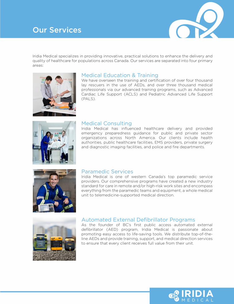
Iridia Medical specializes in providing innovative, practical solutions to enhance the delivery and quality of healthcare for populations across Canada. Our services are separated into four primary areas:
Medical Education & TrainingWe have overseen the training and certification of over four thousand lay rescuers in the use of AEDs, and over three thousand medical professionals via our advanced training programs, such as Advanced Cardiac Life Support (ACLS) and Pediatric Advanced Life Support (PALS).
Medical ConsultingIridia Medical has influenced healthcare delivery and provided emergency preparedness guidance for public and private sector organizations across North America. Our clients include health authorities, public healthcare facilities, EMS providers, private surgery and diagnostic imaging facilities, and police and fire departments.
Paramedic ServicesIridia Medical is one of western Canada’s top paramedic service providers. Our comprehensive programs have created a new industry standard for care in remote and/or high-risk work sites and encompass everything from the paramedic teams and equipment, a whole medical unit to telemedicine-supported medical direction.
Automated External Defibrillator ProgramsAs the founder of BC’s first public access automated external defibrillator (AED) program, Iridia Medical is passionate about promoting easy access to life-saving tools. We distribute top-of-the-line AEDs and provide training, support, and medical direction services to ensure that every client receives full value from their unit.
Our Services
Our Withdrawal & Transfer Policies
Participant requests for transfers, withdrawals, or refunds must be made in writing to Iridia Medical prior to the start of the course.
Please email your written notification to [email protected].
Withdrawals:
• If notice of withdrawal is given more than 14 days prior to the registered course date, the participant will receive a full refund less a 25% administration fee.
• If notice of withdrawal is given less than 14 days prior to the registered course date, the entire tuition or class fee will be non-refundable.
Transfers:
• If notice of transfer is given more than 30 days prior to the registered course date, no transfer fee will be charged.
• If notice of transfer is given from 14-30 days prior to the registered course date a transfer fee of 15% of the total fees will be applied.
• Unfortunately due to our commitment to our instructors, we are unable to accept transfers less than 14 days prior to the registered course date.
• Transfer fees are applicable for each transfer.
Cancellations:
• Iridia Medical reserves the right to cancel a class and refund registration fees due to insufficient registration or other circumstances beyond our control.
• Iridia Medical will provide participants two weeks’ notice of cancellation prior to the course date.
Books:
• All book sales are final.
Page 4
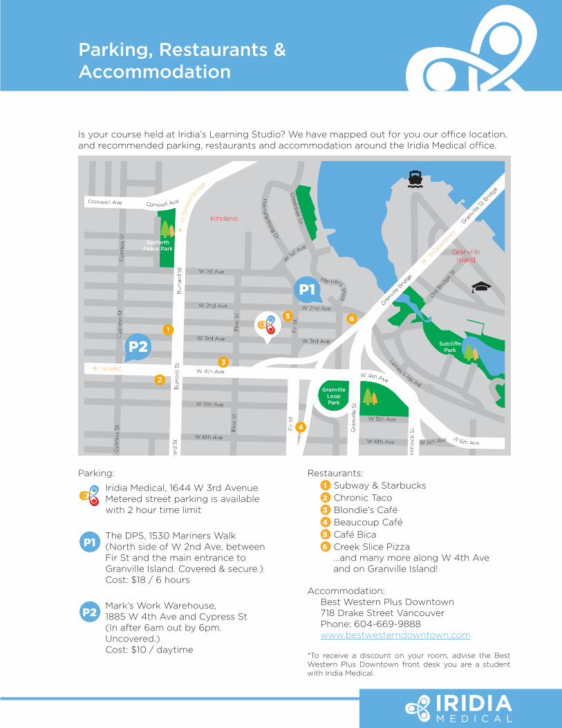
Is your course held at Iridia’s Learning Studio? We have mapped out for you our office location, and recommended parking, restaurants and accommodation around the Iridia Medical office.
Parking, Restaurants & Accommodation
Parking:
Iridia Medical, 1644 W 3rd AvenueMetered street parking is available with 2 hour time limit
P1 The DPS, 1530 Mariners Walk(North side of W 2nd Ave, between Fir St and the main entrance to Granville Island. Covered & secure.)Cost: $18 / 6 hours
P2 Mark’s Work Warehouse,1885 W 4th Ave and Cypress St(In after 6am out by 6pm. Uncovered.)Cost: $10 / daytime
Restaurants:1 Subway & Starbucks2 Chronic Taco3 Blondie’s Café4 Beaucoup Café5 Café Bica6 Creek Slice Pizza …and many more along W 4th Ave
and on Granville Island!
Accommodation:Best Western Plus Downtown718 Drake Street VancouverPhone: 604-669-9888www.bestwesterndowntown.com
*To receive a discount on your room, advise the Best Western Plus Downtown front desk you are a student with Iridia Medical.
ACLS Precourse Preparation
Your success in this course depends on adequate precourse preparation. To best prepare and achieve the objectives of the course, please allow at least eight (8) hours to review the following ACLS resources - available at www.iridiamedical.com.
Mandatory:• Advanced Cardiac Life Support Provider Manual This is the required study guide for the course. This book applies new ACLS concepts to
realistic situations and includes sections on all of the skill and knowledge requirements listed in the objectives. Also included are a quick reference guide and reminder cards, and two ACLS pocket reference cards.
• Handbook of Emergency Cardiovascular Care for Healthcare Providers Highly recommended by our Instructors as a real-life reference tool, this pocket sized,
detailed manual can be used at any time during the course and on the job.
Recommended:• ACLS & Emergency Cardiovascular Care 2011 Essentials for Health Professionals in Hospital
- Adult BLS & ACLS Algorithms This summary document, included with your course package, provides an organized guide
for responding to cardiac emergencies. These essentials identify the core concepts of the course, while the manuals provide reference material for the algorithms.
• Highlights of the 2010 American Heart Association Guidelines for CPR and ECC
• ACLS Written Precourse Self-Assessment
The ACLS core cases will be reviewed during the course, which will assist you in gaining the knowledge and develop the ability to:
1. Recognize and initiate early management of peri-arrest conditions that may result in cardiac arrest or complicate resuscitation outcome.
2. Demonstrate proficiency in providing BLS care, including prioritizing chest compressions and integrating Automatic External Defibrillator use.
3. Recognize and manage respiratory arrest.
4. Recognize and manage cardiac arrest until return of spontaneous circulation, termination of resuscitation, or transfer of care, including immediate post-arrest care.
5. Recognize and initiate early management of Acute Coronary Syndromes (ACS), including appropriate disposition.
6. Recognize and initiate early management of stroke, including appropriate disposition.
7. Demonstrate effective communication as a member or leader of a resuscitation team and recognize the impact of team dynamics on overall team performance.
Page 6
Precourse Preparation Checklist:
Complete a Basic Life Support (BLS) for Healthcare Providers Course
CPR Competency
Understand the 10 Core Cases in the ACLS Provider Manual
Understand the ACLS algorithms for the Core Cases
Complete the ACLS Self-Assessment Tests
To successfully complete the course, you must:
• Demonstrate competency in BLS knowledge and skills. You will be tested on Bag-Mask Ventilation and CPR/AED skills.
• Demonstrate competency in ACLS knowledge and skills through your performance during case scenarios.
• Pass the closed-book, 50 question multiple-choice exam, with a minimum score of 84%. In the incident you are unsuccessful in demonstrating BLS/ACLS practical knowledge and skills during a testable case scenario or do not achieve 84% on the written exam, you will be offered a second attempt at the end of the course.
If either the second attempts are unsuccessful, arrangements can be made with Iridia Medical to participate in further training courses and to repeat the evaluation process.
ACLS Provider Agenda - Day 1
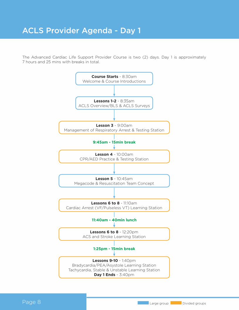
The Advanced Cardiac Life Support Provider Course is two (2) days. Day 1 is approximately 7 hours and 25 mins with breaks in total.
Large group Divided groupsPage 8
Course Starts - 8:30amWelcome & Course Introductions
Lessons 1-2 - 8:35amACLS Overview/BLS & ACLS Surveys
Lesson 4 - 10:00amCPR/AED Practice & Testing Station
Lessons 6 to 8 - 11:10amCardiac Arrest (VF/Pulseless VT) Learning Station
Lesson 3 - 9:00amManagement of Respiratory Arrest & Testing Station
Lessons 6 to 8 - 12:20pmACS and Stroke Learning Station
Lessons 9-10 - 1:40pmBradycardia/PEA/Asystole Learning Station
Tachycardia, Stable & Unstable Learning StationDay 1 Ends - 3:40pm
Lesson 5 - 10:45amMegacode & Resuscitation Team Concept
9:45am - 15min break
1:25pm - 15min break
11:40am - 40min lunch
ACLS Provider Agenda - Day 2
Large group Divided groups
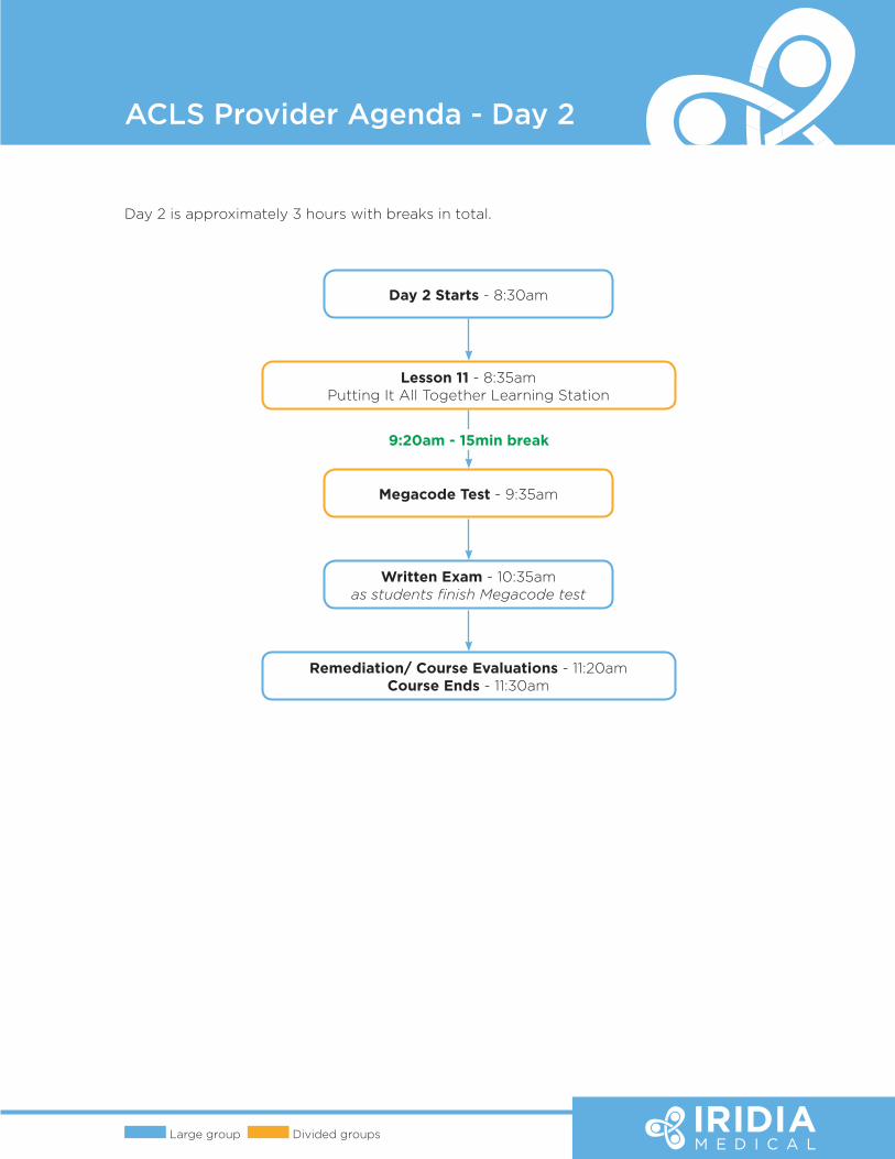
Day 2 is approximately 3 hours with breaks in total.
Day 2 Starts - 8:30am
Lesson 11 - 8:35amPutting It All Together Learning Station
Megacode Test - 9:35am
Written Exam - 10:35amas students finish Megacode test
Remediation/ Course Evaluations - 11:20amCourse Ends - 11:30am
9:20am - 15min break
ACLS Update Agenda
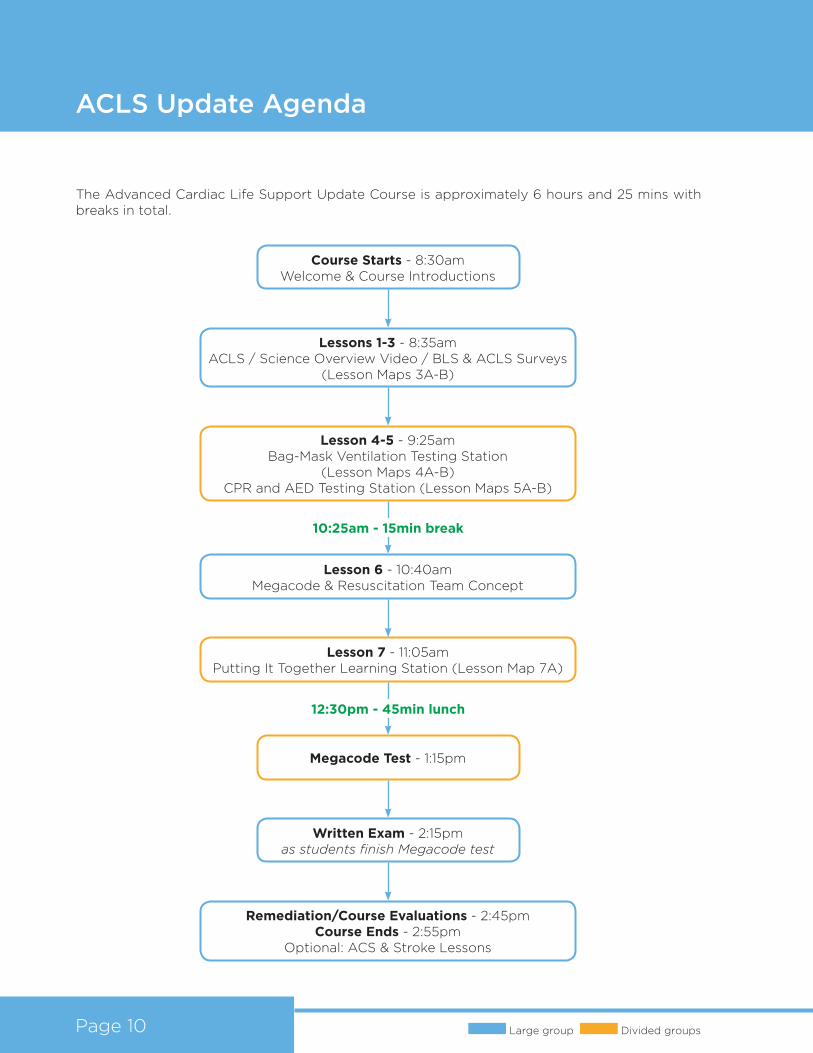
The Advanced Cardiac Life Support Update Course is approximately 6 hours and 25 mins with breaks in total.
Page 10 Large group Divided groups
Course Starts - 8:30amWelcome & Course Introductions
Lessons 1-3 - 8:35amACLS / Science Overview Video / BLS & ACLS Surveys
(Lesson Maps 3A-B)
Lesson 6 - 10:40amMegacode & Resuscitation Team Concept
Megacode Test - 1:15pm
Lesson 4-5 - 9:25amBag-Mask Ventilation Testing Station
(Lesson Maps 4A-B)CPR and AED Testing Station (Lesson Maps 5A-B)
Written Exam - 2:15pmas students finish Megacode test
Remediation/Course Evaluations - 2:45pmCourse Ends - 2:55pm
Optional: ACS & Stroke Lessons
Lesson 7 - 11:05amPutting It Together Learning Station (Lesson Map 7A)
10:25am - 15min break
12:30pm - 45min lunch
Reading
ACLS & EMERGENCY CARDIOVASCULAR
CARE 2011Essentials for Health
Professionals in Hospital

Enabling Peace of Mind
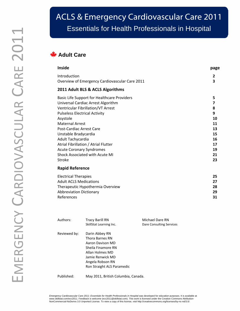
Inside page
Introduction 2 Overview of Emergency Cardiovascular Care 2011 3
2011 Adult BLS & ACLS Algorithms
Basic Life Support for Healthcare Providers 5Universal Cardiac Arrest Algorithm 7Ventricular Fibrillation/VT Arrest 8Pulseless Electrical Activity 9Asystole 10Maternal Arrest 11Post-Cardiac Arrest Care 13Unstable Bradycardia 15Adult Tachycardia 16Atrial Fibrillation / Atrial Flutter 17Acute Coronary Syndromes 19Shock Associated with Acute MI 21 Stroke 23
Rapid Reference
Electrical Therapies 25Adult ACLS Medications 27Therapeutic Hypothermia Overview 28Abbreviation Dictionary 29References 31
ACLS & Emergency Cardiovascular Care 2011 Essentials for Health Professionals in Hospital
EMER
GEN
CY
CA
RD
IOV
ASC
ULA
R C
AR
E 2
01
1
Authors: Tracy Barill RN Michael Dare RNSkillStat Learning Inc. Dare Consulting Services
Reviewed by: Darin Abbey RNThora Barnes RNAaron Davison MDSheila Finamore RNAllan Holmes MDJamie Renwick MDAngela Robson RNRon Straight ALS Paramedic
Published: May 2011, British Columbia, Canada.
Emergency Cardiovascular Care 2011: Essentials for Health Professionals in Hospital was developed for education purposes. It is available at
www.skillstat.com/ecc2011. Feedback is welcome ([email protected]). This work is licensed under the Creative Commons Attribution-
NonCommercial-NoDerivs 3.0 Unported License. To view a copy of this license, visit http://creativecommons.org/licenses/by-nc-nd/3.0/.
Adult Care
Introduction
2Barill/Dare
ECC
20
11
On October 18, 2010 the International Liaison Committee On Resuscitation (ILCOR) released a major 5 year update to the CPR and Emergency Cardiovascular Care (ECC) Guidelines. The American Heart Association (AHA) and the European Resuscitation Council (ERC) in turn released similar interpretations of this release.
These guidelines provide core algorithms to outline key actions and decisions for the immediate care of common cardiovascular emergencies:
· cardiac arrest· post-cardiac arrest· hemodynamically unstable bradycardia and tachycardia· acute coronary syndromes· stroke
These algorithms are central to advanced cardiac life support and pediatric advanced life support courses. Considerable material included in the major release documents is not included in these core algorithms. This makes sense. Most health providers focus their attention on the more likely core subset of possible cardiovascular emergencies.
For advanced care health professionals in hospital, though, their required skills encompass a broader scope of practice, a full complement of therapeutic interventions and a complex array of morbidities. Support documents and courses specific to this setting harness added content to supplement the core algorithms. This ‘best fit’ approach builds on the ‘one-size-fits-all’ core algorithms to ensure optimal care and improved patient outcomes.
ACLS & ECC 2011 Essentials is an education supplement for healthcare professionals tasked with the emergency cardiac care of adults in hospital. This summary of 2010/2011 emergency cardiovascular care guidelines combines recent resuscitation science, suggested procedures and guiding principles into an organized approach to in-hospital emergency cardiovascular care. Canadian Stroke Strategy guidelines and Canadian Cardiovascular Society atrial fibrillation protocols round out this document. We hope that a solid understanding and long term concept adoption of the latest in hospital emergency cardiovascular care science is enhanced with this supplement.
Much thanks go to the advanced care practitioners who reviewed this document. Their significant investments in time and their many suggestions have added to this document. Despite great efforts placed in the creation of these documents, error free results rarely occur despite several reviews and edits. Please direct any suggestions or questions to [email protected].
Both the AHA and the ERC are careful to point out that not all recommendations will apply to all rescuers or all situations. The algorithms included here are not intended to replace established AHA | ERC guidelines or sound medical judgement. Resuscitation science is dynamic, with frequent updates.
Find the official ECC 2010 guidelines, executive summaries and updates online. Links are included below for your convenience.
International Liaison Committee for Resuscitation (http://www.ilcor.org/en/home/)American Heart Association (http://guidelines.ecc.org)
Canadian Cardiovascular Society Atrial Fibrillation Guidelines (http://www.ccs.ca/consensus_conferences/cc_library_e.aspx)
European Resuscitation Council (http://www.cprguidelines.eu/2010/)
For educational purposes only
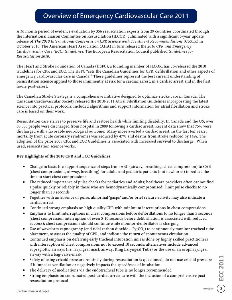
A 36 month period of evidence evaluation by 356 resuscitation experts from 29 countries coordinated through the International Liaison Committee on Resuscitation (ILCOR) culminated with a significant 5-year update release of The 2010 International Consensus on CPR Science with Treatment Recommendations (CoSTR) in October 2010. The American Heart Association (AHA) in turn released the 2010 CPR and Emergency Cardiovascular Care (ECC) Guidelines. The European Resuscitation Council published Guidelines for Resuscitation 2010.
The Heart and Stroke Foundation of Canada (HSFC), a founding member of ILCOR, has co-released the 2010 Guidelines for CPR and ECC. The HSFC “sets the Canadian Guidelines for CPR, defibrillation and other aspects of emergency cardiovascular care in Canada.” These guidelines represent the best current understanding of resuscitation science applied to those imminently at risk for a cardiac arrest, in a cardiac arrest and in the first hours post-arrest.
The Canadian Stroke Strategy is a comprehensive initiative designed to optimize stroke care in Canada. The Canadian Cardiovascular Society released the 2010-2011 Atrial Fibrillation Guidelines incorporating the latest science into practical protocols. Included algorithms and support information for atrial fibrillation and stroke care is based on their work.
Resuscitation care strives to preserve life and restore health while limiting disability. In Canada and the US, over 50 000 people were discharged from hospital in 2009 following a cardiac arrest. Recent data show that 75% were discharged with a favorable neurological outcome. Many more averted a cardiac arrest. In the last ten years, mortality from acute coronary syndromes was reduced by 47% and deaths from stroke reduced by 14%. The adoption of the prior 2005 CPR and ECC Guidelines is associated with increased survival to discharge. When used, resuscitation science works. Key Highlights of the 2010 CPR and ECC Guidelines
· Change in basic life support sequence of steps from ABC (airway, breathing, chest compression) to CAB (chest compressions, airway, breathing) for adults and pediatric patients (not newborns) to reduce the time to start chest compressions
· The reduced importance of pulse checks for pediatrics and adults; healthcare providers often cannot find a pulse quickly or reliably in those who are hemodynamically compromised; limit pulse checks to no longer than 10 seconds
· Together with an absence of pulse, abnormal ‘gasps’ and/or brief seizure activity may also indicate a cardiac arrest
· Continued strong emphasis on high quality CPR with minimum interruptions in chest compressions
· Emphasis to limit interruptions in chest compressions before defibrillations to no longer than 5 seconds (chest compression interruption of even 5-10 seconds before defibrillation is associated with reduced success); chest compressions should continue while monitor-defibrillator is charging
· Use of waveform capnography (end tidal carbon dioxide – PETCO2) to continuously monitor tracheal tube placement, to assess the quality of CPR, and indicate the return of spontaneous circulation
· Continued emphasis on deferring early tracheal intubation unless done by highly skilled practitioners with interruption of chest compressions not to exceed 10 seconds; alternatives include advanced supraglottic airways (i.e. laryngeal mask airway, King Laryngeal Tube) or the use of an oropharyngeal airway with a bag-valve-mask
· Safety of using cricoid pressure routinely during resuscitation is questioned; do not use cricoid pressure if it impedes ventilation or negatively impacts the speed/ease of intubation
· The delivery of medications via the endotracheal tube is no longer recommended
· Strong emphasis on coordinated post-cardiac arrest care with the inclusion of a comprehensive post resuscitation protocol
(continued on next page)
Overview of Emergency Cardiovascular Care 2011
3
ECC
20
11
Barill/Dare
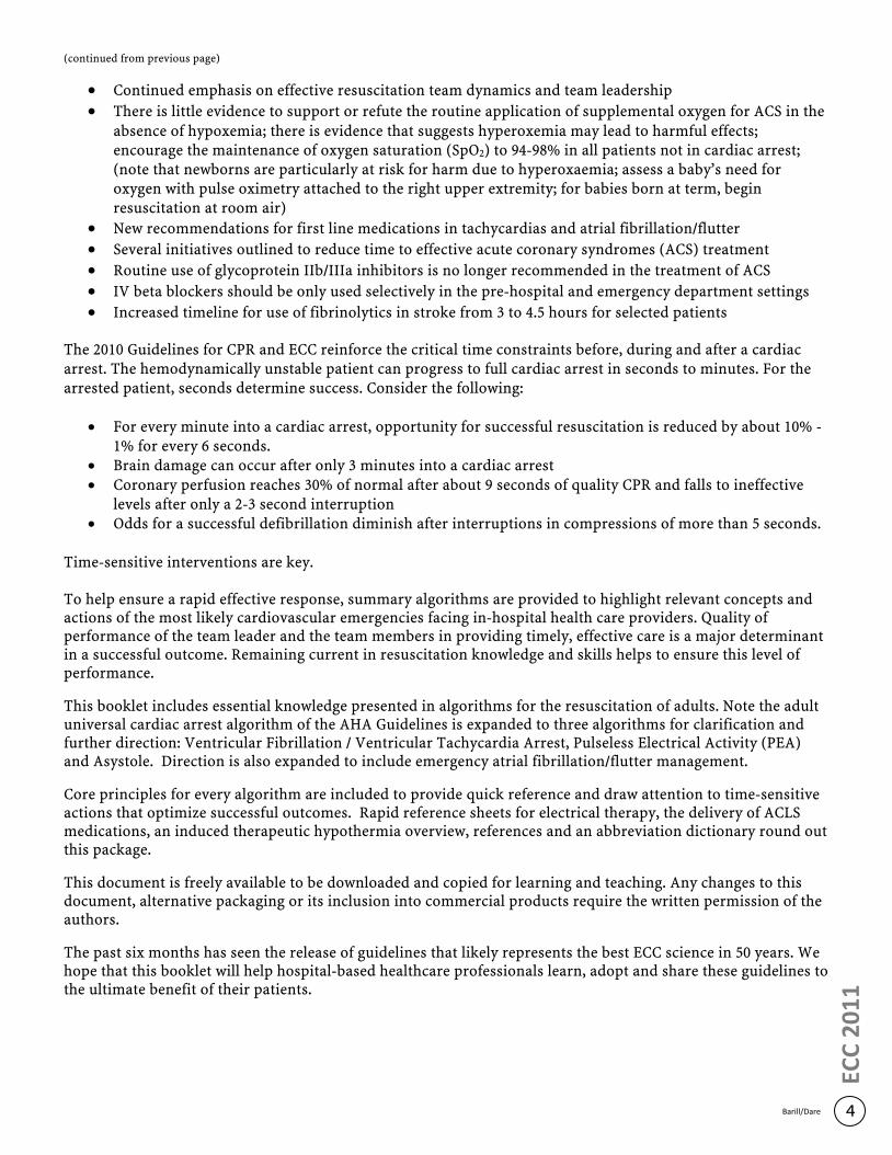
(continued from previous page)
· Continued emphasis on effective resuscitation team dynamics and team leadership
· There is little evidence to support or refute the routine application of supplemental oxygen for ACS in the absence of hypoxemia; there is evidence that suggests hyperoxemia may lead to harmful effects; encourage the maintenance of oxygen saturation (SpO2) to 94-98% in all patients not in cardiac arrest; (note that newborns are particularly at risk for harm due to hyperoxaemia; assess a baby’s need for oxygen with pulse oximetry attached to the right upper extremity; for babies born at term, begin resuscitation at room air)
· New recommendations for first line medications in tachycardias and atrial fibrillation/flutter
· Several initiatives outlined to reduce time to effective acute coronary syndromes (ACS) treatment
· Routine use of glycoprotein IIb/IIIa inhibitors is no longer recommended in the treatment of ACS
· IV beta blockers should be only used selectively in the pre-hospital and emergency department settings
· Increased timeline for use of fibrinolytics in stroke from 3 to 4.5 hours for selected patients
The 2010 Guidelines for CPR and ECC reinforce the critical time constraints before, during and after a cardiac arrest. The hemodynamically unstable patient can progress to full cardiac arrest in seconds to minutes. For the arrested patient, seconds determine success. Consider the following:
· For every minute into a cardiac arrest, opportunity for successful resuscitation is reduced by about 10% - 1% for every 6 seconds.
· Brain damage can occur after only 3 minutes into a cardiac arrest · Coronary perfusion reaches 30% of normal after about 9 seconds of quality CPR and falls to ineffective
levels after only a 2-3 second interruption· Odds for a successful defibrillation diminish after interruptions in compressions of more than 5 seconds.
Time-sensitive interventions are key.
To help ensure a rapid effective response, summary algorithms are provided to highlight relevant concepts and actions of the most likely cardiovascular emergencies facing in-hospital health care providers. Quality of performance of the team leader and the team members in providing timely, effective care is a major determinant in a successful outcome. Remaining current in resuscitation knowledge and skills helps to ensure this level of performance.
This booklet includes essential knowledge presented in algorithms for the resuscitation of adults. Note the adult universal cardiac arrest algorithm of the AHA Guidelines is expanded to three algorithms for clarification and further direction: Ventricular Fibrillation / Ventricular Tachycardia Arrest, Pulseless Electrical Activity (PEA) and Asystole. Direction is also expanded to include emergency atrial fibrillation/flutter management.
Core principles for every algorithm are included to provide quick reference and draw attention to time-sensitive actions that optimize successful outcomes. Rapid reference sheets for electrical therapy, the delivery of ACLS medications, an induced therapeutic hypothermia overview, references and an abbreviation dictionary round out this package.
This document is freely available to be downloaded and copied for learning and teaching. Any changes to this document, alternative packaging or its inclusion into commercial products require the written permission of the authors.
The past six months has seen the release of guidelines that likely represents the best ECC science in 50 years. We hope that this booklet will help hospital-based healthcare professionals learn, adopt and share these guidelines to the ultimate benefit of their patients.
4Barill/Dare
ECC
20
11
5
ECC
20
11
– A
du
lt B
LS
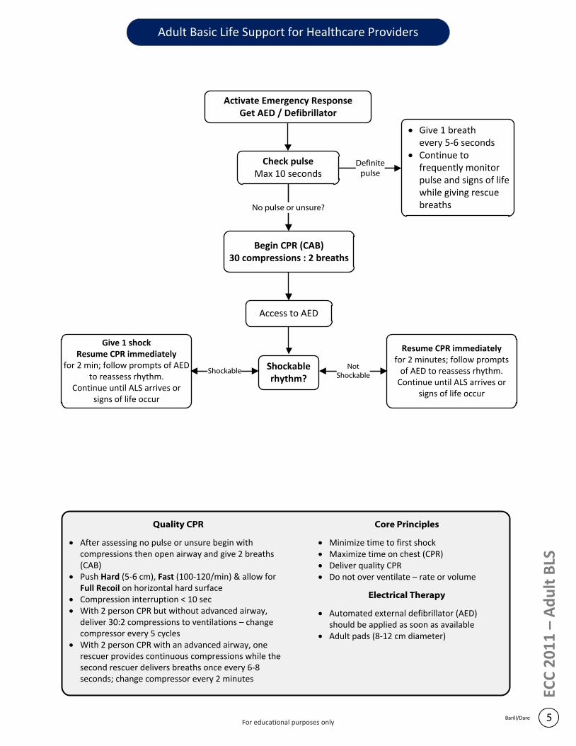
Check pulseMax 10 seconds
Begin CPR (CAB)30 compressions : 2 breaths
Activate Emergency ResponseGet AED / Defibrillator
· Give 1 breath every 5-6 seconds
· Continue to frequently monitor pulse and signs of life while giving rescue breaths
Definite pulse
No pulse or unsure?
Access to AED
Shockable rhythm?
Give 1 shockResume CPR immediately
for 2 min; follow prompts of AED to reassess rhythm.
Continue until ALS arrives or signs of life occur
Resume CPR immediately for 2 minutes; follow prompts
of AED to reassess rhythm.Continue until ALS arrives or
signs of life occur
ShockableNot
Shockable
Quality CPR
· After assessing no pulse or unsure begin with compressions then open airway and give 2 breaths (CAB)
· Push Hard (5-6 cm), Fast (100-120/min) & allow for Full Recoil on horizontal hard surface
· Compression interruption < 10 sec· With 2 person CPR but without advanced airway,
deliver 30:2 compressions to ventilations – change compressor every 5 cycles
· With 2 person CPR with an advanced airway, one rescuer provides continuous compressions while the second rescuer delivers breaths once every 6-8 seconds; change compressor every 2 minutes
Core Principles
· Minimize time to first shock· Maximize time on chest (CPR)· Deliver quality CPR· Do not over ventilate – rate or volume
Electrical Therapy
· Automated external defibrillator (AED) should be applied as soon as available
· Adult pads (8-12 cm diameter)
Adult Basic Life Support for Healthcare Providers
Barill/DareFor educational purposes only
6 CPR & ECC 2011 – Adult BLS
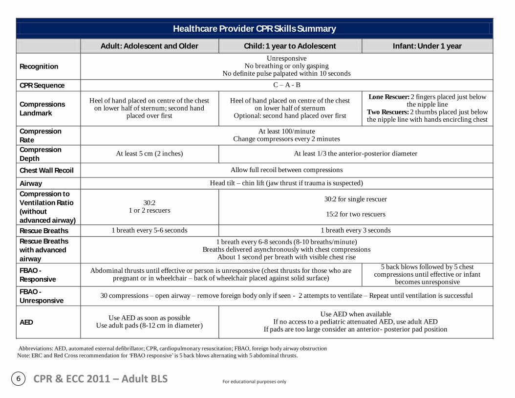
Healthcare Provider CPR Skills Summary
Adult: Adolescent and Older Child: 1 year to Adolescent Infant: Under 1 year
Recognition Unresponsive
No breathing or only gasping No definite pulse palpated within 10 seconds
CPR Sequence C – A - B
Compressions Landmark
Heel of hand placed on centre of the chest on lower half of sternum; second hand
placed over first
Heel of hand placed on centre of the chest on lower half of sternum
Optional: second hand placed over first
Lone Rescuer: 2 fingers placed just below the nipple line
Two Rescuers: 2 thumbs placed just below the nipple line with hands encircling chest
Compression Rate
At least 100/minute Change compressors every 2 minutes
Compression Depth
At least 5 cm (2 inches) At least 1/3 the anterior-posterior diameter
Chest Wall Recoil Allow full recoil between compressions
Airway Head tilt – chin lift (jaw thrust if trauma is suspected)
Compression to Ventilation Ratio (without advanced airway)
30:2 1 or 2 rescuers
30:2 for single rescuer
15:2 for two rescuers
Rescue Breaths 1 breath every 5-6 seconds 1 breath every 3 seconds
Rescue Breaths with advanced airway
1 breath every 6-8 seconds (8-10 breaths/minute) Breaths delivered asynchronously with chest compressions
About 1 second per breath with visible chest rise
FBAO - Responsive
Abdominal thrusts until effective or person is unresponsive (chest thrusts for those who are pregnant or in wheelchair – back of wheelchair placed against solid surface)
5 back blows followed by 5 chest compressions until effective or infant
becomes unresponsive
FBAO - Unresponsive
30 compressions – open airway – remove foreign body only if seen - 2 attempts to ventilate – Repeat until ventilation is successful
AED Use AED as soon as possible
Use adult pads (8-12 cm in diameter)
Use AED when available If no access to a pediatric attenuated AED, use adult AED
If pads are too large consider an anterior- posterior pad position
Abbreviations: AED, automated external defibrillator; CPR, cardiopulmonary resuscitation; FBAO, foreign body airway obstruction
Note: ERC and Red Cross recommendation for ‘FBAO responsive’ is 5 back blows alternating with 5 abdominal thrusts.
For educational purposes only
ECC
20
11
- A
CLS
7
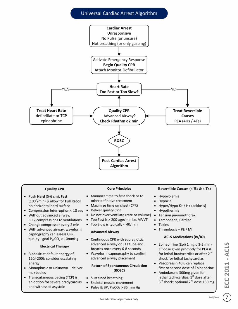
Universal Cardiac Arrest Algorithm
ROSC
Activate Emergency ResponseBegin Quality CPR
Attach Monitor-Defibrillator
Cardiac ArrestUnresponsive
No Pulse (or unsure) Not breathing (or only gasping)
Heart Rate Too Fast or Too Slow?
Post-Cardiac ArrestAlgorithm
Treat Heart Ratedefibrillate or TCP
epinephrine
Treat Reversible Causes
PEA (4Hs / 4Ts)
Quality CPR Advanced Airway?
Check Rhythm q2 min
NOYES
Barill/Dare
Quality CPR
· Push Hard (5-6 cm), Fast (100+/min) & allow for Full Recoil on horizontal hard surface
· Compression interruption < 10 sec· Without advanced airway,
30:2 compressions to ventilations· Change compressor every 2 min· With advanced airway, waveform
capnography can assess CPR quality - goal PETCO2 > 10mmHg
Electrical Therapy
· Biphasic at default energy of 120J-200J; consider escalating energy
· Monophasic or unknown – deliver max Joules
· Transcutaneous pacing (TCP) is an option for severe bradycardias and witnessed asystole
Reversible Causes (4 Hs & 4 Ts)
· Hypovolemia· Hypoxia· Hyper/Hypo K+ / H+ (acidosis)· Hypothermia· Tension pneumothorax· Tamponade, Cardiac· Toxins· Thrombosis – PE / MI
ACLS Medications (IV/IO)
· Epinephrine (Epi) 1 mg q 3-5 min - 1st dose given promptly for PEA & for lethal bradycardias or after 2nd shock for lethal tachycardias
· Vasopressin 40 u can replacefirst or second dose of Epinephrine
· Amiodarone 300mg given for lethal tachycardias; 1st dose after 3rd shock; optional 2nd dose 150 mg
Core Principles
· Minimize time to first shock or to other definitive treatment
· Maximize time on chest (CPR)· Deliver quality CPR· Do not over ventilate (rate or volume)· Too Fast is > 200-age/min i.e. VF/VT· Too Slow is typically < 40/min
Advanced Airway
· Continuous CPR with supraglottic advanced airway or ETT tube and breaths once every 6-8 seconds
· Waveform capnography to confirm advanced airway placement
Return of Spontaneous Circulation(ROSC)
· Sustained breathing· Skeletal muscle movement· Pulse & BP; PETCO2 > 35 mm Hg
For educational purposes only
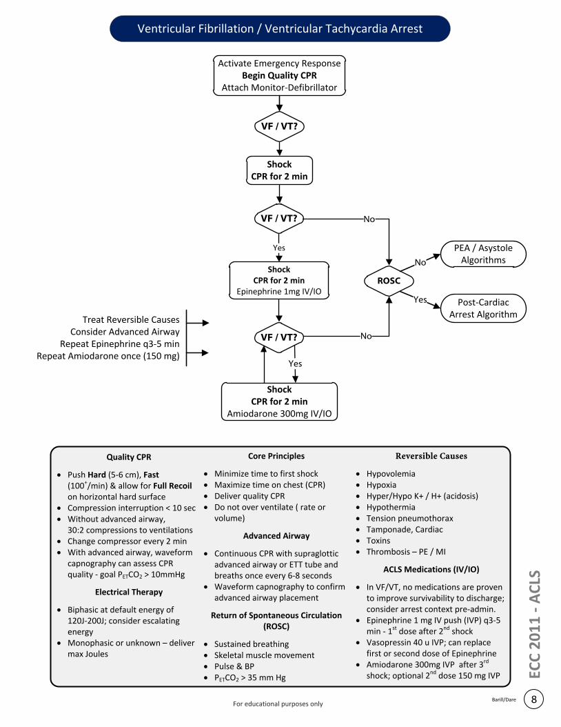
Ventricular Fibrillation / Ventricular Tachycardia Arrest
Activate Emergency ResponseBegin Quality CPR
Attach Monitor-Defibrillator
8
ShockCPR for 2 min
VF / VT?
Yes
ShockCPR for 2 min
Epinephrine 1mg IV/IO
VF / VT?
Yes
ShockCPR for 2 min
Amiodarone 300mg IV/IO
ROSC
PEA / Asystole Algorithms
Post-Cardiac Arrest Algorithm
No
Yes
No
No
Treat Reversible CausesConsider Advanced Airway
Repeat Epinephrine q3-5 min Repeat Amiodarone once (150 mg)
Barill/Dare
ECC
20
11
- A
CLS
VF / VT?
Quality CPR
· Push Hard (5-6 cm), Fast (100+/min) & allow for Full Recoil on horizontal hard surface
· Compression interruption < 10 sec· Without advanced airway,
30:2 compressions to ventilations· Change compressor every 2 min· With advanced airway, waveform
capnography can assess CPR quality - goal PETCO2 > 10mmHg
Electrical Therapy
· Biphasic at default energy of 120J-200J; consider escalating energy
· Monophasic or unknown – deliver max Joules
Reversible Causes
· Hypovolemia· Hypoxia· Hyper/Hypo K+ / H+ (acidosis)· Hypothermia· Tension pneumothorax· Tamponade, Cardiac· Toxins· Thrombosis – PE / MI
ACLS Medications (IV/IO)
· In VF/VT, no medications are proven to improve survivability to discharge; consider arrest context pre-admin.
· Epinephrine 1 mg IV push (IVP) q3-5 min - 1st dose after 2nd shock
· Vasopressin 40 u IVP; can replacefirst or second dose of Epinephrine
· Amiodarone 300mg IVP after 3rd shock; optional 2nd dose 150 mg IVP
Core Principles
· Minimize time to first shock· Maximize time on chest (CPR)· Deliver quality CPR· Do not over ventilate ( rate or
volume)
Advanced Airway
· Continuous CPR with supraglottic advanced airway or ETT tube and breaths once every 6-8 seconds
· Waveform capnography to confirm advanced airway placement
Return of Spontaneous Circulation(ROSC)
· Sustained breathing· Skeletal muscle movement· Pulse & BP · PETCO2 > 35 mm Hg
For educational purposes only
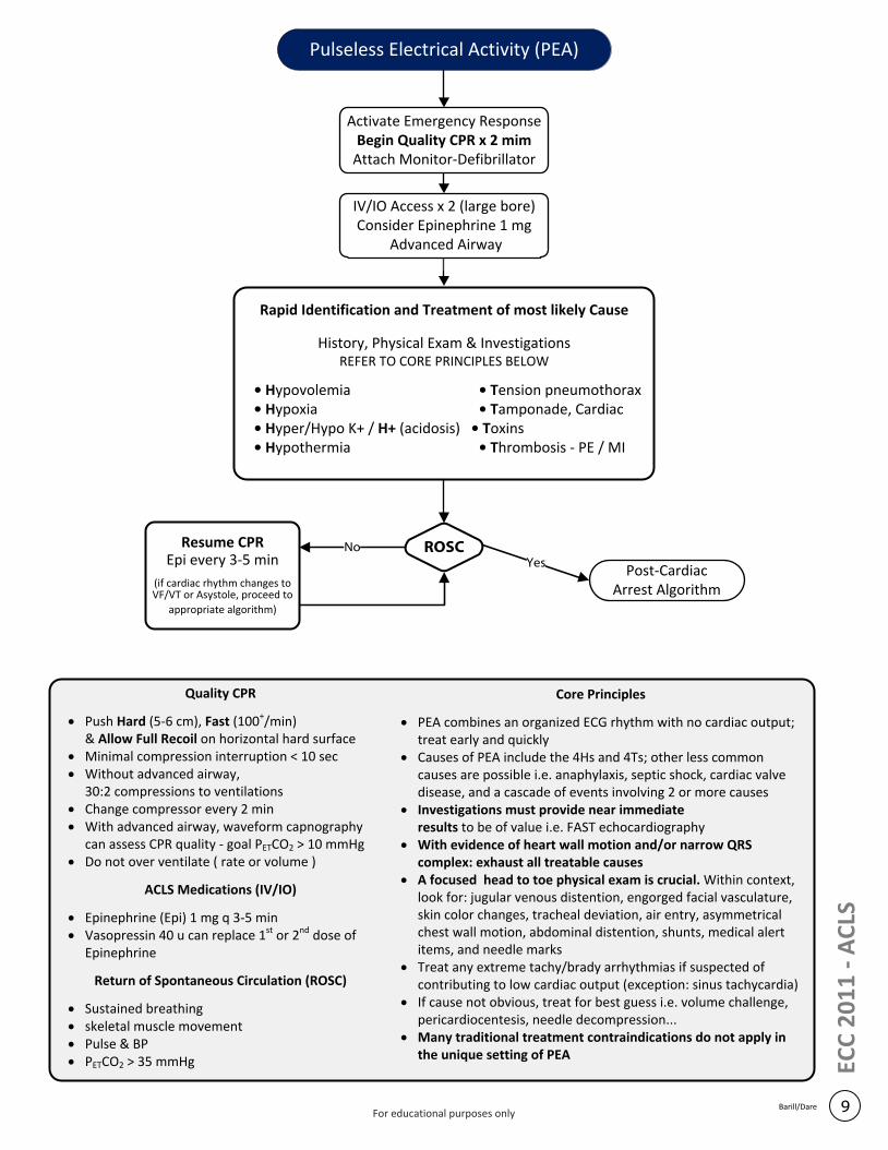
Activate Emergency ResponseBegin Quality CPR x 2 mim
Attach Monitor-Defibrillator
Rapid Identification and Treatment of most likely Cause
History, Physical Exam & InvestigationsREFER TO CORE PRINCIPLES BELOW
• Hypovolemia • Tension pneumothorax• Hypoxia • Tamponade, Cardiac• Hyper/Hypo K+ / H+ (acidosis) • Toxins • Hypothermia • Thrombosis - PE / MI
IV/IO Access x 2 (large bore)Consider Epinephrine 1 mg
Advanced Airway
Post-Cardiac Arrest Algorithm
Resume CPREpi every 3-5 min
(if cardiac rhythm changes toVF/VT or Asystole, proceed to
appropriate algorithm)
ROSCNoYes
Pulseless Electrical Activity (PEA)
9Barill/Dare
ECC
20
11
- A
CLS
Quality CPR
· Push Hard (5-6 cm), Fast (100+/min) & Allow Full Recoil on horizontal hard surface
· Minimal compression interruption < 10 sec· Without advanced airway,
30:2 compressions to ventilations · Change compressor every 2 min· With advanced airway, waveform capnography
can assess CPR quality - goal PETCO2 > 10 mmHg· Do not over ventilate ( rate or volume )
ACLS Medications (IV/IO)
· Epinephrine (Epi) 1 mg q 3-5 min· Vasopressin 40 u can replace 1st or 2nd dose of
Epinephrine
Return of Spontaneous Circulation (ROSC)
· Sustained breathing· skeletal muscle movement· Pulse & BP · PETCO2 > 35 mmHg
Core Principles
· PEA combines an organized ECG rhythm with no cardiac output; treat early and quickly
· Causes of PEA include the 4Hs and 4Ts; other less common causes are possible i.e. anaphylaxis, septic shock, cardiac valve disease, and a cascade of events involving 2 or more causes
· Investigations must provide near immediate results to be of value i.e. FAST echocardiography
· With evidence of heart wall motion and/or narrow QRS complex: exhaust all treatable causes
· A focused head to toe physical exam is crucial. Within context, look for: jugular venous distention, engorged facial vasculature, skin color changes, tracheal deviation, air entry, asymmetrical chest wall motion, abdominal distention, shunts, medical alert items, and needle marks
· Treat any extreme tachy/brady arrhythmias if suspected of contributing to low cardiac output (exception: sinus tachycardia)
· If cause not obvious, treat for best guess i.e. volume challenge, pericardiocentesis, needle decompression...
· Many traditional treatment contraindications do not apply in the unique setting of PEA
For educational purposes only
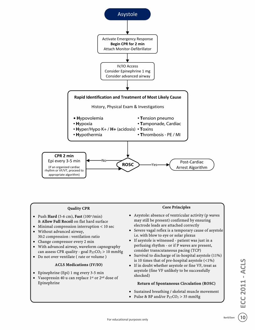
Activate Emergency ResponseBegin CPR for 2 min
Attach Monitor-Defibrillator
Rapid Identification and Treatment of Most Likely Cause
History, Physical Exam & Investigations
• Hypovolemia • Tension pneumo• Hypoxia • Tamponade, Cardiac• Hyper/Hypo K+ / H+ (acidosis) • Toxins • Hypothermia • Thrombosis - PE / MI
IV/IO AccessConsider Epinephrine 1 mg Consider advanced airway
Post-Cardiac Arrest Algorithm
CPR 2 minEpi every 3-5 min
(if an organized cardiac rhythm or VF/VT, proceed to
appropriate algorithm)
ROSC YesNo
Asystole
10Barill/Dare
ECC
20
11
- A
CLS
Quality CPR
· Push Hard (5-6 cm), Fast (100+/min) & Allow Full Recoil on flat hard surface
· Minimal compression interruption < 10 sec· Without advanced airway,
30:2 compression : ventilation ratio· Change compressor every 2 min· With advanced airway, waveform capnography
can assess CPR quality - goal PETCO2 > 10 mmHg· Do not over ventilate ( rate or volume )
ACLS Medications (IV/IO)
· Epinephrine (Epi) 1 mg every 3-5 min· Vasopressin 40 u can replace 1st or 2nd dose of
Epinephrine
Core Principles
· Asystole: absence of ventricular activity (p waves may still be present) confirmed by ensuring electrode leads are attached correctly
· Severe vagal reflex is a temporary cause of asystole i.e. with blow to eye or solar plexus
· If asystole is witnessed - patient was just in a perfusing rhythm - or if P waves are present, consider transcutaneous pacing (TCP)
· Survival to discharge of in-hospital asystole (11%) is 10 times that of pre-hospital asystole (<1%)
· If in doubt whether asystole or fine VF, treat as asystole (fine VF unlikely to be successfully shocked)
Return of Spontaneous Circulation (ROSC)
· Sustained breathing / skeletal muscle movement· Pulse & BP and/or PETCO2 > 35 mmHg
For educational purposes only
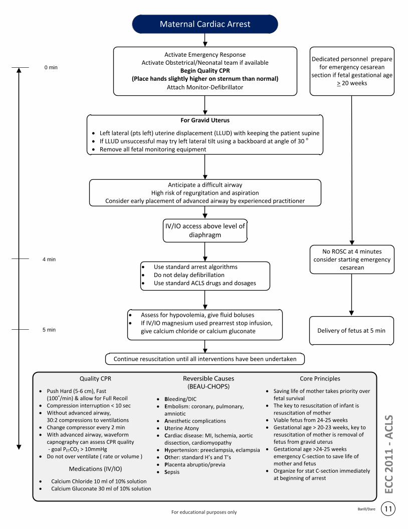
Activate Emergency ResponseActivate Obstetrical/Neonatal team if available
Begin Quality CPR (Place hands slightly higher on sternum than normal)
Attach Monitor-Defibrillator
For Gravid Uterus
· Left lateral (pts left) uterine displacement (LLUD) with keeping the patient supine· If LLUD unsuccessful may try left lateral tilt using a backboard at angle of 30 o · Remove all fetal monitoring equipment
Anticipate a difficult airwayHigh risk of regurgitation and aspiration
Consider early placement of advanced airway by experienced practitioner
IV/IO access above level of diaphragm
· Use standard arrest algorithms· Do not delay defibrillation· Use standard ACLS drugs and dosages
Dedicated personnel prepare for emergency cesarean
section if fetal gestational age > 20 weeks
· Assess for hypovolemia, give fluid boluses· If IV/IO magnesium used prearrest stop infusion,
give calcium chloride or calcium gluconate
0 min
4 min
No ROSC at 4 minutes consider starting emergency
cesarean
5 min Delivery of fetus at 5 min
Continue resuscitation until all interventions have been undertaken
Maternal Cardiac Arrest
11Barill/Dare
ECC
20
11
- A
CLS
Quality CPR
· Push Hard (5-6 cm), Fast (100+/min) & allow for Full Recoil
· Compression interruption < 10 sec· Without advanced airway,
30:2 compressions to ventilations· Change compressor every 2 min· With advanced airway, waveform
capnography can assess CPR quality - goal PETCO2 > 10mmHg
· Do not over ventilate ( rate or volume )
Medications (IV/IO)
· Calcium Chloride 10 ml of 10% solution· Calcium Gluconate 30 ml of 10% solution
Reversible Causes(BEAU-CHOPS)
· Bleeding/DIC· Embolism: coronary, pulmonary,
amniotic· Anesthetic complications· Uterine Atony· Cardiac disease: MI, Ischemia, aortic
dissection, cardiomyopathy· Hypertension: preeclampsia, eclampsia· Other: standard H’s and T’s· Placenta abruptio/previa· Sepsis
Core Principles
· Saving life of mother takes priority over fetal survival
· The key to resuscitation of infant is resuscitation of mother
· Viable fetus from 24-25 weeks· Gestational age > 20-23 weeks, key to
resuscitation of mother is removal of fetus from gravid uterus
· Gestational age >24-25 weeks emergency C-section to save life of mother and fetus
· Organize for stat C-section immediately at beginning of arrest
For educational purposes only

Purposefully left blank
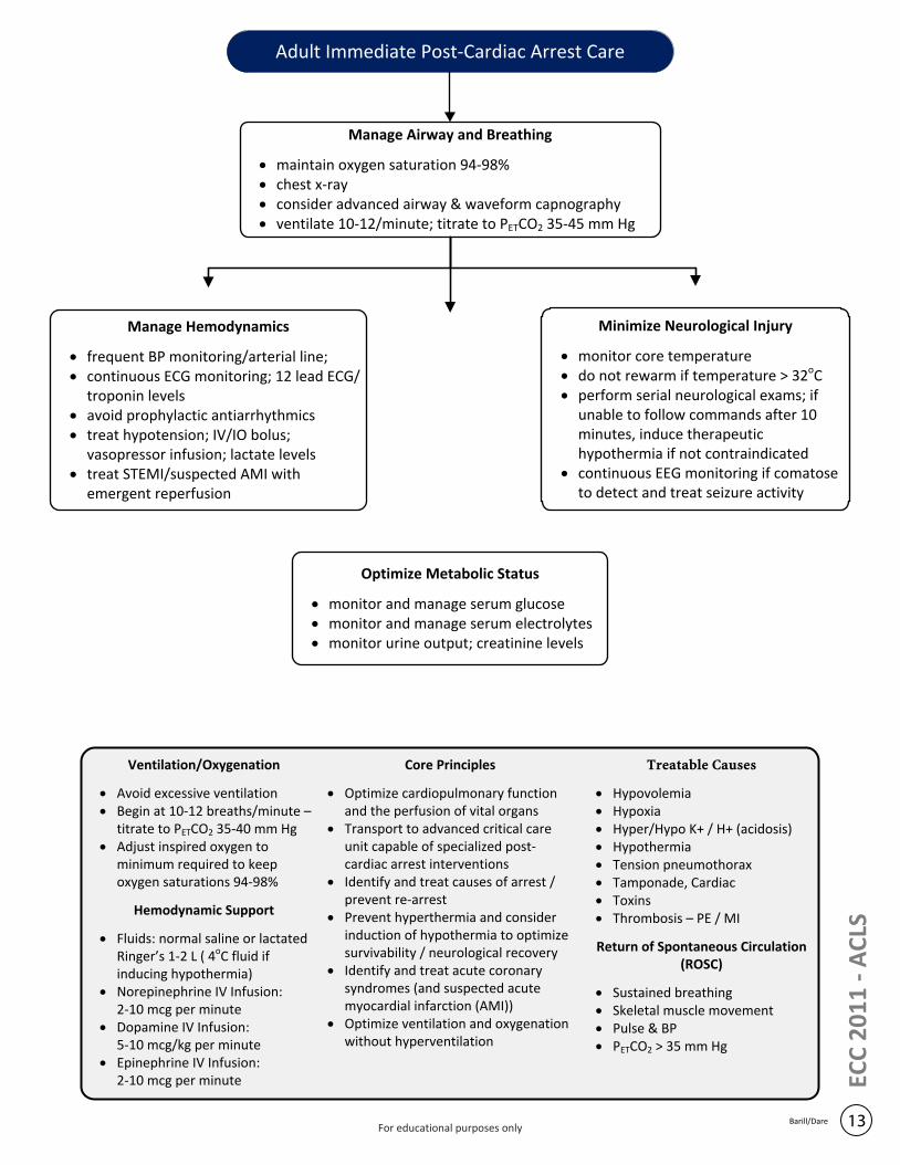
Manage Airway and Breathing
· maintain oxygen saturation 94-98%· chest x-ray· consider advanced airway & waveform capnography· ventilate 10-12/minute; titrate to PETCO2 35-45 mm Hg
Manage Hemodynamics
· frequent BP monitoring/arterial line; · continuous ECG monitoring; 12 lead ECG/
troponin levels· avoid prophylactic antiarrhythmics· treat hypotension; IV/IO bolus;
vasopressor infusion; lactate levels· treat STEMI/suspected AMI with
emergent reperfusion
Ventilation/Oxygenation
· Avoid excessive ventilation· Begin at 10-12 breaths/minute –
titrate to PETCO2 35-40 mm Hg· Adjust inspired oxygen to
minimum required to keep oxygen saturations 94-98%
Hemodynamic Support
· Fluids: normal saline or lactated Ringer’s 1-2 L ( 4oC fluid if inducing hypothermia)
· Norepinephrine IV Infusion: 2-10 mcg per minute
· Dopamine IV Infusion:5-10 mcg/kg per minute
· Epinephrine IV Infusion:2-10 mcg per minute
Treatable Causes
· Hypovolemia· Hypoxia· Hyper/Hypo K+ / H+ (acidosis)· Hypothermia· Tension pneumothorax· Tamponade, Cardiac· Toxins· Thrombosis – PE / MI
Return of Spontaneous Circulation (ROSC)
· Sustained breathing· Skeletal muscle movement· Pulse & BP· PETCO2 > 35 mm Hg
Core Principles
· Optimize cardiopulmonary function and the perfusion of vital organs
· Transport to advanced critical careunit capable of specialized post-cardiac arrest interventions
· Identify and treat causes of arrest / prevent re-arrest
· Prevent hyperthermia and consider induction of hypothermia to optimize survivability / neurological recovery
· Identify and treat acute coronary syndromes (and suspected acute myocardial infarction (AMI))
· Optimize ventilation and oxygenation without hyperventilation
Adult Immediate Post-Cardiac Arrest Care
13Barill/Dare
Minimize Neurological Injury
· monitor core temperature· do not rewarm if temperature > 32oC· perform serial neurological exams; if
unable to follow commands after 10 minutes, induce therapeutic hypothermia if not contraindicated
· continuous EEG monitoring if comatose to detect and treat seizure activity
Optimize Metabolic Status
· monitor and manage serum glucose· monitor and manage serum electrolytes· monitor urine output; creatinine levels
ECC
20
11
- A
CLS
For educational purposes only
Post-Cardiac Arrest Algorithm – A Multisystem Approach
14Barill/Dare
ECC
20
11
- A
CLS
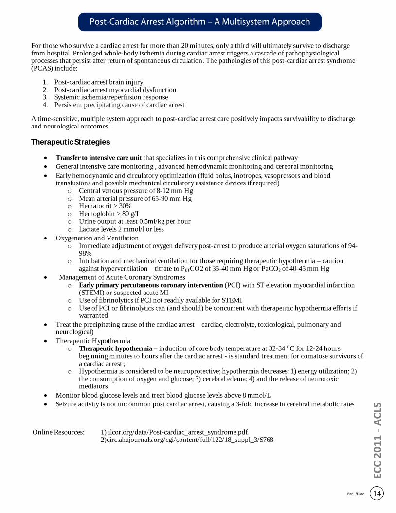
For those who survive a cardiac arrest for more than 20 minutes, only a third will ultimately survive to discharge from hospital. Prolonged whole-body ischemia during cardiac arrest triggers a cascade of pathophysiological processes that persist after return of spontaneous circulation. The pathologies of this post-cardiac arrest syndrome (PCAS) include:
1. Post-cardiac arrest brain injury 2. Post-cardiac arrest myocardial dysfunction 3. Systemic ischemia/reperfusion response 4. Persistent precipitating cause of cardiac arrest
A time-sensitive, multiple system approach to post-cardiac arrest care positively impacts survivability to discharge and neurological outcomes.
Therapeutic Strategies
· Transfer to intensive care unit that specializes in this comprehensive clinical pathway
· General intensive care monitoring , advanced hemodynamic monitoring and cerebral monitoring
· Early hemodynamic and circulatory optimization (fluid bolus, inotropes, vasopressors and blood transfusions and possible mechanical circulatory assistance devices if required)
o Central venous pressure of 8-12 mm Hg o Mean arterial pressure of 65-90 mm Hg o Hematocrit > 30% o Hemoglobin > 80 g/L o Urine output at least 0.5ml/kg per hour o Lactate levels 2 mmol/l or less
· Oxygenation and Ventilation o Immediate adjustment of oxygen delivery post-arrest to produce arterial oxygen saturations of 94-
98% o Intubation and mechanical ventilation for those requiring therapeutic hypothermia – caution
against hyperventilation – titrate to PETCO2 of 35-40 mm Hg or PaCO2 of 40-45 mm Hg
· Management of Acute Coronary Syndromes o Early primary percutaneous coronary intervention (PCI) with ST elevation myocardial infarction
(STEMI) or suspected acute MI o Use of fibrinolytics if PCI not readily available for STEMI o Use of PCI or fibrinolytics can (and should) be concurrent with therapeutic hypothermia efforts if
warranted
· Treat the precipitating cause of the cardiac arrest – cardiac, electrolyte, toxicological, pulmonary and neurological)
· Therapeutic Hypothermia o Therapeutic hypothermia – induction of core body temperature at 32-34 OC for 12-24 hours
beginning minutes to hours after the cardiac arrest - is standard treatment for comatose survivors of a cardiac arrest ;
o Hypothermia is considered to be neuroprotective; hypothermia decreases: 1) energy utilization; 2) the consumption of oxygen and glucose; 3) cerebral edema; 4) and the release of neurotoxic mediators
· Monitor blood glucose levels and treat blood glucose levels above 8 mmol/L
· Seizure activity is not uncommon post cardiac arrest, causing a 3-fold increase in cerebral metabolic rates
Online Resources: 1) ilcor.org/data/Post-cardiac_arrest_syndrome.pdf 2)circ.ahajournals.org/cgi/content/full/122/18_suppl_3/S768
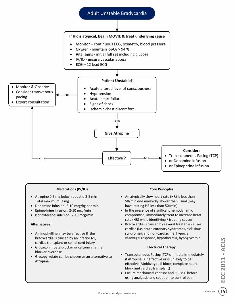
If HR is atypical, begin MOVIE & treat underlying cause
· Monitor – continuous ECG, oximetry, blood pressure· Oxygen - maintain SpO2 > 94 %· Vital signs - initial full set including glucose· IV/IO - ensure vascular access· ECG – 12 lead ECG
Patient Unstable?
· Acute altered level of consciousness· Hypotension· Acute heart failure· Signs of shock· Ischemic chest discomfort
· Monitor & Observe· Consider transvenous
pacing· Expert consultation
Give Atropine
Yes
No
Medications (IV/IO)
· Atropine 0.5 mg bolus, repeat q 3-5 minTotal maximum: 3 mg
· Dopamine infusion: 2-10 mcg/kg per min· Epinephrine infusion: 2-10 mcg/min· Isoproterenol infusion: 2-10 mcg/min
Alternatives:
· Aminophylline may be effective if the bradycardia is caused by an inferior MI, cardiac transplant or spinal cord injury
· Glucagon if beta-blocker or calcium channel blocker overdose
· Glycopyrrolate can be chosen as an alternative to Atropine
Core Principles
· An atypically slow heart rate (HR) is less than50/min and markedly slower than usual (may have resting HR less than 50/min)
· In the presence of significant hemodynamic compromise, immediately treat to increase heart rate (HR) while identifying / treating causes
· Bradycardia is caused by several treatable causes: cardiac (i.e. acute coronary syndromes, sick sinus syndrome), and non-cardiac (i.e. hypoxia, vasovagal response, hypothermia, hypoglycemia)
Electrical Therapy
· Transcutaneous Pacing (TCP): initiate immediately if Atropine is ineffective or is unlikely to be effective (Mobitz type II block, complete heart block and cardiac transplant)
· Ensure mechanical capture and SBP>90 before using analgesia and sedation to control pain
YES Effective ?
Consider:· Transcutaneous Pacing (TCP)· or Dopamine infusion· or Epinephrine infusion
NO
Adult Unstable Bradycardia
15Barill/Dare
ECC
20
11
- A
CLS
For educational purposes only
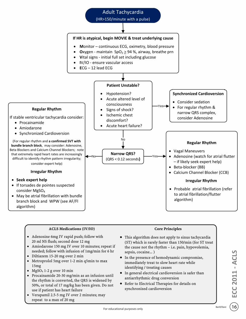
If HR is atypical, begin MOVIE & treat underlying cause
· Monitor – continuous ECG, oximetry, blood pressure· Oxygen - maintain SpO2 > 94 %, airway, breathe prn· Vital signs - initial full set including glucose· IV/IO - ensure vascular access· ECG – 12 lead ECG
Patient Unstable?
· Hypotension?· Acute altered level of
consciousness· Signs of shock?· Ischemic chest
discomfort?· Acute heart failure?
Narrow QRS?(QRS < 0.12 seconds)
Synchronized Cardioversion
· Consider sedation· For regular rhythm &
narrow QRS complex, consider Adenosine
No
Yes
Yes
Regular Rhythm
· Vagal Maneuvers· Adenosine (watch for atrial flutter
– if likely seek expert help)· Beta-blocker (BB)· Calcium Channel Blocker (CCB)
Irregular Rhythm
· Probable atrial fibrillation (refer to atrial fibrillation/flutter algorithm)
No
Regular Rhythm
If stable ventricular tachycardia consider:· Procainamide· Amiodarone· Synchronized Cardioversion
(For regular rhythm and a confirmed SVT with bundle branch block, may consider: Adenosine,
Beta Blockers and Calcium Channel Blockers; note that extremely rapid heart rates are increasingly difficult to identify rhythm pattern irregularity;
consider expert help)
Irregular Rhythm
· Seek expert help· If torsades de pointes suspected
consider MgSO4
· May be atrial fibrillation with bundle branch block and WPW (see AF/Fl algorithm)
Adult Tachycardia(HR>150/minute with a pulse)
16Barill/Dare
ECC
20
11
- A
CLS
ACLS Medications (IV/IO)
· Adenosine 6mg IV rapid push; follow with 20 ml NS flush; second dose 12 mg
· Amiodarone 150 mg IV over 10 minutes; repeat if needed; follow with infusion of 1mg/min for 6 hr
· Diltiazem 15-20 mg over 2 min· Metroprolol 5mg over 1-2 min q5min to max
15mg· MgSO4 1-2 g over 10 min· Procainamide 20-30 mg/min as an infusion until
the rhythm is converted, the QRS is widened by 50%, or total of 17 mg/kg has been given. Do not use if patient has heart failure
· Verapamil 2.5-5 mg IV over 2 minutes; may repeat to a max of 20 mg
Core Principles
· This algorithm does not apply to sinus tachycardia
(ST) which is rarely faster than 150/min (for ST treat the cause not the rhythm – i.e. pain, hypovolemia, sepsis, cocaine... )
· In the presence of hemodynamic compromise,
immediately treat to slow heart rate while identifying / treating causes
· In general electrical cardioversion is safer than
antiarrhythmic drug conversion
· Refer to Electrical Therapies for details on
synchronized cardioversion
For educational purposes only
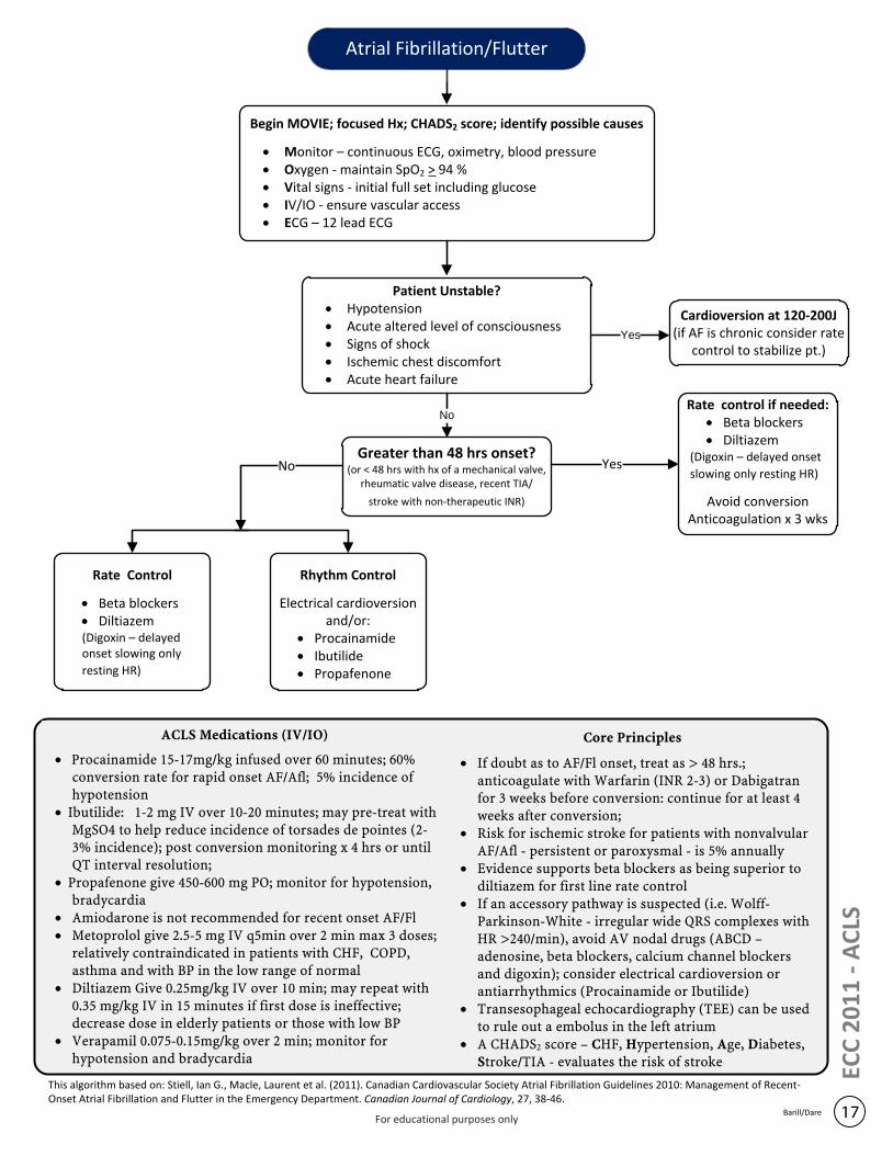
Cardioversion at 120-200J(if AF is chronic consider rate
control to stabilize pt.)
Rate Control
· Beta blockers· Diltiazem(Digoxin – delayed onset slowing only
resting HR)
Rhythm Control
Electrical cardioversion and/or:
· Procainamide· Ibutilide· Propafenone
Rate control if needed:· Beta blockers· Diltiazem
(Digoxin – delayed onset
slowing only resting HR)
Avoid conversionAnticoagulation x 3 wks
ACLS Medications (IV/IO)
· Procainamide 15-17mg/kg infused over 60 minutes; 60% conversion rate for rapid onset AF/Afl; 5% incidence of hypotension
· Ibutilide: 1-2 mg IV over 10-20 minutes; may pre-treat with MgSO4 to help reduce incidence of torsades de pointes (2-3% incidence); post conversion monitoring x 4 hrs or until QT interval resolution;
· Propafenone give 450-600 mg PO; monitor for hypotension, bradycardia
· Amiodarone is not recommended for recent onset AF/Fl · Metoprolol give 2.5-5 mg IV q5min over 2 min max 3 doses;
relatively contraindicated in patients with CHF, COPD, asthma and with BP in the low range of normal
· Diltiazem Give 0.25mg/kg IV over 10 min; may repeat with 0.35 mg/kg IV in 15 minutes if first dose is ineffective; decrease dose in elderly patients or those with low BP
· Verapamil 0.075-0.15mg/kg over 2 min; monitor for hypotension and bradycardia
Core Principles
· If doubt as to AF/Fl onset, treat as > 48 hrs.; anticoagulate with Warfarin (INR 2-3) or Dabigatran for 3 weeks before conversion: continue for at least 4 weeks after conversion;
· Risk for ischemic stroke for patients with nonvalvular AF/Afl - persistent or paroxysmal - is 5% annually
· Evidence supports beta blockers as being superior to diltiazem for first line rate control
· If an accessory pathway is suspected (i.e. Wolff-Parkinson-White - irregular wide QRS complexes with HR >240/min), avoid AV nodal drugs (ABCD – adenosine, beta blockers, calcium channel blockers and digoxin); consider electrical cardioversion or antiarrhythmics (Procainamide or Ibutilide)
· Transesophageal echocardiography (TEE) can be used to rule out a embolus in the left atrium
· A CHADS2 score – CHF, Hypertension, Age, Diabetes, Stroke/TIA - evaluates the risk of stroke
Begin MOVIE; focused Hx; CHADS2 score; identify possible causes
· Monitor – continuous ECG, oximetry, blood pressure· Oxygen - maintain SpO2 > 94 %· Vital signs - initial full set including glucose· IV/IO - ensure vascular access· ECG – 12 lead ECG
Patient Unstable?· Hypotension· Acute altered level of consciousness· Signs of shock· Ischemic chest discomfort· Acute heart failure
Greater than 48 hrs onset?(or < 48 hrs with hx of a mechanical valve,
rheumatic valve disease, recent TIA/
stroke with non-therapeutic INR)
Yes
No
Atrial Fibrillation/Flutter
17Barill/Dare
No Yes
This algorithm based on: Stiell, Ian G., Macle, Laurent et al. (2011). Canadian Cardiovascular Society Atrial Fibrillation Guidelines 2010: Management of Recent-Onset Atrial Fibrillation and Flutter in the Emergency Department. Canadian Journal of Cardiology, 27, 38-46.
ECC
20
11
- A
CLS
For educational purposes only

Purposefully left blank
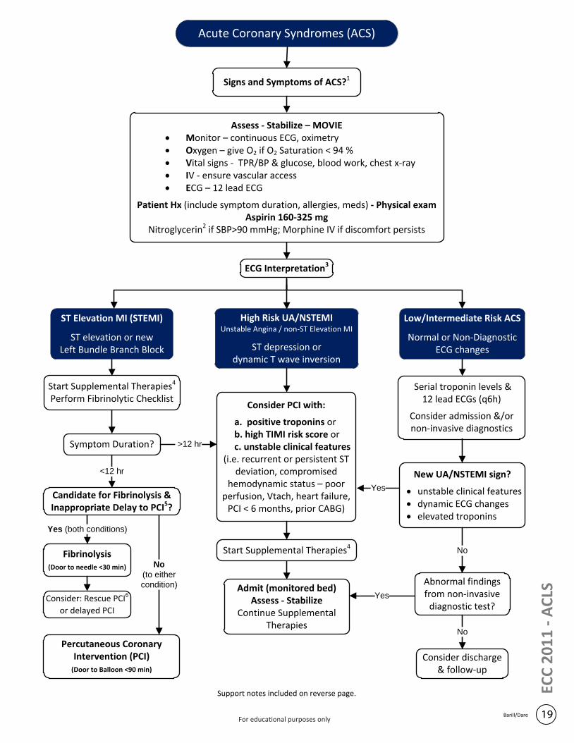
Acute Coronary Syndromes (ACS)
Signs and Symptoms of ACS?1
ECG Interpretation3
Start Supplemental Therapies4
Perform Fibrinolytic Checklist
Assess - Stabilize – MOVIE· Monitor – continuous ECG, oximetry· Oxygen – give O2 if O2 Saturation < 94 % · Vital signs - TPR/BP & glucose, blood work, chest x-ray· IV - ensure vascular access· ECG – 12 lead ECG
Patient Hx (include symptom duration, allergies, meds) - Physical exam Aspirin 160-325 mg
Nitroglycerin2 if SBP>90 mmHg; Morphine IV if discomfort persists
ST Elevation MI (STEMI)
ST elevation or new Left Bundle Branch Block
Support notes included on reverse page.
Candidate for Fibrinolysis & Inappropriate Delay to PCI5?
High Risk UA/NSTEMI Unstable Angina / non-ST Elevation MI
ST depression or dynamic T wave inversion
Low/Intermediate Risk ACS
Normal or Non-Diagnostic ECG changes
Fibrinolysis(Door to needle <30 min)
Yes (both conditions)
Percutaneous Coronary Intervention (PCI)
(Door to Balloon <90 min)
Symptom Duration?
No
(to either
condition)
<12 hr
Consider PCI with:
a. positive troponins or b. high TIMI risk score or c. unstable clinical features
(i.e. recurrent or persistent ST deviation, compromised
hemodynamic status – poor perfusion, Vtach, heart failure,
PCI < 6 months, prior CABG)
Start Supplemental Therapies4
Admit (monitored bed)Assess - Stabilize
Continue Supplemental Therapies
Serial troponin levels & 12 lead ECGs (q6h)
Consider admission &/or non-invasive diagnostics
New UA/NSTEMI sign?
· unstable clinical features · dynamic ECG changes· elevated troponins
Yes
Abnormal findings from non-invasive
diagnostic test?Yes
Consider discharge & follow-up
No
>12 hr
No
Consider: Rescue PCI6
or delayed PCI
19Barill/Dare
ECC
20
11
- A
CLS
For educational purposes only
20Barill/Dare
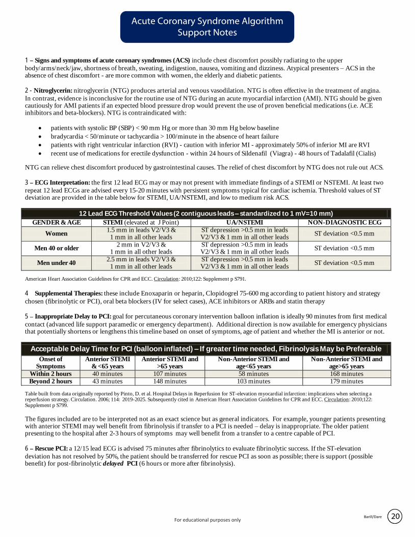
Acute Coronary Syndrome Algorithm Support Notes
20Barill/Dare
1 Signs and symptoms of acute coronary syndromes (ACS) include chest discomfort possibly radiating to the upper
body/arms/neck/jaw, shortness of breath, sweating, indigestion, nausea, vomiting and dizziness. Atypical presenters – ACS in the absence of chest discomfort - are more common with women, the elderly and diabetic patients.
2 - Nitroglycerin: nitroglycerin (NTG) produces arterial and venous vasodilation. NTG is often effective in the treatment of angina.
In contrast, evidence is inconclusive for the routine use of NTG during an acute myocardial infarction (AMI). NTG should be given cautiously for AMI patients if an expected blood pressure drop would prevent the use of proven beneficial medications (i.e. ACE inhibitors and beta-blockers). NTG is contraindicated with:
· patients with systolic BP (SBP) < 90 mm Hg or more than 30 mm Hg below baseline
· bradycardia < 50/minute or tachycardia > 100/minute in the absence of heart failure
· patients with right ventricular infarction (RVI) - caution with inferior MI - approximately 50% of inferior MI are RVI
· recent use of medications for erectile dysfunction - within 24 hours of Sildenafil (Viagra) - 48 hours of Tadalafil (Cialis)
NTG can relieve chest discomfort produced by gastrointestinal causes. The relief of chest discomfort by NTG does not rule out ACS.
3 ECG Interpretation: the first 12 lead ECG may or may not present with immediate findings of a STEMI or NSTEMI. At least two
repeat 12 lead ECGs are advised every 15-20 minutes with persistent symptoms typical for cardiac ischemia. Threshold values of ST deviation are provided in the table below for STEMI, UA/NSTEMI, and low to medium risk ACS.
12 Lead ECG Threshold Values (2 contiguous leads – standardized to 1 mV=10 mm)
GENDER & AGE STEMI (elevated at J Point) UA/NSTEMI NON-DIAGNOSTIC ECG
Women 1.5 mm in leads V2/V3 &
1 mm in all other leads ST depression >0.5 mm in leads V2/V3 & 1 mm in all other leads
ST deviation <0.5 mm
Men 40 or older 2 mm in V2/V3 &
1 mm in all other leads ST depression >0.5 mm in leads V2/V3 & 1 mm in all other leads
ST deviation <0.5 mm
Men under 40 2.5 mm in leads V2/V3 &
1 mm in all other leads ST depression >0.5 mm in leads V2/V3 & 1 mm in all other leads
ST deviation <0.5 mm
American Heart Association Guidelines for CPR and ECC. Circulation: 2010;122: Supplement p S791.
4 Supplemental Therapies: these include Enoxaparin or heparin, Clopidogrel 75-600 mg according to patient history and strategy
chosen (fibrinolytic or PCI), oral beta blockers (IV for select cases), ACE inhibitors or ARBs and statin therapy
5 Inappropriate Delay to PCI: goal for percutaneous coronary intervention balloon inflation is ideally 90 minutes from first medical
contact (advanced life support paramedic or emergency department). Additional direction is now available for emergency physicians that potentially shortens or lengthens this timeline based on onset of symptoms, age of patient and whether the MI is anterior or not.
Acceptable Delay Time for PCI (balloon inflated) – If greater time needed, Fibrinolysis May be Preferable
Onset of Symptoms
Anterior STEMI & <65 years
Anterior STEMI and >65 years
Non-Anterior STEMI and age<65 years
Non-Anterior STEMI and age>65 years
Within 2 hours 40 minutes 107 minutes 58 minutes 168 minutes Beyond 2 hours 43 minutes 148 minutes 103 minutes 179 minutes
Table built from data originally reported by Pinto, D. et al. Hospital Delays in Reperfusion for ST-elevation myocardial infarction: implications when selecting a reperfusion strategy. Circulation. 2006; 114: 2019-2025. Subsequently cited in American Heart Association Guidelines for CPR and ECC. Circulation: 2010;122: Supplement p S799.
The figures included are to be interpreted not as an exact science but as general indicators. For example, younger patients presenting with anterior STEMI may well benefit from fibrinolysis if transfer to a PCI is needed – delay is inappropriate. The older patient presenting to the hospital after 2-3 hours of symptoms may well benefit from a transfer to a centre capable of PCI.
6 Rescue PCI: a 12/15 lead ECG is advised 75 minutes after fibrinolytics to evaluate fibrinolytic success. If the ST-elevation
deviation has not resolved by 50%, the patient should be transferred for rescue PCI as soon as possible; there is support (possible benefit) for post-fibrinolytic delayed PCI (6 hours or more after fibrinolysis).
For educational purposes only
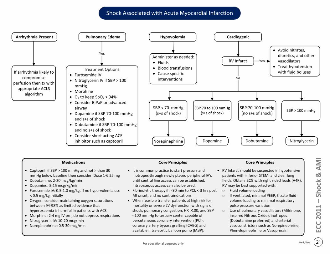
Hypovolemia CardiogenicArrhythmia Present
If arrhythmia likely to compromise
perfusion then tx with appropriate ACLS
algorithm
Pulmonary Edema
Administer as needed:· Fluids· Blood transfusions· Cause specific
interventions
Yes
Treatment Options:· Furosemide IV· Nitroglycerin IV if SBP > 100
mmHg· Morphine· O2 to keep SpO2 > 94%· Consider BiPaP or advanced
airway· Dopamine if SBP 70-100 mmHg
and s+s of shock· Dobutamine if SBP 70-100 mmHg
and no s+s of shock· Consider short acting ACE
inhibitor such as captopril
SBP 70 to 100 mmHg(s+s of shock)
SBP 70-100 mmHg(no s+s of shock)
SBP < 70 mmHg(s+s of shock)
SBP > 100 mmHg
Norepinephrine Dopamine Dobutamine Nitroglycerin
ECC
20
11
– S
ho
ck &
AM
I
RV Infarct
· Avoid nitrates, diuretics, and other vasodilators
· Treat hypotension with fluid boluses
Yes
No
Shock Associated with Acute Myocardial Infarction
21Barill/Dare
Medications
· Captopril: if SBP > 100 mmHg and not > than 30 mmHg below baseline then consider. Dose 1-6.25 mg
· Dobutamine: 2-20 mcg/kg/min· Dopamine: 5-15 mcg/kg/min· Furosemide IV: 0.5-1.0 mg/kg. If no hypervolemia use
< 0.5 mg/kg initially· Oxygen: consider maintaining oxygen saturations
between 94-98% as limited evidence that hyperoxaemia is harmful in patients with ACS
· Morphine: 2-4 mg IV prn, do not depress respirations· Nitroglycerin IV: 10-20 mcg/min· Norepinephrine: 0.5-30 mcg/min
Core Principles
· It is common practice to start pressors and inotropes through newly placed peripheral IV’s until central line access can be established. Intraosseous access can also be used.
· Fibrinolytic therapy if > 90 min to PCI, < 3 hrs post MI onset, and no contraindications.
· When feasible transfer patients at high risk for mortality or severe LV dysfunction with signs of shock, pulmonary congestion, HR >100, and SBP <100 mm Hg to tertiary center capable of percutaneous coronary intervention (PCI), coronary artery bypass grafting (CABG) and available intra-aortic balloon pump (IABP).
Core Principles
· RV Infarct should be suspected in hypotensive patients with inferior STEMI and clear lung fields. Obtain ECG with right sided leads (V4R). RV may be best supported with:o Fluid volume loading o If ventilated, minimal PEEP; titrate fluid
volume loading to minimal respiratory pulse pressure variation
o Use of pulmonary vasodilators (Milrinone, inspired Nitrous Oxide), inotropes (Dobutamine preferred) and arterial vasoconstrictors such as Norepinephrine, Phenylepinephrine or Vasopressin
For educational purposes only

Purposefully left blank
ECC
20
11
- S
tro
ke
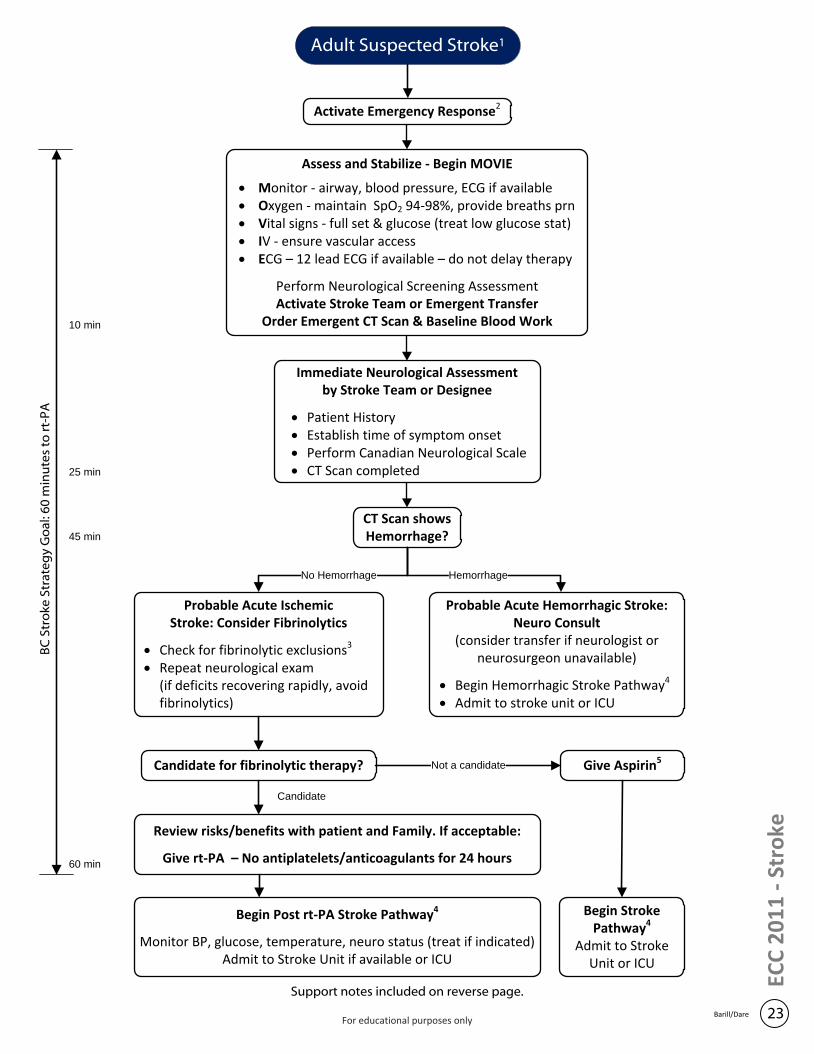
Activate Emergency Response2
Immediate Neurological Assessment by Stroke Team or Designee
· Patient History· Establish time of symptom onset· Perform Canadian Neurological Scale· CT Scan completed
CT Scan shows Hemorrhage?
Candidate for fibrinolytic therapy? Give Aspirin5
Assess and Stabilize - Begin MOVIE
· Monitor - airway, blood pressure, ECG if available· Oxygen - maintain SpO2 94-98%, provide breaths prn· Vital signs - full set & glucose (treat low glucose stat)· IV - ensure vascular access· ECG – 12 lead ECG if available – do not delay therapy
Perform Neurological Screening AssessmentActivate Stroke Team or Emergent Transfer
Order Emergent CT Scan & Baseline Blood Work
Probable Acute Ischemic Stroke: Consider Fibrinolytics
· Check for fibrinolytic exclusions3
· Repeat neurological exam (if deficits recovering rapidly, avoid fibrinolytics)
Probable Acute Hemorrhagic Stroke: Neuro Consult
(consider transfer if neurologist or neurosurgeon unavailable)
· Begin Hemorrhagic Stroke Pathway4
· Admit to stroke unit or ICU
No Hemorrhage Hemorrhage
Not a candidate
Review risks/benefits with patient and Family. If acceptable:
Give rt-PA – No antiplatelets/anticoagulants for 24 hours
Begin Post rt-PA Stroke Pathway4
Monitor BP, glucose, temperature, neuro status (treat if indicated)Admit to Stroke Unit if available or ICU
Support notes included on reverse page.
BC
Str
oke
Str
ate
gy
Go
al: 6
0 m
inu
tes
to r
t-P
A
10 min
25 min
45 min
60 min
Begin Stroke Pathway4
Admit to Stroke Unit or ICU
Candidate
Adult Suspected Stroke1
23Barill/DareFor educational purposes only
Adult Suspected Stroke Algorithm: Support Notes
24Barill/Dare
ECC
20
11
- S
tro
ke
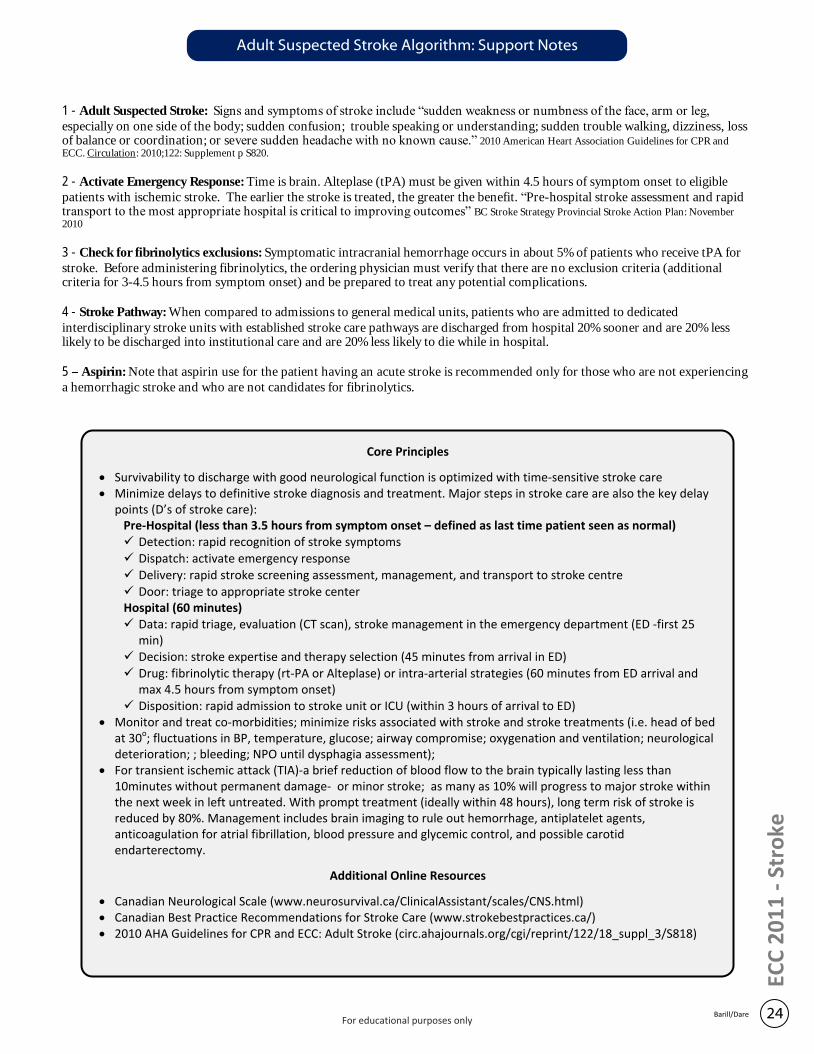
1 - Adult Suspected Stroke: Signs and symptoms of stroke include “sudden weakness or numbness of the face, arm or leg,
especially on one side of the body; sudden confusion; trouble speaking or understanding; sudden trouble walking, dizziness, loss of balance or coordination; or severe sudden headache with no known cause.” 2010 American Heart Association Guidelines for CPR and ECC. Circulation: 2010;122: Supplement p S820.
2 - Activate Emergency Response: Time is brain. Alteplase (tPA) must be given within 4.5 hours of symptom onset to eligible
patients with ischemic stroke. The earlier the stroke is treated, the greater the benefit. “Pre-hospital stroke assessment and rapid transport to the most appropriate hospital is critical to improving outcomes” BC Stroke Strategy Provincial Stroke Action Plan: November 2010
3 - Check for fibrinolytics exclusions: Symptomatic intracranial hemorrhage occurs in about 5% of patients who receive tPA for
stroke. Before administering fibrinolytics, the ordering physician must verify that there are no exclusion criteria (additional criteria for 3-4.5 hours from symptom onset) and be prepared to treat any potential complications.
4 - Stroke Pathway: When compared to admissions to general medical units, patients who are admitted to dedicated
interdisciplinary stroke units with established stroke care pathways are discharged from hospital 20% sooner and are 20% less likely to be discharged into institutional care and are 20% less likely to die while in hospital.
5 Aspirin: Note that aspirin use for the patient having an acute stroke is recommended only for those who are not experiencing
a hemorrhagic stroke and who are not candidates for fibrinolytics.
Core Principles
· Survivability to discharge with good neurological function is optimized with time-sensitive stroke care · Minimize delays to definitive stroke diagnosis and treatment. Major steps in stroke care are also the key delay
points (D’s of stroke care):Pre-Hospital (less than 3.5 hours from symptom onset – defined as last time patient seen as normal)ü Detection: rapid recognition of stroke symptomsü Dispatch: activate emergency responseü Delivery: rapid stroke screening assessment, management, and transport to stroke centreü Door: triage to appropriate stroke center
Hospital (60 minutes)ü Data: rapid triage, evaluation (CT scan), stroke management in the emergency department (ED -first 25
min)ü Decision: stroke expertise and therapy selection (45 minutes from arrival in ED)ü Drug: fibrinolytic therapy (rt-PA or Alteplase) or intra-arterial strategies (60 minutes from ED arrival and
max 4.5 hours from symptom onset)ü Disposition: rapid admission to stroke unit or ICU (within 3 hours of arrival to ED)
· Monitor and treat co-morbidities; minimize risks associated with stroke and stroke treatments (i.e. head of bed at 30o; fluctuations in BP, temperature, glucose; airway compromise; oxygenation and ventilation; neurological deterioration; ; bleeding; NPO until dysphagia assessment);
· For transient ischemic attack (TIA)-a brief reduction of blood flow to the brain typically lasting less than 10minutes without permanent damage- or minor stroke; as many as 10% will progress to major stroke within the next week in left untreated. With prompt treatment (ideally within 48 hours), long term risk of stroke is reduced by 80%. Management includes brain imaging to rule out hemorrhage, antiplatelet agents, anticoagulation for atrial fibrillation, blood pressure and glycemic control, and possible carotid endarterectomy.
Additional Online Resources
· Canadian Neurological Scale (www.neurosurvival.ca/ClinicalAssistant/scales/CNS.html)· Canadian Best Practice Recommendations for Stroke Care (www.strokebestpractices.ca/)· 2010 AHA Guidelines for CPR and ECC: Adult Stroke (circ.ahajournals.org/cgi/reprint/122/18_suppl_3/S818)
For educational purposes only
ECC
20
11
– R
apid
Re
fere
nce
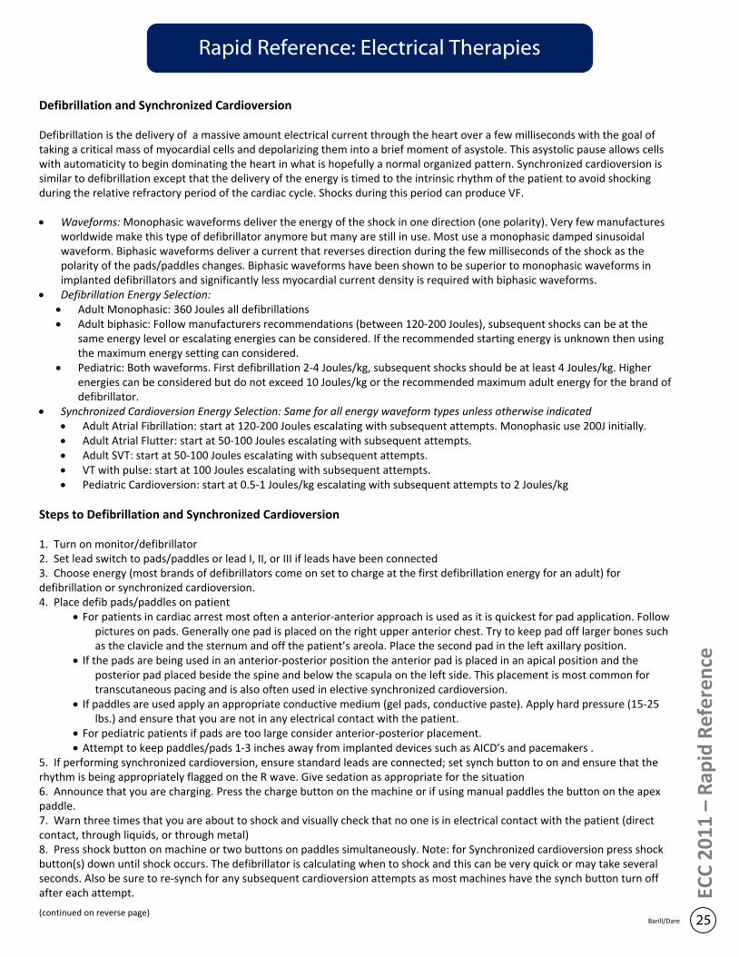
Rapid Reference: Electrical Therapies
Defibrillation and Synchronized Cardioversion
Defibrillation is the delivery of a massive amount electrical current through the heart over a few milliseconds with the goal of taking a critical mass of myocardial cells and depolarizing them into a brief moment of asystole. This asystolic pause allows cells with automaticity to begin dominating the heart in what is hopefully a normal organized pattern. Synchronized cardioversion is similar to defibrillation except that the delivery of the energy is timed to the intrinsic rhythm of the patient to avoid shocking during the relative refractory period of the cardiac cycle. Shocks during this period can produce VF.
· Waveforms: Monophasic waveforms deliver the energy of the shock in one direction (one polarity). Very few manufactures worldwide make this type of defibrillator anymore but many are still in use. Most use a monophasic damped sinusoidal waveform. Biphasic waveforms deliver a current that reverses direction during the few milliseconds of the shock as the polarity of the pads/paddles changes. Biphasic waveforms have been shown to be superior to monophasic waveforms in implanted defibrillators and significantly less myocardial current density is required with biphasic waveforms.
· Defibrillation Energy Selection: · Adult Monophasic: 360 Joules all defibrillations· Adult biphasic: Follow manufacturers recommendations (between 120-200 Joules), subsequent shocks can be at the
same energy level or escalating energies can be considered. If the recommended starting energy is unknown then using the maximum energy setting can considered.
· Pediatric: Both waveforms. First defibrillation 2-4 Joules/kg, subsequent shocks should be at least 4 Joules/kg. Higher energies can be considered but do not exceed 10 Joules/kg or the recommended maximum adult energy for the brand of defibrillator.
· Synchronized Cardioversion Energy Selection: Same for all energy waveform types unless otherwise indicated· Adult Atrial Fibrillation: start at 120-200 Joules escalating with subsequent attempts. Monophasic use 200J initially.· Adult Atrial Flutter: start at 50-100 Joules escalating with subsequent attempts.· Adult SVT: start at 50-100 Joules escalating with subsequent attempts.· VT with pulse: start at 100 Joules escalating with subsequent attempts.· Pediatric Cardioversion: start at 0.5-1 Joules/kg escalating with subsequent attempts to 2 Joules/kg
Steps to Defibrillation and Synchronized Cardioversion
1. Turn on monitor/defibrillator2. Set lead switch to pads/paddles or lead I, II, or III if leads have been connected3. Choose energy (most brands of defibrillators come on set to charge at the first defibrillation energy for an adult) for defibrillation or synchronized cardioversion.4. Place defib pads/paddles on patient
· For patients in cardiac arrest most often a anterior-anterior approach is used as it is quickest for pad application. Follow pictures on pads. Generally one pad is placed on the right upper anterior chest. Try to keep pad off larger bones such as the clavicle and the sternum and off the patient’s areola. Place the second pad in the left axillary position.
· If the pads are being used in an anterior-posterior position the anterior pad is placed in an apical position and the posterior pad placed beside the spine and below the scapula on the left side. This placement is most common for transcutaneous pacing and is also often used in elective synchronized cardioversion.
· If paddles are used apply an appropriate conductive medium (gel pads, conductive paste). Apply hard pressure (15-25 lbs.) and ensure that you are not in any electrical contact with the patient.
· For pediatric patients if pads are too large consider anterior-posterior placement.· Attempt to keep paddles/pads 1-3 inches away from implanted devices such as AICD’s and pacemakers .
5. If performing synchronized cardioversion, ensure standard leads are connected; set synch button to on and ensure that the rhythm is being appropriately flagged on the R wave. Give sedation as appropriate for the situation6. Announce that you are charging. Press the charge button on the machine or if using manual paddles the button on the apex paddle.7. Warn three times that you are about to shock and visually check that no one is in electrical contact with the patient (direct contact, through liquids, or through metal)8. Press shock button on machine or two buttons on paddles simultaneously. Note: for Synchronized cardioversion press shock button(s) down until shock occurs. The defibrillator is calculating when to shock and this can be very quick or may take several seconds. Also be sure to re-synch for any subsequent cardioversion attempts as most machines have the synch button turn off after each attempt.
(continued on reverse page)25Barill/Dare
Rapid Reference: Electrical Therapies
(Continued from previous page)
8. For cardiac arrest situations continue CPR if possible as machine is charged and resume with compressions immediately after the shock to minimize CPR time off chest.
Transcutaneous Pacing (TCP)
Transcutaneous pacing (TCP) is a highly effective emergency method of pacing for severe symptomatic bradycardias. Other methods for increasing heart rate like the use of atropine, dopamine, or epinephrine may also be attempted depending on situational factors and what rhythm the patient is in. This non-targeted method of pacing is unique in that it will also pace skeletal muscle, gut muscle and the diaphragm at the currents needed to capture the myocardium electrically. This can mean significant discomfort for the patient and the need for procedural sedation. The current levels needed to get capture are very high in comparison to other methods of pacing and the aberrancy of the route of conduction from the pads leads to QRS complexes that are very wide and bizarre resembling large PVCs. These observations are all normal and are expected.
Steps to Transcutaneous Pacing
1. Position pads on patient for pacing as indicated by the manufacturer. This is usually an anterior-posterior position. Look for a indicator on the pads to show if one pad is specific to the apical position.2. Ensure standard leads are also connected to the patient3. Turn pacer on4. Most TCP will come on default in demand (synchronous) mode, verify not set in non-demand (asynchronous) mode. This mode is rarely used unless there is a situation where artifact is mistakenly being sensed as intrinsic ECG complexes.5. Set pacer demand rate to 60-80 per minute6. Set current by titrating the mA dial upwards until you have consistent electrical capture as indicated by seeing pacer spikes followed by a new QRS morphology which is very wide with a broad T wave. Buffer the capture current (threshold) by increasing the mA by approximately 10%.7. Check for mechanical capture (paced rhythm produces cardiac output) by assessing distal pulses (at sites where skeletal muscles are not contracting surrounding the vessel), level of consciousness, vital signs and other signs/symptoms of improved infusion.8. Proceed to give analgesia and sedation as needed to keep patient comfortable.9. Arrange transvenous or permanent pacemaker placement as needed
26Barill/Dare
ECC
20
11
– R
apid
Re
fere
nce
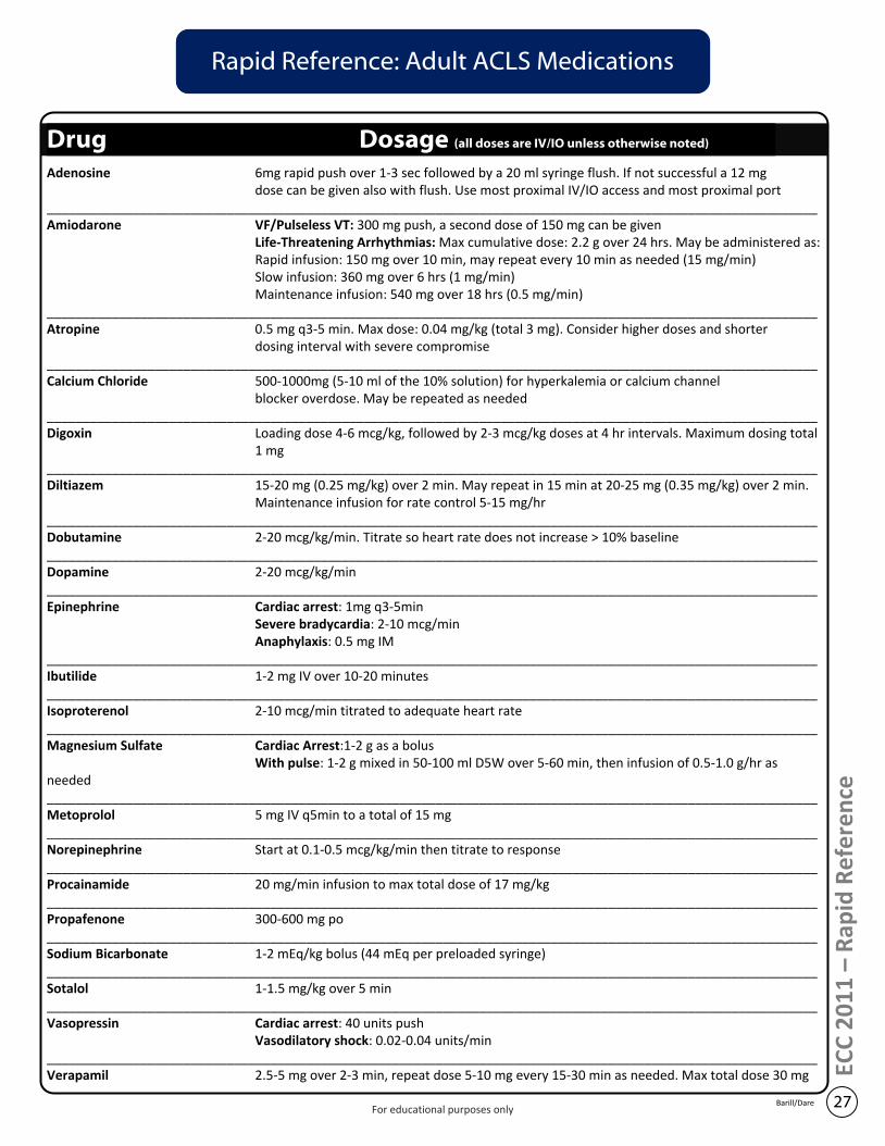
Rapid Reference: Adult ACLS Medications
Drug Dosage (all doses are IV/IO unless otherwise noted)
Adenosine 6mg rapid push over 1-3 sec followed by a 20 ml syringe flush. If not successful a 12 mg dose can be given also with flush. Use most proximal IV/IO access and most proximal port
___________________________________________________________________________________________________________Amiodarone VF/Pulseless VT: 300 mg push, a second dose of 150 mg can be given
Life-Threatening Arrhythmias: Max cumulative dose: 2.2 g over 24 hrs. May be administered as:Rapid infusion: 150 mg over 10 min, may repeat every 10 min as needed (15 mg/min)Slow infusion: 360 mg over 6 hrs (1 mg/min)Maintenance infusion: 540 mg over 18 hrs (0.5 mg/min)
___________________________________________________________________________________________________________Atropine 0.5 mg q3-5 min. Max dose: 0.04 mg/kg (total 3 mg). Consider higher doses and shorter
dosing interval with severe compromise___________________________________________________________________________________________________________Calcium Chloride 500-1000mg (5-10 ml of the 10% solution) for hyperkalemia or calcium channel
blocker overdose. May be repeated as needed___________________________________________________________________________________________________________Digoxin Loading dose 4-6 mcg/kg, followed by 2-3 mcg/kg doses at 4 hr intervals. Maximum dosing total
1 mg___________________________________________________________________________________________________________Diltiazem 15-20 mg (0.25 mg/kg) over 2 min. May repeat in 15 min at 20-25 mg (0.35 mg/kg) over 2 min.
Maintenance infusion for rate control 5-15 mg/hr___________________________________________________________________________________________________________Dobutamine 2-20 mcg/kg/min. Titrate so heart rate does not increase > 10% baseline___________________________________________________________________________________________________________Dopamine 2-20 mcg/kg/min___________________________________________________________________________________________________________Epinephrine Cardiac arrest: 1mg q3-5min
Severe bradycardia: 2-10 mcg/minAnaphylaxis: 0.5 mg IM
___________________________________________________________________________________________________________Ibutilide 1-2 mg IV over 10-20 minutes___________________________________________________________________________________________________________Isoproterenol 2-10 mcg/min titrated to adequate heart rate___________________________________________________________________________________________________________Magnesium Sulfate Cardiac Arrest:1-2 g as a bolus
With pulse: 1-2 g mixed in 50-100 ml D5W over 5-60 min, then infusion of 0.5-1.0 g/hr as needed___________________________________________________________________________________________________________Metoprolol 5 mg IV q5min to a total of 15 mg___________________________________________________________________________________________________________Norepinephrine Start at 0.1-0.5 mcg/kg/min then titrate to response___________________________________________________________________________________________________________Procainamide 20 mg/min infusion to max total dose of 17 mg/kg___________________________________________________________________________________________________________Propafenone 300-600 mg po___________________________________________________________________________________________________________Sodium Bicarbonate 1-2 mEq/kg bolus (44 mEq per preloaded syringe)___________________________________________________________________________________________________________Sotalol 1-1.5 mg/kg over 5 min___________________________________________________________________________________________________________Vasopressin Cardiac arrest: 40 units push
Vasodilatory shock: 0.02-0.04 units/min___________________________________________________________________________________________________________Verapamil 2.5-5 mg over 2-3 min, repeat dose 5-10 mg every 15-30 min as needed. Max total dose 30 mg
27Barill/Dare
ECC
20
11
– R
apid
Re
fere
nce
For educational purposes only
Induced Therapeutic Hypothermia Overview
Brain injury and hemodynamic instability are the main causes of death for survivors of a cardiac arrest. Induced therapeutic hypothermia (ITH) - active cooling to 32-34o Celsius for 24 hours - is shown to improve neurological recovery for those who experience a prolonged cardiac arrest.
ITH is recommended for: survivors of out-of-hospital VF arrests who are unable to follow verbal commands.ITH should be considered for:
· survivors of out-of-hospital cardiac arrest from PEA or Asystole who are unable to follow verbal commands · survivors of in-hospital cardiac arrest of any initial rhythm who are unable to follow verbal commands 10
minutes after the arrestActive rewarming is discouraged for unresponsive cardiac arrest survivors who spontaneously become mildly hypothermic (core temperature >32o Celsius) for the first 48 hours post-arrest.
At the time of writing no universally accepted induced therapeutic hypothermia algorithm has been published by AHA, ERC, or ILCOR. Many regions in Canada have adopted and/or modified the 2005 ITH guidelines of the Canadian Association of Emergency Physicians (CAEP).
Three Phases: Induction, Maintenance and Rewarming
Induction: a combination of external and internal cooling methods are implemented early post-arrest; interventions include rapid infusion (30 ml/kg) of 4oC 0.9% saline, ice packs, cooling blankets, intravascular heat exchangers, ice water gastric/bladder lavage and evaporative cooling techniquesMaintenance: a cooling method is used along with continuous temperature feedback monitoring to keep the core body temperature between 32-34oCRewarming: typically after 24 hours of cooling, rewarming should begin and progress slowly (i.e. 0.25-0.5oC/hour); manage fluctuations in electrolyte concentrations, intravascular volume and metabolic rate that occur with changes in body temperature; prevent hyperthermia by maintaining core temperatures at 36-37.5oC
Considerations
· ITH is an important component of a multisystem approach to post-cardiac arrest care; advanced airway management, hemodynamic stabilization, treatment of acute coronary syndromes and metabolic monitoring (serial lactate, electrolyte, glucose testing) are performed simultaneously
· Cooling suppresses several physiologic pathways that cause delayed (post-arrest) cell death; cooling also reduces cerebral metabolic rate by 6-10% for every 1oC reduction in cerebral temperature
· Shivering is an expected physiologic response to mild hypothermia; shivering increases heat production, thus slowing or preventing cooling; shivering should be treated; ; the threshold temperature for shivering can be decreased with IV sedation; IV boluses of neuromuscular blocking agents (NMBA) may be warranted; note that NMBA can eliminate skeletal muscle movement - the primary indicator of seizures activity (a seizure can triple cerebral metabolic rate); a continuous NBMA infusion requires ongoing EEG monitoring
· Physiologic effects and associated complications of hypothermia include: o increased systemic vascular resistance; continuous ECG monitoring for dysrhythmias; o diuresis; manage hemodynamic status and electrolyte levels o reduced insulin sensitivity and production; monitor for hyperglycemiao possible impaired coagulation; monitor for increased bleedingo impaired immune response; monitor for an increased incidence of infectiono impaired medication elimination (one study showed a 30% reduction in medication clearance); re-evaluate
medication dosing as temperature is reduced· Target ITH temperatures are often maintained with simple interventions such as ongoing sedation, keeping
the patient uncovered and the occasional use of evaporative cooling (a fan directed over wetted skin)· Prognosis of post-cardiac arrest patients treated with ITH can not be reliably predicted for at least 72 hours
28Barill/Dare
ECC
20
11
– R
apid
Re
fere
nce
For educational purposes only
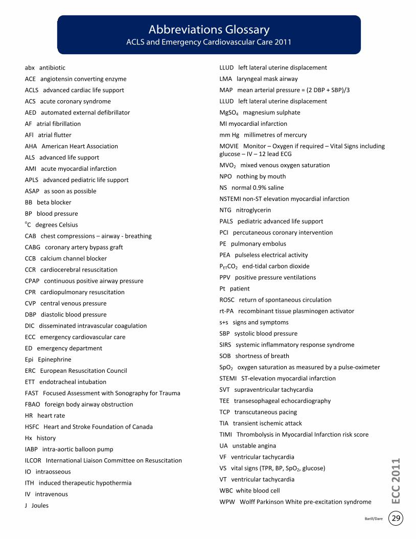
ECC
20
11
29Barill/Dare
Abbreviations GlossaryACLS and Emergency Cardiovascular Care 2011
abx antibiotic
ACE angiotensin converting enzyme
ACLS advanced cardiac life support
ACS acute coronary syndrome
AED automated external defibrillator
AF atrial fibrillation
AFl atrial flutter
AHA American Heart Association
ALS advanced life support
AMI acute myocardial infarction
APLS advanced pediatric life support
ASAP as soon as possible
BB beta blocker
BP blood pressureoC degrees Celsius
CAB chest compressions – airway - breathing
CABG coronary artery bypass graft
CCB calcium channel blocker
CCR cardiocerebral resuscitation
CPAP continuous positive airway pressure
CPR cardiopulmonary resuscitation
CVP central venous pressure
DBP diastolic blood pressure
DIC disseminated intravascular coagulation
ECC emergency cardiovascular care
ED emergency department
Epi Epinephrine
ERC European Resuscitation Council
ETT endotracheal intubation
FAST Focused Assessment with Sonography for Trauma
FBAO foreign body airway obstruction
HR heart rate
HSFC Heart and Stroke Foundation of Canada
Hx history
IABP intra-aortic balloon pump
ILCOR International Liaison Committee on Resuscitation
IO intraosseous
ITH induced therapeutic hypothermia
IV intravenous
J Joules
LLUD left lateral uterine displacement
LMA laryngeal mask airway
MAP mean arterial pressure = (2 DBP + SBP)/3
LLUD left lateral uterine displacement
MgSO4 magnesium sulphate
MI myocardial infarction
mm Hg millimetres of mercury
MOVIE Monitor – Oxygen if required – Vital Signs including glucose – IV – 12 lead ECG
MVO2 mixed venous oxygen saturation
NPO nothing by mouth
NS normal 0.9% saline
NSTEMI non-ST elevation myocardial infarction
NTG nitroglycerin
PALS pediatric advanced life support
PCI percutaneous coronary intervention
PE pulmonary embolus
PEA pulseless electrical activity
PETCO2 end-tidal carbon dioxide
PPV positive pressure ventilations
Pt patient
ROSC return of spontaneous circulation
rt-PA recombinant tissue plasminogen activator
s+s signs and symptoms
SBP systolic blood pressure
SIRS systemic inflammatory response syndrome
SOB shortness of breath
SpO2 oxygen saturation as measured by a pulse-oximeter
STEMI ST-elevation myocardial infarction
SVT supraventricular tachycardia
TEE transesophageal echocardiography
TCP transcutaneous pacing
TIA transient ischemic attack
TIMI Thrombolysis in Myocardial Infarction risk score
UA unstable angina
VF ventricular tachycardia
VS vital signs (TPR, BP, SpO2, glucose)
VT ventricular tachycardia
WBC white blood cell
WPW Wolff Parkinson White pre-excitation syndrome

Purposefully left blank
References
31Barill/Dare
1. American Heart Association. Out-of-Hospital (Sudden) Cardiac Arrest Statistics. (2009). Retrieved from http://www.americanheart.org/downloadable/heart/1236978541670OUT_OF_HOSP.pdf
2. Barill, T. & Dare, M. (2006). Managing Cardiac Emergencies. North Vancouver, BC: SkillStat Press.
3. BC Stroke Strategy. A proposed algorithm for identifying patients with acute cerebrovascular syndrome. (December, 2010). Retrieved from http://www.bcstrokestrategy.ca/documents/BCACVSAlgorithmFinal.pdf .
4. BC Stroke Strategy. Evaluation of TIA Rapid Assessment Clinics. (December, 2010). Retrieved from http://www.bcstrokestrategy.ca/documents/EvaluationofTIAClinicsFinal.pdf .
5. Cairns, J.A., Connolly, S., McMurtry, S. et al. (2011). Canadian Cardiovascular Society Atrial Fibrillation Guidelines 2010: Prevention of stroke and systemic thromboembolism in atrial fibrillation and flutter. Canadian Journal of Cardiology, 27, 74-90.
6. Desbiens, Norman A. (2008). Simplifying the Diagnosis and Management of Pulseless Electrical Activity in Adults: A Qualitative Review. Critical Care Medicine. 36(2), 391-396.
7. Ewy, Gordon A. (2005). Cardiocerebral Resuscitation: The new Cardiopulmonary Resuscitation. Circulation, 111, 2134-2142.
8. Gausche-Hill, M., Fuchs, S. & Yamamoto, L. (Eds.). (2004). APLS: The Pediatric Emergency Medicine Resource. Toronto: Jones and Bartlett Publishers.
9. Hazinski, Mary F. & Field, John M. (Eds.). (2010). 2010 American Heart Association Guidelines for Cardiopulmonary Resuscitation and Emergency Cardiovascular Care. Circulation, 122 (Suppl. 3).
10. Heart and Stroke Foundation of BC and Yukon. BC Stroke Strategy: Provincial Stroke Action Plan. (November 2010). Retrieved from http://www.bcstrokestrategy.ca/documents/ProvincialStrokeActionPlanAppendixA.pdf
11. Hommers, Clare. (2010). Oxygen therapy post-cardiac arrest? The’ Goldilocks’ principle?. Resuscitation, 81, 1605-1606.
12. Kory, P., Weiner, J., Mathew, J. et al. (2011). European Resuscitation Council Guidelines for Resuscitation 2010. Resuscitation, 82(1), 15-20.
13. Kushner, F., Hand, M., King, S.B. et al. (2009). 2009 ACC/AHA guidelines for the management of patients with ST-elevation myocardial infarction and ACC/AHA/SCAI guidelines on percutaneous coronary intervention. Journal of the American College of Cardiologists, 54, 2205-2241.
14. Levy M.M., Fink M.P., Marshall J.C. et al. (2003). 2001 SCCM/ESICM/ACCP/ATS/SIS International Sepsis Definitions Conference. Critical Care Medicine. 31(4),1250-6.
15. Lindsay, P., Bayley, M., Hellings, C. et al. (2008). Canadian best practice recommendations for stroke care (updated 2008). Canadian Medical Association Journal, 179(Suppl. 12), S1-S25.
16. Meaney, P., Nadkarni, V., Kern, K. et al. (2010). Rhythms and outcomes of adult in-hospital cardiac arrest. Journal of Critical Care Medicine, 38(1), 101-108.
17. Nolan, J.P., Hazinski, M.F., Billi, J.E. et al. (2010). 2010 International Consensus on Cardiopulmonary Resuscitation and Emergency Cardiovascular Care Science with Treatment Recommendations . Resuscitation, 81(Supplement e).
18. Nolan, J.P., Soar, J., Deakin, C.D. et al. (2010). European Resuscitation Council Guidelines for Resuscitation 2010. Resuscitation, 81(10). EC
C 2
01
1

19. Nolan, J.P., Neumar, R.W., Adrie, C. et al. (2008). Post-cardiac arrest syndrome: Epidemiology, pathophysiology, treatment, prognostication. A Scientific Statement from the International Liaison Committee on Resuscitation; the American Heart Association Emergency Cardiovascular Care Committee; the Council on Cardiovascular Surgery and Anesthesia; the Council on Cardiopulmonary, Perioperative, and Critical Care; the Council on Clinical Cardiology; the Council on Stroke et al. Resuscitation, 79(9), 350-379.
20. Parkash, R., Verma, A. & Tang, A.S.L. (2010). Persistent atrial fibrillation: current approach and controversies. Current Opinion in Cardiology, 25m 1-7.
21. Pinto, D.S., Kirtane, A.J., Nallamothu, B.K. et al. (2006). Hospital Delays in Reperfusion for ST-Elevation Myocardial Infarction: Implications when selecting a reperfusion strategy. Circulation, 114, 2019-2025.
22. Ralston, M. & Hazinski, M.F. & Schexnayder, S. et al. (2007). Pediatric Emergency Assessment, Recognition, and Stabilization. Dallas, TX: American Heart Association.
23. Ralston, M. & Hazinski, M.F. & Zaritsky, A. et al. (2006). Pediatric Advanced Life Support. Dallas, TX: American Heart Association.
24. Rothwell, P., Giles, M., Chandratheva, A. et al. Effect of urgent treatment of transient ischaemic attack and minor stroke on early recurrent stroke (EXPRESS study): a prospective population-based sequential comparison. Lancet, 370, 1432-42.
25. Sandroni, C., Nolan, J., Cavallaro, F. & Antonelli, M. (2007). In-hospital cardiac arrest: incidence, prognosis and possible measures to improve survival. Intensive Care Medicine, 33, 237-245.
26. Smith, Stephen W. & Whitwam, W. (2006). Acute Coronary Syndromes. Emergency Medical Clinics of North America, 24, 53-89.
27. Stiell, Ian G., Macle, Laurent et al. (2011). Canadian Cardiovascular Society Atrial Fibrillation Guidelines 2010: Management of Recent-Onset Atrial Fibrillation and Flutter in the Emergency Department. Canadian Journal of Cardiology, 27, 38-46.
28. Van de Werf, F.J. (2006). Fine-tuning the selection of a reperfusion strategy. Circulation, 114, 2002-2003.
29. Wann, S., Curtis, A., January, C. et al. (2011). 2011 ACCF/AHA/HRS Focused update on the management of patients with atrial fibrillation (Updating the 2006 Guideline): A Report of the American College of Cardiology Foundation/American Heart Association Task Force on Practice Guidelines. Circulation, 123, 104-123.
30. Wijesinghe, M., Perrin, K., Ranchord, A. et al. (2008). Routine use of oxygen in the treatment of myocardial infarction: systematic review. Heart, 95, 198-202.
31. Wright, R.S., Anderson, J.L., Adams, C.D. et al. (2011). 2011 ACCF/AHA Focused Update incorporated into the ACC/AHA 2007 guidelines for the management of patients with unstable angina/non ST-elevation myocardial infarction. Journal of the American College of Cardiologists, 57 (Suppl. E), e215-e367.
References
32Barill/Dare
ECC
20
11
Notes

Notes
Page 46


Notes
Page 48


Notes
Page 50
Do you have the skills to beat the highest score in ourSMARTMAN COMPRESSION CHALLENGE?
Visit our office and find out!1644 West 3rd Avenue, Vancouver
Let’s Connect!
pinterest.com/iridiamedicalfacebook.com/iridiamedical
gplus.to/iridiamedical
Need to know what’s going on at Iridia?This should be your first stop!
Want to see whats happening at Iridia?
Check out our pinboards.
For the most up-to-date information,head over to our
Twitter feed.
Stop by and say hito the Iridia team on our Google+page.
Have a look under the hood of Iridia Medical!
twitter.com/iridiamedical
blog.iridiamedical.com