Autoimmune Insulin Dependent Diabetes Mellitus (Type 1 Diabetes Mellitus) :

Autoimmune diabetes is a heterogeneous disease that is generally regarded as a condition that presents in child-hood or adolescence. However, a substantial proportion of patients experience onset in adulthood1. In this case, termed adult-onset autoimmune diabetes, the disease is even more heterogeneous than young-onset autoimmune diabetes, as the rate of β-cell destruction is highly variable, which is probably due to differences in the penetrance of genetic and immune factors2–4. In the past few decades, particular attention has been paid to the characterization of adult-onset autoimmune diabetes. Epidemiological studies have highlighted that the majority of patients with onset in adulthood do not require treatment with insulin at the time of diagnosis, and these patients are defined as having latent autoimmune diabetes in adults (LADA)5–7. This term was introduced in the early 1990s to define a subgroup of patients who had non- insulin-requiring diabetes mellitus that was initially thought to be type 2 diabetes mellitus (T2DM) but who had detectable serum immune markers of type 1 diabetes mellitus (T1DM)5,8. Alternative eponyms for this condition have been pro-posed, such as ‘type 1.5 diabetes’ (REF. 9), ‘non- insulin-requiring autoimmune diabetes’ (REF. 10) and ‘slowly progressive insulin-dependent type 1 diabetes’ (REF. 11).
In 2005, the Immunology of Diabetes Society pro-posed three main criteria for the diagnosis of LADA: adult age of onset (>30 years); the presence of any islet autoantibody; and the absence of insulin requirement for at least 6 months after diagnosis12. However, con-troversies regarding these criteria still exist. The major criticism is the subjectivity of the clinician’s decision to start insulin treatment, which is a key factor affecting the classification13. The American Diabetes Association and WHO do not recognize LADA as a distinct entity, as they include LADA within the T1DM classification14,15. Owing to the slow rate of β-cell loss that is observed in LADA4, the period of insulin independence after onset can distinguish patients with LADA from those with classic adult-onset T1DM, who require insulin within 3 months of diagnosis14. The broad definition of LADA12 — which includes any patient with diabetes who does not require insulin and who is positive for any islet auto-antibody, regardless of titre, number or epitope specificity — represents the basic prerequisite for the wide degree of heterogeneity observed in this form of diabetes.
New data that further elucidate the pathophysiology and clinical implications of adult-onset autoimmune diabetes have been reported. Nonetheless, these topics
1Department of Experimental Medicine, Sapienza University, Viale Regina Elena 324, 00161, Rome, Italy.2Department of Medicine, Unit of Endocrinology and Diabetes, University Campus Bio-Medico, Via Álvaro del Portillo 21, 00128, Rome, Italy.
Correspondence to R.B. [email protected]
doi:10.1038/nrendo.2017.99Published online 8 Sep 2017
Adult-onset autoimmune diabetes: current knowledge and implications for managementRaffaella Buzzetti1, Simona Zampetti1 and Ernesto Maddaloni2
Abstract | Adult-onset autoimmune diabetes is a heterogeneous disease that is characterized by a reduced genetic load, a less intensive autoimmune process and a mild metabolic decompensation at onset compared with young-onset type 1 diabetes mellitus (T1DM). The majority of patients with adult-onset autoimmune diabetes do not require insulin treatment for at least 6 months after diagnosis. Such patients are defined as having latent autoimmune diabetes in adults (LADA), which is distinct from classic adult-onset T1DM. The extensive heterogeneity of adult-onset autoimmune diabetes is apparent beyond the distinction between classic adult-onset T1DM and LADA. LADA is characterized by genetic, phenotypic and humoral heterogeneity, encompassing different degrees of insulin resistance and autoimmunity; this heterogeneity is probably a result of different pathological mechanisms, which have implications for treatment. The existence of heterogeneous phenotypes in LADA makes it difficult to establish an a priori treatment algorithm, and therefore, a personalized medicine approach is required. In this Review, we discuss the current understanding and gaps in knowledge regarding the pathophysiology and clinical features of adult-onset autoimmune diabetes and highlight the similarities and differences with classic T1DM and type 2 diabetes mellitus.
NATURE REVIEWS | ENDOCRINOLOGY ADVANCE ONLINE PUBLICATION | 1
REVIEWS
© 2017
Macmillan
Publishers
Limited,
part
of
Springer
Nature.
All
rights
reserved.

are still under debate, and controversies in the classifi-cation of adult-onset autoimmune diabetes remain. In this Review, we report and critically discuss the results of the most recent and relevant studies, emphasizing the differences between T2DM, LADA, young-onset T1DM and adult-onset T1DM. Data regarding the epidemiol-ogy of adult-onset autoimmune diabetes are gathered to define the global picture of this disease. We review the pathogenesis of this disease in depth, highlighting genetic susceptibility and immune features as well as their influ-ence on β-cell function. In addition, the metabolic and clinical features of individuals with LADA from different ethnic groups are summarized, and the data regarding the risk of both microvascular and macrovascular com-plications are discussed. Finally, we review the most recent advances towards a pathophysiology- oriented treatment paradigm for LADA.
EpidemiologyThe growing body of literature on the epidemiology of adult-onset autoimmune diabetes is a testament to the increasing interest in adult-onset autoimmune dia-betes. The available data show that adult-onset T1DM is more common than previously recognized. Indeed, approximately 40% of T1DM cases globally occur in people older than 30 years of age1. In Italy, the incidence of T1DM in individuals aged 30–49 years is similar to that in adolescents aged 15–19 years16. Moreover, a study carried out in Sweden showed that the real incidence of T1DM in individuals aged 15–34 years was twofold to threefold higher than previously reported17. Two large studies published in the past 5 years have investigated the relative frequencies of T1DM autoantibody positivity in classic adult-onset T1DM and LADA and reported 3.3-fold and 12.8-fold higher frequencies of LADA in people of European and Arab origin, respectively6,7. The results of both studies suggest that LADA is the most frequent form of adult-onset autoimmune diabetes. In addition, multicentre studies have reported that 4–14%
of patients with a diagnosis of T2DM are positive for T1DM-associated autoantibodies, which is indicative of a diagnosis of LADA6,7,18–27.
The frequency of patients with T1DM-related autoantibodies among all patients diagnosed with T2DM varies considerably depending on ethnicity (TABLE 1), with the highest rates of LADA reported in people of northern European origins (7–14%)18,19,25,28. The NonInsulin Requiring Autoimmune Diabetes (NIRAD) Study22, carried out in Italy, found that the cumulative frequency of positivity for either glutamic acid decarboxylase (GAD) autoantibodies and/or tyros-ine phosphatase IA-2 (IA-2) autoantibodies was 4.5% in a cohort of 5,330 patients with T2DM22. Testing for zinc transporter 8 (ZnT8) autoantibodies identified an addi-tional 1.4% of patients as autoantibody positive, bring-ing the potential frequency of autoantibody positivity in this T2DM cohort to 5.9%29. A frequency of 9.7% was reported in Action LADA, a European multicentre study that evaluated 6,000 patients with adult-onset diabetes who attended primary and secondary care centres6. In the multicentre LADA China Study, the frequency of autoantibody positivity among adults with T2DM was reported to be 5.9%, which is similar to, or even higher than, the frequency described in European countries26. This observation was quite unexpected, considering that childhood-onset T1DM is rare in China30. Outside of Europe, the highest frequency of autoantibody positiv-ity was reported in Indonesia, with up to 20% of T2DM patients being affected31, whereas the lowest frequen-cies were reported in Alaska32 and Papua New Guinea33. Studies have also reported that African-American, Hispanic and Arab people have a lower prevalence of adult-onset autoimmune diabetes than white people7,34.
The worldwide variance observed in the frequency of autoantibody positivity in patients with T2DM could be primarily due to differences in study design and inclusion criteria (such as age at diagnosis, sex, mode of recruit-ment, number and type of autoantibodies tested and sensitivity and specificity of autoantibody assays) as well as ethnicity. Furthermore, the increasing prevalence of T2DM in some populations could influence the frequency of autoantibody positivity in patients with T2DM35.
Genes, autoimmunity and β cellsAdult-onset autoimmune diabetes has a lower ‘genetic load’ and is characterized by fewer diabetes-associated autoantibodies than young-onset T1DM2,3,36. These genetic and autoimmune characteristics are consistent with a less severe functional deterioration of the β cells at disease onset than in young-onset T1DM3. Compared with young-onset T1DM, LADA represents the other extreme of the autoimmune diabetes spectrum, whereby genetic susceptibility, an autoimmune response and non-insulin-requiring presentation converge in a mild form of diabetes mellitus22 (TABLE 2).
GeneticsIn LADA, genetic susceptibility has been investigated, but only in relation to genes that have been previously associated with young-onset T1DM2,37,38 or T2DM39,40.
Key points
• Adult-onset autoimmune diabetes encompasses a wide spectrum of heterogeneous genotypes and phenotypes, ranging from classic adult-onset type 1 diabetes mellitus to latent autoimmune diabetes in adults (LADA)
• The heterogeneity of LADA arises from its definition as being present in any adult with diabetes who does not require insulin and who is positive for any islet autoantibody, regardless of titre, number or epitope specificity
• The heterogeneity of LADA manifests in different clinical phenotypes, ranging from prevalent insulin resistance to prevalent insulin deficiency, each of which might be associated with different autoimmune and metabolic markers
• Although patients with LADA are leaner and have healthier lipid and blood pressure profiles, evidence shows that there is no difference in cardiovascular outcomes between these patients and those with type 2 diabetes mellitus
• The extensive heterogeneity of adult-onset autoimmune diabetes, and particularly LADA, makes it difficult to determine an a priori algorithm for treatment
• The successful treatment of adult-onset autoimmune diabetes will require a personalized medicine approach that takes into account the intrinsic characteristics of each patient
R E V I E W S
2 | ADVANCE ONLINE PUBLICATION www.nature.com/nrendo
© 2017
Macmillan
Publishers
Limited,
part
of
Springer
Nature.
All
rights
reserved. ©
2017
Macmillan
Publishers
Limited,
part
of
Springer
Nature.
All
rights
reserved.

This approach, which is based on the assumption that adult-onset autoimmune diabetes could have the same components of genetic susceptibility as young-onset T1DM, limits the possibility of discovering novel genes that are associated solely with adult-onset T1DM and/or LADA. In addition, most studies that have analysed genetic susceptibility did not compare patients with T1DM and LADA of the same age. Therefore, the data pertaining to this topic require careful interpretation.
The genes encoding human leukocyte antigen (HLA), cytotoxic T-lymphocyte antigen 4 (CTLA4), tyrosine- protein phosphatase non-receptor type 22 (PTPN22) and insulin (INS) have been associated with adult-onset auto-immune diabetes3. The HLA–DRB1*04–DQB1*0302 and HLA–DRB1*0301–DQB1*0201 haplotypes, which confer the highest susceptibility to T1DM41,42 and show a pro-gressive decrease in frequency with increasing age at dis-ease onset in children and adolescents43,44, were shown to be further decreased in patients with older age at T1DM onset and were present at the lowest frequency in patients with LADA22,45. The risk of T1DM that is conferred by the CTLA4 Ala49Gly polymorphism in exon 1 (REF. 37) did not decrease with age at clinical onset46, suggesting that this polymorphism is also associated with LADA47. The Cys1858Thr single-nucleotide polymorphism in the PTPN22 gene and the INS VNTR I/I genotype — which are both associated with increased susceptibil-ity to T1DM38,48 — were present at a lower frequency in LADA than in young-onset T1DM45,49. In addition, two different markers for the MHC class I polypeptide- related sequence A (MICA) gene, namely, the MICA5 and MICA5.1 alleles, have been shown to be associated with T1DM and LADA, respectively50. The association between MICA5.1 and LADA was later confirmed in a study carried out on patients from Finland51.
It is important to note that most of the studies were carried out in a single population. Furthermore, these studies were not well powered and thus did not have adequate statistical power to detect a true
association. Further studies with a larger number of patients are required to clarify the genetic associations that underpin LADA.
AutoimmunityT1DM is a well-recognized cell-mediated autoimmune disease; in individuals with LADA, the presence of T cells that are reactive to islet-cell proteins provides some evidence of a cell-mediated immune response52. More specifically, the presence of insulitis in LADA (as well as in T1DM) has been demonstrated by pancreatic scintig-raphy with IL-2 radiolabelled with 99mTc, which revealed the presence of activated peripheral blood mono-nuclear cells in both T1DM and LADA53. Importantly, it has been shown that a proportion of patients with phenotypic T2DM who are autoantibody negative display an autoimmune T-cell response, suggesting that these patients have autoantibodies that are currently undefined54.
Islet autoantibodies are thought to be an epiphenome-non rather than key pathogenic factors in islet-cell destruc-tion; however, they are used to discriminate autoimmune from non-autoimmune diabetes. The presence of GAD autoantibodies is not influenced by the age at disease onset and thus represents the most sensitive autoanti- body marker in adult-onset T1DM and LADA18. Other autoantibodies are negatively associated with age at onset; older patients with T1DM are more likely than younger patients to be negative for insulin auto antibodies55, IA-2 and ZnT8 (REFS 55,56), which are each detected only in small percentages of patients with LADA29.
The islet autoantibodies that have been used thus far to identify patients with LADA are those that were first identified in T1DM. Autoantibodies against IA-2 are detected by different radioimmunoassays using different constructs (truncated fragments of the full-length pro-tein that are used as antigens to detect autoantibodies) of the IA-2 protein57. However, different auto antibodies against IA-2 exist, differing according to the protein
Table 1 | The prevalence of patients with T1DM‑related autoantibodies among patients with T2DM
Study Location Type of study Sample size (n)
Age (years)
Measured autoantibodies Frequency of autoantibody positivity (%)
Refs
UKPDS 25 UK Clinical-based 3,672 25–65 GAD and/or ICA 12 18
BOTNIA Study Finland Registry-based 1,122 28–83 GAD and/or IA-2 9.3 19
Eihme Study Japan Clinical-based 4,980 >20 GAD 3.8 20
ADOPT USA, Europe Clinical-based 4,357 30–75 GAD and/or IA-2 4.2 21
NIRAD Study Italy Clinical-based 5,330 30–75 GAD and/or IA-2 4.5 22
HUNT Study Norway Population-based 1,134 ≥20 GAD 10 23
Tianjin China Population-based 8,109 ≥15 GAD 9.2 24
Maioli et al. Sardinia Clinical-based 5,568 35–70 GAD 4.9 25
Action LADA Europe Clinical-based 6,810 30–70 GAD and/or IA-2, ZnT8 9.7 6
LADA China China Clinical-based 5,324 ≥20 GAD 5.9 26
Maddaloni et al. United Arab Emirates Clinical-based 17,072 30–70 GAD and/or IA-2 2.6 7
ADOPT, A Diabetes Outcome Progression Trial; GAD, glutamic acid decarboxylase; HUNT, Nord-Trøndelag Health; IA-2, tyrosine phosphatase IA-2; ICA, islet-cell antibody; LADA, latent autoimmune diabetes in adults; NIRAD, Non Insulin Requiring Autoimmune Diabetes; UKPDS, United Kingdom Prospective Diabetes Study; ZnT8, zinc transporter 8.
R E V I E W S
NATURE REVIEWS | ENDOCRINOLOGY ADVANCE ONLINE PUBLICATION | 3
© 2017
Macmillan
Publishers
Limited,
part
of
Springer
Nature.
All
rights
reserved. ©
2017
Macmillan
Publishers
Limited,
part
of
Springer
Nature.
All
rights
reserved.

epitope that they recognize. The NIRAD Study has shown that distinct constructs of the IA-2 protein might have different diagnostic sensitivities for the detection of IA-2 autoantibodies in T1DM and LADA57. The intracytoplasmatic (IC) IA-2IC(605–979) construct showed the highest immunoreactivity in patients with T1DM, whereas the IA-2(256–760) fragment showed the highest immunoreactivity in patients with LADA57. This con-struct might represent a new sensitive diagnostic marker for the detection of islet autoimmunity in individuals with T2DM, which raises the possibility of the exist-ence of different autoimmune processes that originate in individuals with obesity and insulin resistance58. In addition, the Action LADA study found that the levels of pro- inflammatory cytokines such as IL-6 and tumour necrosis factor and the anti-inflammatory proteins IL-1 receptor antagonist and IL-10 were increased in patients with T2DM compared with individuals with auto-immune diabetes, whereas the levels of these cytokines were similar in LADA and T1DM59.
β‑Cell functionIn young-onset autoimmune diabetes, β-cell function is already severely compromised at diagnosis60. Early impairment of β-cell function is also seen in adult- onset autoimmune diabetes; however, this dysfunction is not as severe as in classic T1DM61. Indeed, a positive correlation between age at diagnosis of autoimmune diabetes and fasting C-peptide levels was reported62. Among adults diagnosed with autoimmune diabetes, those who fulfilled the diagnostic criteria for LADA had higher stimulated levels of C-peptide at all time-points following a mixed-meal tolerance test (MMTT)4. Prospective data from adults with diabetes who were followed up for 12 years from diagnosis showed that those with detectable auto-antibodies had a severe degeneration of β-cell function, but this study did not distinguish individuals with LADA from those with adult-onset T1DM61. However, when
considering only individuals with LADA, C-peptide lev-els seemed to decline slowly4. Overall, residual C-peptide levels reflect the severity of disease progression, and pro-gressive β-cell sparing is observed as the age of onset of autoimmune diabetes increases.
Metabolic and clinical heterogeneityThe intrinsic heterogeneity that is present in LADA arises from the original classification itself 12, which includes any patient with diabetes who is non- insulin requiring and who displays positivity for any islet autoantibody, regardless of titre, number or epitope specificity. Nevertheless, evidence exists showing that autoantibody titre and number are both related to differ-ent metabolic and clinical phenotypes of the disease, as outlined below. Moreover, new data suggest that the type of autoantibody that is present in patients with LADA indicates a different pathophysiology compared with that of classic T1DM.
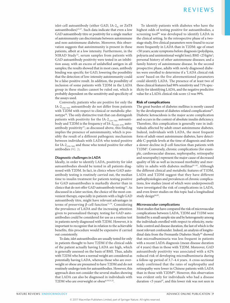
Autoantibody titre and numberThe presence of an insulin-deficient phenotype in patients with LADA who were in the highest tertile for GAD autoantibody levels was initially demonstrated in the BOTNIA Study19. Subsequently, the NIRAD Study22 highlighted the presence of a ‘bimodal distribution’ of the GAD autoantibody titre in patients with LADA — which had a nadir of 32 GAD autoantibody arbitrary units, corresponding to 300 WHO units — that iden-tified two subpopulations, with high and low GAD autoantibody titres (FIG. 1). Based on the method used by Joanes and Gill63, the bimodality of the GAD auto-antibody titre in the NIRAD Study22 showed a bimodal-ity coefficient ≥0.555. The existence of subpopulations with a high or low GAD autoantibody titre has been confirmed by other, independent groups6,25,26; however, some studies did not report a bimodal distribution of the GAD autoantibody titre19.
Table 2 | Genetic, metabolic and clinical features of LADA compared with T1DM and T2DM
Disease feature Disease
T1DM LADA T2DM
Age at diagnosis Childhood to adolescence and rarely in adulthood
>30 years Adulthood and rarely in childhood to adolescence
Onset Acute Rarely acute Slow
Autoimmunity Severely increased Increased No change
Ketosis Frequent Rare Rare
Insulin resistance No change Increased or no change Severely increased
β-Cell function Severely decreased Decreased Increased or no change
Insulin dependence At onset >6 months (even years) after onset
Years after onset
BMI Underweight to normal Normal to overweight Overweight to obese
Risk of the metabolic syndrome
No change Increased Severely increased
HLA susceptibility Severely increased Increased No change
LADA, latent autoimmune diabetes in adults; T1DM, type 1 diabetes mellitus; T2DM, type 2 diabetes mellitus.
R E V I E W S
4 | ADVANCE ONLINE PUBLICATION www.nature.com/nrendo
© 2017
Macmillan
Publishers
Limited,
part
of
Springer
Nature.
All
rights
reserved. ©
2017
Macmillan
Publishers
Limited,
part
of
Springer
Nature.
All
rights
reserved.

Compared with patients with LADA who had a low GAD autoantibody titre, those with a high titre had more severe autoimmunity, which resulted in higher levels of HbA1c, a lower BMI and a lower prevalence of the metabolic syndrome22. In patients with LADA, these GAD-autoantibody titre-dependent differences in clinical and biochemical features were substantiated by genetic studies that reported that the frequencies of high-risk and moderate-risk HLA genotypes decreased linearly from a high to a low GAD autoantibody titre22. Similarly, the PTPN22 risk genotype was also associated with a high GAD autoantibody titre in patients with LADA64. Conversely, the transcription factor 7 like 2 (TCF7L2) risk allele for T2DM was associated with a low, rather than a high, GAD autoantibody titre65. Of note, not all studies have confirmed the association between LADA and the TCF7L2 gene66; this discrepancy might be due to different patient inclusion criteria and the fact that such studies were underpowered. In addi-tion, a higher frequency of autoantibodies associated
with autoimmune diabetes and other organ-specific autoantibodies — including thyroid peroxidase, steroid 21-hydroxylase, tissue transglutaminase and parietal cell autoantibodies — was detected in patients with LADA who had high GAD autoantibody titres than in those with low titres, thus confirming the increased sever-ity of the autoimmune process in the former group of patients67. These findings might have important clinical implications, and we suggest regular screening for other organ-specific autoantibodies (indicative of other auto-immune diseases) in patients with LADA according to the GAD autoantibody titre.
The number of islet autoantibodies in LADA has also been demonstrated to reflect the intensity of the autoimmune response and to predict future insulin defi-ciency18,68. ZnT8 autoantibodies, as a marker in addition to GAD autoantibodies and IA-2 autoantibodies, have enabled stratification of the intensity of the islet auto-immune response, which is a clear reflection of the clinical phenotype of patients with adult-onset diabetes; features of more severe insulin insufficiency are proportional to the number of islet autoantibodies29.
In addition, the United Kingdom Prospective Diabetes Study 25 reported that a high GAD auto antibody titre was associated with an increased risk of insulin require-ment only among patients >55 years old at diagnosis18. However, conflicting results have since been reported; some studies demonstrated that a high titre of GAD autoantibodies was associated with a shorter insulin- free period19,69, whereas other studies did not support this hypothesis25,70. In the NIRAD Study, a high GAD autoantibody titre, BMI <25 kg/m2, positivity for ZnT8 and IA-2IC(605–979) autoantibodies and treatment with sul-fonylurea in the first year after diagnosis led to markedly increased progression to insulin requirement in patients with LADA71.
Autoantibodies and pathophysiologyThe IA-2(256-760) autoantibody, which is present in 30% of patients with GAD autoantibody positivity and in 3.4% of patients who are negative for GAD and IA-2IC(605–979) autoantibodies57, was the only autoantibody that showed increased frequency with increasing BMI in a population of consecutive patients with T2DM58. More interestingly, only patients with T2DM who were pos-itive for the IA-2(256-760) autoantibody showed clinical and metabolic phenotypes that exactly resembled those of patients with classic T2DM and obesity and had a substantially slower progression to insulin requirement within 7 years of follow-up than patients who were GAD autoantibody positive58.
The presence of the IA-2(256-760) autoantibody in patients with diabetes could underlie a pathophysio-logical mechanism resulting in a humoral immune response that is different from the response occurring in ‘classic’ autoimmune diabetes, as it is probably derived from the chronic systemic inflammation that is associ-ated with obesity. Innate and acquired autoimmunity, specifically through the activity of macrophages and self-reactive T cells, contribute to the increased secre-tion of pro- inflammatory cytokines that are involved in
Figure 1 | Bimodal distribution of the glutamic acid decarboxylase autoantibody titre in patients with latent autoimmune diabetes in adults. The bar graph shows the frequency and distribution of the glutamic acid decarboxylase (GAD) autoantibody titre in patients with latent autoimmune diabetes in adults (LADA). The y axis shows the absolute frequency (%) of the values specified on the x axis. The x axis reports the log10-transformed titre of GAD autoantibodies (and the respective titre in arbitrary units (U)) measured in 193 patients with LADA who participated in the Non Insulin Requiring Autoimmune Diabetes (NIRAD) Study22. The GAD autoantibody titre was measured using a radiobinding assay with in vitro-translated 35S-methionine-labelled GAD65 antibody. The results were converted into U by extrapolation from a standard curve with a local standard designated 100 U. The graph illustrates the bimodal distribution of the GAD autoantibody titre in patients with LADA, with a nadir of 32 U, and identifies two subpopulations, with low (blue bars) and high (red bars) GAD autoantibody titres. The bimodality of the GAD autoantibody titre was verified using a bimodality coefficient63, which we determined to be ≥0.555. American Diabetes Association, Buzzetti, R. et al. High titer of autoantibodies to GAD identifies a specific phenotype of adult-onset autoimmune diabetes, Diabetes Care, 30, 932–938, American Diabetes Association, 2007. Copyright and all rights reserved. Material from this publication has been used with the permission of American Diabetes Association.
Nature Reviews | Endocrinology
Abs
olut
e fr
eque
ncy
(%)
GAD autoantibody titre (log10
transformed)
GAD autoantibody titre arbitrary units (U)
0
5
10
10
15
20
25
30
0.5 1.0 1.5 2.0 2.5 3.0
3 32 100 316 1,000
Low GAD autoantibody titre
High GAD autoantibody titre
R E V I E W S
NATURE REVIEWS | ENDOCRINOLOGY ADVANCE ONLINE PUBLICATION | 5
© 2017
Macmillan
Publishers
Limited,
part
of
Springer
Nature.
All
rights
reserved. ©
2017
Macmillan
Publishers
Limited,
part
of
Springer
Nature.
All
rights
reserved.

inflammatory processes72. The resulting inflammation might favour the presentation of extracellular antigens to antigen-presenting cells, thus promoting autoimmune activation72.
The IA-2 autoantibody in T1DM is directed against the intracellular portion of the protein73. However, the epitope recognized by the IA-2(256-760) autoantibody is located in the extracellular domain of IA-2, which is more accessible to autoantibodies than intracellular epitopes. Thus, tissue damage induced by inflamma-tion might trigger an autoimmune response to ‘cryptic’ self-antigens, thereby accelerating β-cell death. This hypothesis is supported by preliminary observations that the IA-2(256-760) autoantibody is detected in patients who are obese and do not have diabetes (R.B., unpublished observations). Therefore, as previously suggested4, the development of autoimmunity in patients with LADA could arise either as a consequence of the chronic inflammatory responses associated with obesity or as a result of a more specific environmental trigger that
can promote the activation of an autoimmune process similar to that which occurs in T1DM74,75 (FIG. 2). This hypothesis is purely speculative but encourages reflec-tion on the existence of autoantibodies in T2DM and on the considerable heterogeneity of the patient population included in the definition of LADA.
The alternative possibility that the IA-2(256-760) auto-antibody could reflect false-positive immunoreactivity was tested in a series of experiments that used different concentrations of unlabelled IA-2(256–760) fragments; the results demonstrated the specificity of antibody bind-ing to 35S-labelled IA-2(256–760) (REF. 57). Furthermore, a new autoantibody against the extracytoplasmatic (EC) IA-2EC(26-577) region can identify a novel antigenic deter-minant within the N terminus of the IA-2 protein76. This autoantibody, whose epitope overlaps with the IA-2(256-760) epitope, can be detected in a subgroup of patients with adult-onset autoimmune diabetes who have a T2DM phenotype and who are negative for conventional islet autoantibodies76. This finding adds further support to the relevance of the N-terminal region of IA-2 in terms of antigenicity in diabetes.
LADA versus T2DMAlthough they are phenotypically distinct when large groups are compared, at the individual level, patients with LADA and patients with T2DM share clinical and metabolic characteristics, making it very difficult to diagnose LADA solely on the basis of the clinical phe-notype. Therefore, individuals with LADA are often misdiagnosed as having T2DM. Several studies have been carried out to investigate the differences between LADA and T2DM with respect to genetic susceptibility and clinical features and have provided insight into how to correctly identify patients with LADA.
Genetic and clinical featuresLADA shares several genetic features with T2DM. As previously mentioned, variants of the TCF7L2 gene are also associated with LADA77; this association is particu-larly strong in patients with a low GAD autoantibody titre65. The FTO66 gene is also associated with LADA, but to a lesser extent than TCF7L2 (REF. 77). These associa-tions suggest that LADA represents a genetic admixture of T1DM and T2DM.
Overall, patients with LADA show metabolic and clinical phenotypes that are substantially different from those of classic T2DM; relative to T2DM, LADA is char-acterized by higher fasting levels of glucose and HbA1c, –a lower prevalence of the metabolic syndrome, a higher fre-quency of thyroid peroxidase autoantibodies22, a higher frequency of HLA risk haplotypes and a consistently greater likelihood of insulin requirement6,71 (TABLE 2). In addition, patients with LADA had a lower stimu-lated C-peptide response at all time-points during an MMTT than individuals with T2DM, indicating reduced β-cell function4. This observation is in agreement with previous studies61,78.
Although they are still present, these differences are less evident in patients with LADA who have a low GAD autoantibody titre or positivity for a single
Nature Reviews | Endocrinology
LADA High degree of heterogeneity
Moderate T1DM genetic susceptibility
T2DM genetic susceptibility
• Normal• Overweight• Obese
Obese
Islet autoimmunity: GAD autoantibody-positive
Islet autoimmunity: IA-2(256-760) autoantibody-positive
β-cell apoptosis
Insulin deficiency Impaired insulin secretion
Accelerated loss of β-cell function
Specific triggerLow-grade inflammation
1 2
Figure 2 | Potential pathological mechanisms of latent autoimmune diabetes in adults. Here, we describe our working hypothesis regarding the pathophysiology of latent autoimmune diabetes in adults (LADA). In patients with moderate genetic susceptibility to type 1 diabetes mellitus (T1DM), specific immunological factors that are not well characterized can trigger an autoimmune process against the islets of Langerhans; this is independent of obesity, as it can occur in individuals with a normal BMI or who are overweight. This autoimmune process is marked by the appearance of glutamic acid decarboxylase (GAD) autoantibodies in the serum. Islet autoimmunity causes β-cell apoptosis, leading to insulin deficiency, which finally causes disease onset (1). LADA might also develop in individuals with obesity who have genetic susceptibility to type 2 diabetes mellitus (T2DM). The low-grade inflammation that characterizes visceral adiposity might trigger a low-grade autoimmune process that leads to the development of less severe islet autoimmunity, marked by the presence of serum autoantibodies against the tyrosine phosphatase IA-2(256-760) construct. This autoimmunity causes accelerated loss of β-cell function and impaired insulin secretion, which, when combined with the insulin resistance that commonly occurs in patients with obesity, causes hyperglycaemia and onset of diabetes mellitus (2). Our proposed pathological pathways explain the heterogeneous metabolic and clinical phenotypes of LADA, ranging from patients who are lean and insulin sensitive to those with obesity and insulin resistance, with a phenotype that is undistinguishable from that of T2DM.
R E V I E W S
6 | ADVANCE ONLINE PUBLICATION www.nature.com/nrendo
© 2017
Macmillan
Publishers
Limited,
part
of
Springer
Nature.
All
rights
reserved. ©
2017
Macmillan
Publishers
Limited,
part
of
Springer
Nature.
All
rights
reserved.
islet-cell autoantibody (either GAD, IA-2IC or ZnT8 autoantibodies)22,27. Such data indicate that even a low GAD autoantibody titre or positivity for a single marker of autoimmunity can discriminate between auto immune and non-autoimmune diabetes. Moreover, this obser-vation suggests that autoimmunity is present in these patients, albeit at a low intensity. Furthermore, in the NIRAD Study22, serum samples from patients with GAD autoantibody positivity were tested in an inhibi-tion assay, with an excess of unlabelled antigen in all samples; the results showed that in most cases, antibody binding was specific for GAD, lowering the possibility that the detection of low-intensity autoimmunity could be a false- positive result. In addition, the possibility of inclusion of some patients with T2DM in the LADA group in these studies cannot be ruled out, which is probably dependent on the sensitivity and specificity of the assays used.
Conversely, patients who are positive for only the IA-2(256-760) autoantibody do not differ from patients with T2DM with respect to clinical or metabolic phe-notype58. The only distinctive trait that can distinguish patients with positivity for the IA-2(256-760) autoanti-body and T2DM is the frequency of IA-2IC(605–979) auto-antibody positivity57; as discussed above, this finding implies the presence of autoimmunity, which is pos-sibly the result of a different pathogenic mechanism between individuals with LADA who tested positive for IA-2(256-760) and those who tested positive for other antibodies (FIG. 2).
Diagnostic challenges in LADAIdeally, in order to identify LADA, positivity for islet autoantibodies should be tested in all patients diag-nosed with T2DM. In fact, in clinics where GAD auto-antibody testing is routinely carried out, the median time to insulin treatment for patients testing positive for GAD autoantibodies is markedly shorter than in clinics that do not offer GAD autoantibody testing13. As discussed in a later section, the choice of the most con-venient therapy, especially in patients with a high GAD autoantibody titre, might have relevant advantages in terms of preserving β-cell function71,79. Considering the prevalence of LADA and the increasing attention given to personalized therapy, testing for GAD auto-antibodies could be considered for use as a routine test in patients newly diagnosed with T2DM. However, it is important to recognize that in relation to the achievable benefits, this procedure would be expensive if carried out consistently.
To date, islet autoantibodies are usually tested for only in patients thought to have T2DM if the clinical odds of the patient actually having LADA are high, which is generally assessed on the basis of BMI. Thus, adults with T2DM who have a normal weight are considered as potentially having LADA, whereas those who are over-weight or obese are presumed to have T2DM and do not routinely undergo tests for autoantibodies. However, this approach does not consider the several studies showing that LADA can also be diagnosed in individuals with T2DM who are overweight or obese6,19,21,22.
To identify patients with diabetes who have the highest odds of testing positive for autoantibodies, a screening tool80 was developed to identify LADA in the clinical setting. In the retrospective phase of a two-stage study, five clinical parameters were found to occur more frequently in LADA than in T2DM: age of onset <50 years; acute symptoms before diagnosis (polydipsia, polyuria and unintentional weight loss); BMI <25 kg⁄m2; personal history of other autoimmune diseases; and a family history of autoimmune disease. In the second prospective phase, adults with newly diagnosed diabe-tes were enrolled to determine if a ‘LADA clinical risk score’ based on the five aforementioned parameters could identify LADA. The presence of at least two of these clinical features had 90% sensitivity and 71% spec-ificity for identifying LADA, and the negative predictive value for a LADA clinical risk score ≤1 was 99%.
Risk of complicationsThe great burden of diabetes mellitus is mostly caused by the development of diabetes-related complications81. Diabetic ketoacidosis is the major acute complication and occurs in the context of absolute insulin deficiency. Therefore, this complication is generally rare in indi-viduals affected by adult-onset autoimmune diabetes. Indeed, individuals with LADA, the most frequent form of adult-onset autoimmune diabetes, have detect-able C-peptide levels at the time of diagnosis and show a slower decline in β-cell function than patients with T1DM4. Conversely, chronic complications (for exam-ple, cardiovascular disease, nephropathy, retinopathy and neuropathy) represent the major cause of decreased quality of life as well as increased morbidity and mor-tality in adults with diabetes mellitus81–83. Although the different clinical and metabolic features of T1DM, LADA and T2DM suggest that they have different pathophysiologies and prevalences of chronic complica-tions, few studies (most of which were underpowered) have investigated the risk of complications in LADA, and even fewer studies on this topic had a longitudinal study design84,85.
Microvascular complicationsMost studies that have compared the risk of micro vascular complications between LADA, T2DM and T1DM were limited by a small sample size and by hetero geneity among the individuals enrolled with respect to ethnicity, meta-bolic control and disease duration, the last of which is the most relevant confounder. Indeed, an analysis of longitu-dinal data from the Fremantle Diabetes Study84 showed that microalbuminuria was less frequent in patients with a recent LADA diagnosis (mean disease duration of 4 years) than in those with T2DM. Moreover, GAD autoantibody positivity was associated with a 62% reduced risk of developing microalbuminuria during a follow-up period of 3.7–4.4 years. A cross-sectional study confirmed that the rates of nephropathy and retinopathy were lower in Chinese patients with LADA than in those with T2DM86. However, this observation was evident only for individuals who had a disease duration <5 years87, and this lower risk was not seen in
R E V I E W S
NATURE REVIEWS | ENDOCRINOLOGY ADVANCE ONLINE PUBLICATION | 7
© 2017
Macmillan
Publishers
Limited,
part
of
Springer
Nature.
All
rights
reserved. ©
2017
Macmillan
Publishers
Limited,
part
of
Springer
Nature.
All
rights
reserved.

patients who had a disease duration >5 years (FIG. 3a,b). Accordingly, studies that evaluated patients with a dis-ease duration >5 years showed that patients with diabetes who were GAD autoantibody positive had equal or even higher rates of microvascular complications than those who were negative for GAD autoantibodies88–90 (FIG. 3a,b).
Prospective data from the BOTNIA Study85 also showed that a disease duration >5.5 years was associated with an increased risk of retinopathy in patients with LADA (FIG. 3a). The low rates of microvascular com-plications observed in patients with LADA who have a short disease duration could in part be explained by the fact that T2DM is usually diagnosed later in the history of the disease than LADA91,92. In fact, individuals with newly diagnosed T2DM are exposed to the detrimen-tal effects of hyperglycaemia on the small retinal and renal blood vessels long before diagnosis, whereas this phenomenon has not been shown in LADA. However, a large Swedish registry study did not report differ-ences between LADA and T2DM with respect to the prevalence of diabetic retinopathy at disease onset, in contrast to previous results93 (FIG. 3a). In addition, no differences in the prevalence of microvascular com-plications were found among patients from South Korea with LADA, T2DM and T1DM94. Most of the studies that have investigated diabetic neuropathy have shown that neuropathy was generally more prevalent in LADA than in T2DM84,85,87,89,94, with the exception of a study by Baum et al.95 (FIG. 3c).
Macrovascular complicationsIndividuals with LADA have a healthier lipid profile and lower blood pressure values, are leaner and have less central adiposity than individuals with T2DM6,7,19,22. Collectively, these factors suggest a lower risk of coro-nary heart disease, stroke and peripheral artery disease
Myhill
et al.8
4
n =
45
Myhill
et al.8
4
n =
45
Myhill
et al.8
4
n =
45
Myhill
et al.8
4
n =
45
Marti
nell et a
l.93
n =
163
Marti
nell et a
l.93
n =
163
Roh et al.9
4
n =
17
Roh et al.9
4
n =
17
Roh et al.9
4
n =
17
Lu et al.8
6 (<5 years)
§
n =
148
Lu et al.8
6 (<5 years)
§
n =
148
Lu et al.8
6 (<5 years)
§
n =
148
Baum et al.9
5
n =
14
Wang et a
l.87 (<
5 years)§
n =
196
Wang et a
l.87
n =
360
Wang et a
l.87
n =
360
Hawa et al.8
8
n =
173
Hawa et al.8
8
n =
173
Balme et a
l.90
n =
24
Arikan et a
l.89
n =
17
Arikan et a
l.89
n =
17
Arikan et a
l.89
n =
17
Isomaa et a
l.85
n =
59
Isomaa et a
l.85
n =
59
Isomaa et a
l.85
n =
59
Isomaa et a
l.85
n =
59
Biesenbach et a
l.124
n =
52
Biesenbach et a
l.124
n =
52
Biesenbach et a
l.124
n =
52
0
20
40
60
80
100
Prev
alen
ce o
f neu
ropa
thy
(%)
0
20
40
60
80
Prev
alen
ce o
f ret
inop
athy
(%)
0
20
40
60
80
100
Prev
alen
ce o
f nep
hrop
athy
* (%
)
0
20
40
60
80
Prev
alen
ce o
f CV
D‡ (%
)
c
d
b
a
Nature Reviews | Endocrinology
<5 years >5 yearsDisease duration
Lu et al.8
6 (≥5 years)
§
n =
171
<5 years >5 yearsDisease duration
>5 yearsDisease duration
Wang et a
l.87 (5–14 years)
§
n =
131
Wang et a
l.87 (>
15 years)§
n =
33
Lu et al.8
6 (≥5 years)
§
n =
171
<5 years >5 yearsDisease duration
Lu et al.8
6 (≥5 years)
§
n =
171
<5 years
LADA T2DM
Figure 3 | Prevalence of chronic diabetes‑related complications in latent autoimmune diabetes in adults and type 2 diabetes mellitus. The y axes depict the prevalence (%) of chronic diabetes-related complications, including retinopathy (part a), nephropathy (part b), neuropathy (part c) and cardiovascular disease (CVD; part d), in patients with latent autoimmune diabetes in adults (LADA; red bars) versus patients with type 2 diabetes mellitus (T2DM; blue bars). The x axes show the studies in which the data were reported. The studies reported on the x axes are stratified by a disease duration threshold of 5 years (depicted with a dashed line). The prevalences of retinopathy and nephropathy are generally lower in LADA (red bars) than in T2DM (blue bars) when the disease duration is <5 years and are equal between the two diseases or higher in LADA than in T2DM when the disease duration is >5 years. Neuropathy is more prevalent in LADA than in T2DM. The prevalence of CVD does not differ between LADA and T2DM. *The rates of microalbuminuria and/or macroalbuminuria are reported. ‡The rates of coronary artery disease are reported. §The data from these studies were split according to years of disease duration and are depicted separately in the graph. The term ‘n’ refers to the number of patients with LADA enrolled in each study.
R E V I E W S
8 | ADVANCE ONLINE PUBLICATION www.nature.com/nrendo
© 2017
Macmillan
Publishers
Limited,
part
of
Springer
Nature.
All
rights
reserved. ©
2017
Macmillan
Publishers
Limited,
part
of
Springer
Nature.
All
rights
reserved.

in patients with LADA than in those who have T2DM. However, the evidence published thus far has failed to support this hypothesis (FIG. 3d). In the BOTNIA Study85, 56% and 5% of the patients with LADA enrolled had a positive history of coronary artery disease and stroke, respectively, after 13 years of disease. These rates were similar to those found in people with T2DM, which were 58% and 7%, respectively85. In the same study, the overall mortality during a follow-up of 5.7 years was 18% in LADA and 20% in T2DM (the difference was not statistically significant), and the cardiovascular mor-tality was 7.4% in LADA and 12.4% in T2DM (the difference was again not statistically significant). The HUNT2 study showed that patients with T2DM who were positive for GAD autoantibodies had a similar risk of all-cause mortality, cardiovascular disease and ischaemic heart disease as individuals with T2DM who were negative for GAD autoantibodies and an increased risk compared with healthy individuals96. The Fremantle Diabetes Study84 also did not show differences in the prevalences of cardiovascular disease and mortality between LADA and T2DM. Moreover, no differences in terms of cardiovascular outcomes were reported in a post-hoc analysis in the Collaborative Atorvastatin Diabetes Study88.
Despite the healthier vascular risk profile of patients with LADA, the similar rates of cardiovascular disease in LADA combined with the evidence that the high mor-tality risk in autoimmune diabetes is not completely explained by hyperglycaemia82 suggest that a different pathophysiological mechanism underlies the develop-ment of cardiovascular disease in LADA compared with T2DM. This hypothesis warrants further investigation of the potential pathways mediating atherogenesis in autoimmune diabetes.
Treatment of LADAAlthough different recommendations exist for the treat-ment of adults with T1DM and T2DM, no specific guide-lines for the treatment of individuals with LADA have been published so far. As a consequence, the treatment of adults with LADA is currently guided by the clinical intuition and expertise of the physician. Owing to both frequent misdiagnosis and a lack of standard treatment options for LADA, most patients with LADA are initially treated with therapies intended for non-autoimmune forms of diabetes mellitus. This approach might result in rapid progression to an insulin-dependent state, espe-cially in patients who are young and lean and who have high GAD autoantibody titres7,71. Nevertheless, several studies have been carried out to test different treatment strategies (TABLE 3).
Addressing insulin deficiencyAs a form of autoimmune diabetes, LADA is character-ized by loss of β-cell mass and function, a crucial patho-logical event that should be managed by therapeutic strategies. Insulin treatment is the most straightforward therapy for replacing the low levels of endogenous insulin caused by β-cell loss. Interventions intended to preserve β-cell function have also been tested in LADA.
Insulin therapy. One of the main unanswered ques-tions about the therapeutic strategies that should be undertaken in patients with LADA is if insulin therapy is really needed as an initial therapy for this form of autoimmune diabetes. Most of the studies investigating this issue were systematically reviewed by the Cochrane Collaboration in 2011, which showed that treatment with sulfonylureas was associated with worse metabolic control than insulin treatment, with earlier progression towards an insulin-dependent state79,97–102. Therefore, it was recommended that sulfonylureas be avoided in favour of early insulin treatment. However, this recom-mendation relies on limited evidence97 and does not take into account the need for a personalized medi-cine approach, which is recommended by the current international guidelines for the treatment of diabetes mellitus in adults103. Our group has shown that pro-gression towards an insulin-dependent state in LADA differs based on clinical and biochemical features7,71. Patients with high GAD autoantibody titres have the highest risk of early insulin dependence. Moreover, a later onset of LADA is associated with a lower risk of needing insulin therapy. This observation raises the question of if early insulin therapy is the best approach in patients with LADA who have a low GAD autoanti-body titre, particularly in older patients, who are at the highest risk of suffering from hypoglycaemia-related adverse effects.
Drugs to preserve β‑cell function in LADA. The control of blood levels of glucose should not be the sole aim of LADA treatments; the preservation of β-cell function by suppressing the autoimmune destruction of β cells and by stimulating β-cell regeneration should also be pursued, taking advantage of the slow progression of LADA104,105. Indeed, preserved insulin-secreting capac-ity is associated with better clinical outcomes and a decreased risk of complications106,107.
Immune intervention trials have also been carried out for LADA and have shown some efficacy in preserving stimulated C-peptide levels and in achieving improved glycaemic control108,109. Vaccination with GAD65 in LADA has shown a good safety profile during a 5-year follow-up period110. The tested molecules, however, are not currently available on the market.
Intriguingly, data suggest that drugs already approved for the treatment of T2DM might be useful for the treat-ment of LADA. Preclinical data from animal models have shown that treatment with glucagon-like peptide 1 (GLP1) induces β-cell self-renewal, reduces β-cell apop-tosis and promotes β-cell neogenesis from ductal cell precursors111. However, in vitro data from human cells have supported only a possible protective effect of incre-tin hormones on human β cells, specifically in terms of reducing apoptosis112, and no randomized controlled tri-als testing the implementation of GLP1 receptor agonists in patients with LADA are available. A large prospective study that evaluated predictors of the glycaemic response to GLP1 receptor agonists found that patients diag-nosed with T2DM who tested positive for GAD and/or IA-2 autoantibodies and who had low C-peptide levels
R E V I E W S
NATURE REVIEWS | ENDOCRINOLOGY ADVANCE ONLINE PUBLICATION | 9
© 2017
Macmillan
Publishers
Limited,
part
of
Springer
Nature.
All
rights
reserved. ©
2017
Macmillan
Publishers
Limited,
part
of
Springer
Nature.
All
rights
reserved.

exhibited a lower reduction in HbA1c than patients who tested negative for autoantibodies and had high C-peptide levels113. This study suggests that auto-antibody positivity and C-peptide levels could be useful for the stratification of patients with diabetes in terms of therapy response; individuals with LADA are less likely to benefit from incretins than patients with T2DM in terms of reductions in levels of HbA1c. However, this study does not exclude the potential benefits of GLP1 receptor agonists in terms of β-cell protection, which is suggested by a mean 17% reduction in daily insulin dose, even in patients who are autoantibody positive and who have C-peptide levels <0.25 pmol/l. Furthermore, other studies have reported some benefits in terms of the preservation of C-peptide levels by using the dipeptidyl peptidase 4 inhibitors sitagliptin, linagliptin and saxagliptin114–116.
Lifestyle factors. Smoking has been hypothesized to be associated with decreased autoimmunity and a larger pancreatic reserve, as suggested by a lower GAD auto-antibody titre, higher C-peptide levels and a reduced risk of developing LADA in smokers than in non- smokers117,118. However, data from larger populations have called this hypothesis into question119. Moreover, an increase in daily alcohol intake has been associated with increased GAD autoantibody levels; however, fur-ther longitudinal studies are needed to better investigate the possible association between alcohol consumption and autoimmunity in adults with diabetes120.
Addressing insulin resistanceIn addition to insulin deficiency, insulin resistance is also a clinical feature of LADA, although it is less severe than in T2DM. Thus, patients with LADA who have
Table 3 | Studies investigating pharmacological therapies for LADA
Study design Drugs tested Study population Main findings Refs
Pilot randomized, open-label study
Insulin versus SU • 10 participants with NIDDM
• Positive for ICAs
• Serum C-peptide response improved significantly within 6 and 12 months in the insulin group and decreased in the SU group.
• Glycaemic control worsened in the SU group, but not in the insulin group.
98
Randomized, multicentre, open-label study
Insulin versus SU 60 participants with diabetes, positive for GAD autoantibodies, disease duration ≤5 years
After a mean follow-up of 57 months, a lower rate of progression to an insulin-dependent state was observed in the insulin group.
99
Randomized, open-label study
Insulin versus diet with or without OHA (metformin and/or SU)
37 participants with NIDDM, positive for ICAs or GAD autoantibodies
• HbA1c levels were stable in the insulin-treated group but increased in the diet ± OHA group.
• No significant differences in C-peptide levels were observed between the two groups.
100
Randomized, open-label study
Rosiglitazone with insulin versus insulin alone
23 participants with LADA, fasting C-peptide levels >0.3 nmol/l
After 12 and 18 months, measures of β-cell function were stable in the group receiving rosiglitazone with insulin but decreased in the insulin-alone group.
121
Phase II, randomized, placebo-controlled, dose-escalation clinical trial
Alum-formulated recombinant GAD65 (GAD65 vaccination)
47 participants with T2DM, GAD autoantibody positive
Fasting C-peptide levels remained stable for 5 years after drug administration in the groups treated with 4, 20 and 100 micrograms of the GAD65 vaccine but decreased in the placebo and 500-microgram groups.
108,110
Longitudinal observational study
GLP1-RA 620 participants with T2DM commencing a GLP1-RA
• GAD or IA-2 islet autoantibody-positive participants had a nonsignificant reduction in the adjusted mean level of HbA1c (−4.6 mmol/mol (95% CI −10.3 to 1.1)), which was lower than the reduction observed in antibody-negative participants.
• Antibody-positive participants experienced a 17% reduction in insulin dose (versus 40% in antibody-negative participants, P < 0.01).
113
Randomized, open-label study
Sitagliptin and insulin versus insulin alone
30 participants with LADA
Measures of β-cell function evaluated after 12 months of treatment were stable in the group receiving sitagliptin and insulin but were decreased in the insulin-alone group.
115
Prespecified exploratory analysis of a randomized controlled trial
Linagliptin versus glimepiride
118 participants with LADA
Fasting C-peptide levels increased from baseline at weeks 28, 52 and 104 in participants treated with linagliptin but decreased in glimepiride-treated patients. No differences in metabolic control were found.
114
Post-hoc analysis of data pooled from five randomized, placebo-controlled, 24-week phase III studies
Saxagliptin 133 participants, GAD autoantibody-positive (98 in the saxagliptin arm and 35 in the placebo arm)
• Saxagliptin was effective in lowering blood glucose levels and was generally well tolerated in GAD autoantibody- positive participants.
• Saxagliptin increased β-cell function in GAD autoantibody- positive participants.
116
GAD, glutamic acid decarboxylase; ICA, islet-cell autoantibody; GLP1-RA, glucagon-like peptide 1 receptor agonist; IA-2, tyrosine phosphatase IA-2; LADA, latent autoimmune diabetes in adults; NIDDM, non-insulin-dependent diabetes mellitus; OHA, oral hypoglycaemic agent; SU, sulfonylurea; T2DM, type 2 diabetes mellitus.
R E V I E W S
10 | ADVANCE ONLINE PUBLICATION www.nature.com/nrendo
© 2017
Macmillan
Publishers
Limited,
part
of
Springer
Nature.
All
rights
reserved. ©
2017
Macmillan
Publishers
Limited,
part
of
Springer
Nature.
All
rights
reserved.

a more pronounced insulin-resistant phenotype might benefit from the addition of insulin sensitizers to insu-lin therapy. However, no randomized controlled trials have tested metformin in LADA; only rosiglitazone has been tested as an insulin sensitizer in LADA, with encouraging results121, demonstrating the potential use of peroxisome proliferator-activated receptor γ agonists.
Clinical outlookIn summary, as LADA has clinical features that are midway between those of T1DM and T2DM, treat-ments aimed at both preserving and restoring β-cell function by suppressing autoimmunity and stimulat-ing β-cell proliferation as well as at improving insulin sensitivity and ameliorating glycaemic control are war-ranted. Accordingly, the use of combination therapies
to target different pathways could be the best choice, but large randomized controlled trials will be required to confirm this hypothesis106. Based on the current evidence, an early start to insulin treatment together with lifestyle interventions (diet and physical activity) could be advised for patients with LADA, especially in younger patients with high GAD autoantibody titres, because such treatment modalities are able to both control hyperglycaemia and preserve β-cell function by reducing β-cell stress and preventing glucotoxicity121,122. Other agents, such as gliptins or insulin sensitizers, could be combined with insulin based on patients’ clin-ical features, such as BMI, HbA1c levels and C-peptide levels. Finally, it should always be remembered that patients with diabetes mellitus, independent of subtype, need a multifactorial approach to treatment that takes into account the complexity of the patient in terms of comorbidities123.
ConclusionsAdult-onset autoimmune diabetes encompasses a wide spectrum of heterogeneous clinical and metabolic phe-notypes, ranging from classic T1DM with onset in adult life to LADA6,19,21,22,25. If we accept the definition whereby LADA can be identified by positivity for any islet autoantibody (not necessarily GAD auto antibodies)12, we have to recognize that this form of diabetes mel-litus also has a considerable degree of heterogeneity. This heterogeneity encompasses not only patients who have clear signs of insulin deficiency, which is usually associated with strong markers of autoimmunity29, but also patients who have a clinical phenotype that resem-bles T2DM but who are still positive for fairly weak markers of autoimmune reactivity57. Therefore, the heterogeneity of LADA could represent a progressive phenotypic spectrum between the two most common forms of diabetes mellitus, T1DM and T2DM75 (FIG. 4). This hypothesis challenges the conventional perception that immune-mediated processes are not relevant to the pathogenesis of T2DM. Further studies are warranted to clarify if the low-grade inflammation that characterizes visceral obesity is linked to an autoimmune process that could influence the understanding of the pathogenesis of T2DM57.
The difficulties in comparing the results of different studies investigating the microvascular and macro-vascular complications associated with adult-onset auto-immune diabetes are partly attributable to the different criteria used to screen patients and to the heterogeneity of the clinical phenotypes of the enrolled patients81,84–94. Overall, studies with larger cohorts and a longitudinal design are needed to investigate both the pathophysiol-ogy and the epidemiology of the complications associated with adult-onset autoimmune diabetes.
We have not yet identified the best therapy for LADA. The identification of efficacious therapies for patients with LADA has been limited by the great degree of vari-ability of the biochemical and clinical features at clinical presentation during patient screening for intervention trials, which makes it difficult to assess the response to therapy79,97–123. Defined categories of disease are
Nature Reviews | Endocrinology
1000
100
1000
0
β-C
ell f
unct
ion
(%)
Insulin sensitivity (%)
T2DM
T1DM
Adult-onset autoimmune diabetes
Adult-onset T1DM
LADA
Autoimmunity (%)
Figure 4 | Interactions between β‑cell function, autoimmunity and insulin sensitivity. β-Cell function, autoimmunity and insulin sensitivity are three key pathological factors that are involved in the pathogenesis of diabetes mellitus. The spectrum of adult-onset diabetes mellitus encompasses all three forms of diabetes, namely, type 1 diabetes mellitus (T1DM), type 2 diabetes mellitus (T2DM) and adult-onset autoimmune diabetes, without defined limits. The figure depicts the continuum (0–100%) of continuously distributed pathological factors and defines the categories of diabetes mellitus according to these variables. Classic T2DM (upper left corner) is characterized by the absence of autoimmunity, well-preserved β-cell function and low insulin sensitivity. Classic T1DM (lower right corner) is characterized by severe autoimmunity, loss of β-cell function and preserved insulin sensitivity. Along the continuum, adult-onset autoimmune diabetes appears midway between the features of T1DM and T2DM, making it difficult to draw defined limits between the different forms of diabetes (dashed lines). Latent autoimmune diabetes in adults (LADA) accounts for the majority of the cases of adult-onset autoimmune diabetes and shows pathological features closer to those of T2DM than to those of adult T1DM, which is more similar to classic T1DM.
R E V I E W S
NATURE REVIEWS | ENDOCRINOLOGY ADVANCE ONLINE PUBLICATION | 11
© 2017
Macmillan
Publishers
Limited,
part
of
Springer
Nature.
All
rights
reserved. ©
2017
Macmillan
Publishers
Limited,
part
of
Springer
Nature.
All
rights
reserved.

commonly needed to offer guidance for the choice of treatment. However, it is difficult to define disease cat-egories for adult-onset autoimmune diabetes owing to the continuous distribution of clinical and metabolic variables (such as autoimmunity, β-cell function and insulin sensitivity). The existence of diverse metabolic
1. Mølbak, A. G., Christau, B., Marner, B., Borch-Johnsen, K. & Nerup, J. Incidence of insulin-dependent diabetes mellitus in age groups over 30 years in Denmark. Diabet. Med. 11, 650–655 (1994).
2. Sabbah, E. et al. Genetic, autoimmune, and clinical characteristics of childhood- and adult-onset type 1 diabetes. Diabetes Care 23, 1326–1332 (2000).
3. Howson, J. M. et al. Genetic analysis of adult-onset autoimmune diabetes. Diabetes 60, 2645–2653 (2011).
4. Hernandez, M. et al. Insulin secretion in patients with latent autoimmune diabetes (LADA): half way between type 1 and type 2 diabetes: action LADA 9. BMC Endocr. Disord. 15, 1 (2015).
5. Tuomi, T. et al. Antibodies to glutamic acid decarboxylase reveal latent autoimmune diabetes mellitus in adults with a non-insulin-dependent onset of disease. Diabetes 42, 359–362 (1993).
6. Hawa, M. I. et al. Adult-onset autoimmune diabetes in Europe is prevalent with a broad clinical phenotype: action LADA 7. Diabetes Care 36, 908–913 (2013).
7. Maddaloni, E. et al. Latent autoimmune diabetes in adults in the United Arab Emirates: clinical features and factors related to insulin-requirement. PLoS ONE 10, e0131837 (2015).
8. Zimmet, P. Z. et al. Latent autoimmune diabetes mellitus in adults (LADA): the role of antibodies to glutamic acid decarboxylase in diagnosis and prediction of insulin dependency. Diabet. Med. 11, 299–303 (1994).
9. Juneja, R. & Palmer, J. P. Type 1 1/2 diabetes: myth or reality? Autoimmunity 29, 65–83 (1999).
10. Pozzilli, P. & Di Mario, U. Autoimmune diabetes not requiring insulin at diagnosis (latent autoimmune diabetes of the adult): definition, characterization, and potential prevention. Diabetes Care 24, 1460–1467 (2001).
11. Kobayashi, T. et al. Insulin intervention to preserve beta cells in slowly progressive insulin-dependent (type 1) diabetes mellitus. Ann. NY Acad. Sci. 958, 117–130 (2001).
12. Fourlanos, S. et al. Latent autoimmune diabetes in adults (LADA) should be less latent. Diabetologia 48, 2206–2212 (2005).
13. Brophy, S. et al. Time to insulin initiation cannot be used in defining latent autoimmune diabetes in adults. Diabetes Care 31, 439–441 (2008).
14. American Diabetes Association. Classification and diagnosis of diabetes. Diabetes Care 40 (Suppl. 1), S11–S24 (2017).
15. Alberti, K. G. & Zimmet, P. Z. Definition, diagnosis and classification of diabetes mellitus and its complications. Part 1: diagnosis and classification of diabetes mellitus provisional report of a WHO consultation. Diabet. Med. 15, 539–553 (1998).
16. Bruno, G. et al. Incidence of type 1 and type 2 diabetes in adults aged 30–49 years: the population-based registry in the province of Turin, Italy. Diabetes Care 28, 2613–2619 (2005).
17. Rawshani, A. et al. The incidence of diabetes among 0–34 year olds in Sweden: new data and better methods. Diabetologia 57, 1375–1381 (2014).
18. Turner, R. et al. UKPDS 25: autoantibodies to islet-cell cytoplasm and glutamic acid decarboxylase for prediction of insulin requirement in type 2 diabetes. UK Prospective Diabetes Study Group. Lancet 350, 1288–1293 (1997).
19. Tuomi, T. et al. Clinical and genetic characteristics of type 2 diabetes with and without GAD antibodies. Diabetes 48, 150–157 (1999).
20. Takeda, H. et al. Clinical, autoimmune, and genetic characteristics of adult-onset diabetic patients with GAD autoantibodies in Japan (Ehime Study). Diabetes Care 25, 995–1001 (2002).
21. Zinman, B. et al. Phenotypic characteristics of GAD antibody-positive recently diagnosed patients with type 2 diabetes in North America and Europe. Diabetes 53, 3193–3200 (2004).
22. Buzzetti, R. et al. High titer of autoantibodies to GAD identifies a specific phenotype of adult-onset autoimmune diabetes. Diabetes Care 30, 932–938 (2007).
23. Radtke, M. A., Midthjell, K., Nilsen, T. I. & Grill, V. Heterogeneity of patients with latent autoimmune diabetes in adults: linkage to autoimmunity is apparent only in those with perceived need for insulin treatment: results from the Nord-Trondelag Health (HUNT) study. Diabetes Care 32, 245–250 (2009).
24. Qi, X. et al. Prevalence and correlates of latent autoimmune diabetes in adults in Tianjin, China: a population-based cross-sectional study. Diabetes Care 34, 66–70 (2011).
25. Maioli, M. et al. Number of autoantibodies and HLA genotype, more than high titers of glutamic acid decarboxylase autoantibodies, predict insulin dependence in latent autoimmune diabetes of adults. Eur. J. Endocrinol. 163, 541–549 (2010).
26. Zhou, Z. et al. Frequency, immunogenetics, and clinical characteristics of latent autoimmune diabetes in China (LADA China Study): a nationwide, multicenter, clinic-based cross-sectional study. Diabetes 62, 543–550 (2013).
27. Tuomi, T. et al. The many faces of diabetes: a disease with increasing heterogeneity. Lancet 383, 1084–1094 (2014).
28. Laugesen, E., Østergaard, J. A. & Leslie, R. D. Latent autoimmune diabetes of the adult: current knowledge and uncertainty. Danish Diabetes Academy Workshop and Workshop Speakers. Diabet. Med. 32, 843–852 (2015).
29. Lampasona, V. et al. Zinc transporter 8 antibodies complement GAD and IA-2 antibodies in the identification and characterization of adult-onset autoimmune diabetes: non insulin requiring autoimmune diabetes (NIRAD) 4. Diabetes Care 33, 104–108 (2010).
30. Yang, Z. et al. Childhood diabetes in China. Enormous variation by place and ethnic group. Diabetes Care 21, 525–529 (1998).
31. Sutanegara, D. & Budhiarta, A. A. The epidemiology and management of diabetes mellitus in Indonesia. Diabetes Res. Clin. Pract. 50, S9–S16 (2000).
32. Mohatt, J., Gilliam, L. K., Bekris, L., Ebbesson, S. & Lernmark, A. Type 1 diabetes-related autoantibodies are rare in Alaska native populations. Int. J. Circumpolar Health 61, 21–31 (2002).
33. Dowse, G. K. et al. Lack of antibodies to glutamic acid decarboxylase in young adults of the high diabetes prevalence Wanigela people of Papua New Guinea. Diabetes Res. Clin. Pract. 24, 195–198 (1994).
34. Barinas-Mitchell, E. et al. Islet cell autoimmunity in a triethnic adult population of the Third National Health and Nutrition Examination Survey. Diabetes 53, 1293–1302 (2004).
35. Temneanu, O. R., Trandafir, L. M. & Purcarea, M. R. Type 2 diabetes mellitus in children and adolescents: a relatively new clinical problem within pediatric practice. J. Med. Life 3, 235–239 (2016).
36. Leslie, R. D., Palmer, J., Schloot, N. C. & Lernmark, A. Diabetes at the crossroads: relevance of disease classification to pathophysiology and treatment. Diabetologia 59, 13–20 (2016).
37. Nisticò, L. et al. The CTLA-4 gene region of chromosome 2q33 is linked to, and associated with, type 1 diabetes. Belgian Diabetes Registry. Hum. Mol. Genet. 5, 1075–1080 (1996).
38. Bottini, N. et al. A functional variant of lymphoid tyrosine phosphatase is associated with type I diabetes. Nat. Genet. 36, 337–338 (2004).
39. Grant, S. F. et al. Variant of transcription factor 7-like 2 (TCF7L2) gene confers risk of type 2 diabetes. Nat. Genet. 38, 320–323 (2006).
40. Frayling, T. M. et al. A common variant in the FTO gene is associated with body mass index and predisposes to childhood and adult obesity. Science 316, 889–894 (2007).
41. Atkinson, M. A. & Eisenbarth, G. S. Type 1 diabetes: new perspectives on disease pathogenesis and treatment. Lancet 358, 221–229 (2001).
42. Leslie, R. D. & Delli Castelli, M. Age-dependent influences on the origins of autoimmune diabetes: evidence and implications. Diabetes 53, 3033–3040 (2004).
43. Caillat-Zucman, S. et al. Age-dependent HLA genetic heterogeneity of type 1 insulin-dependent diabetes mellitus. J. Clin. Invest. 90, 2242–2250 (1992).
44. Petrone, A. et al. Residual insulin secretion at diagnosis of type 1 diabetes is independently associated with both, age of onset and HLA genotype. Diabetes Metab. Res. Rev. 21, 271–275 (2005).
45. Andersen, M. K. et al. Latent autoimmune diabetes in adults differs genetically from classical type 1 diabetes diagnosed after the age of 35 years. Diabetes Care 33, 2062–2064 (2010).
46. Van der Auwera, B. J. et al. CTLA-4 gene polymorphism confers susceptibility to insulin-dependent diabetes mellitus (IDDM) independently from age and from other genetic or immune disease markers. The Belgian Diabetes Registry. Clin. Exp. Immunol.110, 98–103 (1997).
47. Cosentino, A., Gambelunghe, G., Tortoioli, C. & Falorni, A. CTLA-4 gene polymorphism contributes to the genetic risk for latent autoimmune diabetes in adults. Ann. NY Acad. Sci. 958, 337–340 (2002).
48. Bennett, S. T. et al. Insulin VNTR allele-specific effect in type 1 diabetes depends on identity of untransmitted paternal allele. Nat. Genet. 17, 350–352 (1997).
49. Haller, K. et al. Insulin gene VNTR, CTLA-4 + 49A/G and HLA-DQB1 alleles distinguish latent autoimmune diabetes in adults from type 1 diabetes and from type 2 diabetes group. Tissue Antigens 69, 21–27 (2007).
50. Gambelunghe, G. Two distinct MICA gene markers discriminate major autoimmune diabetes types. J. Clin. Endocrinol. Metab. 86, 3754–3760 (2001).
51. Sanjeevi, C. B. Genetics of latent autoimmune diabetes in adults. Ann. NY Acad. Sci. 958, 107–111 (2002).
52. Brooks-Worrell, B. M., Juneja, R., Minokadeh, A., Greenbaum, C. J. & Palmer, J. P. Cellular immune responses to human islet proteins in antibody-positive type 2 diabetic patents. Diabetes 48, 983–988 (1999).
53. Signore, A. et al. Detection of insulitis by pancreatic scintigraphy with 99mTc-labeled IL-2 and MRI in patients with LADA (Action LADA 10). Diabetes Care 38, 652–658 (2015).
54. Brooks-Worrell, B. M., Reichow, J. L., Goel, A., Ismail, H. & Palmer, J. P. Identification of autoantibody-negative autoimmune type 2 diabetic patients. Diabetes Care 34, 168–173 (2011).
55. Graham, J.et al. Genetic effects on age-dependent onset and islet cell autoantibody markers in type 1 diabetes. Diabetes 51, 1346–1355 (2002).
56. Kawasaki, E. Type 1 diabetes and autoimmunity. Clin. Pediatr. Endocrinol. 23, 99–105 (2014).
57. Tiberti, C. et al. Identification of tyrosine phosphatase 2(256–760) construct as a new, sensitive marker for the detection of islet autoimmunity in type 2 diabetic patients: the non-insulin requiring autoimmune diabetes (NIRAD) study 2. Diabetes 57, 1276–1283 (2008).
58. Buzzetti, R. et al. Tyrosine phosphatase-related islet antigen 2(256–760) autoantibodies, the only marker of islet autoimmunity that increases by increasing the degree of BMI in obese subjects with type 2 diabetes. Diabetes Care 38, 513–520 (2015).
59. Pham, M. N. et al. Pro- and anti-inflammatory cytokines in latent autoimmune diabetes in adults, type 1 and type 2 diabetes patients: action LADA 4. Diabetologia 54, 1630–1638 (2011).
60. Ferrannini, E. The stunned beta cell: a brief history. Cell Metab. 11, 349–352 (2010).
61. Stenström, G., Gottsäter, A., Bakhtadze, E., Berger, B. & Sundkvist, G. Latent autoimmune diabetes in adults: definition, prevalence, beta-cell function, and treatment. Diabetes 54 (Suppl. 2), S68–S72 (2005).
and clinical phenotypes in adult-onset autoimmune diabetes makes it difficult to establish an a priori treat-ment algorithm and therefore draws attention to the concept of a personalized medicine approach to ther-apy that takes into account the intrinsic characteristics of each patient.
R E V I E W S
12 | ADVANCE ONLINE PUBLICATION www.nature.com/nrendo
© 2017
Macmillan
Publishers
Limited,
part
of
Springer
Nature.
All
rights
reserved. ©
2017
Macmillan
Publishers
Limited,
part
of
Springer
Nature.
All
rights
reserved.

62. Barker, A. et al. Age-dependent decline of β-cell function in type 1 diabetes after diagnosis: a multi-centre longitudinal study. Diabetes Obes. Metab. 16, 262–267 (2014).
63. Joanes, D. N. & Gill, C. A. Comparing measures of sample skewness and kurtosis. Statistician 47, 183–189 (1998).
64. Petrone, A. et al. The protein tyrosine phosphatase nonreceptor 22 (PTPN22) is associated with high GAD antibody titer in latent autoimmune diabetes in adults: non Insulin Requiring Autoimmune Diabetes (NIRAD) Study 3. Diabetes Care 31, 534–538 (2008).
65. Zampetti, S. et al. Association of TCF7L2 gene variants with low GAD autoantibody titre in LADA subjects (NIRAD Study 5). Diabet. Med. 27, 701–704 (2010).
66. Pettersen, E., Skorpen, F., Kvaløy, K., Midthjell, K. & Grill, V. Genetic heterogeneity in latent autoimmune diabetes is linked to various degrees of autoimmune activity: results from the Nord-Trøndelag Health Study. Diabetes 59, 302–310 (2010).
67. Zampetti, S. et al. GADA titer-related risk for organ-specific autoimmunity in LADA subjects subdivided according to gender (NIRAD study 6). J. Clin. Endocrinol. Metab. 97, 3759–3765 (2012).
68. Bottazzo, G. F. et al. IA-2 antibody prevalence and risk assessment of early insulin requirementin subjects presenting with type 2diabetes (UKPDS 71). Diabetologia 48, 703–708 (2005).
69. Genovese, S. et al. Clinical phenotype and β-cell autoimmunity in Italian patients with adult-onset diabetes. Eur. J. Endocrinol.154, 441–447 (2006).
70. Desai, M. et al. GAD autoantibodies and epitope reactivities persist after diagnosis in latent autoimmune diabetes in adults but do not predict disease progression: UKPDS 77. Diabetologia 50, 2052–2060 (2007).
71. Zampetti, S. et al. High GADA titer increases the risk of insulin requirement in LADA patients: a 7-year follow-up (NIRAD study 7). Eur. J. Endocrinol.171, 697–704 (2014).
72. Itariu, B. K. & Stulnig, T. M. Autoimmune aspects of type 2 diabetes mellitus — a mini-review. Gerontology 60, 189–196 (2014).
73. Zhang, B., Lan, M. S. & Notkins, A. L. Autoantibodies to IA-2 in IDDM: location of major antigenic determinants. Diabetes 46, 40–43 (1997).
74. Brooks-Worrell, B., Narla, R. & Palmer, J. P. Islet autoimmunity in phenotypic type 2 diabetes patients. Diabetes Obes. Metab. 3, 137–140 (2013).
75. Palmer, J. P., Hampe, C. S., Chiu, H., Goel, A. & Brooks-Worrell, B. M. Is latent autoimmune diabetes in adults distinct from type 1 diabetes or just type 1 diabetes at an older age? Diabetes 54, S62–S67 (2005).
76. Acevedo-Calado, M. et al. Identification of unique antigenic determinants in the amino terminus of IA-2 (ICA512) in childhood and adult autoimmune diabetes: new biomarker development. Diabetes Care 40, 561–568 (2017).
77. Andersen, M. K.et al. Type 2 diabetes susceptibility gene variants predispose to adult-onset autoimmune diabetes. Diabetologia 57, 1859–1868 (2014).
78. Carlsson, A., Sundkvist, G., Groop, L. & Tuomi, T. Insulin and glucagon secretion in patients with slowly progressing autoimmune diabetes (LADA). J. Clin. Endocrinol. Metab. 85, 76–80 (2000).
79. Maruyama, T. Multicenter prevention trial of slowly progressive type 1 diabetes with small dose of insulin (the Tokyo study): preliminary report. Ann. NY Acad. Sci. 1005, 362–369 (2003).
80. Fourlanos, S. et al. A clinical screening tool identifies autoimmune diabetes in adults. Diabetes Care 29, 970–975 (2006).
81. Gregg, E. W. et al. Changes in diabetes-related complications in the United States1990–2010. N. Engl. J. Med. 370, 1514–1523 (2014).
82. Lind, M. et al. Glycemic control and excess mortality in type 1 diabetes. N. Engl. J. Med. 371, 1972–1982 (2014).
83. Secrest, A. M., Becker, D. J., Kelsey, S. F., Laporte, R. E. & Orchard, T. J. Cause-specific mortality trends in a large population-based cohort with long-standing childhood-onset type 1 diabetes. Diabetes 59, 3216–3222 (2010).
84. Myhill, P. et al. Chronic complications and mortality in community-based patients with latent autoimmune diabetes in adults: the Fremantle Diabetes Study. Diabet. Med. 25, 1245–1250 (2008).
85. Isomaa, B. et al. Chronic complications in patients with slowly progressing autoimmune type 1 diabetes (LADA). Diabetes Care 22, 1347–1353 (1999).
86. Lu, J. et al. Associations between clinical characteristics and chronic complications in latent autoimmune diabetes in adults and type 2 diabetes. Diabetes. Metab. Res. Rev. 31, 411–420 (2015).
87. Wang, C. et al. Evaluating peripheral nerve function in asymptomatic patients with type 2 diabetes or latent autoimmune diabetes of adults (LADA): results from nerve conduction studies. J. Diabetes Complications 29, 265–269 (2015).
88. Hawa, M. I. et al. LADA and CARDS: a prospective study of clinical outcome in established adult-onset autoimmune diabetes. Diabetes Care 37, 1643–1649 (2014).
89. Arikan, E., Sabuncu, T., Ozer, E. M. & Hatemi, H. The clinical characteristics of latent autoimmune diabetes in adults and its relation with chronic complications in metabolically poor controlled Turkish patients with Type 2 diabetes mellitus. J. Diabetes Complications 19, 254–258 (2005).
90. Balme, M., McAllister, I., Davis, W. A. & Davis, T. M. E. Retinopathy in latent autoimmune diabetes of adults: the Fremantle Diabetes Study. Diabet. Med. 19, 602–605 (2002).
91. Harris, M. I., Klein, R., Welborn, T. A. & Knuiman, M. W. Onset of NIDDM occurs at least 4–7 yr before clinical diagnosis. Diabetes Care 15, 815–819 (1992).
92. Porta, M. et al. Estimating the delay between onset and diagnosis of type 2 diabetesfrom the time course of retinopathy prevalence. Diabetes Care 37, 1668–1674 (2014).
93. Martinell, M. et al. Prevalence and risk factors for diabetic retinopathy at diagnosis (DRAD) in patients recently diagnosed with type 2 diabetes (T2D) or latent autoimmune diabetes in the adult (LADA). J. Diabetes Complications 30, 1456–1461 (2016).
94. Roh, M.-O., Jung, C.-H., Kim, B.-Y., Mok, J.-O. & Kim, C.-H. The prevalence and characteristics of latent autoimmune diabetes in adults (LADA) and its relation with chronic complications in a clinical department of a university hospital in Korea. Acta Diabetol. 50, 129–134 (2013).
95. Baum, P. et al. Diabetic neuropathy in patients with latent autoimmune diabetes of the adult (LADA) compared with patients with type 1 and type 2 diabetes. J. Neurol. 250, 682–687 (2003).
96. Olsson, L. et al. Mortality in adult-onset autoimmune diabetes is associated with poor glycemic control: results from the HUNT Study. Diabetes Care 36, 3971–3978 (2013).
97. Brophy, S., Davies, H., Mannan, S., Brunt, H. & Williams, R. Interventions for latent autoimmune diabetes (LADA) in adults. Cochrane Database Syst. Rev. 9, CD006165 (2011).
98. Kobayashi, T., Nakanishi, K., Murase, T. & Kosaka, K. Small doses of subcutaneous insulin as a strategy for preventing slowly progressive beta-cell failure in islet cell antibody-positive patients with clinical features of NIDDM. Diabetes 45, 622–626 (1996).
99. Maruyama, T. et al. Insulin intervention in slowly progressive insulin-dependent (type 1) diabetes mellitus. J. Clin. Endocrinol. Metab. 93, 2115–2121 (2008).
100. Thunander, M., Thorgeirsson, H., Törn, C., Petersson, C. & Landin-Olsson, M. β-Cell function and metabolic control in latent autoimmune diabetes in adults with early insulin versus conventional treatment: a 3-year follow-up. Eur. J. Endocrinol.164, 239–245 (2011).
101. Zhu, L., Liu, Y., Huang, M., Wei, H. & Liu, Z. Study on improvement of islet beta cell function in patients with latent autoimmune diabetes mellitus in adults by integrative Chinese and Western medicine [Chinese]. Zhongguo Zhong Xi Yi Jie He Za Zhi 24, 581–584 (2004).
102. Davis, T. M. E. et al. Islet autoantibodies in clinically diagnosed type 2 diabetes: prevalence and relationship with metabolic control (UKPDS 70). Diabetologia 48, 695–702 (2005).
103. American Diabetes Association. 7. Approaches to glycemic treatment. Diabetes Care 38, S41–S48 (2015).
104. Cernea, S., Buzzetti, R. & Pozzilli, P. β-Cell protection and therapy for latent autoimmune diabetes in adults. Diabetes Care 32, S246–S252 (2009).
105. Pozzilli, P., Maddaloni, E. & Buzzetti, R. Combination immunotherapies for type 1 diabetes mellitus. Nat. Rev. Endocrinol.11, 289–297 (2015).
106. Rosário, P. W. S. et al. Latent autoimmune diabetes in adults (LADA): usefulness ofanti-GAD antibody titers and benefit of early insulinization. Arq. Bras. Endocrinol. Metabol. 51, 52–58 (2007).
107. Steffes, M. W., Sibley, S., Jackson, M. & Thomas, W. Beta-cell function and thedevelopment of diabetes-related complications in the diabetes control and complicationstrial. Diabetes Care 26, 832–836 (2003).
108. Agardh, C.-D. et al. Clinical evidence for the safety of GAD65 immunomodulation in adult-onset autoimmune diabetes. J. Diabetes Compl. 19, 238–246 (2005).
109. Pozzilli, P. & Guglielmi, C. Immunomodulation for the prevention of SPIDDM and LADA. Ann. NY Acad. Sci. 1079, 90–98 (2006).
110. Agardh, C. D., Lynch, K. F., Palmér, M., Link, K. & Lernmark, A. GAD65 vaccination: 5 years of follow-up in a randomised dose-escalating study in adult-onset autoimmune diabetes. Diabetologia 52, 1363–1368 (2009).
111. Pettus, J., Hirsch, I. & Edelman, S. GLP-1 agonists in type 1 diabetes. Clin. Immunol. 149, 317–323 (2013).
112. Farilla, L. et al. Glucagon-like peptide 1 inhibits cell apoptosis and improves glucose responsiveness of freshly isolated human islets. Endocrinology 144, 5149–5158 (2003).
113. Jones, A. G. et al. Markers of β-cell failure predict poor glycemic response to GLP-1 receptor agonist therapy in type 2 diabetes. Diabetes Care 39, 250–257 (2016).
114. Johansen, O. E. et al. C-Peptide levels in latent autoimmune diabetes in adults treated with linagliptin versus glimepiride: exploratory results from a 2-year double-blind, randomized, controlled study. Diabetes Care 37, e11–e12 (2014).
115. Zhao, Y. et al. Dipeptidyl peptidase 4 inhibitor sitagliptin maintains β-cell function in patients with recent-onset latent autoimmune diabetes in adults: one year prospective study. J. Clin. Endocrinol. Metab. 99, E876–E880 (2014).
116. Buzzetti, R., Pozzilli, P., Frederich, R., Iqbal, N. & Hirshberg, B. Saxagliptin improves glycaemic control and C-peptide secretion in latent autoimmune diabetes in adults (LADA). Diabetes Metab. Res. Rev. 32, 289–296 (2016).
117. Carlsson, S., Midthjell, K., Grill, V. & Nord-Trøndelag study. Smoking is associated with an increased risk of type 2 diabetes but a decreased risk of autoimmune diabetes in adults: an 11-year follow-up of incidence of diabetes in the Nord-Trøndelag study. Diabetologia 47, 1953–1956 (2004).
118. Rasouli, B. et al. Smoking is associated with reduced risk of autoimmune diabetes in adults contrasting with increased risk in overweight men with type 2 diabetes: a 22-year follow-up of the HUNT study. Diabetes Care 36, 604–610 (2013).
119. Rasouli, B. Smoking and the Risk of LADA: results from a Swedish population-based case-control study. Diabetes Care 39, 794–800 (2016).
120. Rasouli, B. Alcohol and the risk for latent autoimmune diabetes in adults: results based on Swedish ESTRID study. Eur. J. Endocrinol. 171, 535–543 (2014).
121. Zhou, Z. et al. Rosiglitazone combined with insulin preserves islet beta cell function in adult-onset latent autoimmune diabetes (LADA). Diabetes Metab. Res. Rev. 21, 203–208 (2005).
122. Argoud, G. M., Schade, D. S. & Eaton, R. P. Insulin suppresses its own secretion in vivo. Diabetes 36, 959–962 (1987).
123. Maddaloni, E. & Pozzilli, P. SMART diabetes: the way to go (Safe and Multifactorial Approach to reduce the Risk for Therapy in diabetes). Endocrine 46, 3–5 (2014).
124. Biesenbach, G., Auinger, M., Clodi, M., Prischl, F. & Kramar, R. Prevalence of LADA and frequency of GAD antibodies in diabetic patients with end-stage renal disease and dialysis treatment in Austria. Nephrol. Dial. Transplant. 20, 559–565 (2005).
AcknowledgementsThe Non Insulin Requiring Autoimmune Diabetes Study was sponsored by ‘Fondazione Diabete e Ricerca’ of the Italian Society of Diabetology.
Author contributionsAll authors contributed equally to all aspects of this manuscript.
Competing interests statementThe authors declare no competing interests.
Publisher’s noteSpringer Nature remains neutral with regard to jurisdictional claims in published maps and institutional affiliations.
R E V I E W S
NATURE REVIEWS | ENDOCRINOLOGY ADVANCE ONLINE PUBLICATION | 13
© 2017
Macmillan
Publishers
Limited,
part
of
Springer
Nature.
All
rights
reserved. ©
2017
Macmillan
Publishers
Limited,
part
of
Springer
Nature.
All
rights
reserved.