ADULT COMNGENITAL HEART DISEASE
67
ADULT CONGENITAL HEART DISEASE Dr O.O. Oni Consultant Cardiologist, Bowen University Teaching Hospital, Ogbomoso
Transcript of ADULT COMNGENITAL HEART DISEASE

ADULT CONGENITAL HEART DISEASE
Dr O.O. Oni
Consultant Cardiologist,
Bowen University Teaching Hospital, Ogbomoso

Outline
• Introduction
• Brief embryology
• Aetiology
• Specific heart diseases• Atrial septal defects• Ventricular septal defects• Coarctation of the aorta• Patent ductus arteriosus• Ebstein’s anormaly
• Summary
• Conclusion

Introduction• Congenital heart diseases(CHD) are heart disorders that children are
born with.
• They are the leading cause of congenital defects and the no. 1 cause of non-infectious mortality in the 1st year of life
• Many congenital heart diseases are not compatible with life, with many infants dying within the first year of life
• However, with advancement in medicine and surgical interventions, many people with complex CHD are now surviving into adulthood
• There is therefore a need to study theses disorders so as to be able to make swift diagnosis and reduce both morbidity and mortality from these conditions.

Cyanotic CHD
• Tetralogy of Fallot
• Transposition of the great arteries
• Tricuspid atresia
• Total anomalous pulmonary drainage
• Common Truncus arteriosus
• Ebstein’s anormaly
• Eisenmenger Physiology

Common Congenital heart diseases
• Bicuspid aortic valve
• Patent foramen ovale
• Atrial Septal defects
• Ventricular septal defects
• Coarctation of the Aorta
• Ebstein’s anormaly
• Tetralogy of Fallot

Atrial Septal Defect

Atrial septal defects
• This is one of the commonest CHD
• Constitutes about 5-10% of all CHD
• Is due to incomplete development of the atrial septum in cardiac morphogenesis
• Septum primum first separates the primitive atrium into two, fusing with the AV endocardial cushions-primum ASD
• Perforations form within the septum primum before it fuses with AV cushions, forming the ostium secundum
• The septum secundum descends on the right of the septum primum and occluded the ostium secundum.-PFO

Primum ASD
• Constitutes 15-20% of all ASDs
• Commoner in males than females
• Involves the AV septal defect, cleft mitral valve and common AV canal
• ECG shows left QRS deviation
• Commonest ASD in down syndrome
Secundum ASD
• The most common ASD(70-80% of all ASDs)
• F:M ratio- 2:1
• ECGs showed incomplete Right bundle branch block and Right QRS axis deviation
• Rheumatic mitral stenosis and ASD: Lutembacher syndrome
• Associated with mitral valve prolapse and other CHD
Sinoseptal defects
• No true communication between the atria
• About 5% of all ASDs
• Communication usually occurs at the level of the SVC, IVC and may possibly involve the unroofing of the coronary sinus.

Ventricular septal defects• Prevalence unknown- most close spontaneously within the first year of life
• May occur in isolation(10% of CHD) or as part of syndromes e.g TOF
• M:F seems equal
• Though essentially pathologic, it could be life-saving
• Types: based on extent of shunt• Restrictive-QP/Qs: ≤1.4:1
• Moderately restrictive 1.4-2.2:1
• Non- restrictive->2.2:1
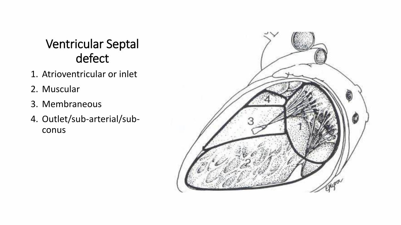
• Types- based on part affected• Membranous:70-80%
• Muscular:5-20%
• Supracristal: 5-7%
• AV or inlet VSD:5-8%

Isolated Ventricular Septal Defect
Ventricular Septal defect
1. Atrioventricular or inlet
2. Muscular
3. Membraneous
4. Outlet/sub-arterial/sub-conus

Pathophysiology of large VSDs
• RV vol. overload
• High output state of the LV
Large VSD
• RV dilatation
• RV hypertrophy
Cardiomegaly• Pulmonary
hypertension
• Eisenmenger syndrome
Cyanotic heart disease

Eisenmenger syndrome
Erythrocytosis
Pulmonary Haemorrhage
Endocarditis
Cerebral abscess
Ventricular arrhythmias
Clinical findings
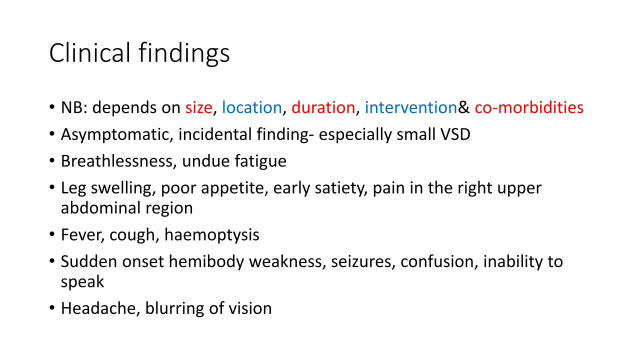
• NB: depends on size, location, duration, intervention& co-morbidities
• Asymptomatic, incidental finding- especially small VSD
• Breathlessness, undue fatigue
• Leg swelling, poor appetite, early satiety, pain in the right upper abdominal region
• Fever, cough, haemoptysis
• Sudden onset hemibody weakness, seizures, confusion, inability to speak
• Headache, blurring of vision


Management• NB: depends on size, location, duration, intervention& co-morbidities
• Adult, symptomatic cases need intervention- Medical ±Transcatheter/Surgical
• Contraindications to surgery:• Pulmonary vascular resistance ≥ 10 wood units
• Irreversible Pulmonary hypertension
• Eisenmenger syndrome
• Refusal to give consent
• Complications• Arrhythmias- both supraventricular& ventricular
• Conduction abnormalities
• Residual leaks- VSD persistence

• Antibiotic prophylaxis is required in• VSD with cyanotic heart disease• VSD with prosthetic material/patch(for the first 6 months
post op)• VSD with residual leaks at the patch site.
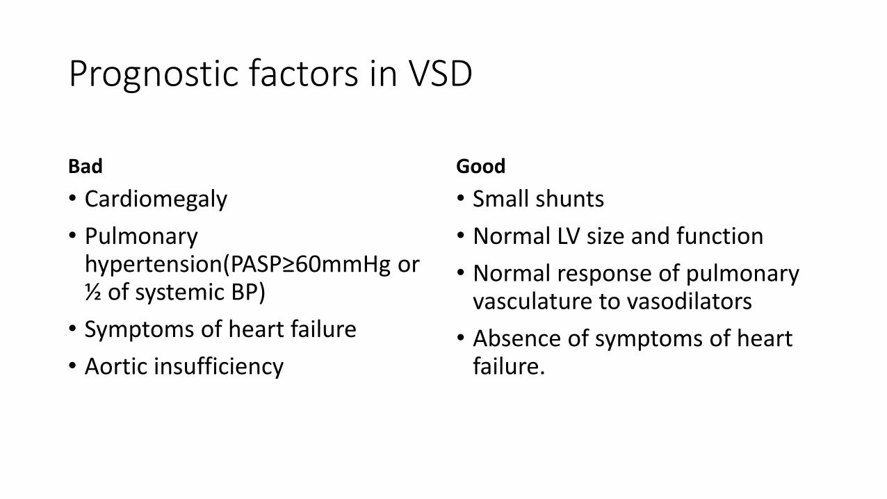
Prognostic factors in VSD
Bad
• Cardiomegaly
• Pulmonary hypertension(PASP≥60mmHg or ½ of systemic BP)
• Symptoms of heart failure
• Aortic insufficiency
Good
• Small shunts
• Normal LV size and function
• Normal response of pulmonary vasculature to vasodilators
• Absence of symptoms of heart failure.

Tetralogy of fallot
• The most common cyanotic CHD(7-10% of all CHD)
• Made up of• Mal-aligned VSD
• RVOT obstruction
• Overriding (or straddling) Aorta
• Right ventricular hypertrophy
• Pentalogy- addition of ASD
• Malignant arrhythmias and heart failure are the commonest causes of death
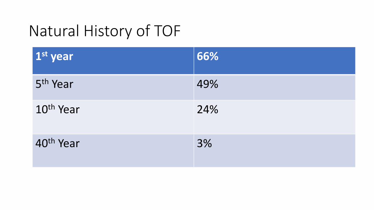
Natural History of TOF
1st year 66%
5th Year 49%
10th Year 24%
40th Year 3%

Tetralogy of Fallot (TOF)
• Named by Etienne-Louis Arthur Fallot in 1888
• Approximately 10% of all complex CHD
• Single developmental error of the terminal portion of the spiral truncoconal septum
• Four distinct components: subpulmonic stenosis, VSD, overriding aorta, and RV hypertrophy
• Often accompanied by other anomalies

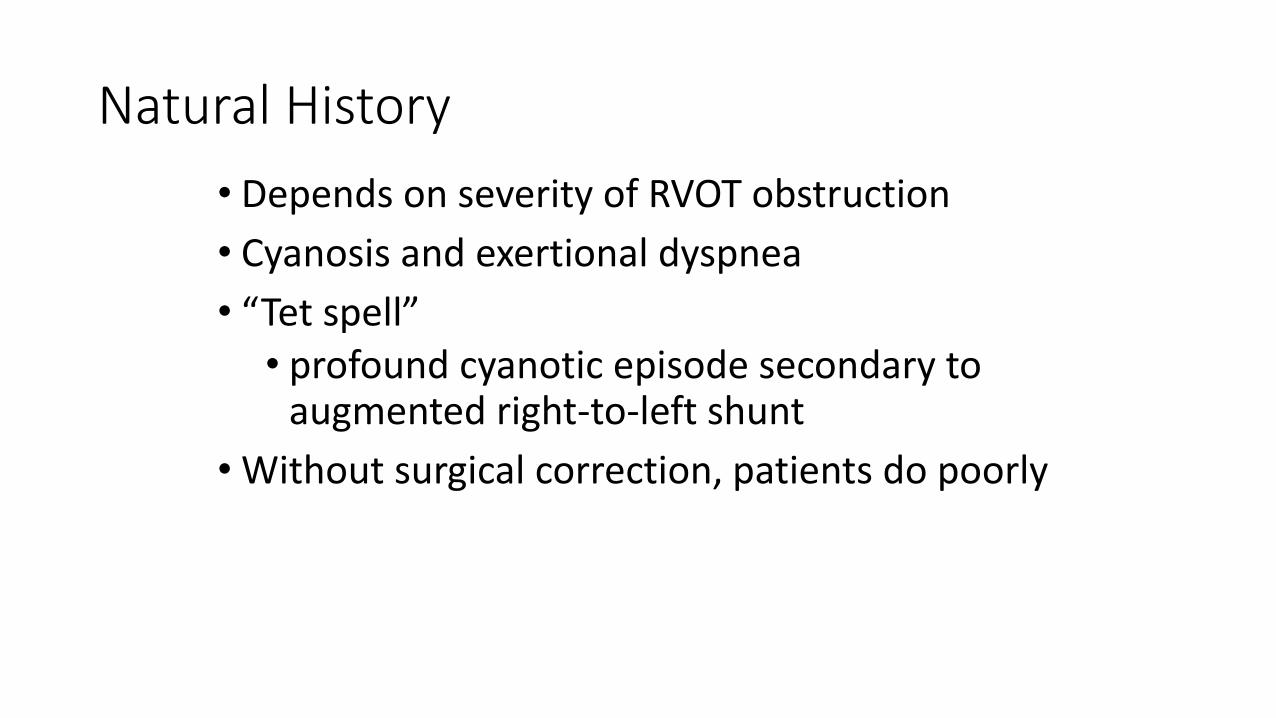
Natural History
• Depends on severity of RVOT obstruction
• Cyanosis and exertional dyspnea
• “Tet spell” • profound cyanotic episode secondary to
augmented right-to-left shunt
• Without surgical correction, patients do poorly

Surgical Repair
• Longest surgical history/most studied outcomes
• Palliative surgical shunts


Surgical Repair
• Longest surgical history/most studied outcomes
• Palliative surgical shunts
• Classic/complete repair• Infundibular muscle resection, VSD patch, & RVOT
repair
• Typically preformed between 4-6 months of age
• Surgical risk: < 5%
• Survival rates: 85% at 30+ years


Adult Presentation
• Repaired• RVOT obstruction
• Pulmonary or tricuspid regurgitation
• LV/RV dysfunction
• Atrial/ventricular arrhythmias
• Unrepaired• Significant morbidity
• Consider later repair

Transposition of theGreat Arteries (TGA)
• Complete TGA or D-TGA• Embryological inversion of the great arteries
• Ventriculoarterial discordance
• First described by Baillie 1797
• Natural history: >90% mortality in infancy
• Incidence: ~5% of congenital heart disease
• Rare association with syndromes or other anomalies
• Male:Female = 2:1
• Possible association with infant of diabetic mother

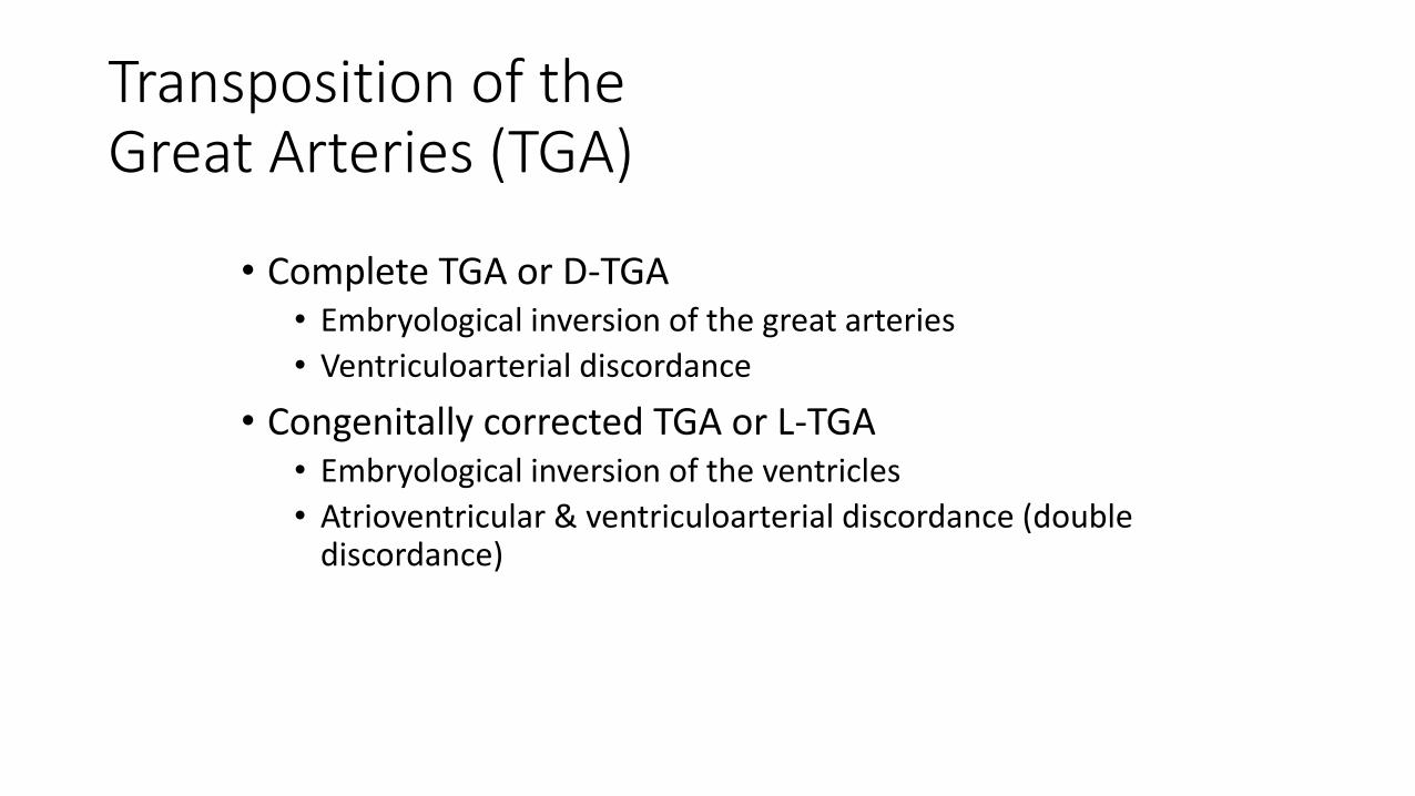
Transposition of theGreat Arteries (TGA)
• Complete TGA or D-TGA• Embryological inversion of the great arteries
• Ventriculoarterial discordance
• Congenitally corrected TGA or L-TGA• Embryological inversion of the ventricles
• Atrioventricular & ventriculoarterial discordance (double discordance)


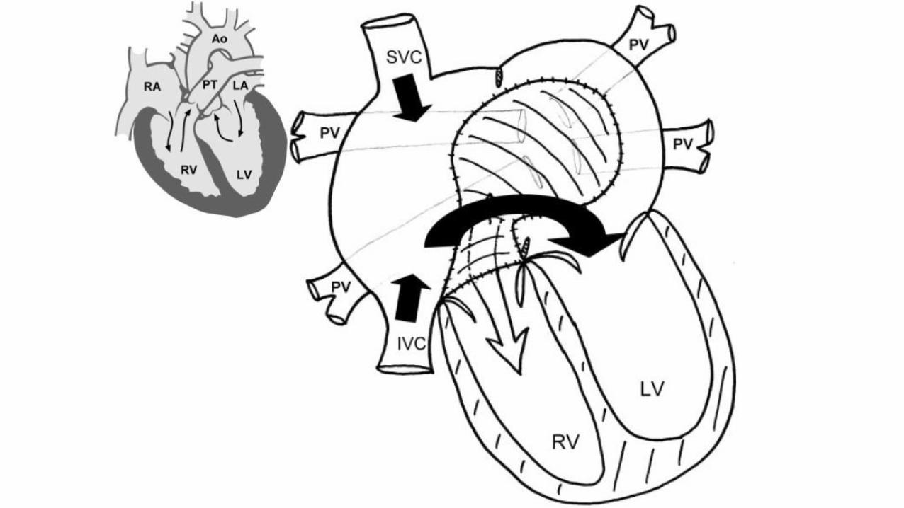
Surgical Repair
• Balloon atrial septostomy• Developed by Rashkind (1965)• Enlarges the atrial communication
• Atrial switch• Performed first by Senning (1958) and later
modified by Mustard (1964)• Atrial baffle is created to direct venous
return to the contralateral ventricle

Adult Presentation
• RV dysfunction
• Tricuspid insufficiency
• Bradyarrhythmias
• Atrial tachycardias
• Obstructed/leaky atrial baffle


Surgical Repair
• Arterial switch• Performed first by Jatene (1976)• Great arteries transected and reattached to
appropriate AV valve• Coronary ostia also transplanted• Surgical treatment of choice• Excellent outcomes so far

Adult Presentation
• Coronary perfusion issues
• Supravalvar aortic and pulmonic stenosis
• Aortic root dilatation and valvular insufficiency
• Branch pulmonary stenosis

Ebstein’s Anormaly
• Rudimentary posterior and septal tricuspid valve cusps
• Large, sail-like anterior tricuspid valve cusp
• Apical displacement of the tricuspid valve apparatus
• Reduction in the right ventricular size and compensatory huge right atrium

Ebstein’s anormaly

Ebstein’s Anormaly

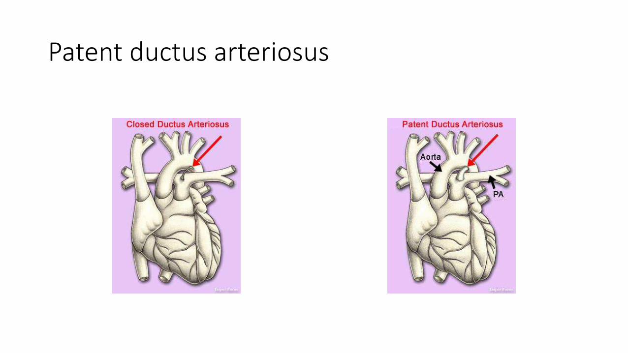
Patent ductus arteriosus
• Persistent connection, usually of the left pulmonary artery and the descending aorta(failure to regress, forming ligamentum arteriosum)
• Occurs in 1 of 2000 life births
• 10-12% of all CHD in infants, rare in adults.
• May occlude spontaneously within the first year of life( 3months in term babies; 12 months in pre-terms)
• Characterized by features of left ventricular volume overload
Risk factors for PDA
• Maternal rubella infection
• Birth in high altitudes
• Premature births
• Female sex( F:M-2:1)
• Genetic factors
• Familial factors(3% higher risk in subsequent offsprings)


Physiology- in utero and after delivery
Pulmonary artery
vasoconstriction
Low O2 Tension
High PGE2
Pulmonary artery
vasodilatation
High O2 tension
Low PGE2
Patent ductus arteriosus

Patent Ductus Arteriosus

PDA classification
Clinical Features• Asymptomatic in 35-40%
• Incidental murmurs increase clinical suspicion-classically, a continuous machinery murmur 1st or 2nd LICS, parasternal edge.
• If symptomatic, • Breathlessness
• Palpitations
• Leg swelling
• Loud P2 and left parasternal heave –signifies pulmonary hypertension
• Finger clubbing/cyanosis of the left hand and both legs-Eisenmenger’s physiology

Investigations
• Chest X- ray: often normal, may show cardiomegaly(dilated LA and LV from volume overload) and increased pulmonary vascular markings
• ECG- non-specific changes
• ECHO: TTE<<TEE in sensitivity(42% to 97%), though almost equal specificity(100%).
• MRI: gives quantitative and qualitative info about the PDA and other congenital abnormalities

PDA Rx- Ligation of the duct
Isolation of the duct Ligation of its ends, then severance

PDA Rx-Coil embolization

Amplatzer ductal occluders
Coarctation of the Aorta
• Occurs in 1 in 1550 autopsy cases
• Commoner in • Males(2:1),
• Caucasians,
• Turner syndrome and
• Bicuspid aortic valve
• Autosomal dominant inheritance has been observed
• Life expectancy-35yrs
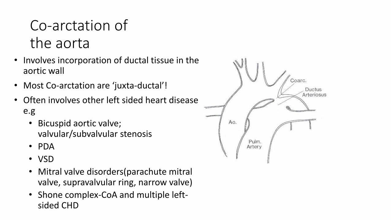
Co-arctation of the aorta
• Involves incorporation of ductal tissue in the aortic wall
• Most Co-arctation are ‘juxta-ductal’!
• Often involves other left sided heart disease e.g
• Bicuspid aortic valve; valvular/subvalvular stenosis
• PDA
• VSD
• Mitral valve disorders(parachute mitral valve, supravalvular ring, narrow valve)
• Shone complex-CoA and multiple left-sided CHD

Co-arctation of the aorta
• Extra cardiac associations-• Intracranial aneurysms• ocular defects• hypospadias • haemangiomas
• Increases the risk for stroke, ischaemic heart disease but not kidney disease

Clinical features
• Femoral pulse- Diminished and Delayed
• The BP is higher in the arms than the legs
• Fundoscopy-Corkscrew arterioles
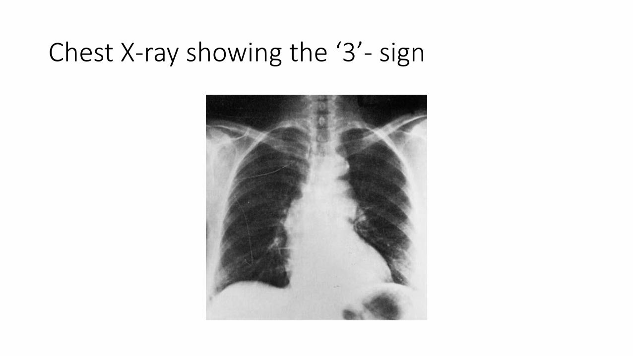
Chest X-ray showing the ‘3’- sign

Co-arctation of the Aorta

Investigations
• MRI- superior to ECHO for imaging the aorta and associated lesions
• Cardiac catheterization: ‘pull-back’ pressure gradient>20mmHg is significant; >50mmHg mandates intervention.

Management• Percutaneous angioplasty
• Baloon angioplasty- procedure of choice for post-surgical re-stenosis
• Complications: stenosis, rupture, aneurysm, dissection
• Stent insertion- intervention of choice in adults and adult-sized adolescents
• Surgery
• Resection with end-to-end anastomoses
• Patch aortoplasty
• Use of subclavian patch
• Complications: spinal cord ischaemia, bowel ischaemia, aneurysm and death
• Medical- largely supportive.
• Antihypertensives

Special Chest X-ray features in CHD
Congenital heart disease X-RAY FeaturesTetralogy of Fallot Boot shaped heart
Total anomalous pulmonary drainage Snowman sign
Partial anomalous pulmonary venous return
Scimitar sign
Ebstein’s anormaly Box shaped heart
Coarctation of the Aorta Number 3 appearance
Transposition of the Great Arteries Egg on a string sign
Endocardial cushion defect Goose neck sign

Summary
• Congenital heart disease are cardiac anormalies that are present at birth, usually as a consequence of maldevelopment.
• These were once exclusively within the domain of the paediatric cardiologists, but these patients are now surviving to adulthood.
• There are classic appearances clinically and with other diagnostic tools which helps with their assessment and diagnosis.
• There is a need to be aware of these pathologies so as to reduce their morbidity and improve their quality of life.

•Thanks for your attention