Bioaccumulation of Heavy Metals by Non-living Rhodococcus ...

APPLIED AND ENVIRONMENTAL MICROBIOLOGY, Sept. 1994, p. 3079-30880099-2240/94/$04.00+0Copyright © 1994, American Society for Microbiology
Adsorption of Rhodococcus Strain GIN-1 (NCIMB 40340) on
Titanium Dioxide and Coal Fly Ash ParticlesY. SHABTAIl* AND G. FLEMINGER2
Program for Biotechnology, Ben-Gurion University of the Negev, Beer-Sheva 84105,1 and Department of MolecularMicrobiology and Biotechnology, Tel-Aviv University, Ramat-Aviv 69978,2 Israel
Received 18 April 1994/Accepted 24 June 1994
Rhiodococcus strain GIN-1 (NCIMB 40340) can be used to enrich and isolate a titanium-rich fraction fromcoal fly ash. The gram-positive bacterium was isolated by its ability to adhere strongly and rapidly to suspendedparticles of pure titanium dioxide or coal fly ash. Adsorption depends on the salt concentration and occurs inseawater. Lowering of the salt concentration or washing of particles with pure water did not, however, causedesorption of the bacteria from TiO2 particles; this was achieved by strong alkaline treatment or combinedtreatment with sodium dodecyl sulfate and urea but not with dilute acids, alcohols, or cationic or nonionicdetergents. The bacterium exhibits higher affinity towards oxides of Ti and Zn than to other oxides with similardistribution of particle size. Moreover, it adheres much faster to TiO2 than to magnetite (Fe304) or A1203.After about 1 min, more than 85% of the cells were adsorbed on TiO2, compared with adsorption of only 10 and8% to magnetite and A1203, respectively. Adsorption of the bacteria on TiO2 occurs over a pH range of 1.0 to9.0 and at temperatures from 4 to over 80°C. Scanning electron microscopy combined with X-ray analysisrevealed preferential adherence of the bacterium to coal ash particles richer in Ti. Stronger adhesion to TiO2was also demonstrated in the translocation of bacteria, preadsorbed on magnetite, to TiO2 particles. Thetemporary co-adhesion to magnetite and TiO2 was exploited for the design of a prototype biomagneticseparation process in which bacterial cells serve as an adhesive mediator between magnetite and TiO2 particlesin a mixture of Al, Si, and Ti oxides that simulates their proportion in the ash.
Coal fly ash (CFA) is produced in large quantities fromcoal-operated power plants. It consists of granular dry powdercomposed of multielemental complexes, generally includingoxides and silicates (1, 5, 6, 11, 16, 18). The chemical compo-sition of the ash depends on the source of the coal and thenature of coal blending before incineration (5, 11, 12, 16).Major elements in the CFA produced from Israeli powerplants include silicon (25 to 32%), aluminum (15 to 18%),calcium and iron (5 to 7%), magnesium (2 to 3%), andtitanium (1 to 2%). Toxic heavy metals such as cadmium andmercury are also present in the ash in small quantities, as inother coal ashes (5, 11, 16). Some particles within the ash arerich in certain elements, e.g., Ca, Fe, or Ti. The CFA powderis relatively inert to leaching and requires extremely acidicconditions (pH below 1) to release soluble metals into anaqueous environment. However, under mild conditions, e.g., inseawater, moderately agitated suspensions release metals suchas Ca, Fe, Ni, Cd, Hg, and Ag, whose local concentrations mayexceed the upper permissible levels. In addition, because of itshigh concentration of potentially useful metals such as alumi-num and titanium, CFA is a source of valuable materials thatcan be extracted before its disposal.Numerous studies have been devoted to analytical aspects of
CFA content, but very few have dealt with exploitation of theash as a source of useful materials (1, 6, 21, 28). Severeecological constraints in Israel, together with the potentialexploitation of CFA, prompted a search for novel treatment ofthe ash before its disposal or safe utilization. Our approach wasto combine biotechnological and chemical procedures in thetreatment of coal ash, with the aim of removing toxic metals
* Corresponding author. Mailing address: Program for Biotechnol-ogy, Ben-Gurion University of the Negev, P.O. Box 653, Beer-Sheva84105, Israel. Phone: 972-7-461799. Fax: 972-7-236446.
and recovering valuable materials from the ash prior to itsdisposal. This work deals with one aspect of the biotechnolog-ical treatment, namely, the extraction of titanium-rich particlesfrom the ash. We describe the isolation of a specific bacterium,its utilization for biosorption of titanium-rich solid particles,and the application of the procedure to a practical separationprocess.
MATERMILS AND METHODS
Microbial strains. The bacterium Rhodococcus sp. strainGIN-1 (NCIMB 40340) was isolated and used in this study andis currently deposited as a patented strain in the NationalCollection of Industrial and Marine Bacteria, Aberdeen, Scot-land, United Kingdom. The following microbial strains wereused for comparison of adsorption to and desorption fromTiO2 and CFA: Acinetobacter calcoaceticus A2 and RAG-1,Bacillus subtilis 168, and Escherichia coli K-12 CHS 57 (kindlydonated from the collection of the Department of MolecularMicrobiology and Biotechnology, Tel-Aviv University, Tel-Aviv, Israel), Pseudomonas aeruginosa YS-7, Saccharomycescerevisiae Y-567, and Candida pseudotropicalis IP-513 from ourlaboratory (Program for Biotechnology, Ben Gurion Univer-sity of the Negev, Beer-Sheva, Israel).
Identification of bacterium. The bacterium was isolated andidentified by the use of a standard identification kit (API 20NE; Biomerieux, Montalie-Vercieu, France). Antibiotic sensi-tivity was determined by the use of standard kits (ATB G- 1401and ATB Staph 1402-OF; Biomerieux). Additional chemotax-onomic analyses were carried out at the National Collection ofIndustrial and Marine Bacteria.Media and growth conditions. The growth medium for
Rhodococcus strain GIN-1 (NCIMB 40340) contained (perliter of deionized water) KCl (45 g), K2HPO4* 3H20 (8.9 g),KH2PO4 (2.9 g), (NH4)2SO4 (4 g), MgSO4 7H20 (0.2 g),
3079
Vol. 60, No. 9
on July 13, 2018 by guesthttp://aem
.asm.org/
Dow
nloaded from

3080 SHABTAI AND FLEMINGER
sodium citrate (2.0 g), yeast extract (8.0 g), and glucose (10.0g). The pH was adjusted to 6.8. Cultures were grown aerobi-cally in shake flasks at 32°C in a gyrotory NBS G-24 shaker(New Brunswick Scientific Ltd.).
Seawater medium for enrichment culture. The medium usedfor the enrichment of adhesive microorganisms in continuousculture was prepared from seawater filtered through a Media-Kap-5 0.2-,um-pore-size hollow-fiber filter (Microgon Inc.,Laguna Hills, Calif.). The filtered seawater was supplementedwith the following ingredients (per liter): K2HPO4 * 3H20, 2.2g; KH2PO4, 0.72 g; (NH4)2S04, 1 g; yeast extract, 2.0 g; andglucose, 5.0 g.
Continuous culture for isolation of adhesive microorgan-isms. Enrichment of adhesive microorganisms was carried outin two consecutive continuous cultures. After enrichment,microbial cells that were retained on TiO2 particles in the firstculture served to inoculate the second continuous culture,which contained CFA. Both cultures were operated continu-ously for 3 weeks after inoculation in a 1-liter fermentor underthe following conditions: dilution rate, 0.1 h-1; agitation, 100rpm; and aeration, 0.5 vol/vol/min. A 5% (wt/vol) suspension ofTiO2 or CFA was mixed into buffered seawater medium.Particle-free medium containing nonadsorbed cells was de-canted from the widened upper section of an inverted U-shaped outlet of the fermentor, and attached cells wereretained inside the fermentor. The primary source of microor-ganisms was a mixture of seawater samples collected fromvarious sites near a power station (Maor David, Hadra, Israel),at which CFA is regularly conveyed, discharged, or dumped.Measurement of growth and quantity of adsorbed cells.
Cultures were monitored spectrophotometrically at 660 nm.The cellular dry weight was determined in cell samples aftercentrifugation (12,000 x g, 10 min). A660 of 1.0 corresponds toabout 0.4 mg of cells (dry weight) ml-'. The amount of cellprotein was determined according to the method of Lowry etal. (19) after alkaline treatment of the cells (0.2 N NaOH,100°C, 20 min). Viability counts were made by employingnutrient agar (Difco) and a spread-plate inoculation technique.Microscopic examinations were also used to verify growth andthe number of microbial strains in the microbial populationduring both stages of continuous culture.Measurement of cell hydrophobicity. A standard assay of
bacterial adhesion to hexadecane droplets was used to evaluatecell hydrophobicity, as previously described (24).CFA. CFA was obtained from Israel Electric Company, Ltd.
Each 30-kg sample of CFA consisted of a blended mixture offour weekly samples collected at Maor David Power Station orRutenberg Power Station, Ashkelon, Israel.Oxide powders. The following analytically graded oxides
were used for bacterial assays of adsorption: AgO, A1203,CdO, GeO2, HgO, MgO, NiO, PbO2, TiO2, ZnO (all obtainedfrom E. Merck AG, Darmstadt, Germany), and magnetite(Fe304; Sigma Chemical Co., St. Louis, Mo.). The particle sizedistribution of each oxide powder was restricted by collectionof a sieved fraction of particles less than 20 pum in size.
Reagents for desorption of attached cells from TiO2. Aque-ous solutions of the following reagents were used for desorp-tion of attached cells from TiO2 particles: up to 4 M NaCl orKCl (Merck); up to 1 N hydrochloric acid (Fluka), up to 0.5 Msulfuric acid (Merck), up to 50% (wt/vol) ethanol or 2-propa-nol (Merck), 1% (wt/vol) sodium dodecyl sulfate (SDS; Sig-ma), 1% (wt/vol) cetyltrimethylammonium bromide (BDH), 8M urea (Sigma), and 0.1 M NaOH (Merck).
Standard adsorption assay. The cell suspension (45 ml) infiltered seawater (or 3% NaCI) containing about 0.8 mg of cells(dry weight) ml- (-0.2 mg of protein ml-') was mixed with
800 mg of oxide or CFA to which 5 ml of filtered seawater hadbeen added. The mixtures were incubated for 1 h at 30°C withgyratory shaking (100 rpm). Samples were taken periodicallyand centrifuged (200 x g, 4 min) to allow efficient separationof free and adsorbed cells (verified by microscopic examina-tion), and the number of free or adsorbed cells was determinedby measurement of cell protein in the separated fractions aftertheir alkaline pretreatment as described above.
Preparation of magnetite biosorbent. Washed cells ofRhodococcus strain GIN-1 were mixed with magnetite at acell-to-magnetite ratio of 1 to 10 (wt/wt) in filtered seawater.The mixture was incubated for 1 h to ensure complete adsorp-tion. The magnetite biosorbent was then collected under amagnetic field of 1 kG with a model EDT magnetic separator(Electromagnetic Davis Tube tester; Eriez ManufacturingCompany, Erie, Pa.) equipped with a rotating glass tubethrough which washing liquids can be flushed or circulated.
Novel biomagnetic separation of TiO2 from a mixture ofoxides. Magnetite biosorbent (20 mg in 2 ml of filteredseawater) was mixed with 200 mg of a mixture of A1203, Si02,and TiO2 (42, 55, and 3% (wt/wt), respectively) suspended in48 ml of filtered seawater. A similar quantity of the magnetitebiosorbent was mixed with each of the oxides separately.Following incubation for 5 min at 32°C with gyratory shaking,each of the mixtures was subjected to a magnetic field of 2 kG,while the mixed suspension was circulated through the rotatingtube by means of a Masterplex peristaltic pump (Cole ParmerInstrument Company, Niles, Ill.) and a pellet was magneticallyimmobilized near the poles of the magnetic fields. The pelletwas then released by interruption of the field and resuspendedin fresh filtered seawater, and the suspension was vigorouslymixed to release the magnetite. A second magnetic separationwas carried out under identical conditions to immobilize themagnetite and wash out a conjugate of the bacterium andoxide. Cell protein was measured to assess the number ofbacteria in each fraction. The elemental content of eachfraction was determined after the oxide pellets were washedwith distilled water.
Determination of elemental content in oxide mixtures andCFA. Elemental analysis was carried out as described byDavison et al. (11). Following a strong acidic digestion, theamounts of the elements in each sample were determined withthe aid of an inductively coupled plasma atomic emissionspectrometer (model Spectroflame; Spectro, Klaeva, Germany).
Scanning electron microscopy coupled with elemental anal-ysis. For specific elemental analysis of particles with andwithout bacteria, electron micrographs were taken followingstandard critical point fixation in a Jeol Electron Microscope(model 840A) equipped with a Link X-Ray Scanning System(High Wycomb, Buckinghamshire, England).
RESULTS
Isolation of bacteria that adhere to titanium dioxide andCFA in continuous culture. A single bacterial species wasenriched in the two-stage continuous culture. It predominantlycolonized the surfaces of the solid particles and adheredstrongly to TiO2 and CFA granules. Viability counts and cellprotein determination indicated that more than 60% of themicrobial cells were associated with the particles. Most of thefree cells also consisted of this bacterium (with other types ofbacteria accounting for less than 0.0001%). After 2 weeks inthe presence of TiO2, other forms of microorganisms (e.g.,yeasts or fungi) were not detected in the continuous culturesdespite their presence in the inocula. This suggests either thatsuch forms could not grow under the existing conditions or that
APPL. ENVIRON. MICROBIOL.
on July 13, 2018 by guesthttp://aem
.asm.org/
Dow
nloaded from

ADSORPTION OF A RHODOCOCCUS SP. TO TiO2 AND COAL FLY ASH
FIG. 1.x 1,200.
Phase-contrast micrographs of Rhodococcus strain GIN-1 cells from exponential phase (A) and stationary phase (B). Magnification,
they failed to adhere and therefore were washed out of thefermentor. During the second stage, i.e., enrichment in thepresence of CFA, the continuously operating culture took onthe characteristics of a pure culture of the Rhodococcus strainGIN-1 bacterium (verified by viability counts and microscopicexamination), presumably possessing advantageous propertiesunder the selective conditions imposed by the CFA.
Identification of isolated bacterium. The isolated bacteriumwas gram positive and strictly aerobic, with irregularly shapedshort or long rod cells (Fig. 1). Its identity as a Rhodococcusspecies was indicated by the use of standard identification kits.The bacterium formed orange-colored colonies on nutrientagar and accordingly was termed Rhodococcus strain GIN-1.Chemotaxonomic analysis of the cell wall composition andfatty acid profile verified the above genus identity. The diaminoacid in the cell wall was meso-diamino pimelic acid (meso-DAP), and mycolic acids were present. The fatty acid profileshowed that the major components are straight-chain satu-rated and unsaturated acids together with branched-chainacids with the CH3 group on C-10, in particular, tuberculosicacid (10-methyloctadecanoic acid). The bacterium exhibited alow protein-to-cell (dry weight) ratio of 0.26 (wt/wt), indicatingthat the cells accumulate other polymers during growth. Thebacterium is capable of growth at temperatures up to 45°C.Some of its physiological properties, the decomposition oforganic compounds as carbon and nitrogen sources, and someof its functional biochemical properties suggested that themost closely fitting profile is that of Rhodococcus rhodochrous(13), despite the atypical finding of maltose, sodium adipate,testosterone, glycerol, and trehalose as the sole sources ofcarbon utilization. The Rhodococcus strain GIN-1 bacteriumwas cultivated readily in a defined medium with glucose as thecarbon source. Small amounts of yeast extracts (-0.4 g/liter)were essential for growth, and larger amounts (>4 g/liter)promoted the growth significantly. The specific requirementsupplied by the yeast extract is not yet known.Growth cycle and adhesive properties of Rhodococcus strain
GIN-1. During a growth cycle, the bacterial cells underwent
morphological changes that affected their adhesiveness andother properties of binding to TiO2 or CFA particles andamong themselves. Fractions of adhesive cells from the differ-ent growth phases are presented in Fig. 2. During the logarith-mic phase, the cells changed from relatively nonadhesive shortrods into highly adhesive, long branched cells. More than 90%of the cells were able to adhere to TiO2. At the late-log andstationary phases, the cells became shorter again, and theiradhesive capacity dropped correspondingly. Less than 40% ofthe stationary phase cells adhered to the oxide particles.
Salt requirement for adsorption. Adsorption of Rhodococ-cus strain GIN-1 to TiO2 or CFA particles occurred only whenthe concentration of salt, e.g., NaCl or KCl, was raised aboveabout 10 mM (Fig. 3). This was observed in the standard assay,in which 31 mg of cells (7.7 mg of cell protein) was blendedwith 800 mg of the oxide. About 95% of the cells were adsorbedon the particles (-9 mg of protein g of oxide-'). No significant
0)E
E.C
co=
U)
CL)
U)
(10.C
0
CU
Time (hours)FIG. 2. Adhesion of Rhodococcus strain GIN-1 cells to TiO2 at
different stages of the growth cycle. Adsorp., adsorption.
VOL. 60, 1994 3081
on July 13, 2018 by guesthttp://aem
.asm.org/
Dow
nloaded from
3082 SHABTAI AND FLEMINGER
10
0
C
0
0)
0
--
0 2 4 8 10 15 20 40 80NaCI (mM)
FIG. 3. Dependence of adherence of Rhodococcus strain GIN-1 toTiO2 on salt concentration.
adsorption was observed in distilled water. Attempts to removethe adsorbed cells by dilution of the salt content or even byextensive washing of the particles with distilled water were
unsuccessful. The maximum weight of cells that can be immo-bilized by TiO2 may reach as high as 0.6 to 0.7 g of cells g ofoxide- '.
Effects of pH and temperature. The effect of pH on adsorp-tion of Rhodococcus strain GIN-1 to TiO2 was examined in thestandard assay in a 3% NaCl solution. The pH levels of thesolutions were adjusted with HCl or NaOH. Adhesion of thecells (-30 mg of cells [dry weight]) was almost unaffected bypH. More than 90% of the cells adhered to the oxide over a
wide pH range of 1 to 9. Adsorption was slightly hindered(lower by -10%) at low pHs of 1 to 3. The weight of cellsadsorbed per gram of TiO2 was independent of temperatureover a range of 4 to 80°C.
Specificity of adsorption. The specificity of adsorption ofRhodococcus strain GIN-1 was examined in two sets of adsorp-tion assays (see Materials and Methods). In the first set ofexperiments, we compared the surface adhesion of Rhodococ-cus strain GIN-1 cells among different oxides. Equal weights ofRhodococcus strain GIN-1 cells (30 mg) were exposed toidentical amounts (800 mg) of each oxide in seawater. The
0
cm
c~ 6-CD
c g CdHg N S n F
0
0.
Ag Cd Hg Ni Si Zn CFA
Al Ge Mg Pb Ti Mag.Type of Oxide
FIG. 4. Adsorption of Rhodococcus strain GIN-1 on various oxides.Mag., magnetite.
a) 1"0
0)._
a)
CDE
0
0~0
Cl)
o
A B C D E F (Microorganism
G H
FIG. 5. Comparison of adsorption of various microorganisms on
TiO2.
relative weights of cells that were adsorbed on each of theoxides after 1 h are presented in Fig. 4, which illustratesmoderate differences in the adhesive capacity of Rhodococcusstrain GIN-1 to the various oxides. The highest degree ofadsorption was obtained with titanium and zinc oxides.
In a second set of experiments, the adhesion of Rhodococcusstrain GIN-1 cells to TiO2 was compared with that of othermicroorganisms. The results (Fig. 5) show a high efficiency ofadsorption of the bacterium on this oxide (more than 97% ofadded cells). The efficiency of adsorption obtained with otherbacteria was much lower (up to 15% of added cells). Most ofthese bacteria died upon adsorption and were easily desorbedduring treatment with mild detergents or acid solutions. Yeaststrains were adsorbed on TiO2 particles to a smaller extentthan the bacteria and could be easily desorbed from theparticles by any of the treatments used.
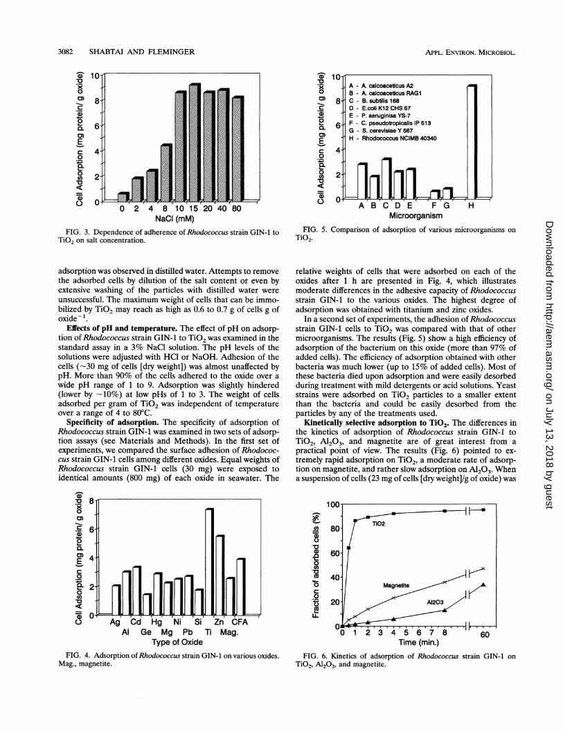
Kinetically selective adsorption to TiO2. The differences inthe kinetics of adsorption of Rhodococcus strain GIN-1 toTiO2, A1203, and magnetite are of great interest from a
practical point of view. The results (Fig. 6) pointed to ex-
tremely rapid adsorption on TiO2, a moderate rate of adsorp-tion on magnetite, and rather slow adsorption on A1203. Whena suspension of cells (23 mg of cells [dry weight]/g of oxide) was
100
X 80-
C.)
0
4600
U)
Time (min.)
FIG. 6. Kinetics of adsorption of Rhodococcus strain GIN-1 on
TiO2, A1203, and magnetite.
0- AA - A. calcoaceticus A2B - A. calcoaceticusRAGi
8- C - B. subtilis 168D - E.coli K12 CHS 57E - P. aeruginisa YS-7
6- F - C. pseudotropicalis IP 513G - S. cerevisiae Y 567H - Rhodococcus NCIMB 40340
4-
2-
APPL. ENvIRON. MICROBIOL.
on July 13, 2018 by guesthttp://aem
.asm.org/
Dow
nloaded from

ADSORPTION OF A RHODOCOCCUS SP. TO TiO2 AND COAL FLY ASH 3083
FIG. 7. Scanning electron micrographs of Rhodococcus strain GIN-1. (A) Free cells; (B) cells with adsorbed TiO2 particles.
mixed with each of the oxides in seawater (at an initial ratio of24 of mg of cells [dry weight]/g of oxide, corresponding to 6 mgof protein/g of oxide), more than 85% of the cells (-20 mg ofcells [dry weight]/g of oxide, or -5 mg of protein/g of oxide)were adsorbed on TiO2 after 1 min. In contrast, about 10% ofthe cells adhered to magnetite and less than 8% of the cellswere adsorbed on A1203 during the same period. Scanningelectron micrographs of the free bacteria and bacteria withadsorbed TiO2 particles are shown in Fig. 7.Adsorption to CFA. Light and electron microscopic obser-
vations of CFA particles following short-term adsorption ofRhodococcus strain GIN-1 cells revealed their nonuniformattachment to the particles (Fig. 8). Combined scanning elec-tron microscopy and X-ray diffraction elemental analysis ofmore than 200 CFA particles with adsorbed bacteria, as shownin Fig. 8, indicated that the surfaces of cell-carrying particleswere at least twice as rich in titanium (2 to 4%) as those ofnaked CFA particles (0.5 to 1.0%). Other elements (e.g., Al,Ca, Fe, and Si) did not appear to vary significantly from theiraverage concentrations in CFA.
VOL. 60, 1994
on July 13, 2018 by guesthttp://aem
.asm.org/
Dow
nloaded from

3084 SHABTAI AND FLEMINGER
FIG. 8. Scanning electron micrographs of Rhodococcus strain GIN-1 cells adsorbed on CFA particles. (A) General view of CFA particles; (B)Rhodococcus strain GIN-1 cells adsorbed selectively on a fraction of titanium-rich particles in CFA; (C) closer view of adsorbed cells.
Desorption of Rhodococcus strain GIN-1 cells from TiO2.The attachment of Rhodococcus strain GIN-1 cells to TiO2appears to be extremely strong. Attempts to achieve desorp-tion of the bound cells from the particles by the use of high orlow salt concentrations or alcohol solutions (Fig. 9), or withdilute hydrochloric or sulfuric acids (results not shown), wereunsuccessful. Treatment with pure SDS or cetyltrimethylam-monium bromide (CTAB) detergents (1% wtlvol) resulted in
release of less than 10% of the attached cells, and the desorbedcells lost their regular shape. Strong alkaline treatment re-moved the bound cells but led to their severe disruption. Highconcentrations of urea (8 M), especially in the presence of 1%SDS, enabled removal of most of the attached cells (>90%).Microscopic examination after such treatment revealed thatmost TiO2 particles were free of cells (data not shown); thiswas further verified by the fact that the amount of cell protein
APPL. ENVIRON. MICROBIOL.
on July 13, 2018 by guesthttp://aem
.asm.org/
Dow
nloaded from

ADSORPTION OF A RHODOCOCCUS SP. TO TiO2 AND COAL FLY ASH 3085
FIG. 8-Continued.
released from the particles corresponded to the amount ini-tially loaded during adsorption.
Translocation of cells from magnetite to TiO2. Rhodococcusstrain GIN-i cells were easily translocated from magnetite toTiO2 particles, but the reverse migration did not occur (Table1). This unidirectional translocation was observed during ex-posure of TiO2 particles to cells that were previously attachedto magnetite. The results presented in Table 1 suggest thatbinding to magnetite is weaker than that to TiO2.
Biomagnetic separation. A fraction rich in titanium oxide(Table 2) was isolated from a mixture of three oxides, SiO2,Al203, and TiO2 (oxide mixture, Table 2), whose elementalproportions simulated that in CFA (see Materials and Meth-
10
0
Ca)
0
<-0
0 10 20 30 40 50 60Time (min.)
FIG. 9. Desorption of bound Rhodococcus strain GIN-1 cells fromTiO2. A, 0.1 M NaOH; *, SDS + urea; O, 8 M urea; X, 1% SDS; +,50% 2-propanol; *, 3% NaCl.
ods). Short-term interaction of the magnetite-bacterium con-jugate (magnetite-biosorbent, Table 2) with the above mixturewas immediately followed by magnetic separation, which leftAl203 and SiO2 unbound (nonadsorbed oxides, Table 2) andenabled us to collect mainly TiO2 associated with the magne-tite-biosorbent (adsorbed oxides, Table 2). The temporarytriple conjugate was resuspended in seawater, and the suspen-sion was vigorously mixed to allow the magnetite to becomedetached from the complex. A second magnetic separationresulted in the immobilization and efficient recovery (>90%)of magnetite in a relatively pure state at the poles of theseparator (released magnetite, Table 2) and, at the same time,enabled us to collect the newly formed conjugate of bacteriumand TiO2 at the bottom of the separator tube. This fractioncontained more than 75% of TiO2 with the attached bacterialcells and with minimum contamination by other oxides (Table2, titanium dioxide-rich fraction). Results obtained with indi-vidual oxides (data not shown) indicated that TiO2 is the onlyoxide immobilized during such short-term biomagnetic sepa-ration and that only minor amounts of the other two oxides areadsorbed by the magnetite-biosorbent. Analyses of cell proteinin each fraction indicated that about 90% of the bacterial cellscould be accounted for in the TiO2 fraction. The rest of thebacterium was found associated with the other oxides, proba-bly A1203, since small amounts were associated with A1203 andnegligible amounts were associated with SiO2 when individualoxides were tested.
DISCUSSION
The isolated bacterium, Rhodococcus strain GIN-i, adheresstrongly to TiO2 and CFA particles. This ability to adhere tosolid surfaces gives the cells a great advantage under thesteady-state conditions in continuous culture, in which themajority of the cells are immobilized on particles and areconstantly growing. The bacterium was isolated from a stream
VOL. 60, 1994
on July 13, 2018 by guesthttp://aem
.asm.org/
Dow
nloaded from

3086 SHABTAI AND FLEMINGER
TABLE 1. Unidirectional translocation of Rhodococcus strainGIN-1 cells from magnetite to TiO2
Adsorption of cells (mg of cell protein/g of oxide)'
Test' Initialc Finald
Magnetite TiO2 Magnetite TiO2
I 0.21 (100) None <0.005 (2.5) 0.19 (90)
II None 0.23 (100) <0.002 (0.9) 0.21 (91)a In test I, the initial adsorption of cells to magnetite is followed by the
addition of TiO2; in test II, the initial adsorption of cells to TiO2 is followed bythe addition of magnetite.
b The percent recovery of cells relative to the initial cell input is shown inparentheses.
c Initial mixture of cells and adsorbing oxide.d Final mixture of cells and two oxides.
of cooling seawater in the vicinity of a coal-fired power station,where it dominated the bacterial population (unpublisheddata). This was not surprising, since conditions in the contin-uous culture in the laboratory resemble those near coal-firedpower stations, where warm streams of cooling seawater,relatively rich in nutrients, suspend and flush CFA and sandparticles into the open sea. Although the growth medium inthe continuous culture was relatively rich in nutrients, only afew other strains of microorganisms were present and theirnumbers were extremely small. This may have resulted fromthe selective pressures imposed (adhesion to TiO2 or CFA inseawater).The isolated bacterium is highly resistant to deleterious
effects that might be caused by binding to the surface of TiO2particles. This oxide binds many biomolecules and may damagebiological cells and even cause severe health problem inanimals (7, 8, 17). In contrast to other bacteria, which eventu-ally died upon adsorption on TiO2 particles, Rhodococcusstrain GIN-1 showed excellent survival and its growth was nothindered in presence of TiO2. This demonstrates its uniqueability to cope with the surface energy of these oxide particles.In such surface-to-surface interactions, the extracellular enve-lope of Rhodococcus strain GIN-1 must provide the cells withadhesive properties and at the same time shield them fromdamage caused during binding to the surface. We found thatthe overall protein content of the bacterium was very low (26%g of protein/g of cells [dry weight]) compared with 55 to 60%in other bacteria, which suggests that the extracellular enve-lope of this bacterium might consist of accumulation of otherpolymers. Analyses carried out to identify the bacterium andpreliminary results obtained during cell fractionation indicated
a relatively high proportion in the bacterial wall of mycolicacids (lipo-oligosaccharidic compounds) associated with hy-drophobic proteins. This is supported by reported findings thatextracellular polysaccharides with hydrophobic ligands mightplay a major role in adhesion and in the protection of oralbacterial flora from TiO2, as previously observed with oralmicroflora (15, 23). Recent studies with Rhodococcus strainsindicate that polysaccharidic capsules, amphiphilic polysaccha-rides, or mycolic acids are involved in the adhesion of variousRhodococcus strains to solid and liquid surfaces (3, 4, 22).The adhesive characteristics of the cell are acquired during
balanced growth, regardless of the location of the bacteria(free or adsorbed). Free Rhodococcus strain GIN-1 cells in thecontinuous culture were adhesive. Moreover, the results con-cerning the growth cycle in a batch culture indicated that theadhesive characteristics of the cells are somehow regulated;the number of adherent cells peaks at the exponential phase ofgrowth and drops when growth is unbalanced at the stationaryphase.The mechanism by which the bacterium adheres to TiO2 or
CFA or regulates its adhesive capacity during a growth cyclehas yet to be elucidated. We have not yet clarified which of thedescribed phenomena, e.g., London-van der Waals, electro-static, and interfacial tension forces (2, 9, 10, 14, 20), areinvolved in the short- and long-term adhesion of Rhodococcusstrain GIN-1. In several Rhodococcus strains, hydrophobic oramphiphilic molecules (e.g., mycolic acids or amphiphilicpolysaccharides) are responsible for the adhesiveness to solidsurfaces (3, 4, 22). As in those cases, the extracellular envelopeof Rhodococcus strain GIN-1 plays a major role in permanentadhesion, possibly through polymeric cross-linking (2, 9, 10).This might explain why mild acids or concentrated alcoholsfailed to remove the bacterium from the particles.
Previous reports (9, 10) have also indicated that cell surfaceproperties determine the capacity and selectivity of adhesionand that modification of the cell surface might affect theiradhesive properties. Changes in hydrophobicity and in struc-tures that establish hydrogen bonding might explain the de-crease in adhesion of stationary cells to TiO2. This notion issupported by the fact that adhesion is strong in the presence ofhigh salt concentrations and that even at low concentrations itis almost irreversible. The measures required for efficientdesorption of the cells (SDS plus 8 M urea) also point to thelikelihood of hydrogen bonding and strong hydrophobic inter-actions. It is therefore hypothesized that a certain hydrophobiccomponent(s) on the surface of the cells participates in a
hydrophobic interaction with TiO2 particles. Release of such a
component, its masking by hydrophilic moieties, or alterationof its structure might reduce the adhesive capacity of the cells
TABLE 2. Biomagnetic separation of TiO2 particles from a mixture of oxides
MixtureCell protein Amt of oxide (mg)b
M(pg)a Fe3O4 A1203 SiO2 TiO2
Oxide mixture 84 (100)C 110 (100) 6 (100)Magnetite-biosorbent 2,230 (100) 19.8 (100)Adsorbed oxidesd 2,100 (94) 19.4 (98) 4.4 (5) 1.2 (1) 5.1 (85)Nonadsorbed oxidese 180 (8) 0.2 (1) 78 (93) 108 (98) 0.7 (12)Titanium dioxide-rich fractione 1,930 (87) 0.2 (1) 1.9 (2) 0.3 (0.3) 4.7 (78)Released magnetitee 27 (1) 18.6 (94) 1.7 (2) 0.6 (0.5) 0.3 (5)
aProtein was measured by the method of Lowry et al. (19) after alkaline pretreatment of samples.b Elemental content was determined with an inductively coupled plasma atomic emission spectrometer and recorded as the content of the corresponding oxide.c Percent recovery of each oxide relative to its input is indicated in parentheses.d First magnetic separation step.e Second magnetic separation step.
APPL. ENVIRON. MICROBIOL.
on July 13, 2018 by guesthttp://aem
.asm.org/
Dow
nloaded from
ADSORPTION OF A RHODOCOCCUS SP. TO TiO2 AND COAL FLY ASH 3087
or play a major role in their detachment from the bindingsurfaces. Evaluation of the hydrophobicity of the cells con-firmed that cells taken from the stationary stage of the growthcycle, which are less adhesive to TiO2, are also less hydropho-bic than those taken from the exponential phase. This findingagrees with results from other laboratories indicating that thehydrophobicity of the Rhodococcus strain plays a major role inits adhesion to solid particles (26, 27).The specificity of adsorption of Rhodococcus strain GIN-1
on solid surfaces appears to be limited, though its capacity foradhesion to TiO2, ZnO, and CFA particles is considerablyhigh. In view of this, the ability of the bacterium to interactrapidly and strongly with some of the CFA particles wassurprising, since these oxides constitute only a very smallproportion of the ash. However, the finding that the shell ofcertain particles in the ash is richer in TiO2 than the core (asindicated by scanning electron microscopy with X-ray analysisand by unpublished data from acid leaching experiments) mayexplain the strong binding of the bacterium to a limitedfraction of the CFA particle.The high affinity of the bacterium toward TiO2 is indicated
by its faster selective adhesion to TiO2 than to A1203 and bythe rapid unidirectional translocation from magnetite to theTiO2 surface. The extremely rapid adhesion to TiO2 comparedwith adhesion to A1203 was the basis for the separation ofthese two oxides.The ability of the bacterium-magnetite conjugate to be
adsorbed rapidly and selectively on TiO2 indicates that thecells serve as multibinding sites for various surfaces. This wasobserved primarily in the independent binding of the bacte-rium to different oxides. The brief temporary stage of dualbinding of the bacterium to magnetite and TiO2 enabled usfirst to separate a triple bacterium-magnetite-TiO2 complexunder the magnetic field. The subsequent detachment of thebacterium from magnetite and its translocation to TiO2 parti-cles facilitated the isolation of a TiO2-rich material from amixture of oxides and the efficient recycling of the magnetite.This biomagnetic separation procedure is the basis of arecently approved patent (25). Biomagnetic separation is cur-rently under investigation on a larger scale, with a view towardestablishing a process for recovery of titanium-rich particlesfrom CFA. One aspect of that study involves the localizationand isolation of the extracellular biomolecules responsible forselectivity of adhesion to the target particles and for protectionof the cells during binding to TiO2. Use of a purified fractionof the adhesive molecules is likely to enhance selectivity ofbinding, with consequent improvement of the separation pro-cedure.
ACKNOWLEDGMENTS
This work was supported by a special grant from the Israel ElectricCompany, Ltd.We thank T. Dando of the National Collection of Industrial and
Marine Bacteria, Aberdeen, Scotland, United Kingdom, for identify-ing and storing the Rhodococcus strain GIN-1 bacterium and Y.Delarea of the Unit of Electron Microscopy, Tel-Aviv University, forvaluable assistance in the scanning electron microscopy and X-raydiffraction analysis of CFA particles and adsorbed bacteria. Specialgratitude is conveyed to S. Smith for excellent editorial assistance.
REFERENCES1. Adriano, D. C., A. L. Page, A. A. Elseewi, A. C. Chang, and I.
Straughan. 1980. Utilization and disposal of fly ash and other coalresidues in terrestrial ecosystems: a review. J. Environ. Qual.9:333-344.
2. Baier, R. E. 1979. Substrata influence on adhesion of microorgan-isms and their resultant new surface properties, p. 60-103. In G.
Bitton and K. C. Marshall (ed.), Adsorption of microorganisms tosurfaces. John Wiley & Sons, New York.
3. Bauer, P., and K Poralla. 1990. The polysaccharide capsule andadhesion of Rhodococcus sp., p. 637-640. In D. Behrens and P.Kramer (ed.), DECHEMA biotechnology conference, vol. 4A.VCH Verlagsgesellschaft, Weinheim, Germany.
4. Bendinger, B., H. M. H. Rijnaart, K. Altendorf, and A. J. B.Zehnder. 1993. Physicochemical cell surface adhesive propertiesof coryneform bacteria related to the presence and chain length ofmycolic acids. Appl. Environ. Microbiol. 59:3973-3997.
5. Block, C., and R. Dams. 1976. Study of fly ash emission duringcombustion of coal. Environ. Sci. Technol. 10:1011-1017.
6. Campbell, J. A., J. C. Laul, K. K. Nielson, and R. D. Smith. 1978.Separation and chemical characterization of finely-sized fly ashparticles. Anal. Chem. 50:1032-1040.
7. Chen, J. L., and W. E. Fayerweather. 1988. Epidemiologic study ofworkers exposed to titanium dioxide. J. Occup. Med. 30:937-942.
8. Connors, K. A., and M. J. Jozwiakowski. 1987. Studies on adsorp-tiochromism. I. Binding of adsorptiochromic spiropyrans to somepharmaceutically useful solids. J. Pharm. Sci. 76:892-897.
9. Corpe, W. A. 1979. Microbial surface components involved inadsorption of microorganisms onto surfaces, p. 106-143. In G.Bitton and K. C. Marshall (ed.), Adsorption of microorganisms tosurfaces. John Wiley & Sons, New York.
10. Daniels, S. L. 1979. Mechanisms involved in sorption of microor-ganisms to solid surfaces, p. 8-57. In G. Bitton and K. C. Marshall(ed.), Adsorption of microorganisms to surfaces. John Wiley &Sons, New York.
11. Davison, R. L., D. F. S. Natusch, J. R. Wallace, and C. A. Evans,Jr. 1974. Trace elements in fly ash: dependence of concentrationon particle size. Environ. Sci. Technol. 8:1107-1113.
12. Dreesen, D. R., E. S. Gladney, J. W. Owens, B. L. Perkins, C. L.Wienke, and L. E. Wangen. 1977. Comparison of levels of traceelements extracted from fly ash and levels found in effluent watersfrom coal-fired power plant. Environ. Sci. Technol. 11:1017-1019.
13. Goodfellow, M. 1986. Genus Rhodococcus Zopf 1891, 28AL, p.1472-1481. In P. H. A. Sneath, N. S. Mair, M. E. Sharpe, and J. G.Holt (ed.), Bergey's manual of systematic bacteriology, vol. 2. TheWilliams & Wilkins Co., Baltimore.
14. Hattori, R., and T. Hattori. 1987. Interaction of microorganismswith a charged surface: a model experiment. Rep. Inst. Agric. Res.36:21-67.
15. Joshi, R. I., and A. Eley. 1988. The in-vitro effect of a titaniumimplant on oral microflora: comparison with other metallic com-
pounds. J. Med. Microbiol. 27:105-107.16. Klein, D. H., A. W. Andren, J. A. Carter, J. F. Emery, C. Feldman,
W. Fulkerson, W. S. Lyon, J. C. Ogle, Y. Taimi, R. I. Van Hook,and N. Bolton. 1975. Pathways of thirty-seven trace elementsthrough coal-fired power plants. Environ. Sci. Technol. 9:973-979.
17. Lee, K. P., N. W. Henry, H. J. Trochimowicz, and C. F. Reinhardt.1986. Pulmonary response to impaired lung clearance in ratsfollowing excessive TiO2 dust deposition. Environ. Res. 41:144-167.
18. Lee, R. E., Jr., H. L. Crist, A. E. Riley, and K. E. Macleod. 1975.Concentration and size distribution of trace metal emissions froma power plant, a steel plant, and a cotton gin. Environ. Sci.Technol. 9:643-647.
19. Lowry, 0. H., N. J. Rosebrough, A. L. Farr, and R. J. Randall.1951. Protein measurement with the Folin phenol reagent. J. Biol.Chem. 193:265-275.
20. Marshall, K. C., and G. Bitton. 1979. Microbial adhesion inperspective, p. 1-5. In G. Bitton and K. C. Marshall (ed.),Adsorption of microorganisms to surfaces. John Wiley & Sons,New York.
21. Martin, J. P., J. Weggel, M. Bruno, and S. Halsely. 1991. The use
of high fly ash concrete for marine structure, p. 54-1 to 54-15. InProceedings of the Ninth International Ash Use Symposium, vol.2. Stabilization and aquatic uses. EPRI GS-7162, vol. 2, Project3176, January 1991. Electrical Power Research Institute.
22. Neu, T. R., and K. Poralla. 1988. An amphiphilic polysaccharidefrom an adhesive Rhodococcus strain. FEMS Microbiol. Lett. 49:389-392.
VOL. 60, 1994
on July 13, 2018 by guesthttp://aem
.asm.org/
Dow
nloaded from

3088 SHABTAI AND FLEMINGER
23. Newman, H. N. 1979. Retention of bacteria on oral surfaces, p.207-251. In G. Bitton and K. C. Marshall (ed.), Adsorption ofmicroorganisms to surfaces. John Wiley & Sons, New York.
24. Rosenberg, M., D. Gutnick, and E. Rosenberg. 1980. Adherence ofbacteria to hydrocarbons: a simple method for measuring cell-surface hydrophobicity. FEMS Microbiol. Lett. 9:29-33.
25. Shabtai, Y., G. Fleminger, and J. C. Fleming. March 1994. U.S.patent 5290697 (corresponds to Israeli patent IL 96611).
26. Stensrom, T. A. 1989. Bacterial hydrophobicity, an overall param-eter for the measurement of adhesion potential to soil particles.
APPL. ENVIRON. MICROBIOL.
Appl. Environ. Microbiol. 55:142-147.27. Van Loosdrecht, M. C. M., J. Lykiema, W. Norde, G. Schraa, and
A. J. B. Zehnder. 1987. The role of cell wall hydrophobicity inadhesion. AppI. Environ. Microbiol. 53:1893-1897.
28. Zimmels, I. Y., G. Shelef, and A. Boas. 1991. Utilization of coal flyash for land reclamation from the sea and offshore islands, p.56-1-56-15. In Proceedings of the Ninth International Ash UseSymposium, vol. 2. Stabilization and aquatic uses. EPRI GS-7162,vol. 2, Project 3176, January 1991. Electrical Power ResearchInstitute.
on July 13, 2018 by guesthttp://aem
.asm.org/
Dow
nloaded from