brochure ita eng - LenzokartTitle brochure_ita_eng.cdr Author Adry Created Date 6/4/2020 11:26:32 AM
Brit. J. industr. Med., I967, 24, 289
A Dry Ashing Method for the Determination of Blood Lead usingCathode Ray Polarography
Comparison with a Wet Ashing Technique
A. A. CERNIK
From the Medical Branch, H.M. Factory Inspectorate, Ministry of Labour, London
A dry ashing method is described for the determination of lead in whole blood. Within the range ofI6-200 ,ig. lead per ioo ml. blood recoveries were 82 to IXo%. The method agreed with a standard wetmethod and is more convenient. Possible sources of error and the uses of the ashing aid are discussed.
Great care is needed in preparing specimens ofblood for the estimation of trace amounts of lead.With 3-5 ml. of blood less than 2 ,ug. of lead maybe present. Such an amount can be estimatedaccurately with modem instruments; but it isessential to avoid both contamination and losses.The organic matter in blood is usually removed
by wet ashing with hot mineral acids. This istedious as it requires close attention at criticalstages, and the reagent blanks may be comparablewith the lead in blood, resulting in low precision.
Organic matter can be removed by dry ashing inwhich the organic matter is removed by atmosphericoxidation. When properly performed in a thermo-static muffle furnace, it needs little attention as theashing can be arranged overnight, the residue iscompletely soluble in the supporting electrolytes,and recoveries are complete because no insolublesalts are present. Ashing aids can be obtainedwhich contain very little lead, so the reagent blanksare low. Temperature control is important. Astemperatures approach 550°C. recoveries fallbecause lead is volatilized and is retained on thesilica crucible (Gorsuch, 1959). Temperaturegradients can exist in thermostatically controlledovens, especially in the back six inches (Hamilton,Minski, and Cleary, I967). Consequently, it ispreferable to ash at temperatures well below 550°C.,and to find a temperature at which fluctuations arenot critical.
Described here is a convenient dry ashingprocedure which compares well for precision witha standard wet ashing procedure.
Materials and Methods
Blood Specimens Venous blood (Io-o ml.) wascollected with a dispQsable plastic syringe and emptiedinto a plastic bottle containing I0-20 mg. disodiumethylenediamine tetra-acetic acid as an anticoagulant.
Polarograph This was the Davis DifferentialCathode-Ray Polarograph Ai66o (Southern Analytical,Camberley, Surrey).
Dry Ashing Procedure
Reagents Magnesium oxide, MgO, Analar;hydrochloric acid, HCI, Analar; lead acetate,Pb(CH3 CO2)2 3H20, Analar (i mg. Pb/ml. in I%acetic acid, v/v); peptone, bacteriological grade (Griffinand George, Ltd.); water, glass distilled and deionizedto better than 2 megohms/cm. (Elgastat).
Method Into a silica crucible, 57 x 37 mm., place250 mg. MgO and 5-o ml. well mixed whole blood.Thoroughly stir the MgO into the blood with a fineglass rod. Wash the blood off the rod with distilledwater. Dry the specimen at I io'C. for four to six hours.When it is dry, set the temperature control to 460°C.so that the temperature increases from IIO°C. tO 460°C.in not less than three hours. Ash for i6 hours at 460°C.Remove the crucible carefully, as any draught causes thelight residue to lift instantly, and place in a fumecupboard to cool to 40-5o°C. Add 2-0 ml. concentratedhydrochloric acid, washing the walls of the cruciblewith the acid. Care is needed as the reaction is vigorous.Leave for at least I0 min., but swirl the acid gentlyaround the walls of the crucible every 3 min. or so duringthis time. Any solid lumps noticed at first will graduallydisintegrate in the acid. If necessary, final solution canbe effected with a glass rod. Deionized water (8 ml.) isadded and mixed, and 4-0 ml. of the solution is placedin a polarograph cell, degassed with 02-free N2for I-2min. and I-2 mg. of peptone are added. Peak wave
289Received for publication October 5, I966.
copyright. on M
ay 23, 2020 by guest. Protected by
http://oem.bm
j.com/
Br J Ind M
ed: first published as 10.1136/oem.24.4.289 on 1 O
ctober 1967. Dow
nloaded from
potential for lead under these conditions is about o 49volt.The polarograph was standardized for each estimation
by adding o.oI ml. of the standard lead solution(i mg./ml.) and re-estimating.
Wet Ashing Procedure A modification of theM.R.C. method for wet ashing of blood (AnalyticalMethods Committee, I959) was used.
Reagents Nitric acid (Analar, s.g. I 42); perchloricacid (Analar, s.g. I 70); hydrochloric acid (Analar, s.g.i i8); hydrogen peroxide (Analar, 30% w/v); glassdistilled water, deionized.
Glassware 25 X ISo mm. rimless Pyrex test tubes.Wash in 50% nitric acid and keep for lead work only.
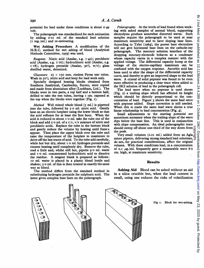
Specially designed heating blocks obtained fromSouthern Analytical, Camberley, Surrey, were copiedand made from aluminium alloy (Luckham, Ltd.). Theblocks were in two parts, a top half and a bottom half,drilled to take the test tubes, leaving i cm. exposed atthe top when the blocks were together (Fig. i).
Method Well mixed whole blood (5 ml.) is pipettedinto the tube, followed by 5 o ml. nitric acid. Gentlyheat on an electric hotplate using the lower block so thatthe acid refluxes for at least the first hour. When theacid is reduced to about i-O ml. take the tube out of theblock and add 5 o ml. of a i /i, v/v mixture of nitric andperchloric acids. Replace the tube in the bottom blockand gently reduce the volume by heating until fume sappear. Then place the upper block over the tube andraise the temperature of the hotplate to maximum todrive off the last traces of acid. To the tube add carefully,while hot but dry, about i o ml. hydrogen peroxide andresume heating until completely dry. Remove the tube,cool a little and, whilst still hot, pipette 9 o ml. waterand I o ml. concentrated hydrochloric acid to dissolvethe residue. A reagent blank is prepared as follows:IO ml. water is placed in a plastic blood bottle andshaken; 5 o ml. of this is then treated in exactly the sameway as blood.The method differs from the standard method in
substituting hydrogen peroxide for sulphuric acid. Thelatter gives complex base lines on the polarograph.
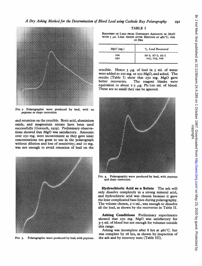
Polarography At the levels of lead found when work-ing with ashed samples of normal blood, supportingelectrolytes produce somewhat distorted waves. Suchsamples require the polarograph to be used at nearmaximum sensitivity and to have sloping base lines.According to Tyler (I964), the purest base electrolytewill not give horizontal base lines on the cathode-raypolarograph. The mercury solution interface of thedropping mercury-electrode behaves as a condenserwhose capacity varies in a complex manner with theapplied voltage. The differential capacity hump at thevoltage of the electro-capillary maximum can beconfused with the oxygen response. Ascorbic acid hasbeen used to alter the shape of the differential capacitycurve, and thereby to give an improved shape to the leadwave. A crystal of solid peptone was found to be evenmore effective in producing a clear trace when added tothe HC1 solution of lead in the polarograph cell.The lead wave when no peptone is used shows
(Fig. 2) a trailing slope which has affected its heightwhich should be directly proportional to the con-centration of lead. Figure 3 shows the same lead wavewith peptone added. Slope correction is still needed.When this is made the same lead wave shows a truelinear relationship to lead concentration (Fig. 4).
Small adjustments to the starting potential aresometimes necessary when the trailing slope of the wavedips below the base line. This is used in conjunctionwith slope compensation. An ideal polarographic traceshould sweep off about one-third of the way down fromthe peak.Very small volumes (o.oi ml.) added from an Agla
micro pipette, delivering strong standard lead solutions,do not, for practical considerations, affect the originalvolume. With these conditions lead, in a concentrationof o.I pg./ml. frequently gave a measurable wave 6,ocm. high, at maximum sensitivity.
Results
Ashing Aid Blood can be ashed without an aidin a silica crucible but, when the lead content issmall, using one reduces the risks of volatilization
Fig i. Block for wet-ashing.
3,CID
CD\o
A. A. Cernik290
copyright. on M
ay 23, 2020 by guest. Protected by
http://oem.bm
j.com/
Br J Ind M
ed: first published as 10.1136/oem.24.4.289 on 1 O
ctober 1967. Dow
nloaded from
A DTy Ashing Method for the Determination of Blood Lead using Cathode Ray Polarography
TABLE I
RECOVERY OF LEAD FROM DIFFERENT AMOUNTS OF MGOWITH 5 jIG. LEAD ADDED AFTER HEATING AT 460°C. FOR
i6 HRS
MgO (mg.) % Lead Recovered
100 92-5, 97-5, 93-5250 105, I03, 100
crucible. Hence 5 pg. of lead in 5 ml. of waterwere added to IOO mg. or 250 MgO, and ashed. Theresults (Table I) show that 250 mg. MgO gavebetter recoveries. The reagent blanks wereequivalent to about 2-5 jig. Pb/ioo ml. of blood.These are so small they can be ignored.
FIG. 2 Polarographic wave produced by lead, with nopeptone or slope correction
and retention on the crucible. Boric acid, aluminiumoxide, and magnesium mitrate have been usedsuccessfully (GTorsuch, 1959). Preliminary observa-tions showed that MgO was satisfactory. Amountsover 250 mg. were inconvenient as they gave ionicconcentrations too great to use in the polarographwithout dilution and loss of sensitivity; and 10o mg.was not enough to avoid retention of lead on the
FIG. 4. Polarographic wave produced by lead, with peptoneand slope correction.
Hydrochloric Acid as a Solute The ash willonly dissolve completely in a strong mineral acid,and hydrochloric acid was chosen because it gavethe least complicated base lines during polarography.The volume chosen, 2 -0 ml., was enough to dissolveall the lead, as shown by the recoveries in Table IL.
Ashing Conditions Preliminary experimentsshowed that 250 mg. MgO was satisfactory for3-5 ml. of blood but not enough for volumes outside
Ashing was incomplete after 8 hrs at46ogC. butwas complete by i6 hrs, as shown by inspection of
FIG. 3. Polarographic wave produced by lead, with peptone. the ash and by recovery tests (Table III).
29I
copyright. on M
ay 23, 2020 by guest. Protected by
http://oem.bm
j.com/
Br J Ind M
ed: first published as 10.1136/oem.24.4.289 on 1 O
ctober 1967. Dow
nloaded from
292 A. A. Cernik
,ol
0 Ca
0 "
0Cu0.4 ~rj
I., CuI.
o4.-3.
o-e
C- 4)
-ov
r, 00A.
C,,
04
eq
0)
00
Co
CY)
-J
0- ,
_w-
0)
(0
It)
CM
j-z
a00-acoI
I I I-F I1 -
0 0 Q0 08 ~ ~~~~~011) 0 U) W
N C1 IWI,
aooi30 lWOOI jed C3Y Br(
-1
L-L-
L-...
M.l
L--,
rr-..
,--r--
I
mI
C=--E
Li
,r---
r-r--
L-1
1-1m
4
0 0
:Fz
U)4 <
L" >3r.9
i
copyright. on M
ay 23, 2020 by guest. Protected by
http://oem.bm
j.com/
Br J Ind M
ed: first published as 10.1136/oem.24.4.289 on 1 O
ctober 1967. Dow
nloaded from
A Dry Ashing Methodfor the Determination of Blood Lead using Cathode Ray Polarography
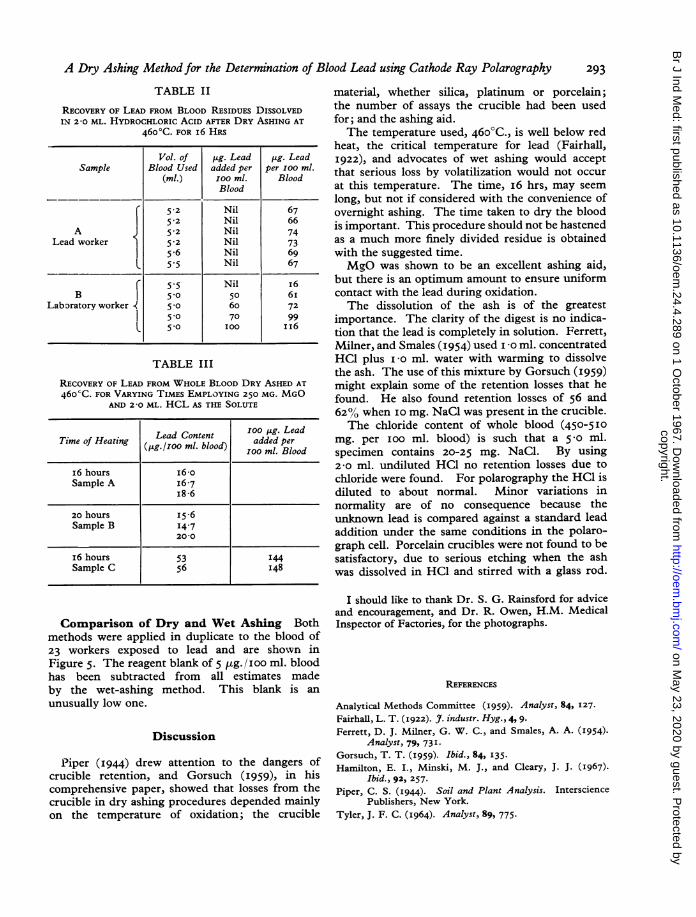
TABLE II
RECOVERY OF LEAD FROM BLOOD RESIDUES DISSOLVEDIN 2-0 ML. HYDROCHLORIC ACID AFTER DRY ASHING AT
46o0C. FOR i6 HRS
Vol. of ,ug. Lead ,tg. LeadSample Blood Used added per per IOO ml.
(ml.) Ioo ml. BloodBlood
5.2 Nil 675.2 Nil 66
A { 52 Nil 74Lead worker 1 52 Nil 73
5.6 Nil 69l 5*5 Nil 67
f 5-5 Nil I6B 5-0 50 6i
Laboratory worker 5.0 60 7250 70 99
L 50 IOO I16
TABLE III
RECOVERY OF LEAD FROM WHOLE BLOOD DRY ASHED AT46ocC. FOR VARYING TIMES EMPLOYING 250 MG. MGO
AND 2-0 ML. HCL AS THE SOLUTE
LeadContent100 jsg. Lead
Time of Heating Lead Content added perQi(g./ioo ml. blood)ioo ml. Blood
I6 hours i6-oSample A I6.7
i8-6
20 hours 15.6Sample B I4.7
20-0
i6 hours 53 144Sample C 56 I48
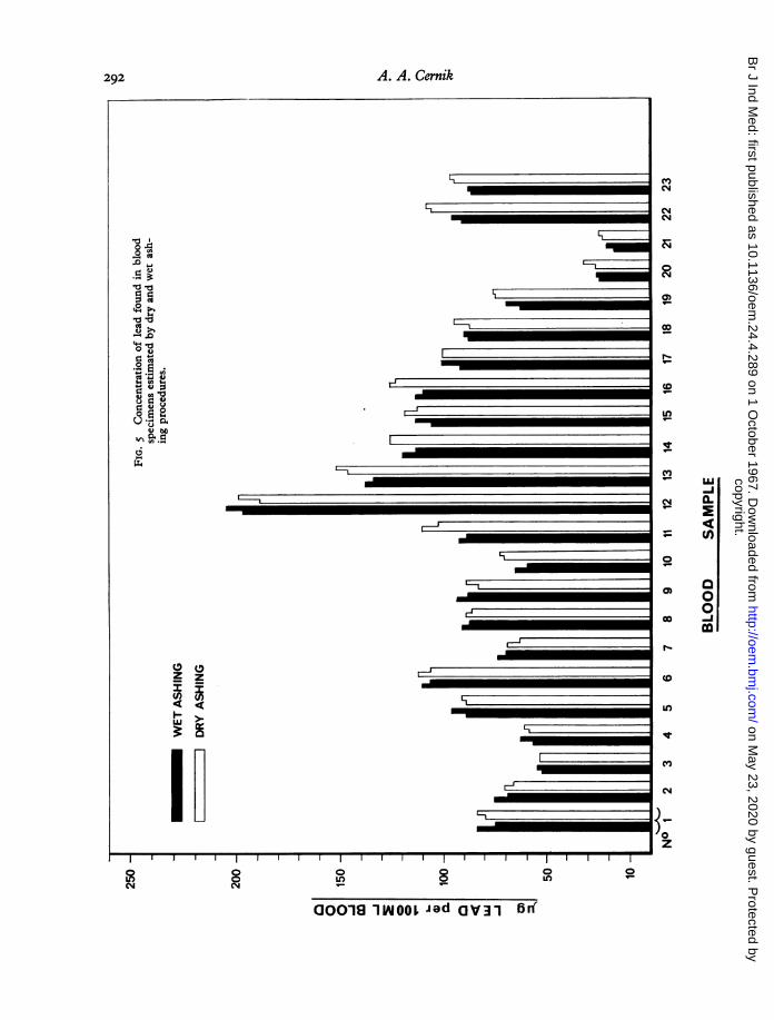
Comparison of Dry and Wet Ashing Bothmethods were applied in duplicate to the blood of23 workers exposed to lead and are shown inFigure 5. The reagent blank of 5 ,tg./ioo ml. bloodhas been subtracted from all estimates madeby the wet-ashing method. This blank is an
unusually low one.
Discussion
Piper (1944) drew attention to the dangers ofcrucible retention, and Gorsuch (I959), in hiscomprehensive paper, showed that losses from thecrucible in dry ashing procedures depended mainlyon the temperature of oxidation; the crucible
material, whether silica, platinum or porcelain;the number of assays the crucible had been usedfor; and the ashing aid.The temperature used, 460°C., is well below red
heat, the critical temperature for lead (Fairhall,1922), and advocates of wet ashing would accept
that serious loss by volatilization would not occur
at this temperature. The time, i6 hrs, may seem
long, but not if considered with the convenience ofovernight ashing. The time taken to dry the bloodis important. This procedure should not be hastenedas a much more finely divided residue is obtainedwith the suggested time.MgO was shown to be an excellent ashing aid,
but there is an optimum amount to ensure uniformcontact with the lead during oxidation.The dissolution of the ash is of the greatest
importance. The clarity of the digest is no indica-tion that the lead is completely in solution. Ferrett,Milner, and Smales (I954) used I 0 ml. concentratedHC1 plus I 0 ml. water with warming to dissolvethe ash. The use of this mixture by Gorsuch (I959)might explain some of the retention losses that hefound. He also found retention losses of 56 and62% when I0 mg. NaCl was present in the crucible.The chloride content of whole blood (450-5I0
mg. per I00 ml. blood) is such that a 5 0 ml.specimen contains 20-25 mg. NaCl. By using2-0 ml. undiluted HC1 no retention losses due to
chloride were found. For polarography the HC1 isdiluted to about normal. Minor variations innormality are of no consequence because theunknown lead is compared against a standard leadaddition under the same conditions in the polaro-graph cell. Porcelain crucibles were not found to besatisfactory, due to serious etching when the ashwas dissolved in HC1 and stirred with a glass rod.
I should like to thank Dr. S. G. Rainsford for adviceand encouragement, and Dr. R. Owen, H.M. MedicalInspector of Factories, for the photographs.
REFERENCES
Analytical Methods Committee (I959). Analyst, 84, 127.
Fairhall, L. T. (I922). J. industr. Hyg., 4, 9.
Ferrett, D. J. Milner, G. W. C., and Smales, A. A. (I954).Analyst, 79, 73I.
Gorsuch, T. T. (1959). Ibid., 84, 135.
Hamilton, E. I., Minski, M. J., and Cleary, J. J. (I967).Ibid., 92, 257.
Piper, C. S. (I944). Soil and Plant Analysis. IntersciencePublishers, New York.
Tyler, J. F. C. (I964). Analyst, 89, 775.
293
copyright. on M
ay 23, 2020 by guest. Protected by
http://oem.bm
j.com/
Br J Ind M
ed: first published as 10.1136/oem.24.4.289 on 1 O
ctober 1967. Dow
nloaded from