
A{DROME - The Podiatry Institute/cite> · CHAPTER
10
CHAPTER II OS TRIGONUM S'\A{DROME Gerard V. Yu, DPl,,t Amanda MeszAros, DPM Theresa L. Scbinke, DPM Michael B. Canales. DPl,t INTRODUCTION The os trigonum is an anatomical variant of the posterior process of the talus w-here a secondary center of ossification located within the lateral tubercle fails to fuse with the main body of the talus. This secondary center generally appears betw-een B-11 ys215 of age and fuses u.ithin 1-3 years of its appearance. Controversy exists as to the trlle natllre of the os trigonum as a distinctly separate entity as opposed to a posttraumatic refltnant of a fractr,rre of the posterolateral process of the talus. Os trigonum syndrome is a symptomatic disruption or inflammatory response within the fibrous tissue, which unites the accessctry ossicle with the posterior process of the talus. The origin and etiology of os trigonum syndrome has been the topic of much clebate amongst both podiatric and orlhopedic physicians. Outlining the somewhat complex derivation of os trigonum syndrome requires a thorough understancling of the anatomy of the posterior ankle. The posterior process of the talus is made up of tu,o tubercles, the medial tubercle and the lateral tubercle. Between these tr-rbercles is a groove that allows the passage of the flexor hallucis longus tenclon from the posterior ankle to enter the tarsal tunnel. The course of the flexor hallucis longus tendon is angled at this level, and its sheath is often chickened to eccommodate the higher degree of friction encountered as it passes through the posterior ankle. The lateral tubercle of the posterior talus is generaily larger, and it is here that the secondary ossification center associated with the development of os trigonum is found. Should this lateral tubercle present as an intact but abnormally elongated process or extension of the posterior talus, it is referred to as Steida's process. When existing as a separate entity, it is referred to as an os trigonum, trigonal process or accessory ossicle. The os trigonum articulates with the talus r.ia a fibrous, fibro-cartilaginous or cartilaginous union. The ossicle also has ligamentous attachments to the main body of the ta1us. The posterolateral process selves as an attachment point for the posterior talofibular ligarnent and the postedor talocalcaneal ligament. Aside from its articulation with the ta1us, the os trigonum communicates superiorly with the posterior capsule of the talo-cr-ural joint, inferiorly u'ith the posterior talocalcaneal ligarnent, medially t itl"r the flexor halh,rcrs longus tendon sheath and lateral1y with the origin of the posterior talo-fibular ligament. The posterior process of the talus derives its bloocl supply via an anastamosis of the medial calcaneal afiery and the communicating branch of the peroneal artery. LITERATURE REVIEW The os trigonum was first described as an accessory ossicle in 180,i by Rosenmuller who identified it basecl on its trigonal contour. In 1882, Shepherd revealed his theory that the os trigonum is not an accessory ossicle created by the failed fusion of a secondary ossification center. Shepherd believed that the os trigonum appears only as a result of a fracture of the posterolateral process of the talus.' In contrast, Rardelen, in 1883, presented a theory that identified the os trigonum as a primitive or intermecliate tarsal bone, retained in a segment of the population. He supportecl this theory on the basis that the ossicle forms from a distinctly separate cartilaginous body that does not fuse with the main caftllaginous body of the talus until the third fetal month.'] Shepherd was challenged by O'Rahilly and Turner in 7953 who concluded that os triS4onum is, indeed a non-unified remnant of the secondary ossification center or nucleus of the posterior ta1us.1,3! McDougall, in 1953 again challenged this view, slrpporting Shepherd's conclusions while speculating that the os trigonum is always secondary to fracture and that its smooth rounded edges are
Transcript of A{DROME - The Podiatry Institute/cite> · CHAPTER

CHAPTER IIOS TRIGONUM S'\A{DROME
Gerard V. Yu, DPl,,tAmanda MeszAros, DPMTheresa L. Scbinke, DPMMichael B. Canales. DPl,t
INTRODUCTION
The os trigonum is an anatomical variant of theposterior process of the talus w-here a secondarycenter of ossification located within the lateraltubercle fails to fuse with the main body of the talus.This secondary center generally appears betw-eenB-11 ys215 of age and fuses u.ithin 1-3 years of itsappearance. Controversy exists as to the trlle natllreof the os trigonum as a distinctly separate entity as
opposed to a posttraumatic refltnant of a fractr,rre ofthe posterolateral process of the talus. Os trigonumsyndrome is a symptomatic disruption orinflammatory response within the fibrous tissue,which unites the accessctry ossicle with the posteriorprocess of the talus. The origin and etiology of ostrigonum syndrome has been the topic of muchclebate amongst both podiatric and orlhopedicphysicians. Outlining the somewhat complexderivation of os trigonum syndrome requires a
thorough understancling of the anatomy of theposterior ankle.
The posterior process of the talus is made upof tu,o tubercles, the medial tubercle and the lateraltubercle. Between these tr-rbercles is a groove thatallows the passage of the flexor hallucis longustenclon from the posterior ankle to enter the tarsaltunnel. The course of the flexor hallucis longustendon is angled at this level, and its sheath is oftenchickened to eccommodate the higher degree offriction encountered as it passes through theposterior ankle. The lateral tubercle of the posteriortalus is generaily larger, and it is here that thesecondary ossification center associated with thedevelopment of os trigonum is found. Should thislateral tubercle present as an intact but abnormallyelongated process or extension of the posteriortalus, it is referred to as Steida's process. Whenexisting as a separate entity, it is referred to as an ostrigonum, trigonal process or accessory ossicle. Theos trigonum articulates with the talus r.ia a fibrous,
fibro-cartilaginous or cartilaginous union. Theossicle also has ligamentous attachments to the mainbody of the ta1us. The posterolateral process selvesas an attachment point for the posterior talofibularligarnent and the postedor talocalcaneal ligament.Aside from its articulation with the ta1us, the ostrigonum communicates superiorly with theposterior capsule of the talo-cr-ural joint, inferiorlyu'ith the posterior talocalcaneal ligarnent, mediallyt itl"r the flexor halh,rcrs longus tendon sheath andlateral1y with the origin of the posterior talo-fibularligament. The posterior process of the talus derivesits bloocl supply via an anastamosis of the medialcalcaneal afiery and the communicating branch ofthe peroneal artery.
LITERATURE REVIEW
The os trigonum was first described as an accessoryossicle in 180,i by Rosenmuller who identified itbasecl on its trigonal contour. In 1882, Shepherdrevealed his theory that the os trigonum is not anaccessory ossicle created by the failed fusion of a
secondary ossification center. Shepherd believedthat the os trigonum appears only as a result of afracture of the posterolateral process of the talus.'
In contrast, Rardelen, in 1883, presented atheory that identified the os trigonum as a primitiveor intermecliate tarsal bone, retained in a segment ofthe population. He supportecl this theory on thebasis that the ossicle forms from a distinctly separatecartilaginous body that does not fuse with the maincaftllaginous body of the talus until the third fetalmonth.'] Shepherd was challenged by O'Rahilly andTurner in 7953 who concluded that os triS4onum is,indeed a non-unified remnant of the secondaryossification center or nucleus of the posterior ta1us.1,3!
McDougall, in 1953 again challenged thisview, slrpporting Shepherd's conclusions whilespeculating that the os trigonum is always secondaryto fracture and that its smooth rounded edges are
CHAPTER 11 55
natllral changes due to erosion of the fracture edgesover time. McDougall also proposed threemechanisms by which fracture of the posterolateraltalar process may occllr, resulting in the appearanceof os trigonum: 1) impingement of the lateraltubercle belween the calcaneus and tibia over timeleads to stress fracture, 2) acute fracture of the lateraltubercle due to sudden, sevcre trxuma with the footplantarflexed, 3) al-ulsion fracture of the lateraltr-rbercle by traction of the posterior talofibularligament n-hile stressed in clorsiflexion.; Recentliterature suppolls the widely accepted view that theos trigonum is a true accessory ossicle whichappears when the secondary ossification center ofthe talus fails to fuse by age 18 years.
PREYALENCE
The incidence of os trigonum in the generalpopulation ranges from 1.7-500/0, 33-5Oo/o of whichpresent bi1ateral1y.6 There does not appear to be an
increased prevalence in women or men; the entitycloes not appear to be more common in cefiain age
groups. Athletes who pafiicipate in spofis thatrequire a high degree of plantarflexory strain, fbrexample, footbal1, soccer, ba1let and other forms ofdance are more prone to both a fiacture of theposterolateral process of the talus (Shepherd's
fracture) and symptomatic os trigonum syndrome.
ETIOLOGY
The etiology of os trigonum syndrome is highlyvariable. It becomes symptomatic during strenuousaciivities and is generally dependent on the positionof the ankle joint during activity. Most commonly, a
symptomatic os trigonum may be attributed torepetitive microtrauma due to impingement of theossicle between the calcaneus and the posterior-inferior aspect of the tibia. Plantarflexion andeversion of the ankle joint will recreate thesymptoms that may be attributed to detachment ofor inflammation within the fibrous junction betweenthe ossicle and the posterior talus. Smaller ossicies
often become symptomatic secondary to bonyimpingement and also to soft tissue compressionas wel1.
Plantarflexing and everting the foot compressesthe accessory bone between the flexor hallucislongus tenclon and the posterior talofibular ligament,resulting not only in an os trigonum syndrome, but
FHL tendonitis as we1l. The FHL tendon passes
thror-rgh a bifurcation of the posterior talo-calcanealligarnent and the posterior tibio-talar ligament.Thickening of the FHL sheath in this area duringcontraction applies pressure directly on the ossicle,aggravatin5l its syndesmosis. Primary FHL tendonitismay also be a contributing factor in symptomatic os
trigonum; in the presence of inflammation, thetendon sheath tends to thicken ancl stenose overtime, creating a medial compression force withflexion of the halh-rx'
In a more acllte setting, forced plantarflexionmay result in fracture of the lateral tubercle orcomplete disruption of the filrror-rs bridge. Fallatexamined this acute injury and its relationship toankle sprains. He reported that less than 1% ofankle pain that is recalcitrant to conselativetherapies following a sprain may be attributed to asymptomatic os trigonum, disrupted at the time ofinitial injury.r Excessive clorsiflexion, too, maycreate a symptomatic os trigonum by causingexcessive traction on the posterior talofibularligament which originates from the lateral tubercle.Traction causes the lateral tubercle to be forcedinferiody, creating a compression force against thecalcaneus. This mechanism of iniury is mostcommon in the supinated, l-iigh-arched foot type.In contrast, a pfonated or planus foot tends tcr
disrupt the synchondrosis because of addecltraction on the origin of the posterior talocalcanealligament. As the talus adducts and plantarflexes inresponse to subtalar and midtarsal joint pronation,the talocalcaneal ligament is stretched, creatinga compression force directly over the ossicle.
Fina11y, direct trauma to the posterior ankle maystimulate an inflammatory response within thefibrous junction.t
CI-A.SSIFICATION
Vatson and Dobas proposed a classification scheme
to categorize alatomical variants with predictedmechanisms of injury.a't Other classification schemes
have not been commonly recognized (Table 1).
CLINICAL FEATI]RES
In the clinical settin!4, the presentation of os
trigonum syndrome is variable based on themechanism of injury. A history of trauma may ormay not be present. Most patients complain of deep,
56 CI]APTER 11
2lching pain in the posterior ankle tl-rat is agglavatedwith weighttearing, especially on Llne\ren terrain.The patient descriltes poststetic dyskinesia, intermit-tent sw-el1ing, pain and stiffness with plantarflexionof the ankle that may 1t :.rcute or insirli<tus in nature,clependent on the type of injurry custAinecl.'i 5re
Physical exam reveals p:rin upon palpation ofthe posterior aspect of the ankle joint, just anteriorto the Achilles tenclon. In the acute serting.ecchymosis rnay be notecl in tl-ris region. Crepitr-rsr-rpon S!| and ankle RONI rnay be inclicative of a
fractured latelal tubercle or loose body secondary tofracture. Pain in the posterolateral ankle is enhancedat encl range of mofion with active and passive anklejoint plant:rrflexion. Passive clorsiflexion of thehalh-rx/FHl manual muscle testing also increlses
Table 1
CI-A,SSIFICATION SCHEMEFOR OS TRIGONUM
I Asyrnptornatic, normal appearance of thelateral tubercle w-ithout clinical conseqllence
II Steida's process- an enlargecl tubercle that is
prone to iniury in pl'.intarflexionIII Os trigonum- an accessory ossicle prone to
irrit:ttion c'lue to l'epctitivc microtralrillaIV Os tligonum- a 'fr-rsecl' accessory ossicle u,-ith a
cartilaginous or synchonclrotic bridge rvith themain body of the talus - tencl to r,rnclergo acutefracture and result in post-traumatic arthritis
tenclerness in the posterior ank1e. Pain is augmentedin activities requiring a greater degree of sr-rbtalarjoint motion, ancl peroneal spasm may result if a
large ossicle has partially obscured or filsecl theposterior subtalar joint facet. Chronic conditions maypresent u,,ith tarsal tunnel-like symptoms due tocontinued inflzrmmation ancl traction along thecollrse of the FHL tendon.j
DIAGNOSIS
A u,ide variety of :rcute ancl chronic conclitions har.ebeen clescribed for the cliff'erential cliagnosis of ostrigonum syndrome. First, a Shepherd's fracture ofthe posterolateral process of tl-re talus n-iust beconsidered. Other common possibilities inclnde a
retrocalcaneal spur. FHL tendonitis andparatenonitis, posterior calcaneal fiactnre, meclial/lateral/posterior rn:rlleolar fracture, osteoafihdtis,Achilles tendonitis, retrocalcaneal bursitis, pafii2l1
ar-ulsion of the Achilles tenclon, Haglr-rnd's clefbrmityand lateral ankle loint or subtalar joint instability.''''Less corlrlon calises of posterior ankle pain includeperoneal tendon subluxzrtion, Cede1l's fractule ofthe posteromedial process of the talus, talsalcoalition. osteochonclral lesion or flake fracture ofthe talar clome, tzrrsal tunnel synclrome, stenosingtenosynovitis of the FHL, pseudomeniscus syndromeof the STJ and peliostitis w-ithin the groove forpassage of the FHL tendon. In pediatric patients,Sever's clisease, or calcaneal apophysitis should alscr
be considered.llo1rThe diagnosis of os trigonum syndrome is not
uncommonly reached by en'rploying a series of racli-ologic evaluations. Plain film radiographs prove zrn
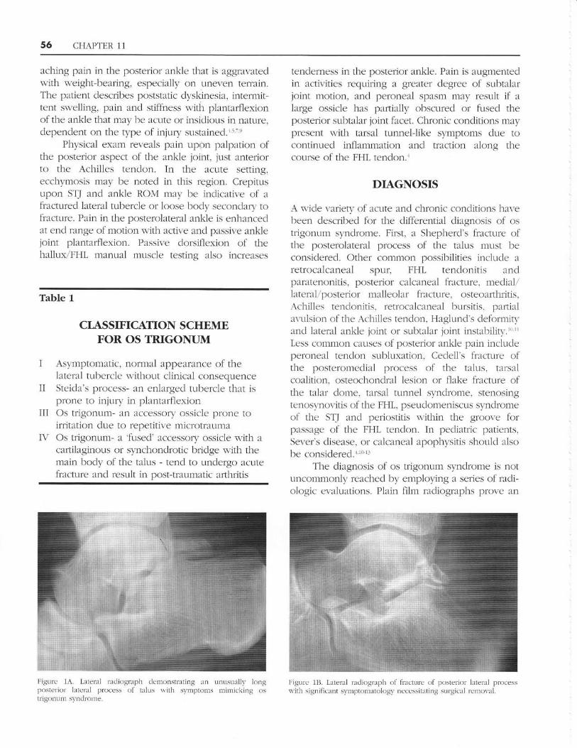
|igrrlc 1B. L:rteral recliogrlph of fracture of posterior' lateral process$-itil si!{nific.Lnt symptorlatolog\'- r'rcccrssitating sr,rrgical remor.al.
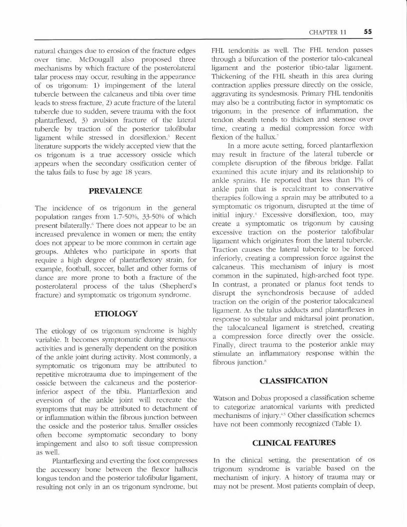
Figurc 1A. L2lterxl lacliolaraph clernonstrating an
l)osterior latcral process of tlhLs n,ith symptornstrigontun sJ,nclrope .
r.rnusuallv krngrninricking os
CHAP'|ER 11 57
extremely r,:rluzrlrle aid in diagnosis; the lateral ancl
oblique vien.s are particLllal'l.v beneficial. Ostrigonum is often an inciclental finding onradiograph, appearing on average in 10% of thepopulation. In symptomaric patients the accessorl,,'
ossicle or elong'atecl Steicla's process is oftenvisualizecl :rlrr:tting the clistal posterior edge of thetibia, r'aising the possibility of posterior ankleimpingement in symptometic patients.rr Hor.eveL.there can be significant difficr-rlty in distinguishing a
tme os trigonum fiom a fiactr-Lrecl lateral trLbercle onplain film (Figr-rre 1). One n'oulc1 expect a trueaccessory ossicle to clisplay a smooth, rouncleclcontour u.hile a fracture frzrgment u.ould clispla.v a
characteristic 'jaggecl' ec1ge, horvever, it has beenshown that over tirne fi'.tcture fragments mav zLppear
smooth as s,'e11 clue to erosive n-ear and tear.- Oyer'time t1-re eroding fracture fragrlents lnav represent a
non-union, ufiere characteristic sclerotic eclges rnal-bc r.isua1izec1. Chao slrggests olrtarning a 3O-clegreesubtalar oblique riiel to diff-erentiate bets-een anos trigonum and an :rcr:te fl-:rctnre.'; Contralateralr.iew-s are not exceptionally useful. althor,rghrecommended, since the os trigonum as u.el1 as
the fracturecl Steida's process may present unil:Lter-ally or bi1aterally.
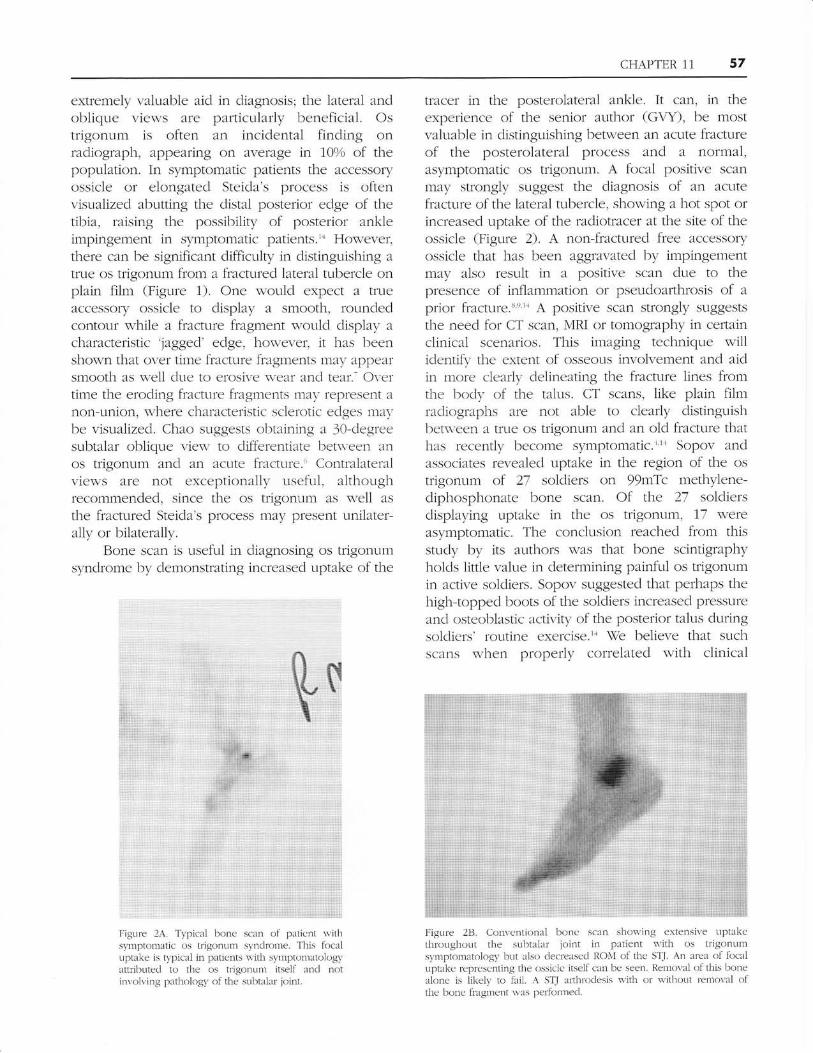
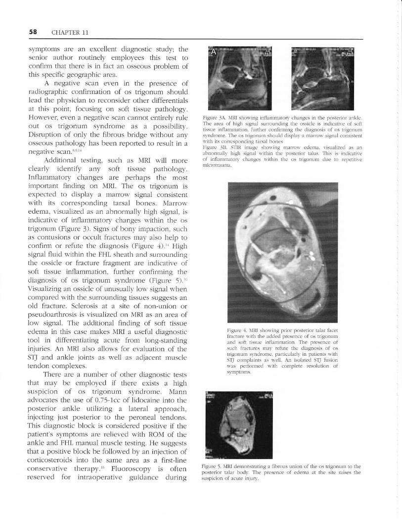
Bone scan is useful in clizrgnosing os trigonuntsynclrone by clemonstrating increased uptake of the
tracer in the posterolzrteral ank1e. It ctn, in theerperience of t1-re senior author (G\Y), be tnostvaluable in clistinguishing betr'veen an acute fractttreof the posterolaterzrl p1'ocess ancl a normal,asymptom2ltic os trigonum. A foczrl positir-e scanmay strongl,v suggest the cliagnosis of zln acLtte
fracture of the 1ater21l tubercle, shor,r,ing a hot spot orincreasecl uptake of the racllotracer at the site of theossicle (Filalrre 2). A non-fiactured free accessoryossicle that has been aggrtr.'ated bv impingenentmay also resuit in a positirre scan cttte to thepresence of inflarnmzLtion or pseucloafihrosis of a
prior fracil-rre.ii'1'' A positive scan strongl), suggests
the need for CT scan. NIRI or tomo5araphy in certainclinical scenarios. This imaging technique rvi1l
iclentifi. thc extent of osseous in-n'olr.erlent :rncl zrid
in more c1carl1. clelineating the fracture lines fiomtl-ic hoclr. of the talus. CT scans, like plain filmracliographs are not able to clearly clistinguishbetn'ecn 2l trlle os trigonum and zrn olcl fiacture thathas recently become symptomatic."' Sopov ancl
associates revealed uptake in the region of the os
trigonum of 27 solcliers on 99mTc methylene-diphosphonatc bone scan. Of the 27 solcliersdisplaying uptake in dre os tdgonum, 17 u-ereasymptomatic. The conclusion reached firtm thisstucly by its authors rras th2tt bone scintigraphyhoids 1itt1e r.alue in cletermining painfirl os trigonumin zrctive soldiers. Sopov sr-rggestecl thilt perhaps thehigh-toppecl boots of the soldiers incteased pressure
and osteoi-rlastic activity of the posterktr talus duringsoldicrs' routine erercise.'' \We believe that sr-rch
scans u.hen properll, correlated u'ith clinic;r1
FigtLre 2B. ConvcnTionxl bonc scan slros'tng extensive LlptakclhfoughoLrt the sttbtalat joint in patient t'ith , ,> Iri!, )ntLllls)'mptolnatok)S1. blrt a]so clecleasccl l{oNI of the STf. An area of fircalupt:Lkc reprcsenting the ossiclc itself c:ttt bc seen. Retnor-a1 of this bonealonc is like1r, to fail. A STJ althrodesis \\.ith or s'ilhout retnor':rl ofthe bonc fiagmer-rt l,as perfbu-necl.
11,11.::,1:;,:1:;1,1;:1;1;l;,11.;1;;,1;;'1],':':;'1:1::,'11
Irigure 2A. Tvpical bonc scan of paljcnl \\,i1hs\lnptolll.Ltic os trigonlrln syndrome. This foc:rlluptake is typic:rl in patients $'ith svn]ptomaLologr'attribute(l Io thc os trigonLrm itsell' :rncl notinr o1r'ing pathologv of the snbtalar joint.
CHAPTER 11
symptoms are an excellent diagnostic study; thesenior zruthor routinely employees this tcst toconfirm that there is in fact an osseor-ts problem ofrhis spet'ilic geograpltic rrea.
A negative scan even in the presence ofradiographic confirmation of os trigonum shouldlead the physician to reconsider other differentialsat this point, fbcusing on soft tissue pathology.However, e\ren a negative scan cannot entirely ruleoLrt os trigonum syndrome 21s a possibility.Disruption of only the fibrous bridge u,.ithout anyosseous pathology has been reported to result in a
negative scan.se t'
Additional testing, such as MRI will moreclearly identify any soft tissue pathology.Inflammatory changes are perhaps the mostimportant finding on MRI. The os trigonum isexpected to display a marron' signal consistentrvith its corresponding tarsal bones. Marrowedema, rrisualizecl as an abnormally high signal, isindicative of inflammatory changes within the ostrigonum (Figr-rre 3). Signs of bony impaction, suchas contusions or occlrlt fractures may also help toconfirm or refute the diagnosis (Figure 4).'' Highsignal fluid within the FHL sheath and surroundingthe ossicle or fractr-rre fiagment are indicative ofsoft tissue inflammation, firrther confirming thediagnosis of os trigonum synchome (Figr-rre 5)."Visualizing an ossicle of unusually lorv signal u.hencompared with the surrounding tissues suggests anold fracture. Sclerosis at a site of non-union orpser-rdoarthrosis is visr-ralized on MRI as an area oflow signal. The additional finding of soft tissueedema in this case makes MRI a useftr1 diagnostictool in clifferentiating acute from long-stanclinginjuries. An MRI also allons for evaluation of theSTJ ancl ankle joints as well as adl'acent muscletendon complexes.
There are a number of other diagnostic teststhat may be employed if there exists a highsuspicion of os trigonum syndrome. Mannadvocates the use of 0.75-1cc of lidocaine into theposterior ankle utilizinla a lateral approach,injecting just posterior to the peroneal tendons.This diagnostic block is considered positive if thepatient's symptoms are relievecl u.ith ROM of theankle and FHL manual muscle testing. He suggeststhat a positive block be follou,-ed by an injection ofcorticosteroids into the same area as a first-lineconservative therapy.'6 Fluoroscopy is oftenreserved for intraoperative guidance during
Figure JA. N'IRI shon'ing inflamrn:Ltory changes in the posterior anklc.The area of high signal sr,rrror-rnding the ossicle is inclicative of softtissne infl:rmmntion. lurther conlirming the diagnosis of os trigonr.Lnrsynclrome. 1'}ie os lrigonum slioulcl displal'a marro\\' sjEanal consistentwith its corrcsponcling t:Lrsal bonesFigr-rre JB. STIII imagc shon,ing ntarro\\' eclema, visualized as anabnorma.llv high signal \\:ithin the posterior talus. This is indicativeof inJlamrnaton' changes within tbc os trigonum dlle ro rcperiti\emicrotrilLlma.
Figurc ,1. r\l1il sl.roning prior posterior talar facetfiacture $'ith the aclclecl prescnce of os trigonun'rancl sofi tissue infl:Lrnmation. The presence ofsuch fractures n'iay refirte the ciiagnosis of ostrigonllm synclromc, particularly in patients s,ithSTJ complaints as u,-e11. An isolatecl STJ fi-rsionn'as pelfbrmecl u.ith complete resoh:tion ofsvmptolns.
Figur-e 5. \,{RI dernonstr:rting a fibrous union of the os trigonllm to theposterior talar body. The presence of eclema i1t the site raises thesr.rspicion of aclLte injurv,

CHAPTER 11 59
excision of the os trigonum w'hen conselativemeasures have failed. llo\\rever, a fluoroscopicallyguidecl cliagnostic block may confirm that thepathology exists purely within the synchondrotic orfibrous bridge connecting the os trigonum to theposterior talus.'r
Stress lateral racliographs are most beneficial inascertaining the exact etiology of the patient'scomplaints. Stress views in extreme dorsiflexion ancl
plantarflexion may visr:ally recreate the mechanismof injury, allowing the interpreter to confirmimpingement of the ossicle and the mobility of ordisruption of the bridge clue to ligamentous traction.Once the diagnosis of os trigonum syndrome has
been made, the physicizrn must cletermine if anyaccompanying patl-rologies exist. For example, FHLtendonitis is a common secondary seqr-re1ae of os
trigonum synclrome. A tenogram can aid inidentifying defects ancl abnormalities within the FHLsheath, however, we believe the MRI is the prefeneclstucly of choice and when properly performecl ancl
interpreted provides unsurpassed cletailed informa-tion as to the type and extent of pathology.
In order to simplily the complex process ofrecognizing and correctly cliagnosing os trigonumsyndrome, Mafiin presented a cliagnostic schematicfor the diagnosis and treatment of the disorder in the
Journal oJ'Foctt Surgety in 1989. His plan begins w-ith
radiographic evah:ation of a pzrtient complaining ofposterior triangle pain. If the plain films suggest a
diagnosis of fracture ancl are consistent u'ith theclinical presentations, then the patient should be
treated with appropriate fracture care. Filmsnon-specific for trauma should be evaluatecl forother possible differential diagnoses andconservative treatment initiated. If pain persists afterconservative therapy, a bone scan should beorderecl. A negative scan should lead to fufiherevaluation with conservative and symptomatictreatment. A positive scan leads to a high suspicionof os trigonum syndrome and should be confirmedwith CT scan: and MRI should zrlso be considered as
a fufiher vaiuable diagnostic imaging str-rdy. A CTnegative for occult fractures or gross osseollspathology should stimulate a more aggressivecourse of conservative therapy and re-evaluation fbra possible inflamed ossicle. A positive CT scanwould show a traumatic fracture or perios[itis orstress fracture of an elongated Steida's process. Ineither case, after failr-rre of a more aggressive course
of conselative therapy, surgical excision of theossicle or fracture fiagment must be considered."
TREATMENT
Os trigonum syndrome, acute fractures of the lateraltubercle and chronic conditions secondaty toprior fracture or repetitive injury are all treatedconservatively and symptomatically fbllou'ing initialdiagnosis. A11 acute fractures and injuries atetreated with 6 weeks non-weightbearing cast
irnmobilization. Mann recommends casting in 10-15
degrees of equinus while most podiatric physicians
believe neutral position is acceptable.'e A11 chronicinjuries should be tre'ated initially with RICE therapy,NSAIDs, stretching exercises, ultrasound ancl
rehabilitation. Iontophoresis with clexamethasone
may also be consiclerecl as a consetwative treatmentrnodality.6 Shoulcl these initial treatment modalitiesfail rn either the acute or chronic setting,corticosteroid injections may prove beneficial.Cotticosteroid injections shoulcl be resen'ed as asecond line therapy because when utilized duringthe first 6-8 weeks of therapy they are implicatecl inthe inhibltion of potential bone healing."i Cautionmust be taken when iniecting cofiicosteroids intothis general area because weakness of the Achillesand flexor hallucis longus may result.6 If syrnptomspersist, an additional 4 weeks of cast immobilizationis recommended.lrr're20 Paulos reports a 330/o success
rate with consetwative therapy aione, and anadditional 10% of patients improved with a second,
more allgressive round of therapy.'o Podiatric and
orthopedic surEleons alike agree that surgicalexcision of the ossicle or fracture fragment shoulcl
be considered fbllowing failurre of 4-6 months ofconsefvative treatment.
When indicated, surgical excision may be
approached in three fllanners: 1) lateral incisionzrlapproach, 2) medial incisional approach,, arthroscopic ercision. The laterzrl incisionalapproach is the most often employed method ofexcising a symptomatic ossicle (Figure 5A) A4-5 cm lateral cun ilinear incision is made posteriorto the peroneal tendons and anterior to thetencloachilles, taking care to avoid the sural nerve;the sural nerve is more readily retracted forward ofthe surgical site minimizing potential injury duringthe surgical procedure (Figure 68). Blunt dissectionis carried down to the deep fascia and the posteriorankle joint capsule is incised to expose the talus.
The ease of dissection al the posterolateralankle offers superior access to the subtalar joint as
well. The FHL tendon should be exposed ancl
identified prior to excising the ossicle. This is

CHAPTER 11
Figurc 0A. Lete[L] incisional appro:rch lbr relIror.al of ,,\ lriijultlLln.'Ihe incision is rn:rcle lnicls,lry. or pr-cfer:rble slightll.posterior to Lltcbisector of the clistlLlice bettecn tfie tencloachillcs ancl the pcrone:rltenclot-rs.
Figure 6C. Iclentification ancl isolariotr of the fiactrlre fragtnent.Iirtir-pation is llrcilit:rtecl 1tv tnanipulating thc bone uith u single skinhook. P:uticul:rr cule shoulcl ltc taken to avo:icl rnjrLrl, to the FIiLtcnclon thiclt is just slightlr'mecli:r1 :rncl cieep to the fiegltent.
accomplishecl by maniplrlating the halluLx Lrnclel'clirect observ:ttion (Figure 6C). Follou,.ingexcision) the talus shoulcl be inspected for sharp orlrregnlar projections ancl the FHL tendon inspectecllor any gross pathology (Figure 7). The FHL rr-Lnnelshor-rld be inspected fbr any loose ltoclies that may1-rave clisplacecl after fiacture.
The latelal incision is a safe approach due tothe lack of large neurovasclrlar str-uctures in the areaanc'l is technically easier to perfbrm thzrn a meclialapproach. A possible conlplication involves stiffhessof the peroneal tendons due to interference insurgery. along u,ith sLtr211 nerve neudtis, alsoseconclary to tr2ruma dr,rfing sufgery. Abrzrmonitz et
Figure 6Ii. Iclentiflcatjcxt ofihe sural nefl'e as it trarcls into the firot.Notc bifttrc:rtion of the nen'e. 'l'ltc ncnc shoulcl be nreticirloush- rnclcarefull,,' retr:lcted anrl protected chrring thc surerrl [, ) .1\ uirll)osl()perati\c cntlxplnent and neuritic svlnpt()nts.
i'igure --r. lntraopcretive x-ra\.s conflnuing retnov:r1 of thc cntirefi:tcture fi:rgnent. The nrain bodl' o1'the trlrLs is cltcckccl celefullv tocnsllre no spilie or rough eclges lern:rin. A srnall h:rnci rrsf is uffittl<in rerrocleling this arca. l)ori cr inrLl-urrcntrtion ir nur reconrtnenclecldue kr potcntial damage to the srLlrounclinc soft tissue.
al ernployecl the lateral incisional approach to excise21 symptomatic os trigonum in 41 patients. The str-rdynoted that sural nen-e iniury was the most commoncomplication, with four patients developingcomplete sensory loss of the nen e.2'
A medial incisional approach is justified inpatients sr-rf'ferinla from concomitant FHL tenclonitis,common among ballet clancers. Here, care nrustbe Ltken to avoicl all structures of the medialneurov:rsclrlzrr br,rnclle ancl the flexor tendons (Figure8A). The medial approach offers better exposure tothe FHL tendon 21nd its sheath, which may bedir.ided if severe tendonitis and stenosis is present(Figr-rre BB). The medial appro2lch may result in less


62 CHAPTER 11
the ankle and subtalar .foints.,, \7e recommenda short period of several weeks of absoluteimmobilization to zrllow healing the tissues and toavoid wound dehiscence.
COMPLICATIONS
Complications of surgical excision inch_rde infection,persistent pain and posttralimatic arthritis followinga fracture. More specifically, postsurgical adhesionsand fibrosis may limit ROM and be a source ofchronic pain. Chronic tendonitis may result clue totrzruma to the flexor hallucis longus tendon dr_rringsurgery or chronic inflammation of rhe tendonsheath due to traction against improperly debridedbony eclges.
Post sllrgical neuritis or other nerwe disorderssuch as neuropraxia and neurontmesis have beenencounterecl in a number of patients whounderw-ent surgery elsewhere. On carefulexamination of these patients, the surgical incision isoften directly overlying the neurovascular bundleand direct insult and injury have been sustained.Complex regional pain syndrome or reflexsympathetic clystrophy syndrome rernain realentities which can result in a chronic painsyndrome in these patients. Any postoperativenerwe disorder can be frustrating, challenging anddiscouraging to both the patient and surgeon. Somepatients will become candiclates for chronic painmanagement proElrams.
Traumatic techniques of resection can alsoresult in injury and damage to the subtalar jointprimarily and less frequently the ankle joint. As aresult. significant degenerative afihritis developsand, if fails to respond to conselvative treatmentmodalities, is 1lkely to require joint afihrodesis.
The prognosis for os trigonum syndrome andacute and chronic fractures of the posterolateral talarprocess is excellent. Approximately 43o/o of al1patients diagnosed with os trigonum syndrome willexperience complete resolution of their symptomswith conselative treatment modalities a1one. Theremainder of patients experience reliel throughsurgical excision with an extremely 1ow rate ofcomplications and chronic intractable pain. Theaverage time to full recovery for an afihroscopicexcision is 3 months, compared to 3-1.2 monthsreported for an open excisional procedr,rre. \With
aggressive physical therapy and rehabilitation,
highly competitive athletes and dancers may attainftr1l recovery within 6-8 weeks.7," The prolongedrecovery and convalescence repofied for opentechniques of resection have not been theexperience of the senior author (G\Y), although wehave seen several patients who have suffered. It isour belief that this occltrs as a result of traumatictechnique with injr-rry to the cutaneolts nelves oneither the rnedial or lateral aspect with or withoutconcomitant injllry to the deeper soft tissues result-ing in an excessive amount of postoperative scartisstre and ldhesions.
DISCUSSION
The cliagnosis and management of an os trigonumsynclrome or related entity involving the posteriorprocess of the talus can be clifficult ancl challenging.Often considerable time has transpired sincethe original inciting event or injr-rry. Freqr,rentlymisdiagnosis has lec1 to extensive conservativetreatment, and in some cases, surgiczrl intelention,of an all together different clinical entity with noimprovement in the orlginal symptomatology.Patients, not uncommonly, are very frustrated,anxious and at times even hostile over their currentcondition, especially when it precludes them fromparticipating in spolts activities. \7e have seenseveral pzrtients who have, unfofiunately, experi-enced this outcome.
A careful cletailed history and physicalexamination shoulcl raise the index of suspicion forthis clinical entiry. Conventional radiographs willeither confirm or refute the ciinical suspicions.In either scenario, further diagnostic workup orconfirmation can be done with specialized str-rdiesincluding a conventional bone scan, CT scan orMRI . A series of diagnostic and therapeutic blockswith long acting 1ocal anesthetics, with or withoutshort acting corlicosteroicl, accurately delivered tothe posterior ankle 1'oint arezr, shoulcl readily confirmthe diagnosis.
Conseruative treatment should be aggressiveand includes a combination of immobilizationfollowed by physical therapy. Various pharmaco-logic agents may prove beneficial including systcmicand locally injected cofiicosteroids combined withlong acting local anesthetics and NSAIDs. The earlierthe correct diagnosis is established and the soonerconservative treatment instituted, the more likelv it
CHAPTER 11 63
is to be successful. Radiographic healing or failureto heal does not necessarily correlate with clinicaloutcomesi treatmenl must not be based upon theradiographic presentation of this entity. \7henpatients fail to responcl to conselvative treatment,surgical exploration with excision is necessary.
Carefill, meticulous and complete excision ofthe fragment of bone must be achieved to ensllrea successful clinical outcome and resolution ofsymptoms. In some cases, exploration of the FHLtendon itself with debridement and or repair is
necessary. Rarely will joint fusion be required as adefinitive treatment.
The senior author believes that the singlemost important requisite lor successful outcomewith this type of surgery is a thorotigh workingknowledge of the anatomy of the ankle andsubtalar joints, including the periarticr,rlarstructures. An awareness of the various nen/estrlctures, especially the sural and posterior tibialneffes will ensure a low incidence of nerve injury.Meticulous, detailed, precise minimally traumatictechnique then paves the way to a successfulexcision of the pathologic bone without damage tosurrounding structures. Hemostasis is critical dr-rringand following the slrr5aery. A tourniquet is routinelyemployed during this procedure but is releasedprior to closure to ensure maximum hemostasis.
REFERENCES
1. X{uramoto J, Ferkel R. Arthroscopic ercision of the os trigonum:a new technique with preliminarr,, clinical rcsults. Fctctt Ankle1997;78:777-84.
2. Burcziu NJ. Carclinal E, Hobclen R, Anbin B. Posterior ankleimpin€leflrent syndrome: NIR image finclings in scr-cn prtisnts.Rcrdiologlt 2000;275 :497 -503.
3. Keiikian H, Kelikian A. Os tri€lonum. Disr.trders clf the Ankle.Philacleiphia;WB Saunders; 1985.
4. O'Rahi1ly R. A sllnrey of carpal and tarsal anomalies../.Bo/rcJoitltSurg Am 7953:35:626 42.
5. Os trigonum,/iiaciures of the talus. In llanks AS. Downey NI,
Martin D, Nliller S, edrtors: llcGlctmtry's Comprehensiue I'extbookol Foot dn.J Ankle Suryery. Philaclelphia: Lippincott, Sflilliarns &\firilkins: 2001.
6. Le TA. Joseph PM. Common exostectomies ol the rearfoot. ClirtPodial t i, .l4t'J \u tg 10oI C:6Ul-lJ
7. Chao -W. Os trigonum. I-oot Ankle Clin 2004;9:78f -96.B. Blake RL, Lallas PJ. Ferguson H. The os trigonllm s1-ndrome. A
literature rev:ien . /,,12 Pocliann Mecl Assoc 7992;82:L54-67.!. $flenig JA. Os tdgonr-lrn syndrome. J Am Podiatryt Mecl Assoc
1990r80:278-82.10. Jimenez C. Torres E. Posterior Ankle pain in ihc recre:rtional ath
lete. Phls Sport< MtJ lo')h;l(':ij.
Nlenclez-Castillo A, Burcl TA. R:Lcliologic case stucly: os rrigonumsyndrome. Oft bop 7999;22:1.208-2.Nlyerson M. Os trigonurn sr'rndrome. Foor and ankle disorders.Philadelphia: \VB Saunclcrs; 2000,Hamilton WG. Stenosing tenosynovitis of the FHL tendon andposterior impingement upon the os trigonum in ballet dancers.Foctt Ankle 1982;3:74-80.GiufTrida AY, Lin SS. Abidi N, Berberian -iil, Berkman A, BehrensFF. Pseudo os trigonlrm sign: m:issecl posteromecliai talzLr f:rcctfracture. L-oot Ankle Int 2003:21:542.Scurran R. I-ractlrres of thc posteroiateral process of the tal-rs.?c.tot and Ankle Trau.ma. Neu. York: Churchill Livingstone; 1996.Sopov V, Liberson. A, Groshar, D. Bone scintigraphic findings ofos tdgonr-lm: a prospcctive sttLd1,6f 100 solcliers on:rctive duty.F-octt Ankle Int 2000;21:823.Nlann R, Coughlin M. Fractr-rre-clislocations of the Lrlus. SurgeryoJ tbe FooL ancl At'tkle. Si. Louis:Nlosbyr 1999.
Jones DNI, Saltzman CL. El-Khoury G. Thc diagnosis of OTS witha fluoroscopically controlled injection of local anesthetic. Ios,-aOfibop J 1999;19:122-6.Martin BF. Posterior triangle pain; the os trigonllm. J F-ctol Surgery198928:31 2-8.Mann R. Coughlin l,{. Os trigonum sl,ndrome. Surge.y cf'tbefootancl ankle. St. Louis: l4osby; 1999.Paulos, N{D. Johnson, NltD, Noyes, NID. Postcriclr Compafilnentfiactures of thc ankle: a commonl_v missed athletic tnjury. AmJSpotls Med 1983;11 4)9-43.Abramorvitz Y, \{rollstein R, Barzilay Y, London E, Matan Y,Shabat S, Nyska NI; Outcorne of rcsection of a symptomatic ostrigonlLn.I. ./ Boz e .Joint Su rg Aln 2003 ;85 : 1 05 1.'Wredmark T, Carlsteclt CA. Os trigonllm svndrone: a clinicalentity in ballet d:rncers. Foot Attkle 7997;L7:t+ll1-6Lon'rbardi CM, Silhanek AD. Connolly FG. Nloclified arthroscopicercision of the symptomatic os tri€Jonum and release of the flexorhallucis longus tenclon: operative technique and case study.
.l l;oot Ankle Surg, 7999;38:J17-51.Kalasick D. Schlr,'citzer NIE. The Os tdgonum sy-ndrome: imrgingt'eattrrcs. A m.l Roent 7996;166:22-5.Goossens NI, Destoop N, Claessens H, Van der StraetenC. Posterior subtalar joint athrography. A useful tool in thediagnosis of hindfoot clisordcrs. Clin OrthoP 7989:249:218 55.Havens RT, Kaloogian I{. Thul JR, Hoflman S. A corrcl:rtionbctwccn os trigonum synclrome and tarsal tunnel syndrome./Ant Podiatry Mr'J As.:oc lox6:-rr: ti0- t.
Nlolay M, Wilson ll. Lasker A. Os trigonum synclrome in a patientwith multiple epiphyscal dysplasia. ./ -F'oo t Surger! 1982 ; 2 1 : lo5-8.X{oellcr FA. The os trigonum syndrome.,/,4r2 Podiatty Med Assoc
797 3:63:491-507 .
Galinski A. Os trigonr-rm :1s a cause of tarsal coai:ltrcn. J AmPod iatry Med A sso c 1c)7 9 ;69 L91. -6.Nloeller F. The os trigonlrm sl,ndrome. J Am Podiahy Med Assoc197 3;63:191-)0L.CrirnJ, Cracchiolo A. Hall. Os trigonum syndrome Imaging cj'the.fbctt artrl ankle. Lnrted Kingdol'r: Lippincott-Raven; 1996.
Os trig<rnr.rm syndrome. ',Vheeless' Textbook of Ofibopedic-\.Ar.rihblc .rt w$ \\.rrtq(lme(lia.( urn.Reinh:ruz R. The significance of os irigonum. J Fa()t Sutg1979;18:61-3.Minoves M. Cornp:rny A, Ardcvol J, Nlatas 1iR, Costansa JM,Setoain J. Domenech-Torne FNl: Redionuclide Im.rging inBilatelal Os Tugonum Syndrome in :l Young Athlete. Clin NuclMecl 2007;26:237.Kim DH, Berkor.itz MJ: Pseudo Os Trigonum Sign: N{issedPosteromedial Talar F'acet Fractlue, Giufliida, AY, et el. FoolAnkle htt 20011251372.
20
27
22
11
12,
13.
14
15
16.
11
18.
19
26
23
24
33
34
30
)6
25
2f
28
29
31
32
35


![jflif{s k ltj| ]bg @)&!/cite> · h= Rffn' cf=j= sf ] >fj0f](https://static.fdocuments.in/doc/165x107/5d52159288c993bf198b707c/jflifs-k-ltj-bg-h-rffn-cfj-sf-fj0f-b-lv-h-i7dd-o-jo-fdf.jpg)


