
Special situations in tonsil and Adenoid disorder Special situations in tonsil and Adenoid disorder

J clin. Path., 1972, 13, 301-305
Adenoid basal carcinoma of the cervix uteri'K. B. SHILKIN
From the Department of Pathology, University of Singapore, Singapore
SYNOPSIS This paper describes a case of adenoid basal carcinoma of the uterine cervix, occurringin a woman aged 78 years. In view of the rarity and the limited knowledge of this group of neo-
plasms, details of the clinical history, the histological appearances, and the histochemical reactionsare presented. An attempt is made to define the histogenesis of the tumour and some points on
nomenclature are discussed. It is suggested that the tumour arises from multipotential cervicalbasal cells and that the adenoid structures rather than being true glands are formed by degenerationof the connective tissue stroma. On the basis of a review of reported cases it seems possible thatthis tumour will be shown to be one of low-grade malignancy.
In recent years an unusual and rare type of tumourof the uterine cervix has been described. Its histo-logical features are distinctive and different frompreviously recognized epithelial tumours of this site.Brief mention of this class of neoplasm is made in astandard text (Novak and Woodruff, 1967) where thetitle 'adenoid basal carcinoma', subtitled parentheti-cally as 'cylindroma', is used. Further confusionconcerning nomenclature is apparent in the medicalliterature where the tumour has been labelledvariously as cylindroma (Tchertkoff and Sedlis,1962), adenoid cystic (pseudoadenomatous) basalcell carcinoma (Gould, Hinerman, Batsakis, andBeamer, 1963), squamous and adenoid cystic basalcell carcinoma (Moss and Collins, 1964), adenoidcystic carcinoma (Bell, 1971; Benitez, Rodriguez,Rodriguez-Cuevas, and Barbosa Chaivez, 1969;Dahlin, 1966; Grafton, Willis, Martin, and Mathews,1968; McGee, Flowers, and Tatum, 1965), and aden-oid-basal carcinoma (Baggish andWoodruff, 1966).Atotal of 12 cases have been documented. Whetherthese 12 tumours are identical is difficult to assess,nevertheless they appear to comprise a separategroup of neoplasms of the cervix. Their histogenesisis open to speculation and is likely to remain un-certain until more tumours of this type are studied.The purpose of this report is to describe a further
example of what is preferably termed, for reasons tobe presented, an 'adenoid basal carcinoma'. Theclinical, histopathological, and histochemicalfeatures are described and some aspects of histo-genesis are discussed.
'Address for reprints: Dr K. B. Shilkin, Department of MorbidAnatomy, King's College Hospital Medical School, Denmark Hill,London SE5, UK.Received for publication 21 October 1971.
301
Case Report
The patient was a 78-year-old Chinese who com-plained of vaginal bleeding for four days. She hadborne three children and since the menopause,many years previously, had no gynaecologicalsymptoms other than that with which she nowpresented. Examination of the cervix revealed a poly-poid mass of 3 cm involving and protruding fromthe external os. Both anterior and posterior lips ofthe cervix were distended by the tumour. The bodyof the uterus was not affected, being small andmobile. The pelvic adnexal structures were normal.No further relevant features were found on generalexamination. Clinically, she was regarded as havingcarcinoma of the cervix, early stage II. A smallbiopsy of the mass was reported as malignant,prompting a second procedure to remove, pervagina, as much of the tumour as was easily access-ible. Subsequently a short course of deep x-raytherapy was given, followed by two insertions ofradium, each of 20 mg for 72 hours, at an intervalof one week. One year after her first admission tohospital she again had vaginal bleeding. The lumenof the uterus, including the cervical part, was noweroded and contained soft tumour tissue, confirmedhistologically. Local extension was not detected andit was thought she did not have metastases. Anothercourse of radiotherapy was given. She died at homethree months later, some 16 months from the firstonset of symptoms. A necropsy was not performedand the exact cause of death was uncertain.
PATHOLOGYThe initial biopsy was small and reported as acarcinoma, type not specified. The tissue removed at
on March 6, 2020 by guest. P
rotected by copyright.http://jcp.bm
j.com/
J Clin P
athol: first published as 10.1136/jcp.25.4.301 on 1 April 1972. D
ownloaded from

K. B. Shilkin
the second biopsy was polypoid, soft, white or palegrey, and had several haemorrhagic zones. Therewere variable and complex microscopic appearances.The major part was made up of a lace-like pattern oftrabeculae (Fig. 1) or adenoid formations (Fig. 2)lying in a loose connective tissue stroma. Many ofthe trabeculae were two cells thick as were the partywalls of interconnecting adenoid structures. Some-times an almost regular honeycomb pattern wasformed (Fig. 3). In all these areas the cells were of afairly uniform basal type, being cuboidal, with around deeply basophilic nucleus and scant eosino-philic cytoplasm. There were frequent mitoses butotherwise only moderate cellular pleomorphism. Theconnective tissue was an important component andformed the overall background in which the tumourwas embedded. Tongues of stroma protruded intocellular areas and lay on either side of the trabeculaeand cell cords. The composition of the centres of theadenoid structures ranged from fibroblastic cellularconnective tissue to completely acellular material ofa granular or homogeneous mucinous nature.Another different pattern consisted of solid islandsof basal cells which aligned at the edges to form a
Fig. 1 The tumour in this area has a lacijorm arrange-
ment of cell cords lying in a loose stroma(Hand E, x 106).-
roughly palisaded rim. These areas merged with thepreviously described zones on the one hand and withyet another pattern of more overtly squamous areason the other. The surface of the tumour also varied,with some parts consisting of relatively normalcervical squamous epithelium and others where theconfiguration was similar to that ofcarcinoma in situ.Of particular note were several areas where the neo-plastic proliferation seemed to arise in the basal layerto extend downwards into the underlying cervicalstroma (Fig. 4). The biopsy taken a year after radia-tion therapy was of more solid tissue. Again therewere solid basal cell areas and although adenoid andlaciform arrangements were not prominent therewere thin cords and small nests of basal type cells.Squamous areas were again evident.
HISTOCHEMISTRYA variety of histochemical techniques were employedto assess the nature of any intracellular substancesand of the material in the centres of the adenoidstructures and in the stroma. The methods for muco-polysaccharides have been previously outlined (Tockand Shilkin, 1970). Stains for lipids were performed
Fig. 2 Adenoidformations are predominant in this area.Most lumens contain stroma, some are acellular(H and E, x 46).
302
on March 6, 2020 by guest. P
rotected by copyright.http://jcp.bm
j.com/
J Clin P
athol: first published as 10.1136/jcp.25.4.301 on 1 April 1972. D
ownloaded from
Adenoid basal carcinoma of the cervix uteri
q. -.
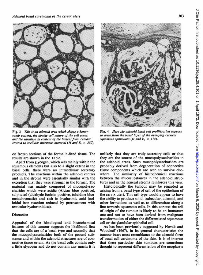
Fig. 3 This is an adenoid area which shows a honey-comb pattern, the double cell nature of the cell cords,and the variation in content of the lumens from cellularstroma to acellular mucinous material (H and E, x 250).
on frozen sections of the formalin-fixed tissue. Theresults are shown in the Table.
Apart from glycogen, which was mainly within thesquamous elements but also to a slight extent in thebasal cells, there were no intracellular secretoryproducts. The reactions within the adenoid centresand in the stroma were essentially similar with theexception that they were stronger in the former. Thematerial was mainly composed of mucopolysac-charides which were acidic (Alcian blue positive),sulphated (aldehyde-fuchsin positive, toluidine blue-metachromatic) and rich in hyaluronic acid (col-loidal iron reaction reduced by pretreatment withtesticular hyaluronidase).
Discussion
Appraisal of the histological and histochemicalfeatures of this tumour suggests the likelihood firstthat the cells are of a basal type and secondly thatthe mucopolysaccharides both of the ground sub-stance and within the adenoid structures are of con-nective tissue origin. As the basal cells contain onlya little glycogen and do not contain any mucin it is
Fig. 4 Here the adenoid basal cell proliferation appearsto arise from the basal layer of the overlying cervicalsquamous epithelium (H and E, x 114).
unlikely that they are truly secretory cells or thatthey are the source of the mucopolysaccharides inthe adenoid areas. Such mucopolysaccharides areprobably derived from degeneration of connectivetissue components which are seen to survive else-where. The similarity of histochemical reactionsbetween the mucosubstances in the adenoid struc-tures and in the general stroma reinforces this view.
Histologically the tumour may be regarded asarising from a basal type of cell of the epithelium ofthe cervix uteri. This cell type would appear to havethe ability to produce solid, trabecular, adenoid, andother formations as well as to differentiate along aline towards squamous cells. In this context the cellof origin of the tumour is likely to be an immatureone and not to have been derived from malignanttransformation of either the differentiated squamouscell or the glandular epithelial cell.As has been previously suggested by Novak and
Woodruff (1967), in its general characteristics thetumour bears most resemblance to the adenoid formof basal cell carcinoma of the skin. It is of interestthat these particular skin tumours are sometimesthought to represent differentiation of the neoplastic
303
xq-,,,. 11le
Aa
on March 6, 2020 by guest. P
rotected by copyright.http://jcp.bm
j.com/
J Clin P
athol: first published as 10.1136/jcp.25.4.301 on 1 April 1972. D
ownloaded from

Stain Basal Cells Squamous Cells Central Adenoid StromaMaterial
PAS + ± ±Pas after diastase + +MC + +Ast B + + +AB - - + ++AB-PAS - ++ +++ ++
(red) (blue) (blue)AB after ribonuclease - + + + + +AB after acid hydrolysis - + + + + +AB after neuraminidase - _ + + + + +AF - - +++ ±TB pH 2-0 - ++ +
(red) (red)CI -~++ +Cl after testicular hyaluronidase _ + +Oil Red 0Sudan Black B - -
Sudan III and IV - _ ± i
Table Histochemical resultsThe + sign indicates strength of colour reaction: + + +± strong; + + moderate; + = weak; ± - trace;- negative.PAS - periodic acid-Schiff; MC mucicarmine; Ast B Astra blue; AB = Alcian blue; AF -aldehyde-fuchsin; TB = toluidine blue;
Cl = colloidal iron.
basal cells towards a sweat gland structure (Lever,1967). In the cervix, of course, sweat glands do notnormally occur. Nevertheless metaplastic reactionsto produce sweat glands as well as sebaceous glandand hair follicles do occur on rare occasions (Willis,1958) and so serve to emphasize the multipotentialityof the cells of the cervical epithelium.With regard to nomenclature the term adenoid
basal carcinoma is to be preferred. As this tumourdoes not contain myo-epithelial cells and its maincell type is non-secretory it is not identical to the trueadenoid cystic carcinomas which occur in the sali-vary glands and elsewhere. Moreover the mucopoly-saccharides of this tumour, being connective tissuein origin, differ from those in the salivary type oftumour where epithelial mucins are prominent(Azzopardi and Smith, 1959). The origin, postulatedhere, of adenoid basal carcinoma of the cervix frombasal cells is in contrast to the histogenesis ofadenoid cystic carcinomas which are believed to bederived from the intercalated duct cell (Hoshino andYamamoto, 1970). Thus the term 'adenoid cysticcarcinoma' which has been used in reference to thistumour has several disadvantages. The use ofcylindroma is probably also unsound as it is in anycase, as Willis (1967) states, a 'meaningless nick-name' for the true adenoid cystic carcinoma.Cylindroma of the skin, to which lesion only is thisname correctly applied, is a different tumourhistomorphologically.The behaviour of the adenoid basal carcinoma of
the cervix is not yet established. Review of the 12reported cases does not provide a full picture asadequate long-term follow up has, for several
reasons, not always been documented. Despite thisit is perhaps significant that in only two cases wasthere spread of tumour beyond the immediatevicinity of the cervix. In one of these the spread wasby direct extension to adjacent pelvic structures andin the other in addition to local pelvic spread therewas a solitary metastatic lesion in an axillary lymphnode, being the only distant metastasis of the entirereported series. The tumour may well prove to beone of low-grade malignancy.
The author is grateful to Professor K. Shanmugarat-nam and Associate Professor E. P. C. Tock foradvice, Mr. K. N. Rai for technical help, and Mr. G.Harwood for photography.
References
Azzopardi, J. G., and Smith, 0. D. (1959). Salivary gland tumoursand their mucins. J. Path. Bact., 77, 131-140.
Baggish, M. S., and Woodruff, J. D. (1966). Adenoid-basal carcinomaof the cervix. Obstet. and Gynec., 28, 213-218.
Bell, J. R. (I1970). Adenoid cystic carcinoma of cervix (Abstr.).Pathology, 3, 60-61.
Benitez, E., Rodriguez, H. A., Rodriguez-Cuevas, H., and BarbosaChavez, G. (1969). Adenoid cystic carcinoma of the uterinecervix: report of a case and review of four cases. Obstet. andGynec., 33, 757-762.
Dahlin, D. C. (1966). 'Case No. 9.' In Proceedings from the Seminarof the Houston Society of Clinical Pathologists.
Gould, S. E., Hinerman, D. L., Batsakis, J. G., and Beamer, P. R.(1963). Diagnostic patterns: lesions of the cervix. Amer. J. clin.Path., 40, 537-540.
Grafton, W. D., Willis, G. W., Martin, J. R., and Mathews, W. R.(1968). Adenoid cystic carcinoma of the cervix: a report oftwo cases. J. L. med. Soc., 120, 276-277.
Hoshino, M., and Yamamoto, I. (1970). Ultrastructure of adenoidcystic carcinoma. Cancer (Philad.), 25, 186-198.
Lever, W. F. (1967). Histopathology of the Skin, 4th ed., pp. 535-536.Lippincott, Philadelphia. Pitman, London.
McGee, J. A., Flowers, C. E., and Tatum, B. S. (1965). Adenoid cystic
304 K. B. Shilkin
on March 6, 2020 by guest. P
rotected by copyright.http://jcp.bm
j.com/
J Clin P
athol: first published as 10.1136/jcp.25.4.301 on 1 April 1972. D
ownloaded from

Adenoid basal carcinoma of the cervix uteri
carcinoma of the cervix. Report of a case. Obstet. and Gynec.,26, 356-358.
Moss, L. D., and Collins, D. N. (1964). Squamous and adenoid cysticbasal cell carcinoma ofthe cervix uteri. Amer. J. Obstet. Gynec.,88,86-90.
Novak, E. R., and Woodruff, J. D. (1967). Gynecologic and ObstetricPathology, 6th ed. Saunders, Philadelphia.
Tchertkoff, V., and Sedlis, A. (1962). Cylindroma of the cervix. Amer.
305
J. Obstet. Gynec., 84, 749-752.Tock, E. P. C., and Shilkin, K. B. (1970). Histochemical study of
mucosubstances and glycogen of the postmenopausal humancervix uteri. Amer. J. Obstet. Gynec., 107, 194-201.
Willis, R. A. (1958). The Borderland of Embryology and Pathology,pp. 520-552. Butterworth, London.
Willis, R. A. (1967). Pathology of Tumours, 4th ed., p. 327. Butter-worth, London.
on March 6, 2020 by guest. P
rotected by copyright.http://jcp.bm
j.com/
J Clin P
athol: first published as 10.1136/jcp.25.4.301 on 1 April 1972. D
ownloaded from


