Adaptor complex AP2/PICALM, through interaction with LC3 ...€¦ · Adaptor complex AP2/PICALM,...
6
Adaptor complex AP2/PICALM, through interaction with LC3, targets Alzheimer’s APP-CTF for terminal degradation via autophagy Yuan Tian, Jerry C. Chang, Emily Y. Fan, Marc Flajolet 1 , and Paul Greengard 1 Laboratory of Molecular and Cellular Neuroscience, The Rockefeller University, New York, NY 10065 Contributed by Paul Greengard, August 27, 2013 (sent for review July 16, 2013) The hallmarks of Alzheimer’s disease (AD) are the aggregates of amyloid-β (Aβ) peptides and tau protein. Autophagy is a major cel- lular pathway leading to the removal of aggregated proteins. We have reported recently that autophagy was responsible for amyloid precursor protein cleaved C-terminal fragment (APP-CTF) degradation and amyloid β clearance in an Atg5-dependent manner. Here we aimed to elucidate the molecular mechanism by which autophagy mediates the degradation of APP-CTF and the clearance of amyloid β. Through affinity purification followed by mass spectrum analysis, we identified adaptor protein (AP) 2 together with phosphatidylinositol clathrin assembly lymphoid-myeloid leukemia (PICALM) as binding proteins of microtubule-associated protein 1 light chain 3 (LC3). Further analysis showed that AP2 regulated the cellular levels of APP-CTF. Knockdown of AP2 reduced autophagy-mediated APP- CTF degradation. Immunoprecipitation and live imaging analysis demonstrated that AP2 and PICALM cross-link LC3 with APP-CTF. These data suggest that the AP-2/PICALM complex functions as an autophagic cargo receptor for the recognition and shipment of APP-CTF from the endocytic pathway to the LC3-marked autopha- gic degradation pathway. This molecular mechanism linking AP2/ PICALM and AD is consistent with genetic evidence indicating a role for PICALM as a risk factor for AD. endocytosis | trafficking | aggregate removal A hallmark pathological feature of Alzheimer’s disease (AD) is the amyloid plaque made of aggregated amyloid β (Aβ) peptides. Among genetic mutations identified in familial AD patients, large numbers are associated with accumulation of Aβ peptides. These peptides are generated via sequential proteolysis of amyloid precursor protein (APP) during the course of its trafficking along the secretory pathway. APP is sequentially matured in the endoplasmic reticulum (ER) and Golgi apparatus and then delivered to the plasma membrane through the trans- Golgi networks. Within minutes of arrival at the cell surface, APP is internalized in clathrin-coated vesicles through endocy- tosis (1) via a tetrapeptide motif, YENP, located at the carboxyl terminus of APP (2). The internalized APP is then delivered to endosomes, where it is processed first by β-secretase, also known as BACE1, and then by γ-secretase, to generate Aβ peptides (1, 3). Adaptor protein 2 (AP2) is a well-characterized complex in- volved in clathrin-mediated endocytosis. It is heterotetrameric and consists of four subunits AP2A1, AP2B1, AP2M1, and AP2S1 (α, β, μ, and σ). It was suggested that the α subunit, AP2A1, mediates the binding to the plasma membrane and acts as a scaffold for endocytic accessory proteins. The μ subunit, AP2M1, is responsible for cargo selection through the recogni- tion of a YxxФ signal (where Ф is a bulky hydrophobic residue) in the cytosolic region of type I transmembrane proteins during endocytosis (4–6). Phosphatidylinositol clathrin assembly lym- phoid-myeloid leukemia (PICALM) is a cytoplasmic adaptor protein that also plays a critical role in clathrin-mediated en- docytosis. It binds to clathrin, phosphatidylinositol, and AP2 to aid in the formation of clathrin-coated pits (7–9). PICALM was also identified by genomewide association studies (GWAS) as a significant risk factor for AD (10). However, the pathogenic mechanism for PICALM as an AD risk factor is unclear. Macroautophagy, hereafter referred to as autophagy, is a highly conserved catabolic process in which proteins and organelles are engulfed in double-membraned vacuoles called autophagosomes and then transported to lysosomes for degradation. The mem- brane origins of autophagosomes are unclear but may include ER (11–13), mitochondria (14), and plasma membrane (15). During the formation of autophagosomes, the small cytosolic ubiquitin-like microtubule-associated protein 1 light chain 3 (LC3-I) is processed and conjugated to phosphatidylethanol- amine (PE) to form lipidated LC3 (LC3-II). LC3-II is specifically targeted to elongating preautophagosomal structures and remains on mature autophagosomes until it is degraded by the fusion to lysosomes (16). Lipidated LC3-II serves as a docking site for specific cargo receptors, such as SQSTM1/p62, NBR1, Nix, NDP52, and OPTN (17–21). They all bind to LC3 through an LC3-interacting region (LIR) to recruit specific cargos for degradation. We and others have reported that autophagy regulates the levels of Aβ peptides (22–24). Furthermore, we showed that the clearance of Aβ peptides results from the degradation of the precursor amyloid precursor protein cleaved C-terminal fragment (APP-CTF) via autophagy. However, the molecular mechanism by which autophagy leads to the down-regulation of the membrane- bound APP-CTF and, in turn, of Aβ is not known. We report here that AP2 functions as an LC3 receptor, which shuttles APP-CTF from the endocytic pathway to autophagosomes for degradation. Significance β-Amyloid aggregates are often found in the brains of Alz- heimer’s patients. We have previously reported that β-amyloid levels can be down-regulated through activation of autophagy, a process involving the degradation of unnecessary or harmful proteins. However, the underlying mechanism of β-amyloid autophagy-mediated degradation is unknown. Here we dem- onstrate that the complex adaptor protein 2/phosphatidylino- sitol clathrin assembly lymphoid-myeloid leukemia (AP2/ PICALM), via an interaction with a β-amyloid precursor, bridges β-amyloid degradation and autophagy. This work reveals mechanistic steps for the targeting of the β-amyloid precursor for degradation via autophagy and supports a genome-wide association study identifying PICALM as a risk factor for Alz- heimer’s disease (AD). Altogether, these findings support the notion that activating autophagy is a valid approach for the AD field, which urgently needs novel therapeutic strategies. Author contributions: Y.T., M.F., and P.G. designed research; Y.T., J.C.C., and E.Y.F. per- formed research; J.C.C. contributed new reagents/analytic tools; Y.T., M.F., and P.G. ana- lyzed data; and Y.T., M.F., and P.G. wrote the paper. The authors declare no conflict of interest. 1 To whom correspondence may be addressed. E-mail: [email protected] or fl[email protected]. This article contains supporting information online at www.pnas.org/lookup/suppl/doi:10. 1073/pnas.1315110110/-/DCSupplemental. www.pnas.org/cgi/doi/10.1073/pnas.1315110110 PNAS | October 15, 2013 | vol. 110 | no. 42 | 17071–17076 NEUROSCIENCE
Transcript of Adaptor complex AP2/PICALM, through interaction with LC3 ...€¦ · Adaptor complex AP2/PICALM,...

Adaptor complex AP2/PICALM, through interactionwith LC3, targets Alzheimer’s APP-CTF for terminaldegradation via autophagyYuan Tian, Jerry C. Chang, Emily Y. Fan, Marc Flajolet1, and Paul Greengard1
Laboratory of Molecular and Cellular Neuroscience, The Rockefeller University, New York, NY 10065
Contributed by Paul Greengard, August 27, 2013 (sent for review July 16, 2013)
The hallmarks of Alzheimer’s disease (AD) are the aggregates ofamyloid-β (Aβ) peptides and tau protein. Autophagy is a major cel-lular pathway leading to the removal of aggregated proteins. Wehave reported recently that autophagy was responsible for amyloidprecursor protein cleaved C-terminal fragment (APP-CTF) degradationand amyloid β clearance in an Atg5-dependent manner. Here weaimed to elucidate the molecular mechanism by which autophagymediates the degradation of APP-CTF and the clearance of amyloid β.Through affinity purification followed bymass spectrum analysis, weidentified adaptor protein (AP) 2 together with phosphatidylinositolclathrin assembly lymphoid-myeloid leukemia (PICALM) as bindingproteins of microtubule-associated protein 1 light chain 3 (LC3).Further analysis showed that AP2 regulated the cellular levels ofAPP-CTF. Knockdown of AP2 reduced autophagy-mediated APP-CTF degradation. Immunoprecipitation and live imaging analysisdemonstrated that AP2 and PICALM cross-link LC3 with APP-CTF.These data suggest that the AP-2/PICALM complex functions as anautophagic cargo receptor for the recognition and shipment ofAPP-CTF from the endocytic pathway to the LC3-marked autopha-gic degradation pathway. This molecular mechanism linking AP2/PICALM and AD is consistent with genetic evidence indicatinga role for PICALM as a risk factor for AD.
endocytosis | trafficking | aggregate removal
Ahallmark pathological feature of Alzheimer’s disease (AD)is the amyloid plaque made of aggregated amyloid β (Aβ)
peptides. Among genetic mutations identified in familial ADpatients, large numbers are associated with accumulation of Aβpeptides. These peptides are generated via sequential proteolysisof amyloid precursor protein (APP) during the course of itstrafficking along the secretory pathway. APP is sequentiallymatured in the endoplasmic reticulum (ER) and Golgi apparatusand then delivered to the plasma membrane through the trans-Golgi networks. Within minutes of arrival at the cell surface,APP is internalized in clathrin-coated vesicles through endocy-tosis (1) via a tetrapeptide motif, YENP, located at the carboxylterminus of APP (2). The internalized APP is then delivered toendosomes, where it is processed first by β-secretase, also knownas BACE1, and then by γ-secretase, to generate Aβ peptides (1, 3).Adaptor protein 2 (AP2) is a well-characterized complex in-
volved in clathrin-mediated endocytosis. It is heterotetramericand consists of four subunits AP2A1, AP2B1, AP2M1, andAP2S1 (α, β, μ, and σ). It was suggested that the α subunit,AP2A1, mediates the binding to the plasma membrane and actsas a scaffold for endocytic accessory proteins. The μ subunit,AP2M1, is responsible for cargo selection through the recogni-tion of a YxxФ signal (where Ф is a bulky hydrophobic residue) inthe cytosolic region of type I transmembrane proteins duringendocytosis (4–6). Phosphatidylinositol clathrin assembly lym-phoid-myeloid leukemia (PICALM) is a cytoplasmic adaptorprotein that also plays a critical role in clathrin-mediated en-docytosis. It binds to clathrin, phosphatidylinositol, and AP2 toaid in the formation of clathrin-coated pits (7–9). PICALM wasalso identified by genomewide association studies (GWAS) as
a significant risk factor for AD (10). However, the pathogenicmechanism for PICALM as an AD risk factor is unclear.Macroautophagy, hereafter referred to as autophagy, is a highly
conserved catabolic process in which proteins and organelles areengulfed in double-membraned vacuoles called autophagosomesand then transported to lysosomes for degradation. The mem-brane origins of autophagosomes are unclear but may includeER (11–13), mitochondria (14), and plasma membrane (15).During the formation of autophagosomes, the small cytosolicubiquitin-like microtubule-associated protein 1 light chain 3(LC3-I) is processed and conjugated to phosphatidylethanol-amine (PE) to form lipidated LC3 (LC3-II). LC3-II is specificallytargeted to elongating preautophagosomal structures and remainson mature autophagosomes until it is degraded by the fusion tolysosomes (16). Lipidated LC3-II serves as a docking site for specificcargo receptors, such as SQSTM1/p62, NBR1, Nix, NDP52, andOPTN (17–21). They all bind to LC3 through an LC3-interactingregion (LIR) to recruit specific cargos for degradation.We and others have reported that autophagy regulates the
levels of Aβ peptides (22–24). Furthermore, we showed thatthe clearance of Aβ peptides results from the degradation of theprecursor amyloid precursor protein cleaved C-terminal fragment(APP-CTF) via autophagy. However, the molecular mechanismby which autophagy leads to the down-regulation of the membrane-bound APP-CTF and, in turn, of Aβ is not known. We report herethat AP2 functions as an LC3 receptor, which shuttles APP-CTFfrom the endocytic pathway to autophagosomes for degradation.
Significance
β-Amyloid aggregates are often found in the brains of Alz-heimer’s patients. We have previously reported that β-amyloidlevels can be down-regulated through activation of autophagy,a process involving the degradation of unnecessary or harmfulproteins. However, the underlying mechanism of β-amyloidautophagy-mediated degradation is unknown. Here we dem-onstrate that the complex adaptor protein 2/phosphatidylino-sitol clathrin assembly lymphoid-myeloid leukemia (AP2/PICALM), via an interaction with a β-amyloid precursor, bridgesβ-amyloid degradation and autophagy. This work revealsmechanistic steps for the targeting of the β-amyloid precursorfor degradation via autophagy and supports a genome-wideassociation study identifying PICALM as a risk factor for Alz-heimer’s disease (AD). Altogether, these findings support thenotion that activating autophagy is a valid approach for the ADfield, which urgently needs novel therapeutic strategies.
Author contributions: Y.T., M.F., and P.G. designed research; Y.T., J.C.C., and E.Y.F. per-formed research; J.C.C. contributed new reagents/analytic tools; Y.T., M.F., and P.G. ana-lyzed data; and Y.T., M.F., and P.G. wrote the paper.
The authors declare no conflict of interest.1To whom correspondence may be addressed. E-mail: [email protected] [email protected].
This article contains supporting information online at www.pnas.org/lookup/suppl/doi:10.1073/pnas.1315110110/-/DCSupplemental.
www.pnas.org/cgi/doi/10.1073/pnas.1315110110 PNAS | October 15, 2013 | vol. 110 | no. 42 | 17071–17076
NEU
ROSC
IENCE

ResultsIdentification of AP2 as an Interacting Partner of LC3. We havepreviously shown that APP-CTF is degraded through autophagy.To understand the underlying mechanism, we sought to identifyfactors that interact with LC3 and, therefore, might mediate thetargeting of APP-CTFs to autophagosomes. Affinity purificationfollowed by mass spectrometry (MS) analysis was performed inHeLa cells stably expressing LC3 fused with eGFP (eGFP-LC3).As shown by the heatmap of MS analysis in Fig. 1A, variouspolypeptides were shown to be associated with LC3. Interest-ingly, a large number of the LC3 interacting factors identified areinvolved in intracellular trafficking, such as clathrin heavy chain 1(CLTC), clathrin heavy chain like 1 (CLTCL1), clathrin inter-actor 1 (CLINT1), adaptor protein α and β subunit (AP2A1/A2/B1), phosphatidylinositol-4-phosphate 3-kinase C2 domain-containing alpha polypeptide (PIK3C2A), FYVE and coiled-coildomain containing 1 (FYCO1), and protein transport proteinSec16A (SEC16A). Sequestosome 1 (SQSTM1, also named p62),a previously known LC3 partner, was also recovered by our MSanalysis (Fig. 1A).Given the well-established role of the AP2 complex in endo-
cytosis, a critical event for the processing of APP, we chose tofocus on this complex. The association of AP2A1 with LC3 wasconfirmed through immunoprecipitation (IP) experiments byusing an anti-GFP antibody and HeLa cells expressing eGFP-LC3 lysates (Fig. 1B). AP2M1, another subunit of AP2, as well asPICALM, a protein reported to interact with AP2 and play a rolein AP2-mediated endocytosis, were also confirmed to bind toLC3 (Fig. 1B). Epidermal growth factor receptor (EGFR), whichalso undergoes endocytosis like APP, was used as a negativecontrol and did not coimmunoprecipitate with LC3 (Fig. 1B).The interaction of AP2M1 and LC3 was further confirmedthrough IP with an antibody against endogenous LC3 using thesame AD double transgenic mice (APPswe/PS1ΔE9) (Fig. 1C) asthe ones used for APP-CTF IPs (see Fig. 3C).
AP2 Is Required for the Cellular Turnover of APP-CTF. Prompted bythe importance of AP2 in endocytosis and the role that endo-cytosis plays in the regulation of the proteolytic processing ofAPP, we next asked whether AP2 is involved in the regulation ofAPP-CTF levels. To this end, using siRNA, we silenced AP2A1in N2a cells stably expressing APP at full length, and found thatknockdown of AP2A1 led to a dramatic increase in the levels ofAPP-CTF (both APP-βCTF and APP-αCTF) (Fig. 2A). Theaccumulation of APP-CTF was unlikely mediated by γ-secretase,because no change in the level of PS1-NTF, the catalytic subunitof γ-secretase, was detected upon AP2A1 knockdown (Fig. 2A).A similar up-regulation of Aβ40 peptide production was seen inthe AP2A1 knocked-down cells (Fig. 2B). It has been reportedthat inhibition of APP endocytosis by mutation of its internalizationsignal “YENP” increased soluble sAPPα fragment secretion andreduced Aβ production (3). This accumulation of APP-CTF andAβ40 peptide upon knockdown of AP2A1 strongly suggestedthat AP2 not only functioned upstream at the endocytosis level,but also downstream of the production of APP-βCTF. With itsability to interact with LC3 (Fig. 1), we asked whether AP2 isinvolved in autophagy-mediated degradation of APP-CTF. Upontreatment with small molecule enhancer of rapamycin 28(SMER28), APP-CTF was dramatically diminished in N2a APPcells (Fig. 2C), consistent with our previous finding (24). How-ever, when AP2A1 expression was reduced by siRNA, the down-regulation of APP-CTFs by SMER28 was largely inhibited (Fig.2C). Quantification of the level of APP-CTF showed that, al-though SMER28 treatment resulted in an approximate 80%reduction in APP-CTF in cells treated with control siRNA, it ledto a lesser decrease after AP2A1 knockdown (approximately40%) (Fig. 2D). These results suggest that AP2 function is re-quired for the degradation of APP-CTF by autophagy. We nexttested whether AP2 and PICALM themselves are regulated byautophagy. The levels of APP-CTFs were greatly reduced uponautophagy induction by using SMER28 or starvation, but notthe levels of EGFR, AP2, or PICALM (Fig. 2E). Furthermore,
AP2M1
LC3
A
C
B
CADIGF2R
TP53BP1SEC16A
FYCO1PIK3C2A
MVP
AP2A1
AP2A2CLTCL1
EML3
NEK9
CLTC
HADHA
KEAP1
CLINT1
NIPSNAP1PRDX4GBAS
SQSTM1
MAP1LC3B
AP2B1
S1 S5S4S3S2 S6LC3 Mock LC3 Mock LC3 Mock LC3 Mock LC3 Mock LC3 Mock
Input
Input
IgG
IgG
anti-G
FP
anti-L
C3
MASCOT score
0-100
3,201-6,4001,601-3,200801-1,600401-800201-400101-200
6,401-12,800 eGFP-LC3
AP2A1
AP2M1
PICALM
EGFR
IP: eGFP-LC3
Mouse brain IP: LC3
Fig. 1. Identification of AP2 as an LC3 binding protein. (A) HeLa cells stably expressing eGFP-LC3 were lysed and immunoprecipitated (IP) with mouse IgG ormonoclonal GFP antibody. Eluates were resolved by SDS/PAGE and stained by colonial blue. Six bands (S1 to S6) from top to bottom of each IP were subjectedto liquid chromatography-mass spectrometry. Proteins associated with LC3 were displayed as a heatmap based on their MASCOT score. The presence of thesame polypeptides found in mock IP is also shown. (B) IP experiments with monoclonal GFP antibody were carried out as in A. (C) IP experiments with LC3antibody using brain extracts from AD double transgenic mice, followed by immunoblotting using antibodies as indicated.
17072 | www.pnas.org/cgi/doi/10.1073/pnas.1315110110 Tian et al.
Bafilomycin A1 treatment, which inhibits autophagy, led to anaccumulation of APP-CTFs but not EGFR, AP2A1, or PICALM(Fig. 2E). Taken together, these results indicate that althoughAP2 and PICALM bind to LC3, they do not seem to be directsubstrates of autophagy.
AP2 Cross-Links LC3 with APP-CTF. Previous studies have shown thatseveral factors, including SQSTM1/p62, function as bridgingmolecules to deliver certain cargos to autophagosomes for deg-radation via LC3. We next addressed the possibility that AP2plays a similar role in the autophagy-mediated degradation ofAPP-CTFs by targeting APP-CTF–containing vacuoles to LC3-marked autophagosomes. We first examined whether AP2 interactswith APP-CTFs. As shown in Fig. 3A, IP of βCTF using 6E10antibody in N2a cells stably expressing βCTF recovered AP2A1and AP2M1, two subunits of AP2, together with LC3, but notSQSTM1/p62 (Fig. 3A). In addition, using AP2M1 antibody toIP endogenous AP2M1, we found it interacted with PICALM,EGFR, βCTF, and LC3 (Fig. 3B). Furthermore, the interaction
of βCTF and AP2M1 was also detected in AD double transgenicmouse brain extracts (Fig. 3C).The ability of AP2 to interact with βCTF and with LC3 sug-
gests the possibility that these proteins may form a complex. Infact, this hypothesis is supported by subcellular fractionation ofN2a APP cells, which showed that AP2A1, LC3-II, and APP-CTF localized in the same subcellular compartment (Fig. S1A).In addition, glycerol gradient analysis of the FLAG affinity pu-rified LC3 complexes showed colocalization of AP2A1 andAP2M1 with a portion of LC3 as well as APP-CTFs (Fig. S1B).To further confirm the interaction of AP2, βCTF and LC3, wetook advantage of the existence of a conserved tetra-peptidemotif, “YKFF,” as an AP2 recognition signal on the C terminusof APP (Fig. 3D). We generated constructs with a deletion ofa short C-terminal region containing this motif, named APPΔC.IP experiments were then performed in cells transfected withAPPΔC or full-length APP (referred as APP). Whereas APP wasable to coimmunoprecipitate with AP2A1 by using either 6E10antibody or RU-369 antibody, which respectively recognize theN- and C-terminal regions of APP-βCTF, APPΔC failed to do so(Fig. 3E), indicating that this YKFF motif is required for theinteraction between APP-CTFs and AP2. More importantly,APPΔC lost its ability to interact with LC3 in contrast to APP(Fig. 3E), further emphasizing the requirement of AP2 in com-plex formation to link APP-CTF to LC3. To rule out the pos-sibility that deletion of the YKFF motif affected APPΔCendocytosis and, therefore, its binding to LC3, we performedcell-surface biotinylation experiments of APP and APPΔC.When APP and APPΔC were expressed at the same levels, thesame amounts of proteins were biotinylated on the cell surface(Fig. S2), indicating that deletion of the YKFF motif did notinterfere with endocytosis of APPΔC.Most of the known autophagy receptors and adaptors bind to
LC3 via an LIR containing a consensus sequence “W/Y/F-X-X-I/L”(17–21). For example, a “WTHL” sequence is present in SQSTM1/p62 (Fig. 3F). We found that AP2A1 contains a similar LIR motif,which is “WKQL” (Fig. 3F). Once this motif in AP2A1 was mu-tated to alanine repeats (Fig. 3E, referred to as AP2A1 Mut), LC3could no longer coimmunoprecipitate with HA-tagged AP2A1(Fig. 3G). Taken together, our biochemical analysis delineateda sequence in AP2 that serves as an intermediate to link APP-CTFsand LC3, therefore potentially facilitating the fusion of APP-CTF–residing vesicles with LC3-containing autophagosomes.
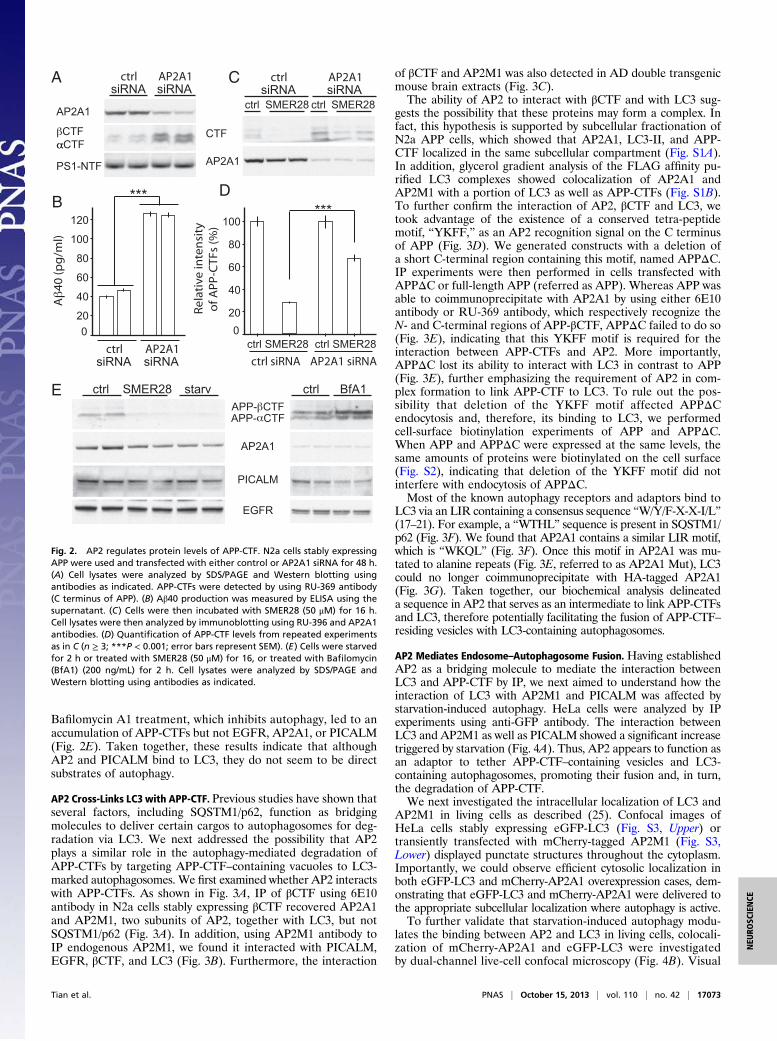
AP2 Mediates Endosome–Autophagosome Fusion. Having establishedAP2 as a bridging molecule to mediate the interaction betweenLC3 and APP-CTF by IP, we next aimed to understand how theinteraction of LC3 with AP2M1 and PICALM was affected bystarvation-induced autophagy. HeLa cells were analyzed by IPexperiments using anti-GFP antibody. The interaction betweenLC3 and AP2M1 as well as PICALM showed a significant increasetriggered by starvation (Fig. 4A). Thus, AP2 appears to function asan adaptor to tether APP-CTF–containing vesicles and LC3-containing autophagosomes, promoting their fusion and, in turn,the degradation of APP-CTF.We next investigated the intracellular localization of LC3 and
AP2M1 in living cells as described (25). Confocal images ofHeLa cells stably expressing eGFP-LC3 (Fig. S3, Upper) ortransiently transfected with mCherry-tagged AP2M1 (Fig. S3,Lower) displayed punctate structures throughout the cytoplasm.Importantly, we could observe efficient cytosolic localization inboth eGFP-LC3 and mCherry-AP2A1 overexpression cases, dem-onstrating that eGFP-LC3 and mCherry-AP2A1 were delivered tothe appropriate subcellular localization where autophagy is active.To further validate that starvation-induced autophagy modu-
lates the binding between AP2 and LC3 in living cells, colocali-zation of mCherry-AP2A1 and eGFP-LC3 were investigatedby dual-channel live-cell confocal microscopy (Fig. 4B). Visual
ctrl AP2A1
AP2A1
CTFCTF
PS1-NTF
siRNA
A)l
m/g
p(04
***
ctrl SMER28 starv ctrlAPP- CTF
AP2A1
PICALM
EGFR
APP- CTF
BfA1
B
AsiRNA
CTF
AP2A1
C ctrl siRNA
AP2A1siRNA
ctrl siRNA
AP2A1siRNA
0
120
100
80
60
40
20
ctrl SMER28 ctrl SMER28
yti snet
nievit al e
R)
%(sFT
C-P
PAf
o***
D
ctrl siRNA AP2A1 siRNA
0
100
80
60
40
20
ctrl SMER28 ctrl SMER28
E
Fig. 2. AP2 regulates protein levels of APP-CTF. N2a cells stably expressingAPP were used and transfected with either control or AP2A1 siRNA for 48 h.(A) Cell lysates were analyzed by SDS/PAGE and Western blotting usingantibodies as indicated. APP-CTFs were detected by using RU-369 antibody(C terminus of APP). (B) Aβ40 production was measured by ELISA using thesupernatant. (C) Cells were then incubated with SMER28 (50 μM) for 16 h.Cell lysates were then analyzed by immunoblotting using RU-396 and AP2A1antibodies. (D) Quantification of APP-CTF levels from repeated experimentsas in C (n ≥ 3; ***P < 0.001; error bars represent SEM). (E) Cells were starvedfor 2 h or treated with SMER28 (50 μM) for 16, or treated with Bafilomycin(BfA1) (200 ng/mL) for 2 h. Cell lysates were analyzed by SDS/PAGE andWestern blotting using antibodies as indicated.
Tian et al. PNAS | October 15, 2013 | vol. 110 | no. 42 | 17073
NEU
ROSC
IENCE
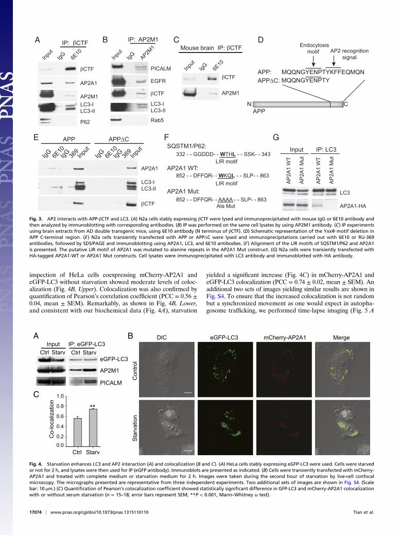
inspection of HeLa cells coexpressing mCherry-AP2A1 andeGFP-LC3 without starvation showed moderate levels of coloc-alization (Fig. 4B, Upper). Colocalization was also confirmed byquantification of Pearson’s correlation coefficient (PCC = 0.56 ±0.04, mean ± SEM). Remarkably, as shown in Fig. 4B, Lower,and consistent with our biochemical data (Fig. 4A), starvation
yielded a significant increase (Fig. 4C) in mCherry-AP2A1 andeGFP-LC3 colocalization (PCC = 0.74 ± 0.02, mean ± SEM). Anadditional two sets of images yielding similar results are shown inFig. S4. To ensure that the increased colocalization is not randombut a synchronized movement as one would expect in autopha-gosome trafficking, we performed time-lapse imaging (Fig. 5 A
IP: CTF
AP2A1
LC3-ILC3-II
CTF
P62
AP2M1
Mouse brain IP: CTF IP: AP2M1
PICALM
EGFR
LC3-ILC3-II
Rab5
BA C D
E
Input
6E10
IgG Input
AP2M1
IgG
CTF
Input
6E10
IgG
CTF
AP2M1
GF
APP:APPC:
APPCN
MQQNGYENPTYKFFEQMQNMQQNGYENPTY
Endocytosis motif AP2 recognition
signal
IgG IgG6E10
369
Input
CTF
AP2A1
LC3-ILC3-II
APP APPC
IgG IgG6E10
369
Input
AP2A1-HA
Input IP: LC3
LC3
SQSTM1/P62:
AP2A1 WT:
AP2A1 Mut:
332 - - GGDDD- - WTHL - - SSK- - 343
852 - - DFFQR- - WKQL - - SLP- - 863
852 - - DFFQR- - AAAA - - SLP- - 863
LIR motif
LIR motif
Ala Mut
AP
2A1
WT
AP
2A1
Mut
AP
2A1
WT
AP
2A1
Mut
Fig. 3. AP2 interacts with APP-βCTF and LC3. (A) N2a cells stably expressing βCTF were lysed and immunoprecipitated with mouse IgG or 6E10 antibody andthen analyzed by immunoblotting with corresponding antibodies. (B) IP was performed on the same cell lysates by using AP2M1 antibody. (C) IP experimentsusing brain extracts from AD double transgenic mice, using 6E10 antibody (N terminus of βCTF). (D) Schematic representation of the YxxФ motif deletion inAPP C-terminal region. (E) N2a cells transiently transfected with APP or APPΔC were lysed and immunoprecipitations carried out with 6E10 or RU-369antibodies, followed by SDS/PAGE and immunoblotting using AP2A1, LC3, and 6E10 antibodies. (F) Alignment of the LIR motifs of SQSTM1/P62 and AP2A1is presented. The putative LIR motif of AP2A1 was mutated to alanine repeats in the AP2A1 Mut construct. (G) N2a cells were transiently transfected withHA-tagged AP2A1-WT or AP2A1 Mut constructs. Cell lysates were immunoprecipitated with LC3 antibody and immunoblotted with HA antibody.
DIC eGFP-LC3 mCherry-AP2A1 Merge
Con
trol
Sta
rvat
ion
BA
C
CtrlIP: eGFP-LC3
StarvInput
Ctrl StarveGFP-LC3
AP2M1
PICALM
Ctrl Starv
Co-
loca
lizat
ion
0.0
0.8
0.6
0.4
1.0
0.2
**
Fig. 4. Starvation enhances LC3 and AP2 interaction (A) and colocalization (B and C). (A) HeLa cells stably expressing eGFP-LC3 were used. Cells were starvedor not for 2 h, and lysates were then used for IP (eGFP antibody). Immunoblots are presented as indicated. (B) Cells were transiently transfected with mCherry-AP2A1 and treated with complete medium or starvation medium for 2 h. Images were taken during the second hour of starvation by live-cell confocalmicroscopy. The micrographs presented are representative from three independent experiments. Two additional sets of images are shown in Fig. S4. (Scalebar: 10 μm.) (C) Quantification of Pearson’s colocalization coefficient showed statistically significant difference in GFP-LC3 and mCherry-AP2A1 colocalizationwith or without serum starvation (n = 15–18; error bars represent SEM; **P < 0.001, Mann–Whitney u test).
17074 | www.pnas.org/cgi/doi/10.1073/pnas.1315110110 Tian et al.

and B). Indeed, we were able to follow the synchronizedmovement of mCherry-AP2A1 and eGFP-LC3 for at least 200 s(Fig. 5C and Movie S1). Taken together, the enhanced in-teraction between LC3 and AP2 as well as PICALM uponinduction of autophagy by starvation provides a mechanisticexplanation as to how autophagy leads to down-regulation ofAPP-CTFs (Fig. S5).
DiscussionOur previous work showed that APP-CTF and Aβ peptides canbe targeted for removal by autophagy in an Atg5-dependentmanner (24). A small molecule enhancer of autophagy (SMER28)promotes this process and reduces the levels of Aβ (24).Autophagy is known to degrade intracellular aggregation-proneproteins. How autophagy reduces membrane-bound APP-CTFand secreted Aβ peptides is largely unknown. The present workidentified AP2 as a mediator that bridges the APP endocyticpathway with the autophagic pathway. AP2 binds to the APP Cterminus during endocytosis and brings APP-CTF to autopha-gosomes via direct binding to LC3 through LIR. Therefore, AP2,functioning as an LC3 receptor, specifically targets APP-CTF toautophagy for degradation. Most APP-βCTF is produced in theendocytic pathway (1). When endocytic APP-βCTF is degradedby autophagy, Aβ levels are greatly reduced. It has been knownthat AP complexes are important for vesicular transport andcargo selection (26). Therefore, it is not surprising to find thatAP2 is used as an LC3 cargo receptor.Recent mounting evidence has indicated that autophagy deg-
radation is more selective than initially thought (27). The sup-porting evidence includes the continuing discovery of specificautophagy receptors that are responsible for recruiting specificcargos to the site of autophagosomes. Thus far, several autoph-agy receptors have been identified, such as SQSTM1/p62, NBR1,Nix, NDP52, and OPTN (17–21). Indeed, it has been shown thatthey regulate the selective degradation of damaged organelles,protein aggregates, and pathogens. Our discovery of AP2 asanother autophagy receptor, which selectively mediates the deg-radation of APP-CTF, further supports the notion of targeted
elimination of unwanted components by autophagy. This studyalso proposes a mechanism by which autophagy is involved in Aβremoval. Impaired or disabled autophagy has been linked tovarious human pathologies, including neurodegenerative dis-eases (28). In our study, we found that PICALM, a knownbinding partner of AP2 involved in clathrin-mediated endocy-tosis, was also recruited to LC3 marked autophagosomes alongwith AP2 and APP-CTF. Because enhanced autophagy increasesthe binding of PICALM to autophagosomes, we speculate thatPICALM might have an important function in the clearance ofAPP-βCTF and, in turn, in the clearance of Aβ via autophagy.However, many questions remain to be resolved, includingthe precise role of PICALM in the bridging of APP-CTF toautophagy. This work, together with the fact that PICALM wasidentified as a risk factor for AD by GWAS (10), highlights thecrucial role of PICALM in APP metabolism and opens a thera-peutic avenue for AD intervention.
Materials and MethodsReagents. Antibodies were diluted 1:1,000 in 5% (wt/vol) milk unless speci-fied. Commercially available antibodies are listed in SI Materials and Meth-ods. RU-369, a rabbit polyclonal antibody that recognizes the C-terminal ofAPP695 (29); Ab14 antiserum targeting residues 1–25 of PS1-NTF (30); andmonoclonal GFP antibody was produced by the monoclonal antibody corefacility at Memorial Sloan–Kettering Cancer Center. Compound SMER28 waspurchased from EMD Chemicals.
Cell Culture and siRNA. N2a cells stably expressing APP were maintained inmedium containing 50%DMEM and 50%Opti-MEM, supplemented with 5%FBS (Invitrogen) plus 400 μg/mL geneticin. HeLa cells stably expressing eGFP-LC3 were maintained in DMEM with 10% FBS plus 400 μg/mL geneticin. ThesiRNA for AP2A1 was purchased from Dharmacon (On-TARGETplus Set of4 siRNAs J-055895-05). The control siRNA was purchased from Dharmacon(On-TARGET plus GAPD Control siRNA D-001830-02-05).
Coimmunoprecipitation. All coimmunoprecipitation experiments using celllysates or brain lysates were performed by using the co-ImmunoprecipitationKit from Invitrogen (Invitrogen) according to manufacturer’s protocol.Briefly, antibodies conjugated to Dynabeads were incubated with lysatessolubilized in a lysis buffer [0.5% Triton (Sigma-Aldrich, St. Louis)] for 30 min.
Fluo
resc
ence
(A. U
.)
BA
C
eGFP-LC3 MergemCherry-AP2A1 Distance (µm)
0 2 4 6 8 10
20
0
40
60
80
100
0 5 10 20 30 50 100 200 (S)
Fig. 5. Starvation-induced autophagy mediates LC3 and AP2 colocalization with time-scale on the order of a few hundred seconds. (A) Representative imagesof live HeLa cells expressing eGFP-LC3 and mCherry-AP2A1 upon 1-h serum starvation treatment. The white box highlights the colocalization of eGFP-LC3 andmCherry-AP2A1. (Scale bar: 10 μm.) (B) Fluorescent intensity profiles of the arrow in the white box indicate the position of a line scan, where two of threemCherry-AP2A1 (red line) punctate structures displayed high colocalization with eGFP-LC3 (green line). (C) Time-lapse images of the white box (A) showing thateGFP-LC3 and mCherry-AP2A1 present an increased colocalization following starvation. Note that the synchronized movement of eGFP-LC3 and mCherry-AP2A1(dashed circles) lasted for at least 200 s. (Scale bar: 5 μm.)
Tian et al. PNAS | October 15, 2013 | vol. 110 | no. 42 | 17075
NEU
ROSC
IENCE
Subcellular Fractionation. For sucrose density gradient fractionation, cellswere prepared as described (31). Briefly, cells were homogenized by usinga stainless steel ball-bearing homogenizer. Homogenates were loaded ontop of a step sucrose gradient (1 mL at 2 M, 4 mL at 1.3 M, 3.5 mL at 1.16 M,and 2.0 mL at 0.8 M). Gradients were centrifuged (2.5 h at 390,000 × g), and1-mL fractions were collected and assayed by Western blot.
Microscopy. Live-cell confocal images were obtained on a Leica TCS SP8confocal imaging system equipped with a 63×/1.4 numerical aperture oil-immersion objective lens. The temperature-controlled stage of the confocalmicroscope was maintained at 32–35 °C. Cells for live-cell imaging wereseeded on poly-D-lysine–coated MatTek glass bottom culture dishes. Mi-croscopy setup and imaging acquisition were performed as described (32).Time-lapse fluorescent images were acquired at 5-s intervals, following themovement of eGFP-LC3 and mCherry-AP2A1. To minimize photodamage
and photobleaching, we restricted time-lapsed confocal imaging to a periodof 300 s.
Statistical Analyses for Colocalization. A minimum of 15 sets of micrographsfrom three independent experiments were used for colocalization analyses.Pearson’s colocalization coefficient was calculated by using Image-Pro Plus(Version 6.0). Mann–Whitney test was performed to determine statisticalsignificance. Data were analyzed in SigmaPlot (Version 11.0).
ACKNOWLEDGMENTS. We thank Drs. Kaye Thomas and Alison North fortechnical assistance; Drs. Aviva Tolkovsky and Zhengyu Yue for providing theHeLa eGFP-LC3 cell line; and Drs. Victor Bustos, John Steele, and WilliamNetzer for critical discussion. This work was supported by the Fisher Centerfor Alzheimer’s Research Foundation and National Institutes of Health GrantAG09464 (to P.G. and M.F.).
1. Haass C, Kaether C, Thinakaran G, Sisodia S (2012) Trafficking and proteolytic pro-cessing of APP. Cold Spring Harb Perspect Med 2(5):a006270.
2. Perez RG, et al. (1999) Mutagenesis identifies new signals for beta-amyloid precursorprotein endocytosis, turnover, and the generation of secreted fragments, includingAbeta42. J Biol Chem 274(27):18851–18856.
3. Koo EH, Squazzo SL, Selkoe DJ, Koo CH (1996) Trafficking of cell-surface amyloidbeta-protein precursor. I. Secretion, endocytosis and recycling as detected by labeledmonoclonal antibody. J Cell Sci 109(Pt 5):991–998.
4. Canfield WM, Johnson KF, Ye RD, Gregory W, Kornfeld S (1991) Localization of thesignal for rapid internalization of the bovine cation-independent mannose 6-phos-phate/insulin-like growth factor-II receptor to amino acids 24-29 of the cytoplasmictail. J Biol Chem 266(9):5682–5688.
5. Jadot M, Canfield WM, Gregory W, Kornfeld S (1992) Characterization of the signalfor rapid internalization of the bovine mannose 6-phosphate/insulin-like growthfactor-II receptor. J Biol Chem 267(16):11069–11077.
6. Ohno H, et al. (1995) Interaction of tyrosine-based sorting signals with clathrin-associated proteins. Science 269(5232):1872–1875.
7. Dreyling MH, et al. (1996) The t(10;11)(p13;q14) in the U937 cell line results in thefusion of the AF10 gene and CALM, encoding a new member of the AP-3 clathrinassembly protein family. Proc Natl Acad Sci USA 93(10):4804–4809.
8. Tebar F, Bohlander SK, Sorkin A (1999) Clathrin assembly lymphoid myeloid leukemia(CALM) protein: Localization in endocytic-coated pits, interactions with clathrin, andthe impact of overexpression on clathrin-mediated traffic. Mol Biol Cell 10(8):2687–2702.
9. Meyerholz A, et al. (2005) Effect of clathrin assembly lymphoid myeloid leukemiaprotein depletion on clathrin coat formation. Traffic 6(12):1225–1234.
10. Harold D, et al. (2009) Genome-wide association study identifies variants at CLU andPICALM associated with Alzheimer’s disease. Nat Genet 41(10):1088–1093.
11. Axe EL, et al. (2008) Autophagosome formation from membrane compartments en-riched in phosphatidylinositol 3-phosphate and dynamically connected to the endo-plasmic reticulum. J Cell Biol 182(4):685–701.
12. Hayashi-Nishino M, et al. (2010) Electron tomography reveals the endoplasmic re-ticulum as a membrane source for autophagosome formation. Autophagy 6(2):301–303.
13. Ylä-Anttila P, Vihinen H, Jokitalo E, Eskelinen EL (2009) 3D tomography revealsconnections between the phagophore and endoplasmic reticulum. Autophagy 5(8):1180–1185.
14. Hailey DW, et al. (2010) Mitochondria supply membranes for autophagosome bio-genesis during starvation. Cell 141(4):656–667.
15. Ravikumar B, Moreau K, Jahreiss L, Puri C, Rubinsztein DC (2010) Plasma membranecontributes to the formation of pre-autophagosomal structures. Nat Cell Biol 12(8):747–757.
16. Ohsumi Y, Mizushima N (2004) Two ubiquitin-like conjugation systems essential for
autophagy. Semin Cell Dev Biol 15(2):231–236.17. Kirkin V, et al. (2009) A role for NBR1 in autophagosomal degradation of ubiquiti-
nated substrates. Mol Cell 33(4):505–516.18. Mostowy S, et al. (2011) p62 and NDP52 proteins target intracytosolic Shigella and
Listeria to different autophagy pathways. J Biol Chem 286(30):26987–26995.19. Novak I, et al. (2010) Nix is a selective autophagy receptor for mitochondrial clear-
ance. EMBO Rep 11(1):45–51.20. Pankiv S, et al. (2007) p62/SQSTM1 binds directly to Atg8/LC3 to facilitate degradation
of ubiquitinated protein aggregates by autophagy. J Biol Chem 282(33):24131–24145.21. Wild P, et al. (2011) Phosphorylation of the autophagy receptor optineurin restricts
Salmonella growth. Science 333(6039):228–233.22. Vingtdeux V, et al. (2011) Novel synthetic small-molecule activators of AMPK
as enhancers of autophagy and amyloid-β peptide degradation. FASEB J 25(1):
219–231.23. Spilman P, et al. (2010) Inhibition of mTOR by rapamycin abolishes cognitive deficits
and reduces amyloid-beta levels in a mouse model of Alzheimer’s disease. PLoS ONE
5(4):e9979.24. Tian Y, Bustos V, Flajolet M, Greengard P (2011) A small-molecule enhancer of au-
tophagy decreases levels of Abeta and APP-CTF via Atg5-dependent autophagy
pathway. FASEB J 25(6):1934–1942.25. Bampton ET, Goemans CG, Niranjan D, Mizushima N, Tolkovsky AM (2005) The dy-
namics of autophagy visualized in live cells: From autophagosome formation to fu-
sion with endo/lysosomes. Autophagy 1(1):23–36.26. Nakatsu F, Ohno H (2003) Adaptor protein complexes as the key regulators of protein
sorting in the post-Golgi network. Cell Struct Funct 28(5):419–429.27. Fimia GM, Kroemer G, Piacentini M (2013) Molecular mechanisms of selective au-
tophagy. Cell Death Differ 20(1):1–2.28. Wolfe DM, et al. (2013) Autophagy failure in Alzheimer’s disease and the role of
defective lysosomal acidification. Eur J Neurosci 37(12):1949–1961.29. Netzer WJ, et al. (2010) Lowering beta-amyloid levels rescues learning and memory in
a Down syndrome mouse model. PLoS ONE 5(6):e10943.30. Thinakaran G, et al. (1996) Endoproteolysis of presenilin 1 and accumulation of
processed derivatives in vivo. Neuron 17(1):181–190.31. Wang H, et al. (2004) Presenilins and gamma-secretase inhibitors affect intracellular
trafficking and cell surface localization of the gamma-secretase complex components.
J Biol Chem 279(39):40560–40566.32. Chang JC, et al. (2012) Single molecule analysis of serotonin transporter regulation
using antagonist-conjugated quantum dots reveals restricted, p38 MAPK-dependent
mobilization underlying uptake activation. J Neurosci 32(26):8919–8929.
17076 | www.pnas.org/cgi/doi/10.1073/pnas.1315110110 Tian et al.