Adaptive Changes in Early and Late Blind: A fMRI Study of...
19
Adaptive Changes in Early and Late Blind: A fMRI Study of Braille Reading H. BURTON, 1,3 A. Z. SNYDER, 2,3 T. E. CONTURO, 3 E. AKBUDAK, 3 J. M. OLLINGER, 3 AND M. E. RAICHLE 1–3 1 Department of Anatomy and Neurobiology, 2 Department of Neurology and Neurological Surgery, and 3 Department of Radiology, Washington University School of Medicine, St. Louis, Missouri 63110 Received 5 April 2001; accepted in final form 20 September 2001 Burton, H., A. Z. Snyder, T. E. Conturo, E. Akbudak, J. M. Ollinger, and M. E. Raichle. Adaptive changes in early and late blind: a fMRI study of Braille reading. J Neurophysiol 87: 589 – 607, 2002; 10.1152/jn. 00285.2001. Braille reading depends on remarkable adaptations that connect the somatosensory system to language. We hypothesized that the pattern of cortical activations in blind individuals reading Braille would reflect these adaptations. Activations in visual (occipital-temporal), fron- tal-language, and somatosensory cortex in blind individuals reading Braille were examined for evidence of differences relative to previously reported studies of sighted subjects reading print or receiving tactile stimulation. Nine congenitally blind and seven late-onset blind subjects were studied with fMRI as they covertly performed verb generation in response to reading Braille embossed nouns. The control task was reading the nonlexical Braille string “######”. This study emphasized image analysis in individual subjects rather than pooled data. Group differences were examined by comparing magnitudes and spatial extent of activated regions first determined to be significant using the general linear model. The major adaptive change was robust activation of visual cortex despite the complete absence of vision in all subjects. This included foci in peri-calcarine, lingual, cuneus and fusiform cortex, and in the lateral and superior occipital gyri encompassing primary (V1), secondary (V2), and higher tier (VP, V4v, LO and possibly V3A) visual areas previously identified in sighted subjects. Subjects who never had vision differed from late blind subjects in showing even greater activity in occipital- temporal cortex, provisionally corresponding to V5/MT and V8. In addition, the early blind had stronger activation of occipital cortex located contralateral to the hand used for reading Braille. Responses in frontal and parietal cortex were nearly identical in both subject groups. There was no evidence of modifications in frontal cortex language areas (infe- rior frontal gyrus and dorsolateral prefrontal cortex). Surprisingly, there was also no evidence of an adaptive expansion of the somatosensory or primary motor cortex dedicated to the Braille reading finger(s). Lack of evidence for an expected enlargement of the somatosensory representa- tion may have resulted from balanced tactile stimulation and gross motor demands during Braille reading of nouns and the control fields. Extensive engagement of visual cortex without vision is discussed in reference to the special demands of Braille reading. It is argued that these responses may represent critical language processing mechanisms normally present in visual cortex. INTRODUCTION A growing body of work suggests that blind individuals use areas of the cerebral cortex normally reserved for vision during Braille reading and other nonvisual tasks involving tactile discrimination. Initial evidence appeared in functional neuro- imaging studies with positron emission tomography (PET) (Sadato et al. 1996, 1998) and experiments using transcranial magnetic stimulation (TMS) of occipital cortex (Cohen et al. 1997, 1999). Several questions remain especially concerning differences between early blind subjects, i.e., persons blind since birth or early childhood, versus persons who lost sight after having learned to read print. Specifically, occipital TMS disrupted Braille reading and tactile discrimination of em- bossed capital letters (Cohen et al. 1997, 1999). These effects occurred only in early blind individuals. Sadato and colleagues also reported blood flow increases in both striate and extrastri- ate visual cortex induced by performance of similar tactile tasks in early but not late blind individuals (Sadato et al. 1996, 1998). Such differences are possibly predictable given the known developmental dependence of the visual system on experience during the early critical period. Another PET study, however, found activation of extrastriate cortex in early blind, but striate cortex in late blind subjects during a language task incorporating Braille reading (Bu ¨ chel et al. 1998a). The present work used functional magnetic resonance imaging (fMRI) to study the effect of age at onset of blindness on visual cortex responses during performance of a language task involving Braille reading. We especially examined possible differences in activation of primary (e.g., striate cortex) and higher tier (e.g., extrastriate) visual areas in subjects with early versus late onset blindness. Another important question is whether blood flow changes in visual cortex of blind individuals reflect specific function- ality. An alternative interpretation is that these responses are nonspecific excessive modulations consequent to early sensory deprivation. This view garners some support from finding of above normal metabolic rates for glucose in visual cortices of early blind subjects (De Volder et al. 1997; Wanet-Defalque et al. 1988). In addition, absent specificity or diversity of func- tions has been proposed to explain recordings of slow negative potentials over visual cortex of blind subjects during an atten- tion or arousing task that was unrelated to reading (Roder et al. 1997). Early blindness might leave visual cortex immature and Address for reprint requests: H. Burton, Dept. of Anatomy and Neurobiol- ogy, Washington University School of Medicine, 660 S. Euclid Ave., St. Louis, MO 63110 (E-mail: [email protected]). The costs of publication of this article were defrayed in part by the payment of page charges. The article must therefore be hereby marked ‘‘advertisement’’ in accordance with 18 U.S.C. Section 1734 solely to indicate this fact. J Neurophysiol 87: 589 – 607, 2002; 10.1152/jn.00285.2001. 589 0022-3077/02 $5.00 Copyright © 2002 The American Physiological Society www.jn.org
Transcript of Adaptive Changes in Early and Late Blind: A fMRI Study of...
Adaptive Changes in Early and Late Blind: A fMRI Study ofBraille Reading
H. BURTON,1,3 A. Z. SNYDER,2,3 T. E. CONTURO,3 E. AKBUDAK,3 J. M. OLLINGER,3 ANDM. E. RAICHLE1–3
1Department of Anatomy and Neurobiology, 2Department of Neurology and Neurological Surgery, and 3Department ofRadiology, Washington University School of Medicine, St. Louis, Missouri 63110
Received 5 April 2001; accepted in final form 20 September 2001
Burton, H., A. Z. Snyder, T. E. Conturo, E. Akbudak, J. M. Ollinger,and M. E. Raichle. Adaptive changes in early and late blind: a fMRIstudy of Braille reading.J Neurophysiol 87: 589–607, 2002; 10.1152/jn.00285.2001. Braille reading depends on remarkable adaptations thatconnect the somatosensory system to language. We hypothesized that thepattern of cortical activations in blind individuals reading Braille wouldreflect these adaptations. Activations in visual (occipital-temporal), fron-tal-language, and somatosensory cortex in blind individuals readingBraille were examined for evidence of differences relative to previouslyreported studies of sighted subjects reading print or receiving tactilestimulation. Nine congenitally blind and seven late-onset blind subjectswere studied with fMRI as they covertly performed verb generation inresponse to reading Braille embossed nouns. The control task was readingthe nonlexical Braille string “######”. This study emphasized imageanalysis in individual subjects rather than pooled data. Group differenceswere examined by comparing magnitudes and spatial extent of activatedregions first determined to be significant using the general linear model.The major adaptive change was robust activation of visual cortex despitethe complete absence of vision in all subjects. This included foci inperi-calcarine, lingual, cuneus and fusiform cortex, and in the lateral andsuperior occipital gyri encompassing primary (V1), secondary (V2), andhigher tier (VP, V4v, LO and possibly V3A) visual areas previouslyidentified in sighted subjects. Subjects who never had vision differedfrom late blind subjects in showing even greater activity in occipital-temporal cortex, provisionally corresponding to V5/MT and V8. Inaddition, the early blind had stronger activation of occipital cortex locatedcontralateral to the hand used for reading Braille. Responses in frontaland parietal cortex were nearly identical in both subject groups. Therewas no evidence of modifications in frontal cortex language areas (infe-rior frontal gyrus and dorsolateral prefrontal cortex). Surprisingly, therewas also no evidence of an adaptive expansion of the somatosensory orprimary motor cortex dedicated to the Braille reading finger(s). Lack ofevidence for an expected enlargement of the somatosensory representa-tion may have resulted from balanced tactile stimulation and gross motordemands during Braille reading of nouns and the control fields. Extensiveengagement of visual cortex without vision is discussed in reference tothe special demands of Braille reading. It is argued that these responsesmay represent critical language processing mechanisms normally presentin visual cortex.
I N T R O D U C T I O N
A growing body of work suggests that blind individuals useareas of the cerebral cortex normally reserved for vision during
Braille reading and other nonvisual tasks involving tactilediscrimination. Initial evidence appeared in functional neuro-imaging studies with positron emission tomography (PET)(Sadato et al. 1996, 1998) and experiments using transcranialmagnetic stimulation (TMS) of occipital cortex (Cohen et al.1997, 1999). Several questions remain especially concerningdifferences between early blind subjects, i.e., persons blindsince birth or early childhood, versus persons who lost sightafter having learned to read print. Specifically, occipital TMSdisrupted Braille reading and tactile discrimination of em-bossed capital letters (Cohen et al. 1997, 1999). These effectsoccurred only in early blind individuals. Sadato and colleaguesalso reported blood flow increases in both striate and extrastri-ate visual cortex induced by performance of similar tactiletasks in early but not late blind individuals (Sadato et al. 1996,1998). Such differences are possibly predictable given theknown developmental dependence of the visual system onexperience during the early critical period. Another PET study,however, found activation of extrastriate cortex in early blind,but striate cortex in late blind subjects during a language taskincorporating Braille reading (Bu¨chel et al. 1998a). The presentwork used functional magnetic resonance imaging (fMRI) tostudy the effect of age at onset of blindness on visual cortexresponses during performance of a language task involvingBraille reading. We especially examined possible differencesin activation of primary (e.g., striate cortex) and higher tier(e.g., extrastriate) visual areas in subjects with early versus lateonset blindness.
Another important question is whether blood flow changesin visual cortex of blind individuals reflect specific function-ality. An alternative interpretation is that these responses arenonspecific excessive modulations consequent to early sensorydeprivation. This view garners some support from finding ofabove normal metabolic rates for glucose in visual cortices ofearly blind subjects (De Volder et al. 1997; Wanet-Defalque etal. 1988). In addition, absent specificity or diversity of func-tions has been proposed to explain recordings of slow negativepotentials over visual cortex of blind subjects during an atten-tion or arousing task that was unrelated to reading (Roder et al.1997). Early blindness might leave visual cortex immature and
Address for reprint requests: H. Burton, Dept. of Anatomy and Neurobiol-ogy, Washington University School of Medicine, 660 S. Euclid Ave., St.Louis, MO 63110 (E-mail: [email protected]).
The costs of publication of this article were defrayed in part by the paymentof page charges. The article must therefore be hereby marked ‘‘advertisement’’in accordance with 18 U.S.C. Section 1734 solely to indicate this fact.
J Neurophysiol87: 589–607, 2002; 10.1152/jn.00285.2001.
5890022-3077/02 $5.00 Copyright © 2002 The American Physiological Societywww.jn.org

prone to abnormal responses (Snyder and Shapley 1979) be-cause of absent pruning of normally expressed exuberant syn-apses, and an excess of retained excitatory connections (Roderet al. 1997). We attempted to address this question by carefullycorrelating the distribution of active cortex with detailed anal-yses of the underlying anatomy. Specificity may be inferred byshowing a close correspondence between active regions andanatomy. To achieve this goal, the protocol was designed toprovide statistically reliable results within individual subjects,thereby allowing optimal inspection of the anatomy of activefoci.
A second goal was to study the correspondence betweenactive foci in occipital cortex of blind individuals and themultiple visual areas of sighted subjects (Felleman and VanEssen 1991). FMRI studies in humans have reinforced a modelof the visual cortex consisting of a distributed network ofspecialized regions each with its own functions (DeYoe et al.1996; Dumoulin et al. 2000; Engel et al. 1997; Hadjikhani et al.1998; Sereno et al. 1995; Tootell et al. 1996, 1997). Findingevidence of activity circumscribed to anatomically distinctportions of visual cortex in blind individuals might suggestfunctional specialization like that attributed to correspondingregions in sighted individuals. The presence of separable fociprovides evidence that activity in visual cortex of blind peopleis specific as opposed to a pathological consequence of visualdeprivation. Detailed comparisons were imperfect in previousneuroimaging studies because the PET-based data relied onaverages across subjects, which limit the correlation of corticalanatomy with activation patterns.
To view all potentially affected cortical areas, we employedwhole brain scanning without a priori selection of particularregions of interest. We also used a repeated measures designwith enough trials to obtain sufficient statistical power todelineate significant activity patterns in individual subjects. Anemphasis on within subject analyses is especially importantdue to differences in Braille reading fluency, education, chro-nological age, and age of blindness. For example, potentialdifferences in Braille reading strategies between early versuslate blind individuals might affect the distribution of activity invisual cortex. The obtained, high resolution correspondencebetween anatomy and activated regions from within subjectanalyses also aided comparisons between these data and pre-viously identified, and functionally interpreted, foci from manystudies in sighted subjects.
Another addressed question concerns the cortical represen-tation of language in blind individuals and the possible depen-dence of this representation on age at onset of blindness. Wehypothesized that the same functional anatomy should be seenin blind and sighted individuals because the lexical and seman-tic aspects of language should be comparable irrespective ofthe sensory channel used to convey orthographic information.In sighted individuals, where language tasks involve similarorthographic-lexical operations, different laboratories concurin identifying activity increases in discrete areas in left inferiorand dorsolateral frontal cortex (Binder et al. 1997; Demonet etal. 1994; Fiez et al. 1995; Kelly et al. 1998; Klein et al. 1995;McCarthy et al. 1993; Paulesu et al. 1997; Petersen and Fiez1993; Phelps et al. 1997; Poldrack et al. 1999; Rumsey et al.1997; Zatorre et al. 1996). Tasks dominated by phonologicalfeatures activate posterior language areas, especially left pari-
eto-temporal and superior temporal regions (Buchel et al.1998; Rumsey et al. 1997).
Several factors, however, might lead to variations in acti-vated language areas in blind people. First, reading by touchdramatically differs from reading of print (Millar 1997). Forexample, most Braille readers use two hands, which mightinfluence the left dominance of language areas in blind indi-viduals. Countervailing this possibility is that only one handreads while the other acts as a place marker (Millar 1997).Second, phonological associations are the only way to learnBraille without sight, which might be reflected by adaptivechanges in the language areas dominated by phonics. Thischange, however, possibly manifests only in individuals withearly onset blindness because they never have rememberedvisual associations with letter shapes when learning Braillethrough phonics. Countering the notion of possible differencesin the activation of the phonologically dominant language areasis that fluent Braille reading does not involve phonologicalcoding (Millar 1984, 1987; Nolan and Kederis 1969; Pring1985, 1994).
We selected verb generation for Braille nouns as the lan-guage paradigm because this task has been extensively studiedin sighted subjects reading print (see reviews in Gabrieli et al.1998; Seger et al. 1999) and provides a potent language task inthe broadest sense. Generating verbs for presented nouns incomparison to a nonlexical or minimal language control stim-ulus reliably produces robust functional responses. We chosenot to use a multiple level language task design with a factorialparadigm of paired contrasts (i.e., reading words with andwithout verb generation, reading pseudo-words, etc). The ob-jective of this study was to assess functional reorganization dueto long term blindness. It was not an experiment to study theorganization of language processing.
We also considered whether changes occurred in the acti-vated extent or components of the somatosensory system be-cause of the intense dependence on tactile perceptions whenreading by touch. We hypothesized expansion of the represen-tation for the Braille reading finger in the somatosensory cortexgiven prior evidence of remarkable plasticity in this cortex(Merzenich and Jenkins 1993; Pons et al. 1991; Ramachandran1996, 1998). Countering this idea was a study which showedthat blind individuals had greater difficulty detecting nearthreshold tactile stimulation of adjacent digit tips normallyused in reading Braille. They interpreted this as evidence of adisorganized representation for these digits in somatosensorycortex (Sterr et al. 1998).
For corollary reasons that Braille reading involves substan-tial use of fine finger movements, we hypothesized expansionof the finger-hand area in the cortical motor areas. A priorstudy with TMS found a use dependent expansion of a lowerthreshold region over the motor representation of the readingfingers in early blind, fluent Braille readers (Pascual-Leone etal. 1995). In addition, due to differences in reading skillsbetween most early and late blind individuals, we hypothesizeddistinctions in the extent and nature of activated cortical motorareas in these two groups. Less proficient Braille readers pre-sumably attend to global-holistic letter or word shapes, infor-mation obtained through more frequent and sequential trape-zoidal shaped up/down movements across Braille cells (Millar1984, 1987; Nolan and Kederis 1969; Pring 1985, 1994). The
590 BURTON, SNYDER, CONTURO, AKBUDAK, OLLINGER, AND RAICHLE
J Neurophysiol • VOL 87 • JANUARY 2002 • www.jn.org
lowest level operation during fluent Braille reading involvesprocessing lateral dot-gap shearing density within individualBraille cells (Millar 1987; Pring 1994). This information arisesthrough distinctively smooth, continuous movements acrossthe Braille field. Thus differences in motor cortex activationsmight reflect these distinguishing motor behaviors in early andlate blind Braille readers.
M E T H O D S
Subjects
Sixteen blind, proficient Braille readers from the greater St. Louiscommunity volunteered and were paid for their participation. Subjectsprovided informed consent in accordance with guidelines approved bythe Human Studies Committee of Washington University. We ob-tained a detailed neurologic history in each subject using a standard-ized questionnaire. Only neurologically normal (excepting visualfunction) subjects were scanned. We report here functional data onlyfrom subjects with normal anatomical MR images. Nine (4 female, 5male; aged 34–67, Avg 44.78) were early-blind having no sight atbirth or by 5 yr of age. Seven (4 female, 3 male; aged 36–66, Avg49.14) were late blind having lost sight after an average age of 12.7 yr(range 10–25); one subject retained sufficient vision to read largeprint.
We assessed handedness with a modified Edinburgh handednessinventory (previously validated questions 1,2,5,7,11,15 and 23 inRaczkowski et al. 1974). When required to read Braille with one hand,all but one early and one late blind subject used their right hand. Onlythe early blind left-hand reader was left-handed.
All subjects had read Braille for more than a decade (Table 1) andmany currently did so one or more hours daily. Reading proficiencywas measured using a standard 266 word Braille text. Early-blindsubjects read more rapidly on average than late-blind (Table 1).Allowances for these different reading speeds were made during theMR scans (see following text).
Task and test apparatus
Single words and control fields in Braille II spelling were presentedto subjects using a MR compatible device. Braille-embossed paperwas threaded through an extended, two-chamber Plexiglas box (ap-
proximately 8 feet �1 ft �4 inches) supported over the subjects asthey lay in the scanner. The stimuli were organized as a single columnof words and control fields segmented into three practice and eight testruns. Each fMRI run contained 128 stimuli: 8 control Braille fieldsfollowed by 20 groups of three words followed by three controls (seefollowing text). The paper was manually advanced in synchrony withscanner frames (Fig. 1) to present each Braille field in a 3 by 1 inchreading window suspended over the subject’s waist.
During practice trials the subjects learned to complete the followingsequence taking 3–5 s per Braille field. The reading finger(s) of thepreferred hand rested on smooth paper at the left edge of the readingwindow. Reading was initiated on sensing the end of paper movementby moving the finger rightward. Braille embossing started 4 cm from
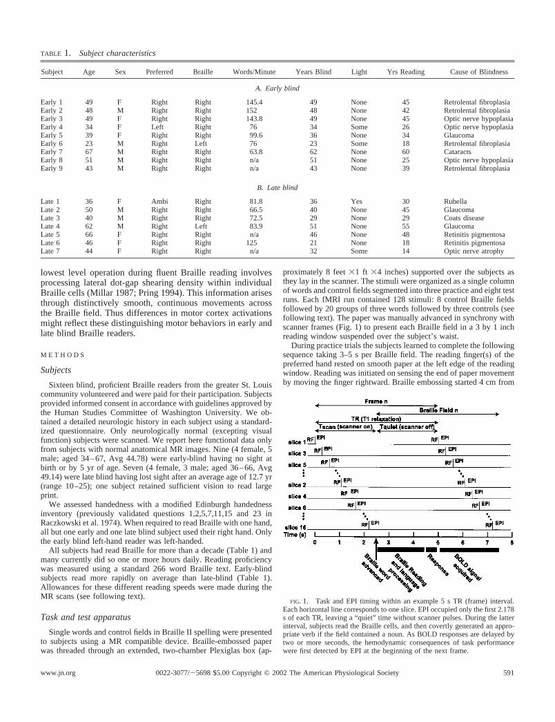
TABLE 1. Subject characteristics
Subject Age Sex Preferred Braille Words/Minute Years Blind Light Yrs Reading Cause of Blindness
A. Early blind
Early 1 49 F Right Right 145.4 49 None 45 Retrolental fibroplasiaEarly 2 48 M Right Right 152 48 None 42 Retrolental fibroplasiaEarly 3 49 F Right Right 143.8 49 None 45 Optic nerve hypoplasiaEarly 4 34 F Left Right 76 34 Some 26 Optic nerve hypoplasiaEarly 5 39 F Right Right 99.6 36 None 34 GlaucomaEarly 6 23 M Right Left 76 23 Some 18 Retrolental fibroplasiaEarly 7 67 M Right Right 63.8 62 None 60 CataractsEarly 8 51 M Right Right n/a 51 None 25 Optic nerve hypoplasiaEarly 9 43 M Right Right n/a 43 None 39 Retrolental fibroplasia
B. Late blind
Late 1 36 F Ambi Right 81.8 36 Yes 30 RubellaLate 2 50 M Right Right 66.5 40 None 45 GlaucomaLate 3 40 M Right Right 72.5 29 None 29 Coats diseaseLate 4 62 M Right Left 83.9 51 None 55 GlaucomaLate 5 66 F Right Right n/a 46 None 48 Retinitis pigmentosaLate 6 46 F Right Right 125 21 None 18 Retinitis pigmentosaLate 7 44 F Right Right n/a 32 Some 14 Optic nerve atrophy
FIG. 1. Task and EPI timing within an example 5 s TR (frame) interval.Each horizontal line corresponds to one slice. EPI occupied only the first 2.178s of each TR, leaving a “quiet” time without scanner pulses. During the latterinterval, subjects read the Braille cells, and then covertly generated an appro-priate verb if the field contained a noun. As BOLD responses are delayed bytwo or more seconds, the hemodynamic consequences of task performancewere first detected by EPI at the beginning of the next frame.
5910022-3077/�5698 $5.00 Copyright © 2002 The American Physiological Societywww.jn.org

the left edge of the reading window. After reading and appropriateresponding, the hand returned to the left edge of the reading windowto wait for the next stimulus. We instructed the subjects always totouch the entire Braille field even if they identified the contents priorto the end. There were no restrictions on Braille reading strategiesexcept to use only one hand and minimize arm movements. Mostsubjects made one pass across most words with their right index fingerwhile abducting the wrist. Re-reading occurred occasionally.
The word stimuli were concrete and abstract nouns (mean wordlength � 5.8 letters). The control stimulus was the Braille pattern forsix number signs (“######” ). The task was generation of a compatibleverb for each noun (e.g., “bake” for “cake” ). Explicit instructions toread the noun and generate a verb were given to the subjects beforeeach run. The 480 nouns were selected to maximize variety ofcompatible verbs without regard to word frequency. The list wassimilar to one used previously (Snyder et al. 1995). Each nounoccurred once to minimize practice effects. The explicit instruction forthe control stimulus was “ to empty your mind and think of nothing.”[Subjects understood this instruction to mean that they were not toperform a lexical task in response to the control field.] As the samecontrol field was repeated throughout the study, and as all fluentBraille readers understand that the Braille symbol for “#” normallyprecedes numbers, identifying the control task was trivial for thesereaders. They instantly recognized the control field and proceeded totouch it as they would a regular word but without doing a lexicallanguage task. In practice sessions subjects read and respondedovertly. During fMRI the responses were covert.
The control field (“######” ) was designed to balance the wordstimuli in somatosensory content and gross motor demands. Readingboth types of stimuli required the same orienting of the reading fingerin the display window, initiation of hand movements in response topaper advancement, and attention to the spatial extent of the Braillefield. The processing engendered by the control field was likelyautomatic as this stimulus was presented throughout the experiment.During the practice runs it was observed that the late-blind subjectsmade more micro-movements over words than the control fields(Millar 1997). The early blind subjects usually read all fields using thesame smooth motion.
Confining the paradigm to a single contrast ensured that the quan-tity of fMRI data obtained in each subject had statistical powersufficient for image analyses within individuals. This objective wasimportant because variability in Braille reading skills, education, andage of the two groups of blind individuals might have compromisedanalyses based solely on averaged images. The least quantity of datacollected from a given subject was 80 trials (1 trial � three generateframes � three control frames). Most subjects provided twice thisamount.
MRI acquisition
Functional MR scans (fMRI) were collected on a Siemens 1.5 TeslaVision scanner, using a custom, single-shot asymmetric spin-echo,echo-planar (EPI) sequence sensitive to blood oxygenation level-dependent (BOLD) contrast. We used a 64 � 64 image matrix, oversampled to reduce noise, blipped readout (Howseman et al. 1988) anddirect 2D-FT reconstruction. The field of view was 240 mm (3.75 �3.75 mm in-plane pixels). This maximized signal to noise sensitivityat a T2* evolution time of 50 ms from a flip angle � 90° (Conturo etal. 1996; Ogawa et al. 1990). Whole brain coverage was obtained with16 contiguous 8 mm slices. Reconstruction, transfer, and storagefollowing a 128-frame fMRI acquisition run took 2 min. Up to 8 fMRIruns were acquired in a 2.5 h session including anatomical imaging.We held EPI to 2.178 s to minimize the effects of inflowing blood,head movement (Friston et al. 1996), and to allow a sufficiently longquiet interval (see Fig. 1) for Braille reading.
EPI occupied only the first 2.178 s of each frame (Fig. 1) leaving
the remaining time quiet during which the subjects read the Braillefield and covertly responded. The induced BOLD responses weredetected in subsequent frames. In most subjects the frame TR was 5 s.We extended the frame TR to not more than 7 s according to thecapacity of the slower readers to keep up with the task.1 This ensuredthat each subject completed word reading before the start of the nextframe. The inequality of TR over subjects precluded estimation ofaveraged event related response time courses. Nevertheless, the re-sponse profiles (intensity as a function of frame) were similar in allsubjects (see Fig. 7) although the trials were of unequal duration (i.e.,30 to 42 s).
PrefMRI structural imaging included a coarse (2 mm cubic voxel,79 s scan) magnetization prepared rapid gradient echo (MP-RAGE)scan which was used to automatically compute standard fMRI sliceprescriptions parallel to the anterior commissure-posterior commis-sure plane. A fast T2-weighted spin echo (SE) image (1 � 1 � 8 mm,TR � 3800 ms, TE � 22 ms) also was acquired using the sameprescriptions. A fine (1 � 1 � 1.25 mm) T1 weighted sagittalMP-RAGE (TR � 9.7 ms, TE � 4 msec, flip angle � 12°, TI � 300ms) was used for definitive atlas transformation and ROI analysis. Asequence of affine transforms (first frame EPI to SE to fine MP-RAGEto atlas representative target MP-RAGE) was computed and combinedby matrix multiplication. Reslicing the functional data in conformitywith the atlas then involved only one interpolation. For cross-modal(e.g., steady state EPI to T2) image registration, we locally developedan algorithm (related to the method of Andersson et al. 1995), whichhas comparable or better precision than AIR (Woods et al. 1993). Weenabled in-plane stretch partially to compensate for EPI distortions(particularly in the phase encoding direction). The above describedEPI-anatomical registration scheme is demonstrated in a previouspublication (Ojemann et al. 1997).
Image analysis
The data were subjected to a two-stage analysis. The first stageincluded preliminary processing of the images and estimation ofresponse magnitudes for each subject. The second stage used thesemagnitudes in a random effects model. Preliminary processing in-volved 1) compensation for systematic slice-dependent time shifts(136 ms/slice), 2) elimination of systematic odd/even slice intensitydifferences due to interpolated acquisition, and 3) realignment of alldata acquired in each subject within and across runs to compensate forrigid body motion (Ojemann et al. 1997). The data then were trans-formed to atlas space, interpolated to isotropic 2 mm voxels, andsmoothed using a 2-voxel Gaussian kernel. For each subject, per voxelresponse time-courses were computed using the general linear model(Friston et al. 1995; Worsley and Friston 1995). The variance of thedata at each voxel was estimated from the residuals. Each six-frametrial (3 verb generate frames followed by 3 control frames) was treatedas a single event. Overall activation magnitudes were computed bycross-correlating estimated time-courses with a delayed gamma func-tion for the hemodynamic response (Boynton et al. 1996; Dale andBuckner 1997), which was convolved with a boxcar function positedto model the duration of neuronal firing that follows stimulus duration(Ollinger et al. 2001). The ratio of these magnitudes to their SD wasused to compute t statistics. These t-statistics were then “Gaussian-ized,” i.e., transformed to normally distributed statistics with the samesignificance probabilities. These Z-score statistical maps were cor-rected for multiple comparisons using a Monte Carlo simulated dis-tribution, and inspected using a Z-score threshold of 4.5 over aminimum of 3 contiguous voxels (P � 0.05) (Forman et al. 1995).
In the second stage of the analysis, we first defined regions from
1 These different TR times also varied the sensitivity across subjects becauseof more complete T1 relaxation at longer TRs. However, sensitivity declinedonly 0.9% between 5 and 7 s TRs.
592 BURTON, SNYDER, CONTURO, AKBUDAK, OLLINGER, AND RAICHLE
J Neurophysiol • VOL 87 • JANUARY 2002 • www.jn.org

average maps across all right-hand readers from each group. Theseaverage images were calculated by summing across the Z-score re-sults from individual subjects within early and late blind groups anddividing the sums per voxel by the square root of the sample size. Thisyielded composite images for each group. Next, using interactiveimage display software (ANALYZE, Biodynamics Research Unit,Mayo Clinic, Rochester, MN), we established 3-D regions of interest(ROI) centered on these local average Z-score maxima. Again usingthe ROI option in ANALYZE, the regions defined from the averageZ-score maps were used for initial identification of comparable loci inthe Z-score images from each subject. We adjusted the boundaries ofthese regions to conform to the cortical anatomy and observed distri-bution of activity in each subject. An automatic search routine deter-mined the centers of mass, Z-score peaks and stereotaxic coordinatesof these maxima in each of the defined 3-D regions for every subject.The dependent measures of spatial extent in 2 mm voxels, average %MR signal change per voxel, and time-course of %MR signal changewere separately calculated for each defined region in each subject.These individual subject values then were analyzed using a randomeffects model and standard GLM ANOVA methods and post hoct-tests (Tukey) in relation to subject, frame, and group variables foreach region.
R E S U L T S
The results detailed below were obtained in 7 early and 7late blind individuals. Essentially the same distribution of focalresponses was found in two additional early blind subjectsstudied using a different task protocol (6 frames generate verbfollowed by 6 frames control) and analysis strategy. Nearlytwo dozen distinct regions with significantly increased BOLDsignals are present in our data (Table 2). The distribution ofactive regions generally is similar in the early and late blindsubjects. Figures 4–6 show averaged results from the six right
hand, early-blind and all late blind readers. Figures 2 and 3include separate images from each of the left-hand readers.Table 2 identifies the regions as cross-referenced in the figures,lists peak coordinates in the average Z-score images, andprovides Brodmann area (BA) designations according to theatlases of Talairach and Damasio (Damasio 1995; Talairachand Tournoux 1988). As our data are exclusively imagingrather than histological, we regard these designations as pro-visional. We categorize the activated regions using broad func-tional labels following a variety of previous imaging studies insighted individuals. The DISCUSSION relates our interpretation ofthese functional designations to the relevant literature.
Significant task related BOLD decreases also were foundand were similar in the two subject groups. Additional detailsconcerning the negative BOLD responses will be presented ina subsequent paper.
We first describe active regions in, especially, primary visualcortex of individual early and late blind subjects. The follow-ing presents average images that compare the wider distribu-tion of active regions for each group. Last are the analyses ofspatial extent and time course of BOLD responses measuredwithin individually determined regions.
Active regions in primary and secondary visual cortex
Both groups have bilateral BOLD responses in posterior andmedial occipital cortex, i.e., peri-calcarine regions BA 17 and18. The latter includes lower and upper banks of the calcarinesulcus and the immediately adjoining lingual and cuneus gyri.Labels for these regions are, respectively, LBCS and UBCS inTable 2. As illustrated by subjects Early 1–5, all right-handreading subjects have significantly greater activity over much
TABLE 2. List of cortical regions and the atlas coordinates of peaks from averaged Z-score images from early and late blind individualswho read with their right hand
Regions BA*
Early Blind Late Blind
x y z Z Score x y z Z Score
1 Lower calcarine (LBCS) 17 �5 �83 �4 23.94 �1 �93 �6 8.12 Lower calcarine (LBCS) 17 11 �87 �6 14.26 11 �89 �8 14.33 Upper calcarine (UBCS) 17 �15 �91 6 23.14 �1 �93 �2 9.74 Upper calcarine (UBCS) 17 11 �87 �2 13.8 9 �91 �4 14.85 Cuneus (Cun) 18 �7 �95 12 25.626 Cuneus 18 11 �89 14 11.477 Lat occipital G (LOG) 18 �17 �93 4 25.3 �29 �91 2 12.128 Lat occipital G 18 31 �87 6 17.48 27 �89 4 15.39 Fusiform gyrus (FG) 19 �41 �69 �10 31.74 �31 �73 �18 14.61
10 Fusiform gyrus 19 31 �59 �22 16.1 29 �67 �18 1511 Ventral intraparietal S (vIPS) 19 �23 �75 22 32.10 �25 �67 22 16.0112 Ventral intraparietal S 19 27 �71 30 17.61 23 �67 34 19.1513 Inferior temporal G (ITG) 19,37 �39 �71 �8 30.82 �43 �57 �16 14.114 Inferior temporal G 37 43 �61 �12 15.62 45 �67 �20 9.915 Mid temporal G (MTG) 21 �45 �51 2 17.5816 Postcentral S (PoCS) 40 �49 �39 48 13.91 �35 �39 36 15.3617 Inferior frontal G (IFG) 45,47 �41 29 8 22.05 �41 29 8 11.418 Inferior/mid frontal G (I/MFG) 9,46 �49 13 28 23.04 �43 7 28 19.519 Pre-central S (PreCS) 6,8 �51 1 38 28.06 �43 3 28 22.9820 Pre-central S 6 33 3 30 13.1621 Cingulate G (CG) 24,32 �5 9 44 22.97 �7 11 42 12.7722 Cingulate S 24,32 5 7 42 16.4123 Medial sup frontal G (mSFG) 6 �5 �1 54 26.85 �1 1 54 17.73
* BA designates Brodmann Area labeled in Talairach atlas (Talairach and Tournoux 1988 #107) for nearest section with corresponding coordinates.
593FMRI OF BRAILLE READING
J Neurophysiol • VOL 87 • JANUARY 2002 • www.jn.org
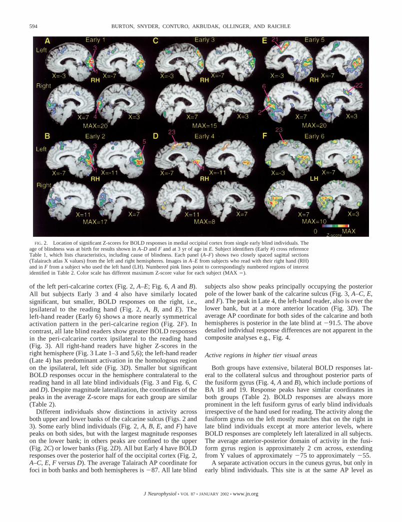
of the left peri-calcarine cortex (Fig. 2, A–E; Fig. 6, A and B).All but subjects Early 3 and 4 also have similarly locatedsignificant, but smaller, BOLD responses on the right, i.e.,ipsilateral to the reading hand (Fig. 2, A, B, and E). Theleft-hand reader (Early 6) shows a more nearly symmetricalactivation pattern in the peri-calcarine region (Fig. 2F). Incontrast, all late blind readers show greater BOLD responsesin the peri-calcarine cortex ipsilateral to the reading hand(Fig. 3). All right-hand readers have higher Z-scores in theright hemisphere (Fig. 3 Late 1–3 and 5,6); the left-hand reader(Late 4) has predominant activation in the homologous regionon the ipsilateral, left side (Fig. 3D). Smaller but significantBOLD responses occur in the hemisphere contralateral to thereading hand in all late blind individuals (Fig. 3 and Fig. 6, Cand D). Despite magnitude lateralization, the coordinates of thepeaks in the average Z-score maps for each group are similar(Table 2).
Different individuals show distinctions in activity acrossboth upper and lower banks of the calcarine sulcus (Figs. 2 and3). Some early blind individuals (Fig. 2, A, B, E, and F) havepeaks on both sides, but with the largest magnitude responseson the lower bank; in others peaks are confined to the upper(Fig. 2C) or lower banks (Fig. 2D). All but Early 4 have BOLDresponses over the posterior half of the occipital cortex (Fig. 2,A–C, E, F versus D). The average Talairach AP coordinate forfoci in both banks and both hemispheres is �87. All late blind
subjects also show peaks principally occupying the posteriorpole of the lower bank of the calcarine sulcus (Fig. 3, A–C, E,and F). The peak in Late 4, the left-hand reader, also is over thelower bank, but at a more anterior location (Fig. 3D). Theaverage AP coordinate for both sides of the calcarine and bothhemispheres is posterior in the late blind at �91.5. The abovedetailed individual response differences are not apparent in thecomposite analyses e.g., Fig. 4.
Active regions in higher tier visual areas
Both groups have extensive, bilateral BOLD responses lat-eral to the collateral sulcus and throughout posterior parts ofthe fusiform gyrus (Fig. 4, A and B), which include portions ofBA 18 and 19. Response peaks have similar coordinates inboth groups (Table 2). BOLD responses are always moreprominent in the left fusiform gyrus of early blind individualsirrespective of the hand used for reading. The activity along thefusiform gyrus on the left mostly matches that on the right inlate blind individuals except at more anterior levels, whereBOLD responses are completely left lateralized in all subjects.The average anterior-posterior domain of activity in the fusi-form gyrus region is approximately 2 cm across, extendingfrom Y values of approximately �75 to approximately �55.
A separate activation occurs in the cuneus gyrus, but only inearly blind individuals. This site is at the same AP level as
FIG. 2. Location of significant Z-scores for BOLD responses in medial occipital cortex from single early blind individuals. Theage of blindness was at birth for results shown in A–D and F and at 3 yr of age in E. Subject identifiers (Early #) cross referenceTable 1, which lists characteristics, including cause of blindness. Each panel (A–F) shows two closely spaced sagittal sections(Talairach atlas X values) from the left and right hemispheres. Images in A–E from subjects who read with their right hand (RH)and in F from a subject who used the left hand (LH). Numbered pink lines point to correspondingly numbered regions of interestidentified in Table 2. Color scale has different maximum Z-score value for each subject (MAX �).
594 BURTON, SNYDER, CONTURO, AKBUDAK, OLLINGER, AND RAICHLE
J Neurophysiol • VOL 87 • JANUARY 2002 • www.jn.org

responses on the upper bank of the calcarine sulcus, especiallyon the left (Table 2). This isolated cuneus gyral region alwaysis in cortex medial to the parietal occipital sulcus, whichseparates it from the more lateral BOLD responses within theintraparietal sulcus. A good example of this functional anat-omy is seen in subject Early 2 (Fig. 2B, X � �11).
Close to the occipital pole in both groups are separable peaksin the lateral occipital gyrus (LOG) (BA 18 in Table 2). TheseBOLD responses are lateral to foci along the calcarine sulcusand cuneus gyrus, and lateral and posterior to those in theposterior portion of the fusiform gyrus (Fig. 4D). Responsesoccur bilaterally in both subject groups with peaks at similarcoordinates (Table 2).
Summary of activity in occipital cortex
Activated foci occupy upper and lower banks of the calcar-ine sulcus and adjoining cortex on the lingual and cuneus gyri.In most subjects the lower bank (upper visual field represen-tation in sighted humans) response is greater than the upperbank response. BOLD responses are prominent along the in-ferior surface of the occipital lobe within the collateral sulcus,fusiform gyrus and temporo-occipital sulcus. Activation withinthe fusiform gyrus extends anterior toward the temporal lobe.In posterior occipital cortex, active regions include inferiorparts of the lateral occipital gyrus. Both groups show bilateral
foci. However, the early blind show more extensive activationof peri-calcarine cortex in the hemisphere contralateral to thereading hand. Right- and left-hand, late blind readers havelarger responses in the hemisphere ipsilateral to the readinghand. Both the number of foci and the spatial extent (seefollowing text) of peri-calcarine foci are greater in early blindin comparison to late blind subjects.
Active visual areas in temporal cortex
Early blind individuals show BOLD responses in the leftinferior temporal gyrus and sulcus (BA 19, 37; Fig. 4C; 5C:Early, X � �43). These mostly are laterally contiguous butseparable from the activations in the fusiform gyrus. As illus-trated by the Z-score peaks in Table 2, responses of nearly halfthe magnitude are found on the right in early blind or in bothhemispheres in the late blind. A medial-lateral separation be-tween the fusiform and inferior temporal gyrus foci is best seenwhen the BOLD responses are smaller (e.g., the right hemi-sphere of early blind and both sides in the late blind, Table 2).The medial-lateral coordinates of the peaks in the left FG andleft ITG from the average Z-score maps are similar and withinthe margins of error in the results from early blind individuals.In these cases BOLD responses are greater and fill much of theventral occipital and inferior temporal cortex. Sulcal anatomyis more distinguishable in individual subjects, and in these the
FIG. 3. Location of significant Z-scores for BOLD responses in medial occipital cortex from single late blind individuals.Subject identifiers (Late #) cross reference to Table 1, which lists characteristics, including cause of blindness. The age of blindnessonset ranged from 6 (A) to 25 (F) years of age. Each panel (A–F) shows two closely spaced sagittal sections as in Fig. 2. Imagesin A–C, E and F from subjects who read with their right hand (RH) and in D from a subject who used the left hand (LH). Numberedpink lines point to correspondingly numbered regions of interest identified in Table 2. Color scale has different maximum Z-scorevalue for each subject (MAX �).
595FMRI OF BRAILLE READING
J Neurophysiol • VOL 87 • JANUARY 2002 • www.jn.org

peak site of activation in the fusiform gyrus always lay be-tween the collateral and, at different anterior-posterior posi-tions, the temporal-occipital or inferior temporal sulci. Thepeaks in the inferior temporal gyrus always occur lateral to theinferior temporal sulcus.
An exclusively early blind response appears in the medialtemporal gyrus (MTG). It is anterior, lateral and superior to thefocus in the inferior temporal gyrus (ITG) and mostly occupiesBA 21. Sagittal sections of the early blind average image (Fig.5A, C: X � �43 and �51) show this focus over posteriorportions of MTG. There is a variable superior extension intothe superior temporal gyrus in some subjects. As in the coronalsection in Fig. 4C, this MTG region may be a superior exten-sion of the ITG focus. However, the MTG peak is �1 cmsuperior and anterior to that in the ITG (Table 2).
Active visual attention areas in parietal-occipital cortex
The BOLD responses in the lateral occipital gyri are con-tiguous with inferior and posterior extensions of bilateral fociwithin the intraparietal sulci (IPS) in both groups (Fig. 4, E andF). The average spatial extent of the IPS region is large. Itmainly involves BA 19, and is similar across groups (Fig.7B1). The IPS region contains one dominant peak at nearly thesame coordinates in both groups, which is ventrally situatedclose to the junction with the occipital cortex (Table 2 and Fig.4, E and F). The BOLD response magnitudes near this ven-trally located peak are also similar across the groups (Fig. 4, E
and F and Fig. 7B2). These responses extend within the pos-terior portion of the IPS toward the superior surface of thebrain. A hint of a second focus exists at this more dorsal site,but it is not sufficiently separated to be distinguished from theventral peak.
Active regions in somatosensory cortex
A third parietal region is seen on the left in both groups. Itis a separable anterior extension of the larger posterior region.It mainly occupies the postcentral sulcus (Fig. 4F: Z � 32).Portions of this anterior parietal region extend onto the post-central gyrus (BA 2; Fig. 5A, Z � 42; and all sagittal sections).Spatial coordinates of peak Z-scores (Table 2) are similar in thetwo groups. BOLD responses occur between the postcentralsulcal focus (label #16 in the figures and Table 2) and the largerresponses within the intraparietal sulcus (label #11 in Table 2and Fig. 4F, Z � 32; Fig. 5A, Z � 42). This activity appearsonly on the left, near the anterior extension of the intraparietalsulcus. A small local maximum exists on the average Z-scoreimages (Figs. 4F and 5A) but it is not separable from thesurrounding larger responses. This region occupies part of thesuperior parietal lobule normally labeled BA 7.
A small region with a barely significant Z-score appearswithin the depths of the central sulcus in some late blindindividuals. No subject shows a significant response within theparietal operculum along the upper bank of the lateral sulcus.
FIG. 4. Location of significant average Z-scoresfor BOLD responses mainly in extrastriate and occip-ital-parietal cortex. A, C, and E: data taken from 6early, right-hand Braille readers. B, D, and F: datataken from 7 late blind subjects (6 right- and 1 left-hand Braille readers). Activation data overlaid on av-erage structural images from the same subjects. Allimages aligned and interpolated to atlas coordinatesusing 2 mm voxels. Paired images in each panel showcoronal (on left) and transverse (on right) slicesthrough or near peak Z-scores for the numbered re-gions of interest identified in Table 2. Pink lines showthe registration of the alternate slice orientation. Colorscale has different maximum Z-score value for eachgroup of subjects (MAX �).
596 BURTON, SNYDER, CONTURO, AKBUDAK, OLLINGER, AND RAICHLE
J Neurophysiol • VOL 87 • JANUARY 2002 • www.jn.org

Language areas in frontal cortex
Both groups show multiple foci over the lateral frontalconvexity. The most inferior location of these activations ex-tends to the left frontal operculum and adjoining inferior fron-tal gyrus (Fig. 5C: IFG) including parts of Brodmann areas 45and 47. A second region lies in the dorsolateral frontal cortex.The latter activations occupy both the superior-posterior part ofthe inferior frontal gyrus and inferior part of the middle frontalgyrus (Fig. 5B: I/MFG) through Brodmann areas 9 and 46. Thedistribution of these two regions and the coordinates of theirpeaks (Table 2) are similar across the groups irrespective of thehand used for Braille reading. Several late blind individualsalso show additional, symmetrically located low Z-score re-
sponses on the right, which is reflected in the average imagesacross all subjects (Fig. 5: A, Z � 42; B, Z � 28; C, Z � 14).
Active regions in premotor cortex
A third, more posterior site of activation in frontal cortex ofboth groups occupies the left precentral sulcus and neighboringprecentral gyrus where it mostly is within Brodmann area 6(Fig. 5A: PrCS; Table 2). This activated region extends for-wards to the middle frontal gyrus where it probably includespart of Brodmann area 8.
Medial frontal cortex, especially on the left, contains mul-tiple BOLD responses (Fig. 6; Fig. 5: A, Y � �3; B, Y � 3).Both groups have one focus that is posterior and superior (BA
FIG. 5. Location of significant average Z-scores for BOLDresponses mainly in frontal cortex. Left three images for eachpanel, A–C, show slices at three orientations for averages (Z-scores and corresponding structural images) from the same 6early and 7 late blind subjects shown in Fig. 4. Data shown areat or near peak activations overlying the precentral sulcus (PrCS)(A), the inferior/middle frontal gyri (I/MFG) (B), and the inferiorfrontal gyrus (IFG) (C). Sagittal slices shown on the far righttaken from averages for the same early or late blind individuals.The data from these two were added and averaged to create theimages shown on the left. Numbered regions of interest identifiedin Table 2. Perpendicular pink lines show the registration of thealternate two slice orientations. Color scale has different maxi-mum Z-score value for averages across all or within each groupof subjects (MAX �).
597FMRI OF BRAILLE READING
J Neurophysiol • VOL 87 • JANUARY 2002 • www.jn.org

6; Table 2) to a second, more anterior and inferior site on therostral cingulate gyrus (BA 24,32; Table 2). Two foci are foundin every early blind (Fig. 2, A, B, D–F) and nearly all late blindsubjects (Fig. 3, A, B, and D). The larger, posterior one extendsover the medial superior frontal gyrus (mSFG); the smalleranterior focus occupies the cingulate gyrus and sulcus bilater-ally (Table 2, Fig. 6, B–D). In addition, late blind subjects
show a third focus between the two more prominent medialfrontal sites (Fig. 6C).
Spatial extent of BOLD responses
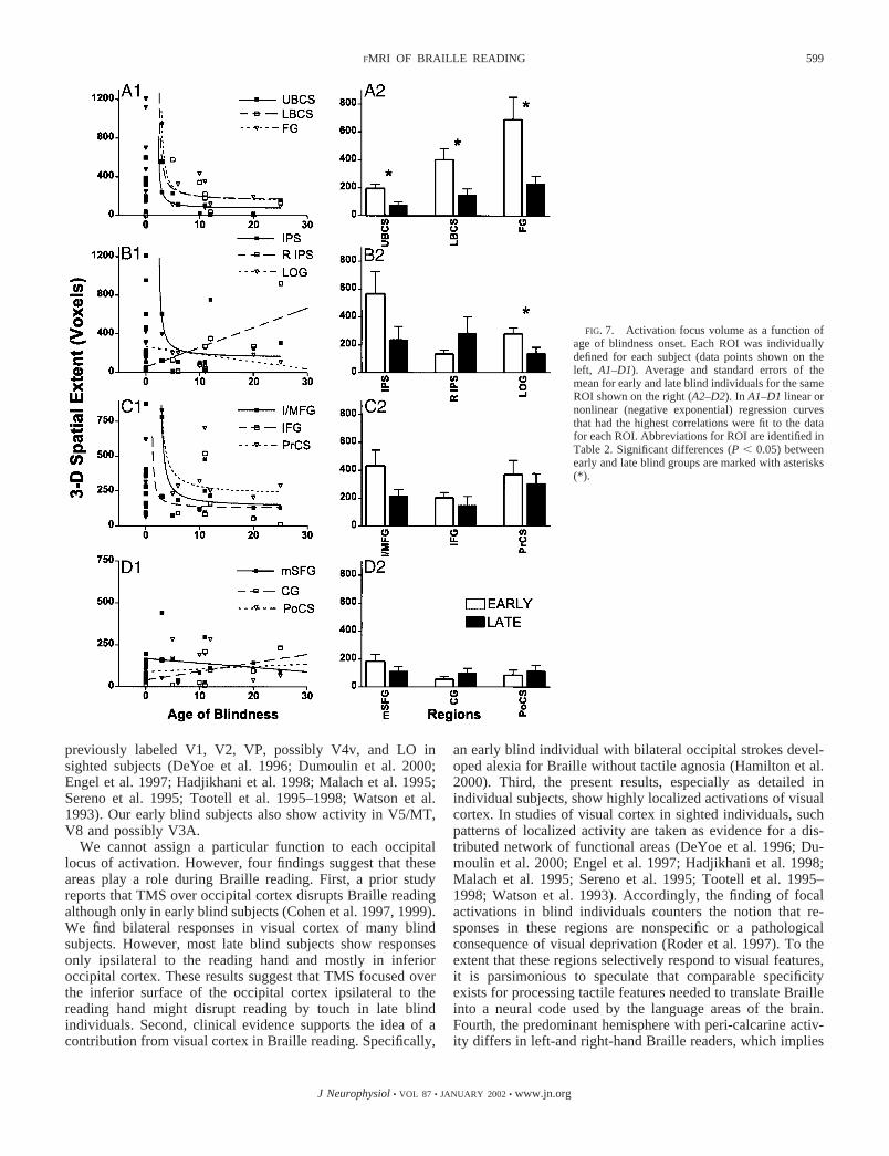
The spatial extent of BOLD responses is significantly greaterin the early blind only in part of visual cortex. This differenceis seen in active regions in upper and lower banks of thecalcarine sulcus. The distinctions between groups persist evenwhen comparing dominant contralateral responses in peri-cal-carine cortex for the early blind to the ipsilateral responses inthe late blind (Fig. 7A2, UBCS and LBCS). The spatial extentof the active region over FG and LOG also are significantlygreater in the early blind (Fig. 7, A2 and B2). In each of theseregions, the spatial extent for individuals who lost sight afterthe age of three declines precipitously (Fig. 7, A1 and B1). Theresults from UBCS, LBCS and FG are best fit with a negativeexponential, nonlinear regression function. A negative linearregression is the best fit for the data from LOG. The spatialextent in all other active regions are not significantly differentbetween the groups. However, several regions (IPS, I/MFG,and PrCS), whose data are best fit by a negative exponentialfunction (Fig. 7, B1 and C1), show a trend of larger spatialextent in early blind individuals.
Time course of BOLD responses
The time course of BOLD signal modulation in all subjectsand all regions exhibited a common pattern characterized bysignal increase during the three verb generation trials anddecrease during the three control trials. The BOLD modulationpeak generally appears 1 to 2 frames after the task switch, i.e.,during frames 2–3 and 5–6. There is no evidence of a timecourse dependence on locus. The pattern of BOLD modulationas a function of frame is the same in both groups despite amodest increase in TR for the slowest late blind readers (Fig.8). The small standard error bars in these plots further attests toresponse profile consistency over all subjects.
D I S C U S S I O N
Our task paradigm (covertly generate verb for Braille em-bossed noun versus read Braille “######” ) induced an exten-sive array of BOLD responses in both early and late blindindividuals. Activated regions include visual, visual/attention,somatomotor, and language areas in frontal cortex. What fol-lows is an attempt to explain these results in terms of func-tionality identifiable in sighted individuals. We suggest that, inresponse to blindness, specialized areas retain their intrinsicmechanisms, which become adapted to the challenge of read-ing by touch. We argue that visual cortex recodes sensoryinformation into a format used by the language areas of thebrain.
Regions usually activated by visual stimuli insighted subjects
OCCIPITAL CORTEX. We find extensive activation of visualcortex in early and late blind individuals, generally in agree-ment with prior PET studies (Buchel et al. 1998; Sadato et al.1996, 1998). Involved cortex in both groups includes regions
FIG. 6. Location of significant average Z-scores for BOLD responsesmainly in medial frontal cortex in 6 early (A and B) and 7 late (C and D) blindsubjects (same subjects as in Fig. 4). Activation data overlaid on averagestructural images from the same subjects. Paired images in each panel showsagittal (on left) and coronal (on right) slices through or near peak Z-scores forthe numbered regions of interest identified in Table 2. Pink lines show theregistration of the alternate slice orientation. Color scale has different maxi-mum Z-score value for each group of subjects (MAX �).
598 BURTON, SNYDER, CONTURO, AKBUDAK, OLLINGER, AND RAICHLE
J Neurophysiol • VOL 87 • JANUARY 2002 • www.jn.org
previously labeled V1, V2, VP, possibly V4v, and LO insighted subjects (DeYoe et al. 1996; Dumoulin et al. 2000;Engel et al. 1997; Hadjikhani et al. 1998; Malach et al. 1995;Sereno et al. 1995; Tootell et al. 1995–1998; Watson et al.1993). Our early blind subjects also show activity in V5/MT,V8 and possibly V3A.
We cannot assign a particular function to each occipitallocus of activation. However, four findings suggest that theseareas play a role during Braille reading. First, a prior studyreports that TMS over occipital cortex disrupts Braille readingalthough only in early blind subjects (Cohen et al. 1997, 1999).We find bilateral responses in visual cortex of many blindsubjects. However, most late blind subjects show responsesonly ipsilateral to the reading hand and mostly in inferioroccipital cortex. These results suggest that TMS focused overthe inferior surface of the occipital cortex ipsilateral to thereading hand might disrupt reading by touch in late blindindividuals. Second, clinical evidence supports the idea of acontribution from visual cortex in Braille reading. Specifically,
an early blind individual with bilateral occipital strokes devel-oped alexia for Braille without tactile agnosia (Hamilton et al.2000). Third, the present results, especially as detailed inindividual subjects, show highly localized activations of visualcortex. In studies of visual cortex in sighted individuals, suchpatterns of localized activity are taken as evidence for a dis-tributed network of functional areas (DeYoe et al. 1996; Du-moulin et al. 2000; Engel et al. 1997; Hadjikhani et al. 1998;Malach et al. 1995; Sereno et al. 1995; Tootell et al. 1995–1998; Watson et al. 1993). Accordingly, the finding of focalactivations in blind individuals counters the notion that re-sponses in these regions are nonspecific or a pathologicalconsequence of visual deprivation (Roder et al. 1997). To theextent that these regions selectively respond to visual features,it is parsimonious to speculate that comparable specificityexists for processing tactile features needed to translate Brailleinto a neural code used by the language areas of the brain.Fourth, the predominant hemisphere with peri-calcarine activ-ity differs in left-and right-hand Braille readers, which implies
FIG. 7. Activation focus volume as a function ofage of blindness onset. Each ROI was individuallydefined for each subject (data points shown on theleft, A1–D1). Average and standard errors of themean for early and late blind individuals for the sameROI shown on the right (A2–D2). In A1–D1 linear ornonlinear (negative exponential) regression curvesthat had the highest correlations were fit to the datafor each ROI. Abbreviations for ROI are identified inTable 2. Significant differences (P � 0.05) betweenearly and late blind groups are marked with asterisks(*).
599FMRI OF BRAILLE READING
J Neurophysiol • VOL 87 • JANUARY 2002 • www.jn.org

specific early processing of tactile information from the read-ing hand.
The within subject analyses show that, despite individualvariability, blind subjects in both groups exhibit active regionsin peri-calcarine cortex. Most subjects show responses over thelower bank of the calcarine and much of the inferior occipitalcortex. The superior foci are more restricted except for exten-sions onto surface cortex (i.e., cuneus gyrus) at the occipitalpole. The across subject variance does not necessarily indicatefunctional inconstancy. A recent quantitative histological anal-ysis of Brodmann areas 17 and 18 (i.e., V1 and V2) reportedthat substantial variability normally occurs in sighted individ-uals (Amunts et al. 2000). Comparable variability is likely inblind individuals.
The peri-calcarine BOLD responses occupy predominantlywhat is, in sighted subjects, the representation of the uppervisual field. The extent of activation superior to the calcarinesulcus is less clear. All late blind and most early blind indi-viduals show little or no activation of V3, which normallyrepresents lower visual space. However, the within subjectanalyses in early blind individuals show some activity withinthat part of the cuneus gyrus previously identified as V3A(Tootell et al. 1997). The active region corresponds best to thatportion of V3A dedicated to upper visual space, i.e., moreinferior, lateral domain of V3A. Thus the same visual repre-sentation appears to be engaged in different subdivisions ofvisual cortex. This correspondence further argues against thenotion that the responses in these regions are nonspecific. The
FIG. 8. Time course of BOLD responses per voxel in se-lected ROI for 7 early and 7 late blind individuals. For eachgroup, data includes responses from 6 right- and 1 left-handreaders. Data for each frame shows mean and standard error ofthe mean. Average percentage change in MR signal was ob-tained in the selected ROI defined in individual subjects andthese values were then averaged as shown.
600 BURTON, SNYDER, CONTURO, AKBUDAK, OLLINGER, AND RAICHLE
J Neurophysiol • VOL 87 • JANUARY 2002 • www.jn.org

explanation for the predominant activation of “upper visualspace” representations is uncertain. Possibly, it is due to thesupine position assumed during the scans.
The greater spatial extent of activity in primary and second-ary visual cortex in early blind subjects (Fig. 7) appears to berelated to age of blindness onset and not Braille reading flu-ency. Nonparametric tests (Spearman rank correlation coeffi-cient and Kendall’s �) show no concordance between Braillereading rates and the spatial extent or magnitude of MR re-sponses in visual cortex. However, the best late-blind Braillereader (Late 6) shows primary visual cortex activity that iscomparable to that observed in nearly all but the least fluentearly blind readers. Extensive training was responsible for thissubject’s fluency. However, before suggesting that intensiverehabilitation in late blind might yield more responses in visualcortex, additional testing is needed with early and late blindpeople who are paired for Braille reading fluency.
OCCIPITAL-TEMPORAL CORTEX. Early and late blind individualsshow activity within the lateral edge of the fusiform gyrus, andstill further lateral in the inferior temporal sulcus and gyrus.There was a significantly greater spatial extent of this focus inearly blind individuals. The responses in lateral FG overlapwith a region at 42.8 � 2.7, �72.7 � 8, �18.2 � 9.8, whichis a lateral occipital center (LO) for object identifications insighted subjects (Malach et al. 1995; Tootell et al. 1996). Inaddition, an object processing region, previously localizedwithin the left posterior basal temporal/fusiform region ofsighted subjects, and which encompasses posterior Brodmannarea 37 (Moore and Price 1999), also corresponds with acti-vated regions in the present study.
It is possible that the basis for this overlap with objectrecognition centers in sighted subjects is in relation to process-ing Braille fields as objects. First, prior studies show that fluentBraille readers recognize whole words (Krueger 1982), whichreflects a conceptualization of the word as a single object.Braille reading also entails projecting personal contact with thedot patterns, which, unlike print reading, involves obtaining athree dimensional percept of the Braille field. Therefore wesurmise that activity in LO and posterior parts of area 37 inboth subject groups might arise from a complex series of steps:touching, recognizing and identifying the word Braille fields asparticular noun objects. These operations must precede gener-ating an appropriate verb.
We observed no responses in an anterior-medial extension ofthe fusiform gyrus (Brodmann area 20/37). Previous reportsbased on PET list this site as a recognition center for integrat-ing tactile information in word reading in early blind subjects(�40, �38, �16 in Buchel et al. 1998b). PET studies insighted subjects report blood flow increases in the same regionduring tasks requiring semantic relative to perceptual informa-tion such as viewing meaningful words or objects (�32, �40,�20 in Moore and Price 1999). The absence of such responsesin the present data may be attributable to “blind spots” inherentin EPI (Ojemann et al. 1997).
One additional extension of activated occipital-temporal cor-tex occurs along the lateral surface of the inferior temporalgyrus in every early blind and no late blind individual. Thisactivity reaches the ascending limb of the inferior temporalsulcus. This sulcal landmark provides a unique identifier for
the motion selective MT/V5 (Dumoulin et al. 2000; Tootell etal. 1995; Watson et al. 1993).
We speculate that characteristics of fluent Braille reading,especially in early blind individuals, might be responsible forthese responses in MT/V5. Thus because V5 is known torespond especially to coherent motion (Tootell et al. 1996), V5might be particularly suited to process tactile contrast arisingfrom hand motion across different words and parts of words inBraille reading. Tactile contrasts are likely important whenreading Braille, because, contrary to earlier ideas (Loomis1981), hand movements across Braille cells create a texturalimage of the word that is based on lateral dot-gap shearingdensity within individual Braille cells (Millar 1984, 1986,1987, 1997; Nolan and Kederis 1969; Pring 1985, 1994). Thesensory information about the orthographic characteristics ofindividual Braille cell letters is processed serially and at a lowspatial frequency. Fluent Braille reading involves selection ofhand motion and direction that scans across the spatial form ofindividual letters.
Another extension of activation within the inferior temporalgyrus occurs anterior and inferior to the major foci in thefusiform gyrus. This region includes the coordinates of whatHadjikhani and colleagues describe as a major color processingcenter in sighted subjects, V8 at �33, �65, �14 (Hadjikhaniet al. 1998). Unlike the responses to color existing in lower tiervisual areas, individuals with lesions affecting V8 lack theability to process the spectral complexity of a scene (Had-jikhani et al. 1998). In reading Braille the spectral dimensionsof roughness affect judgements about textural tactile differ-ences for different letters in a word (Millar 1986, 1987, 1997).We hypothesize that mechanisms related to analyzing spectraof a varying tactile surface, when applied to the tactile domainof Braille, might explain the activity detected in V8 in earlyblind subjects.
Our late blind subjects, with one exception, read less flu-ently. Reading in these cases possibly involves determining thestructure of individual Braille cells rather than a holistic, tex-ture perception of letters, combinations of letters or wholewords. Thus less fluent Braille reading is affected more byorthographic elements (Millar 1997). A consequence mightthen be greater reliance on activity in lower tier visual areas asopposed to V5/MT in late-blind individuals. Why these re-sponses occur predominantly in the hemisphere ipsilateral tothe reading hand remains a puzzle.
Parietal regions activated by attention in sighted subjects
IPS REGIONS. The present study finds the largest magnitudeBOLD responses in the same part of the ventral intraparietalsulcus (vIPS) identified in sighted subjects performing eye-movements (Petit and Haxby 1999), detecting and attending tovisual motion (Shulman et al. 1999; Tootell et al. 1995), andvoluntary orienting of attention to visual space (Corbetta et al.1998a,b, 2000). A separate focus in pIPS is not apparent,although extension of the BOLD responses (Z-scores �15),with a peak in vIPS, includes the atlas coordinates of the sitespreviously described as pIPS in sighted subjects. We observeno activations in anterior IPS (Corbetta 1998, 2000; Shulmanet al. 1999). In the light of the above-cited literature, wesuggest that Braille reading induces left-lateralized prefrontal
601FMRI OF BRAILLE READING
J Neurophysiol • VOL 87 • JANUARY 2002 • www.jn.org

modulation of spatial attention and voluntary orientating to-ward the reading hand (fingers) as it moves across successiveBraille cells. Additional experiments will be required to disso-ciate spatial attention, tactile perception and language pro-cesses in IPS.
Our data includes the intriguing observation of opposite IPSlateralization in early versus late blind individuals. We findgreater bilateral symmetry in the size of the vIPS foci in righthand, late blind readers compared with predominantly con-tralateral, left sided sites in right hand, early blind readers. Inthe left-hand, early blind reader, the activated site is also onlyon the contralateral, right side; while the late blind, left-handreader has bilateral foci, but the larger site is on the right. Thesegroup differences might reflect an under developed sense ofprojected external space in early blind individuals (Millar1994). Thus the regional asymmetry in vIPS activations in theearly blind might indicate a relatively greater awareness ofimmediate personal space. Late blind individuals, through priorvisual experience, possibly project more readily into a widerspatial environment, which could explain the more symmetri-cal, bilateral activation of vIPS in late blind individuals. Thusin late blind a combination of voluntary attention to personaland extra-personal space might contribute to contralateral re-sponses for personal space with respect to the reading hand,and a right hemisphere dominance for wider extra-personalspace that contains the Braille field.
POSTCENTRAL SULCUS. Responses centered around the leftpostcentral sulcus coincide with a region modulated by atten-tion to tactile stimuli on the right hand, and which lies anteriorand lateral to visual attention regions within the IPS (Burton etal. 1999). It is certain that our subjects attended, as readingBraille is difficult even under optimal conditions. Accordingly,we interpret the left postcentral sulcus activations as due totactile attention to the Braille fields during word reading.
Regions activated by touch
We find little or no BOLD modulation in most somatosen-sory cortical regions including the parietal operculum, i.e.,second somatosensory cortical area (S2). Only scant responsesoccur within the central sulcus, i.e., in primary somatosensorycortex (S1). In view of the remarkable plasticity of somatosen-sory cortex (Buonomano and Merzenich 1998; Pons et al.1991; Ramachandran 1996, 1998), it would be reasonable tosuppose that some component of the adaptations underlyingBraille skill should be manifest in this part of the brain.Normally sighted subjects who receive tactile stimulation showrobust activations in portions of anterior and lateral parietalcortex (Burton 2001) and suppression of visual cortex activity(Drevets et al. 1995). Touching Braille cells clearly providespotent somatosensory stimulation of the skin and, therefore wemight expect comparable cortical responses in these subjects.The minimal responses observed do not exclude the possibilitythat Braille reading finger(s) have an expanded cortical repre-sentation.
There are two possible explanations for the unexpectedlysmall responses in somatosensory cortex. A procedural expla-nation suggests that when the subjects touched the controlfields they successfully balanced tactile stimulation and grossmotor behavior with the demands of word reading. We in-
structed our subjects to duplicate their Braille word readingstrategies as they touched the control fields. Greater imbalancein tactile stimulation between activation and control trialscould account for previous reports of increased blood flow insomatosensory and motor cortex of early blind individuals(Sadato et al. 1998).
Alternatively, it is may be that, in adapting to read Braille,blind individuals redirect tactile discrimination processingaway from primary and secondary somatosensory areas to thevisual cortex, possibly through dorsal parietal association ar-eas. This question could theoretically be illuminated by study-ing sighted subjects reading Braille, in which a more balancedactivation of somatosensory cortex with minimal or no re-sponses in visual cortex might be expected. Unfortunately, noone with these skills was available in our local community.Many of our late blind subjects had normal visual experiencesfor a decade or longer. These subjects showed slightly greateractivation of somatosensory cortex, which could be viewed asevidence of reduced adaptations to Braille reading. However,again, a procedural explanation is more likely because inreading Braille less fluently, these subjects touched the Braillecells for words with more exaggerated movements comparedwith the control fields. Early blind subjects showed similarsmooth sweeping movements for words and control fields.
An observation potentially related to the question of adap-tive changes associated with Braille reading is that, in sightedsubjects, visual cortex normally is suppressed during tactilediscrimination tasks (Drevets et al. 1995). Even where a tactiletask engages a portion of parietal-occipital cortex in sightedsubjects (Sathian et al. 1997), primary visual cortex shows noactivity while somatosensory cortex exhibits expected re-sponses. The normal suppression of visual cortex by tactilestimulation clearly does not occur during fluent Braille readingin blind individuals.
Regions activated by motor behavior
PREMOTOR CORTICAL REGIONS. We find no BOLD modulationin the anterior bank of the central sulcus, i.e., primary motorcortex as traditionally defined (Cramer et al. 1999; Crespo-Facorro et al. 1999; Fink et al. 1997). However, extensiveresponses occur throughout several previously identified non-primary motor areas (Fink et al. 1997). In all subjects, thelargest BOLD responses in the frontal cortex occur on thelateral convexity on the left side. This activation is in the leftlateral premotor cortex regardless of the hand used for Braillereading.
We find bilateral activation around the precentral sulcus verynear the area identified as the frontal eye fields (Corbetta et al.1998; Petit and Haxby 1999). The major portion of the presentactivation, however, mostly borders the lateral and anteriorportion of the previously described eye movement relatedresponse. The main peak of the PrCS region is within acomponent of premotor cortex, which suggests these responsesreflect motor planning. Especially in our early blind subjects,this response overlaps part of the left PrCS region described ina study of complex object manipulations (Binkofski et al.1999) or simple flexion-extension movements of the right handand fingers (Fink et al. 1997). The center of activity in our dataare superior and posterior to the object manipulation area of
602 BURTON, SNYDER, CONTURO, AKBUDAK, OLLINGER, AND RAICHLE
J Neurophysiol • VOL 87 • JANUARY 2002 • www.jn.org

Binkofski et al. (1999). These differences may reflect minordifferences in the target images used to compute atlas trans-formations. This response most likely is attributable to activa-tion of the premotor representation for the fingertips.
Braille reading ordinarily is accompanied by a slight in-crease in the downward pressure exerted by the reading fingeras it moves across each Braille cell (Millar 1997). This likelyserves to increase stimulation of the slowly adapting pressurereceptors, thereby increasing the transmission of salient infor-mation. It is possible that our readers omitted this action asthey touched the control field because the content was entirelypredictable. In any case, consistent motor differences betweenthe word and control fields, whatever they were, manifested aspremotor but not primary motor activation.
Activity exclusively in the left PrCS, even in left-handreaders, suggests that the PrCS region may not just encodemotor behavior. In a study of complex object manipulations,maximum responses were observed in cortex contralateral tothe used hand (Binkofski et al. 1999). In contrast, a similar lefthemisphere specialization exists in deaf subjects using signlanguage (Neville et al. 1998). The left dominance with Braillereading might therefore reflect some abstraction of the learnedspecial motor behavior for language functions, which are nor-mally dominant in the left hemisphere.
Activity along the medial wall of premotor areas could arisefrom particular motor programs needed for Braille reading.The more posterior and superior region in this study hascoordinates that place it posterior to the vertical anterior com-missure line (Vca), which centers it within the domain of thearm representation of the supplementary motor area (SMA)(Fink et al. 1997; Kwan et al. 2000; Picard and Strick 1996;Thickbroom et al. 2000). SMA is especially active in sequen-tial movements such as typing that are also integrated withreceived sensory information (Gordon et al. 1998). This isexactly what occurs during Braille reading.
Prior human imaging studies distinguish between SMAproper, which is engaged by simple, automatic movements,and a more anterior, inferior pre-SMA region, which servesmore complex movements, possibly associated with skill ac-quisition (Picard and Strick 1996). More intense activation ofthese medial motor areas also occurs for motor tasks timedunpredictably to external events (Thickbroom et al. 2000).Only late blind subjects show a focus just anterior to Vca,within the pre-SMA area. Most of our late blind subjects areless fluent Braille readers and probably use slightly differentreading strategies. Fluent readers exhibit rapid, smooth, rela-tively stereotyped horizontal scans across Braille fields. DuringBraille learning, reading with less fluency, or reading physi-cally degraded Braille cells, readers make minute trapezoidal,up-down motions over each Braille cell during horizontal scan-ning (Millar 1987, 1997). Such selective motor behavior hasless predictable timing, and might require more complex motorprograms, which are known to activate a pre-SMA area (Finket al. 1997; Thickbroom et al. 2000). This might explain thegreater activation of this region in late blind subjects.
Both subject groups show activation of the cingulate sulcusand gyrus nearly 1 cm anterior to Vca and �1 cm inferior tothe SMA site. The coordinates of this focus place it within aposterior part of the anterior cingulate cortex previously asso-ciated with motor behaviors (Fink et al. 1997; Kwan et al.
2000; Picard and Strick 1996) similar to those used in Braillereading. Other processes which may be invoked to account foractivation in this region include cognition (Whalen et al. 1998),verb generation (Petersen and Fiez 1993; Raichle et al. 1994)and attention (Burton et al. 1999; Corbetta et al. 1991). Thepresent paradigm does not provide a means of distinguishingbetween these possibilities. The activated zone is more than 3cm posterior to anterior cingulate regions associated with effort(Whalen et al. 1998) and pain (Kwan et al. 2000). It is thereforereasonable to suggest that our task paradigm did not invokemodulation of emotional processes.
Language areas in frontal and temporal cortex
The subject of word reading overall and verb generation inparticular have been under continuous inquiry in the neuroim-aging (for reviews see Fiez and Petersen 1998; Gabrieli et al.1998) and neuropsychology literature (Illes et al. 1999;Thompson-Schill et al. 1998) since the verb generation taskwas first employed in functional imaging (Petersen 1988,1989). The task of verb generation, while admittedly linguis-tically complex, consistently produces left lateralized re-sponses in right-handed individuals and, when performed na-ively (Raichle et al. 1994), activates traditional languagerelated areas of the cerebral cortex. We employed this robustlanguage task as a means of obtaining an overall assessment ofthe distribution of language processing in blind subjects. Wedid not propose to isolate linguistic, memory, motor and atten-tion processes, as has been done by others and ourselves.Rather, we merely sought to determine if the functional anat-omy of language is different in blind individuals.
PREFRONTAL CORTEX. We believe the results clearly demon-strate that blind subjects use the same general neural process-ing architecture employed by sighted individuals when per-forming the verb generation task. The presently observedfunctional anatomy replicates prior descriptions of left prefron-tal activations observed in a variety of language tasks (Fiez1997; Gabrieli et al. 1998; Illes et al. 1999; Petersen and Fiez1993; Poldrack et al. 1999). All subjects show two distinctactivations in prefrontal cortex. One focus resides entirelywithin the inferior frontal gyrus in Brodmann areas 45 and 47.The other focus spreads across the inferior frontal and middlefrontal gyri on the dorsolateral convexity with its peak centeredin the middle frontal gyrus. This region includes portions ofBrodmann areas 9 and 46 according to the Damasio atlas(Damasio 1995).
In studies with sighted subjects, inferior prefrontal activity ismost prominent when contrasting semantic to nonsemanticprocessing even when the latter is a more difficult task (re-viewed in Gabrieli et al. 1998; Poldrack et al. 1999). This viewsupports earlier PET studies (Fiez 1997). The prefrontal foci inthe present study are essentially coextensive with these tworegions. This raises the question as to whether the current tasksimilarly is divisible into semantic and phonological processes.
Verb generation is a language task with substantial semanticprocessing (Fiez 1997; Gabrieli et al. 1998; Illes et al. 1999;Petersen and Fiez 1993; Seger et al. 1999). In the present studywe contrast verb generation with reading and interpreting theBraille embossed field for the number sign. Normally, whenthe Braille symbol for a number sign appears first in a word,
603FMRI OF BRAILLE READING
J Neurophysiol • VOL 87 • JANUARY 2002 • www.jn.org

readers know the following Braille cells represent numbers.Thus detecting the number symbol involves some semanticprocessing. However, by presenting these strings of numbersigns during the initial eight baseline scans, priming effects(Raichle et al. 1994; Seger et al. 1999) produce instant recog-nition, and minimal semantic demands when similar Braillefields alternate with words within the runs. Therefore thesemantic demands of verb generation were predominant.
Word reading involves phonological processes (Fiez et al.1997, 1998; Gabrieli 1998). The dorsolateral prefrontal regionalso is activated by word/object encoding tasks (Kelly et al.1998) and object naming (Moore and Price 1999). Such en-coding must precede the semantic aspects of verb generation.Although the present experiment does not attempt to differen-tiate various language operations, our results do suggest that, inblindness, early or late, there is no reorganization of semanticor phonological representations.
In the absence of sight, phonology and phonemics providethe only basis for associating the tactile feel of a Braille wordand its meaning (Millar 1975a,b, 1984, 1997; Nolan and Ke-deris 1969; Pring 1982, 1985). However, in skilled readers,attention to phonology interferes with fluent Braille reading(Millar 1975a,b, 1997; Pring 1982). In the light of this obser-vation, it is possible that early blind Braille readers, being lessdependent on phonology, should show relatively reduced dor-solateral prefrontal responses. This result is not evident. Theabsence of distinctions between early and late blind individualsin dorsolateral prefrontal activity possibly relates to prior find-ings that this same region is also particularly concerned withword/object encoding (Kelly et al. 1998) or specific word orobject naming (Moore and Price 1999). Our subjects mustencode the words into some object or concept for, respectively,concrete or abstract nouns before doing the semantic task ofgenerating a verb. Thus activation of this dorsolateral region(and, for similar reasons noted above, posterior basal occipitaltemporal cortex) possibly occurs because of word encodingrather than phonological processing.
POSTERIOR TEMPORAL CORTEX. Only early blind individualsshow activation of the middle temporal gyrus (BA 21). Thereis no consensus regarding which linguistic operations are mostspecific to this area (Binder et al. 1997; Fiez et al. 1996;Petersen and Fiez 1993; Pugh et al. 1996; Vandenberghe et al.1996; Warburton et al. 1996; Zatorre et al. 1996). Rumsey andcolleagues suggest that the posterior temporal region is espe-cially engaged in letter identification, i.e., processing of visualorthography. The detailed structure of a Braille cell (i.e., thelocation of dots in the left or right column and in upper orlower rows) arguably is comparable to the arrangement ofintersecting line segments which make up letters. The BA 21activation, however, is absent in late blind subjects who aremore likely to attend to the shapes of individual Braille cells(see above). This response is present in the early blind whoperceive Braille more holistically. Early blind individuals at-tend to shifting textural patterns formed by the different Brailleletters, and possibly process an overall orthography similar tothe holistic interpretations of letters in reading print. Suchholistic analyses would require the integrative mechanismsmore likely in higher visual/language areas in posterior tem-poral cortex.
Conclusions
We confirm earlier reports of visual cortex activation byBraille reading. Two entrenched ideas make this finding sur-prising. First, based on its neuroanatomical connections, par-ticularly those involving the thalamus, this cortex is visual.Second, loss of vision presumably renders the affected regioninoperable. Yet, blind subjects show clear, well localized ac-tivation of both lower and higher tier visual areas. The lowertier responses in large measure correspond to retinotopicallyorganized visual cortex subregions in sighted individuals.These findings indicate a need to reexamine the contribution ofoccipital cortex to reading. One possibility is that blind Braillereaders use occipital cortex in a novel way, unlike the use ofthis cortex for early vision in sighted people. Another possi-bility is to suggest that mechanisms and connections withinoccipital cortex are necessary for encoding orthography (visualor tactile) into information used by language areas in frontal,and possibly temporoparietal, cortex. Accordingly, the activityin occipital cortex of blind individuals is not a totally noveladaptation. Rather, it represents a function already present insighted individuals, namely recoding information for languageareas. Loss of vision does not remove this role from occipitalcortex, which accounts for the fact that this region is active inblind individuals.
Each of the many additional foci of significant activity inparietal, frontal and temporal cortex possibly relates to char-acteristic features of Braille reading. Thus the necessity offocusing attention on the fingertips when touching Braillemight be expected to engage parietal attention regions. We alsosuggest that minute movement strategies used when touchingBraille cells selectively might increase activity in medial, fron-tal premotor areas. Braille reading obviously is at least in parta somatomotor task. However, the contribution of primarysomatomotor cortex evidently can be balanced by a suitablyimplemented low-level control task. Potential differences inthe reading strategies of early and late blind individuals pos-sibly explains the distinctions in activated temporo-occipitalfoci including MT/V5, V8 and BA 21.
The verb generation task used in this study matches semanticand phonological features extensively studied in sighted indi-viduals. There is a corresponding match of the observed left-lateralized prefrontal functional anatomy. We also suggest thatword recognition precedes the semantic processing in the verbgeneration task, and possibly that touching Braille cells in-volves object recognition. Thus word and object encoding focipreviously described in sighted subjects in dorsolateral pre-frontal cortex and ventral occipital temporal cortex are corre-spondingly engaged in blind individuals during Braille reading.
P. Schonlau, the Braille Reading Teacher at the Missouri School for theBlind, provided invaluable assistance, advice, and subject recruitment through-out this project. We acknowledge N. Thompson for tireless processing of thedata and J. Kreitler for the design and construction of the Braille stimuluspresentation device. We also thank our colleagues, Drs. M. Corbetta, G.Shulman, and D. Van Essen for thoughtful comments, perspectives, and accessto their own data.
This work was supported by National Institute of Neurological Disordersand Stroke Grants NS-37237 to H. Burton and NS-06833 to M. Raichle and bythe McDonnell Center for Higher Brain Function.
604 BURTON, SNYDER, CONTURO, AKBUDAK, OLLINGER, AND RAICHLE
J Neurophysiol • VOL 87 • JANUARY 2002 • www.jn.org

REFERENCES
AMUNTS K, MALIKOVIC A, MOHLBERG H, SCHORMANN T, AND ZILLES K.Brodmann’s areas 17 and 18 brought into stereotaxic space-where and howvariable? Neuroimage 11: 66–84, 2000.
ANDERSSON JL, SUNDIN A, AND VALIND S. A method for coregistration of PETand MR brain images. J Nucl Med 36: 1307–1315, 1995.
BINDER JR, FROST JA, HAMMEKE TA, COX RW, RAO SM, AND PRIETO T.Human brain language areas identified by functional magnetic resonanceimaging. J Neurosci 17: 353–362, 1997.
BINKOFSKI F, BUCCINO G, POSSE S, SEITZ RJ, RIZZOLATTI G, AND FREUND H. Afronto-parietal circuit for object manipulation in man: evidence from anfMRI-study. Eur J Neurosci 11: 3276–3286, 1999.
BOYNTON GM, ENGEL SA, GLOVER GH, AND HEEGER DJ. Linear systemsanalysis of functional magnetic resonance imaging in human V1. J Neurosci16: 4207–4221, 1996.
BUCHEL C, PRICE C, FRACKOWIAK RS, AND FRISTON K. Different activationpatterns in the visual cortex of late and congenitally blind subjects. Brain121: 409–419, 1998a.
BUCHEL C, PRICE C, AND FRISTON K. A multimodal language region in theventral visual pathway. Nature 394: 274–277, 1998b.
BUONOMANO DV AND MERZENICH MM. Cortical plasticity: from synapses tomaps. Annu Rev Neurosci 21: 149–186, 1998.
BURTON H. Cerebral cortex regions devoted to the somatosensory system:results from brain imaging studies in humans. In: The SomatosensorySystem: Deciphering the Brain’s Own Body Image, edited by R. Nelson.Boca Raton: CRC, 2001, p. 27–72.
BURTON H, ABEND NS, MACLEOD A-MK, SINCLAIR RJ, SNYDER AZ, AND
RAICHLE ME. Tactile attention tasks enhance activation in somatosensoryregions of parietal cortex: a positron emission tomography study. CerebCortex 9: 662–674, 1999.
COHEN LG, CELNIK P, PASCUAL-LEONE A, CORWELL B, FAIZ L, DAMBROSIA J,HONDA M, SADATO N, GERLOFF C, CATALA MD, AND HALLETT M. Func-tional relevance of cross-modal plasticity in blind humans. Nature 389:180–183, 1997.
COHEN LG, WEEKS RA, SADATO N, CELNIK P, ISHII K, AND HALLETT M. Periodof susceptibility for cross-modal plasticity in the blind. Ann Neurol 45:451–460, 1999.
CONTURO TE, MCKINSTRY RM, AKBUDAK E, SNYDER AZ, YANG T, AND
RAICHLE ME. Sensitivity optimization and experimental design in fMRI. SocNeurosci Abstr 22: 7, 1996.
CORBETTA M, AKBUDAK E, CONTURO TE, SNYDER AZ, OLLINGER JM, DRURY
HA, LINENWEBER MR, PETERSEN SE, RAICHLE ME, VAN ESSEN DC, AND
SHULMAN GL. A common network of functional areas for attention and eyemovements. Neuron 21: 761–773, 1998.
CORBETTA M, KINCADE JM, OLLINGER JM, MCAVOY MP, AND SHULMAN GL.Voluntary orienting is dissociated from target detection in human posteriorparietal cortex. Nature Neurosci 3: 292–297, 2000.
CORBETTA M, MIEZIN FM, DOBMEYER S, SHULMAN GL, AND PETERSEN SE.Selective and divided attention during visual discriminations of shape, color,and speed: functional anatomy by positron emission tomography. J Neurosci11: 2383–402, 1991.
CRAMER SC, FINKLESTEIN SP, SCHAECHTER JD, BUSH G, AND ROSEN BR.Activation of distinct motor cortex regions during ipsilateral and contralat-eral finger movements. J Neurophysiol 81: 383–387, 1999.
CRESPO-FACORRO B, KIM JJ, ANDREASEN NC, O’LEARY DS, WISER AK,BAILEY JM, HARRIS G, AND MAGNOTTA VA. Human frontal cortex: anMRI-based parcellation method. Neuroimage 10: 500–519, 1999.
DALE AM AND BUCKNER RL. Selective averaging of rapidly presented indi-vidual trials using fMRI. Hum Brain Mapp 5: 329–340, 1997.
DAMASIO H. Human Brain Anatomy in Computerized Images. Oxford: Oxford,1995.
DE VOLDER AG, BOL A, BLIN J, ROBERT A, ARNO P, GRANDIN C, MICHEL C,AND VERAART C. Brain energy metabolism in early blind subjects: neuralactivity in the visual cortex. Brain Res 750: 235–244, 1997.
DEMONET JF, PRICE C, WISE R, AND FRACKOWIAK RS. A PET study ofcognitive strategies in normal subjects during language tasks. Influence ofphonetic ambiguity and sequence processing on phoneme monitoring. Brain117: 671–682, 1994.
DEYOE EA, CARMAN GJ, BANDETTINI P, GLICKMAN S, WIESER J, COX R,MILLER D, AND NEITZ J. Mapping striate and extrastriate visual areas inhuman cerebral cortex. Proc Natl Acad Sci USA 93: 2382–2386, 1996.
DREVETS WC, BURTON H, VIDEEN TO, SNYDER AZ, SIMPSON JR JR, AND
RAICHLE ME. Blood flow changes in human somatosensory cortex duringanticipated stimulation. Nature 373: 249–252, 1995.
DUMOULIN SO, BITTAR RG, KABANI NJ, BAKER CL JR, LE GOUALHER G,BRUCE PIKE G, AND EVANS AC. A new anatomical landmark for reliableidentification of human area V5/MT: a quantitative analysis of sulcal pat-terning. Cereb Cortex 10: 454–463, 2000.
ENGEL SA, GLOVER GH, AND WANDELL BA. Retinotopic organization inhuman visual cortex and the spatial precision of functional MRI. CerebCortex 7: 181–192, 1997.
FELLEMAN DJ AND VAN ESSEN DC. Distributed hierarchical processing in theprimate cerebral cortex. Cereb Cortex 1: 1–47, 1991.
FIEZ JA. Phonology, semantics, and the role of the left inferior prefrontalcortex. Hum Brain Mapp 5: 79–83, 1997.
FIEZ JA AND PETERSEN SE. Neuroimaging studies of word reading. Proc NatlAcad Sci (USA) 95: 914–921, 1998.
FIEZ JA, RAICHLE ME, BALOTA DA, TALLAL P, AND PETERSEN SE. PETactivation of posterior temporal regions during auditory word presentationand verb generation. Cereb Cortex 6: 1–10, 1996.
FIEZ JA, TALLAL P, RAICHLE ME, MIEZIN FM, KATZ WF, DOBMEYER S, AND
PETERSEN SE. PET studies of auditory and phonological processing: effectsof stimulus type and task conditions. J Cogn Neurosci 7: 357–375, 1995.
FINK GR, FRACKOWIAK RS, PIETRZYK U, AND PASSINGHAM RE. Multiplenonprimary motor areas in the human cortex. J Neurophysiol 77: 2164–2174, 1997.
FORMAN SD, COHEN JD, FITZGERALD M, EDDY WF, MINTUN MA, AND NOLL
DC. Improved assessment of significant activation in functional magneticresonance imaging (fMRI): use of a cluster-size threshold. Magn Reson Med33: 636–647, 1995.
FRISTON K, HOLMES A, WORSLEY K, POLINE J, FRITH C, AND FRACKOWIAK R.Statistical parametric maps in functional imaging: a general linear approach.Hum Brain Mapp 2: 189–210, 1995.
FRISTON KJ, WILLIAMS S, HOWARD R, FRACKOWIAK RS, AND TURNER R.Movement-related effects in fMRI time-series. Magn Reson Med 35: 346–55, 1996.
GABRIELI JD, POLDRACK RA, AND DESMOND JE. The role of left prefrontalcortex in language and memory. Proc Natl Acad Sci USA 95: 906–913,1998.
GORDON AM, LEE JH, FLAMENT D, UGURBIL K, AND EBNER TJ. Functionalmagnetic resonance imaging of motor, sensory, and posterior parietal cor-tical areas during performance of sequential typing movements. Exp BrainRes 121: 153–166, 1998.
HADJIKHANI N, LIU AK, DALE AM, CAVANAGH P, AND TOOTELL RB. Retino-topy and color sensitivity in human visual cortical area V8 [see comments].Nature Neurosci 1: 235–241, 1998.
HAMILTON R, KEENAN JP, CATALA M, AND PASCUAL-LEONE A. Alexia forBraille following bilateral occipital stroke in an early blind woman. Neuro-report 11: 237–240, 2000.
HOWSEMAN AW, STEHLING MK, CHAPMAN B, COXON R, TURNER R, ORDIDGE
RJ, CAWLEY MG, GLOVER P, MANSFIELD P, AND COUPLAND RE. Improve-ments in snap-shot nuclear magnetic resonance imaging. Brit J Radiol 61:822–828, 1988.
ILLES J, FRANCIS WS, DESMOND JE, GABRIELI JD, GLOVER GH, POLDRACK R,LEE CJ, AND WAGNER AD. Convergent cortical representation of semanticprocessing in bilinguals. Brain Lang 70: 347–363, 1999.
KELLY WM, MIEZIN FM, MCDERMOTT KB, BUCKNER RL, RAICHLE ME, COHEN
NJ, OLLINGER JM, AKBUDAK E, CONTURO TE, SNYDER AZ, AND PETERSEN
SE. Hemispheric specialization in human dorsal frontal cortex and medialtemporal lobe for verbal and nonverbal memory encoding. Neuron 20:927–936, 1998.
KLEIN D, MILNER B, ZATORRE RJ, MEYER E, AND EVANS AC. The neuralsubstrates underlying word generation: a bilingual functional-imaging study.Proc Natl Acad Sci USA 92: 2899–2903, 1995.
KRUEGER LE. A word-superiority effect with print and braille characters.Percept Psychophys 31: 345–352, 1982.
KWAN CL, CRAWLEY AP, MIKULIS DJ, AND DAVIS KD. An fMRI study of theanterior cingulate cortex and surrounding medial wall activations evoked bynoxious cutaneous heat and cold stimuli. Pain 85: 359–374, 2000.
LOOMIS JM. On the tangibility of letters and braille. Percept Psychophys 29:37–46, 1981.
LOOMIS JM. Counterexample to the hypothesis of functional similarity betweentactile and visual pattern perception. Percept Psychophys 54: 179–184,1993.
605FMRI OF BRAILLE READING
J Neurophysiol • VOL 87 • JANUARY 2002 • www.jn.org

MALACH R, REPPAS JB, BENSON RR, KWONG KK, JIANG H, KENNEDY WA,LEDDEN PJ, BRADY TJ, ROSEN BR, AND TOOTELL RB. Object-related activityrevealed by functional magnetic resonance imaging in human occipitalcortex. Proc Natl Acad Sci USA 92: 8135–8139, 1995.
MCCARTHY G, BLAMIRE AM, ROTHMAN DL, GRUETTER R, AND SHULMAN RG.Echo-planar magnetic resonance imaging studies of frontal cortex activationduring word generation in humans. Proc Natl Acad Sci USA 90: 4952–4956,1993.
MERZENICH MM AND JENKINS WM. Reorganization of cortical representationsof the hand following alterations of skin inputs induced by nerve injury, skinisland transfers, and experience. J Hand Ther 6: 89–104, 1993.
MILLAR S. Effects of phonological and tactual similarity on serial object recallby blind and sighted children. Cortex 11: 170–180, 1975a.
MILLAR S. Effects of tactual and phonological similarity on the recall of brailleletters by blind children. British Journal of Psychology 66: 193–201, 1975b.
MILLAR S. Strategy choices by young Braille readers. Perception 13: 567–579,1984.
MILLAR S. Aspects of size, shape and texture in touch: redundancy andinterference in children’s discrimination of raised dot patterns. J ChildPsychol Psychiat 27: 367–381, 1986.
MILLAR S. Perceptual and task factors in fluent braille. Perception 16: 521–536, 1987.
MILLAR S. Understanding and Representing Space. Theory and EvidenceFrom Studies With Blind and Sighted Children. Oxford: Clarendon Press,1994.
MILLAR S. Reading By Touch. London: Routledge, 1997.MOORE CJ AND PRICE CJ. Three distinct ventral occipitotemporal regions for
reading and object naming. Neuroimage 10: 181–192, 1999.NEVILLE HJ, BAVELIER D, CORINA D, RAUSCHECKER J, KARNI A, LALWANI A,
BRAUN A, CLARK V, JEZZARD P, AND TURNER R. Cerebral organization forlanguage in deaf and hearing subjects: biological constraints and effects ofexperience. Proc Natl Acad Sci USA 95: 922–929, 1998.
NOLAN C AND KEDERIS C. Perceptual Factors in Braille Word Recognition.New York: American Foundation for the Blind, 1969.
OGAWA S, LEE TM, KAY AR, AND TANK DW. Brain magnetic resonanceimaging with contrast dependent on blood level oxygenation. Proc Nat AcadSci 87: 9868–9872, 1990.
OJEMANN JG, AKBUDAK E, SNYDER AZ, MCKINSTRY RC, RAICHLE ME, AND
CONTURO TE. Anatomic localization and quantitative analysis of gradientrefocused echo-planar fMRI susceptibility artifacts. Neuroimage 6: 156–167, 1997.
OLLINGER JM, CORBETTA M, AND SHULMAN GL. Separating processes within atrial in event-related functional MRI II: Analysis. Neuroimage 13: 218–229,2001.
PASCUAL-LEONE A, WASSERMANN EM, SADATO N, AND HALLETT M. The roleof reading activity on the modulation of motor cortical outputs to the readinghand in Braille readers. Ann Neurol 38: 910–915, 1995.
PAULESU E, GOLDACRE B, SCIFO P, CAPPA S, GILARDI M, CASTIGLIONI I, PERANI
D, AND FAZIO F. Functional heterogeneity of left inferior frontal cortex asrevealed by fMRI. Neuroreport 8: 2011–2016, 1997.
PETERSEN S AND FIEZ J. The processing of single words studied with positronemission tomography. Annu Rev Neurosci 16: 509–530, 1993.
PETERSEN SE, FOX PT, POSNER MI, MINTUN M, AND RAICHLE ME. Positronemission tomographic studies of the cortical anatomy of single-word pro-cessing. Nature 331: 585–589, 1988.
PETERSEN SE, FOX PT, POSNER MI, MINTUN MA, AND RAICHLE ME. Positronemission tomographic studies of the processing of single words. J CognNeurosci 1: 153–170, 1989.
PETIT L AND HAXBY JV. Functional anatomy of pursuit eye movements inhumans as revealed by fMRI. J Neurophysiol 82: 463–471, 1999.
PHELPS E, HYDER F, BLAMIRE A, AND SHULMAN R. FMRI of the prefrontalcortex during overt verbal fluency. Neuroreport 8: 561–565, 1997.
PICARD N AND STRICK PL. Motor areas of the medial wall: a review of theirlocation and functional activation. Cereb Cortex 6: 342–353, 1996.
POLDRACK RA, WAGNER AD, PRULL MW, DESMOND JE, GLOVER GH, AND
GABRIELI JD. Functional specialization for semantic and phonological pro-cessing in the left inferior prefrontal cortex. Neuroimage 10: 15–35, 1999.
PONS TP, GARRAGHTY PE, OMMAYA AK, KAAS JH, TAUB E, AND MISHKIN M.Massive cortical reorganization after sensory deafferentation in adult ma-caques. Science 252: 1857–1860, 1991.
PRING L. Phonological and tactual coding of Braille by blind children. Br JPsychol 73: 351–359, 1982.
PRING L. Processes involved in braille reading. J Vis Impair and Blind 79:252–255, 1985.
PRING L. Touch and go: learning to read braille. Read Res Quart 29: 67–74,1994.
PUGH KR, SHAYWITZ BA, SHAYWITZ SE, CONSTABLE RT, SKUDLARSKI P,FULBRIGHT RK, BRONEN RA, SHANKWEILER DP, KATZ L, FLETCHER JM, AND
GORE JC. Cerebral organization of component processes in reading. Brain119: 1221–1238, 1996.
RACZKOWSKI D, KALAT JW, AND NEBES R. Reliability and validity of somehandedness questionnaire items. Neuropsychologia 12: 43–47, 1974.
RAICHLE ME, FIEZ JA, VIDEEN TO, MACLEOD AM, PARDO JV, FOX PT, AND
PETERSEN SE. Practice-related changes in human brain functional anatomyduring nonmotor learning. Cereb Cortex 4: 8–26, 1994.
RAMACHANDRAN VS AND HIRSTEIN W. The perception of phantom limbs. TheD. O. Hebb lecture. Brain 121: 1603–1630, 1998.
RAMACHANDRAN VS AND ROGERS-RAMACHANDRAN D. Synaesthesia in phan-tom limbs induced with mirrors. Proc R Soc Lond B Biol Sci 263: 377–386,1996.
RODER B, ROSLER F, AND HENNIGHAUSEN E. Different cortical activationpatterns in blind and sighted humans during encoding and transformation ofhaptic images. Psychophysiology 34: 292–307, 1997.
RUMSEY JM, HORWITZ B, DONOHUE BC, NACE K, MAISOG JM, AND ANDREA-SON P. Phonological and orthographic components of word recognition. APET- rCBF study. Brain 120: 739–759, 1997.
SADATO N, PASCUAL-LEONE A, GRAFMAN J, DEIBER MP, IBANEZ V, AND
HALLETT M. Neural networks for Braille reading by the blind. Brain 121:1213–1229, 1998.
SADATO N, PASCUAL-LEONE A, GRAFMAN J, IBANEZ V, DEIBER MP, DOLD G,AND HALLETT M. Activation of the primary visual cortex by Braille readingin blind subjects. Nature 380: 526–528, 1996.
SATHIAN K, ZANGALADZE A, HOFFMAN J, AND GRAFTON S. Feeling with themind’s eye. Neuroreport 8: 3877–3881, 1997.
SEGER CA, RABIN LA, DESMOND JE, AND GABRIELI JD. Verb generationpriming involves conceptual implicit memory. Brain Cogn 41: 150–177,1999.
SERENO MI, DALE AM, REPPAS JB, KWONG KK, BELLIVEAU JW, BRADY TJ,ROSEN BR, AND TOOTELL RB. Borders of multiple visual areas in humansrevealed by functional magnetic resonance imaging [see comments]. Science268: 889–893, 1995.
SHULMAN GL, OLLINGER JM, AKBUDAK E, CONTURO TE, SNYDER AZ, PE-TERSEN SE, AND CORBETTA M. Areas involved in encoding and applyingdirectional expectations to moving objects. J Neurosci 19: 9480–9496,1999.
SNYDER A AND SHAPLEY R. Deficits in the visual evoked potentials of cats asa result of visual deprivation. Exp Brain Res 37: 73–86, 1979.
SNYDER AZ, ABDULLAEV YG, POSNER MI, AND RAICHLE ME. Scalp electricalpotentials reflect regional cerebral blood flow responses during processingof written words. Proc Natl Acad Sci USA 92: 1689–1693, 1995.
STERR A, MULLER MM, ELBERT T, ROCKSTROH B, PANTEV C, AND TAUB E.Perceptual correlates of changes in cortical representation of fingers in blindmultifinger Braille readers. J Neurosci 18: 4417–4423, 1998.
TALAIRACH J AND TOURNOUX P. Coplanar Stereotaxic Atlas of the HumanBrain. New York: Thieme Medical, 1988.
THICKBROOM GW, BYRNES ML, SACCO P, GHOSH S, MORRIS IT, AND MASTA-GLIA FL. The role of the supplementary motor area in externally timedmovement: the influence of predictability of movement timing. Brain Res874: 233–241, 2000.
THOMPSON-SCHILL S, SWICK D, FARAH M, D’ESPOSITO M, KAN I, AND KNIGHT
RT. Verb generation in patients with focal lesions: a neuropyschological testlf neuroimaging findings. Proc Natl Acad Sci USA 95: 15855–15860, 1998.
TOOTELL RB, DALE AM, SERENO MI, AND MALACH R. New images fromhuman visual cortex. Trends Neurosci 19: 481–489, 1996.
TOOTELL RB, HADJIKHANI N, HALL EK, MARRETT S, VANDUFFEL W, VAUGHAN
JT, AND DALE AM. The retinotopy of visual spatial attention. Neuron 21:1409–1422, 1998.
TOOTELL RB, MENDOLA JD, HADJIKHANI NK, LEDDEN PJ, LIU AK, REPPAS JB,SERENO MI, AND DALE AM. Functional analysis of V3A and related areas inhuman visual cortex. J Neurosci 17: 7060–7078, 1997.
TOOTELL RB, REPPAS JB, KWONG KK, MALACH R, BORN RT, BRADY TJ,ROSEN BR, AND BELLIVEAU JW. Functional analysis of human MT andrelated visual cortical areas using magnetic resonance imaging. J Neurosci15: 3215–3230, 1995.
606 BURTON, SNYDER, CONTURO, AKBUDAK, OLLINGER, AND RAICHLE
J Neurophysiol • VOL 87 • JANUARY 2002 • www.jn.org

VANDENBERGHE R, PRICE C, WISE R, JOSEPHS O, AND FRACKOWIAK RS.Functional anatomy of a common semantic system for words and pictures.Nature 383: 254–256, 1996.
WANET-DEFALQUE MC, VERAART C, DE VOLDER A, METZ R, MICHEL C,DOOMS G, AND GOFFINET A. High metabolic activity in the visual cortex ofearly blind human subjects. Brain Res 446: 369–373, 1988.
WARBURTON E, WISE R, CJ P, WEILLER C, HADAR U, RAMSAY S, AND
FRACKOWIAK R. Noun and verb retrieval by normal subjects studied withPET. Brain 119: 159–179, 1996.
WATSON JDG, MYERS R, FRACKOWIAK RSJ, HAJNAL JV, WOODS RP, MAZZI-OTTA JC, SHIPP S, AND ZEKI S. Area V5 of the human brain: evidence froma combined study using positron emission tomography and magnetic reso-nance imaging. Cereb Cortex 3: 79–94, 1993.
WHALEN PJ, BUSH G, MCNALLY RJ, WILHELM S, MCINERNEY SC, JENIKE MA,AND RAUCH SL. The emotional counting Stroop paradigm: a functionalmagnetic resonance imaging probe of the anterior cingulate affective divi-sion. Biol Psychiatry 44: 1219–1228, 1998.
WOODS RP, MAZZIOTTA JC, AND CHERRY SR. MRI-PET registration withautomated algorithm. J Comput Assist Tomogr 17: 536–546, 1993.
WORSLEY K AND FRISTON K. Analysis of fMRI time-series revisited—again.NeuroImage 2: 173–181, 1995.
ZATORRE RJ, MEYER E, GJEDDE A, AND EVANS AC. PET studies of phoneticprocessing of speech: review, replication, and reanalysis. Cereb Cortex 6:21–30, 1996.
ZWIERS MP, VAN OPSTAL AJ, AND CRUYSBERG JR. A spatial hearing deficit inearly-blind humans. J Neurosci 21 (RC142): 1–5, 2001.
607FMRI OF BRAILLE READING
J Neurophysiol • VOL 87 • JANUARY 2002 • www.jn.org