Adaptations for nutrition part 1 - WJEC
2
Multicellular organisms Organism Gut Hydra - single food source Undifferentiated, sac-like gut with a single opening. Earthworm - varied foods A tube gut with different openings for ingestion and egestion and specialised regions for the digestion of different food. Earthworm - varied foods A tube gut with different openings for ingestion and egestion and specialised regions for the digestion of different food. Human - omnivorous diet outside inside Specialised regions of gut. The wall of the gut contains the following layers: serosa circular muscle longitudal muscle sub-mucosa mucosa epithelium muscle layers Types of nutrition Autotrophic – makes complex organic molecules from simple inorganic ones. Photoautotrophic - Use light as a source of energy for synthesis of food. Chemoautotrophic - Oxidise inorganic molecules to provide energy for the synthesis of food. Heterotrophic – consume complex organic food molecules. Saprophytic- External digestion of food using secretion of enzymes followed by absorption of the products of digestion into the organism, e.g Fungi. Holozoic - Internal digestion of food, involves ingestion, absorption, assimilation and egestion. Unicellular organisms 1. Amoeba pseudopodia move around prey and enclose it in a food vacuole. 2. Enzymes are released and prey digested. 3. Products of digestion are absorbed into the cytoplasm and the undissolved waste left behind as the amoeba moves on. pseudopodia food cup food vacuole prey food vacuole prey Amoeba Adaptations for nutrition part 1 Layer Features Serosa Tough outer coat of connective tissue. Muscle Longitudinal muscle contracts to shorten the gut and circular muscle contracts to reduce diameter. These waves of contraction called peristalsis force food along the gut. Submucosa Contains blood and lymph vessels to remove digested food products. Mucosa Inner layer, secretes mucus for lubrication. In some areas secretes digestive juices in others absorbs products. Epithelium Layer of cells in contact with food. Types of protease Proteases called endopeptidases hydrolyse peptide bods between specific amino acids in the middle of the polypeptide chain to form peptides. Exopeptidases hydrolyse peptide bonds on the end of peptides, from the free amino end or the free carboxyl end. Buccal cavity - Mechanical digestion of food occurs here. The tongue moves food to the cutting and grinding surfaces of the teeth. Chemical digestion of starch and glycogen into maltose by the enzyme amylase. Saliva moistens food and also maintains the pH for the enzyme. The tongue then rolls the food into a bolus which is swallowed. Liver - Produces bile. Bile emulsifies lipids to increase the surface area available for lipase enzymes to digest them. It also neutralises stomach acid to create a slightly alkaline pH in the duodenum for the pancreatic enzymes. Gall bladder - Stores the bile before delivering it to the duodenum via the bile duct. Duodenum - Further digestion occurs on the epithelial cells of the villi. • Sucrose digested by sucrase into glucose and fructose. • Maltose digested by maltase into alpha glucose. • Lactose digested by lactase into glucose and galactose. • Further digestion of polypeptides by endopeptidases and exopeptidases. Oesophagus – peristaltic waves of muscle contraction push the bolus of food down to the stomach. Mucus lubricates the way. Stomach - Gastric glands in the mucosa produce gastric juice. The Oxyntic cells produce hydrochloric acid (HCl) that kills bacteria and lowers the pH to 2. The chief or peptic cells produce pepsinogen, the inactive precursor of the endopeptidase enzyme, pepsin. This is activated by the HCl. Finally, the goblet cells produce mucus to protect the stomach lining. Pancreas - produces enzymes that are transported to the duodenum via the pancreatic duct. Carbohydrase - pancreatic amylase. Protease – trypsinogen that is activated into the endopeptidase Trypsin by enterokinase in the duodenum. Pancreatic lipase enzymes digest triglycerides into monoglycerides and eventually glycerol and fatty acids. A villus - The villi increase the surface area in the small intestine for absorption of digested food into the blood. Rectum and anus Capillary lacteal columnar epithelium Ileum • Amino acids are actively transported into the epithelial cells of the villi, facilitated diffusion then occurs into the capillaries in the villi. • Glucose and other monosaccharides move into epithelial cells by co-transport with sodium ions, facilitated diffusion then occurs into the capillaries in the villi. • Fatty acids and glycerol diffuse into epithelial cells and are reassembled into triglycerides and carried by the lacteal to the lymphatic system.
Transcript of Adaptations for nutrition part 1 - WJEC

Multicellular organisms
Organism Gut
Hydra - single food source Undifferentiated, sac-like gut with a single opening.
Earthworm - varied foods A tube gut with different openings for ingestion and egestion and specialised regions for the digestion of different food.
Earthworm - varied foods A tube gut with different openings for ingestion and egestion and specialised regions for the digestion of different food.
Human - omnivorous diet
outs
ide
insi
de
Specialised regions of gut. The wall of the gut contains the following layers:
serosa
circular muscle
longitudal muscle
sub-mucosa
mucosa
epithelium
muscle layers
Types of nutritionAutotrophic – makes complex organic molecules from simple inorganic ones.
Photoautotrophic - Use light as a source of energy for synthesis of food.
Chemoautotrophic - Oxidise inorganic molecules to provide energy for the synthesis of food.
Heterotrophic – consume complex organic food molecules.
Saprophytic- External digestion of food using secretion of enzymes followed by absorption of the products of digestion into the organism, e.g Fungi.
Holozoic - Internal digestion of food, involves ingestion, absorption, assimilation and egestion.
Unicellular organisms1. Amoeba pseudopodia move around prey and enclose it in a food vacuole.2. Enzymes are released and prey digested.3. Products of digestion are absorbed into the cytoplasm and the undissolved waste left behind as the amoeba moves on.
pseudopodia
food cup
food vacuole
prey
food vacuole
prey
Amoeba
Adaptations for nutrition part 1
Layer Features
Serosa Tough outer coat of connective tissue.
Muscle Longitudinal muscle contracts to shorten the gut and circular muscle contracts to reduce diameter. These waves of contraction called peristalsis force food along the gut.
Submucosa Contains blood and lymph vessels to remove digested food products.
Mucosa Inner layer, secretes mucus for lubrication. In some areas secretes digestive juices in others absorbs products.
Epithelium Layer of cells in contact with food.
Types of proteaseProteases called endopeptidases hydrolyse peptide bods between specific amino acids in the middle of the polypeptide chain to form peptides. Exopeptidases hydrolyse peptide bonds on the end of peptides, from the free amino end or the free carboxyl end.
Buccal cavity - Mechanical digestion of food occurs here. The tongue moves food to the cutting and grinding surfaces of the teeth. Chemical digestion of starch and glycogen into maltose by the enzyme amylase. Saliva moistens food and also maintains the pH for the enzyme. The tongue then rolls the food into a bolus which is swallowed.
Liver - Produces bile. Bile emulsifies lipids to increase the surface area available for lipase enzymes to digest them. It also neutralises stomach acid to create a slightly alkaline pH in the duodenum for the pancreatic enzymes.
Gall bladder - Stores the bile before delivering it to the duodenum via the bile duct.
Duodenum - Further digestion occurs on the epithelial cells of the villi.• Sucrose digested by sucrase into glucose and fructose.• Maltose digested by maltase into alpha glucose.• Lactose digested by lactase into glucose and galactose.• Further digestion of polypeptides by endopeptidases and exopeptidases.
Oesophagus – peristaltic waves of muscle contraction push the bolus of food down to the stomach. Mucus lubricates the way.
Stomach - Gastric glands in the mucosa produce gastric juice. The Oxyntic cells produce hydrochloric acid (HCl) that kills bacteria and lowers the pH to 2. The chief or peptic cells produce pepsinogen, the inactive precursor of the endopeptidase enzyme, pepsin. This is activated by the HCl. Finally, the goblet cells produce mucus to protect the stomach lining.
Pancreas - produces enzymes that are transported to the duodenum via the pancreatic duct. Carbohydrase - pancreatic amylase. Protease – trypsinogen that is activated into the endopeptidase Trypsin by enterokinase in the duodenum. Pancreatic lipase enzymes digest triglycerides into monoglycerides and eventually glycerol and fatty acids.
A villus - The villi increase the surface area in the small intestine for absorption of digested food into the blood.
Rectum and anus
Capillary
lacteal
columnar epithelium
Ileum• Amino acids are actively transported into the epithelial cells of the villi, facilitated diffusion then occurs into the capillaries in the villi.• Glucose and other monosaccharides move into epithelial cells by co-transport with sodium ions, facilitated diffusion then occurs into the capillaries in the villi.• Fatty acids and glycerol diffuse into epithelial cells and are reassembled into triglycerides and carried by the lacteal to the lymphatic system.
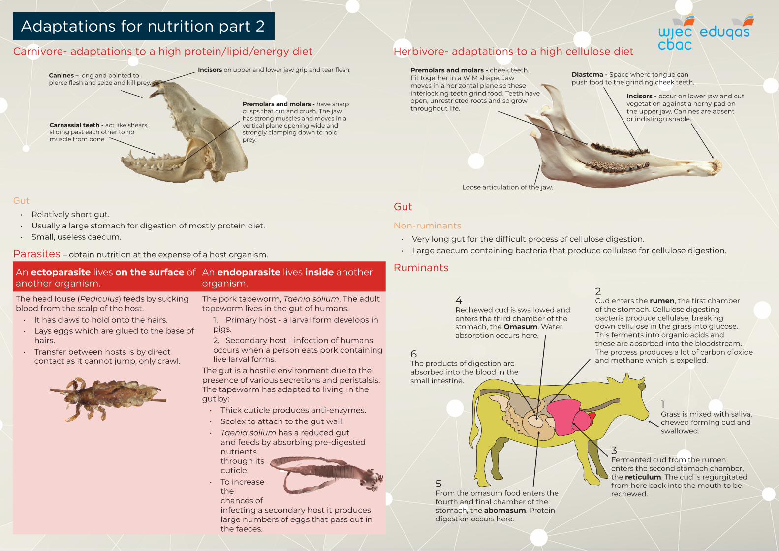
Parasites – obtain nutrition at the expense of a host organism.
An ectoparasite lives on the surface of another organism.
An endoparasite lives inside another organism.
The head louse (Pediculus) feeds by sucking blood from the scalp of the host.
• It has claws to hold onto the hairs. • Lays eggs which are glued to the base of
hairs. • Transfer between hosts is by direct
contact as it cannot jump, only crawl.
The pork tapeworm, Taenia solium. The adult tapeworm lives in the gut of humans.
1. Primary host - a larval form develops in pigs.2. Secondary host - infection of humans occurs when a person eats pork containing live larval forms.
The gut is a hostile environment due to the presence of various secretions and peristalsis. The tapeworm has adapted to living in the gut by:
• Thick cuticle produces anti-enzymes.• Scolex to attach to the gut wall. • Taenia solium has a reduced gut
and feeds by absorbing pre-digested nutrients through its cuticle.
• To increase the chances of infecting a secondary host it produces large numbers of eggs that pass out in the faeces.
Herbivore- adaptations to a high cellulose diet
Diastema - Space where tongue can push food to the grinding cheek teeth.
Premolars and molars - cheek teeth. Fit together in a W M shape. Jaw moves in a horizontal plane so these interlocking teeth grind food. Teeth have open, unrestricted roots and so grow throughout life.
Loose articulation of the jaw.
Incisors - occur on lower jaw and cut vegetation against a horny pad on the upper jaw. Canines are absent or indistinguishable.
Carnivore- adaptations to a high protein/lipid/energy diet
Incisors on upper and lower jaw grip and tear flesh.Canines – long and pointed to pierce flesh and seize and kill prey.
Carnassial teeth - act like shears, sliding past each other to rip muscle from bone.
Premolars and molars - have sharp cusps that cut and crush. The jaw has strong muscles and moves in a vertical plane opening wide and strongly clamping down to hold prey.
Gut • Relatively short gut.• Usually a large stomach for digestion of mostly protein diet.• Small, useless caecum.
Adaptations for nutrition part 2
Gut
Non-ruminants• Very long gut for the difficult process of cellulose digestion.• Large caecum containing bacteria that produce cellulase for cellulose digestion.
Ruminants
1Grass is mixed with saliva, chewed forming cud and swallowed.
2Cud enters the rumen, the first chamber of the stomach. Cellulose digesting bacteria produce cellulase, breaking down cellulose in the grass into glucose. This ferments into organic acids and these are absorbed into the bloodstream. The process produces a lot of carbon dioxide and methane which is expelled.
4Rechewed cud is swallowed and enters the third chamber of the stomach, the Omasum. Water absorption occurs here.
6The products of digestion are absorbed into the blood in the small intestine.
3Fermented cud from the rumen enters the second stomach chamber, the reticulum. The cud is regurgitated from here back into the mouth to be rechewed.
5From the omasum food enters the fourth and final chamber of the stomach, the abomasum. Protein digestion occurs here.