Acute Stroke Biomarkers: Are We There Yet?
16
REVIEW published: 05 February 2021 doi: 10.3389/fneur.2021.619721 Frontiers in Neurology | www.frontiersin.org 1 February 2021 | Volume 12 | Article 619721 Edited by: Emmanuel Pinteaux, The University of Manchester, United Kingdom Reviewed by: Theodoros Karapanayiotides, Aristotle University of Thessaloniki, Greece Jialing Liu, University of California, San Francisco, United States *Correspondence: Marie Dagonnier [email protected] Specialty section: This article was submitted to Stroke, a section of the journal Frontiers in Neurology Received: 21 October 2020 Accepted: 14 January 2021 Published: 05 February 2021 Citation: Dagonnier M, Donnan GA, Davis SM, Dewey HM and Howells DW (2021) Acute Stroke Biomarkers: Are We There Yet? Front. Neurol. 12:619721. doi: 10.3389/fneur.2021.619721 Acute Stroke Biomarkers: Are We There Yet? Marie Dagonnier 1,2 *, Geoffrey A. Donnan 1,3 , Stephen M. Davis 3 , Helen M. Dewey 1,4 and David W. Howells 1,5 1 Stroke Division, Melbourne Brain Centre, The Florey Institute of Neuroscience and Mental Health, Melbourne, VIC, Australia, 2 Department of Neurology, Ambroise Paré Hospital, Mons, Belgium, 3 Melbourne Brain Centre at the Royal Melbourne Hospital and University of Melbourne, Melbourne, VIC, Australia, 4 Eastern Health Clinical School, Monash University, Melbourne, VIC, Australia, 5 Faculty of Health, School of Medicine, University of Tasmania, Hobart, TAS, Australia Background: Distinguishing between stroke subtypes and knowing the time of stroke onset are critical in clinical practice. Thrombolysis and thrombectomy are very effective treatments in selected patients with acute ischemic stroke. Neuroimaging helps decide who should be treated and how they should be treated but is expensive, not always available and can have contraindications. These limitations contribute to the under use of these reperfusion therapies. Aim: An alternative approach in acute stroke diagnosis is to identify blood biomarkers which reflect the body’s response to the damage caused by the different types of stroke. Specific blood biomarkers capable of differentiating ischemic from hemorrhagic stroke and mimics, identifying large vessel occlusion and capable of predicting stroke onset time would expedite diagnosis and increase eligibility for reperfusion therapies. Summary of Review: To date, measurements of candidate biomarkers have usually occurred beyond the time window for thrombolysis. Nevertheless, some candidate markers of brain tissue damage, particularly the highly abundant glial structural proteins like GFAP and S100β and the matrix protein MMP-9 offer promising results. Grouping of biomarkers in panels can offer additional specificity and sensitivity for ischemic stroke diagnosis. Unbiased “omics” approaches have great potential for biomarker identification because of greater gene, protein, and metabolite coverage but seem unlikely to be the detection methodology of choice because of their inherent cost. Conclusion: To date, despite the evolution of the techniques used in their evaluation, no individual candidate or multimarker panel has proven to have adequate performance for use in an acute clinical setting where decisions about an individual patient are being made. Timing of biomarker measurement, particularly early when decision making is most important, requires urgent and systematic study. Keywords: stroke, biomarker, review, microarray, acute BIOMARKERS Use of the term “biomarker” describes measures of biological function was first seen in Medline is 1977 and has exploded in the last decade (1). A US National Institutes of Health working group defined a biomarker as: “a characteristic that is objectively measured and evaluated as an indicator of normal biological processes, pathogenic processes, or pharmacologic responses to a therapeutic
Transcript of Acute Stroke Biomarkers: Are We There Yet?

REVIEWpublished: 05 February 2021
doi: 10.3389/fneur.2021.619721
Frontiers in Neurology | www.frontiersin.org 1 February 2021 | Volume 12 | Article 619721
Edited by:
Emmanuel Pinteaux,
The University of Manchester,
United Kingdom
Reviewed by:
Theodoros Karapanayiotides,
Aristotle University of
Thessaloniki, Greece
Jialing Liu,
University of California, San Francisco,
United States
*Correspondence:
Marie Dagonnier
Specialty section:
This article was submitted to
Stroke,
a section of the journal
Frontiers in Neurology
Received: 21 October 2020
Accepted: 14 January 2021
Published: 05 February 2021
Citation:
Dagonnier M, Donnan GA, Davis SM,
Dewey HM and Howells DW (2021)
Acute Stroke Biomarkers: Are We
There Yet? Front. Neurol. 12:619721.
doi: 10.3389/fneur.2021.619721
Acute Stroke Biomarkers: Are WeThere Yet?Marie Dagonnier 1,2*, Geoffrey A. Donnan 1,3, Stephen M. Davis 3, Helen M. Dewey 1,4 and
David W. Howells 1,5
1 Stroke Division, Melbourne Brain Centre, The Florey Institute of Neuroscience and Mental Health, Melbourne, VIC, Australia,2Department of Neurology, Ambroise Paré Hospital, Mons, Belgium, 3Melbourne Brain Centre at the Royal Melbourne
Hospital and University of Melbourne, Melbourne, VIC, Australia, 4 Eastern Health Clinical School, Monash University,
Melbourne, VIC, Australia, 5 Faculty of Health, School of Medicine, University of Tasmania, Hobart, TAS, Australia
Background: Distinguishing between stroke subtypes and knowing the time of stroke
onset are critical in clinical practice. Thrombolysis and thrombectomy are very effective
treatments in selected patients with acute ischemic stroke. Neuroimaging helps decide
who should be treated and how they should be treated but is expensive, not always
available and can have contraindications. These limitations contribute to the under use
of these reperfusion therapies.
Aim: An alternative approach in acute stroke diagnosis is to identify blood biomarkers
which reflect the body’s response to the damage caused by the different types of stroke.
Specific blood biomarkers capable of differentiating ischemic from hemorrhagic stroke
and mimics, identifying large vessel occlusion and capable of predicting stroke onset
time would expedite diagnosis and increase eligibility for reperfusion therapies.
Summary of Review: To date, measurements of candidate biomarkers have usually
occurred beyond the time window for thrombolysis. Nevertheless, some candidate
markers of brain tissue damage, particularly the highly abundant glial structural proteins
like GFAP and S100β and the matrix protein MMP-9 offer promising results. Grouping
of biomarkers in panels can offer additional specificity and sensitivity for ischemic stroke
diagnosis. Unbiased “omics” approaches have great potential for biomarker identification
because of greater gene, protein, and metabolite coverage but seem unlikely to be the
detection methodology of choice because of their inherent cost.
Conclusion: To date, despite the evolution of the techniques used in their evaluation, no
individual candidate or multimarker panel has proven to have adequate performance for
use in an acute clinical setting where decisions about an individual patient are being
made. Timing of biomarker measurement, particularly early when decision making is
most important, requires urgent and systematic study.
Keywords: stroke, biomarker, review, microarray, acute
BIOMARKERS
Use of the term “biomarker” describes measures of biological function was first seen in Medline is1977 and has exploded in the last decade (1). A US National Institutes of Health working groupdefined a biomarker as: “a characteristic that is objectively measured and evaluated as an indicatorof normal biological processes, pathogenic processes, or pharmacologic responses to a therapeutic
Dagonnier et al. Acute Stroke Biomarkers
intervention” (2). While the term “biomarker” can includeclinical or imaging measurements, it is usually reserved fordescribing molecules found in bodily fluids (1).
Biomarkers such as cardiac troponin, creatine kinase, or D-dimer are used in practice in the emergency department forthe diagnosis and early management of the life-threateningconditions including myocardial infarction or pulmonaryembolism. Indeed, D-dimer measurements are used for theexclusion of a diagnosis of pulmonary embolism with asensitivity of 96%. A negative D-dimer test will virtually ruleout thromboembolism (3). Cardiac troponin (and especially the Iisoform) is used routinely to diagnose myocardial infarction witha sensitivity of more than 90% for a cut off value of 0.04 ng/ml (4).
Other biomarkers are used as tools for disease stating (e.g.,carcinoembryonic antigen-125 for cancers), for classificationof disease severity (e.g., blood prostate-specific antigenconcentration to indicate prostate cancer growth andmetastasis), to assess disease prognosis (e.g., measurementof tumor shrinkage) or to aid therapeutic monitoring (e.g., bloodcholesterol concentrations during therapy to reduce the risk ofheart disease) (2).
THE NEED FOR ACUTE STROKEBIOMARKERS
Five interventions improve outcome in patients with ischemicstroke. These are thrombolysis with recombinant tissueplasminogen activator (rt-PA) (5), aspirin given within 48 h(6), management of the patients within a dedicated stroke unit(7), hemicraniectomy (8), and more recently endovascular clotretrieval (9).
Thrombolysis is currently recommended for IS patients thatpresent within 4.5 h of stroke onset. Advanced neuroimagingallows extension of this time window up to 9 h and inclusionof patients that wake up with stroke symptoms if salvageablebrain tissue can be identified (10, 11). Nevertheless, thrombolysisis disappointingly infrequent in patients with acute ischemicstroke. Indeed, <10% of ischemic stroke patients receive thistherapy in most centers and no more than a third in the bestperforming centers (12–16). The main reasons for this underuseare uncertainty about stroke type, how long the ischemia hasbeen present diagnosis and the associated risks of cerebralbleeding (17–21).
Thrombectomy is currently recommended in IS patients (afteror independently from rt-PA) with evidence of large vesselproximal anterior circulation occlusion andwithin 6 (or 24 hwithadvanced imaging selection) of symptoms onset (9, 22–24). Thisrevolutionary treatment is unfortunately not in more widespreaduse than thrombolysis as it is estimated that fewer than 10% ofacute IS patients would meet the eligibility criteria and not allstroke centers have sufficient resources and expertise to deliverthis therapy (25, 26).
Brain imaging currently plays a critical biomarker role in acutestroke management as it is the only proven way to differentiateischemic from hemorrhagic stroke. Advanced perfusion imagingcan also be used to help select patients that might benefit from
rt-PA or thrombectomy under specific circumstances (10, 11, 23,24). Nevertheless, imaging cost, availability, contraindications,as well as the level of expertise required to interpret advancedimaging results, restricts the global use of reperfusion therapies.
Other less expensive more and accessible stroke biomarkersdetected in the blood would be an important addition to thestroke clinician’s armory.
An ideal stroke biomarker(s) should be able, with highspecificity and sensitivity, to differentiate hemorrhagic andischemic stroke (and clearly distinguish them from strokemimics). They should predict stroke prognosis, facilitatetherapeutic stratification and therapeutic monitoring, forexample by indicating risk of hemorrhagic transformation afterstroke or after rt-PA treatment. Moreover, if repeated measurescan be made in a clinically useful time frame, specific strokebiomarkers could act as a “stroke clock” to aid in assessing timeof stroke onset to increase the number of IS able to benefit fromtreatment with rt-PA, especially those who wake-up with stroke.
With the advent of mechanical thrombectomy, brain imagingwith vascular sequences has become a de facto standard inthe management of an acute stroke. Nevertheless, a biomarkerthat provides the same information would facilitate and fastenthe access to therapies. It would have the potential to aidearly identification and pre-hospital stratification of ischemicstroke patients. Indeed, biomarker stratification of the differentclasses of stroke patients in a pre-hospital setting would facilitatedirecting them to a hospital where thrombectomy is performedwithout losing crucial time by performing brain imaging inthe nearest hospital and then transferring the patient to thecomprehensive stroke center. It is known that substantial delaysof 110–128min are associated with secondary transfer vs. thedirect approach (27).
Over 150 candidate stroke biomarkers have been studied forroles ranging from diagnosis to long term prognosis (28–35).
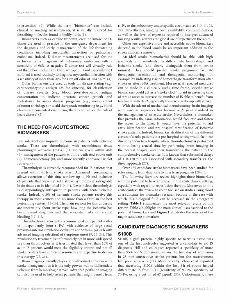
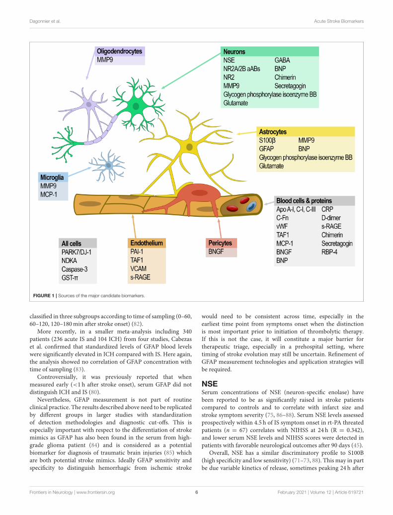
The following literature review highlights those biomarkerswith the potential to have an impact in the acute clinical setting,especially with regard to reperfusion therapy. Moreover, in thisacute context, the review has been focused on studies using bloodas a substrate for biomarker research because of the ease withwhich this biological fluid can be accessed in the emergencysetting. Table 1 summarizes the most relevant results of thisreview. Table 2 highlights the main clinical uses ascribed to thepotential biomarkers and Figure 1 illustrates the sources of themajor candidate biomarkers.
CANDIDATE DIAGNOSTIC BIOMARKERS
S100BS100B, a glial protein, highly specific to nervous tissue, wasone of the first molecules suggested as a candidate to aid ISdiagnosis. Hill and colleagues reported a specificity of morethan 95% for S100B measured on the first day of admissionin 28 non-consecutive stroke patients but the measurementhad poor sensitivity (71). More recently, Zhou et al. reportedthat measuring S100B within the first 6 h of stroke helpeddifferentiate IS from ICH (sensitivity of 95.7%, specificity of70.4%, using a cut-off of 67 pg/ml) (36). Unfortunately, these
Frontiers in Neurology | www.frontiersin.org 2 February 2021 | Volume 12 | Article 619721

Dagonnier et al. Acute Stroke Biomarkers
TABLE 1 | Summary of the most relevant studies and results of stroke biomarkers.
Biomarker Function tested Timing of sampling Sensitivity (%) Specificity (%) Cut off value n References
S100B Differentiation between
IS and ICH
Within 6 h of symptom
onset
95.7 70.4 67 pg/ml 71 IS and 46 ICH (36)
Differentiation between
stroke and mimics
24 h after symptom
onset
94.4 31.8 0.0415 ng/ml 31 IS and 22 mimics (37)
Risk of hemorrhagic
transformation after
rt-PA
Within 6 h of symptom
onset
82 46 >0.23 g/l 275 rt-PA treated IS (38)
Risk of malignant
oedema
At 8, 12, 16, 20, and
24 h after symptom
onset
75 (at 12 h)
94 (at 24 h)
80 (at 12 h)
83 (at 24 h)
>0.35 g/l (at 12 h)
>1.03 g/l (at 24 h)
16 malignant,
35 non-malignant
(39)
GFAP Differentiation between
IS and ICH
Within 4.5 h of symptom
onset
84.2 96.3 2.9 ng/l 163 IS, 39 ICH and 3
mimics
(40)
Differentiation between
IS and ICH
Between 2 and 6 h of
symptom onset
86 76.9 0.7 ng/ml 65 IS and 43 ICH (41)
Differentiation between
IS and ICH
Within 4.5 h of symptom
onset
61 96 34 ng/ml 79 IS and 45 ICH (42)
Differentiation between
IS and ICH
Within 6 h of symptom
onset
77.8 94.2 0.03 g/l 146 I and 46 ICH (43)
Differentiation between
IS, ICH and mimics
Within 6 h of symptom
onset
91 97 0.43 ng/ml 121 IS, 34 ICH, 31
mimics, 5 SAH, and 79
controls
(44)
NSE Favorable outcome after
rt-PA
Within 4.5 h of symptom
onset
77.1 59.4 13.90 ng/ml 67 rt-PA treated IS (45)
MMP-9 Risk of hemorrhagic
transformation after
rt-PA
Within 3 h of symptom
onset
92 74 >140 ng/ml 134 rt-PA treated IS (46)
NR2A/2B aAbs Differentiation between
stroke and controls
Within 3 h of symptom
onset
95 97 2.0 µg/l 31 IS, 56 TIAs, and 255
controls
(47)
NR2 Differentiation between
stroke, mimics and
controls
Within 72 h of symptom
onset
92 96 1.0 µg/l 101 IS, 91 non-stroke and
52 controls
(48)
Apo C-III Differentiation between
IS and ICH
Within 6 h of symptom
onset
94 87 36 16 IS and 15 ICH (49)
Apo C-I Differentiation between
IS and ICH
Within 6 h of symptom
onset
94 73 60 16 IS and 15 ICH (49)
Apo B Differentiation between
stroke and controls
After a period of
overnight fasting for 12 h
96 94 144 mg/dl 50 strokes and 50
controls
(50)
Apo A-I Differentiation between
stroke and controls
After a period of
overnight fasting for 12 h
88 86 114 mg/dl 50 strokes and 50
controls
(50)
Apo B/Apo A-I Differentiation between
stroke and controls
After a period of
overnight fasting for 12 h
98 96 1.2 50 strokes and 50
controls
(50)
PARK 7 Differentiation between
stroke and controls
On admission (median
of 17 h after symptom
onset)
AUC = 0.897; OR = 1.087 – 72 strokes and 78
controls
(51)
NDKA Differentiation between
stroke and controls
On admission (median
of 17 h after symptom
onset)
AUC = 0.462; OR = 0.882 – 72 strokes and 78
controls
(51)
Glycogen
phosphorylase
isoenzyme BB
Differentiation between
stroke and controls
Within 12 h of symptom
onset
93 93 7.0 ng/ml 172 IS and 133 controls (52)
c-Fn Risk of hemorrhagic
transformation after
rt-PA
Within 3 h of symptom
onset
100 60 3.6µg/ml 27 rt-PA treated IS (46)
Risk of malignant
oedema
On admission (mean
time of 6–7 h after
symptom onset)
90 100 >16.6µg/ml 40 malignant and 35
non-malignant
(53)
(Continued)
Frontiers in Neurology | www.frontiersin.org 3 February 2021 | Volume 12 | Article 619721

Dagonnier et al. Acute Stroke Biomarkers
TABLE 1 | Continued
Biomarker Function tested Timing of sampling Sensitivity (%) Specificity (%) Cut off value n References
PAI-1 and TAFI Risk of hemorrhagic
transformation after
rt-PA
Within 3 h of symptom
onset
75 97.6 PAI-1 <21.4 ng/mL
and TAFI >180%
77 rt-PA treated IS (54)
Glutamate Risk of early neurological
deterioration
On admission (mean
time of 9–10 h after
symptom onset)
81 87 >200 µmol/l 27 progressing and 86
non-progressing lacunar
strokes
(55)
GABA Risk of early neurological
deterioration
On admission (mean
time of 9–10 h after
symptom onset)
96 94 <240 nmol/l 27 progressing and 86
non-progressing lacunar
strokes
(55)
Combination of
S100B, BNGF,
vWF, MMP-9, and
MCP-1
Differentiation between
stroke and controls
Within 12 h of symptom
onset
91 97 – 223 strokes and 214
controls
(56)
Combination of
S100B, vWF,
MMP-9, and
VCAM
Differentiation between
stroke and controls
Within 24 h of symptom
onset
90 90 – 65 IS and 157 controls (57)
Combination of
S100B, MMP-9,
D-dimer, BNP, and
CRP
Differentiation between
stroke and controls
Within 6 h of symptom
onset
81 70 – 130 patients with focal
neurological deficit
(58)
Combination of
S100B, MMP-9,
D-dimer, and BNP
Differentiation between
stroke and controls
Within 24 h of symptom
onset
86 37 – 1,100 patients with focal
neurological deficit
(59)
Combination of
MMP-9, D-Dimer,
sRAGE,
caspase-3,
chimerin, and
secretagogin
Differentiation between
stroke and mimics
Within 24 h of symptom
onset
Overall accuracy of the model:
0.91
– 915 strokes and 90
mimics
(60)
S100B and
sRAGE
Differentiation between
IS and ICH
Within 3 and 6 h of
symptom onset
22.7 80.2 S100B >96 pg/ml
and sRAGE
<0.97 ng/ml
776 IS and 139 ICH (61)
GFAP and RBP4 Differentiation between
IS and ICH
Within 6 h of symptom
onset
– 100 GFAP <0.07 ng/ml
and RBP4 >61
lg/mL
38 IS and 28 ICH (62)
Panel of 22 genes Differentiation between
stroke and controls
<24, 24–48, >48 h after
symptom onset
78 80 – 20 IS and 20 controls (63)
Panel of 18 genes Differentiation between
stroke and controls
Within 3 h, at 5 h and at
24 h after symptom
onset
With accuracy in 66% within 3 h,
86% at 5 h and 100% at 24 h
– 15 IS and 8 controls (64)
Panel of 23 genes Differentiation between
stroke etiologies
Within 3 h, at 5 h and at
25 h after symptom
onset
95.2 95.2 – 15 IS (65)
Panel of 40 genes Differentiation between
cardio-embolic and
large vessel strokes
At 3 h, at 5 h and at 25 h
after symptom onset
>90 >90 – 76 IS (66)
Panel of 34 genes Differentiation between
TIA and patient with
CVD
From 9 to 68 h (mean
35 h) after symptom
onset
100 100 – 26 TIAs and 26 controls (67)
Panel of 26 genes Differentiation between
IS or TIA and controls
Within 72 h of symptom
onset
89 89 – 94 IS, 26 TIAs, and 44
controls
(68)
Panel of 41 genes Differentiation between
lacunar and non-lacunar
strokes
Within 72 h of symptom
onset
>90 >90 – 30 lacunar and 86
non-lacunar strokes
(69)
GST-π Discrimination between
early (<3 h) and late (3 h)
presentation of stroke
Within 3 h and after 3 h
of symptom onset
AUC = 0.79; OR = 10 17.7 µg/l 103 IS and 132 controls (70)
Frontiers in Neurology | www.frontiersin.org 4 February 2021 | Volume 12 | Article 619721

Dagonnier et al. Acute Stroke Biomarkers
TABLE 2 | Main clinical uses and their linked potential biomarkers.
Differentiation between
stroke and controls
S100B
NSE
NR2A/2B aAbs
Apo B, Apo A-I, and Apo B/Apo A-I ratio
CRP
P-Selectin
Homocysteine
BNP
D-dimer
Combination of S100B, MMP-9, vWF, BNGF,
and MCP-1
Combination of S100B, MMP-9, vWF, and
VCAM
Combination of S100B, MMP-9, D-dimer, BNP,
and CRP
Combination of S100B, MMP-9, D-dimer, and
BNP
Combination of MMP-9, D-Dimer, sRAGE,
caspase-3, chimerin, and secretagogin
Panel of genes
Differentiation between IS
and ICH
S100B
GFAP
Apo C-I and Apo C-III
BNP
S100B and sRAGE
GFAP and RBP4
Risk of hemorrhage after
rt-PA
S100B
NSE
MMP-9
c-Fn
PAI-1 and TAFI
Correlation with
hemorrhage volume
GFAP
Correlation with stroke
severity and infarct size
S100B
NSE
MMP-9
Correlation with favorable
neurological outcome
NSE
Acting as a stroke clock NSE
NR2
GST-π
PARK7
NDKA
Risk of early neurological
deterioration
MMP-9
Glutamate
IL-6
TNF-α
ICAM-1
Risk of malignant oedemaS100B
MMP-9
c-Fn
Stroke etiology Panel of genes
results were not substantiated by Gonzalez-García’s 2012 studywhere S100B, measured between 8 and 48 h of symptom onsetwas significantly elevated in stroke compared to controls butfailed to differentiate between IS and ICH and did not correlatewith stroke severity on admission (72) despite other studiessuggesting that S100B concentrations correlated with strokeseverity and size of infarction (73–75).
Serum S100B concentration measured 24 h after symptomonset is significantly higher in stroke patients (posteriorcirculation IS or infratentorial ICH, no distinction was made inthe analysis) than in controls or in patients with vertigo fromnon-vascular causes (37). However, in addition to prolonged anddelayed release into the blood after stroke, S100B levels are alsoincreased in other neurological pathologies such as traumaticbrain injuries and extracranial malignancies (76, 77).
Never-the-less, elevated S100B concentration (>0.23 g/l) hasbeen associated with hemorrhage risk due to rt-PA treatment(specificity of 82%, sensitivity 46%) (38) suggesting with furtherwork, this biomarker may have some utility.
GFAPGFAP (glial fibrillary acidic protein), another glial protein specificto astrocytes (78), is the best candidate to date for differentiatinghemorrhage and ischemic stroke. Based on detection of delayedGFAP release in patients with ischemic stroke (maximumconcentration reached 2–4 days after ischemic stroke onset),Foerch and his team studied this molecule in different clinicalsettings and showed promising results (79–81). In a multicenterclinical study of 205 patients (163 with IS, 39 with ICHand three stroke mimics) diagnostic accuracy was high fordifferentiating intracerebral hemorrhage from ischemic stroke byGFAP immunoassay on a single blood sample obtained within4.5 h of symptom onset. Using GFAP cut-off of 2.9 ng/l provideda specificity of 96.3% and a sensitivity of 84.2% for distinguishingICH, IS and stroke mimics (40). In addition, the levels ofGFAP were shown to be correlated with the hemorrhage volume(40, 79, 80).
Several studies have confirmed the potential for serum GFAPto distinguish IS and ICH. Xiong et al. showed that the GFAPconcentration in blood collected within 2–6 h after symptomonset was significantly higher in ICH (n = 43) than IS (n =
65) patients, with 86 and 76.9% sensitivity and specificity of,respectively, using a cut-off point of 0.7 ng/ml) (41). Ren et al.replicated these findings using a GFAP cut off value of 0.34 ng/mlwith 61% sensitivity and 96% specificity in a 4.5 h time windowfrom symptoms onset (42). A later study measuring serum GFAPin 46 ICH and 146 IS patients reported 77.8 and 94.2% sensitivityand specificity distinguishing the two stroke subtypes using acut-off value of 0.03 g/l within 6 h of diagnosis (43).
Similarly, Katsanos et al. reported that in samples frompatients presenting within 6 h from symptoms onset, significantlyraised median plasma GFAP concentrations detected in ICHvs. IS, stroke mimetics, and controls. A cut-off of 0.43 ng/mLprovided the best threshold for differentiation between ICHand AIS (sensitivity of 91% and specificity of 97%). They alsodescribed that the best timing of sampling to allow optimaldifferential diagnostic between IS and ICH was in the secondhour from symptom onset (44).
A meta-analysis including nearly 1,300 patients confirmedthe potential of measuring GFAP in the blood in the earlyphase of stroke (samples drawn <3 h from symptoms onset),to discriminate IS, ICH and mimics. Interestingly, there was nosignificant difference in diagnosis accuracy when patients were
Frontiers in Neurology | www.frontiersin.org 5 February 2021 | Volume 12 | Article 619721
Dagonnier et al. Acute Stroke Biomarkers
FIGURE 1 | Sources of the major candidate biomarkers.
classified in three subgroups according to time of sampling (0–60,60–120, 120–180min after stroke onset) (82).
More recently, in a smaller meta-analysis including 340patients (236 acute IS and 104 ICH) from four studies, Cabezaset al. confirmed that standardized levels of GFAP blood levelswere significantly elevated in ICH compared with IS. Here again,the analysis showed no correlation of GFAP concentration withtime of sampling (83).
Controversially, it was previously reported that whenmeasured early (<1 h after stroke onset), serum GFAP did notdistinguish ICH and IS (80).
Nevertheless, GFAP measurement is not part of routineclinical practice. The results described above need to be replicatedby different groups in larger studies with standardizationof detection methodologies and diagnostic cut-offs. This isespecially important with respect to the differentiation of strokemimics as GFAP has also been found in the serum from high-grade glioma patient (84) and is considered as a potentialbiomarker for diagnosis of traumatic brain injuries (85) whichare both potential stroke mimics. Ideally GFAP sensitivity andspecificity to distinguish hemorrhagic from ischemic stroke
would need to be consistent across time, especially in theearliest time point from symptoms onset when the distinctionis most important prior to initiation of thrombolytic therapy.If this is not the case, it will constitute a major barrier fortherapeutic triage, especially in a prehospital setting, wheretiming of stroke evolution may still be uncertain. Refinement ofGFAP measurement technologies and application strategies willbe required.
NSESerum concentrations of NSE (neuron-specific enolase) havebeen reported to be as significantly raised in stroke patientscompared to controls and to correlate with infarct size andstroke symptom severity (75, 86–88). Serum NSE levels assessedprospectively within 4.5 h of IS symptom onset in rt-PA threatedpatients (n = 67) correlates with NIHSS at 24 h (R = 0.342),and lower serum NSE levels and NIHSS scores were detected inpatients with favorable neurological outcomes after 90 days (45).
Overall, NSE has a similar discriminatory profile to S100B(high specificity and low sensitivity) (71–73, 88). This may in partbe due variable kinetics of release, sometimes peaking 24 h after
Frontiers in Neurology | www.frontiersin.org 6 February 2021 | Volume 12 | Article 619721

Dagonnier et al. Acute Stroke Biomarkers
stroke (89, 90). Interestingly, Kim et al. showed that IS patientsidentified as having a second peak of serum NSE (20% of thestudied population) were more at risk of developing hemorrhagictransformation (OR = 6.8) (90). Therefore, while NSE is notcurrently recommended for the diagnosis of acute stroke it maystill have clinical potential.
MMP-9Expectations have been high for MMP-9 (matrixmetalloproteinase-9) as a stroke diagnostic biomarker becauseof its role in response to brain injury via its involvementin extracellular matrix degradation. MMP-9 concentrationsmeasured acutely have been linked increased to infarct size,worse neurological outcome, and complications of hemorrhagictransformation (46, 91–96). Serum MMP-9 concentrations≥140 ng/ml were shown to predict hemorrhagic transformationin rt-PA treated ischemic stroke patients (sensitivity 92%,specificity 74%) (46). Six other studies confirmed the correlationbetween MMP-9 concentration and increased bleeding riskafter rt-PA (96). Similarly, Barr et al. identified an associationbetween elevated serum concentrations of MMP-9 and bloodbrain barrier disruption which is a key feature of hemorrhagictransformation (97). However, the rise of MMP-9 is not specificto ischemic stroke, moreover its concentration is reported topeak at 24 h post stroke (96), too late for making decisions aboutthrombolysis, and standardization of MMP-9 measurements andexperimental replication are still required.
NMDA-RAutoantibodies to the glutamate NMDA-R (N-methyl-Daspartate receptors; NR2A/NR2B subunits) associated withneurotoxicity are elevated after stroke and distinguish IS patients(n = 31) from controls 3 h after symptoms onset with 97%sensitivity and 98% specificity (47). In a different cohort, plasmalevels of NMDA-R NR2A were also shown to be elevated inischemic strokes when there was no difference observed inpatients with cerebral hemorrhage in comparison to controls(98). Criticism for the potential use of NMDA-R antibodiesfor the diagnosis of ischemic stroke were raised as NMDA-R antibodies have also been detected in patients with priorstroke hypertension, atherosclerosis, epilepsy, systemic lupuserythematous, and encephalitis (47, 98–100). Nevertheless, morerecently, NR2 peptide (a product of degradation of NMDA-R) inblood has been reported to distinguish IS from stroke mimetics,patients with vascular risk factors and controls with 92 and 96%sensitivity and specificity, respectively (48). On the negativeside, NR2 levels might not be increased in lacunar and smallcortical strokes (48). Interestingly, detection of NMDA-R NR2Aantibodies and NR2 concentrations might have a temporalprofile after ischemic stroke (with a peak after 12 h) (48, 98)that might contribute to pinpointing a patients stage of strokeevolution but these results need to be validated in specificallydesigned studies.
Apo-lipoproteinsSome members of the apo-lipoprotein family have also beentested as potential biomarkers for IS diagnosis. Apo C-I and Apo
C-III concentrations were found to be increased in IS comparedto ICH within 6 h of symptom onset and both were reported tohave the potential to discriminate IS from ICH. For Apo C-IIIthis was achieved with 94 and 87% sensitivity and specificity,respectively (49). A panel of nine apo-lipoproteins was testedas a tool to distinguish IS and ICH patients within the firstweek after symptom onset using a mass spectrometry assay. ApoC-I and Apo C-III reported to provide the best classificationpower as individual markers but combining Apo C-III andApo A-I provided the best discrimination overall (AUC = 0.92)(101). Unfortunately, these results were not confirmed by Walshand colleagues who looked at a broader panel which includedparaoxonase-1, MMP- 3 and 9 and Apo A-I, C-I, and C-III fortheir ability to distinguish between IS, ICH patients and controlson blood samples obtained within 12 h of symptom onset. Inthis cohort, the levels of Apo A-I, Apo C-I, and paraoxonase-1,were shown to be lower in IS than in ICH patients with the othercandidates having no discriminatory value (102). It is intriguingto speculate that this stark difference might be accounted for bya temporal component to the expression profile that might itselfbe useful.
Others have taken a ratio-metric approach to the use ofapo-lipoprotein family members as stroke biomarkers. As et al.reported that Apo B concentrations and the Apo B/Apo A-Iratio were significantly elevated while levels of Apo A-I wassignificantly decreased in IS patients compared to controls.All three-potential biomarker tests were reported to have ahigh specificity and sensitivity to discriminate stroke patients(between 86 and 98%) (50). The Apo B/Apo A-I ratio has alsobeen associated with early neurological deterioration in largeartery atherosclerotic stroke, this was not found for other strokesubtypes (103).
OthersOther less well-studied candidates may also have merit. Forexample, Allard et al. described first in 2005 the potentialof PARK7 and NDKA (nucleoside diphosphate kinase A) asbiomarkers for stroke diagnosis as their plasma concentrationsincreased early after symptom onset (29, 104). However, theirspecificity and sensitivity as markers were dependent on thediagnostic cut-off values used (104) and the results still need to bereplicated. Tulantched et al. later specified that PARK7 seemed tohave a better prognostic value thanNDKA, both in sensitivity andspecificity. Once more, collection time in this prospective studywas late after stroke onset with a median of 17 h (51).
More recently, a prospective study found that glycogenphosphorylase isoenzyme BB measurements were able todiscriminate between 172 IS and 133 controls with 93%sensitivity and specificity (cut-off of 7.0 ng/mL and sample drawnwithin 12 h of onset) (52). Nevertheless, glycogen phosphorylasewhich metabolizes glycogen to provide glucose-1-phosphate torestore energy stores has also been identified as a potential markerof ischemic myocardial injury (105, 106).
A meta-analysis interrogating over 130 biomarkers publishedby Hasan et al. in 2012, concluded that C-reactive protein (CRP),P-selectin and homocysteine were the only three biomarkersable to significantly differentiate ischemic stroke from healthy
Frontiers in Neurology | www.frontiersin.org 7 February 2021 | Volume 12 | Article 619721

Dagonnier et al. Acute Stroke Biomarkers
patients (28). Nevertheless, once more, these three moleculeshave a low specificity for ischemic stroke and therefore precludetheir diagnostic use in acute stroke situations.
A systematic review performed by Misra et al. identified 10single biomarkers and seven biomarker panels with a potentialfor differentiating IS and ICH. Once more, GFAP appeared toperform well, either as a single marker or in association eitherwith the Activated Protein C- Protein C Inhibitor Complex(APC-PCI) or with the Retinol Binding Protein 4 (RBP4).Nevertheless, because the time of sampling was outside of thetime window for practical acute stroke intervention (31), theirclinical utility is still unclear. In another systematic review,Monbailliu and colleagues identified a different pairing ofdiagnostic biomarkers for consideration. BNP and S100 were theonly two blood-based proteins biomarkers in their study thatcould differentiate IS from ICH, stroke mimetics and healthycontrol subjects (32).
The most recent meta-analysis published in 2020 analyzed 25biomarkers across 40 studies and over 5,000 IS, 750 ICH, 550mimics, and 1,770 healthy controls on samples collected within24 h of symptoms onset. BNP, MMP-9, D-Dimer were identifiedto significantly differentiate the different patient groups whileGFAP was successful to differentiate IS from ICH within 6 h.S100B, caspase-3 and NSE only distinguished IS from strokemimics. Nevertheless, the authors highlighted that 67% of thestudies included had only moderate study quality suggesting theneed for further well-conducted studies (107).
While these markers all offer promise as diagnostic aids, untillarger validation studies tease out the reproducibility of diagnosis,specificity in different patient groups and the role of samplingwindow in the value of the measurements, the current level ofuncertainty does not recommend their immediate clinical use.
BIOMARKERS OF DISEASEPROGRESSION
In addition to the previously mentioned MMP-9, NSE, andS100B, other molecules are linked with increased bleedingrisk after IS. Plasma levels of c-Fn (cellular-fibronectin),which reflect vascular damage, have been associated with thedevelopment of hemorrhagic transformation following t-PA use(108). When evaluated in a second cohort of 27 subjects, serumc-Fn ≥3.6µg/ml identified hemorrhagic transformation with asensitivity of 100% but specificity of 60% (46). Combining c-Fnwith MMP-9 allowed detection of hemorrhagic transformationwith 92% sensitivity, 87% specificity, and a positive predictivevalue of 41% (46). Reduced levels of PAI-1 (plasminogenactivator inhibitor) and higher levels of TAFI (thrombin-activated fibrinolysis inhibitor), two endogenous fibrinolysisinhibitors, have been associated with symptomatic intracranialhemorrhage after thrombolysis therapy. When combined, PAI-1 level <21.4 ng/ml and a TAFI level >180% predictedsymptomatic intracranial hemorrhage after rt-PA (sensitivity andspecificity of 75 and 97.6%, respectively) (54).
Several biomarkers have been associated with earlyneurological deterioration (END). This has been defined as
neurological worsening between 48 and 72 h after admission andoccurs in one third ischemic stroke patients (109). Cytotoxicmechanisms mediated by glutamate, nitric oxide, and cytokinesand endothelial-leukocyte adhesion molecules have beenproposed as mediators of progression of tissue damage (110).
High plasma glutamate concentrations have been correlatedwith neurological worsening and infarct growth at 72 h afterstroke onset (55, 110). Plasma glutamate concentrations of>200 µmol/l on admission have a positive predictive value forneurological deterioration at 48 h after lacunar infarction of67% (55). Plasma GABA levels <240 nmol/l on admission alsohad a positive predictive value for neurological deteriorationat 48 h after lacunar infarction of 84% (55). Higher levels ofinflammatory markers such as ferritin, IL-6 (interleukine-6),TNF-α (tumor necrosis factor-α) and ICAM-1 (intercellularadhesion molecule-1) were also shown to be associated with earlyneurological worsening (110–112).
Space-occupying brain oedema (also called malignantoedema), an early life-threatening problem in patients withlarge hemispheric stroke, has been shown to be predicted by anelevated plasma S100B level (>0.35 g/l) with a 75% sensitivityand a 80% specificity at 12 h after stroke and even more at24 h (94 and 83% sensitivity and specificity, respectively) (39).c-Fn and MMP-9 concentrations have also been found to besignificantly higher in patients with malignant MCA (m-MCA)infarction than in controls. c-Fn concentrations of >16.6µg/mlprovided a 90% sensitivity and 100% specificity with 89 and100% negative and positive predictive values, respectively, forprediction of m-MCA infarction (53).
While more work is needed to determine precisely whenin a patient’s clinical course these measurements first providevaluable information about that individual’s likely outcome, theirgenerally high sensitivity and specificity suggest they will findclinical utility.
BIOMARKERS PANELS
To better account for the molecular complexity of the ischemiccascade and increase the sensitivity and specificity of biomarkersas diagnosis tools, many researchers have also investigatedbiomarker panels, evaluating multiple molecules simultaneouslyinstead of looking for a single biomarker. In a systematic review,Whiteley et al. identified seven panels of biomarkers testedas ischemic stroke diagnostic tools. The main criticisms werethat the multimarker panel studies did not provide regressionequations for stroke prediction and that a variety of cut-offvalues were used for the same biomarker. Moreover, the samplecollection time points usually occurred outside the windowwhere treatment was possible (29).
Reynolds et al. assessed plasma from 223 stroke patients(including IS, ICH and subarachnoid hemorrhage) and214 healthy controls for more than 50 serum biomarkersusing ELISAs (enzyme-linked immunosorbent assay). Thecombination of S100B, B-type neurotrophic growth factor(BNGF), von Willebrand factor (vWF), MMP-9, and monocytechemotactic protein-1 (MCP-1) provided diagnosis of stroke
Frontiers in Neurology | www.frontiersin.org 8 February 2021 | Volume 12 | Article 619721

Dagonnier et al. Acute Stroke Biomarkers
within 12 h after symptom onset with a 91% sensitivity and a97% specificity (56). A related panel of S100B, MMP-9, vWF, andvascular cell adhesion molecule (VCAM) studied by the samegroup of researchers in 65 suspected ischemic stroke patientsand 157 controls within 24 h of symptoms provided a sensitivityand specificity of 90% (57).
In 130 patients with acute focal neurologic deficits admittedwithin 6 h of onset of symptoms, a panel including D-dimer,CRP, B-type natriuretic protein (BNP), MMP-9, and S100B waspredictive of ischemic stroke with sensitivity and specificity of81 and 70%, respectively (58). While less specific and sensitivethan the preceding panels, its time window of application is moreappropriate to the acute stroke setting (6 vs. 24 h). However,when the same panel of markers, excluding CRP, was tested ina prospective multicenter trial of more than 1,100 patients whopresented with symptoms suggestive of stroke, a 86% sensitivityand 37% specificity were achieved for distinguishing stroke frommimics in the first 24 h after symptom onset (59).
In a study published in 2011, Montaner et al. tested, in anED setting, a panel of blood biomarkers including CRP, S100B,MMP-9, a soluble receptor for advanced glycation end products(sRAGE), D-Dimer, brain natriuretic peptide (BNP), caspase-3, neurotrophin-3, chimerin, and secretagogin. They identifiedlevels of caspase-3, D-dimer, sRAGE, chimerin, secretagogin,and MMP-9 as independent predictors of stroke vs. mimics.Moreover, they reported a predictive probability for identifyingstroke of 99.01% by combining set cut-off values of these sixbiomarkers (60). The same team have also demonstrated, ina cohort of 915 stroke patients, that just S100B and sRAGE,could distinguish between IS and ICH with an AUC of 0.76 forblood samples obtained within 3 h after symptom onset. Thiswas confirmed in blood samples obtained within 6 h of symptomonset (61).
More recently, measurement of retinol binding protein 4(RBP4) (with a cut off value >61 g/ml) and GFAP (with a cut offvalue of<0.07 ng/ml) was shown to distinguish IS from ICHwitha specificity of 100% in a cohort of 38 IS and 28 ICH samples (62).
In the STROKE-CHIP study, a prospective multicenter studyof over 1,300 patients, published in 2017, Bustamante et al.studied a panel of 21 biomarkers selected from prior studiesand published literature (including S100B, cFn, NSE, MMP-9e.g.) on blood samples collected immediately upon arrival ofpatients presenting within 6 h after symptom onset. None ofthese biomarkers were able to provide an accurate hyperacutedifferential diagnosis of stroke (113).
While adding complexity to the laboratory work required,panels of markers appear to have the potential to offer significantimprovements in specificity and sensitivity. However, furthervalidation is still clearly required.
THE mRNA REVOLUTION
The development of oligonucleotide microarray techniques, andmore recently RNAseq, opened new perspectives in the quest fordiscovery of specific acute stroke biomarkers. These techniquesallow unbiased investigation of the entire transcriptome as RNA
shed from damaged or communicating cells, or contained withinthe cells of the immune system, the body’s own “first responders”to injury. In addition, changes in mRNA expression occurvery quickly often before changes of protein expression can bedetected (114). This suits perfectly the acute ischemic strokesetting where “time is brain.”
Tang and colleagues reported a blood genomic responsespecific to ischemic stroke on blood samples collected at 24 hfrom rats subject to MCAo, sham surgery, and naïve controls.Twenty five genes were shown to be significantly (more than2-fold) over expressed in rat blood 24 h after induction ofischemia while 98 had decreased significantly in comparison tocontrols (115).
Using blood samples collected from 20 patients with ischemicstroke and in 20 controls and stratified for sampling time(<24 h n = 7, 24–48 h n = 10, and >48 h n = 3), Moore andcolleagues found that, after correction for multiple comparisons,190 genes were differentially expressed (comparing stroke andcontrol). Moreover, a panel of 22 genes identified as comingfrom peripheral mononuclear cells differentiated ischemic strokefrom controls with 78 and 80% sensitivity and specificity,respectively (63).
When bloods were sequentially collected within 3 h, at5 h and at 24 h from eight controls and 15 ischemic strokepatients [initially enrolled in the Combination approach to Lysisutilizing Eptifibatide And Recombinant tissue-type plasminogenActivator (CLEAR) trial], 104 genes were identified to have a1.5-fold change between ischemic stroke and controls at 3 h,1,106 at 5 h and 906 at 24 h. An 18-gene panel distinguishedbetween ischemic stroke and controls with accuracy in 75% ofthe cases or more at all 3 different time points (64). Genesincluded in this panel reflected the involvement of inflammationin the ischemic pathway but were different from thoseidentified by Moore.
When samples from the CLEAR trial were used to exploreRNA expression after different ischemic stroke etiologies, 77genes showed at least a 1.5-fold change in expression betweenlarge vessel occlusion and cardioembolic strokes. Twenty threeof these genes could distinguish these etiologies with >95%sensitivity and specificity (65).
However, when RNA isolated from the peripheral bloodmononuclear cells of acute ischemic stroke patients, strokesurvivors and patients with acute traumatic brain injury wasanalyzed (cohort n = 15–20, sampling time: 24–27 h after eventonset), no significant differences in single genes expressionwere identified between these groups. Nevertheless, expressionof PDE4D (phosphodiesterase 4 D), an enzyme metabolizingcyclic adenosine monophosphate in inflammatory cells, wassignificantly different between acute ischemic stroke patients andhealthy controls with cardiovascular risk factors (116).
A retrospective case-control study of 39 ischemic strokepatients and 25 controls (sampling time 10 ± 6.5 h), identifiednine genes whose expression was significantly different in strokepatients and involvement of toll-like receptor signaling in theischemic cascade (117). Five of these nine genes; MMP9, ARG1,CA4, LY96, and S100A12, had previously been reported asspecific for stroke (64).
Frontiers in Neurology | www.frontiersin.org 9 February 2021 | Volume 12 | Article 619721
Dagonnier et al. Acute Stroke Biomarkers
In a larger cohort of 194 blood samples collected at 3, 5,and 24 h after stroke from 76 patients with acute IS, a 40-genepanel distinguished cardio-embolic from large vessel strokes with>95% sensitivity and specificity. In addition, a 37-gene panelwas identified to be able to differentiate atrial fibrillation fromnon-atrial fibrillation causes of cardioembolic stroke with >90%sensitivity and specificity (66).
Zhan and colleagues took a different approach and comparedTIAwith ischemic stroke. In rats they showed that only brief focalischemia was needed to induce the majority of changes caused byischemic stroke (118).When the same group compared the bloodexpression profiles of TIA patients (n = 26) and control subjectswith vascular risk factor but without symptomatic cardiovasculardisease (n = 26), they identified 449 genes that distinguishedbetween the two groups. Thirty-four genes separated TIAs fromcontrols with 100% sensitivity and specificity. In addition, twodifferent patterns of gene expression were identified by clusteranalysis for the TIA patients suggesting a heterogeneous responseto the event between patients and a possible relation with a higherrisk of stroke (67).
These findings were soon tested in a bigger cohort by Jicklingand colleagues. In 164 blood samples collected within 72 h ofsymptom onset from stroke, TIA and control patients, 145 geneswere differentially expressed between TIA and controls and 413genes were significantly different between IS and controls. Moreimportantly, 74 of the 145 genes identified in the TIA groupwere also found in ischemic stroke patients. Twenty six of these74 common genes were used as a panel to distinguish strokeand TIA from controls with 89% sensitivity and specificity.Pathways analysis revealed that the genes common to strokeand TIA were involved in innate and adaptive immune systemsactivation involving B-cells and granulocytes (68). Unfortunately,the authors did not reveal the composition of their 26-gene panel,so comparison with the 34-genes panel identified earlier by Zhanet al. is not possible.
Jickling et al. also evaluated the gene expression profile oflacunar strokes. In a cohort of 30 lacunar and 86 non-lacunarstrokes (with blood sampling within 72 h of stroke onset), theyidentified a 41 genes discriminating lacunar and non-lacunarstroke with >90% sensitivity and specificity (69).
In 2012, Oh et al. performed microarray analysis on bloodsamples collected from 12 ischemic stroke patients and 12controls (sampling time 12.7 ± 5.3 h after stroke onset). Theyidentified 88 transcripts with a 1.5-fold change in ischemic strokecompared to controls and 11 transcripts with 2-fold difference(including MMP9, Il1R2). Then, they validated the expressionof the three most differently expressed genes (MMP9, Il18RAP,and GNLY) by quantitative polymerase chain reaction (qPCR).In another cohort of 120 ischemic stroke patients and 82 controls(sampling time 10.4 ± 9.7 h). MMP9 concentrations measuredusing ELISA were significantly greater in IS compared to controlsbut did not to correlate with infarct volume (119).
When quantitative PCR was used validate the expressionprofiles of 40 candidate biomarkers identified in previousstudies (63, 64, 117) in 18 ischemic stroke patients and 15controls (median time of blood sampling 36 h), 16 geneswere significantly upregulated in ischemic stroke patients in
comparison to controls. Six gene clusters were reported todiscriminate between stroke and controls and one of them,containing seven transcripts, was reported to show high accuracyfor stroke classification (120).
In common with the candidate protein biomarker studiesdescribed earlier, the investigators for the transcriptome studiessummarized above have tended (samples from the CLEAR trialare an obvious exception) to perform analyses relatively latein stroke evolution, when diagnosis is generally already certainand decisions about therapy already made. Moreover, there hasbeen little emphasis on distinguishing ischemic and hemorrhagicstroke, or identifying genes that might identify a heightened riskof bleeding. These gaps in the analysis are surprising. Arraytechnologies also lend themselves to collaborative re-analysis,indeed many publishers stipulate that array data be made freelyavailable. It is therefore also surprising that pooled analysis of theavailable data has not yet been performed.
BIOMARKERS OF A STROKE CLOCK
As mentioned above, stroke biomarker discovery has rarelyfocused on early temporal change, despite the dynamiccharacteristics of stroke. The possibility that changes inexpression of candidate biomarkers with time might help predictstroke evolution and act as a biological stroke clock whichcould allow more patients to be recruited to thrombolysis islargely unstudied.
In serial blood samples collected at 3, 6, 12, 18, 24, 48, 72, 96,and 120 h after onset of stroke symptoms, NSE concentration,measured by immune-assay, rose in the first 2–3 h, then felluntil 12 h before a second elevation that was maintained untilmeasurement ended on day 5. Tau concentration showed acontinuous increase from admission onward (87).
During a study evaluating the diagnostic performance of29 pre-selected molecules within the therapeutic windowfor thrombolysis in 103 stroke and 132 control patients,glutathione S-transferase-π (GST-π), an enzyme providingprotection against oxidative stress, was the most significantlyelevated molecule in stroke patient blood. Importantly, GST-π measurement allowed the discrimination of early (<3 h) andlate (>3 h) presentations of stroke in 90% of the cases witha cut-off value of 17.7 µg/l. Indeed, GST-π concentration wasalmost immediately after stroke with increases detected within3 h after symptom onset and within 1 h in some. Importantly,GST-π concentration decreased rapidly after 3 h reaching aconcentration close to normal levels by 6 h after stroke symptomsonset. When GST-π was measured in a cohort of thrombolysedstroke patients (blood collected within 3 h after stroke onset, n= 100), its concentration was elevated above the threshold of17.7 µg/l in 98% of the cases. A similar but less striking patternwas observed for PARK7and NDKA (70).
Conversely, in plasma samples collected at 12, 24, and 48 hafter symptoms onset in 39 patients with ischemic stroke, whileMMP-9 concentrations were greater in stroke patients than thereference interval for healthy controls, no significant changeswere reported over time (95).
Frontiers in Neurology | www.frontiersin.org 10 February 2021 | Volume 12 | Article 619721

Dagonnier et al. Acute Stroke Biomarkers
Others have collected human blood samples sequentially inthe same patient early after stroke, but the analysis focused oncreation of a diagnostic tool able to differentiate IS patients fromcontrols and blood samples were not collected within 3 h after theischemic event (results presented previously) (64).
To date, these are the only investigations identifying bloodborn biomarkers with a potential to contribute to developmentof a stroke clock and a potential ability to discriminate eligible vs.ineligible patients for reperfusion therapy.
Nevertheless, clinical trials for the discovery of diagnosticstroke biomarkers suitable for use in the hyperacute phaseof the disease are underway. Some of these trials hope toidentify biomarkers that will aid stroke diagnosis on admissionto the clinic.
The multicenter, observational Biomarkers of Acute StrokeEtiology (BASE) study aims to identify biomarkers defining acuteIS etiology and is recruited patients presenting within 24 h ofsymptom onset. Blood samples are being obtained on arrival and24, and 48 h later, and gene expression profiling is being used toidentify biomarker candidates of stroke (121).
Results of the innovative Blood And Clot ThrombectomyRegistry And Collaboration (BACTRAC) trial could also leadto new findings in the stroke biomarker field. Fraser et al.aim to collect intracranial thrombus material and arterial bloodcollected before, after and during mechanical thrombectomy toallow gene expression and proteomic analysis of the early humanmolecular response to ischemic stroke (122).
The Helsinki Ultra-acute Stroke Biomarker Study evensampled in a pre-hospital setting via blood samples taken byemergency medical service clinicians during transit to analyzeGFAP and NR2 peptide levels explore novel markers. Therecruitment phase is over but the study has yet to report on theprimary outcomes (123).
CONCLUSIONS
Improving in patient outcomes in acute stroke requires a rapidand accurate diagnosis of stroke and its subtypes. A biomarkerthat could differentiate between hemorrhagic and ischemicstroke or risk of subsequent bleeding would, in theory, permitwidespread initiation of thrombolysis in the ambulance and savevaluable time and brain tissue.
Markers of brain tissue damage, particularly the highlyabundant glial structural proteins like GFAP and S100β andthe matrix protein MMP-9 offer this promise but have not yetbeen systematically evaluated at the earliest time points whichmatter most. To date, other highly abundant structural proteinssuch as those characteristic of axons, dendrites, and synapsesor oligodendrocyte processes have rarely been considered forthis role with the exception of the NR2 degradation productof the NMDA receptor and PARK7 which has a specific anti-oxidant role.
Whether such molecules will be able to rule out strokemimics which also damage the structure of the brain remainsto be determined. In this regard, the circulating apolipoproteins(Apo A1, Apo C1, and Apo C111) and c-FN, PAI-1, and TAFI
which may specifically react to the hematological changes of ahemorrhagic stroke or hemorrhagic transformation, respectively,require further study. Accurate prediction of poor outcome afterstroke would help patients, their families and clinicians to makeearly and informed decision about choices between rehabilitationand palliative care.
The suggestion that autoantibodies to NMDA receptors mighthelp in this task raises the question of whether their presencein ischemic stroke signifies previous undetected ischemic eventsand thus heightened stroke risk. Patients with acute minor IS orTIA are at risk of further occlusive vascular events, particularlyrecurrence of stroke (124, 125). Prognostic scores based onclinical characteristics observed when first assessed, such as theABCD2 score (126), tend to predict early stroke recurrence riskbut they do not discriminate perfectly between those individualswho will have a recurrent stroke and those who will not (127).Specific biomarkers which helped stratify this risk would be ofconsiderable value but might be of little use in diagnosis of firstever stroke.
If selecting candidate biomarkers based on prior knowledgeof involvement in stroke pathophysiology has yet to provesuccessful, the high costs of “omic” discovery strategies haslimited the scope of their use and is still in its infancy. Developingpanels of markers from either source and developing ratiometricapproaches to analysis seem to offer the hope of significantlybetter specificity and sensitivity.
For both strategies, most measurements made to date havebeen performed later than the clinically critical thrombolysisand thrombectomy time window. Timing of biomarkermeasurement, particularly early when decision making is mostimportant, requires urgent and systematic study. The kinetics ofchange may be revealing in their own right and, if a biomarkerstroke clock can be constructed, might dramatically broaden theutility of thrombolysis and thrombectomy.
The recent discoveries in advanced cerebral imaging and thesubsequent extension of time window for both thrombolysis andthrombectomy highlight that specific biomarkers of penumbrawould be even more crucial than biomarkers of time fortherapeutic decision making in the acute setting. Researchcombining imaging and biological biomarkers is needed.
The ultimate aim of the stroke biomarker research is thedevelopment of a point of care device. A quick and reliablebedside biomarker assessment would revolutionize the acutestroke management. It could potentially expedite the diagnosisof ischemic stroke by making the imaging step redundantand aid the clinical decision-making (even in a prehospitalsetting). It will reduce time from symptoms to initiation ofreperfusion therapies. Specific biomarkers could also be usedfor pre-hospital stratification of important subgroups. Indeed,they might help identify patients with large vessel occlusionand facilitate direct access to comprehensive stroke centers andtimely thrombectomy. Stroke biomarkers could help to resolvethe mothership vs. drip and ship dilemma.
Most of the candidate biomarkers described in this reviewhave been detected by what are best described as research tools(e.g., ELISA, Western Blotting, Mass Spectrometry, Gene array,RNASeq) which have inherently long lead times before a result
Frontiers in Neurology | www.frontiersin.org 11 February 2021 | Volume 12 | Article 619721

Dagonnier et al. Acute Stroke Biomarkers
might be available for a clinician to use. However, a range of assaysystems are capable of providing results within minutes, both ina laboratory and point of care setting.
Good examples of rapid assays that could be adapted for strokebiomarker detection include a range of widely used clinical testsbased on the principles of sandwich ELISA, in which a targetprotein is first captured to the surface of the assay device andthen detected by a second antibody bearing an easily detectedlabel (128). Perhaps the best known of these are pregnancytests that detect human chorionic gonadotrophin within a fewminutes of sample application (129). Numerous point of careimmune assays for biomarker detection are currently underevaluation (130).
Other assay methodologies also have potential for rapiddetection of stroke biomarkers. For example, blood glucose canbe detected even more rapidly (5 s) by using electrochemicaldetection of the reaction products of glucose oxidase activity(131). Miniaturization now also makes highly sensitive andselective and rapid analyte detection by a range of massspectrometry protocols possible, even at the bedside (132). Evennucleic acid biomarkers can now be detected within minutes,with recent publications reporting completion of 30 qPCR cycles
within 54 s (133), certainly fast enough for stroke diagnosticsif the promise of portable devices that might be used at thebedside (134) are realized. Moreover, nanotechnology offersthe promise of highly multiplexed biosensors capable of rapidsimultaneous analysis of large panels of biomarkers (135), animportant consideration if multiple analytes must be assessed toprovide stroke diagnosis and prognosis.
However, it has to be concluded that none of the candidatemarkers described in this review have entered routine clinical usedespite their obvious promise. More work is required before livescan depend on such measurements.
AUTHOR CONTRIBUTIONS
DH conceived the manuscript. MD wrote the original draft.MD, GD, SD, HD, and DH edited the document. All authorscontributed to the article and approved the submitted version.
ACKNOWLEDGMENTS
The authors would like to thank Dr. Jo-Maree Courtney(University of Tasmania) for the figure illustration.
REFERENCES
1. Whiteley W, Tian Y, Jickling GC. Blood biomarkers in stroke:
research and clinical practice. Int J Stroke. (2012) 7:435–9.
doi: 10.1111/j.1747-4949.2012.00784.x
2. Biomarkers Definitions Working Group. Biomarkers and surrogate
endpoints: preferred definitions and conceptual framework. Clin Pharmacol
Ther. (2001) 69:89–95. doi: 10.1067/mcp.2001.113989
3. Quinn DA, Fogel RB, Smith CD, Laposata M, Taylor Thompson B, Johnson
SM, et al. D-dimers in the diagnosis of pulmonary embolism. Am J Respir
Crit Care Med. (1999) 159:1445–9. doi: 10.1164/ajrccm.159.5.9808094
4. Daubert MA, Jeremias A. The utility of troponin measurement to detect
myocardial infarction: review of the current findings. Vasc Health Risk
Manage. (2010) 7:691–9. doi: 10.2147/VHRM.S5306
5. Hacke W, Donnan G, Fieschi C, Kaste M, von Kummer R, Broderick J,
et al. Association of outcome with early stroke treatment: pooled analysis of
ATLANTIS, ECASS, and NINDS rt-PA stroke trials. Lancet. (2004) 363:768–
74. doi: 10.1016/S0140-6736(04)15692-4
6. Chen ZM, Sandercock P, Pan HC, Counsell C, Collins R, Liu LS,
et al. Indications for early aspirin use in acute ischemic stroke: a
combined analysis of 40 000 randomized patients from the chinese acute
stroke trial and the international stroke trial. Stroke. (2000) 31:1240–9.
doi: 10.1161/01.STR.31.6.1240
7. Langhorne P, Williams BO, Gilchrist W, Howie K. Do stroke units save lives?
Lancet. (1993) 342:395–8. doi: 10.1016/0140-6736(93)92813-9
8. Hofmeijer J, Kappelle LJ, Algra A, Amelink GJ, van Gijn J, van der
Worp HB. Surgical decompression for space-occupying cerebral infarction
(the Hemicraniectomy After Middle Cerebral Artery infarction with Life-
threatening Edema Trial [HAMLET]): a multicentre, open, randomised trial.
Lancet Neurol. (2009) 8:326–33. doi: 10.1016/S1474-4422(09)70047-X
9. Goyal M, Menon BK, van Zwam WH, Dippel DWJ, Mitchell PJ, Demchuk
AM, et al. Endovascular thrombectomy after large-vessel ischaemic stroke: a
meta-analysis of individual patient data from five randomised trials. Lancet.
(2016) 387:1723–31. doi: 10.1016/S0140-6736(16)00163-X
10. Ma H, Campbell BCV, Parsons M, Churilov L, Levi C, Hsu CY, et al.
Thrombolysis guided by perfusion imaging up to 9 hours after onset of
stroke. N Engl J Med. (2019) 380:1795–803. doi: 10.1056/NEJMoa1813046
11. Thomalla G, Simonsen CZ, Boutitie F, Andersen G, Berthezene Y, Cheng B,
et al. MRI-guided thrombolysis for stroke with unknown time of onset. N
Engl J Med. (2018) 379:611–22. doi: 10.1056/NEJMoa1804355
12. Minnerup J, Wersching H, Ringelstein EB, Schilling M, Schäbitz WR,
Wellmann J, et al. Impact of the extended thrombolysis time window on
the proportion of recombinant tissue-type plasminogen activator-treated
stroke patients and on door-to-needle time. Stroke. (2011) 42:2838–43.
doi: 10.1161/STROKEAHA.111.616565
13. Moradiya Y, Levine S. Comparison of short-term outcomes of thrombolysis
for in-hospital stroke and out-of-hospital stroke in United States. Stroke.
(2013) 44:1903–8. doi: 10.1161/STROKEAHA.113.000945
14. Dalloz MA, Bottin L, Muresan IP, Favrole P, Foulon S, Levy P, et al.
Thrombolysis rate and impact of a stroke code: a French hospital
experience and a systematic review. J Neurol Sci. (2012) 314:120–5.
doi: 10.1016/j.jns.2011.10.009
15. Krogias C, Bartig D, Kitzrow M, Weber R, Eyding J. Trends of hospitalized
acute stroke care in Germany from clinical trials to bedside. Comparison of
nation-wide administrative data 2008-2012. J Neurol Sci. (2014) 345:202–8.
doi: 10.1016/j.jns.2014.07.048
16. Moey A, Hamilton-Bruce M, Howell S, Leyden J, Chong W, Dodd L,
et al. Significant increase in thrombolysis therapy rates for stroke in South
Australia. Int J Stroke. (2015) 10:E49. doi: 10.1111/ijs.12498
17. Howells DW, Donnan GA. Where will the next generation of
stroke treatments come from? PLoS Med. (2010) 7:e1000224.
doi: 10.1371/journal.pmed.1000224
18. Barber PA, Zhang J, Demchuk AM, Hill MD, Buchan AM. Why are stroke
patients excluded from TPA therapy? An analysis of patient eligibility.
Neurology. (2001) 56:1015–20. doi: 10.1212/WNL.56.8.1015
19. Fink JN, Kumar S, Horkan C, Linfante I, Selim MH, Caplan LR,
et al. The stroke patient who woke up. Clinical and radiological
features, including diffusion and perfusion MRI. Stroke. (2002) 33:988–93.
doi: 10.1161/01.STR.0000014585.17714.67
20. Faiz KW, Sundseth A, Thommessen B, Ronning OM. Reasons for low
thrombolysis rate in a Norwegian ischemic stroke population. Neurol Sci.
(2014) 35:1977–82. doi: 10.1007/s10072-014-1876-4
21. Albers GW, Amarenco P, Easton JD, Sacco RL, Teal P. Antithrombotic
and thrombolytic therapy for ischemic stroke: American College of Chest
Frontiers in Neurology | www.frontiersin.org 12 February 2021 | Volume 12 | Article 619721
Dagonnier et al. Acute Stroke Biomarkers
Physicians evidence-based clinical practice guidelines (8th Edition). Chest.
(2008) 133(6 Suppl):630S−69S. doi: 10.1378/chest.08-0720
22. Powers WJ, Derdeyn CP, Biller J, Coffey CS, Hoh BL, Jauch EC, et al. 2015
American Heart Association/American Stroke Association focused update
of the 2013 guidelines for the early management of patients with acute
ischemic stroke regarding endovascular treatment: a guideline for healthcare
professionals from the American Heart Association/American Stroke
Association. Stroke. (2015) 46:3020–35. doi: 10.1161/STR.0000000000000074
23. Nogueira RG, Jadhav AP, Haussen DC, Bonafe A, Budzik RF, Bhuva P, et al.
Thrombectomy 6 to 24 hours after stroke with a mismatch between deficit
and infarct. N Engl J Med. (2018) 378:11–21. doi: 10.1056/NEJMoa1706442
24. Albers GW, Marks MP, Kemp S, Christensen S, Tsai JP, Ortega-Gutierrez S,
et al. Thrombectomy for stroke at 6 to 16 hours with selection by perfusion
imaging. N Engl J Med. (2018) 378:708–18. doi: 10.1056/NEJMoa1713973
25. Chia NH, Leyden JM, Newbury J, Jannes J, Kleinig TJ. Determining
the number of ischemic strokes potentially eligible for endovascular
thrombectomy: a population-based study. Stroke. (2016) 47:1377–80.
doi: 10.1161/STROKEAHA.116.013165
26. McMeekin P, White P, James MA, Price CI, Flynn D, Ford GA. Estimating
the number of UK stroke patients eligible for endovascular thrombectomy.
Euro Stroke J. (2017) 2:319–26. doi: 10.1177/2396987317733343
27. Froehler MT, Saver JL, Zaidat OO, Jahan R, Aziz-Sultan MA, Klucznik
RP, et al. Interhospital transfer before thrombectomy is associated
with delayed treatment and worse outcome in the STRATIS registry
(systematic evaluation of patients treated with neurothrombectomy
devices for acute ischemic stroke). Circulation. (2017) 136:2311–21.
doi: 10.1161/CIRCULATIONAHA.117.028920
28. Hasan N, McColgan P, Bentley P, Edwards RJ, Sharma P. Towards
the identification of blood biomarkers for acute stroke in humans: a
comprehensive systematic review. Br J Clin Pharmacol. (2012) 74:230–40.
doi: 10.1111/j.1365-2125.2012.04212.x
29. Whiteley W, Tseng MC, Sandercock P. Blood biomarkers in the diagnosis
of ischemic stroke: a systematic review. Stroke. (2008) 39:2902–9.
doi: 10.1161/STROKEAHA.107.511261
30. Whiteley W, Chong WL, Sengupta A, Sandercock P. Blood markers for the
prognosis of ischemic stroke: a systematic review. Stroke. (2009) 40:e380–9.
doi: 10.1161/STROKEAHA.108.528752
31. Misra S, Kumar A, Kumar P, Yadav AK, Mohania D, Pandit AK, et al. Blood-
based protein biomarkers for stroke differentiation: a systematic review.
Proteomics Clin Appl. (2017) 11. doi: 10.1002/prca.201700007
32. Monbailliu T, Goossens J, Hachimi-Idrissi S. Blood protein biomarkers as
diagnostic tool for ischemic stroke: a systematic review. BiomarkMed. (2017)
11:503–12. doi: 10.2217/bmm-2016-0232
33. Simats A, Garcia-Berrocoso T, Montaner J. Neuroinflammatory biomarkers:
from stroke diagnosis and prognosis to therapy. Biochim Biophys Acta. (2016)
1862:411–24. doi: 10.1016/j.bbadis.2015.10.025
34. Glushakova O, Glushakov A, Miller E, Valadka A, Hayes R. Biomarkers
for acute diagnosis and management of stroke in neurointensive care units.
Brain Circ. (2016) 2:28. doi: 10.4103/2394-8108.178546
35. Kamtchum-Tatuene J, Jickling GC. Blood biomarkers for stroke
diagnosis and management. NeuroMolecular Med. (2019) 21:344–68.
doi: 10.1007/s12017-019-08530-0
36. Zhou S, Bao J, Wang Y, Pan S. S100beta as a biomarker for differential
diagnosis of intracerebral hemorrhage and ischemic stroke. Neurol Res.
(2016) 38:327–32. doi: 10.1080/01616412.2016.1152675
37. Purrucker JC, Herrmann O, Lutsch JK, Zorn M, Schwaninger M, Bruckner
T, et al. Serum protein S100beta is a diagnostic biomarker for distinguishing
posterior circulation stroke from vertigo of nonvascular causes. Euro Neurol.
(2014) 72:278–84. doi: 10.1159/000363569
38. Foerch C, Wunderlich M, Dvorak F, Humpich M, Kahles T, Goertler M,
et al. Elevated serum S100B levels indicate a higher risk of hemorrhagic
transformation after thrombolytic therapy in acute stroke. Stroke. (2007)
38:2491–5. doi: 10.1161/STROKEAHA.106.480111
39. Foerch C, Otto B, Singer OC, Neumann-Haefelin T, Yan B, Berkefeld J,
et al. Serum S100B predicts a malignant course of infarction in patients
with acute middle cerebral artery occlusion. Stroke. (2004) 35:2160–4.
doi: 10.1161/01.STR.0000138730.03264.ac
40. Foerch C, Niessner M, Back T, Bauerle M, De Marchis GM, Ferbert
A, et al. Diagnostic accuracy of plasma glial fibrillary acidic protein
for differentiating intracerebral hemorrhage and cerebral ischemia in
patients with symptoms of acute stroke. Clin Chem. (2012) 58:237–45.
doi: 10.1373/clinchem.2011.172676
41. Xiong L, Yang Y, Zhang M, Xu W. The use of serum glial fibrillary acidic
protein test as a promising tool for intracerebral hemorrhage diagnosis in
Chinese patients and prediction of the short-term functional outcomes.
Neurol Sci. (2015) 36:2081–7. doi: 10.1007/s10072-015-2317-8
42. Ren C, Kobeissy F, Alawieh A, Li N, Li N, Zibara K, et al. Assessment of
serum UCH-L1 and GFAP in acute stroke patients. Sci Rep. (2016) 6:24588.
doi: 10.1038/srep24588
43. Luger S, Witsch J, Dietz A, Hamann GF, Minnerup J, Schneider H, et al.
Glial fibrillary acidic protein serum levels distinguish between intracerebral
hemorrhage and cerebral ischemia in the early phase of stroke. Clin Chem.
(2017) 63:377–85. doi: 10.1373/clinchem.2016.263335
44. Katsanos AH, Makris K, Stefani D, Koniari K, Gialouri E, Lelekis
M, et al. Plasma glial fibrillary acidic protein in the differential
diagnosis of intracerebral hemorrhage. Stroke Res Treat. (2017) 48:2586–88.
doi: 10.1161/STROKEAHA.117.018409
45. Lu K, Xu X, Cui S, Wang F, Zhang B, Zhao Y. Serum neuron specific
enolase level as a predictor of prognosis in acute ischemic stroke
patients after intravenous thrombolysis. J Neurol Sci. (2015) 359:202–6.
doi: 10.1016/j.jns.2015.10.034
46. Castellanos M, Sobrino T, Millan M, Garcia M, Arenillas J, Nombela F,
et al. Serum cellular fibronectin and matrix metalloproteinase-9 as screening
biomarkers for the prediction of parenchymal hematoma after thrombolytic
therapy in acute ischemic stroke: a multicenter confirmatory study. Stroke.
(2007) 38:1855–9. doi: 10.1161/STROKEAHA.106.481556
47. Dambinova S, Khounteev G, Izykenova G, Zavolokov I, Ilyukhina A,
Skoromets A. Blood test detecting autoantibodies to N-methyl-D-aspartate
neuroreceptors for evaluation of patients with transient ischemic attack and
stroke. Clin Chem. (2003) 49:1752–62. doi: 10.1373/49.10.1752
48. Dambinova S, Bettermann K, Glynn T, Tews M, Olson D, Weissman J,
et al. Diagnostic potential of the NMDA receptor peptide assay for acute
ischemic stroke. PLoS ONE. (2012) 7:e42362. doi: 10.1371/journal.pone.
0042362
49. Allard L, Lescuyer P, Burgess J, Leung KY, Ward M, Walter N, et al.
ApoC-I and ApoC-III as potential plasmatic markers to distinguish
between ischemic and hemorrhagic stroke. Proteomics. (2004) 4:2242–51.
doi: 10.1002/pmic.200300809
50. As S, Sahukar S, Murthy J, Kumar K. A study of serum apolipoprotein A1,
apolipoprotein B and lipid profile in stroke. J Clin Diagnostic Res. (2013)
7:1303–6. doi: 10.7860/JCDR/2013/5269.3123
51. Tulantched MDS, Min Z, Feng WX. Comparison of plasma PARK7 and
NDKA diagnostic value in acute stroke. Future Sci OA. (2019) 5:FSO375.
doi: 10.2144/fsoa-2018-0080
52. Park KY, Ay I, Avery R, Caceres JA, Siket MS, Pontes-Neto OM,
et al. New biomarker for acute ischaemic stroke: plasma glycogen
phosphorylase isoenzyme BB. J Neurol Neurosurg Psychiatry. (2018) 89:404–
9. doi: 10.1136/jnnp-2017-316084
53. Serena J, Blanco M, Castellanos M, Silva Y, Vivancos J, Moro
MA, et al. The prediction of malignant cerebral infarction by
molecular brain barrier disruption markers. Stroke. (2005) 36:1921–6.
doi: 10.1161/01.STR.0000177870.14967.94
54. Ribo M, Montaner J, Molina CA, Arenillas JF, Santamarina E,
Quintana M, et al. Admission fibrinolytic profile is associated
with symptomatic hemorrhagic transformation in stroke patients
treated with tissue plasminogen activator. Stroke. (2004) 35:2123–7.
doi: 10.1161/01.STR.0000137608.73660.4c
55. Serena J, Leira R, Castillo J, Pumar J, Castellanos M, Dávalos A.
Neurological deterioration in acute lacunar infarctions: the role of
excitatory and inhibitory neurotransmitters. Stroke. (2001) 32:1154–61.
doi: 10.1161/01.STR.32.5.1154
56. Reynolds MA, Kirchick HJ, Dahlen JR, Anderberg JM, McPherson PH,
Nakamura KK, et al. Early biomarkers of stroke. Clin Chem. (2003) 49:1733–
9. doi: 10.1373/49.10.1733
Frontiers in Neurology | www.frontiersin.org 13 February 2021 | Volume 12 | Article 619721

Dagonnier et al. Acute Stroke Biomarkers
57. Lynch JR, Blessing R, White WD, Grocott HP, Newman MF, Laskowitz
DT. Novel diagnostic test for acute stroke. Stroke. (2004) 35:57–63.
doi: 10.1161/01.STR.0000105927.62344.4C
58. Laskowitz DT, Blessing R, Floyd J, White WD, Lynch JR. Panel
of biomarkers predicts stroke. Ann N Y Acad Sci. (2005) 30.
doi: 10.1111/j.1749-6632.2005.tb00006.x
59. Laskowitz DT, Kasner SE, Saver J, Remmel KS, Jauch EC. Clinical usefulness
of a biomarker-based diagnostic test for acute stroke: the Biomarker Rapid
Assessment in Ischemic Injury (BRAIN) study. Stroke. (2009) 40:77–85.
doi: 10.1161/STROKEAHA.108.516377
60. Montaner J, Mendioroz M, Ribo M, Delgado P, Quintana M, Penalba
A, et al. A panel of biomarkers including caspase-3 and D-dimer
may differentiate acute stroke from stroke-mimicking conditions
in the emergency department. J Internal Med. (2011) 270:166–74.
doi: 10.1111/j.1365-2796.2010.02329.x
61. Montaner J, Mendioroz M, Delgado P, Garcia-Berrocoso T, Giralt D, Merino
C, et al. Differentiating ischemic from hemorrhagic stroke using plasma
biomarkers: the S100B/RAGE pathway. J Proteomics. (2012) 75:4758–65.
doi: 10.1016/j.jprot.2012.01.033
62. Llombart V, Garcia-Berrocoso T, Bustamante A, Giralt D, Rodriguez-
Luna D, Muchada M, et al. Plasmatic retinol-binding protein 4 and
glial fibrillary acidic protein as biomarkers to differentiate ischemic
stroke and intracerebral hemorrhage. J Neurochem. (2016) 136:416–24.
doi: 10.1111/jnc.13419
63. Moore DF, Li H, Jeffries N,Wright V, Cooper RA Jr, Elkahloun A, et al. Using
peripheral blood mononuclear cells to determine a gene expression profile of
acute ischemic stroke: a pilot investigation. Circulation. (2005) 111:212–21.
doi: 10.1161/01.CIR.0000152105.79665.C6
64. Tang Y, Xu H, Du X, Lit L, Walker W, Lu A, et al. Gene expression in
blood changes rapidly in neutrophils and monocytes after ischemic stroke in
humans: a microarray study. J Cereb Blood Flow Metab. (2006) 26:1089–102.
doi: 10.1038/sj.jcbfm.9600264
65. Xu H, Tang Y, Liu DZ, Ran R, Ander BP, Apperson M, et al. Gene expression
in peripheral blood differs after cardioembolic compared with large-vessel
atherosclerotic stroke: biomarkers for the etiology of ischemic stroke. J Cereb
Blood Flow Metab. (2008) 28:1320–8. doi: 10.1038/jcbfm.2008.22
66. Jickling GC, Xu H, Stamova B, Ander BP, Zhan X, Tian Y, et al. Signatures of
cardioembolic and large-vessel ischemic stroke. Ann Neurol. (2010) 68:681–
92. doi: 10.1002/ana.22187
67. Zhan X, Jickling G, Tian Y, Stamova B, Xu H, Ander B, et al. Transient
ischemic attacks characterized by RNA profiles in blood. Neurology. (2011)
77:1718–24. doi: 10.1212/WNL.0b013e318236eee6
68. Jickling GC, Zhan X, Stamova B, Ander BP, Tian Y, Liu D, et al. Ischemic
transient neurological events identified by immune response to cerebral
ischemia. Stroke. (2012) 43:1006–12. doi: 10.1161/STROKEAHA.111.638577
69. Jickling GC, Stamova B, Ander BP, Zhan X, Tian Y, Liu D, et al.
Profiles of lacunar and nonlacunar stroke. Ann Neurol. (2011) 70:477–85.
doi: 10.1002/ana.22497
70. Turck N, Robin X, Walter N, Fouda C, Hainard A, Sztajzel R, et al. Blood
glutathione S-transferase-pi as a time indicator of stroke onset. PLoS ONE.
(2012) 7:e43830. doi: 10.1371/journal.pone.0043830
71. Hill MD, Jackowski G, Bayer N, Lawrence M, Jaeschke R. Biochemical
markers in acute ischemic stroke. CMAJ. (2000) 162:1139–40.
72. Gonzalez-Garcia S, Gonzalez-Quevedo A, Fernandez-Concepcion
O, Pena-Sanchez M, Menendez-Sainz C, Hernandez-Diaz Z, et al.
Short-term prognostic value of serum neuron specific enolase and
S100B in acute stroke patients. Clin Biochem. (2012) 45:1302–7.
doi: 10.1016/j.clinbiochem.2012.07.094
73. Jauch EC, Lindsell C, Broderick J, Fagan SC, Tilley BC, Levine SR.
Association of serial biochemical markers with acute ischemic stroke:
the National Institute of Neurological Disorders and Stroke recombinant
tissue plasminogen activator Stroke Study. Stroke. (2006) 37:2508–13.
doi: 10.1161/01.STR.0000242290.01174.9e
74. Foerch C, Singer O, Neumann-Haefelin T, du Mesnil de Rochemont R,
Steinmetz H, Sitzer M. Evaluation of serum S100B as a surrogate marker
for long-term outcome and infarct volume in acute middle cerebral artery
infarction. Arch Neurol. (2005) 62:1130–4. doi: 10.1001/archneur.62.7.1130
75. Wunderlich MT, Wallesch CW, Goertler M. Release of neurobiochemical
markers of brain damage is related to the neurovascular status on admission
and the site of arterial occlusion in acute ischemic stroke. J Neurol Sci. (2004)
227:49–53. doi: 10.1016/j.jns.2004.08.005
76. Ishiguro Y, Kato K, Ito T, Nagaya M. Determination of three enolase
isozymes and S-100 protein in various tumors in children. Cancer Res.
(1983) 42:6080–4.
77. Raabe A, Grolms C, Keller M, Döhnert J, Sorge O, Seifert V. Correlation
of computed tomography findings and serum brain damage markers
following severe head injury. Acta Neurochir. (1998) 140:787–91.
doi: 10.1007/s007010050180
78. Eng LF, Ghirnikar RS, Lee YL. Glial fibrillary acidic protein: GFAP-
thirty-one years (1969-2000). Neurochem Res. (2000) 25:1439–51.
doi: 10.1023/A:1007677003387
79. Foerch C, Curdt I, Yan B, Dvorak F, Hermans M, Berkefeld J, et al. Serum
glial fibrillary acidic protein as a biomarker for intracerebral haemorrhage in
patients with acute stroke. J Neurol Neurosurg Psychiatry. (2006) 77:181–4.
doi: 10.1136/jnnp.2005.074823
80. Dvorak F, Haberer I, Sitzer M, Foerch C. Characterisation of the diagnostic
window of serum glial fibrillary acidic protein for the differentiation of
intracerebral haemorrhage and ischaemic stroke. Cerebrovasc Dis. (2009)
27:37–41. doi: 10.1159/000172632
81. Herrmann M, Vos P, Wunderlich MT, de Bruijn CHMM, Lamers KJB.
Release of glial tissue-specific proteins after acute stroke: a comparative
analysis of serum concentrations of protein S-100B and glial fibrillary acidic
protein. Stroke. (2000) 31:2670–7. doi: 10.1161/01.STR.31.11.2670
82. Perry LA, Lucarelli T, Penny-Dimri JC, McInnes MD, Mondello S,
Bustamante A, et al. Glial fibrillary acidic protein for the early diagnosis of
intracerebral hemorrhage: systematic review and meta-analysis of diagnostic
test accuracy. Int J Stroke. (2019) 14:390–9. doi: 10.1177/1747493018806167
83. Cabezas JA, Bustamante A, Giannini N, Pecharroman E, Katsanos AH,
Tsivgoulis G, et al. Discriminative value of glial fibrillar acidic protein (GFAP)
as a diagnostic tool in acute stroke. Individual patient data meta-analysis. J
Investig Med. (2020) 68:1379–85. doi: 10.1136/jim-2020-001432
84. Jung CS, Foerch C, Schanzer A, Heck A, Plate KH, Seifert V, et al. Serum
GFAP is a diagnostic marker for glioblastoma multiforme. Brain. (2007)
130(Pt 12):3336–41. doi: 10.1093/brain/awm263
85. Schiff L, Hadker N, Weiser S, Rausch C. A literature review of the feasibility
of glial fibrillary acidic protein as a biomarker for stroke and traumatic brain
injury.Mol Diagn Ther. (2012) 16:79–92. doi: 10.1007/BF03256432
86. Missler U, Wiesmann M, Friedrich C, Kaps M. S-100 protein and
neuron-specific enolase concentrations in blood as indicators of infarction
volume and prognosis in acute ischemic stroke. Stroke. (1997) 28:1956–60.
doi: 10.1161/01.STR.28.10.1956
87. Wunderlich MT, Lins H, Skalej M, Wallesch CW, Goertler M. Neuron-
specific enolase and tau protein as neurobiochemical markers of neuronal
damage are related to early clinical course and long-term outcome
in acute ischemic stroke. Clin Neurol Neurosurg. (2006) 108:558–63.
doi: 10.1016/j.clineuro.2005.12.006
88. Singh HV, Pandey A, Shrivastava AK, Raizada A, Singh SK, Singh N.
Prognostic value of neuron specific enolase and IL-10 in ischemic stroke and
its correlation with degree of neurological deficit. Clin Chim Acta. (2013)
419:136–8. doi: 10.1016/j.cca.2013.02.014
89. Anand N, Stead LG. Neuron-specific enolase as a marker for acute
ischemic stroke: a systematic review. Cerebrovasc Dis. (2005) 20:213–9.
doi: 10.1159/000087701
90. Kim BJ, Kim YJ, Ahn SH, Kim NY, Kang DW, Kim JS, et al. The second
elevation of neuron-specific enolase peak after ischemic stroke is associated
with hemorrhagic transformation. J Stroke Cerebrovasc Dis. (2014) 23:2437–
43. doi: 10.1016/j.jstrokecerebrovasdis.2014.05.020
91. Montaner J, Alvarez-Sabin J, Molina CA, Angles A, Abilleira S, Arenillas
J, et al. Matrix metalloproteinase expression is related to hemorrhagic
transformation after cardioembolic stroke. Stroke. (2001) 32:2762–7.
doi: 10.1161/hs1201.99512
92. Rosell A, Alvarez-Sabin J, Arenillas JF, Rovira A, Delgado P, Fernandez-
Cadenas I, et al. A matrix metalloproteinase protein array reveals a
strong relation between MMP-9 and MMP-13 with diffusion-weighted
Frontiers in Neurology | www.frontiersin.org 14 February 2021 | Volume 12 | Article 619721
Dagonnier et al. Acute Stroke Biomarkers
image lesion increase in human stroke. Stroke. (2005) 36:1415–20.
doi: 10.1161/01.STR.0000170641.01047.cc
93. Alvarez-Sabin J, Delgado P, Abilleira S, Molina CA, Arenillas J, Ribo
M, et al. Temporal profile of matrix metalloproteinases and their
inhibitors after spontaneous intracerebral hemorrhage: relationship
to clinical and radiological outcome. Stroke. (2004) 35:1316–22.
doi: 10.1161/01.STR.0000126827.69286.90
94. Montaner J, Molina CA, Monasterio J, Abilleira S, Arenillas JF, Ribó M,
et al. Matrix metalloproteinase-9 pretreatment level predicts intracranial
hemorrhagic complications after thrombolysis in human stroke. Circulation.
(2003) 107:598–603. doi: 10.1161/01.CIR.0000046451.38849.90
95. Montaner J, Alvarez-Sabín J, Molina C, Anglés A, Abilleira S, Arenillas J,
et al. Matrix metalloproteinase expression after human cardioembolic stroke:
temporal profile and relation to neurological impairment. Stroke. (2001)
32:1759–66. doi: 10.1161/01.STR.32.8.1759
96. Ramos-Fernandez M, Bellolio MF, Stead LG. Matrix metalloproteinase-9 as
a marker for acute ischemic stroke: a systematic review. J Stroke Cerebrovasc
Dis. (2011) 20:47–54. doi: 10.1016/j.jstrokecerebrovasdis.2009.10.008
97. Barr TL, Latour LL, Lee KY, Schaewe TJ, Luby M, Chang GS, et al.
Blood-brain barrier disruption in humans is independently associated
with increased matrix metalloproteinase-9. Stroke. (2010) 41:e123–8.
doi: 10.1161/STROKEAHA.109.570515
98. Dambinova S, Khounteev G, Skoromets A. Multiple panel of
biomarkers for TIA/stroke evaluation. Stroke. (2002) 33:1181–2.
doi: 10.1161/01.STR.0000014922.83673.86
99. Ganor Y, Goldberg-Stern H, Lerman-Sagie T, Teichberg V, Levite
M. Autoimmune epilepsy: distinct subpopulations of epilepsy patients
harbor serum autoantibodies to either glutamate/AMPA receptor GluR3,
glutamate/NMDA receptor subunit NR2A or double-stranded DNA.
Epilepsy Res. (2005) 65:11–22. doi: 10.1016/j.eplepsyres.2005.03.011
100. Husebye E, Sthoeger Z, Dayan M, Zinger H, Elbirt D, Levite M, et al.
Autoantibodies to a NR2A peptide of the glutamate/NMDA receptor in
sera of patients with systemic lupus erythematosus. Ann Rheum Dis. (2005)
64:1210–3. doi: 10.1136/ard.2004.029280
101. Lopez MF, Sarracino DA, Prakash A, Athanas M, Krastins B, Rezai T, et al.
Discrimination of ischemic and hemorrhagic strokes using a multiplexed,
mass spectrometry-based assay for serum apolipoproteins coupled to
multi-marker ROC algorithm. Proteomics Clin Appl. (2012) 6:190–200.
doi: 10.1002/prca.201100041
102. Walsh KB, Hart K, Roll S, Sperling M, Unruh D, Davidson WS, et al.
Apolipoprotein A-I and paraoxonase-1 are potential blood biomarkers
for ischemic stroke diagnosis. J Stroke Cerebrovasc Dis. (2016) 25:1360–5.
doi: 10.1016/j.jstrokecerebrovasdis.2016.02.027
103. RyuWS, Schellingerhout D, Jeong SW, Nahrendorf M, Kim DE. Association
between serum lipid profiles and early neurological deterioration in
acute ischemic stroke. J Stroke Cerebrovasc Dis. (2016) 25:2024–30.
doi: 10.1016/j.jstrokecerebrovasdis.2016.05.009
104. Allard L, Burkhard PR, Lescuyer P, Burgess JA, Walter N, Hochstrasser
DF, et al. PARK7 and nucleoside diphosphate kinase A as plasma
markers for the early diagnosis of stroke. Clin Chem. (2005) 51:2043–51.
doi: 10.1373/clinchem.2005.053942
105. Krause EG, Rabitzsch G, Noll F, Mair J, Puschendorf B. Glycogen
phosphorylase isoenzyme BB in diagnosis of myocardial ischaemic
injury and infarction. Mol Cell Biochem. (1996) 160–1:289–95.
doi: 10.1007/BF00240061
106. Lippi G, Mattiuzzi C, Comelli I, Cervellin G. Glycogen phosphorylase
isoenzyme BB in the diagnosis of acute myocardial infarction: a meta-
analysis. Biochem Med. (2013) 23:78–82. doi: 10.11613/BM.2013.010
107. Misra S, Montaner J, Ramiro L, Arora R, Talwar P, Nath M, et al. Blood
biomarkers for the diagnosis and differentiation of stroke: a systematic
review and meta-analysis. Int J Stroke. (2020) Aug 3:1747493020946157.
108. Castellanos M, Leira R, Serena J, BlancoM, Pedraza S, Castillo J, et al. Plasma
cellular-fibronectin concentration predicts hemorrhagic transformation after
thrombolytic therapy in acute ischemic stroke. Stroke. (2004) 35:1671–6.
doi: 10.1161/01.STR.0000131656.47979.39
109. DavalosM, Toni D, Iweins F, Lesaffre E, Bastianello S, Castillo J. Neurological
deterioration in acute ischemic stroke potential predictors and associated
factors in the European Cooperative Acute Stroke Study (ECASS) I. Stroke.
(1999) 30:2631–6. doi: 10.1161/01.STR.30.12.2631
110. Castellanos M, Sobrino T, Pedraza S, Moldes O, Pumar J, Silva
Y, et al. High plasma glutamate concentrations are associated with
infarct growth in acute ischemic stroke. Neurology. (2008) 71:1862–8.
doi: 10.1212/01.wnl.0000326064.42186.7e
111. Castellanos M, Castillo J, García M, Leira R, Serena J, Chamorro
A, et al. Inflammation-mediated damage in progressing lacunar
infarctions: a potential therapeutic target. Stroke. (2002) 33:982–7.
doi: 10.1161/hs0402.105339
112. Dávalos A, Castillo J, Marrugat J, Fernandez-Real J, Armengou A, Cacabelos
P, et al. Body iron stores and early neurologic deterioration in acute cerebral
infarction. Neurology. (2000) 54:1568–74. doi: 10.1212/WNL.54.8.1568
113. Bustamante A, López-Cancio E, Pich S, Penalba A, Giralt D,
García-Berrocoso T, et al. Blood biomarkers for the early diagnosis
of stroke: the stroke-chip study. Stroke. (2017) 48:2419–25.
doi: 10.1161/STROKEAHA.117.017076
114. Sharp FR, Jickling GC, Stamova B, Tian Y, Zhan X, Liu D, et al.
Molecular markers and mechanisms of stroke: RNA studies of blood
in animals and humans. J Cereb Blood Flow Metab. (2011) 31:1513–31.
doi: 10.1038/jcbfm.2011.45
115. Tang Y, Lu A, Aronow BJ, Sharp FR. Blood genomic responses differ after
stroke, seizures, hypoglycemia, and hypoxia: blood genomic fingerprints of
disease. Ann Neurol. (2001) 50:699–707. doi: 10.1002/ana.10042
116. Grond-Ginsbach C, Hummel M, Wiest T, Horstmann S, Pfleger K,
Hergenhahn M, et al. Gene expression in human peripheral blood
mononuclear cells upon acute ischemic stroke. J Neurol. (2008) 255:723–31.
doi: 10.1007/s00415-008-0784-z
117. Barr TL, Conley Y, Ding J, Dillman A, Warach S, Singleton A,
et al. Genomic biomarkers and cellular pathways of ischemic stroke
by RNA gene expression profiling. Neurology. (2010) 75:1009–14.
doi: 10.1212/WNL.0b013e3181f2b37f
118. Zhan X, Ander BP, Jickling G, Turner R, Stamova B, Xu H, et al. Brief
focal cerebral ischemia that simulates transient ischemic attacks in humans
regulates gene expression in rat peripheral blood. J Cereb Blood Flow Metab.
(2010) 30:110–8. doi: 10.1038/jcbfm.2009.189
119. Oh SH, Kim OJ, Shin DA, Song J, Yoo H, Kim YK, et al. Alteration of
immunologic responses on peripheral blood in the acute phase of ischemic
stroke: blood genomic profiling study. J Neuroimmunol. (2012) 249:60–5.
doi: 10.1016/j.jneuroim.2012.04.005
120. Adamski MG, Li Y, Wagner E, Yu H, Seales-Bailey C, Soper SA, et al.
Expression profile based gene clusters for ischemic stroke detection.
Genomics. (2014) 104:163–9. doi: 10.1016/j.ygeno.2014.08.004
121. Jauch EC, Barreto AD, Broderick JP, Char DM, Cucchiara BL, Devlin TG,
et al. Biomarkers of acute stroke etiology (BASE) study methodology. Transl
Stroke Res. (2017) 8:424–8. doi: 10.1007/s12975-017-0537-3
122. Fraser JF, Collier LA, Gorman AA, Martha SR, Salmeron KE, Trout AL, et al.
The blood and clot thrombectomy registry and collaboration (BACTRAC)
protocol: novel method for evaluating human stroke. J Neurointerv Surg.
(2019) 11:265–70. doi: 10.1136/neurintsurg-2018-014118
123. Mattila OS, Harve H, Pihlasviita S, Ritvonen J, Sibolt G, Pystynen M,
et al. Ultra-acute diagnostics for stroke: large-scale implementation of
prehospital biomarker sampling. Acta Neurol Scand. (2017) 136:17–23.
doi: 10.1111/ane.12687
124. Rothwell PM, Giles MF, Chandratheva A, Marquardt L, Geraghty O,
Redgrave JN, et al. Effect of urgent treatment of transient ischaemic attack
and minor stroke on early recurrent stroke (EXPRESS study): a prospective
population-based sequential comparison. Lancet. (2007) 370:1432–342.
doi: 10.1016/S0140-6736(07)61448-2
125. Ois A, Gomis M, Rodriguez-Campello A, Cuadrado-Godia E, Jimenez-
Conde J, Pont-Sunyer C, et al. Factors associated with a high risk of
recurrence in patients with transient ischemic attack or minor stroke. Stroke.
(2008) 39:1717–21. doi: 10.1161/STROKEAHA.107.505438
126. Johnston SC, Rothwell PM, Nguyen-Huynh MN, Giles MF, Elkins JS,
Bernstein AL, et al. Validation and refinement of scores to predict very
early stroke risk after transient ischaemic attack. Lancet. (2007) 369:283–92.
doi: 10.1016/S0140-6736(07)60150-0
Frontiers in Neurology | www.frontiersin.org 15 February 2021 | Volume 12 | Article 619721

Dagonnier et al. Acute Stroke Biomarkers
127. Chandratheva A, Geraghty OC, Rothwell PM. Poor performance of
current prognostic scores for early risk of recurrence after minor
stroke. Stroke. (2011) 42:632–7. doi: 10.1161/STROKEAHA.110.
593301
128. Alhajj M, Farhana A. Enzyme Linked Immunosorbent Assay. StatPearls.
Treasure Island, FL: StatPearls Publishing (2020).
129. Legoupil C, Enderle I, Le Baccon FA, Bendavid C, Peltier L, Bauville E, et al.
Performance of a quick pregnancy test on whole blood in early pregnancy
units: a prospective cohort study. Euro J Emerg Med. (2019) 26:105–11.
doi: 10.1097/MEJ.0000000000000501
130. Pecoraro V, Banfi G, Germagnoli L, Trenti T. A systematic evaluation
of immunoassay point-of-care testing to define impact on patients’
outcomes. Ann Clin Biochem. (2017) 54:420–31. doi: 10.1177/0004563217
694377
131. Setford S, Grady M, Phillips S, Miller L, Mackintosh S, Cameron H,
et al. Seven-year surveillance of the clinical performance of a blood
glucose test strip product. J Diabetes Sci Technol. (2017) 11:1155–62.
doi: 10.1177/1932296817703133
132. Banerjee S. Empowering clinical diagnostics with mass spectrometry. ACS
Omega. (2020) 5:2041–8. doi: 10.1021/acsomega.9b03764
133. Roche PJR, NajihM, Lee SS, Beitel LK, Carnevale ML, Paliouras M, et al. Real
time plasmonic qPCR: how fast is ultra-fast? 30 cycles in 54 seconds. Analyst.
(2017) 142:1746–55. doi: 10.1039/C7AN00304H
134. Jie J, Hu S, Liu W, Wei Q, Huang Y, Yuan X, et al. Portable and battery-
powered PCR device for DNA amplification and fluorescence detection.
Sensors. (2020) 20:2627. doi: 10.3390/s20092627
135. Liu R, Ye X, Cui T. Recent progress of biomarker detection sensors. Research.
(2020) 2020:7949037. doi: 10.34133/2020/7949037
Conflict of Interest: The authors declare that the research was conducted in the
absence of any commercial or financial relationships that could be construed as a
potential conflict of interest.
Copyright © 2021 Dagonnier, Donnan, Davis, Dewey and Howells. This is an open-
access article distributed under the terms of the Creative Commons Attribution
License (CC BY). The use, distribution or reproduction in other forums is permitted,
provided the original author(s) and the copyright owner(s) are credited and that the
original publication in this journal is cited, in accordance with accepted academic
practice. No use, distribution or reproduction is permitted which does not comply
with these terms.
Frontiers in Neurology | www.frontiersin.org 16 February 2021 | Volume 12 | Article 619721