Acute Right Lower Abdomen in a Patient with a History of...
5
Case Report Acute Right Lower Abdomen in a Patient with a History of Gastrointestinal Stromal Tumor Dabanjan Bandyopadhyay 1,2 and Hugo J. R. Bonatti 1,3 1 University of Maryland Community Medical Group, Surgical Care, 500 Cadmus Lane, Easton, MD 21601, USA 2 Osteopathic University of Pikeville, KY, USA 3 Meritus Surgical Specialists, Hagerstown, MD, USA Correspondence should be addressed to Dabanjan Bandyopadhyay; [email protected] Received 26 July 2018; Revised 30 December 2018; Accepted 29 January 2019; Published 21 February 2019 Academic Editor: Alexander R. Novotny Copyright © 2019 Dabanjan Bandyopadhyay and Hugo J. R. Bonatti. This is an open access article distributed under the Creative Commons Attribution License, which permits unrestricted use, distribution, and reproduction in any medium, provided the original work is properly cited. Background. Gastrointestinal stromal tumor (GIST) is an uncommon tumor of the GI tract usually seen in elderly patients, often difficult to diagnose because of the unspecific symptoms such as abdominal pain and fullness. Recurrent GIST may have an even more obscure clinical presentation. Case Report. A 44-year-old female with a history of surgically treated GIST presented to the emergency room complaining of acute onset sharp RLQ pain, nausea, and vomiting. Clinically, she had RLQ tenderness with rebound, her WBC was elevated, and CT scan showed signs of appendicitis and also soft tissue masses suspicious for malignancy. After contemplating treatment options including antibiotics and further workup, it was recommended to proceed with surgery. Laparoscopy showed a thickened appendix with nodular infiltration and multiple mass-like lesions in the RLQ not amendable to minimal invasive resection. From a lower midline incision, an open appendectomy and excision of multiple masses in the terminal ileum and in the abdominal wall were done. Narrow-based Meckel’s diverticulum with multiple nodular lesions was also removed. Pathology identified appendicitis and serosal involvement of GIST in all specimens staining positive for CD68, CD117, and vimentin. The patient was started on imatinib and remained recurrence-free after 6 months. Conclusions. This case illustrates a rare presentation of acute symptomatic recurrent metastatic GIST. Our patient was unusually young, and GIST recurrence presented with acute RLQ pain suggestive for acute appendicitis and also involved Meckel’s diverticulum. Surgical debulking followed by imatinib seems to be a reasonable approach in such cases. 1. Introduction Gastrointestinal stromal tumor (GIST) comprises less than 1% of all GI tumors [1]. Histologically, they look similar to a smooth muscle cell tumor; however, they originate from the interstitial cells of Cajal (ICC). Approximately, 60% of GISTs originate from the stomach with the small bowel being the second most common site affected. Primary appendiceal GIST is extremely rare [2, 3]. The CD117 gene is positive in the majority of GISTs. This marker is used as a key indicator in diagnosis, and this is also the target of the tyrosinase kinase inhibitor (TKI) imatinib, which is used for systemic treat- ment in the neoadjuvant, adjuvant, and metastatic setting. Second-line treatment with newer TKIs, such as sunitinib and regorafenib, is available for imatinib-resistant GISTs, and new molecular-targeted therapies are on the horizon [4, 5]. GIST is usually seen in elderly patients who frequently present with nonspecific symptoms such as abdominal pain and abdominal fullness. Increasingly, GIST is an incidental finding on CT scan done for other indications. Treatment of the tumor is based on staging according to size and the mitotic index [6]. Surgical resection remains the first step in isolated lesions, with TKIs being powerful agents to control growth of the tumor [4, 6, 7]. GIST usually spreads intra- peritoneally and to the liver, while lymph node involvement is rare. Acute appendicitis may be treated with antibiotics alone; however, most surgeons still consider laparoscopic appen- dectomy the treatment of choice for most individuals [8]. CT scan is highly accurate in diagnosing appendicitis, and Hindawi Case Reports in Surgery Volume 2019, Article ID 6091515, 4 pages https://doi.org/10.1155/2019/6091515
Transcript of Acute Right Lower Abdomen in a Patient with a History of...

Case ReportAcute Right Lower Abdomen in a Patient with a History ofGastrointestinal Stromal Tumor
Dabanjan Bandyopadhyay 1,2 and Hugo J. R. Bonatti 1,3
1University of Maryland Community Medical Group, Surgical Care, 500 Cadmus Lane, Easton, MD 21601, USA2Osteopathic University of Pikeville, KY, USA3Meritus Surgical Specialists, Hagerstown, MD, USA
Correspondence should be addressed to Dabanjan Bandyopadhyay; [email protected]
Received 26 July 2018; Revised 30 December 2018; Accepted 29 January 2019; Published 21 February 2019
Academic Editor: Alexander R. Novotny
Copyright © 2019 Dabanjan Bandyopadhyay and Hugo J. R. Bonatti. This is an open access article distributed under the CreativeCommons Attribution License, which permits unrestricted use, distribution, and reproduction in any medium, provided theoriginal work is properly cited.
Background. Gastrointestinal stromal tumor (GIST) is an uncommon tumor of the GI tract usually seen in elderly patients, oftendifficult to diagnose because of the unspecific symptoms such as abdominal pain and fullness. Recurrent GIST may have an evenmore obscure clinical presentation. Case Report. A 44-year-old female with a history of surgically treated GIST presented to theemergency room complaining of acute onset sharp RLQ pain, nausea, and vomiting. Clinically, she had RLQ tenderness withrebound, her WBC was elevated, and CT scan showed signs of appendicitis and also soft tissue masses suspicious formalignancy. After contemplating treatment options including antibiotics and further workup, it was recommended to proceedwith surgery. Laparoscopy showed a thickened appendix with nodular infiltration and multiple mass-like lesions in the RLQ notamendable to minimal invasive resection. From a lower midline incision, an open appendectomy and excision of multiplemasses in the terminal ileum and in the abdominal wall were done. Narrow-based Meckel’s diverticulum with multiple nodularlesions was also removed. Pathology identified appendicitis and serosal involvement of GIST in all specimens staining positivefor CD68, CD117, and vimentin. The patient was started on imatinib and remained recurrence-free after 6 months.Conclusions. This case illustrates a rare presentation of acute symptomatic recurrent metastatic GIST. Our patient wasunusually young, and GIST recurrence presented with acute RLQ pain suggestive for acute appendicitis and also involvedMeckel’s diverticulum. Surgical debulking followed by imatinib seems to be a reasonable approach in such cases.
1. Introduction
Gastrointestinal stromal tumor (GIST) comprises less than1% of all GI tumors [1]. Histologically, they look similar toa smooth muscle cell tumor; however, they originate fromthe interstitial cells of Cajal (ICC). Approximately, 60% ofGISTs originate from the stomach with the small bowel beingthe second most common site affected. Primary appendicealGIST is extremely rare [2, 3]. The CD117 gene is positive inthe majority of GISTs. This marker is used as a key indicatorin diagnosis, and this is also the target of the tyrosinase kinaseinhibitor (TKI) imatinib, which is used for systemic treat-ment in the neoadjuvant, adjuvant, and metastatic setting.Second-line treatment with newer TKIs, such as sunitiniband regorafenib, is available for imatinib-resistant GISTs,
and new molecular-targeted therapies are on the horizon[4, 5]. GIST is usually seen in elderly patients who frequentlypresent with nonspecific symptoms such as abdominal painand abdominal fullness. Increasingly, GIST is an incidentalfinding on CT scan done for other indications. Treatmentof the tumor is based on staging according to size and themitotic index [6]. Surgical resection remains the first step inisolated lesions, with TKIs being powerful agents to controlgrowth of the tumor [4, 6, 7]. GIST usually spreads intra-peritoneally and to the liver, while lymph node involvementis rare.
Acute appendicitis may be treated with antibiotics alone;however, most surgeons still consider laparoscopic appen-dectomy the treatment of choice for most individuals [8].CT scan is highly accurate in diagnosing appendicitis, and
HindawiCase Reports in SurgeryVolume 2019, Article ID 6091515, 4 pageshttps://doi.org/10.1155/2019/6091515

also, other pathologies in the case of RLQ pain may bedetected. Primary, recurrent, and metastatic GIST mayinvolve RLQ structures including the appendix and Meckel’sdiverticulum. If such a diverticulum is found incidentallyduring any abdominal surgery, it should be removed in orderto prevent subsequent complications. Primary GIST inMeckel’s diverticulum has been reported [9, 10], howevernot so in a recurrent GIST.
We herein report the rare case of recurrent metastaticGIST to both the appendix and Meckel’s diverticulum in apatient presenting with RLQ pain and appendicitis onCT scan.
2. Case Report
A 44-year-old female presented to the emergency roomwith sudden onset sharp right lower quadrant pain, nausea,and vomiting. On physical exam, she was positive forMcBurney’s, Rovsing’s, psoas, and obturator signs. Her whiteblood cell count was elevated at 16.5. CT scan showed signsof appendicitis as well as suspicious intraabdominal softtissue masses (Figure 1). Five years earlier, she presented toa different hospital with diffuse abdominal pain, and CT scanidentified a small bowel lesion; endoscopic biopsy showedGIST. She underwent SB resection without complications.No pathology was available, and according to the patient,no adjuvant chemotherapy was given; on her one-yearfollow-up CT scan, no evidence for tumor recurrence wasfound with no additional oncologic follow-up. She remainedsymptom-free for the next several years, until this episode.
Secondary to the CT findings suggestive for appendicitis,nonoperative management and further workup were con-templated. The RLQ masses were most concerning for recur-rent GIST, and after discussion with the patient, indicationfor surgery was made. On diagnostic laparoscopy, the appen-dix wall was found thickened and acutely inflamed withnodular lesions. In addition, multiple up to 2 cm in diameternodules on the terminal ileum and the parietal peritoneum ofthe anterior and lateral abdominal walls were visualized(Figure 2). One larger nodule in the right pelvis could notbe mobilized. The liver was found to be free of any lesions.Due to the volume of the separate masses and inability tomobilize the large right pelvic nodule out of the pelvis, thecase was converted to laparotomy. A lower midline incisionwas made. The appendix was mobilized, the mesoappendix
was secured using a stapler, and the appendix was resectedat the base and handed off for pathology. All visible noduleswere then removed from the peritoneum, and a partialresection of the terminal ileum was performed. When thesmall bowel was run from the terminal ileum to the jejunum,at 100 cm from the TI, narrow-based Meckel’s diverticulumwith multiple nodular lesions was found. The diverticulumwas resected at the base using a stapler. On pathology, acuteappendicitis was seen; however, also serosal involvement ofmetastatic GIST was noted in all specimens including theappendix and the Meckel diverticulum. The tumor stainedpositive for CD68, CD117, and vimentin.
The patient had an uneventful recovery and wasdischarged from the hospital on postoperative day five. Thepatient was started on imatinib, and at the 6-month interval,she was doing well without any complaints; CT scan showedno evidence for recurrent GIST. She was then lost tofollow-up when she moved.
3. Discussion
This is a rare presentation of GIST, in an unusually youngpatient with simultaneous GIST lesions to the appendixand a Meckel diverticulum amongst other intraabdominalsites. The patient presented with RLQ pain, elevated WBC,and CT scan findings suggesting acute appendicitis. At timeof presentation to our ER, the patient was not aware ofpossible recurrent GIST; however, such pathology wasimmediately considered.
Operative management remains the primary treatmentmodality for low-grade GIST tumors. For high-risk tumors,the recurrence rate after surgical resection alone remainshigh [11] with recurrent GIST having the same risk profileas metastatic tumor, for which the first line of treatment isimatinib [11]. Surgical options for recurrent GIST arelimited, but tumor debulking has remained a good option.Imatinib alone in this setting provides a low completeresponse rate but is associated with a good disease controlrate [11]. The current recommendation for initial treatmentof recurrent GIST in a stable patient is imatinib. In symptom-atic patients such as ours, surgery should be considered toreduce the tumor burden and to treat and/or prevent surgicalcomplications such as obstruction, perforation, and hemor-rhage [12]. Nonoperative management and further workupwere contemplated; however, we opted for laparoscopy given
(a) (b)
Figure 1: (a, b) CT scan: signs of appendicitis (stranding, diameter 9 mm), suspicious soft tissue masses.
2 Case Reports in Surgery
the patient acute clinical symptoms and imaging and labora-tory findings. Even in the setting of recurrent metastaticGIST, acute appendicitis may still develop independentlyalthough this is probably extremely rare. The appendixshowed signs of acute inflammation on laparoscopy, andpathology confirmed the presence of appendicitis in addition
to GIST involvement. It is impossible to determine if this wasan independent finding or if this was triggered by spread ofthe GIST to the appendix. Once it was determined that theprocedure could not safely be finished in a minimally inva-sive fashion, a laparotomy was made with extensive tumordebulking and resection of incidentally found Meckel’s
(a)
(b)
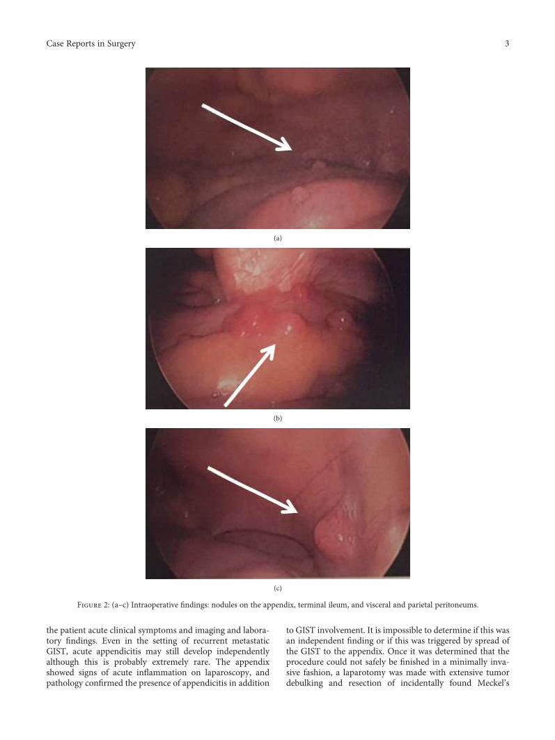
(c)
Figure 2: (a–c) Intraoperative findings: nodules on the appendix, terminal ileum, and visceral and parietal peritoneums.
3Case Reports in Surgery

diverticulum which was also involved in the metastaticdisease. Pathology confirmed GIST in all specimens; thetumor stained positive for CD117, and imatinib was started.GIST tumors are often fragile, and care should be taken inresection—as tumor rupture or bleeding leads to an increasedrisk of recurrence [11]. Reduction of the tumor burdenfollowed by administration of a TKI has remained theaccepted strategy for metastatic GIST necessitating surgicalintervention [5, 7]. Prognosis is acceptable with manypatients surviving for more than 5 years [6].
In patients with a history of GIST who present withabdominal symptoms, recurrent disease should be consid-ered. Metastatic and primary GISTs may present similar toacute appendicitis [2, 3]; even perforation of GIST withinMeckel’s diverticulum has been described [10]. Nevertheless,spread of GIST both to the appendix and Meckel’s diverticu-lum is a very rare condition with as yet undefined guidelinesfor surgical intervention.
Disclosure
Parts of this manuscript were presented at the 37th meetingof the Surgical Infection Society in May 2017.
Conflicts of Interest
No conflicts of interest exist; we have nothing to disclose, andthe article is not in review at another journal.
References
[1] M. Miettinen and J. Lasota, “Gastrointestinal stromal tumors –definition, clinical, histological, immunohistochemical, andmolecular genetic features and differential diagnosis,” Virch-ows Archiv, vol. 438, no. 1, pp. 1–12, 2001.
[2] A. Agaimy, A. F. Pelz, P.Wieacker, A. Roessner, P. H.Wunsch,and R. Schneider-Stock, “Gastrointestinal stromal tumors ofthe vermiform appendix: clinicopathologic, immunohisto-chemical, and molecular study of 2 cases with literaturereview,”Human Pathology, vol. 39, no. 8, pp. 1252–1257, 2008.
[3] M. Bouassida, M. F. Chtourou, E. Chalbi et al., “AppendicealGIST: report of an exceptional case and review of the littera-ture,” Pan African Medical Journal, vol. 15, p. 85, 2013.
[4] T. Ishikawa, T. Kanda, H. Kameyama, and T. Wakai,“Neoadjuvant therapy for gastrointestinal stromal tumor,”Translational Gastroenterology and Hepatology, vol. 3, p. 3,2018.
[5] T. Nishida, T. Doi, and Y. Naito, “Tyrosine kinase inhibitors inthe treatment of unresectable or metastatic gastrointesti-nal stromal tumors,” Expert Opinion on Pharmacotherapy,vol. 15, no. 14, pp. 1979–1989, 2014.
[6] R. Cirocchi, E. Farinella, F. La Mura et al., “Efficacy of surgeryand imatinib mesylate in the treatment of advanced gastroin-testinal stromal tumor: a systematic review,” Tumori Journal,vol. 96, no. 3, pp. 392–399, 2010.
[7] A. Poveda, X. Garcia Del Muro, J. A. Lopez-Guerrero et al.,“GEIS guidelines for gastrointestinal sarcomas (GIST),”Cancer Treatment Reviews, vol. 55, pp. 107–119, 2017.
[8] C. B. Horn, D. Tian, G. V. Bochicchio, and I. R. Turnbull,“Incidence, demographics, and outcomes of nonoperative
management of appendicitis in the United States,” Journal ofSurgical Research, vol. 223, pp. 251–258, 2018.
[9] A. C. Berry, R. Nakshabendi, O. Kanar, and S. Hamer, “Meckeldiverticulum harboring a rare gastrointestinal stromal tumor,”The Ochsner Journal, vol. 17, no. 1, pp. 121–123, 2017.
[10] E. M. Lopez-Tomassetti Fernandez, J. R. Hernandez Hernan-dez, and V. Nunez Jorge, “Perforated gastrointestinal stromaltumor in Meckel’s diverticulum treated laparoscopically,”Asian Journal of Endoscopic Surgery, vol. 6, no. 2, pp. 126–129, 2013.
[11] I. Deshaies, J. Cherenfant, N. J. Gusani et al., “Gastrointestinalstromal tumor (GIST) recurrence following surgery: review ofthe clinical utility of imatinib treatment,” Therapeutics andClinical Risk Management, vol. 6, pp. 453–458, 2010.
[12] A. D. Ucar, E. Oymaci, E. B. Carti et al., “Characteristics ofemergency gastrointestinal stromal tumor (GIST),” Hepato-gastroenterology, vol. 62, no. 139, pp. 635–640, 2015.
4 Case Reports in Surgery

Stem Cells International
Hindawiwww.hindawi.com Volume 2018
Hindawiwww.hindawi.com Volume 2018
MEDIATORSINFLAMMATION
of
EndocrinologyInternational Journal of
Hindawiwww.hindawi.com Volume 2018
Hindawiwww.hindawi.com Volume 2018
Disease Markers
Hindawiwww.hindawi.com Volume 2018
BioMed Research International
OncologyJournal of
Hindawiwww.hindawi.com Volume 2013
Hindawiwww.hindawi.com Volume 2018
Oxidative Medicine and Cellular Longevity
Hindawiwww.hindawi.com Volume 2018
PPAR Research
Hindawi Publishing Corporation http://www.hindawi.com Volume 2013Hindawiwww.hindawi.com
The Scientific World Journal
Volume 2018
Immunology ResearchHindawiwww.hindawi.com Volume 2018
Journal of
ObesityJournal of
Hindawiwww.hindawi.com Volume 2018
Hindawiwww.hindawi.com Volume 2018
Computational and Mathematical Methods in Medicine
Hindawiwww.hindawi.com Volume 2018
Behavioural Neurology
OphthalmologyJournal of
Hindawiwww.hindawi.com Volume 2018
Diabetes ResearchJournal of
Hindawiwww.hindawi.com Volume 2018
Hindawiwww.hindawi.com Volume 2018
Research and TreatmentAIDS
Hindawiwww.hindawi.com Volume 2018
Gastroenterology Research and Practice
Hindawiwww.hindawi.com Volume 2018
Parkinson’s Disease
Evidence-Based Complementary andAlternative Medicine
Volume 2018Hindawiwww.hindawi.com
Submit your manuscripts atwww.hindawi.com


