Acute Myocarditis in Emergency Medicinecdn.intechweb.org/pdfs/21879.pdf3 Acute Myocarditis in...
19
3 Acute Myocarditis in Emergency Medicine Yukihiro Ikegami and Choichiro Tase Fukushima Medical University, Japan 1. Introduction Emergency doctors provide primary care to many patients with an acute-onset condition in the emergency room (ER) every day. We occasionally must address circulatory failure in patients that ranges from “acutely developing” to “severe.” Usually, it is not difficult to diagnose or to choose the proper therapeutic procedures, because most of these cases are caused by heart diseases, such as ischemic heart disease or an arrhythmia. However, in some cases the causes of circulatory failure cannot be determined immediately. It is known that cardiomyopathies account for some of those undiagnosed cases. Classically, cardiomyopathies have been regarded as idiopathic myocardial diseases because of the difficulty of detecting their etiology or the mechanisms causing the problem. Recent developments in biochemical technology have provided the option of approaching these unknown mechanisms using genetic analyses (Richardson et al., 1996). Cardiomyopathies are presently classified into several groups, defined by the cause, tissue type, and clinical course. In 2006, a committee of the American Heart Association (AHA) advocated new criteria for cardiomyopathies. With those criteria, primary cardiomyopathies are classified into a genetic type, a mixed type, and an acquired type (Maron et al., 2006). We encounter several types of cardiomyopathy in the ER. Immediate adequate primary treatment of these cases must be prudent because circulatory insufficiency can rapidly progress to cardiac arrest. In most cases, myocarditis is caused by an inflammatory response and is classified as an acquired type of primary cardiomyopathy. It is also categorized as either acute or chronic. The comprehensive concept of chronic myocarditis has not yet been established because cases of chronic myocarditis have not been sufficiently reported. For most cases of chronic myocarditis, the causes and developmental mechanisms remain unclear. We have only practical guidelines for treating chronic myocarditis in Japan (JCS, 1996). These patients usually have long-term therapeutic histories. Therefore, the patients’ own doctors who are familiar with their clinical histories should be responsible for treatment in the ER. On the other hand, most cases of acute myocarditis are of sudden onset, and severe cases are taken to the ER by the emergency medical service. Doctors in the ER must then be responsible for the primary treatment of these patients. Because there is the risk of sudden development of circulatory insufficiency with acute myocarditis, immediate and adequate initial treatment is necessary for survival. www.intechopen.com
Transcript of Acute Myocarditis in Emergency Medicinecdn.intechweb.org/pdfs/21879.pdf3 Acute Myocarditis in...

3
Acute Myocarditis in Emergency Medicine
Yukihiro Ikegami and Choichiro Tase Fukushima Medical University,
Japan
1. Introduction
Emergency doctors provide primary care to many patients with an acute-onset condition in the emergency room (ER) every day. We occasionally must address circulatory failure in patients that ranges from “acutely developing” to “severe.” Usually, it is not difficult to diagnose or to choose the proper therapeutic procedures, because most of these cases are caused by heart diseases, such as ischemic heart disease or an arrhythmia. However, in some cases the causes of circulatory failure cannot be determined immediately. It is known that cardiomyopathies account for some of those undiagnosed cases. Classically, cardiomyopathies have been regarded as idiopathic myocardial diseases because of the difficulty of detecting their etiology or the mechanisms causing the problem. Recent developments in biochemical technology have provided the option of approaching these unknown mechanisms using genetic analyses (Richardson et al., 1996). Cardiomyopathies are presently classified into several groups, defined by the cause, tissue type, and clinical course. In 2006, a committee of the American Heart Association (AHA) advocated new criteria for cardiomyopathies. With those criteria, primary cardiomyopathies are classified into a genetic type, a mixed type, and an acquired type (Maron et al., 2006). We encounter several types of cardiomyopathy in the ER. Immediate adequate primary treatment of these cases must be prudent because circulatory insufficiency can rapidly progress to cardiac arrest. In most cases, myocarditis is caused by an inflammatory response and is classified as an
acquired type of primary cardiomyopathy. It is also categorized as either acute or chronic.
The comprehensive concept of chronic myocarditis has not yet been established because
cases of chronic myocarditis have not been sufficiently reported. For most cases of chronic
myocarditis, the causes and developmental mechanisms remain unclear.
We have only practical guidelines for treating chronic myocarditis in Japan (JCS, 1996).
These patients usually have long-term therapeutic histories. Therefore, the patients’ own
doctors who are familiar with their clinical histories should be responsible for treatment in
the ER. On the other hand, most cases of acute myocarditis are of sudden onset, and severe
cases are taken to the ER by the emergency medical service. Doctors in the ER must then be
responsible for the primary treatment of these patients. Because there is the risk of sudden
development of circulatory insufficiency with acute myocarditis, immediate and adequate
initial treatment is necessary for survival.
www.intechopen.com

Myocarditis
70
In this chapter, we explain the causes, mechanisms as presently known, classification, and clinical course of myocarditis. Additionally, we outline the recommended treatment for the acute phase of myocarditis.
2. Epidemiology and prognosis
The clinical course of myocarditis varies. The symptoms range widely, from slight to severe, and it is difficult to investigate myocarditis epidemiologically. In 2002 in Japan, a large-scale investigation for cardiomyopathy was performed and reported that cardiomyopathy was found in 21.3 per 100,000 population (Miura et al., 2002). However, the subjects of this investigation were consulted patients with data provided by hospitals. It is thought that actual morbidity is higher, because there are many non-consulted persons with occult cardiomyopathy, such as early stage dilated cardiomyopathy or hypertrophic cardiomyopathy with no symptoms. Therefore, we still cannot grasp the exact morbidity of myocarditis in Japan. Okada et al. (1989) reported that among 10,000 autopsied cases during 1958–1978 there were 115 cases of myocarditis. In another report, occult myocarditis was found in 0.6% of autopsied cases in which cardiac disease had not detected while the patients were alive (Feely et al., 2000). Therefore, it should be recognized that myocarditis is not clinically infrequent and includes a mild type that displays no symptoms. The mortality rate for myocarditis has not been established. An investigative committee of the Ministry of Public Welfare and Labor in Japan reported in 1986 that 13 (4.7%) of 274 patients died within 1 month in a small-scale investigation targeting acute-phase myocarditis (Kawamura et al., 1986). Among those cases, the causes of death were cardiogenic shock in six cases (46%), congestive heart failure in five cases (38%), and complete atrioventricular block in two cases (15%). Intensive treatment for severe cardiac dysfunction and fatal arrhythmias are important to reduce mortality during the acute phase of severe myocarditis.
3. Cause of myocarditis and classification
Myocarditis is classified by cause, clinical type, and pathological picture. In clinical medicine, myocarditis presenting as a sudden development should be attended to because any delay of treatment presents a risk of fatal circulatory failure. It should be recognized that there is different prognosis depending on the clinical type of myocarditis. The classification of myocarditis is shown in Table 1.
3.1 Causes
Several causes or mechanisms of the development of myocarditis have been detected, among which infection is the most the frequent and important. In earlier years, myocarditis was often caused by rheumatic fever and diphtheria. More recently, in advanced nations it is mostly caused by viral infections. Bowles et al. (1986) reported that Coxsackie B virus was detected in patients with myocarditis using a dot blot hybridization method. Coxsackie virus, an enterovirus belonging to the picornavirus family, was extracted from patients with polio in 1982 and is particularly compatible with myocardium (Clements, 1993). Half of group A types and all of group B types of the Coxsackie virus cause myocarditis accompanied by symptoms of the common cold. Presently, it is possible to detect many viruses with polymerase chain reaction or in situ hybridization. Therefore, we can detect myocarditis due to viral infection in most cases. In
www.intechopen.com

Acute Myocarditis in Emergency Medicine
71
Causes Histological classification Clinical classification
Infection Lymphocytic myocarditis Acute type
Virus Giant cell myocarditis Fulminant type
Bacterium Eosinophilic myocarditis
Chronic type (including persistent symptoms type and unremarkable symptoms type)
Fungus Granulomatous myocarditis
Rickettsia
Spirochete
Parasite
Others
Drugs, chemical materials
Allergy, autoimmunity
Collagen disease
Kawasaki disease
Sarcoidosis
Radiation
Heat stroke
Unknown (idiopathic)
Table 1. classification of myocarditis. modified from the guideline for the diagnosis and treatment of myocarditis (JCS, 2005).
1. Nonsteroidal anti-inflammatory drugs
Indomethacin, oxyphenbutazone, phenylbutazone
2. Psychotropic drugs
(Tricyclic) antidepressant: imipramine, clomipramine, amitriptyline
Antimanic: lithium carbonate, lithium oxalic acid
Antiepileptic: phenytoin, carbamazepine
3. Diuretic: acetazolamide, hydrochlorothiazide, spironolactone, chlorthalidone
4. Depressor: methyldopa
5. Anticancer drug: adriamycin, daunorubicin, mitoxantrone
6. Antibiotics
Amphotericin B, penicillin, ampicillin, tetracycline
Chloramphenicol, streptomycin
7. Sulfaminum: sulfadiazine, sulfisoxazole
8. Antiphthisic: isoniazid (INH), para-aminosalicylic acid (PAS)
9. Biological agents: tetanus toxoid, α-interferon, interleukin 2 (IL-2)
10. Antidiabetic: sulfonylurea
11. Others: catecholamines, cocaine, amphetamine, arsenic
Table 2. Drugs that cause myocarditis.
www.intechopen.com

Myocarditis
72
recent reports, adenovirus, sharing the same receptors as the Coxsackie virus, parvovirus B19, human herpes virus 6, and hepatitis C virus, are known to cause myocarditis (Okabe et al., 1997). Myocarditis does not always develop in patients infected by a virus compatible with the myocardium (e.g., the Coxsackie virus). In such cases, symptoms of myocarditis are unremarkable or slight, and the mechanism of development of myocarditis remains unknown. Based on the results in experimental animal models, Cupta et al. (2008) suggested a developmental pattern of myocardial damage by an infective virus and the host’s reactions as follows: (1) initial infection of virus in myocardial cells; (2) innate immune reaction; (3) defense by adaptive immunity; and (4) recovery of inflammatory reaction. Several kinds of drugs, external physical stimulations such as exposure to irradiation or excessive heat, metabolic disorders, pregnancy, collagen diseases, sarcoidosis, and Kawasaki disease are important causes of myocarditis. It should be well noted that drug-induced myocarditis, particular in Japan, Europe, and the United States, is caused by many of the drugs that are administered or prescribed to patients in advanced countries. Drugs that cause myocarditis are shown in Table 2.
3.2 Histological classification Histologically, myocarditis is classified as lymphocytic myocarditis, giant cell myocarditis, eosinophilic leukocyte myocarditis, and granuloma myocarditis. Most cases of lymphocytic myocarditis are caused by infection. Other types are due to materials toxic to the myocardium, a drug allergy, or an autoimmune reaction.
3.2.1 Lymphocytic myocarditis Lymphocytic myocarditis is the most frequent histological type, and most cases are caused by a viral infection. Kodama et al. found that 85% of patients with myocarditis were diagnosed with the lymphocytic variety (Kodama et al., 2001). The primary symptomsfever and pharyngeal painare not specific for myocarditis during the early phase, but symptoms of circulatory failure, such as edema, are specific and are observed during the developed phase. The severity of circulatory failure ranges widely from slight to fulminant.
3.2.2 Giant cell myocarditis Giant cell myocarditis is encountered infrequently. Clinically, it has a poor prognosis because it can easily progress to fulminant myocarditis. After the onset, circulatory failure develops suddenly and in many cases rapidly progresses to a fatal state. It is suggested that an allergic reaction or an autoimmune reaction participates in this development. Infiltration by inflammatory cells and multinucleated cells are observed in the myocardium (Davidoff et al., 1991). Cardiac function markedly deteriorates with massive necrosis of the myocardium. Identification of multinucleated cells is necessary for a definitive diagnosis. It is difficult to decide on the timing of a biopsy because multinucleated cells are seen only during the acute phase. In patients presenting with a chronic course, cardiac sarcoidosis must be excluded (Shield et al., 2002).
3.2.3 Eosinophilic myocarditis
The clinical symptoms of eosinophilic myocarditisfever, pharyngeal pain, coughduring the early phase are similar to those seen with myocarditis due to a viral infection. The
www.intechopen.com

Acute Myocarditis in Emergency Medicine
73
differential diagnosis between viral myocarditis and eosinophilic myocarditis is often impossible because the eosinophil count is normal during the early phase of eosinophilic myocarditis (Morimoto et al., 2003). White blood cells (WBCs) gradually increase with the development of an inflammatory reaction; and materials toxic to the myocardium released from eosinophilic leukocytes cause severe contraction of the myocardium as the eosinophil count exceeds 500/nm3. In most cases, the increase in eosinophils is detected in a peripheral blood examination. However, some patients who suffer from circulatory insufficiency exhibit a delayed increase in eosinophils (Gets et al., 1991). Therefore, we should perform more frequent eosinophil counts during the acute phase. Infiltration of eosinophilic leukocytes, degranulation, and destruction of myocardium are observed in the biopsy specimen. The main causes are an allergic reaction, drugs, and parasitic infection. Most cases, however, are treated as idiopathic myocarditis (Forrester et al., 1976). It has been reported that the mortality rate among patients with acute-phase eosinophilic myocarditis is approximately 7% (Morimot et al., 2003), (Mori et al., 2004).
3.3 Clinical classification
Myocarditis is clinically classified as acute or chronic. Since it is possible to detect the exact onset of acute myocarditis, it is easier to estimate the circulatory parameters for diagnosis and establish the strategy for treatment of acute myocarditis than for chronic myocarditis. In some cases of acute myocarditis, we treat it as fulminant myocarditis that has developed suddenly and may progress to severe circulatory failure during the acute phase (Aoyama et al., 2002). The worldwide morbidity and mortality rates associated with fulminant myocarditis are not yet clear. Cupta et al. (2008) reported that the frequency of fulminant myocarditis is 10% among all cases of acute myocarditis in the United States. The global concept of chronic myocarditis has not been established, although several studies have suggested that viral infection (Fujioka et al., 2000) or autoimmunity (Lauer et al., 2000) play a role in its development. There are clinically two types of chronic myocarditis, and they have different clinical courses. One type has persistent, continuous symptoms, and the other has unremarkable symptoms (JCS, 1996). In an investigation of clinical types of myocarditis in 48 patients reported by Kodama et al. (2001), nine were the acute type, 21 were the fulminant type, three were the chronic persistent symptom type, and 15 were the chronic unremarkable symptom type. The mortality rates during the first admission were 22% for the acute type, 43% for the fulminant type, 33% for the chronic persistent symptom type, and 40% for the chronic unremarkable symptom type. The long-term prognosis for patients who recovered from their cardiac dysfunction during the early phase was good. However, the long-term prognosis for patients who were diagnosed with the chronic, unremarkable symptom type was not good because they developed irreversible circulatory dysfunction, such as dilated cardiomyopathy.
4. Clinical symptoms and diagnosis
4.1 Clinical symptoms and physical symptoms
Clinically, symptoms range from slight problemsfever, pharyngeal pain, cough, vomiting,
diarrhea, and arthropathies, as observed with the common coldto severe circulatory failure. Therefore, we cannot list any symptoms characteristic of acute-phase myocarditis. In
www.intechopen.com

Myocarditis
74
cases of established cardiac dysfunction, patients suffer from dyspnea, edema, cyanosis, palpitations due to hypoxia or arrhythmia, and other severe symptoms such as loss of consciousness and cramping. Whenever we treat acute-onset patients who present with symptoms of cardiac dysfunction in the ER, we maintain the suspicion that they are in the pre-developmental phase of severe myocarditis. Among the physical symptoms of myocarditis, dysfunction of the heart’s conduction system, which occurs in 60%–80% of patients with myocarditis, is critical. Particularly notable is the 30% incidence of severe bradycardia requiring temporary pacing in those with fulminant myocarditis (Kawamura et al., 1986). A galloping rhythm, a heart murmur caused by backflow in the atrioventricular valves, and pulmonary moist rales are found in patients who are developing heart failure. Pericardial and pleural stridulation can be auscultated in patients with pericarditis or pleuritis. Furthermore, pericardial effusion or cardiac tamponade is observed in some cases of developmental pericarditis.
4.2 Blood examinations
Inflammatory reactions such as increased WBCs, an increased erythrocyte sedimentation rate (ESR), or an increased C-reactive protein (CRP) level are detected in blood examinations during the early phase. Additionally, creatine kinase MB (CPK-MB) and cardiac troponin T assays, which are elevated in the presence of myocardial damage, are useful. Troponin T sensitivity is particularly high, and the severity of the myocardial damage can be estimated by a fixed quantity analysis (Lauer et al., 1997). Troponin T continuously increases with development of cardiac dysfunction and maintains a high peak value in those with fulminant myocarditis.
4.3 Chest radiography
Cardiac dilatation is present in 70% of chest radiography examinations, and pulmonary congestion or pleural effusion is often present in patients with severe heart failure. It should be noted that cardiac dilatation and pulmonary congestion are not remarkable in some cases of myocarditis, which causes mainly right ventricular failure (McFalls and van Suylen, 1993).
4.4 Electrocardiography
Various abnormal changes are revealed by electrocardiography (ECG), although none of the
changes are specific for myocarditis. ECG is not an invasive procedure, and it has the benefit
of simplicity of performance. The sensitivity is high, and any changes on the ECG tracing
probably enhance with the development of myocarditis even if only slight changes are
detected during the early phase. ECG should be repeated in the case of suspected
myocarditis. Limited elevation of ST-T, mainly observed with acute myocardial infarction
(AMI), is present in some patient with myocarditis. Elevation of ST-T in all leads is present
in cases complicated by pericarditis. Bundle branch block or atrioventricular block is present
in cases complicated by dysfunction of the heart’s conduction system. It should be noted
that the change to a wide QRS complex on the ECG tracing suggests the development of
cardiac dysfunction. Because there is the risk of sudden changes with fatal ventricular
tachycardia (VT) or ventricular fibrillation (VF) in the case of frequent arrhythmias,
continuous ECG monitoring is necessary.
www.intechopen.com

Acute Myocarditis in Emergency Medicine
75
4.5 Echocardiography
Temporal thickening of the ventricular wall and deterioration of ventricular wall motion are present at inflammatory sites. All-round centripetal thickening, diffuse deterioration of wall motion, and stenosis of the intracardiac space are present in typical cases (Hiramatsu et al., 2001). In the cases of severe circulatory failure or fulminant myocarditis, multiple left ventricular thrombi are frequently caused by the deterioration of all-round wall motion. Continuous wall motion dysfunction causes diffuse thinning of the wall and/or ventricular aneurysms, and dilatation of the left ventricle develops. Ultimately, there is no morphological difference from dilated cardiomyopathy. Diagnosis by exclusion of ischemic heart disease is necessary in patients with wall thickening or wall motion deterioration.
4.6 Cardiac magnetic resonance
Cardiac magnetic resonance (CMR) is a noninvasive, useful examination for myocarditis (JCS, 2011). It can be used to estimate the morphological changes in the ventricles, contractive function, perfusion in the myocardium, and histological characteristics in one performance. In myocarditis, hyperemia and capillary leakage in the cardiac microcirculation are caused by an inflammatory reaction. The site of inflammation in the myocardium has high signal intensity on T1-weighted magnetic resonance imaging (MRI) several minutes after gadolinium contrast enhancement during the acute phase. It is suggested that the changes of microcirculation caused by an inflammatory reaction can be directly visualized with CMR (Friedrich et al., 1998). In many cases, widespread edema in the myocardium is caused during the acute phase. High signal intensity on T2-weighted images is present in 36% of myocarditis diagnosed by the Dallas criteria (Aretz et al., 1987), (Mahrholdt et al., 2004). In those patients, follow-up CMR, performed a year later, shows reduced left ventricular capacity (Zagrosesek et al., 2008). We can predict the histological changes and prognosis for cardiac function using CMR.
4.7 Radioisotope examination
Gallium-67 (67Ga) is specific for infiltration of large monocytes, but it does not have high sensitivity (O’Connel et al., 1984). Pyrophosphate scintigraphy using technetium-99m (99mTc) is sensitive and accumulates at the inflammatory site in the myocardium (Morguet et al., 1994).
4.8 Cardiac catheterization and endomyocardial biopsy
We perform cardiac catheterization for the differential diagnosis during the acute phase if
the patient’s circulatory condition can tolerate it. We first exclude significant coronary
stenosis by coronary angiography and then perform endomyocardial biopsy (Sekiguchi et
al., 1980). The endomyocardial biopsy is now the most important and reliable technique for
a definitive diagnosis. However, we often cannot obtain samples of the lesion site because
the inflammatory reaction in the myocardium occurs inhomogeneously in most cases
(Baughman, 2006). Cooper et al. (2007) reported that cardiac tamponade or ventricular
perforation occurs at the time of sampling with a 0.1%–0.5% frequency.
4.9 Detection of viruses
In cases of suspected viral myocarditis, we measure the antibody titer using paired sera collected at a more than a 2-week interval. The reliable positive ratio is only 10%, and the
www.intechopen.com
Myocarditis
76
capability to detect infected organs is not available. A definitive diagnosis is possible if we can directly detect the original viruses using a polymerase chain reaction or in situ hybridization. However, these techniques are not yet approved as standard examinations because their results vary widely depending on the institution in which they are performed.
4.10 Other diagnostic factors
In cases of suspected drug-induced myocarditis, we narrow down the list of causative drugs by detailed interviews with the patient regarding his or her clinical history. We can then identify the causative drug by a drug-induced lymphocyte stimulation test. Soluble Fas and Fas ligand (Fuse et al., 2000), interleukin-10 (Nishii et al., 2004), and tenascin-C (Imanaka-Yoshida et al., 2002) may be used in upcoming tests for diagnosing myocarditis. Guidelines for diagnosis are presented in Table 3. The basic concept is to exclude ischemic heart disease and confirm an active lesion site by endomyocardial biopsy. It is currently impossible to detect the cause in most cases. Clinically, it is best to provide the primary care that is given to patients suspected of having myocarditis caused by a viral infection.
5. Development and strategy for treatment during the acute phase
Generally, the clinical conditions of patients are similar in many cases of myocarditis. A toxic protein produced by the infecting virus destroys the myocardial dystrophin complex within several days after onset of the myocarditis. It has been noted that this mechanism causes severe myocardial dysfunction accompanied by widespread myocardial cell death (Bandorff et al., 1999). Silver and Kowaldzuk (1989) reported that viral infection directly causes widespread myocardial ischemia by microvascular spasm. After the viral infection is established, the immune response produces inflammatory cytokines in large quantities (Fairweather et al., 2005). This cytokine network originally plays a role in prophylaxis against the viral infection. Inflammatory cytokines such as interleukins 1 and 2 and tumor necrosis factor-α eliminate infected viruses by activating macrophages, lymphocytes, and endothelial cells. However, excessive cytokine release damages myocardial cells and causes myocardial dysfunction (Kawai, 1999). Additionally, inducible nitric oxide (NO) synthase (iNOS) induced by activated macrophages acts to encourage NO to eliminate infected viruses. It has been reported that excessive release of NO strongly damages myocardial cells (Mikami et al., 1996). Infiltration by inflammatory cells, including T cells and natural killer cells, peaks 7–14 days after viral infection and causes widespread necrosis of myocardial cells (Seko et al., 1993). As already noted, infected virus and released inflammatory cytokines are the main mechanisms in the development of myocardial dysfunction during the acute phase. Severe deciduation of myocardial cells causes pump failure, which progresses to fatal circulatory collapse. It is important to remember that cardiac dysfunction associated with myocarditis is reversible in many cases. Full recovery of cardiopulmonary function can be expected if the patient’s life support is adequately performed. Therefore, we compress the strategy for treatment into three stages to give patients suffering from myocarditis the best chance for survival. In most cases, the first strategic issue is intervention regarding the cause. Unfortunately, it is impossible to provide antibiotic therapy because effective antiviral drugs to address viral myocarditis have not been developed. There is a risk of further viral propagation when
www.intechopen.com

Acute Myocarditis in Emergency Medicine
77
1. Primary symptoms (nonspecific symptoms in most cases)
Symptoms that appear with the common cold (fever, headache, cough, pharyngeal pain)
Digestive symptoms (nausea, vomiting, diarrhea, abdominal pain)
Others (eruption, arthralgia, myalgia)
Note: Some patients are found in sudden cardiac arrest 2. Physical findings
Tachycardia, bradycardia, arrhythmia, weak heart sounds
Galloping rhythm (III, IV sounds), pericardial friction murmur
Systolic murmur 3. Abnormality of ECG: various changes
Atrioventricular block, wide QRS complex, reduction of height in R wave, abnormal Q wave
Change of ST-T level, low-voltage wave, frequent premature contractions
Supraventricular tachycardia, atrial fibrillation, sinus arrest
Ventricular tachycardia, ventricular fibrillation, asystole 4. Echocardiography
Focal or diffuse thickening of the ventricular wall
Focal or diffuse deterioration of ventricular wall motion
Stenosis of intracardiac space
Pericardial effusion 5. Blood examinations
Detection of creatine kinase MB (CPK-MB)
Detection troponin T
Inflammatory reaction (increased WBCs, CRP level)
Note: Troponin T is sensitive using whole blood during the acute phase 6. Items 2–5 (above) change within several hours. Therefore, continuous observation is
necessary. Bradycardia, wide QRS complex, frequent premature contractions, enhanced thickening of the ventricular wall and deterioration of ventricular wall motion, and continuous high troponin T levels are dangerous symptoms of fatal circulatory crisis.
7. A differential diagnosis of acute myocardial infarction (AMI) is necessary. 8. Endocardial biopsy provides a definitive diagnosis, but AMI cannot be excluded if
tissue images are not obtained.
Diagnostic criteria in tissue image:
Infiltration of large and small monocytes
Rupture, fusion, or disappearance of myocardial cells
Edema or fibrotic changes in interstitial tissue 9. More than four-fold change of viral antibody titer in paired sera is adequate for viral
detection. Polymerase chain reaction is effective for diagnosing a viral infection.
Additionally, virus isolation or detection of viral antigen from a throat swab, urine,
feces, blood, or particularly pericardial effusion or myocardial tissue are direct
evidence of the diagnosis.
Table 3. Guidelines for diagnosing myocarditis. Modified from the guideline for diagnosis and treatment of myocarditis (JCS, 2005).
www.intechopen.com

Myocarditis
78
administering anti-inflammatory drugs such as immunosuppressants and steroids. On the other hand, we can expect to reverse cardiac dysfunction by initially giving anti-inflammatory drugs because it has been reported that allergic and autoimmune reactions strongly participate in the development of the giant cell myocarditis and eosinophilic myocarditis, both unusual forms. Therefore, early and adequate ascertainment of the cause is the maximum priority. Presently, the performance of myocardial biopsy is limited. Steroids should not be selected as a first choice even if the patient is in a shock state. The second issue is to provide cardiopulmonary support during continuing fatal circulatory failure. During the acute phase of severe myocarditis, there is the risk of cardiogenic shock, complete atrioventricular block, fatal arrhythmia, and/or sudden cardiac arrest at any time. Therefore, most patients require intensive care. Drug therapy for myocarditis is no different from that for usual heart failure. Catecholamines, a diuretic, or both are administered following Forrester’s classification. Mechanical cardiopulmonary life support should be immediately introduced when the circulatory insufficiency cannot be reversed with drugs. Full recovery of cardiac function is expected by advanced life support within several days in patients with acute myocarditis. The third issue is to control the inflammatory reaction. If the actions of excessive inflammatory cytokines and NO are reduced, cardiac dysfunction is expected to be reversed during the acute phase. Although treatment using high doses of steroid, high doses of γ-globulin, and plasma exchange have been tried and evaluated, we have no evidence of their effectiveness.
5.1 Cardiopulmonary support Immediate cardiac resuscitative support must be provided if the patients with fulminant myocarditis are to survive. Delay of treatment results in fatal circulatory collapse. Although the short-term mortality rate for fulminant myocarditis is generally high, the long-term functional prognosis for patients who survive the acute circulatory crisis is good compared with that for dilated cardiomyopathy (McCarthy et al., 2000). The Scientific Committee of the Japanese Circulation performed a retrospective follow-up survey on severe fulminant myocarditis (Aoyama et al., 2002). The cases of 52 patients who required percutaneous cardiopulmonary support (PCPS) because of severe circulatory failure were investigated. The mortality rate for the acute phase was 40.4% (21/52). Among the 31 surviving patients, 30 (96.8%) had fully recovered from their cardiac dysfunction. McCarthy et al. (2000) noted that cardiac function had been maintained in good condition for a long time in more than 90% of patients who had recovered from fulminant myocarditis. Therefore, the “bridge” treatment of using mechanical cardiopulmonary support to avoid multiple organ dysfunction caused by hypoperfusion is an important treatment strategy during the acute phase of fulminant myocarditis. Patients who have survived on mechanical support, such as percutaneous cardiopulmonary support or a ventricular assist system, have also been reported (Chen et al, 2005), (Topkara et al., 2006).
5.1.1 Percutaneous cardiopulmonary support
A guideline for the use of percutaneous cardiopulmonary support (PCPS) has been formulated in Japan (Fig. 1) (Aoyama et al., 2002). In suspected cases of low cardiac output due to pump dysfunction, we may apply PCPS in accordance with continuous monitoring of the circulatory condition. Important clinical parameters to examine when making the decision of whether to use PCS include the urinary volume, SvO2 (< 60%), development of
www.intechopen.com

Acute Myocarditis in Emergency Medicine
79
Fig. 1. Modified algorithm of PCPS for fulminant myocarditis (Aoyama et al, 2002).
metabolic acidosis, and multiple organ dysfunction suggested by blood chemistry examinations. The initial flow of PCPS should be established at 3.0–3.5 L/min. Concomitant use of intra-aortic balloon pumping (IABP) provides the benefits of reduced afterload,
www.intechopen.com
Myocarditis
80
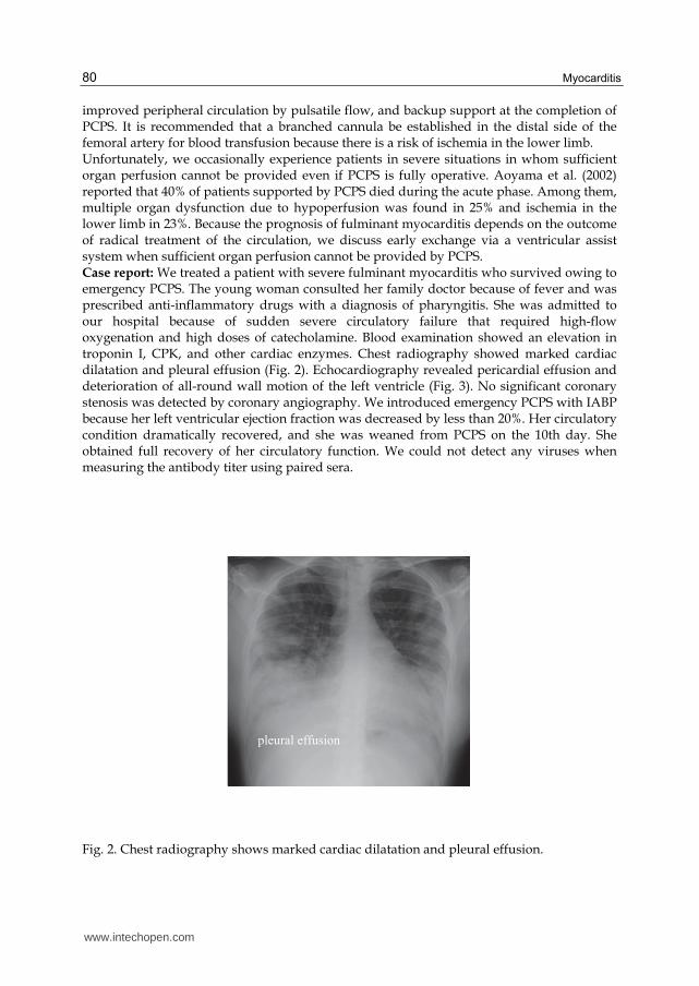
improved peripheral circulation by pulsatile flow, and backup support at the completion of PCPS. It is recommended that a branched cannula be established in the distal side of the femoral artery for blood transfusion because there is a risk of ischemia in the lower limb. Unfortunately, we occasionally experience patients in severe situations in whom sufficient organ perfusion cannot be provided even if PCPS is fully operative. Aoyama et al. (2002) reported that 40% of patients supported by PCPS died during the acute phase. Among them, multiple organ dysfunction due to hypoperfusion was found in 25% and ischemia in the lower limb in 23%. Because the prognosis of fulminant myocarditis depends on the outcome of radical treatment of the circulation, we discuss early exchange via a ventricular assist system when sufficient organ perfusion cannot be provided by PCPS. Case report: We treated a patient with severe fulminant myocarditis who survived owing to emergency PCPS. The young woman consulted her family doctor because of fever and was prescribed anti-inflammatory drugs with a diagnosis of pharyngitis. She was admitted to our hospital because of sudden severe circulatory failure that required high-flow oxygenation and high doses of catecholamine. Blood examination showed an elevation in troponin I, CPK, and other cardiac enzymes. Chest radiography showed marked cardiac dilatation and pleural effusion (Fig. 2). Echocardiography revealed pericardial effusion and deterioration of all-round wall motion of the left ventricle (Fig. 3). No significant coronary stenosis was detected by coronary angiography. We introduced emergency PCPS with IABP because her left ventricular ejection fraction was decreased by less than 20%. Her circulatory condition dramatically recovered, and she was weaned from PCPS on the 10th day. She obtained full recovery of her circulatory function. We could not detect any viruses when measuring the antibody titer using paired sera.
Fig. 2. Chest radiography shows marked cardiac dilatation and pleural effusion.
pleural effusion
www.intechopen.com

Acute Myocarditis in Emergency Medicine
81
Fig. 3. Echocardiography shows pericardial effusion and deterioration of the all-round wall motion of the left ventricle.
5.1.2 Ventricular assist system
There is the risk of severe pulmonary congestion due to PCPS in cases of extreme deterioration of left ventricular function. In such cases, the patient should be switched to a ventricular assist system (VAS) before organ dysfunction develops. Grinda et al. (2004) reported that they introduced VAS in five cases of severe fulminant myocarditis and obtained good outcomes. Recently, the clinical effectiveness of modified VAS using an extracorporeally established centrifugal pump was advocated by John et al. (2007) in the United States. Modified VAS has combined the advantages of minimally invasive extracorporeal membrane oxygenation and high efficiency.
5.2 Immunoregulation therapy
Generally, the time limit for continuous mechanical circulatory support, including PCPS or VAS, is approximately 1 week. We withdraw the system even if recovery from circulatory failure is incomplete. In such cases, introduction of immunoregulation therapy is discussed. Many case reports have asserted the effectiveness of immunoregulation therapy. Although there is no established evidence, we believe that immunoregulation is acceptable in intractable cases because no radical treatments are presently available.
5.2.1 High-dose γ-globulin
The effectiveness of high-dose γ-globulin was reported by Takada et al. (1995). It is expected that γ-globulin counteracts the actions of the infective viruses and reduces suppression of cardiac function by inflammatory cytokines during the acute phase. γ-Globulin intensifies patients’ immune competence, and therefore complications, such as an infection compromised by steroid administration, do not take hold. The mechanisms of high-dose administration have not been completely clarified, although several hypotheses have been suggested: (1) it functions as a neutralizing antibody; (2) it has an anti-inflammatory effect, reducing the release of inflammatory cytokines induced by a combination of the Fc part of γ-globulin and the suppressive Fc receptor of macrophages; and (3) it has the effect of anti-activation on activated complement (Rosen, 1993). However, it is not strongly recommended because there is no evidence that has been confirmed by large clinical trials. Finally, cardiac function has not recovered in several cases with its use.
pericardial effusion
www.intechopen.com

Myocarditis
82
5.2.2 High-dose steroids
The effectiveness of steroids as anti-inflammatory and immunosuppressant drugs has been widely accepted. Its effectiveness when treating patients with fulminant myocarditis has been also reported in many studies (Ino et al., 1995). However, administration of steroid was not proved effective in patients with lymphocytic myocarditis in a clinical trial (Mason et al., 1995). We would not hastily administer steroids to patients with acute-phase myocarditis with a suspected viral infection. On the other hand, it is expected that administration of steroids alleviates cardiac dysfunction in patients with giant cell myocarditis and eosinophilic myocarditis (Cooper et al., 1997). Particularly, high-dose steroids should be given prior to other treatments in patients with the fulminant type of giant cell myocarditis.
6. Conclusions
It is not easy to explain myocarditis concisely and clearly because there are varieties of causes, clinical types, clinical courses, and the severity of circulatory failure. Some patients present with common cold-like symptoms, whereas others require mechanical circulatory support in the ER because of a suddenly developing circulatory crisis. Information of the patient’s background and clinical history is essential when deciding on a treatment plan for myocarditis in most cases, although we are sometimes unexpectedly confronted with emergency conditions regarding these patients in the ER. Currently, the most troublesome issue in the course of treating myocarditis is when to perform a myocardial biopsy for a definitive diagnosis. Although noninvasive diagnostic methods such as CMR have been developed to reduce the risk of serious complications and physical strain on the patient, an effective diagnosis cannot be established during the acute phase. Regarding the treatment for myocarditis, we cannot presently exclude original causes in many cases and can only provide unpredictable “bridge” support, such as PCPS or VAS. Many patients with myocarditis can survive if we remember the possibility of myocarditis in the differential diagnosis and provide immediate, adequate treatment. When we face illness of unknown origin in patients with a severe arrhythmia or circulatory failure, we should immediately assemble the medical staff and prepare cardiac support. Any delay in treatment can allow abrupt deterioration of the circulation. Additionally, we should establish a system of simultaneous processing of the histological diagnosis to decide on the propriety of immunoregulation therapy.
7. References
Aoyama, N., Izumi, T., Hiramori, K., et al. (2002). Japanese investigations of fulminant myocarditis: national survey of fulminant myocarditis in Japan: therapeutic guidelines and long-term prognosis of using percutaneous cardiopulmonary support for fulminant myocarditis (special report from a scientific committee). Circ J, 66, 133-144.
Aretz, H.T., Billingham, M.E., Edwards, W.D., et al. (1987). Myocarditis: a histopathologic definition and classification. Am J Cardiovasc Pathol, 1, 3-14.
Bandorff, C., Lee, G.H., Lamphear, B.J., et al. (1999). Enteroviral protease 2A cleaves dystrophin: evidence of cytoskeletal disruption in an acquired cardiomyopathy. Nat Med, 5, 320-326.
www.intechopen.com

Acute Myocarditis in Emergency Medicine
83
Baughman, K.L. (2006). Diagnosis of myocarditis: death of Dallas criteria. Circulation, 113, 593-595.
Bowles, N.E., Richardson, P.J., Olsen, E.G., et al. (1986). Detection of Coxsackie-B-virus-specific RNA sequences in myocardial biopsy samples from patients with myocarditis and dilated cardiomyopathy. Lancet, 17, 1120-1123.
Chen, Y.S., & Yu, H.Y. (2005). Choice of mechanical support for fulminant myocarditis: ECMO vs. VAD? Eur J Cardiothorac Surg, 27, 921-932.
Clements, G.B. (1993). Characteristics of viruses inducing cardiac disease, In: Viral Infection of the Heart, J.E. Banatvala (Ed.), l-21, Hodder & Stoughton, London.
Cooper, L.T., Berry, G.J., & Shabetai, R. (1997). Idiopathic giant-cell myocarditis: natural history and treatment. N Engl J Med, 336, 1860-1866.
Cooper, L.T., Baughman, K.L., Feldman, A.M., et al. (2007). The role of endomyocardial biopsy in the management of cardiovascular disease: a scientific statement from the American Heart Association, the American College of Cardiology, and the European Society of Cardiology; endorsed by the Heart Failure Society of America and the Heart Failure Association of the European Society of Cardiology. J Am Coll Cardiol, 50, 1914-1931.
Cupta, S., Markham, D.W., & Drazner, M.H. (2008). Mammen fulminant myocarditis. Nat Clin Pract Cardiovasc, 11, 693-706.
Davidoff, R., Palacios, I., Southern, J., et al. (1991). Giant cell versus lymphocytic myocarditis: a comparison of their clinical features and long-term outcomes. Circulation, 83, 953-961.
Fairweather, D., Frisancho-Kiss, S., & Rose, N.R. (2005). Viruses as adjuvants for autoimmunity: evidence from Coxsackie-virus-induced myocarditis. Rev Med Virol,15, 17-27.
Feely, K.M., Harris, J., & Suvarna, S.K. (2000). Necropsy diagnosis of myocarditis: a retrospective study using CD45RO immunohistochemistry. J Clin Pathol, 53, 147-149.
Felker, G.M., Thompson, R.E., Hare, J.M., et al. (2000). Underlying causes and long-term survival in patients with initially unexplained cardiomyopathy. N Engl J Med, 342, 1077-1084.
Forrester, J.S., Diamond, G., Chatterjee, K., et al. (1976). Medical therapy of acute myocardial infarction by application hemodynamic subsets (second of two parts). N Engl J Med, 295, 1404-1413.
Friedrich, M.G., Strohm, O., Schulz-Menger, J., et al. (1998). Contrast media-enhanced magnetic resonance imaging visualizes myocardial changes in the course of viral myocarditis. Circulation, 97, 1802-1809.
Fujioka, S., Kitaura, Y., Ukimura, A., et al. (2000). Evaluation of viral infection in the myocardium of patients with idiopathic dilated cardiomyopathy. J Am Coll Cardiol, 36, 1920-1926.
Fuse, K., Kodama, M., Okura, Y., et al. (2000). Predictors of disease course in patients with acute myocarditis. Circulation, 102, 2829-2835.
Getz, M. A., Subramanian, R., Logemann, T., et al. (1991). Acute necrotizing eosinophilic myocarditis as a manifestation of severe hypersensitivity myocarditis. Antemortem diagnosis and successful treatment. Ann Intern Med, 201-202.
www.intechopen.com

Myocarditis
84
Grinda, J.M., Chevalier, P., D’Attellis, N., et al. (2004). Fulminant myocarditis in adults and children: bi-ventricular assist device for recovery. Eur J Cardiothorac Surg, 26, 1169-1173.
Hiramitsu, S., Morimoto, S., Kato, S., et al. (2001). Transient ventricular wall thickening in acute myocarditis: a serial echocardiographic and histological study. Jpn Circ J, 65, 2001.
Imanaka-Yoshida, K., Hiroe, M., Yasutomi, Y., et al. (2002). Tenascin-C is a useful marker for disease activity in myocarditis. J Pathol, 197, 388-394.
Ino, T., Okubo, M., Akimoto, K., et al. (1995). Corticosteroid therapy for ventricular tachycardia in children with silent lymphocytic myocarditis. J Pediatr, 126, 304-308.
Japanese Circulation Society (JCS) Task Force Committee on Chronic Myocarditis. (1996). Guideline for diagnosing chronic myocarditis. Jpn Circ J, 60, 263-264.
Japanese Circulation Society; Joint Working Group for Guidelines for Diagnosis and Treatment of Cardiovascular Diseases. (2005). Guidelines for diagnosis and treatment for myocarditis (JCS 2004). J Cardiol, 45, 377-384.
Japanese Circulation Society joint working group. (2011). Guidelines for Diagnosis and Treatment of Myocarditis (JCS 2009). Circ J, 75, 734-743.
John, R., Liao, K., Lietz, K., et al. (2007). Experience with the Levitronix CentriMag circulatory support system as a bridge to decision in patients with refractory acute cardiogenic shock and multisystem organ failure. J Thorac Cardiovasc Surg, 134, 351-358.
Kawai, C. (1999). From myocarditis to cardiomyopathy: mechanisms of inflammation and cell death: learning from the past for the future. Circulation, 99, 1091-1100.
Kawamura, K., Kitaura, Y., Deguchi H., et al. (1986). National questionnaire survey of viral or idiopathic myocarditis: 3rd report (Japanese), pp 23-26. Kodama, M., Oda, H., Okabe, M., et al. (2001). Early and long-term mortality of the clinical
subtypes of myocarditis. Jpn Circ J, 65, 961-964. Lauer, B., Niederau, C., Kuhl, U., et al. (1997). Cardiac troponin T in patients with clinically
suspected myocarditis. J Am Coll Cardiol, 30, 1354-1359. Lauer, B., Schannwell, M., Kuhl, U., et al. Antimyosin autoantibodies are associated with
deterioration of systolic and diastolic left ventricular function in patients with chronic myocarditis. J Am Coll Cardiol, 35, 11-18.
Mahrholdt, H., Goedecke, C., Wagner, A., et al. (2004). Cardiovascular magnetic resonance assessment of human myocarditis: a comparison to histology and molecular pathology. Circulation, 109, 1250-1258.
Maron, B.J., Towbin, J. A., Thiene, G., et al (2006). Contemporary definitions and classification of the cardiomyopathies: an American Heart Association Scientific Statement from the Council on Clinical Cardiology, Heart Failure and Transplantation Committee; Quality of Care and Outcomes Research and Functional Genomics and Translational Biology Interdisciplinary Working Groups; and Council on Epidemiology and Prevention. Circulation, 113, 1807-1816.
Mason, J.W., O’Connell, J.B., Herskowitz, A., et al. (1995). A clinical trial of immunosuppressive therapy for myocarditis. N Engl J Med, 333, 269-275.
McCarthy, R., 3rd, Boehmer, J.P., Hruban, R.H., et al. (2000). Long-term outcome of fulminant myocarditis as compared with acute (nonfluminant) myocarditis. N Engl J Med, 342, 690-695.
www.intechopen.com

Acute Myocarditis in Emergency Medicine
85
McFalls, E.O., van Suylen, R.J. (1993). Myocarditis as a cause of primary right ventricular failure. Chest, 103, 1607-1608.
McNamara, D.M., Holubkov, R., Starling, R.C., et al. (2001). Controlled trial of intravenous immune globulin in recent-onset dilated cardiomyopathy. Circulation, 103, 2254-2259.
Mikami, S., Kawashima, S., Kanazawa, K., et al. (1996). Expression of nitric oxide synthase in a murine model of viral myocarditis induced by coxsackievirus B3. Biochem Biophys Res Commun, 220, 983-989.
Miura, K., Nakagawa, H., Morikawa, Y., et al. (2002). Epidemiology of idiopathic cardiomyopathy in Japan: results from a nationwide survey. Heart, 87, 126-130.
Mori, N., Morimoto, S., Hiramatsu, S., et al. (2004). Clinical pictures of 35 cases with eosinophilic myocarditis. Circ J, 68(suppl), 244.
Morimoto, S., Kubo, N., Hiramatsu, S., et al. (2003). Changes in the peripheral eosinophil count in patients with acute eosinophilic myocarditis. Heart Vessels, 18, 193-196.
Morguet, A.J., Munz, D.L., Kreuzer, H., et al. (1994). Scintigraphic detection of inflammatory heart disease. Eur J Nucl Med, 21, 666-674.
Nishii, M., Inomata, T., Takehana, H., et al. (2004). Serum levels of interleukin-10 on admission as a prognostic predictor of human fulminant myocarditis. J Am Coll Cardiol, 44, 1292-1297.
O’Connel, J.B., Henkin, R.E., Robinson, J.A., et al. (1984). Gallium-67 imaging in patients with dilated cardiomyopathy and biopsy-proven myocarditis. Circulation, 70, 58-62.
Okabe, M., Fukuda, K., Arakawa, K., et al. (1997). Chronic variant of myocarditis associated with hepatitis C virus infection. Circulation, 96, 22-24.
Okada, R., Kawai, S., & Kasuya, R. (1989). Nonspecific myocarditis: a statistical and clinicopathological study of autopsy cases. Jpn Circ J, 53, 40-48.
Richardson, P., McKenna, W., Bristow, M., et al (1996). Report of the 1995 World Health Organization/International Society and Federation of Cardiology Task Force on the Definition and Classification of Cardiomyopathies. Circulation, 93, 841-842.
Rosen, F.S. (1993). Putative mechanisms of the effect of intravenous gamma-globulin. Clin Immunol Immunopathol, 67, S41-S43.
Sekiguchi, M., Hiroe, M., Take, M., et al. (1980). Clinical and histopathological profile of sarcoidosis of the heart and acute idiopathic myocarditis: concepts through a study employing endomyocardial biopsy. II. Myocarditis. Jpn Circ J, 44, 264-273.
Seko, Y., Matsuda, H., Kato, K., et al. (1993). Expression of intercellular adhesion molecule-1 in murine hearts with acute myocarditis caused by coxsackievirus B3. J Clin Invest, 91, 1327-1336.
Shields, R.C., Tazelaar H.D., Berry, G.J., et al. (2002). The role of right ventricular endomyocardial biopsy for idiopathic giant cell myocarditis. J Cardiac Fail, 8, 74-78.
Silver, M.A., Kowalczyk, D. (1989). Coronary microvascular narrowing in acute murine coxsackie B3 myocarditis. Am Heart J, 118, 173-174.
Takada, H., Kishimoto, C., & Hiraoka, Y. (1995). Therapy with immunoglobulin suppresses myocarditis in a murine coxsakievirus B3 model: antiviral and anti-inflammatory effects. Circulation, 92, 1604-1611.
Topkara V.K., Dang, N.C., Barili, F., et al. (2006). Ventricular assist device use for the treatment of acute viral myocarditis. J Thorac Cardiovasc Surg, 131, 1190-1191.
www.intechopen.com

Myocarditis
86
Zagrosek, A., Wassmuth, R., Abdel-Aty, H., et al. (2008). Relation between myocardial edema and myocardial mass during the acute and convalescent phase of myocarditis: a CMR study. J Cardiovasc Magn Reson, 10,19.
www.intechopen.com

MyocarditisEdited by Dr. Daniela Cihakova
ISBN 978-953-307-289-0Hard cover, 428 pagesPublisher InTechPublished online 19, October, 2011Published in print edition October, 2011
InTech EuropeUniversity Campus STeP Ri Slavka Krautzeka 83/A 51000 Rijeka, Croatia Phone: +385 (51) 770 447 Fax: +385 (51) 686 166www.intechopen.com
InTech ChinaUnit 405, Office Block, Hotel Equatorial Shanghai No.65, Yan An Road (West), Shanghai, 200040, China
Phone: +86-21-62489820 Fax: +86-21-62489821
Myocarditis, the inflammation of the heart muscle, could be in some cases serious and potentially fataldisease. This book is a comprehensive compilation of studies from leading international experts on variousaspects of myocarditis. The first section of the book provides a clinical perspective on the disease. It containscomprehensive reviews of the causes of myocarditis, its classification, diagnosis, and treatment. It alsoincludes reviews of Perimyocarditis; Chagas’ chronic myocarditis, and myocarditis in HIV-positive patients.The second section of the book focuses on the pathogenesis of myocarditis, discussing pathways andmechanisms activated during viral infection and host immune response during myocarditis. The third, and final,section discusses new findings in the pathogenesis that may lead to new directions for clinical diagnosis,including use of new biomarkers, and new treatments of myocarditis.
How to referenceIn order to correctly reference this scholarly work, feel free to copy and paste the following:
Yukihiro Ikegami and Choichiro Tase (2011). Acute Myocarditis in Emergency Medicine, Myocarditis, Dr.Daniela Cihakova (Ed.), ISBN: 978-953-307-289-0, InTech, Available from:http://www.intechopen.com/books/myocarditis/acute-myocarditis-in-emergency-medicine