Isuog practice guidelines performance of first trimester fetal ultrasound scan

Acute Fetal Demise with First TrimesterMaternal Infection Resulting fromListeria monocytogenes in a NonhumanPrimate Model
Bryce Wolfe,a,b Gregory J. Wiepz,a,c Michele Schotzko,a Gennadiy I. Bondarenko,a
Maureen Durning,a Heather A. Simmons,a Andres Mejia,a Nancy G. Faith,d
Emmanuel Sampene,e Marulasiddappa Suresh,d Sophia Kathariou,g
Charles J. Czuprynski,d Thaddeus G. Golosa,c,f
Wisconsin National Primate Research Center, University of Wisconsin—Madison, Madison, Wisconsin, USAa;Departments of Pathology and Laboratory Medicine,b Comparative Biosciences,c Pathobiological Sciences,d
Biostatistics and Medical Informatics,e and Obstetrics and Gynecology,f University of Wisconsin—Madison,Madison, Wisconsin, USA; Department of Food, Bioprocessing and Nutrition Sciences, North Carolina StateUniversity, Raleigh, North Carolina, USAg
ABSTRACT Infection with Listeria monocytogenes during pregnancy is associatedwith miscarriage, preterm birth, and neonatal complications, including sepsis andmeningitis. While the risk of these conditions is thought to be greatest during thethird trimester of pregnancy, the determinants of fetoplacental susceptibility to in-fection, the contribution of gestational age, and the in vivo progression of disease atthe maternal-fetal interface are poorly understood. We developed a nonhuman pri-mate model of listeriosis to better understand antecedents of adverse pregnancyoutcomes in early pregnancy. Four pregnant cynomolgus macaques (Macaca fascicu-laris) received a single intragastric inoculation between days 36 and 46 of gestationwith 107 CFU of an L. monocytogenes strain isolated from a previous cluster of hu-man listeriosis cases that resulted in adverse pregnancy outcomes. Fecal shedding,maternal bacteremia, and fetal demise were consistently noted within 7 to 13 days.Biopsy specimens of maternal liver, spleen, and lymph node displayed variable in-flammation and relatively low bacterial burden. In comparison, we observed greaterbacterial burden in the decidua and placenta and the highest burden in fetal tissues.Histopathology indicated vasculitis, fibrinoid necrosis, and thrombosis of the decid-ual spiral arteries, acute chorioamnionitis and villitis in the placenta, and hematoge-nous infection of the fetus. Vascular pathology suggests early impact of L. monocyto-genes infection on spiral arteries in the decidua, which we hypothesize precipitatessubsequent placentitis and fetal demise. These results demonstrate that L. monocyto-genes tropism for the maternal reproductive tract results in infection of the decidua,placenta, and the fetus itself during the first trimester of pregnancy.
IMPORTANCE Although listeriosis is known to cause significant fetal morbidity andmortality, it is typically recognized in the third trimester of human pregnancy. Its im-pact on early pregnancy is poorly defined. Here we provide evidence that exposureto L. monocytogenes in the first trimester poses a greater risk of fetal loss than cur-rently appreciated. Similarities in human and nonhuman primate placentation, physi-ology, and reproductive immunology make this work highly relevant to humanpregnancy. We highlight the concept that the maternal immune response that pro-tects the mother from serious disease is unable to protect the fetus, a concept rele-vant to classic TORCH (toxoplasmosis, other, rubella, cytomegalovirus, and herpes)infections and newly illuminated by current Zika virus outbreaks. Studies with thismodel, using the well-understood organism L. monocytogenes, will permit precise
Received 20 October 2016 Accepted 23January 2017 Published 21 February 2017
Citation Wolfe B, Wiepz GJ, Schotzko M,Bondarenko GI, Durning M, Simmons HA, MejiaA, Faith NG, Sampene E, Suresh M, Kathariou S,Czuprynski CJ, Golos TG. 2017. Acute fetaldemise with first trimester maternal infectionresulting from Listeria monocytogenes in anonhuman primate model. mBio 8:e01938-16.https://doi.org/10.1128/mBio.01938-16.
Invited Editor Jorgen Johansson, UmeaUniversity
Editor Scott J. Hultgren, WashingtonUniversity School of Medicine
Copyright © 2017 Wolfe et al. This is an open-access article distributed under the terms ofthe Creative Commons Attribution 4.0International license.
Address correspondence to Thaddeus G. Golos,[email protected].
This article is a direct contribution from aFellow of the American Academy ofMicrobiology. External solicited reviewers:Nancy Freitag, University of Illinois at Chicago;Anna Bakardjiev, University of California, SanFrancisco.
RESEARCH ARTICLE
crossm
January/February 2017 Volume 8 Issue 1 e01938-16 ® mbio.asm.org 1

analysis of host-pathogen interactions at the maternal-fetal interface and have broadsignificance to both recognized and emerging infections in the setting of pregnancy.
Listeria monocytogenes is an environmentally ubiquitous bacterium that causesfoodborne illness. While infection in healthy individuals is generally not associated
with significant disease, it poses a substantial risk for specific populations, includingimmunocompromised, elderly, or pregnant individuals. Listeriosis during pregnancy isgenerally associated with a spectrum of adverse outcomes during the third trimester,including miscarriage, preterm labor, stillbirth, and neonatal infection (1). Inflammationof fetal membranes and placental villitis are noted in these cases (2), but the actualcourse of events at the maternal-fetal interface that cause fetal demise are poorlyunderstood. In the clinic, human placental tissues generally are not available until afteran adverse outcome, at which point fetal infection has progressed for an indeterminateperiod of time. In vitro studies with tissue explants from human first (3) or third (4)trimester placentas concluded that either extravillous trophoblasts (3) or syncytiotro-phoblasts (4) are likely the primary route of placental infection. However, in vitro studiesare limited in reproducing the physiological and immunological complexity at thematernal-fetal interface, and it is unclear how infection proceeds in vivo. Growingrecognition of the impact of microbial infections on fetal well-being (e.g., Zika virus orTORCH [toxoplasmosis, other, rubella, cytomegalovirus, and herpes] infections) (5, 6)underscores the importance of gaining a better mechanistic understanding of suchevents. The aim of this study was to establish a nonhuman primate model to map theoutcome of infection with L. monocytogenes during early pregnancy. Like humans,macaques and other nonhuman primates have villous hemochorial placentas andinvasive extravillous fetal trophoblasts that actively remodel maternal spiral arteries inthe decidua. Their similar endocrine, reproductive, and immune systems make nonhu-man primates a highly relevant model for examining the interplay of maternal and fetalresponses to infection during pregnancy. Remarkably, first trimester inoculation ofcynomolgus macaques consistently resulted in maternal and fetal infection followed byfetal death within 7 to 13 days postinoculation. This contrasts with the generalassumption that listeriosis is of greatest concern during the third trimester of preg-nancy. Microbial analysis of collected tissues revealed that the decidua basalis andplacental bed as well as the placenta had significant bacterial burden. These findingssuggest that maternal infection affects both the placenta—the presumptive target oflisteriosis during human pregnancy—and the maternal reproductive tract.
RESULTSPhysiological responses to L. monocytogenes inoculation. Table S1 in the sup-
plemental material summarizes the infection schedule, dose, and vital signs of eachanimal. Four cynomolgus macaques were inoculated in early gestation. Each animalreceived ~107 CFU of L. monocytogenes in 10 ml of whipping cream through a softintragastric feeding tube while under sedation. This strain of L. monocytogenes wasassociated with a listeriosis outbreak that resulted in adverse pregnancy outcomes in 11pregnant women (7). Following inoculation, animals were observed to ensure bacteriawere not lost due to emesis. Subsequently, animals were monitored for changes inbehavior, defecation, and food intake. There were no notable behavioral changesassociated with L. monocytogenes administration. Two of the animals that receivedL. monocytogenes had mild fever (body temperature of �38.89°C) in the days prior tofetal demise.
Fecal shedding. L. monocytogenes was not detectable in any fecal samples prior to
inoculation and was first detectable in the stool between 1 and 5 days postinoculationin 3 out of 4 animals. In 2 animals, there was intermittent shedding of bacteria withdetection of colonies up to 3 weeks postinoculation (Fig. 1A and B). L. monocytogeneswas not detected at any date in feces from one animal (cy25), although this animal had
Wolfe et al. ®
January/February 2017 Volume 8 Issue 1 e01938-16 mbio.asm.org 2

both bacteremia and fetal demise. Control animals did not shed L. monocytogenes atany time.
Bacteremia. Peripheral blood samples were collected to monitor bacteremia pre-and postinoculation. There was no detectable growth in preinoculation samples.Bacteremia was detectable in all animals in at least one sample within 4 to 14 days ofinoculation (Fig. 1A and B).
Hematology. Complete blood cell counts were within reference ranges for femalecynomolgus macaques, except for elevated numbers of monocytes near the day ofsurgery (see Fig. S1 in the supplemental material). One animal that received L. mono-cytogenes had elevated lymphocytes, and 2 of the 4 animals had elevated basophils incomparison to control animals, but there were no statistically significant differencesbetween pre- and postinoculation levels of any leukocyte subset within individualanimals. The differences observed among subjects most likely relate to individualvariation in a genetically diverse animal model rather than response to infection.
Pregnancy outcome. Fetal demise occurred within 7 to 13 days postinfection in allfour pregnancies. Figure 1 summarizes each animal’s gestation day at inoculation,postinfection fecal shedding of L. monocytogenes, peripheral blood bacteremia, day ofsurgical collection of tissues when loss of fetal heartbeat was detected by ultrasound,and estimated day of fetal death based on the date of the last documented fetalheartbeat and the degree of autolysis observed during histological examination.
Tissue bacterial burden. Bacteriological analysis of maternal biopsy specimens andfetal tissue homogenates demonstrated remarkable similarity across all pregnancies(Fig. 2), with average bacterial burdens of �105 CFU/g in maternal nonreproductivetissues of lymph node, spleen, and liver, 2 � 106 CFU/g in placental bed/myometrium,2 � 107 CFU/g in umbilical cord and amniotic fluid, 1 � 108 CFU/g in decidua andplacenta, and �108 CFU/g in fetal tissues.
Control inoculations. Two dams in the first trimester of pregnancy were fed 10 mlof whipping cream without L. monocytogenes to serve as gestational age-matchednegative controls and were monitored by the same parameters as Listeria-inoculatedanimals. Uterotomy was performed at gestation days 51 (cy27) and 55 (cy26) forremoval of the fetoplacental unit. Maternal and fetal tissues were processed in the samemanner as experimental animals. L. monocytogenes was not detected in feces, blood,maternal tissues, or fetal tissues.
FIG 1 (A) Timeline of events and laboratory analyses for all 4 animals (ID no. cy19, cy21, cy22, and cy25) and key to symbols for eventson the timeline. Positive blood cultures, negative blood cultures, positive fecal cultures, and estimated day of fetal demise are shown.Overlapping symbols indicate concurrent events. The gestation day at inoculation (d40, d46, d36, and d39) is given after each animal ID.(B) The bar graphs indicate fecal shedding of L. monocytogenes for individual animals. The x axis depicts the days postinoculation, withsymbols indicating events as shown in panel A. The arrow on each graph indicates the day of surgical collection of fetal tissues andmaternal biopsy specimens. The y axis quantifies fecal shedding in CFU per gram. The absence of a bar denotes that a sample wascollected but no L. monocytogenes was detectable on that date.
Listeria monocytogenes in Nonhuman Primate Pregnancy ®
January/February 2017 Volume 8 Issue 1 e01938-16 mbio.asm.org 3

Gross examination. At surgery, the placental discs from infected animals werenoted to be mottled yellow-white to red-purple with focal to multifocal areas ofhemorrhage and coalescing areas of pallor (see Fig. S2 in the supplemental material).Umbilical cords were edematous, with vessels visible through the serosal surface. Fetaltissues were variably edematous and fragile, especially the liver. The lungs were mildlymottled. The fetus of one animal, cy19, had mild to moderate fetal tissue autolysiscompatible with fetal demise approximately 18 to 24 h prior to collection. No pathol-ogies were noted in control animals.
Histopathology at the maternal-fetal interface. Placental bed and decidual spec-imens demonstrated significantly greater bacterial burden than other maternal tissues,which correlated with histologic lesions of multifocal suppurative endometritis anddeciduitis (Fig. 3A) with sporadic Gram-positive intralesional bacteria (Fig. 3B). Decidualtissues from all animals demonstrated vascular pathology on histologic examination(Fig. 3C). Decidual spiral arteries, normally remodeled by endovascular trophoblasts,demonstrated vasculitis, necrotic fibrosis, and thrombosis highly atypical for earlypregnancy (8) (Fig. 3D). Gram-positive rods were observed within maternal spiralarteries (Fig. 3B). Suppurative necrosis and edema within the decidua basalis weremarked, in contrast to control tissues (Fig. 3E and F). The cytotrophoblastic shell thatanchors the placenta and demarcates the boundary between maternal decidua basalisand fetal villi (9) demonstrated neutrophilic infiltration, multifocal necrosis of anchoringvilli and syncytium, and multiple microabscesses (Fig. 4A and B). Histologic examinationof placental specimens included hematoxylin and eosin (H&E), Gram stain, and Listeria-specific immunofluorescence. There was mild to severe multifocal necrosuppurativevillitis, intervillositis (Fig. 4C and D), and vasculitis with necrosis of villous vessels andintralesional bacteria. Fetal membranes contained Gram-positive and listeria O antigen-positive rods with severe diffuse necrotizing and suppurative chorioamnionitis withmoderate edema (Fig. 5A to D; see Fig. S3C in the supplemental material). The umbilicalcord had moderate diffuse edema, mild multifocal neutrophilic and plasmacytic vasc-ulitis and perivasculitis, thrombophlebitis, and mild multifocal vascular fibrinoid necro-sis with intralesional bacteria (Fig. 5E).
Maternal extrauterine histopathology. Maternal extrauterine tissue biopsy spec-imens demonstrated variable and relatively low levels of infection as determined bybacteriological assay (Fig. 2). Tissue CFU per gram ranged between 0 and 105 in liverand between 102 and 104 in lymph node and spleen. cy19 and cy22 had moderatediffuse neutrophilia in the red pulp of the maternal spleen and mild diffuse suppurativehepatitis with single-cell necrosis, focal vasculitis, and mild diffuse glycogen vacuoliza-
FIG 2 Maternal and fetal tissue burden of L. monocytogenes. The chart represents data from 4 animals.Tissues are shown on the y axis, and the x axis shows the CFU per gram on a log scale. The vertical blacklines indicate the mean CFU per gram for each tissue, and horizontal gray lines indicate the range.Individual animals are noted in the legend.
Wolfe et al. ®
January/February 2017 Volume 8 Issue 1 e01938-16 mbio.asm.org 4

tion. cy25 had mild diffuse suppurative splenitis and mild suppurative and lymphoe-osinophilic hepatitis.
DISCUSSION
While it is recognized that adverse pregnancy outcomes associated with maternallisteriosis typically include placental infection, the precise pathogenesis of placentalinfection and subsequent fetal infection remains incompletely described. This studydemonstrates that within 7 to 13 days following first trimester intragastric inoculation,there is modest infection of maternal extrauterine tissues with extraordinarily highnumbers of L. monocytogenes in the decidua and placenta. In addition, degenerativechanges in spiral arteries were associated with the implantation site within 8 dayspostinoculation. These results reveal a remarkable tropism, attack rate, and consistentpathophysiology in first trimester listeriosis.
It was previously reported that adverse outcomes occurred in approximately 30% ofmacaques infected with L. monocytogenes at approximately day 110 of gestation (10).Animals with stillbirth shed L. monocytogenes from the gastrointestinal tract for a longerperiod of time than animals with normal birth. These authors estimated that the fetal50% lethal dose (LD50) was 1 � 107 organisms. Consistent with our study, these authorsdid not observe outward signs of maternal illness or significant changes in blood cellcounts. In contrast to these third trimester experimental infections, all four animals
FIG 3 Histopathology of placental bed and decidua in L. monocytogenes-infected animals. (A) Diffusesuppurative deciduitis. H&E, 200-�m scale bar. (B) Intravascular Gram-positive rods in maternal decidualspiral artery (arrow). Gram stain, 200-�m scale bar. (C) Diffuse suppurative deciduitis with vasculitis of thematernal spiral arteries. Arrows indicate perivascular inflammation. An asterisk denotes the vessel shownin panel D. H&E, 400-�m scale bar. (D) Higher magnification of the spiral artery indicated by the asteriskin panel C with transmural suppurative vasculitis (V) and fibrinoid necrosis (F). H&E, 200-�m scale bar. (E)Normal decidua (D) with cytotrophoblast shell (CTB), placental villi (V), and intact syncytial layer (S) froma control animal. H&E, 400-�m scale bar. (F) Listeria-infected decidua with suppurative necrosis, diffuseedema, and syncytial degeneration. H&E, 400-�m scale bar.
Listeria monocytogenes in Nonhuman Primate Pregnancy ®
January/February 2017 Volume 8 Issue 1 e01938-16 mbio.asm.org 5
infected in the first trimester of pregnancy in the present study rapidly progressed tofetal demise. Three animals displayed fecal shedding of L. monocytogenes, and allanimals had positive blood cultures in conjunction with fetal demise. It is possible thatthe strain of L. monocytogenes employed in the present study is more virulent thanstrains used in prior macaque studies. However, our observation of unremarkablepregnancy progression and lack of fetal or placental infection following a third trimes-ter (day 110) inoculation with this strain suggests that gestational age may influence
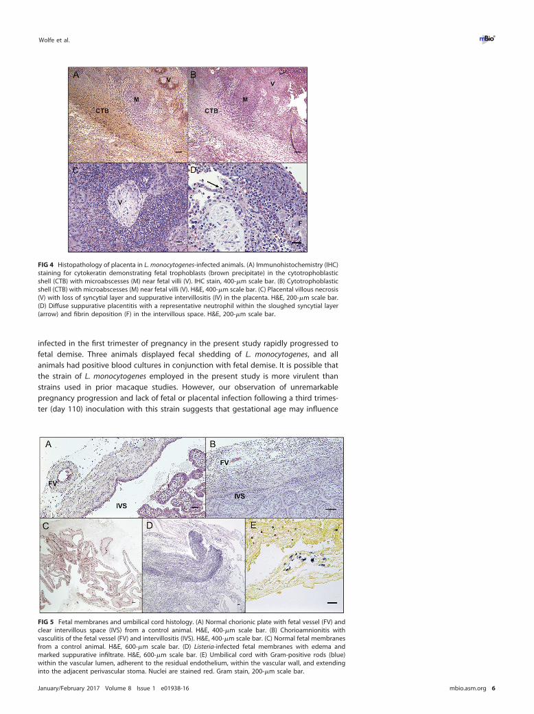
FIG 5 Fetal membranes and umbilical cord histology. (A) Normal chorionic plate with fetal vessel (FV) andclear intervillous space (IVS) from a control animal. H&E, 400-�m scale bar. (B) Chorioamnionitis withvasculitis of the fetal vessel (FV) and intervillositis (IVS). H&E, 400-�m scale bar. (C) Normal fetal membranesfrom a control animal. H&E, 600-�m scale bar. (D) Listeria-infected fetal membranes with edema andmarked suppurative infiltrate. H&E, 600-�m scale bar. (E) Umbilical cord with Gram-positive rods (blue)within the vascular lumen, adherent to the residual endothelium, within the vascular wall, and extendinginto the adjacent perivascular stoma. Nuclei are stained red. Gram stain, 200-�m scale bar.
FIG 4 Histopathology of placenta in L. monocytogenes-infected animals. (A) Immunohistochemistry (IHC)staining for cytokeratin demonstrating fetal trophoblasts (brown precipitate) in the cytotrophoblasticshell (CTB) with microabscesses (M) near fetal villi (V). IHC stain, 400-�m scale bar. (B) Cytotrophoblasticshell (CTB) with microabscesses (M) near fetal villi (V). H&E, 400-�m scale bar. (C) Placental villous necrosis(V) with loss of syncytial layer and suppurative intervillositis (IV) in the placenta. H&E, 200-�m scale bar.(D) Diffuse suppurative placentitis with a representative neutrophil within the sloughed syncytial layer(arrow) and fibrin deposition (F) in the intervillous space. H&E, 200-�m scale bar.
Wolfe et al. ®
January/February 2017 Volume 8 Issue 1 e01938-16 mbio.asm.org 6

maternal-fetal susceptibility to infection. Thus, it appears that the maternal-fetal inter-face in early pregnancy in the macaque is exquisitely sensitive to listeriosis.
Only a few reports of pregnancy outcome in nonhuman primates with listeriosis areavailable (11), and none examined early gestational experimental infection. Similarly,there are few reports of pregnancy outcome in early gestation with human cases oflisteriosis (1, 12, 13). Because the majority of case reports come from third trimester andneonatal infections, listeriosis is often characterized as a concern for late pregnancy (14,15). We suggest that the number of first trimester miscarriages due to infection byL. monocytogenes is underreported because maternal listeriosis is often asymptomaticand unlikely to raise suspicion of infection (16). As maternal complications and fetal lossin late pregnancy will require medical attention, they are more likely to be reported.
The ability to study early pregnancy loss in a nonhuman primate model withplacentation and gestation highly similar to those of humans is invaluable. Collectionof discrete tissue samples and monitoring of maternal and fetal biomarkers throughoutpregnancy are not possible in human studies, and the prolonged incubation periodsseen in human outbreaks (17, 18) make it difficult to accurately determine exposuredose or timing of infection. Controlling these parameters in a nonhuman primatemodel allows precise monitoring of disease progression. Subsequent studies to char-acterize immune cell activation at the maternal-fetal interface, ontogeny of pathogen-esis, and consequences of reexposure to L. monocytogenes will shed additional light onmechanisms of fetal loss in listeriosis.
How L. monocytogenes enters the placenta remains incompletely understood. As afoodborne pathogen, L. monocytogenes trafficks from the maternal gastrointestinaltract to maternal organs, including the spleen, liver, and lymph nodes, before coloniz-ing the reproductive tract. Nonpregnant women without comorbidities are as resistantto infection as other healthy individuals; however, even healthy pregnant women havean increased risk of listeriosis (19, 20). Bakardjiev et al. demonstrated in a guinea pigmodel that only a few bacteria need reach the placenta to proliferate and thendisseminate back to the maternal organs, because the placenta provides an immune-privileged reservoir for replication (21). Placentitis is a hallmark of listeriosis, but it ispossible that the placenta may be an organ of collateral damage as well as a proximaltarget of L. monocytogenes. Poulsen et al. previously reported that LM2203 colonizesthe uteri of nonpregnant as well as pregnant mice and suggested that fetal loss maybe a consequence of preferential trafficking to the uterus (22). Early in gestation,fetus-derived extravillous trophoblast cells invade the decidua and remodel the ma-ternal vasculature to ensure a sufficient exchange of nutrients and oxygen betweenmother and fetus. These structural changes may facilitate colonization of the uterus andpotentially give entry to the fetal vasculature that underlies the syncytiotrophoblast,which itself has been found to be restrictive to infection (23, 24). As in humans,nonhuman primate placentas are comprised of cotyledons, or lobules, which contain amain stem villus with its branches (25). Remarkably, in animal cy19, the placentalcotyledon associated with an individual disrupted spiral artery (Fig. 3C) had concomi-tant villitis.
One means of bacterial entry into the placenta may be through degeneration of thesyncytiotrophoblast layer during the acute inflammatory response to L. monocytogenesat the maternal-fetal interface. The nonpregnant endometrium and pregnant deciduacontain a resident maternal immune cell population that has been found to regulateL. monocytogenes infection in murine models of early pregnancy (26). In mice, bacterialburden was associated with restriction of cytotoxic T cells and macrophages from thedecidua, suggesting that maternal immune tolerance toward the fetus may allow forunchecked bacterial proliferation. A distinct advantage of this nonhuman primatemodel is that macaque and human decidua contain highly similar subsets of NK cells,macrophages, and regulatory T cells, which are thought to play crucial roles inremodeling of decidual spiral arteries and maintenance of maternal tolerance towardfetal tissues (27–30). Given the extensive neutrophilic infiltrate noted in our study, wepropose that L. monocytogenes elicits a strong proinflammatory response in the non-
Listeria monocytogenes in Nonhuman Primate Pregnancy ®
January/February 2017 Volume 8 Issue 1 e01938-16 mbio.asm.org 7

human primate decidua that resolves maternal infection but results in damage to theintegrity of the maternal-fetal interface, thus allowing L. monocytogenes to traverse theplacental barrier and access the fetus. An overview of the macaque maternal-fetalinterface during the first trimester is presented in Fig. 6.
To our knowledge, this is the first study to document the outcome and histopa-thology of early pregnancy L. monocytogenes infection in a nonhuman primate model.From the present findings, we conclude that listeriosis in first trimester pregnancy leadsto damage to decidual, placental, and fetal tissues followed by rapid fetal demise. Inaddition, the presence of bacteria within intervillous spaces (maternal circulation),villous capillaries, the umbilical cord, fetal liver, and fetal lungs (fetal circulation) ishighly suggestive of a hematogenous route of fetal infection. Since it was not possibleto determine the relative timing of decidual and placental infection in the presentstudy, future studies tracing the spatiotemporal movement of L. monocytogenesthrough the nonhuman primate maternal-fetal interface will be useful in defining thepathogenesis of bacterial transmission to the fetus.
MATERIALS AND METHODSAnimals and breeding. Adult female cynomolgus monkeys (Macaca fascicularis) were purchased
from commercial vendors and maintained at the Wisconsin National Primate Research Center. Allexperimental procedures were performed in accordance with the NIH guidelines for care and use oflaboratory animals and under the approval of the University of Wisconsin Graduate School Animal Careand Use Committee. Female monkeys were cohoused with compatible males and observed daily formenses and copulation. Pregnancy was detected by ultrasound examination of the uterus approximately18 to 20 days following the predicted day of ovulation. The day of gestation when pregnancy wasdetected was estimated based on previous experience and published data describing cynomolgusmacaque fetal size during gestation (31). Ultrasound examination of the uterus was done weekly orbiweekly until the day of inoculation with L. monocytogenes and after inoculation on schedules describedbelow.
Inoculation with L. monocytogenes. At varied days of gestation in the first trimester (days 36, 39,40, and 46 [term is day 165]), monkeys were sedated, the uterus was examined by ultrasound to confirma viable pregnancy, and approximately 107 CFU log-phase cells of strain LM2203 (alternative designation,WS1) from a 2000 outbreak of listeriosis among pregnant women in Winston-Salem, NC (7), wereadministered in 10 ml of whipping cream via a soft intragastric feeding tube as previously described (32).Two monkeys were given a Listeria-free whipping cream inoculum in an identical manner at gestationdays 38 and 41 and were included as uninfected controls for histological and physiological comparison.A portion of the inoculum was diluted, plated on blood agar, and incubated at 37°C to confirm preciselythe CFU per milliliter of L. monocytogenes given to each animal.
FIG 6 Overview of the macaque maternal-fetal interface in the first trimester of pregnancy. Specificstructures of the placenta and decidua are highlighted.
Wolfe et al. ®
January/February 2017 Volume 8 Issue 1 e01938-16 mbio.asm.org 8

Fecal shedding. Before and after inoculation, fecal samples were collected from cage pans foranalysis of fecal shedding of L. monocytogenes. Schedules of sample collection are described in thelegends to Fig. 1 and 2. Serial dilutions of fecal samples were plated in duplicate on modified Oxford agarplates (33), and the number of colonies was determined using ImageJ colony-counting software after 24to 48 h of incubation at 37°C.
Bacteremia monitoring. Peripheral blood samples were collected periodically for aerobic andanaerobic culture to detect bacteremia as previously described (34) and processed on a BD Bactec 9050blood culture system (BD Diagnostic Systems, Sparks, MD) in the Clinical Pathology Laboratory at theUniversity of Wisconsin—Madison School of Veterinary Medicine. Aseptically inoculated pediatric BactecPeds Plus/F blood culture bottles (BD Diagnostic Systems, Sparks, MD) were incubated until a positivesignal was observed or for a maximum of 5 days. Bottles that were not positive at the end of 5 days wereGram stained and subcultured to verify the absence of bacterial growth.
Surgery and tissue processing. Ultrasound examination of the uterus was done 1 to 3 times perweek after L. monocytogenes dosing to monitor fetal well-being and confirm fetal heartbeat and umbilicalblood flow. When fetal demise was indicated by absence of heartbeat, fetal and maternal tissues weresurgically collected at laparotomy. These were survival surgeries for the dams. For products of gestation,the entire conceptus (decidua, placenta, fetal membranes, umbilical cord, amniotic fluid, and fetus) wasremoved by uterotomy. Biopsy specimens of the placental bed (uterine placental attachment sitecontaining deep decidua basalis, myometrium, and uterine serosa), maternal liver, spleen, and amesenteric lymph node were collected aseptically. The fetus was dissected into 3- to 4-mm coronalsegments, and alternating segments were fixed and embedded for histology (see below), or homoge-nized for bacteriological analysis on blood agar plates as previously described (35).
Histology. Dissected tissues were fixed in 2 to 4% paraformaldehyde for 24 to 72 h, rinsed inphosphate-buffered saline (PBS), and stored in 70% ethanol until processed and embedded in paraffin.Paraffin sections (5 �m) were stained with hematoxylin and eosin (H&E) and Gram stained using standardmethods. Fetal trophoblasts in the decidua were visualized by chromogenic immunohistochemistry withanticytokeratin (CAM 5.2; 1 �g/ml [Becton, Dickinson]) using previously reported methods (36).
Statistical analysis. Analysis of the data for fecal and tissue CFU per gram was performed usingGraphPad Prism version 5.0 (GraphPad Software, Inc., La Jolla, CA). For maternal vital signs and peripheralblood leukocyte data, means � standard deviations (SD) were calculated for each group, and thestatistical significance of the differences between pairs of groups was assessed, as well as the differenceswithin individual preinfection versus postinfection measurements, using a mixed-effects logistic regres-sion model with statistical consult at the UW Institute for Clinical and Translational Research.
SUPPLEMENTAL MATERIALSupplemental material for this article may be found at https://doi.org/10.1128/
mBio.01938-16.TABLE S1, DOCX file, 0.1 MB.FIG S1, TIF file, 0.4 MB.FIG S2, TIF file, 1.5 MB.FIG S3, TIF file, 2.7 MB.TEXT S1, DOCX file, 0.01 MB.
ACKNOWLEDGMENTSWe thank the WNPRC Veterinary, Scientific Protocol Implementation, and Pathology
Services staff for assistance with animal procedures, including breeding, ultrasoundmonitoring, and sample collection. We thank J. D. Sauer of UW—Madison’s Departmentof Medical Microbiology and Immunology for a critical reading of the manuscript, FayeHartmann of UW—Madison Veterinary Care’s Diagnostic Microbiology for assistancewith blood cultures, Michael Meyer and Troy Thoong for assistance with specimenprocessing, and Beth Gray of the UW—Madison Research Animal Resources Center forassistance and training with Gram staining.
This work was supported by NIH grants R01 HD37120, R21 AI100156, and R01AI107157 to T. G. Golos, P51 OD011106 to the WNPRC, the Renk Endowed Laboratoryfor Food Safety and the Food Research Institute to C. J. Czuprynski, and bridge supportto T. G. Golos from the UW—Madison Department of Obstetrics and Gynecology andSchool of Veterinary Medicine. T. G. Golos was supported in part by a Vilas Life CycleProfessorship. This research was conducted in part at a facility constructed with supportfrom Research Facilities Improvement Program grant no. RR15459-01 and RR020141-01.Statistical consultation was supported by the Clinical and Translational Science Award(CTSA) program through the NIH National Center for Advancing Translational Sciences(NCATS), grant UL1TR000427. The content is solely the responsibility of the authors anddoes not necessarily represent the official views of ORIP or the NIH.
Listeria monocytogenes in Nonhuman Primate Pregnancy ®
January/February 2017 Volume 8 Issue 1 e01938-16 mbio.asm.org 9

REFERENCES1. Lamont RF, Sobel J, Mazaki-Tovi S, Kusanovic JP, Vaisbuch E, Kim SK,
Uldbjerg N, Romero R. 2011. Listeriosis in human pregnancy: a system-atic review. J Perinat Med 39:227–236. https://doi.org/10.1515/jpm.2011.035.
2. Topalovski M, Samuel Yang SS, Boonpasat Y. 1993. Listeriosis of theplacenta: clinicopathologic study of seven cases. Am J Obstet Gynecol169:616 – 620. https://doi.org/10.1016/0002-9378(93)90632-S.
3. Robbins JR, Skrzypczynska KM, Zeldovich VB, Kapidzic M, Bakardjiev AI.2010. Placental syncytiotrophoblast constitutes a major barrier to verti-cal transmission of Listeria monocytogenes. PLoS Pathog 6:e100732.https://doi.org/10.1371/journal.ppat.1000732.
4. Lecuit M, Nelson DM, Smith SD, Khun H, Huerre M, Vacher-Lavenu M-C,Gordon JI, Cossart P. 2004. Targeting and crossing of the human mater-nofetal barrier by Listeria monocytogenes: role of internalin interactionwith trophoblast E-cadherin. Proc Natl Acad Sci U S A 101:6152– 6157.https://doi.org/10.1073/pnas.0401434101.
5. Oladapo OT, Souza JP, De Mucio B, de León RGP, Perea W, GülmezogluAM. 2016. WHO interim guidance on pregnancy management in thecontext of Zika virus infection. Lancet Glob Health 4:e510 – e511. https://doi.org/10.1016/S2214-109X(16)30098-5.
6. Adams Waldorf KM, McAdams RM. 2013. Influence of infection duringpregnancy on fetal development. Reproduction 146:R151–R162. https://doi.org/10.1530/REP-13-0232.
7. MacDonald PD, Whitwam RE, Boggs JD, MacCormack JN, Anderson KL,Reardon JW, Saah JR, Graves LM, Hunter SB, Sobel J. 2005. Outbreak oflisteriosis among Mexican immigrants as a result of consumption ofillicitly produced Mexican-style cheese. Clin Infect Dis 40:677– 682.https://doi.org/10.1086/427803.
8. Bondarenko GI, Durning M, Golos TG. 2012. Immunomorphologicalchanges in the rhesus monkey endometrium and decidua during themenstrual cycle and early pregnancy. Am J Reprod Immunol 68:309 –321. https://doi.org/10.1111/j.1600-0897.2012.01174.x.
9. Enders AC, King BF. 1991. Early stages of trophoblastic invasion of thematernal vascular system during implantation in the macaque andbaboon. Am J Anat 192:329 –346. https://doi.org/10.1002/aja.1001920403.
10. Smith MA, Takeuchi K, Anderson G, Ware GO, McClure HM, RaybourneRB, Mytle N, Doyle MP. 2008. Dose-response model for Listeriamonocytogenes-induced stillbirths in nonhuman primates. Infect Immun76:726 –731. https://doi.org/10.1128/IAI.01366-06.
11. Egal ES, Ardeshir A, Mariano FV, Gondak RO, Montalli VA, dos Santos HT,Canfield DR, Yee J, Lemoy MJ, Lerche NW, Tarara RP. 2015. Contributionof endemic listeriosis to spontaneous abortion and stillbirth in a largeoutdoor-housed colony of rhesus macaques (Macaca mulatta). J AmAssoc Lab Anim Sci 54:399 – 404.
12. Goddijn M, Schipper HG, Spanjaard L, Wolf H. 1997. First trimesterListeria monocytogenes septicemia. Infect Dis Obstet Gynecol 5:219 –221.
13. Boucher M, Yonekura ML. 1986. Perinatal listeriosis (early-onset): corre-lation of antenatal manifestations and neonatal outcome. Obstet Gyne-col 68:593–597.
14. Smith JL. 1999. Foodborne infections during pregnancy. J Food Prot62:818 – 829. https://doi.org/10.4315/0362-028X-62.7.818.
15. Kourtis AP, Read JS, Jamieson DJ. 2014. Pregnancy and infection. N EnglJ Med 370:2211–2218. https://doi.org/10.1056/NEJMra1213566.
16. Doyle ME. 2001. Virulence characteristics of Listeria monocytogenes. FRIBrief https://fri.wisc.edu/files/Briefs_File/virulencelmono.pdf.
17. Riedo FX, Pinner RW, Tosca ML, Cartter ML, Graves LM, Reeves MW,Weaver RE, Plikaytis BD, Broome C. 1994. A point-source foodbornelisteriosis outbreak: documented incubation period and possible mildillness. J Infect Dis 170:693– 696. https://doi.org/10.1093/infdis/170.3.693.
18. Goulet V, King LA, Vaillant V, de Valk H. 2013. What is the incubationperiod for listeriosis? BMC Infect Dis 13:11. https://doi.org/10.1186/1471-2334-13-11.
19. Janakiraman V. 2008. Listeriosis in pregnancy: diagnosis, treatment, andprevention. Rev Obstet Gynecol 1:179 –185.
20. Mylonakis E, Paliou M, Hohmann EL, Calderwood SB, Wing EJ. 2002.Listeriosis during pregnancy: a case series and review of 222 cases.Medicine 81:260 –269. https://doi.org/10.1097/00005792-200207000-00002.
21. Bakardjiev AI, Theriot JA, Portnoy DA. 2006. Listeria monocytogenestraffics from maternal organs to the placenta and back. PLoS Pathog2:e66. https://doi.org/10.1371/journal.ppat.0020066.
22. Poulsen KP, Faith NG, Steinberg H, Czuprynski CJ. 2011. Pregnancyreduces the genetic resistance of C57BL/6 mice to Listeria monocyto-genes infection by intragastric inoculation. Microb Pathog 50:360 –366.https://doi.org/10.1016/j.micpath.2011.02.003.
23. Cao B, Mysorekar IU. 2014. Intracellular bacteria in placental basal platelocalize to extravillous trophoblasts. Placenta 35:139 –142. https://doi.org/10.1016/j.placenta.2013.12.007.
24. Robbins JR, Bakardjiev AI. 2012. Pathogens and the placental fortress.Curr Opin Microbiol 15:36 – 43. https://doi.org/10.1016/j.mib.2011.11.006.
25. Baergen RN. 2011. Manual of pathology of the human placenta, 2nd ed,p 1–544. Springer, Berlin, Germany.
26. Qiu X, Zhu L, Pollard JW. 2009. Colony-stimulating factor-1-dependentmacrophage functions regulate the maternal decidua immune re-sponses against Listeria monocytogenes infections during early gestationin mice. Infect Immun 77:85–97. https://doi.org/10.1128/IAI.01022-08.
27. Riley JK, Yokoyama WM. 2008. NK cell tolerance and the maternal-fetalinterface. Am J Reprod Immunol 59:371–387. https://doi.org/10.1111/j.1600-0897.2008.00593.x.
28. Smith SD, Dunk CE, Aplin JD, Harris LK, Jones RL. 2009. Evidence forimmune cell involvement in decidual spiral arteriole remodeling in earlyhuman pregnancy. Am J Pathol 174:1959 –1971. https://doi.org/10.2353/ajpath.2009.080995.
29. Dambaeva SV, Durning M, Rozner AE, Golos TG. 2012. Immunopheno-type and cytokine profiles of rhesus monkey CD56bright and CD56dimdecidual natural killer cells. Biol Reprod 86:1–10. https://doi.org/10.1095/biolreprod.111.094383.
30. Lash GE, Pitman H, Morgan HL, Innes BA, Agwu CN, Bulmer JN. 2016.Decidual macrophages: key regulators of vascular remodeling in humanpregnancy. J Leukoc Biol 100:315–325. https://doi.org/10.1189/jlb.1A0815-351R.
31. Tarantal AF, Hendrickx AG. 1988. Characterization of prenatal growthand development in the crab-eating macaque (Macaca fascicularis) byultrasound. Anat Rec 222:177–184. https://doi.org/10.1002/ar.1092220210.
32. Smith MA, Takeuchi K, Brackett RE, McClure HM, Raybourne RB, WilliamsKM, Babu US, Ware GO, Broderson JR, Doyle MP. 2003. Nonhumanprimate model for Listeria monocytogenes-induced stillbirths. Infect Im-mun 71:1574 –1579. https://doi.org/10.1128/IAI.71.3.1574-1579.2003.
33. Kang DH, Fung DYC. 1999. Thin agar layer method for recovery ofheat-injured Listeria monocytogenes. J Food Prot 62:1346 –1349. https://doi.org/10.4315/0362-028X-62.11.1346.
34. Lancaster DP. 2015. Blood volume required for detection of low levelsand ultralow levels of organisms responsible for neonatal bacteremia byuse of Bactec Peds Plus/F, Plus Aerobic/F medium, and the BD Bactec FXsystem: an in vitro study. J Clin Microbiol 53:3609 –3613. https://doi.org/10.1128/JCM.01706-15.
35. Poulsen KP, Faith NG, Steinberg H, Czuprynski CJ. 2013. Bacterial loadand inflammation in fetal tissues is not dependent on IL-17a or IL-22 in10 –14 day pregnant mice infected with Listeria monocytogenes. MicrobPathog 56:47–52. https://doi.org/10.1016/j.micpath.2012.11.003.
36. Golos TG, Bondarenko GI, Breburda EE, Dambaeva SV, Durning M,Slukvin II. 2006. Immune and trophoblast cells at the rhesus monkeymaternal-fetal interface. Methods Mol Med 122:93–108.
Wolfe et al. ®
January/February 2017 Volume 8 Issue 1 e01938-16 mbio.asm.org 10