
ACUTE AND CHRONIC OSTEOMYELITIS - medsyllabus.org file4 Introduction-•Oldest known evidence of...
63
ACUTE AND CHRONIC OSTEOMYELITIS
Transcript of ACUTE AND CHRONIC OSTEOMYELITIS - medsyllabus.org file4 Introduction-•Oldest known evidence of...

ACUTE AND CHRONIC OSTEOMYELITIS

DEFINITION
⦿ Inflammation of the bone caused by an infecting organism
HISTORY
In the early 1900’s about 20% of patients with osteomyelitis died and patients who survived had significant morbidity.

4
Introduction-
• Oldest known evidence of osteomyelitis fractured spine of dimetrodon permian reptile 291-250 million years ago
• Hippocrates 460-370 BC infection after fracture
• Nelaton credited with introducing the term osteomyelitis in 1844

INTRODUCTION ⦿ The key to successful management is
early diagnosis and appropriate surgical and antimicrobial treatment.
⦿ A multi disciplinary approach is required, involving an orthopaedic surgeon, an infectious disease specialist, and a plastic surgeon in complex cases with significant soft tissue loss.

CLASSIFICATION 1) The duration - acute, subacute
and chronic
2) Mechanism of infection – exogenous or hematogenous
3) The type of host response to the infection- pyogenic or non pyogenic

ACUTE HEMATOGENOUS OSTEOMYELITIS⦿ Most common type of bone
infection, usually seen in children⦿ Decrease in incidence, could be due
to higher standard of living and improved hygiene.
⦿ Bimodal distribution- younger than 2 years, and 8-12 years
⦿ More common in males
ACUTE HEMATOGENOUS OSTEOMYELITIS-
CAUSES⦿ Caused by a bacteraemia
⦿ Bacteriological seeding of bone generally is associated with other factors such as localized trauma, chronic illness, malnutrition or an inadequate immune system.

ACUTE HEMATOGENOUS OSTEOMYELITISPATHOPHYSIOLOGY
⦿ In children the infection generally involves the metaphyses of rapidly growing long bones
⦿ Bacterial seeding leads to an inflammatory reaction which can cause local ischaemic necrosis of bone and subsequent abscess formation

ACUTE HEMATOGENOUS OSTEOMYELITIS
PATHOPHYSIOLOGY ⦿ As the abscess enlarges,
intramedullary pressure increases causing cortical ischaemia, which may allow purulent material to escape through the cortex into the subperoisteal space.

ACUTE HEMATOGENOUS OSTEOMYELITISPATHOPHYSIOLOGY
⦿ A subperisoteal abscess then develops
⦿ If left untreated this process eventually results in extensive sequetra formation and chronic osteomyelitis

ACUTE HEMATOGENOUS OSTEOMYELITISPATHOPHYSIOLOGY
⦿ In children younger than 2 years, blood vessels cross the physis, thus epiphysis may be involved
⦿ Limb shortening or angular deformity may occur
ACUTE HEMATOGENOUS OSTEOMYELITIS
PATHOPHYSIOLOGY ⦿ Joint may be involved in some cases-
hip joint most common, especially for intraarticular physes- proximal humerus,radial neck, distal fibula
⦿ Metaphysis has relatively fewer phagocytic cells than the physis or diaphysis, hence more infection here

ACUTE HEMATOGENOUS OSTEOMYELITISPATHOPHYSIOLOGY ⦿ In children older than 2 years the
physis effectively acts as a barrier to the spread of a metaphyseal abscess
⦿ Metaphyseal cortex thicker, hence diaphysis more at risk
⦿ After physes are closed acute hematogenous osteomyelitis is much less common

ACUTE HEMATOGENOUS OSTEOMYELITISPATHOPHYSIOLOGY ⦿ After the physes are closed,
infection can extend directly from the metaphysis into the epiphysis and involve the joint
⦿ Septic arthritis resulting from acute hematogenous osteomyelitis generally is seen only in infants and adults.

ACUTE HEMATOGENOUS OSTEOMYELITISMICROBIAL PATTERN⦿ Staphylococcus aureus most common
in older children and adults⦿ Gram negative bacteria- increasing
trend- vertebral⦿ Pseudomonas most common in
intravenous drug abusers⦿ Salmonella in sicke cell⦿ Fungal infections in chronically ill
patients on long term intravenous therapy.
ACUTE HEMATOGENOUS OSTEOMYELITISMICROBIAL PATTERN⦿ Infants- staph aureus most common
but group B streptococcus and gram negative coliforms
⦿ Prematures staph aureus andgram negative organisms
⦿ Hemophilus influenzae primarily in children 6 months to 4 years old, incidence decreased dramatically by immunizations

ACUTE HEMATOGENOUS OSTEOMYELITISMICROBIAL PATTERN
Ngetich, 2002 found that of children presenting with haematogenous osteomyelitis in Kenyatta national hospital the commonest isolated organism was staphylococcus aureus accounting for 29 (60.4%)of the cases, 15 (60%) of these were MRSA strains

ACUTE HEMATOGENOUS OSTEOMYELITISDIAGNOSIS⦿ History and physical examination◼ Fever and malaise◼Pain and local tenderness◼ Sweliing◼Compartment syndrome in children

ACUTE HEMATOGENOUS OSTEOMYELITISDIAGNOSIS⦿ Laboratory tests◼White blood cell count◼ Erythrocyte sedimentation rate◼C-reactive protein
• checked very 2- 3 days post treatment initiation
◼Aspiration for suspected abscess
ACUTE HEMATOGENOUS OSTEOMYELITISDIAGNOSIS
⦿ Plain radiographs
⦿ Technetium-99m bone scan +/- MRI
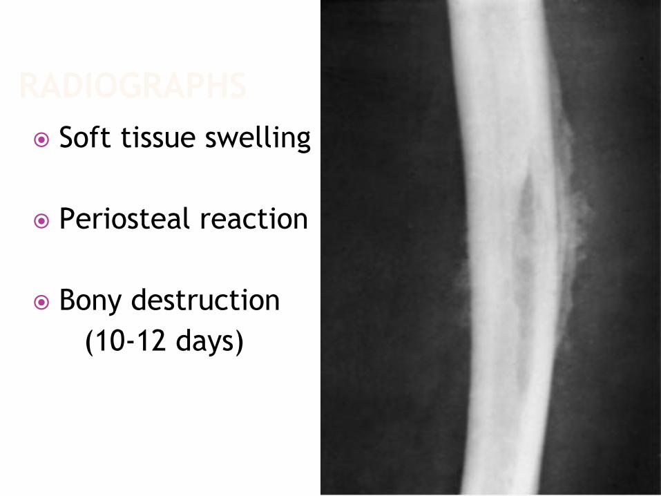
RADIOGRAPHS ⦿ Soft tissue swelling
⦿ Periosteal reaction
⦿ Bony destruction (10-12 days)
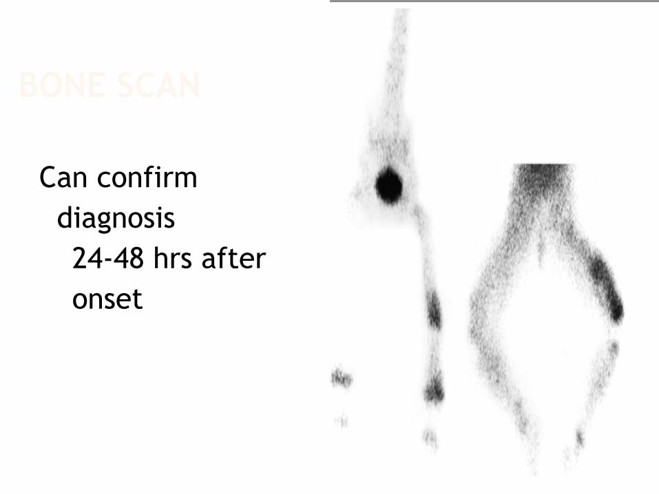
BONE SCAN
Can confirm diagnosis
24-48 hrs after onset

ACUTE HEMATOGENOUS OSTEOMYELITIS
TREATMENT
⦿ Surgery and antibiotic treatment are complementary, in some cases antibiotics alone may cure the disease.
⦿ Choice of antibiotics is based on the highest bacteriocidal activity, the least toxicity and the lowest cost
ACUTE HEMATOGENOUS OSTEOMYELITIS
TREATMENT⦿ Nade’s 5 principles of treatment
1. An appropriate antibiotic is effective before pus formation
2. Antibiotics do not sterilize avascular tissues or abscesses and such areas require surgical removal
ACUTE HEMATOGENOUS OSTEOMYELITISTREATMENT- NADES PRINCIPLES
3. If such removal is effective, antibiotics should prevent their reformation and primary wound closure should be safe
4. Surgery should not damage already ischaemic bone and soft tissue
5. Antibiotics should be continued after surgery
ACUTE HEMATOGENOUS OSTEOMYELITISTREATMENT
⦿ The two main indications for surgery in acute hematogenous osteomyelitis are:1. The presence of an abscess requiring
drainage2. Failure of the patient to improve
despite appropriate intravenous antibiotic treatment

ACUTE HEMATOGENOUS OSTEOMYELITISTREATMENT- SURGERY⦿ The objective of surgery is to drain any
abscess cavity and remove all non viable or necrotic tissue
⦿ Subperiosteal abscess in an infant-several small holes drilled through the cortex into the medullary canal
⦿ If intramedullary pus is found, a small window of bone is removed
⦿ Skin is closed loosely over drains and the limb splinted

ACUTE HEMATOGENOUS OSTEOMYELITISTREATMENT
⦿ Generally a 6 week course of intravenous antibiotics is given
⦿ Orthopedic and infectious disease followup is continued for at least 1 year
SUBACUTE HEMATOGENOUS OSTEOMYELITIS
⦿ More insidious onset and lacks severity of symptoms
⦿ Indolent course hence diagnosis delayed for more than two weeks.

SUBACUTE HEMATOGENOUS OSTEOMYELITIS
CLINICAL FEATURES⦿ The indolent course of subacute
osteomyelitis is due to:◼ increased host resistance◼ decreased bacterial virulence◼ administration of antibiotics before the
onset of symptoms⦿ Systemic signs and symptoms are minimal⦿ Temperature is only mildly elevated⦿ Mild to moderate pain

SUBACUTE HEMATOGENOUS OSTEOMYELITIS
INVESTIGATIONS⦿ White blood cell counts are generally normal
⦿ ESR is elevated in only 50% of patients
⦿ Blood cultures are usually negative
⦿ Plain radiographs and bone scans generally are positive

SUBACUTE HEMATOGENOUS OSTEOMYELITIS
INVESTIGATIONS
⦿ S. Aureus and Staphylococcus epidermidis are the predominant organisms identified in subacute osteomyelitis

SUBACUTE HEMATOGENOUS OSTEOMYELITIS
BRODIE ABSCESS
⦿ Localized form of subacute osteomyelitis occuring most commonly in the long bones of the lower extremeties
⦿ Intermittent pain of long duration is most times the presenting compliant, along with tenderness over the affected area

BRODIE ABSCESS

SUBACUTE HEMATOGENOUS OSTEOMYELITISBRODIE ABSCESS⦿ On plain radiographs appears as a lytic lesion
with a rim of sclerotic bone
⦿ S aureus is cultured in 50% of patients and in 20% the culture is negative
⦿ The condition requires open biopsy with curetage to make the diagnosis
⦿ The wound should be closed loosely over a drain

SUBACUTE HEMATOGENOUS OSTEOMYELITISGLEDHILL CLASSIFICATION

SUBACUTE HEMATOGENOUS OSTEOMYELITISTREATMENT⦿ Biopsy and curettage followed by treatment
with appropriate antibiotics for all lesions that seem to be aggressive
⦿ For lesions that seem to be a simple abscess in the epiphysis or metaphysis biopsy is not recommended- IV antibiotics for 48 hrs followed by a 6 week course of oral antibiotics

CHRONIC OSTEOMYELITIS⦿ Hallmark is infected dead bone
within a compromised soft tissue envelope
⦿ The infected foci within the bone are surrounded by sclerotic, relatively avascular bone covered by a thickened periosteum and scarred muscle and subcutaneous tissue

COM
⦿ Sinus track cultures usually do not corelate with cultures obtained at bone biopsy

COM IN KNH
⦿ 2008-= 108⦿ 2009 = 79⦿ 2010-= 99⦿ 2011-= 79⦿ 2012= 53

CLASSIFICATION OF COM

ANATOMICAL CLASSIFICATION
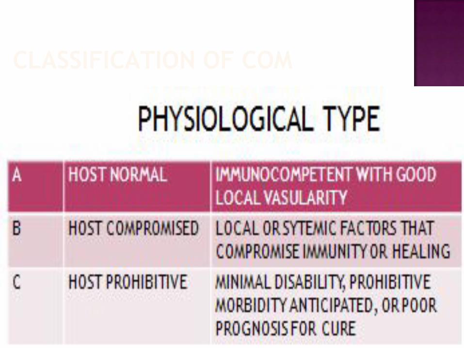
CLASSIFICATION OF COM

DIAGNOSIS COM
⦿ Based on ◼Clinical◼ laboratory and◼ imaging studies

CLINICAL EVALUATION COM
⦿ Skin and soft tissue integrity⦿ Tenderness ⦿ Bone stability⦿ Neurovascular status of limb⦿ Presence of sinus
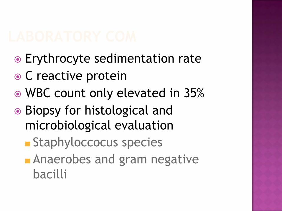
LABORATORY COM ⦿ Erythrocyte sedimentation rate⦿ C reactive protein⦿ WBC count only elevated in 35%⦿ Biopsy for histological and
microbiological evaluation◼ Staphyloccocus species◼Anaerobes and gram negative
bacilli

ORGANISMS IN COM⦿ Girasi, 1981 found that the
commonest organisms found at the orthopaedic unit at Kenyatta national hospital, then in kabete was staphylococcus aureus which was resistant to penicillin and ampicillin

IMAGING STUDIES IN COM⦿ Plain X rays◼Cortical destruction◼Periosteal reaction◼Sequestra ◼Sinography

SINOGRAPHY

IMAGING -
⦿ Isotopic bone scanning more useful in acute than in chronic osteomyelitis
⦿ Gallium scans increased uptake in areas where leucocytes and bacteria accumulate. Normal scan excludes osteomyelitis
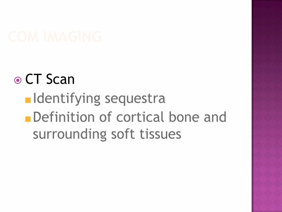
COM IMAGING
⦿ CT Scan◼ Identifying sequestra◼Definition of cortical bone and surrounding soft tissues

COM IMAGING⦿ MRI◼ Shows margins of bone and soft
tissue oedema◼ Evaluate recurrence of infection
after 1 year◼Rim sign- well defined rim of high
signal intensity surrounding the focus of active disease
◼ Sinus tracks and cellulitis
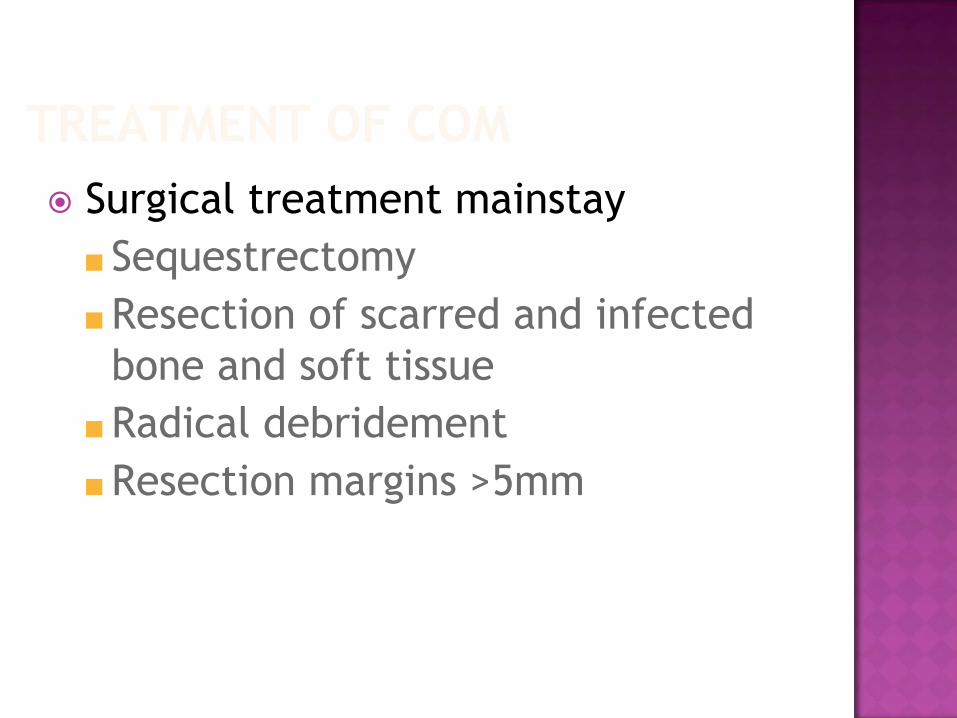
TREATMENT OF COM⦿ Surgical treatment mainstay◼ Sequestrectomy◼Resection of scarred and infected
bone and soft tissue◼Radical debridement◼Resection margins >5mm
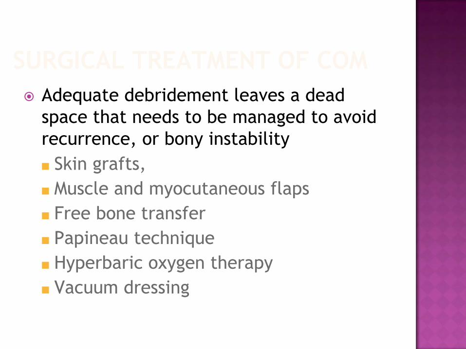
SURGICAL TREATMENT OF COM⦿ Adequate debridement leaves a dead
space that needs to be managed to avoid recurrence, or bony instability◼ Skin grafts,◼ Muscle and myocutaneous flaps◼ Free bone transfer◼ Papineau technique◼ Hyperbaric oxygen therapy◼ Vacuum dressing

TREATMENT OF COM⦿ Antibiotic duration is controversial◼ 6 week is the traditional duration◼ 1 week IV, 6 weeks of oral therapy◼ Antibiotic polymethyl methacrylate
(PMMA) beads as a temporary filler of dead space
◼ Biodegradable antibiotic delivery system
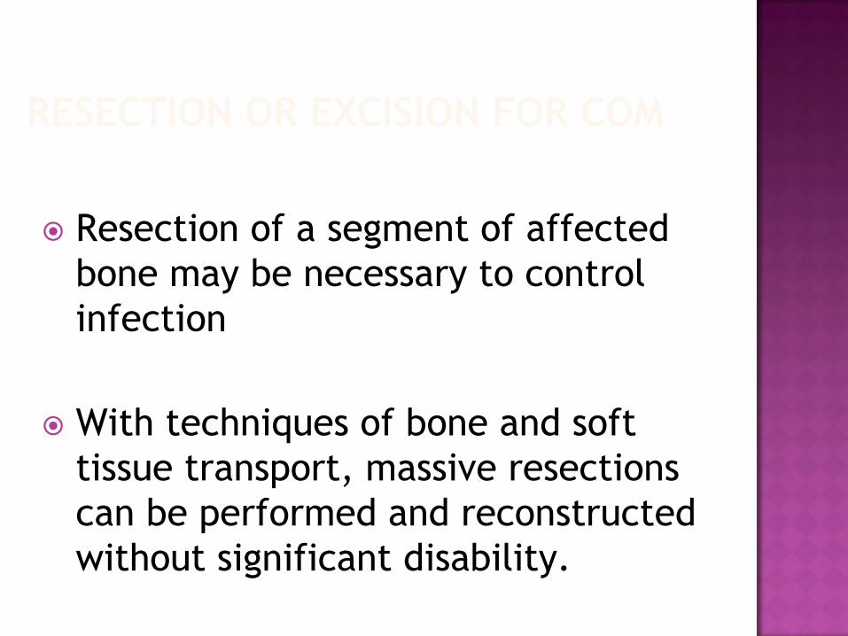
RESECTION OR EXCISION FOR COM
⦿ Resection of a segment of affected bone may be necessary to control infection
⦿ With techniques of bone and soft tissue transport, massive resections can be performed and reconstructed without significant disability.

RESECTION OR EXCISION FOR COM

AMPUTATION FOR OSTEOMYELITIS⦿ Amputation indications include◼ Arterial insufficiency◼ Major nerve paralysis◼ Non functional limb-stiffness,
contracture◼ Malignant change
⦿ Prevalence of maliganacy arising from COM reported as 0.2 to 1.6% of cases.
⦿ Most are squamous cell carcinoma, also reticulum cell carcinoma,fibrosarcoma
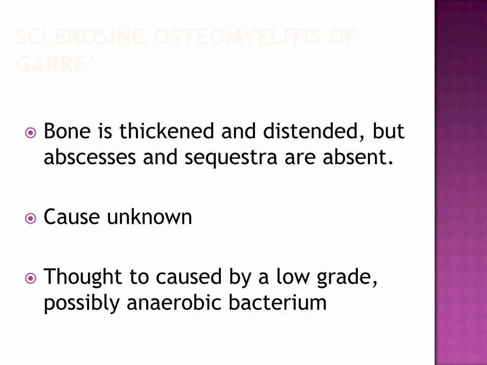
SCLEROSING OSTEOMYELITIS OF GARRE’
⦿ Bone is thickened and distended, but abscesses and sequestra are absent.
⦿ Cause unknown
⦿ Thought to caused by a low grade, possibly anaerobic bacterium

SCLEROSING OSTEOMYELITIS OF GARRE’

REFERENCES ⦿ Canale Terry and Beaty James (2007) Campbell’s
Operative Orthopaedics, Philadelphia, Mosby⦿ Ben Mbonye-Girasi (1981) Mode of Presentation and
End results of Management of Haematogenous Osteomyelitis at the Orthopaedic Unit Kenyatta National Hospital over a Five Year Period. Nairobi : unpublished masters in medicine project, School of Medicine, University of Nairobi
⦿ Issac K Ngetich (2002) A Study of Haematogenous Osteomyelitis in Children in Kenyatta National Hospital Kenya. Nairobi : unpublished masters in medicine project, School of Medicine, University of Nairobi
⦿ Lewis R P, Sutter V L and Finegold S M (1978) Bone Infections Involving Anaerobic Bacteria. Baltimore pub med PMID 207946


![Periacetabular Brucella Osteomyelitis - file.scirp.org · spondylitis, bursitis, tenosynovitis and osteomyelitis [3-6]. Brucella osteomyelitis may appear as a radiolucent area and](https://static.fdocuments.in/doc/165x107/5d52ce1188c993277b8b9aaa/periacetabular-brucella-osteomyelitis-filescirporg-spondylitis-bursitis.jpg)


