ACUTE ABDOMEN: AN OVERVIEW · ACUTE ABDOMEN: AN OVERVIEW Dr. S. Nag, M.D. ... Incidence of specific...
28
ACUTE ABDOMEN: AN OVERVIEW Dr. S. Nag, M.D. (Ob/ Gyn) Dr. N. Bhattacharya, M.D., M.S., D.Sc., FACS (USA) Vidyasagar Hospital Kolkata
Transcript of ACUTE ABDOMEN: AN OVERVIEW · ACUTE ABDOMEN: AN OVERVIEW Dr. S. Nag, M.D. ... Incidence of specific...
ACUTE ABDOMEN:
AN OVERVIEW
Dr. S. Nag, M.D. (Ob/ Gyn)
Dr. N. Bhattacharya, M.D., M.S., D.Sc.,
FACS (USA)
Vidyasagar Hospital
Kolkata
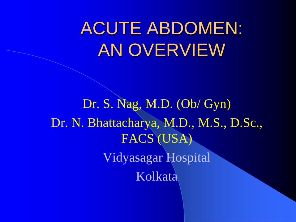
Abdominal plain films are essential for accurate assessment of the acute abdomen. In many cases, they may confirm the presence of a perforated viscus, colonic obstruction, or other abnormality requiring immediate surgical intervention. Alternatively, they may suggest relatively benign disease and help avoid unnecessary operations in these patients. In either case, emergency room physicians should benefit greatly from a systematic approach to the plain film diagnosis of the acute abdomen.
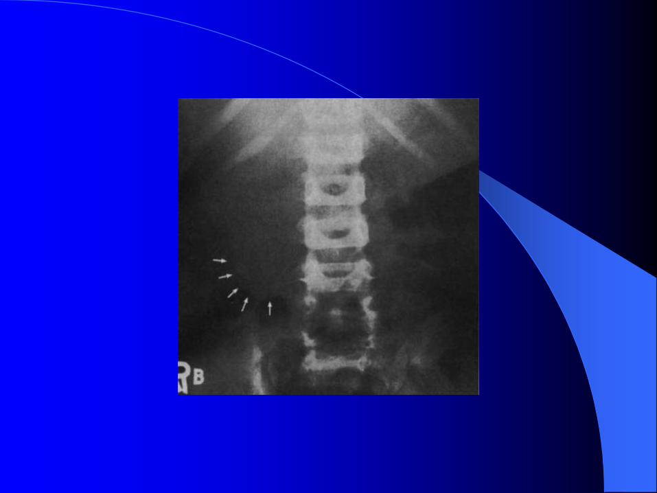
Acute abdominal disorders are common reasons for consultation at the emergency department. The diagnosis of all acute abdominal disorders begins with a careful history and physical examination. When appropriate, the clinical examination should be supplemented by conventional plain abdominal radiography. Gastrointestinal perforation and obstruction are very commonly encountered in the diagnosis of acute abdomen. Plain abdominal radiographs are the initial diagnostic methods of choice. In some circumstances, ultrasonography and CT may be valuable for the evaluation of the cause of abdominal disorder.

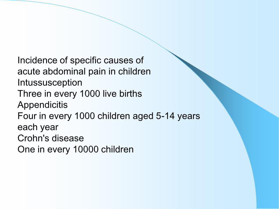
Incidence of specific causes of
acute abdominal pain in children
Intussusception
Three in every 1000 live births
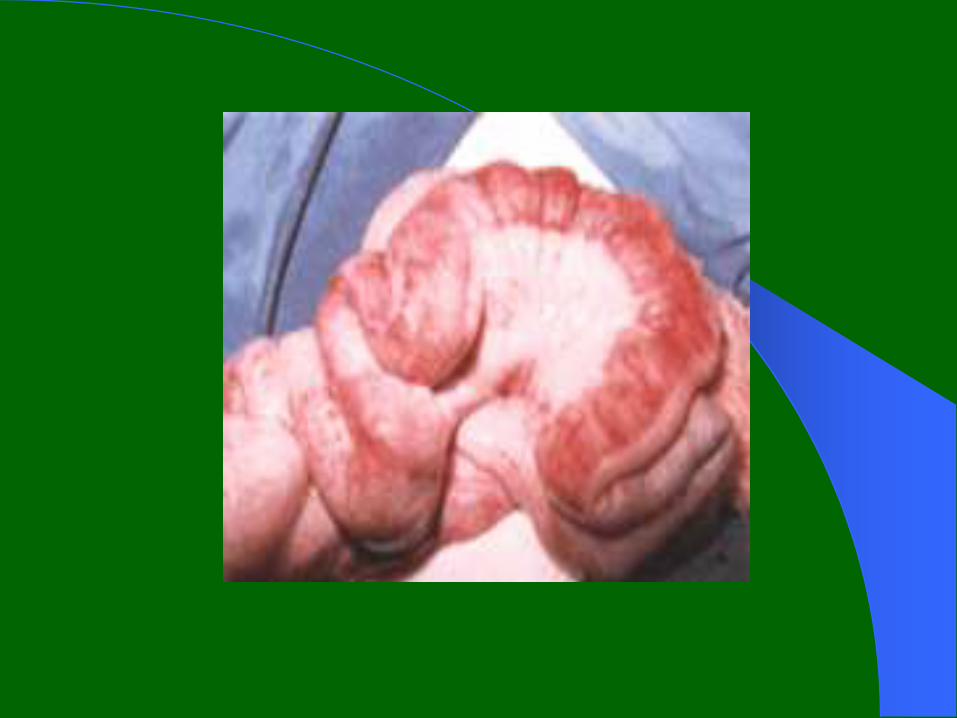
Appendicitis
Four in every 1000 children aged 5-14 years
each year
Crohn's disease
One in every 10000 children
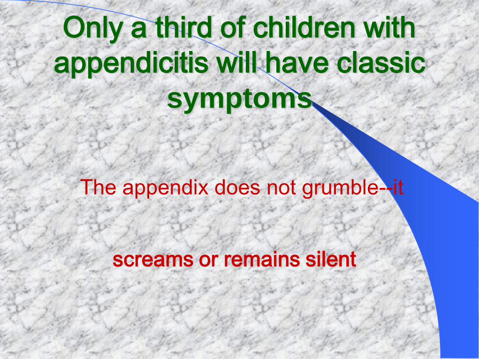
Only a third of children with
appendicitis will have classic
symptoms
screams or remains silent
The appendix does not grumble--it
Causes of acute abdominal pain in childrenCommon causes* Appendicitis * Non-specific
abdominal pain
,
contd.
Uncommon causes
crisis, gall stones, pancreatitis, tonsillitis, otitis media, acute hapatitis, acute
porphyria, intestinal bands, malrotation, ureteric calculi, urinary tract
infection, pneumonia, peptic ulcer disease, psychogenic, Henoch-Schonlein
purpura, intussusception, yersinia infection, obstructed inguinal hernia ,
Meckel's diverticulitis, mesenteric adenitis, Crohn's disease, sickle cell
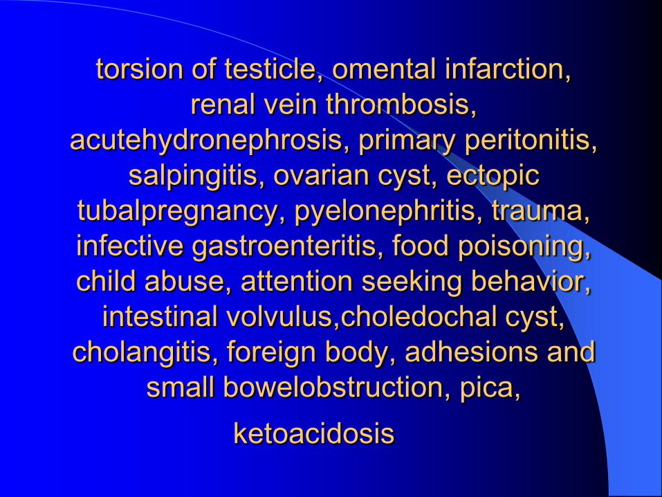
torsion of testicle, omental infarction,
renal vein thrombosis,
acutehydronephrosis, primary peritonitis,
salpingitis, ovarian cyst, ectopic
tubalpregnancy, pyelonephritis, trauma,
infective gastroenteritis, food poisoning,
child abuse, attention seeking behavior,
intestinal volvulus,choledochal cyst,
cholangitis, foreign body, adhesions and
small bowelobstruction, pica,
ketoacidosis

Spiral CT and multidetector-row CT
diagnosis of perforation of the small
intestine caused by ingested foreign
bodies.
Coulier B, Tancredi MH, Ramboux A. Department of Diagnostic Radiology, Clinique
St Luc, Rue St Luc 8, Bouge, Namur, Belgium. [email protected]
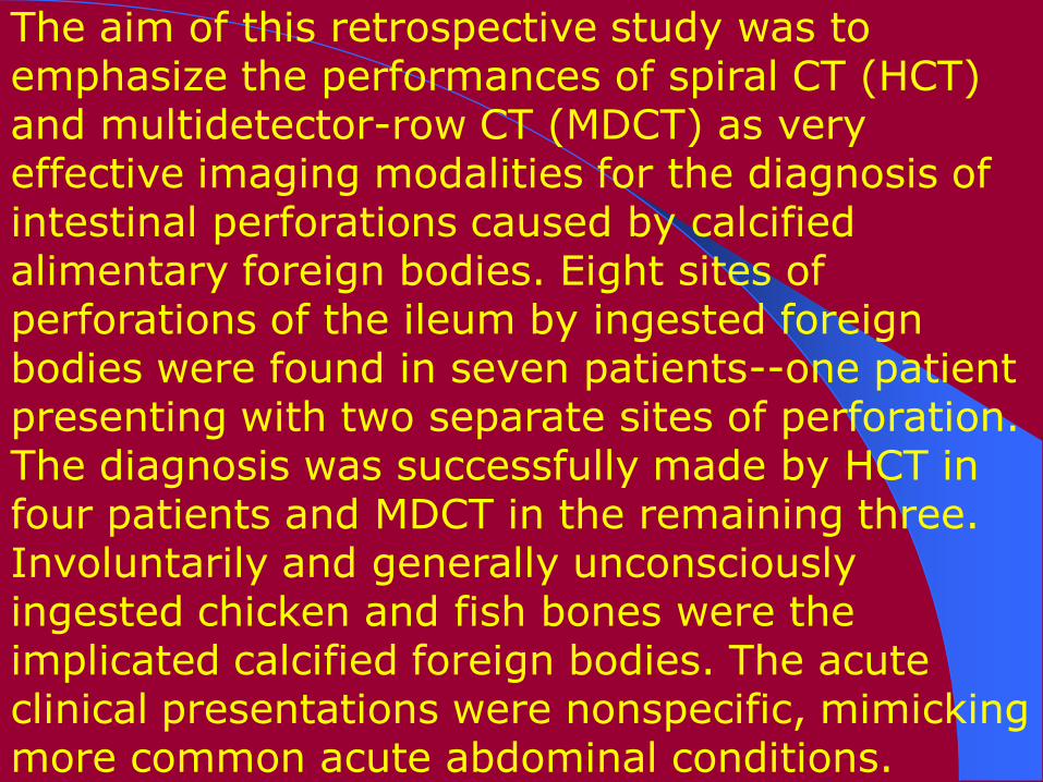
The aim of this retrospective study was to emphasize the performances of spiral CT (HCT) and multidetector-row CT (MDCT) as very effective imaging modalities for the diagnosis of intestinal perforations caused by calcified alimentary foreign bodies. Eight sites of perforations of the ileum by ingested foreign bodies were found in seven patients--one patient presenting with two separate sites of perforation. The diagnosis was successfully made by HCT in four patients and MDCT in the remaining three. Involuntarily and generally unconsciously ingested chicken and fish bones were the implicated calcified foreign bodies. The acute clinical presentations were nonspecific, mimicking more common acute abdominal conditions.

A thickened intestinal segment (7/8 sites) with
localized pneumoperitoneum (4/8 sites), surrounded
by fatty infiltration (4/8 sites) and associated with
already present or developing obstruction or sub-
obstruction (5/7 patients) were the most common CT
signs, but the definite diagnosis was clearly made by
the identification of the calcified foreign bodies (7/7
patients). In each patient, this identification was only
possible thanks to the scrupulous analysis of very
thin overlapping reconstructions obtained not only in
the perforation sites (6/8 sites), but also through the
entire abdomen (2/8 sites). Our report emphasizes
the high performances of CTA and MDCT in
identifying intestinal perforation caused by calcified
alimentary foreign bodies. Moreover, the high
specificity of the CT diagnosis made it possible to
avoid surgical exploration in three patients.
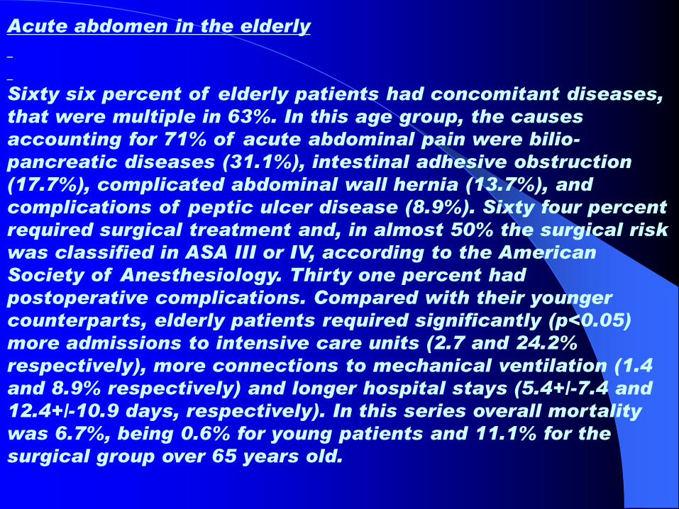
Acute abdomen in the elderly
Sixty six percent of elderly patients had concomitant diseases,
that were multiple in 63%. In this age group, the causes
accounting for 71% of acute abdominal pain were bilio-
pancreatic diseases (31.1%), intestinal adhesive obstruction
(17.7%), complicated abdominal wall hernia (13.7%), and
complications of peptic ulcer disease (8.9%). Sixty four percent
required surgical treatment and, in almost 50% the surgical risk
was classified in ASA III or IV, according to the American
Society of Anesthesiology. Thirty one percent had
postoperative complications. Compared with their younger
counterparts, elderly patients required significantly (p<0.05)
more admissions to intensive care units (2.7 and 24.2%
respectively), more connections to mechanical ventilation (1.4
and 8.9% respectively) and longer hospital stays (5.4+/-7.4 and
12.4+/-10.9 days, respectively). In this series overall mortality
was 6.7%, being 0.6% for young patients and 11.1% for the
surgical group over 65 years old.
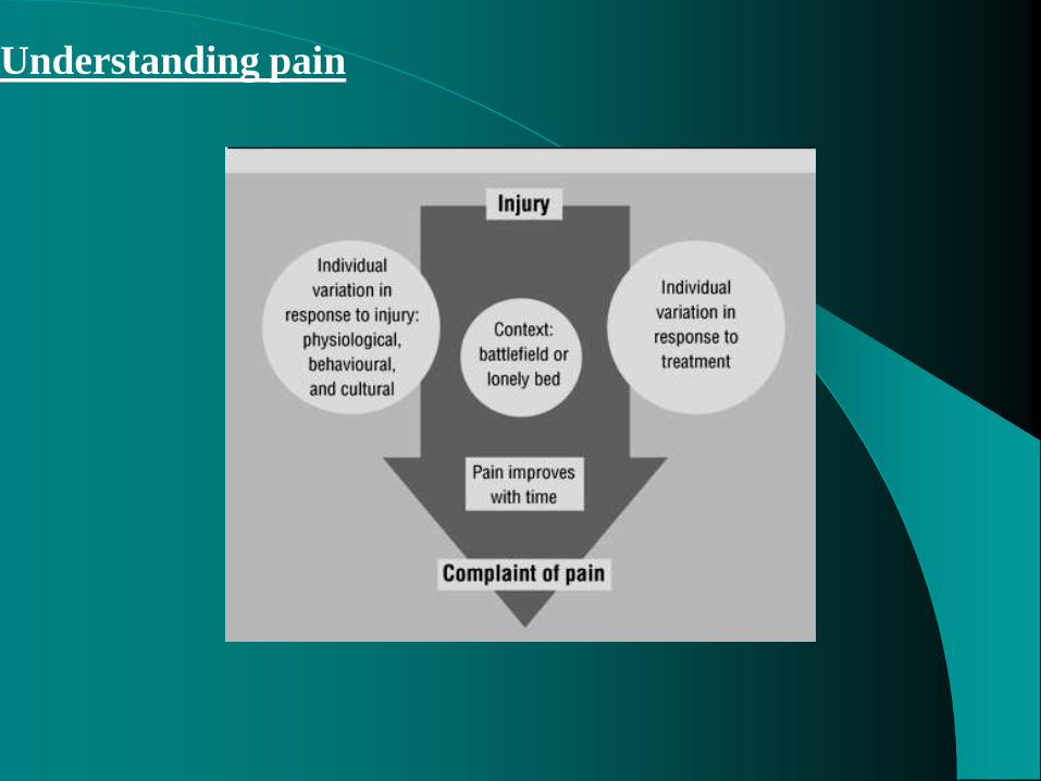
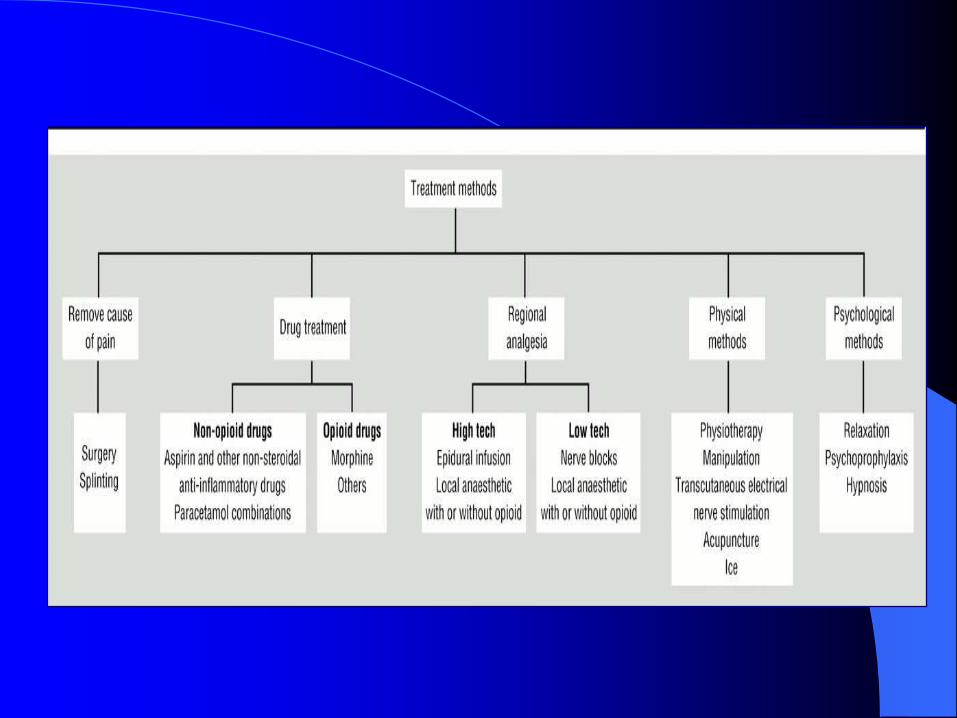
Understanding pain
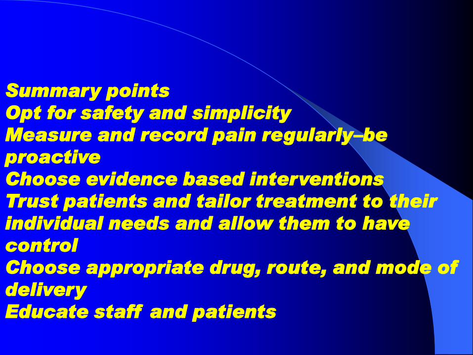
Summary points
Opt for safety and simplicity
Measure and record pain regularly–be
proactive
Choose evidence based interventions
Trust patients and tailor treatment to their
individual needs and allow them to have
control
Choose appropriate drug, route, and mode of
delivery
Educate staff and patients

Settings where pain is a problem
•After operations: inpatient; day surgery;
wound dressing
•Medical illness: myocardial infarction; sickle
cell crisis; renal colic
•Musculoskeletal disease: acute low back
pain; rheumatoid arthritis
•Cancer
•Trauma
•Burns
•Childbirth
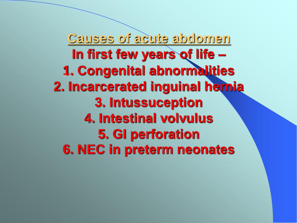
Causes of acute abdomen
In first few years of life –
1. Congenital abnormalities
2. Incarcerated inguinal hernia
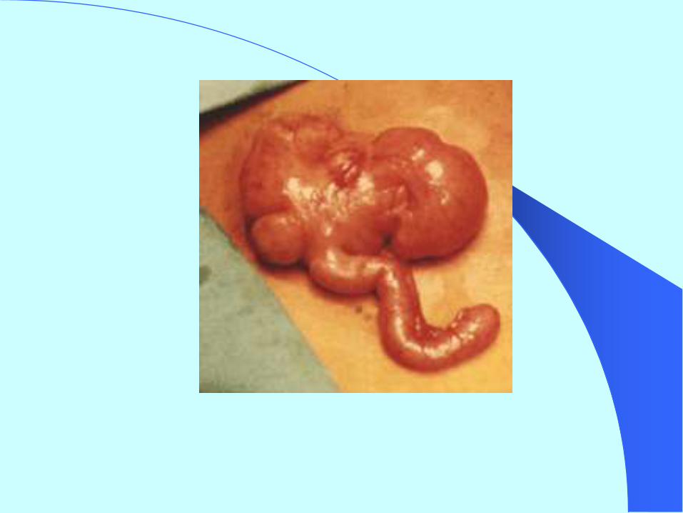
3. Intussuception
4. Intestinal volvulus
5. GI perforation6. NEC in preterm neonates
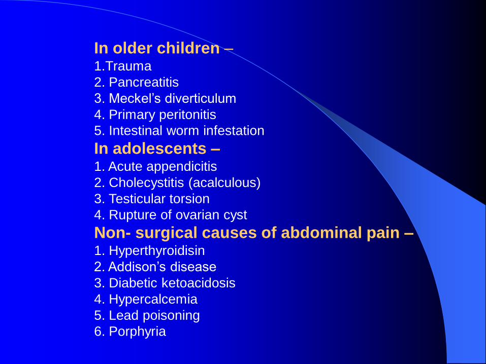
In older children –1.Trauma
2. Pancreatitis
3. Meckel’s diverticulum
4. Primary peritonitis
5. Intestinal worm infestation
In adolescents –1. Acute appendicitis
2. Cholecystitis (acalculous)
3. Testicular torsion
4. Rupture of ovarian cyst
Non- surgical causes of abdominal pain –1. Hyperthyroidisin
2. Addison’s disease
3. Diabetic ketoacidosis
4. Hypercalcemia
5. Lead poisoning
6. Porphyria
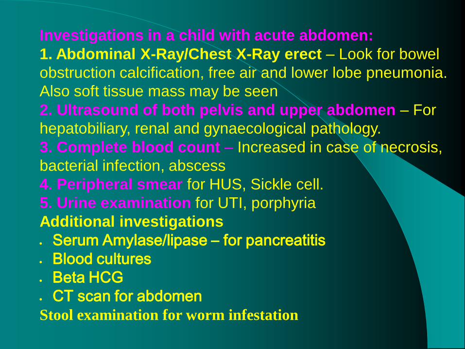
Investigations in a child with acute abdomen:
1. Abdominal X-Ray/Chest X-Ray erect – Look for bowel
obstruction calcification, free air and lower lobe pneumonia.
Also soft tissue mass may be seen
2. Ultrasound of both pelvis and upper abdomen – For
hepatobiliary, renal and gynaecological pathology.
3. Complete blood count – Increased in case of necrosis,
bacterial infection, abscess
4. Peripheral smear for HUS, Sickle cell.
5. Urine examination for UTI, porphyria
Additional investigations
Serum Amylase/lipase – for pancreatitis
Blood cultures
Beta HCG
CT scan for abdomen
Stool examination for worm infestation

Typical presenting clinical characteristics of
appendicitis in infants and children
Diagnosi
s
Age/S
ex
History Physical
Examinat
ion
Lab
Analysis
Radiology
(Abdomen
)
Appendic
itis
Peak:
10-12
years
M:F=3
:2
Periumbi
lical pain
(early)
followed
by
vomiting
and
localized
right
lower
quadrant
pain.
- Fever
>100.5
degree F.
-
Localized
right
lower
quadrant
peritonitis
Increase
d WBC
(>
10000/c
umm)
X-Ray
- Concave
curvature
of spine to
the right.
- Presence
of faecolith
in 5–10 %
USG
- Pericolic
/appendice
a fluid
and/or
edema.
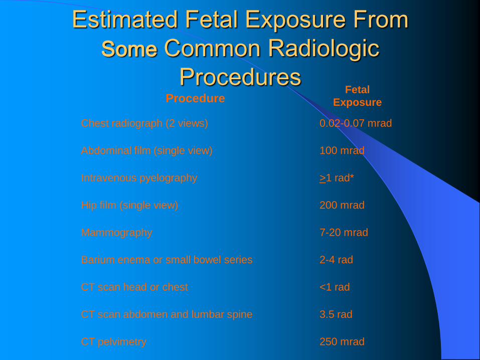
Estimated Fetal Exposure From
Some Common Radiologic
Procedures Procedure
Fetal
Exposure
Chest radiograph (2 views) 0.02-0.07 mrad
Abdominal film (single view) 100 mrad
Intravenous pyelography >1 rad*
Hip film (single view) 200 mrad
Mammography 7-20 mrad
Barium enema or small bowel series 2-4 rad
CT scan head or chest <1 rad
CT scan abdomen and lumbar spine 3.5 rad
CT pelvimetry 250 mrad

Thank You