Acuson X500 DICOM Conformance Statement - Siemens Medical
142
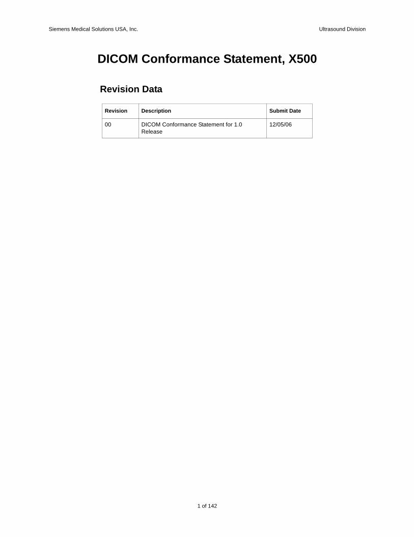
Siemens Medical Solutions USA, Inc. Ultrasound Division 1 of 142 DICOM Conformance Statement, X500 Revision Data Revision Description Submit Date 00 DICOM Conformance Statement for 1.0 Release 12/05/06
Transcript of Acuson X500 DICOM Conformance Statement - Siemens Medical
Siemens Medical Solutions USA, Inc. Ultrasound Division
DICOM Conformance Statement, X500
Revision Data
Revision Description Submit Date
00 DICOM Conformance Statement for 1.0 Release
12/05/06
1 of 142

Siemens Medical Solutions USA, Inc. Ultrasound Division
2 of 142
Siemens Medical Solutions USA, Inc. Ultrasound Division
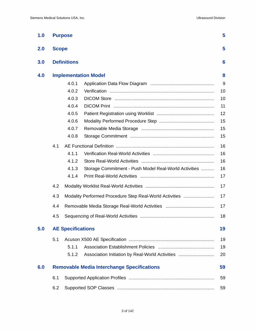
1.0 Purpose 5
2.0 Scope 5
3.0 Definitions 6
4.0 Implementation Model 84.0.1 Application Data Flow Diagram .................................................. 94.0.2 Verification .................................................................................. 104.0.3 DICOM Store .............................................................................. 104.0.4 DICOM Print ............................................................................... 114.0.5 Patient Registration using Worklist ............................................. 124.0.6 Modality Performed Procedure Step ........................................... 154.0.7 Removable Media Storage ......................................................... 154.0.8 Storage Commitment .................................................................. 15
4.1 AE Functional Definition ............................................................................. 164.1.1 Verification Real-World Activities ................................................ 164.1.2 Store Real-World Activities ......................................................... 164.1.3 Storage Commitment - Push Model Real-World Activities .......... 164.1.4 Print Real-World Activities .......................................................... 17
4.2 Modality Worklist Real-World Activities ...................................................... 17
4.3 Modality Performed Procedure Step Real-World Activities ........................ 17
4.4 Removable Media Storage Real-World Activities ...................................... 17
4.5 Sequencing of Real-World Activities .......................................................... 18
5.0 AE Specifications 19
5.1 Acuson X500 AE Specification ................................................................... 195.1.1 Association Establishment Policies ............................................ 195.1.2 Association Initiation by Real-World Activities ............................ 20
6.0 Removable Media Interchange Specifications 59
6.1 Supported Application Profiles ................................................................... 59
6.2 Supported SOP Classes ............................................................................ 59
3 of 142

Siemens Medical Solutions USA, Inc. Ultrasound Division
6.2.1 Supported SOP Classes and Transfer Syntaxes ........................ 59
6.3 Information Object Definition and DICOMDIR Keys .................................. 606.3.1 DICOM File Meta Information ..................................................... 606.3.2 Basic Directory Information Object Definitions - File-set Identification
Module 616.3.3 Basic Directory Information Object Definitions - Directory Identification
Module 616.3.4 Physical Storage Media and Media Formats .............................. 62
7.0 Communication Profiles 63
7.1 TCP/IP Stack Supported ............................................................................ 637.1.1 Physical Media Supported .......................................................... 637.1.2 Chapter Extensions/Specializations/Privatizations ..................... 63
8.0 Configuration 64
8.1 General System Configuration ................................................................... 648.1.1 Hospital Name ............................................................................ 64
8.2 DICOM Network Configuration .................................................................. 648.2.1 Local ........................................................................................... 648.2.2 Remote ....................................................................................... 65
8.3 External Equipment Configuration ............................................................. 69
8.4 Support of Extended Character Sets ......................................................... 69
9.0 Security 70
9.1 Security Profiles ......................................................................................... 70
9.2 Association Level Security ......................................................................... 70
9.3 Application Level Security .......................................................................... 70
9.4 Virus Protection .......................................................................................... 70
10.0 Appendix A: Mapping for Obstetric DICOM SR 71
11.0 Appendix B: Mapping for Cardiac DICOM SR 108
4 of 142
Siemens Medical Solutions USA, Inc. Ultrasound Division
1.0 PurposeThis document describes the conformance to the ACR-NEMA DICOM 3.0 Standard by the Acuson X500 ultrasound system software version 1.0 from Siemens Medical Solutions USA, Inc. Ultrasound Division. It shall establish the conformance specifications for this system only, and does not apply to other products offered by Siemens Medical Solutions USA, Inc., or its affil-iates.
The Acuson X500 system is a device that generates ultrasound images that can be sent using DICOM standard protocols and definitions to other DICOM compliant devices that support SOP classes as defined in Table 2 in this document.
2.0 ScopeThe DICOM standard provides a well-defined set of structures and proto-cols that allow inter-operability to a wide variety of medical imaging devices.
When configured with the DICOM option, the Acuson X500 system pro-vides support for essential services related to ultrasound scanning and con-nectivity to DICOM compliant devices. Acuson X500 system products will not support all features supported by the DICOM standard. This document clearly states the DICOM services and data classes that are supported by the applications included with the Acuson X500. The intent of this document is to allow users and other vendors who also conform to the DICOM stan-dard to exchange information within the specific context of those elements of the DICOM standard that Acuson X500 system supports.
This document is written with respect to the adopted portions of the DICOM standard, Revision 3. The following sections of this document follow the outline specified in the DICOM Standard NEMA publication PS3.2.1
1 Second part of the DICOM standard: NEMA Standards Publication PS 3.2-2003, Digital Imaging and Communications in Medicine (DICOM), Part 2: Conformance
5 of 142
142
Siemens Medical Solutions USA, Inc. Ultrasound Division
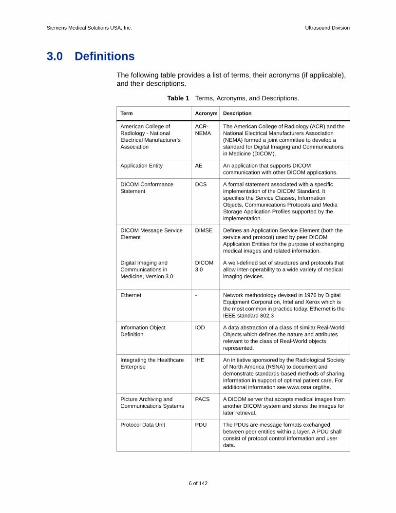
3.0 DefinitionsThe following table provides a list of terms, their acronyms (if applicable), and their descriptions.
Table 1 Terms, Acronyms, and Descriptions.
Term Acronym Description
American College of Radiology - National Electrical Manufacturer's Association
ACR-NEMA
The American College of Radiology (ACR) and the National Electrical Manufacturers Association (NEMA) formed a joint committee to develop a standard for Digital Imaging and Communications in Medicine (DICOM).
Application Entity AE An application that supports DICOM communication with other DICOM applications.
DICOM Conformance Statement
DCS A formal statement associated with a specific implementation of the DICOM Standard. It specifies the Service Classes, Information Objects, Communications Protocols and Media Storage Application Profiles supported by the implementation.
DICOM Message Service Element
DIMSE Defines an Application Service Element (both the service and protocol) used by peer DICOM Application Entities for the purpose of exchanging medical images and related information.
Digital Imaging and Communications in Medicine, Version 3.0
DICOM 3.0
A well-defined set of structures and protocols that allow inter-operability to a wide variety of medical imaging devices.
Ethernet - Network methodology devised in 1976 by Digital Equipment Corporation, Intel and Xerox which is the most common in practice today. Ethernet is the IEEE standard 802.3
Information Object Definition
IOD A data abstraction of a class of similar Real-World Objects which defines the nature and attributes relevant to the class of Real-World objects represented.
Integrating the Healthcare Enterprise
IHE An initiative sponsored by the Radiological Society of North America (RSNA) to document and demonstrate standards-based methods of sharing information in support of optimal patient care. For additional information see www.rsna.org/ihe.
Picture Archiving and Communications Systems
PACS A DICOM server that accepts medical images from another DICOM system and stores the images for later retrieval.
Protocol Data Unit PDU The PDUs are message formats exchanged between peer entities within a layer. A PDU shall consist of protocol control information and user data.
6 of 142
Siemens Medical Solutions USA, Inc. Ultrasound Division
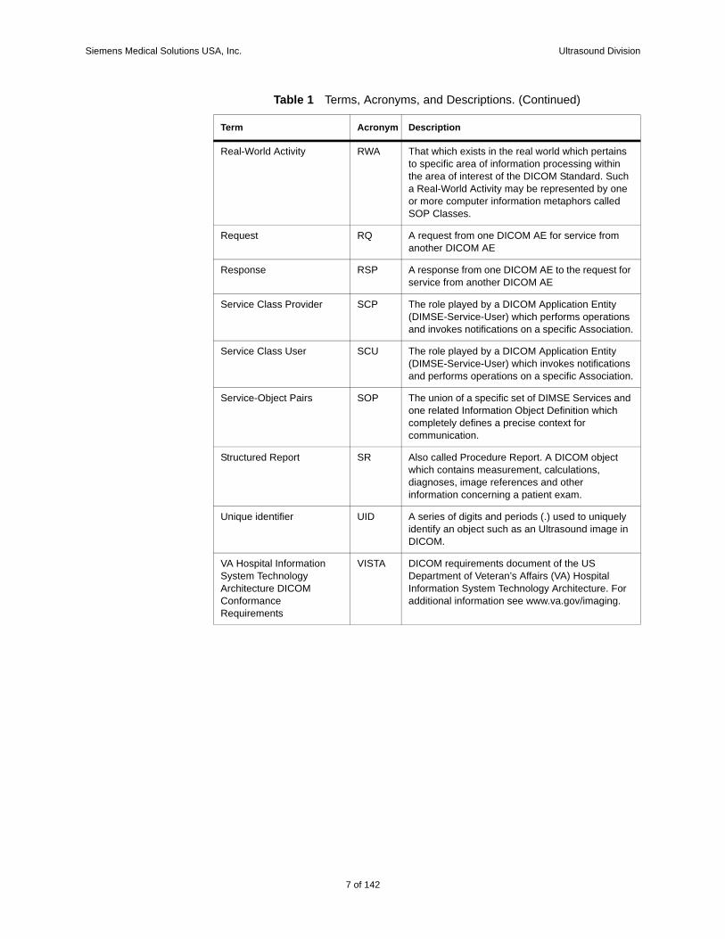
Real-World Activity RWA That which exists in the real world which pertains to specific area of information processing within the area of interest of the DICOM Standard. Such a Real-World Activity may be represented by one or more computer information metaphors called SOP Classes.
Request RQ A request from one DICOM AE for service from another DICOM AE
Response RSP A response from one DICOM AE to the request for service from another DICOM AE
Service Class Provider SCP The role played by a DICOM Application Entity (DIMSE-Service-User) which performs operations and invokes notifications on a specific Association.
Service Class User SCU The role played by a DICOM Application Entity (DIMSE-Service-User) which invokes notifications and performs operations on a specific Association.
Service-Object Pairs SOP The union of a specific set of DIMSE Services and one related Information Object Definition which completely defines a precise context for communication.
Structured Report SR Also called Procedure Report. A DICOM object which contains measurement, calculations, diagnoses, image references and other information concerning a patient exam.
Unique identifier UID A series of digits and periods (.) used to uniquely identify an object such as an Ultrasound image in DICOM.
VA Hospital Information System Technology Architecture DICOM Conformance Requirements
VISTA DICOM requirements document of the US Department of Veteran’s Affairs (VA) Hospital Information System Technology Architecture. For additional information see www.va.gov/imaging.
Table 1 Terms, Acronyms, and Descriptions. (Continued)
Term Acronym Description
7 of 142
142
Siemens Medical Solutions USA, Inc. Ultrasound Division
4.0 Implementation ModelAcuson X500 system users can store images and other data directly on the Acuson X500 system hard disk. Images and structured reports can be exported to a DICOM archive server or workstation on a network. In the fol-lowing sections, Acuson X500 system Real World Activities are indicated by “Real World Activity” name while “X500 AE” indicates the invoked Appli-cation Entity. Similarly, the activities associated with service providers are indicated as “Real World Service Activity.”
8 of 142
Siemens Medical Solutions USA, Inc. Ultrasound Division
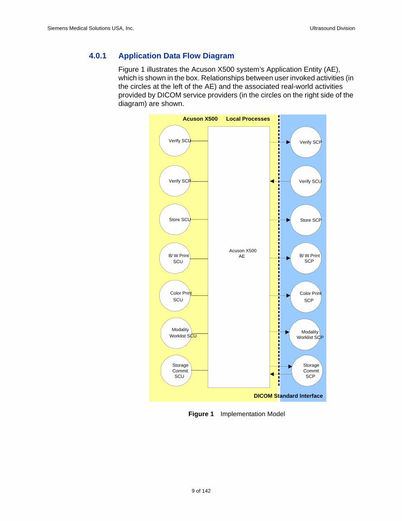
4.0.1 Application Data Flow DiagramFigure 1 illustrates the Acuson X500 system’s Application Entity (AE), which is shown in the box. Relationships between user invoked activities (in the circles at the left of the AE) and the associated real-world activities provided by DICOM service providers (in the circles on the right side of the diagram) are shown.
Figure 1 Implementation Model
Acuson X500AE
Verify SCU
Store SCU
B/ W PrintSCU
Color PrintSCU
Verify SCP
Store SCP
B/ W PrintSCP
Color PrintSCP
Modality
StorageCommitSCU
ModalityWorklist SCP
StorageCommitSCP
DICOM Standard Interface
Acuson X500 Local Processes
Verify SCP Verify SCU
Worklist SCU
9 of 142
142
Siemens Medical Solutions USA, Inc. Ultrasound Division
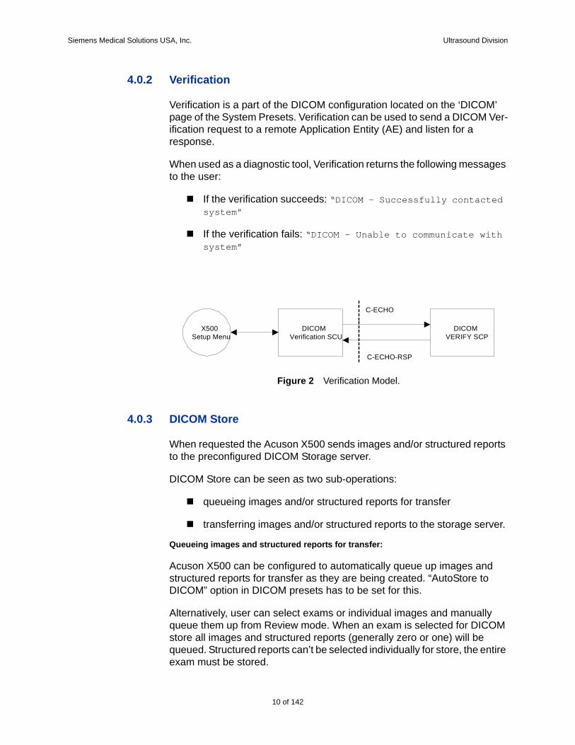
4.0.2 Verification
Verification is a part of the DICOM configuration located on the ‘DICOM’ page of the System Presets. Verification can be used to send a DICOM Ver-ification request to a remote Application Entity (AE) and listen for a response.
When used as a diagnostic tool, Verification returns the following messages to the user:
If the verification succeeds: “DICOM - Successfully contacted system”
If the verification fails: “DICOM - Unable to communicate with system”
4.0.3 DICOM Store
When requested the Acuson X500 sends images and/or structured reports to the preconfigured DICOM Storage server.
DICOM Store can be seen as two sub-operations:
queueing images and/or structured reports for transfer
transferring images and/or structured reports to the storage server.
Queueing images and structured reports for transfer:
Acuson X500 can be configured to automatically queue up images and structured reports for transfer as they are being created. “AutoStore to DICOM” option in DICOM presets has to be set for this.
Alternatively, user can select exams or individual images and manually queue them up from Review mode. When an exam is selected for DICOM store all images and structured reports (generally zero or one) will be queued. Structured reports can’t be selected individually for store, the entire exam must be stored.
Figure 2 Verification Model.
X500Setup Menu
DICOMVerification SCU
DICOMVERIFY SCP
C-ECHO
C-ECHO-RSP
10 of 142

Siemens Medical Solutions USA, Inc. Ultrasound Division
Transfer of images and structured reports to the storage server:
Further, once images and/or structured reports are queued they may be immediately transferred or delayed till the end of study using the transfer storage configuration.
Acuson X500 supports two storage configurations: “Store At End of Exam” and “Store During Exam”.
If the storage configuration is set to “Store At End of Exam” transfer attempts begin when the user selects “Close Study” or “New Patient”.
If the storage configuration is set to “Store During Exam”, transfer attempts to destination devices begin immediately after they are queued.
For both “Store At End of Exam” and “Store During Exam” settings, image and/or structured report transfer will be delayed if the Acuson X500 is busy performing another DICOM Store operation.
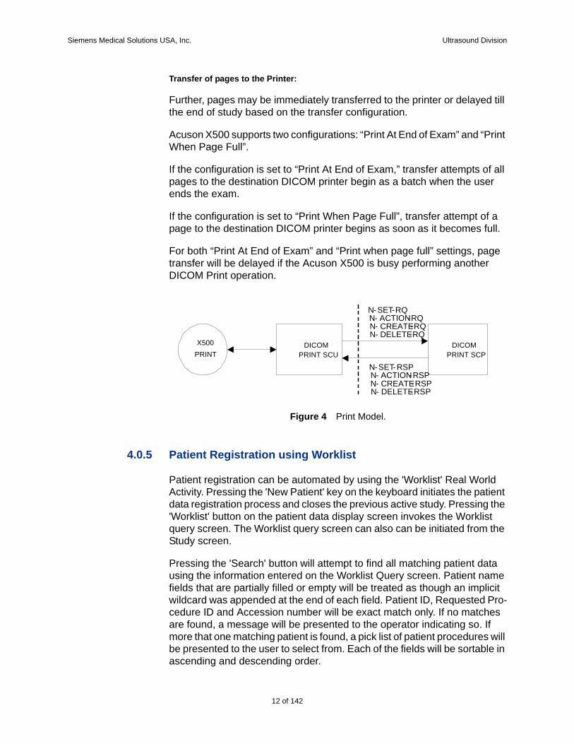
4.0.4 DICOM Print
Acuson X500 system is capable of grayscale (B/W) and color printing.
When requested, single frame images will be printed to a pre-configured DICOM network printer.
DICOM Print can be seen as two sub-operations:
paging images for transfer
transferring pages to printer
Paging images for transfer:
Acuson X500 can be configured to automatically queue up images to be printed on B/W Printer and/or Color printer as they are being created.
Alternatively, user can select exams or individual images and manually queue them up from Review mode for print.
Every image queued up is added into a page in the respective printer layout (DICOM B/W Printer Layout or DICOM Color Printer Layout).
Figure 3 Store Model.
STOREDICOM
STORE SCUDICOM
STORE SCP
C-STORE-RQ
C-STORE-RSP
X500
11 of 142
142
Siemens Medical Solutions USA, Inc. Ultrasound Division
Transfer of pages to the Printer:
Further, pages may be immediately transferred to the printer or delayed till the end of study based on the transfer configuration.
Acuson X500 supports two configurations: “Print At End of Exam” and “Print When Page Full”.
If the configuration is set to “Print At End of Exam,” transfer attempts of all pages to the destination DICOM printer begin as a batch when the user ends the exam.
If the configuration is set to “Print When Page Full”, transfer attempt of a page to the destination DICOM printer begins as soon as it becomes full.
For both “Print At End of Exam” and “Print when page full” settings, page transfer will be delayed if the Acuson X500 is busy performing another DICOM Print operation.
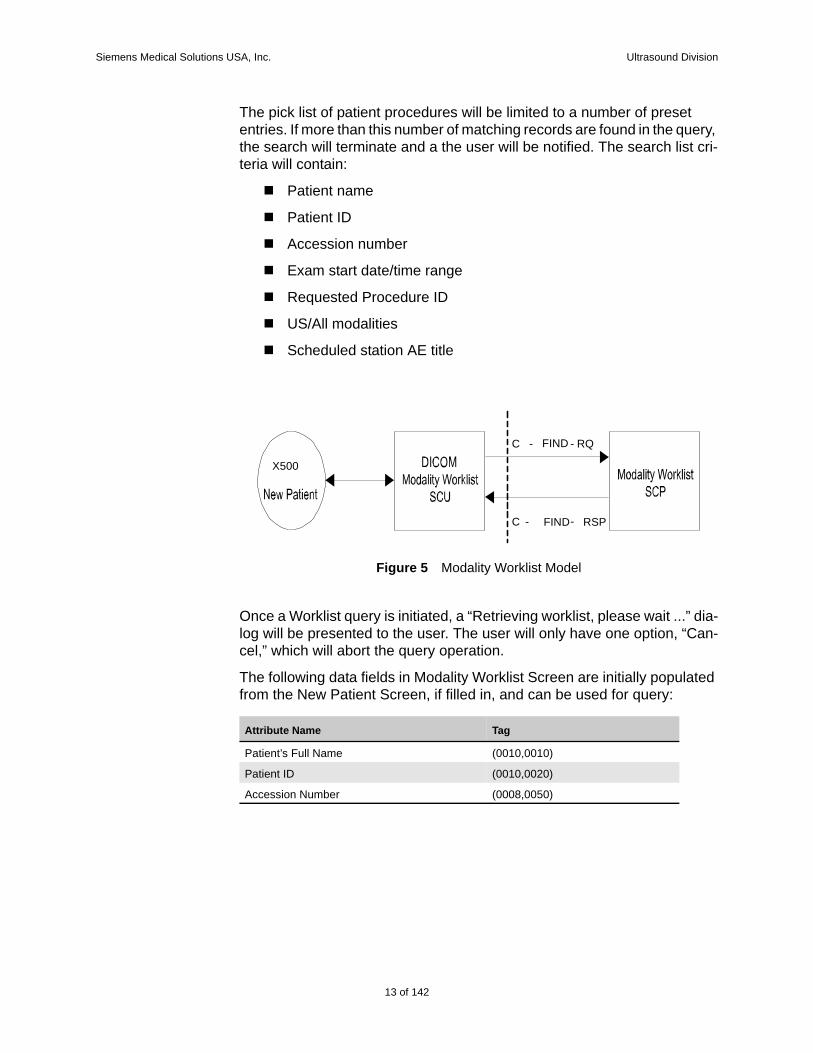
4.0.5 Patient Registration using Worklist
Patient registration can be automated by using the 'Worklist' Real World Activity. Pressing the 'New Patient' key on the keyboard initiates the patient data registration process and closes the previous active study. Pressing the 'Worklist' button on the patient data display screen invokes the Worklist query screen. The Worklist query screen can also can be initiated from the Study screen.
Pressing the 'Search' button will attempt to find all matching patient data using the information entered on the Worklist Query screen. Patient name fields that are partially filled or empty will be treated as though an implicit wildcard was appended at the end of each field. Patient ID, Requested Pro-cedure ID and Accession number will be exact match only. If no matches are found, a message will be presented to the operator indicating so. If more that one matching patient is found, a pick list of patient procedures will be presented to the user to select from. Each of the fields will be sortable in ascending and descending order.
Figure 4 Print Model.
X500
PRINT SCU
N-SET-RQN- ACTION-RQN- CREATE-RQN- DELETE-RQ
N-SET-RSPN- ACTION-RSPN- CREATE-RSPN- DELETE-RSP
DICOMPRINT
DICOMPRINT SCP
12 of 142
Siemens Medical Solutions USA, Inc. Ultrasound Division
The pick list of patient procedures will be limited to a number of preset entries. If more than this number of matching records are found in the query, the search will terminate and a the user will be notified. The search list cri-teria will contain:
Patient name
Patient ID
Accession number
Exam start date/time range
Requested Procedure ID
US/All modalities
Scheduled station AE title
Once a Worklist query is initiated, a “Retrieving worklist, please wait ...” dia-log will be presented to the user. The user will only have one option, “Can-cel,” which will abort the query operation.
The following data fields in Modality Worklist Screen are initially populated from the New Patient Screen, if filled in, and can be used for query:
Figure 5 Modality Worklist Model
X500
FIND - RQ
C - FIND- RSP
-C
Attribute Name Tag
Patient’s Full Name (0010,0010)
Patient ID (0010,0020)
Accession Number (0008,0050)
13 of 142
142
Siemens Medical Solutions USA, Inc. Ultrasound Division
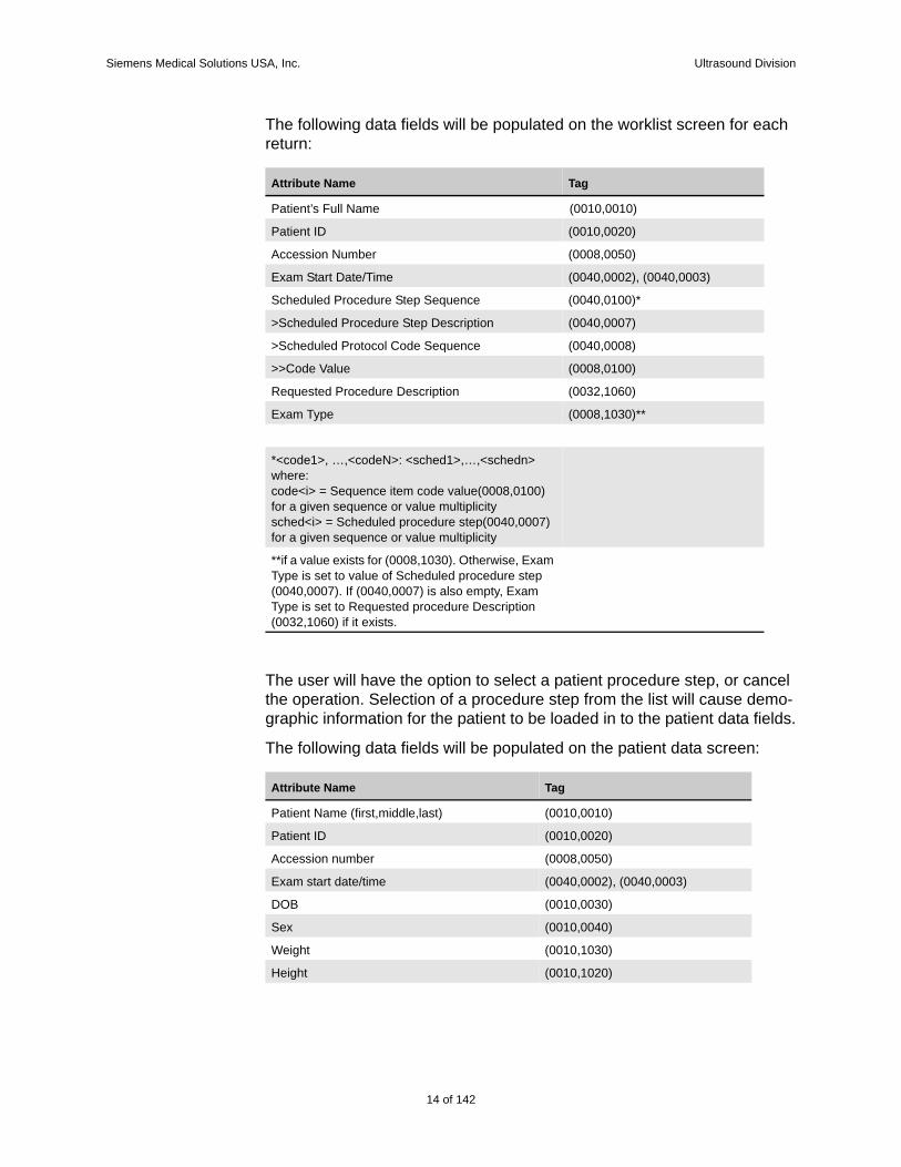
The following data fields will be populated on the worklist screen for each return:
The user will have the option to select a patient procedure step, or cancel the operation. Selection of a procedure step from the list will cause demo-graphic information for the patient to be loaded in to the patient data fields.
The following data fields will be populated on the patient data screen:
Attribute Name Tag
Patient’s Full Name (0010,0010)
Patient ID (0010,0020)
Accession Number (0008,0050)
Exam Start Date/Time (0040,0002), (0040,0003)
Scheduled Procedure Step Sequence (0040,0100)*
>Scheduled Procedure Step Description (0040,0007)
>Scheduled Protocol Code Sequence (0040,0008)
>>Code Value (0008,0100)
Requested Procedure Description (0032,1060)
Exam Type (0008,1030)**
*<code1>, …,<codeN>: <sched1>,…,<schedn>where:code<i> = Sequence item code value(0008,0100) for a given sequence or value multiplicitysched<i> = Scheduled procedure step(0040,0007) for a given sequence or value multiplicity
**if a value exists for (0008,1030). Otherwise, Exam Type is set to value of Scheduled procedure step (0040,0007). If (0040,0007) is also empty, Exam Type is set to Requested procedure Description (0032,1060) if it exists.
Attribute Name Tag
Patient Name (first,middle,last) (0010,0010)
Patient ID (0010,0020)
Accession number (0008,0050)
Exam start date/time (0040,0002), (0040,0003)
DOB (0010,0030)
Sex (0010,0040)
Weight (0010,1030)
Height (0010,1020)
14 of 142
Siemens Medical Solutions USA, Inc. Ultrasound Division
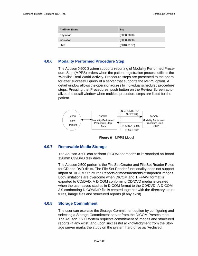
4.0.6 Modality Performed Procedure Step
The Acuson X500 System supports reporting of Modality Performed Proce-dure Step (MPPS) orders when the patient registration process utilizes the ‘Worklist’ Real World Activity. Procedure steps are presented to the opera-tor after successful query of a server that supports the MPPS option. A detail window allows the operator access to individual scheduled procedure steps. Pressing the ‘Procedures’ push button on the Review Screen actu-alizes the detail window when multiple procedure steps are listed for the patient.
Figure 6 MPPS Model
4.0.7 Removable Media Storage
The Acuson X500 can perform DICOM operations to its standard on-board 120mm CD/DVD disk drive.
The Acuson X500 performs the File Set Creator and File Set Reader Roles for CD and DVD disks. The File Set Reader functionality does not support import of DICOM Structured Reports or measurements of imported images. Both limitations are overcome when DICOM and TIFF/AVI format is exported to CD/DVD. A DICOM conforming CD/DVD media is created when the user saves studies in DICOM format to the CD/DVD. A DICOM 3.0 conforming DICOMDIR file is created together with the directory struc-tures, image files and structured reports (if any exist).
4.0.8 Storage Commitment
The user can exercise the Storage Commitment option by configuring and selecting a Storage Commitment server from the DICOM Presets menu. The Acuson X500 system requests commitment of images and structured reports (if any exist) and upon successful acknowledgment from the Stor-age server marks the study on the system hard drive as ‘Archived’.
Physician (0008,0090)
Indication (0080,1080)
LMP (0010,21D0)
Attribute Name Tag
NewPatient
DICOMModality Performed
Procedure StepSCU
DICOMModality Performed
Procedure StepSCP
N-CREATE-RQN-SET-RQ
N-CREATE-RSPN-SET-RSP
X500
15 of 142
142
Siemens Medical Solutions USA, Inc. Ultrasound Division
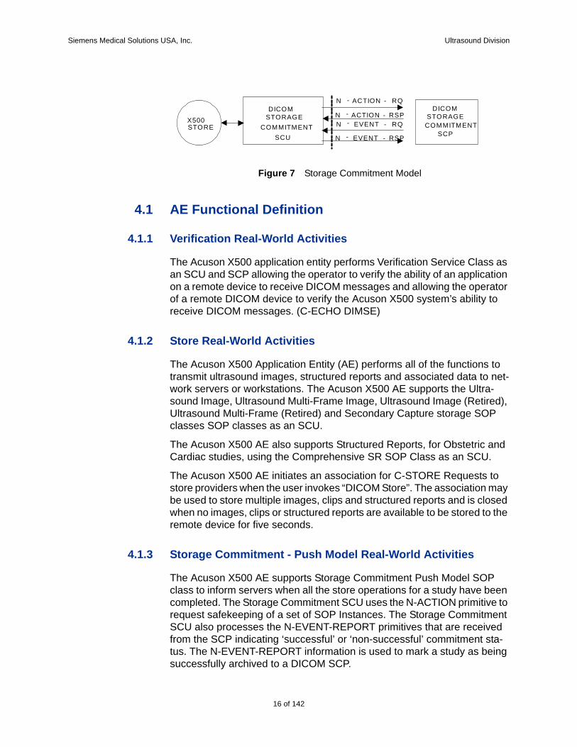
Figure 7 Storage Commitment Model
4.1 AE Functional Definition
4.1.1 Verification Real-World Activities
The Acuson X500 application entity performs Verification Service Class as an SCU and SCP allowing the operator to verify the ability of an application on a remote device to receive DICOM messages and allowing the operator of a remote DICOM device to verify the Acuson X500 system’s ability to receive DICOM messages. (C-ECHO DIMSE)
4.1.2 Store Real-World Activities
The Acuson X500 Application Entity (AE) performs all of the functions to transmit ultrasound images, structured reports and associated data to net-work servers or workstations. The Acuson X500 AE supports the Ultra-sound Image, Ultrasound Multi-Frame Image, Ultrasound Image (Retired), Ultrasound Multi-Frame (Retired) and Secondary Capture storage SOP classes SOP classes as an SCU.
The Acuson X500 AE also supports Structured Reports, for Obstetric and Cardiac studies, using the Comprehensive SR SOP Class as an SCU.
The Acuson X500 AE initiates an association for C-STORE Requests to store providers when the user invokes “DICOM Store”. The association may be used to store multiple images, clips and structured reports and is closed when no images, clips or structured reports are available to be stored to the remote device for five seconds.
4.1.3 Storage Commitment - Push Model Real-World Activities
The Acuson X500 AE supports Storage Commitment Push Model SOP class to inform servers when all the store operations for a study have been completed. The Storage Commitment SCU uses the N-ACTION primitive to request safekeeping of a set of SOP Instances. The Storage Commitment SCU also processes the N-EVENT-REPORT primitives that are received from the SCP indicating ‘successful’ or ‘non-successful’ commitment sta-tus. The N-EVENT-REPORT information is used to mark a study as being successfully archived to a DICOM SCP.
STORE
DICOMSTORAGE
COMMITMENTSCU
X500
N - ACTION - RQ
N - ACTION - RSPN - EVENT - RQ
N - EVENT - RSP
DICOMSTORAGE
COMMITMENTSCP
16 of 142
Siemens Medical Solutions USA, Inc. Ultrasound Division
The successful commit status and archival indication on the X500 does not ensure permanent archival of the images and Structured Reports. The operations performed by the SCP are dependent on its capabilities and configuration.
4.1.4 Print Real-World ActivitiesThe Acuson X500 AE provides all aspects of the Print Management SCU. The Acuson X500 AE initiates an association to the printer when the user invokes “DICOM Print”. The association may be used to print multiple pages and is closed when no pages are available to be printed to the remote device for five seconds.
4.2 Modality Worklist Real-World ActivitiesThe Acuson X500 AE supports the DICOM Basic Worklist Management Service as an SCU. The AE initiates an association to the active Worklist server when a Worklist query is selected (via the “Worklist” button). The association is closed upon the completion of each query. A preset maxi-mum number of matching results is accepted, at which point, the Acuson X500 AE issues a C-CANCEL-RQ request.
4.3 Modality Performed Procedure Step Real-World ActivitiesThe Acuson X500 AE supports Modality Performed Procedure Step (MPPS) in the role of SCU. The Acuson X500 is capable of displaying scheduled procedure steps via the User Interface (UI) for Modality Per-formed Procedure Step. The operator can select a single PPS. The opera-tor can notify the MPPS server that a MPPS is ‘In Progress’, ‘Discontinued’ or ‘Completed’.
4.4 Removable Media Storage Real-World ActivitiesThe Acuson X500 AE provides a standard implementation of DICOM Store to CD or DVD. The Acuson X500 AE selects one or more studies and exports the same to CD or DVD. Acuson X500 AE creates a DICOM File Format Image File for every image, clip and structured report in each of the selected studies.
A DICOMDIR file is created along with the files.
Measurements are not supported on imported images unless TIFF/AVI for-mat is exported.
The DICOM SR cannot be imported from media unless the TIFF/AVI format is exported along with the DICOM SR.
17 of 142
142

Siemens Medical Solutions USA, Inc. Ultrasound Division
4.5 Sequencing of Real-World ActivitiesPrint, Store, Echo, Worklist, Storage Commit and MPPS commands can be transmitted simultaneously within the limits described below.
Storage Commit
The Storage Commitment (if enabled) command is sent in the following sit-uations:
a. On series close, when all images have previously stored successfully.
b. The series is closed before all images are stored successfully, all previ-ous stores have succeeded and the last image stores successfully.
c. The series is closed before all images are stored successfully, at least one store has succeeded, at least one store has failed and the last store with non-zero retry count fails or succeeds.
d. A series has been partially committed as in c. Later, due to "Retry Job” button press on the Store Status UI screen the store jobs are retried. Another Storage Commit is sent when at least one store has succeeded and the last store with non-zero retry count fails or succeeds.
MPPS
The MPPS (if enabled) command is sent in the following situations:
a. N-CREATE command is sent whenever a new procedure step is selected. The state of the MPPS command is set to “In-Progress”.
b. N-SET command is sent when the Procedure Step is closed by the user pressing either the Completed or Discontinued button on the Close Proce-dure dialog. The state of the MPPS command is set, according to the state (Completed or Discontinued) set by the user.
18 of 142
Siemens Medical Solutions USA, Inc. Ultrasound Division
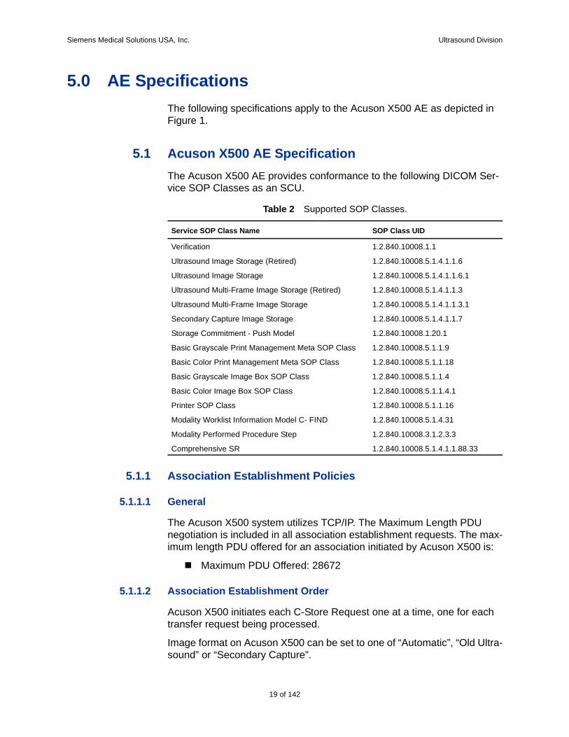
5.0 AE SpecificationsThe following specifications apply to the Acuson X500 AE as depicted in Figure 1.
5.1 Acuson X500 AE SpecificationThe Acuson X500 AE provides conformance to the following DICOM Ser-vice SOP Classes as an SCU.
5.1.1 Association Establishment Policies
5.1.1.1 General
The Acuson X500 system utilizes TCP/IP. The Maximum Length PDU negotiation is included in all association establishment requests. The max-imum length PDU offered for an association initiated by Acuson X500 is:
Maximum PDU Offered: 28672
5.1.1.2 Association Establishment Order
Acuson X500 initiates each C-Store Request one at a time, one for each transfer request being processed.
Image format on Acuson X500 can be set to one of “Automatic”, “Old Ultra-sound” or “Secondary Capture”.
Table 2 Supported SOP Classes.
Service SOP Class Name SOP Class UID
Verification 1.2.840.10008.1.1
Ultrasound Image Storage (Retired) 1.2.840.10008.5.1.4.1.1.6
Ultrasound Image Storage 1.2.840.10008.5.1.4.1.1.6.1
Ultrasound Multi-Frame Image Storage (Retired) 1.2.840.10008.5.1.4.1.1.3
Ultrasound Multi-Frame Image Storage 1.2.840.10008.5.1.4.1.1.3.1
Secondary Capture Image Storage 1.2.840.10008.5.1.4.1.1.7
Storage Commitment - Push Model 1.2.840.10008.1.20.1
Basic Grayscale Print Management Meta SOP Class 1.2.840.10008.5.1.1.9
Basic Color Print Management Meta SOP Class 1.2.840.10008.5.1.1.18
Basic Grayscale Image Box SOP Class 1.2.840.10008.5.1.1.4
Basic Color Image Box SOP Class 1.2.840.10008.5.1.1.4.1
Printer SOP Class 1.2.840.10008.5.1.1.16
Modality Worklist Information Model C- FIND 1.2.840.10008.5.1.4.31
Modality Performed Procedure Step 1.2.840.10008.3.1.2.3.3
Comprehensive SR 1.2.840.10008.5.1.4.1.1.88.33
19 of 142
142
Siemens Medical Solutions USA, Inc. Ultrasound Division
For the “Automatic” setting, Acuson X500 proposes Ultrasound Multi-Frame Image, Ultrasound Image, Ultrasound Multi-Frame Image (Retired), Ultrasound Image (Retired), Secondary Capture Image and Comprehen-sive SR sequentially.
For the “Old Ultrasound” setting, Acuson X500 proposes Ultrasound Multi-Frame Image (Retired), Ultrasound Image (Retired), Secondary Capture and Comprehensive SR Image to be negotiated sequentially.
For the “Secondary Capture” setting, Acuson X500 proposes Secondary Capture Image and Comprehensive SR to be negotiated sequentially.
5.1.1.3 Asynchronous Nature
All associations use the default synchronous mode of operation. Asynchro-nous Operations Window negotiations are not supported on the Acuson X500 system.
5.1.1.4 Implementation Identifying Information
Implementation Class UID: “1.3.12.2.1107.5.5.5” (See below).
Implementation Version Name: “MergeCOM3_351MergeCOM3_351”
Siemens has provided registration for all Siemens Medical Solutions Groups. This unique Class UID is defined as:
“1.3.12.2.1107.5.5.product”
Where the interpretation is:
1. = International Standards Organization (ISO)
3. = International branch of ISO
12.2.1107.5. = Assigned to Siemens-UB MED
5. = Ultrasound Modality (SMS-UG)
Product = 5 - DICOM implementation for SONOLINE G20, G40, G50, G60 S, X300 and Acuson CV70, X500
5.1.2 Association Initiation by Real-World Activities
5.1.2.1 Real World Activity – Verification
The Acuson X500 is capable of supporting Verification service class as SCU or SCP. Verification can be initiated as a singular event from the Sys-tems Presets menu to any configured SCP that supports Verification.
20 of 142
Siemens Medical Solutions USA, Inc. Ultrasound Division
Proposed Presentation Contexts – Verification
The Acuson X500 will propose Presentation contexts as shown in table 3.
5.1.2.2 Real World Activity – Store
Acuson X500 facilitates users to store images and structured reports as they are being created or later in review mode.
Queueing images during acquisition:
“Autostore to DICOM” option in DICOM presets has to be set. One or more of “Print/Store 1”, “Print/Store 2” and “Clip Store” keys on the control panel can be configured for Store (Disk Store, D.Store, Clip capture). When the user presses one of the configured keys, an image or clip is acquired, stored on the hard disk and queued up to be transferred to the storage server. Structured reports, if any, will be stored automatically after the study is closed and each time the report is modified after study close.
Queueing images and structured reports in Review mode:
User can select one or more closed studies and queue them up for Storage. The DICOM Store button is available in Review screen for this operation. All images and structured reports (if any) are stored. The study must be closed to generate a structured report.
Transfer of images to the storage server:
See section 4.1.2.
Associated Real World Activities
When images and/or structured reports are transferred from the hard disk to a DICOM Store SCP, the system establishes an association between the Acuson X500 AE and the configured DICOM device. The association may be used to store multiple images and/or structured reports and is closed when no images or structured reports are available to be stored to the remote device for five seconds.
Table 3 Verification Presentation Context.
Abstract Syntax Transfer SyntaxRole Extended
NegotiationName UID Name List UID List
Verification 1.2.840.10008.1.1 Implicit VR Little Endian
1.2.840.10008.1.2 SCU/SCP
None
Verification 1.2.840.10008.1.1 Explicit VR Little Endian
1.2.840.10008.1.2.1 SCU/SCP
None
Verification 1.2.840.10008.1.1 Explicit VR Big Endian
1.2.840.10008.1.2.2 SCU/SCP
None
21 of 142
142
Siemens Medical Solutions USA, Inc. Ultrasound Division
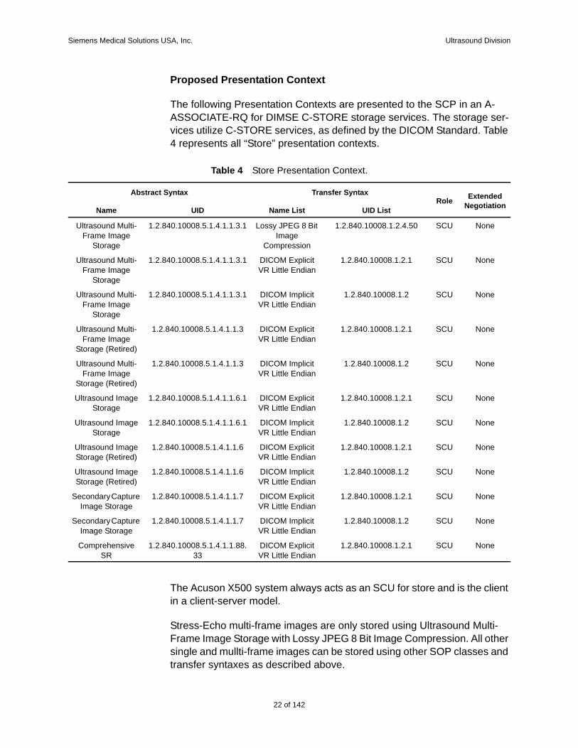
Proposed Presentation Context
The following Presentation Contexts are presented to the SCP in an A-ASSOCIATE-RQ for DIMSE C-STORE storage services. The storage ser-vices utilize C-STORE services, as defined by the DICOM Standard. Table 4 represents all “Store” presentation contexts.
The Acuson X500 system always acts as an SCU for store and is the client in a client-server model.
Stress-Echo multi-frame images are only stored using Ultrasound Multi-Frame Image Storage with Lossy JPEG 8 Bit Image Compression. All other single and mullti-frame images can be stored using other SOP classes and transfer syntaxes as described above.
Table 4 Store Presentation Context.
Abstract Syntax Transfer SyntaxRole Extended
NegotiationName UID Name List UID List
Ultrasound Multi-Frame Image
Storage
1.2.840.10008.5.1.4.1.1.3.1 Lossy JPEG 8 Bit Image
Compression
1.2.840.10008.1.2.4.50 SCU None
Ultrasound Multi-Frame Image
Storage
1.2.840.10008.5.1.4.1.1.3.1 DICOM Explicit VR Little Endian
1.2.840.10008.1.2.1 SCU None
Ultrasound Multi-Frame Image
Storage
1.2.840.10008.5.1.4.1.1.3.1 DICOM Implicit VR Little Endian
1.2.840.10008.1.2 SCU None
Ultrasound Multi-Frame Image
Storage (Retired)
1.2.840.10008.5.1.4.1.1.3 DICOM Explicit VR Little Endian
1.2.840.10008.1.2.1 SCU None
Ultrasound Multi-Frame Image
Storage (Retired)
1.2.840.10008.5.1.4.1.1.3 DICOM Implicit VR Little Endian
1.2.840.10008.1.2 SCU None
Ultrasound Image Storage
1.2.840.10008.5.1.4.1.1.6.1 DICOM Explicit VR Little Endian
1.2.840.10008.1.2.1 SCU None
Ultrasound Image Storage
1.2.840.10008.5.1.4.1.1.6.1 DICOM Implicit VR Little Endian
1.2.840.10008.1.2 SCU None
Ultrasound Image Storage (Retired)
1.2.840.10008.5.1.4.1.1.6 DICOM Explicit VR Little Endian
1.2.840.10008.1.2.1 SCU None
Ultrasound Image Storage (Retired)
1.2.840.10008.5.1.4.1.1.6 DICOM Implicit VR Little Endian
1.2.840.10008.1.2 SCU None
Secondary Capture Image Storage
1.2.840.10008.5.1.4.1.1.7 DICOM Explicit VR Little Endian
1.2.840.10008.1.2.1 SCU None
Secondary Capture Image Storage
1.2.840.10008.5.1.4.1.1.7 DICOM Implicit VR Little Endian
1.2.840.10008.1.2 SCU None
Comprehensive SR
1.2.840.10008.5.1.4.1.1.88.33
DICOM Explicit VR Little Endian
1.2.840.10008.1.2.1 SCU None
22 of 142
Siemens Medical Solutions USA, Inc. Ultrasound Division
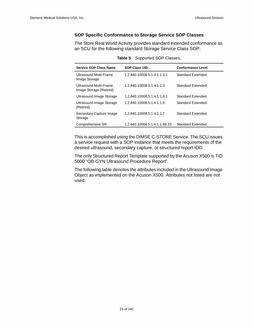
SOP Specific Conformance to Storage Service SOP Classes
The Store Real World Activity provides standard extended conformance as an SCU for the following standard Storage Service Class SOP:
This is accomplished using the DIMSE C-STORE Service. The SCU issues a service request with a SOP instance that meets the requirements of the desired ultrasound, secondary capture, or structured report IOD.
The only Structured Report Template supported by the Acuson X500 is TID 5000 “OB-GYN Ultrasound Procedure Report”.
The following table denotes the attributes included in the Ultrasound Image Object as implemented on the Acuson X500. Attributes not listed are not used.
Table 5 Supported SOP Classes.
Service SOP Class Name SOP Class UID Conformance Level
Ultrasound Multi-Frame Image Storage
1.2.840.10008.5.1.4.1.1.3.1 Standard Extended
Ultrasound Multi-Frame Image Storage (Retired)
1.2.840.10008.5.1.4.1.1.3 Standard Extended
Ultrasound Image Storage 1.2.840.10008.5.1.4.1.1.6.1 Standard Extended
Ultrasound Image Storage (Retired)
1.2.840.10008.5.1.4.1.1.6 Standard Extended
Secondary Capture Image Storage
1.2.840.10008.5.1.4.1.1.7 Standard Extended
Comprehensive SR 1.2.840.10008.5.1.4.1.1.88.33 Standard Extended
23 of 142
142
Siemens Medical Solutions USA, Inc. Ultrasound Division
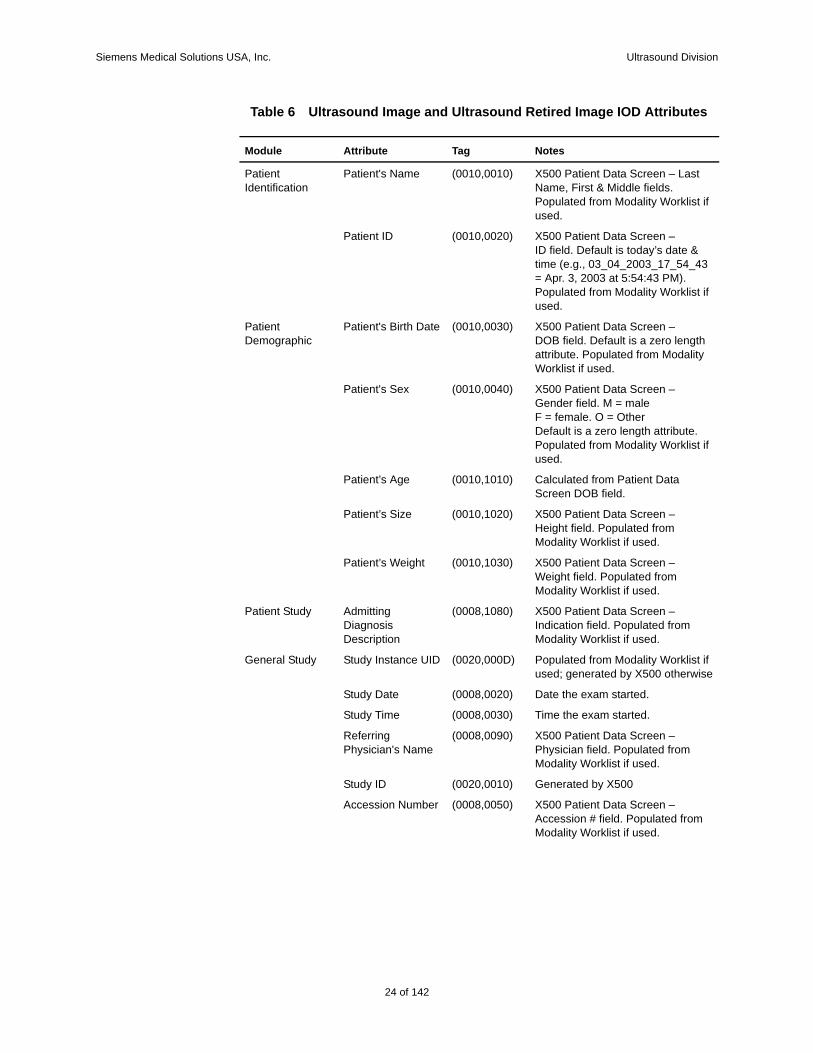
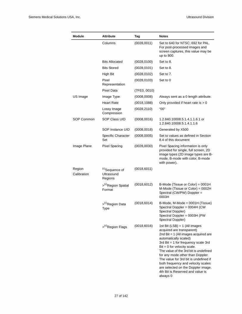
Table 6 Ultrasound Image and Ultrasound Retired Image IOD Attributes
Module Attribute Tag Notes
Patient Identification
Patient's Name (0010,0010) X500 Patient Data Screen – Last Name, First & Middle fields. Populated from Modality Worklist if used.
Patient ID (0010,0020) X500 Patient Data Screen –ID field. Default is today’s date & time (e.g., 03_04_2003_17_54_43 = Apr. 3, 2003 at 5:54:43 PM). Populated from Modality Worklist if used.
Patient Demographic
Patient's Birth Date (0010,0030) X500 Patient Data Screen –DOB field. Default is a zero length attribute. Populated from Modality Worklist if used.
Patient's Sex (0010,0040) X500 Patient Data Screen –Gender field. M = maleF = female. O = Other Default is a zero length attribute. Populated from Modality Worklist if used.
Patient’s Age (0010,1010) Calculated from Patient Data Screen DOB field.
Patient’s Size (0010,1020) X500 Patient Data Screen –Height field. Populated from Modality Worklist if used.
Patient’s Weight (0010,1030) X500 Patient Data Screen –Weight field. Populated from Modality Worklist if used.
Patient Study Admitting Diagnosis Description
(0008,1080) X500 Patient Data Screen – Indication field. Populated from Modality Worklist if used.
General Study Study Instance UID (0020,000D) Populated from Modality Worklist if used; generated by X500 otherwise
Study Date (0008,0020) Date the exam started.
Study Time (0008,0030) Time the exam started.
Referring Physician's Name
(0008,0090) X500 Patient Data Screen – Physician field. Populated from Modality Worklist if used.
Study ID (0020,0010) Generated by X500
Accession Number (0008,0050) X500 Patient Data Screen – Accession # field. Populated from Modality Worklist if used.
24 of 142
Siemens Medical Solutions USA, Inc. Ultrasound Division
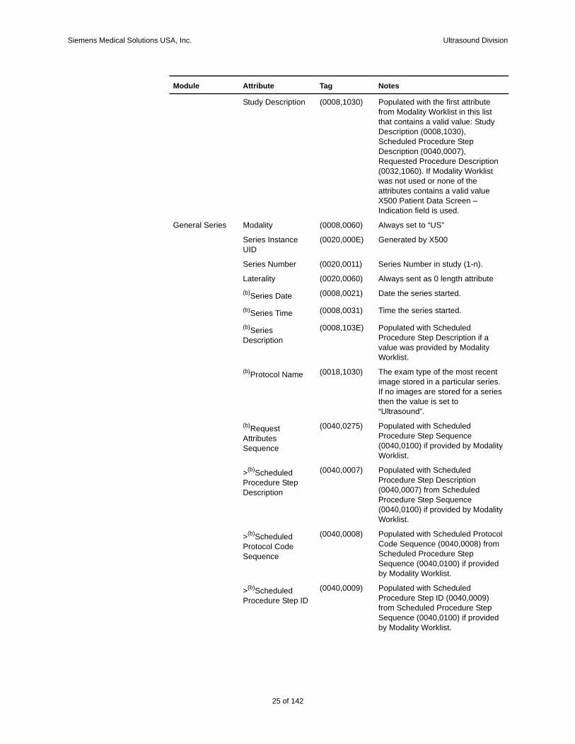
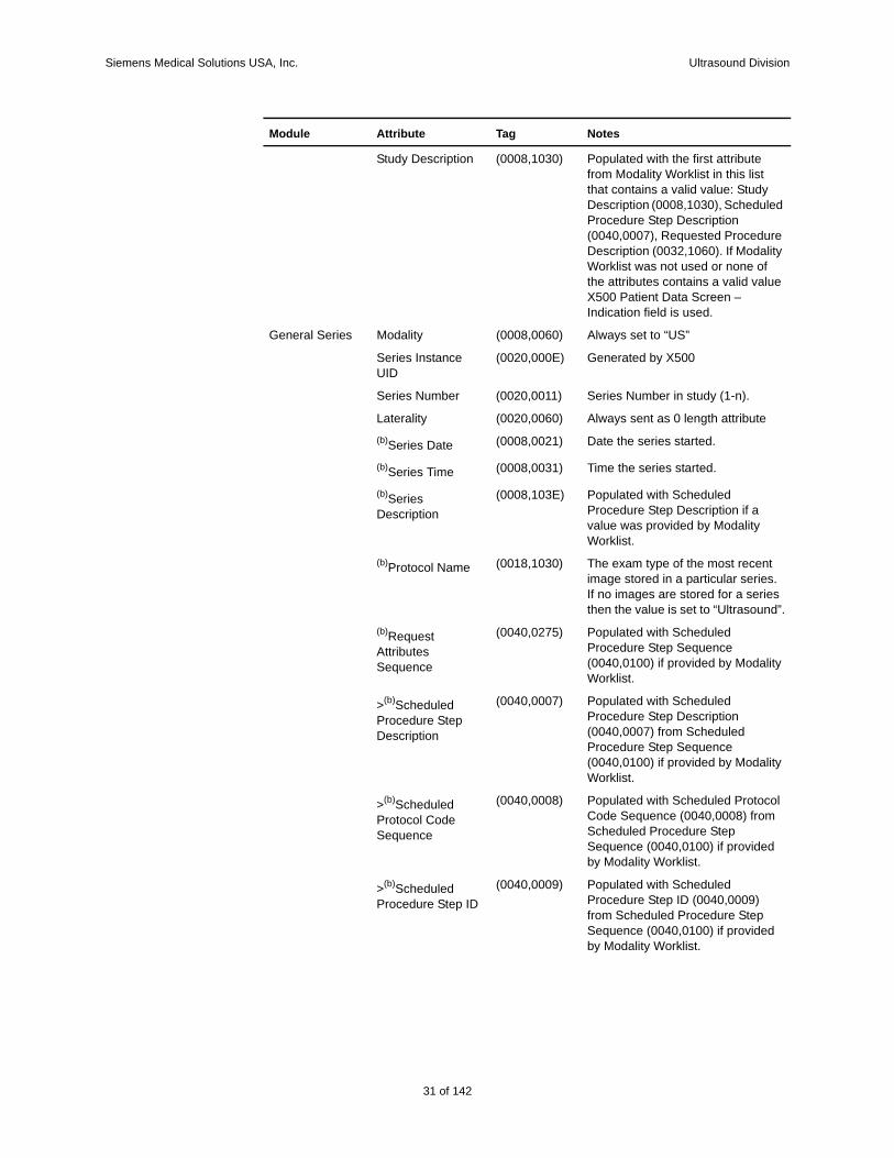
Study Description (0008,1030) Populated with the first attribute from Modality Worklist in this list that contains a valid value: Study Description (0008,1030), Scheduled Procedure Step Description (0040,0007), Requested Procedure Description (0032,1060). If Modality Worklist was not used or none of the attributes contains a valid value X500 Patient Data Screen – Indication field is used.
General Series Modality (0008,0060) Always set to “US”
Series Instance UID
(0020,000E) Generated by X500
Series Number (0020,0011) Series Number in study (1-n).
Laterality (0020,0060) Always sent as 0 length attribute
(b)Series Date (0008,0021) Date the series started.
(b)Series Time (0008,0031) Time the series started.
(b)Series Description
(0008,103E) Populated with Scheduled Procedure Step Description if a value was provided by Modality Worklist.
(b)Protocol Name (0018,1030) The exam type of the most recent image stored in a particular series. If no images are stored for a series then the value is set to “Ultrasound”.
(b)Request Attributes Sequence
(0040,0275) Populated with Scheduled Procedure Step Sequence (0040,0100) if provided by Modality Worklist.
>(b)Scheduled Procedure Step Description
(0040,0007) Populated with Scheduled Procedure Step Description (0040,0007) from Scheduled Procedure Step Sequence (0040,0100) if provided by Modality Worklist.
>(b)Scheduled Protocol Code Sequence
(0040,0008) Populated with Scheduled Protocol Code Sequence (0040,0008) from Scheduled Procedure Step Sequence (0040,0100) if provided by Modality Worklist.
>(b)Scheduled Procedure Step ID
(0040,0009) Populated with Scheduled Procedure Step ID (0040,0009) from Scheduled Procedure Step Sequence (0040,0100) if provided by Modality Worklist.
Module Attribute Tag Notes
25 of 142
142
Siemens Medical Solutions USA, Inc. Ultrasound Division
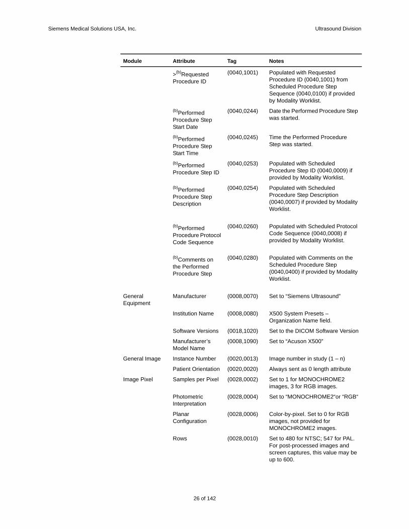
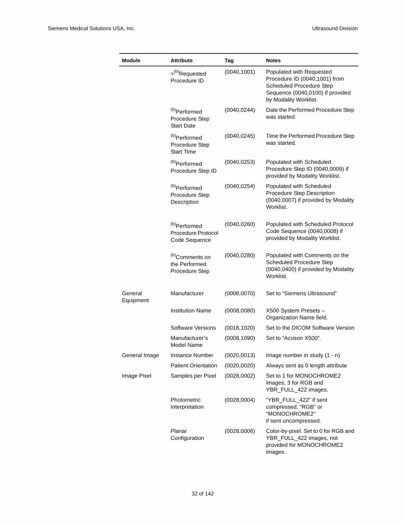
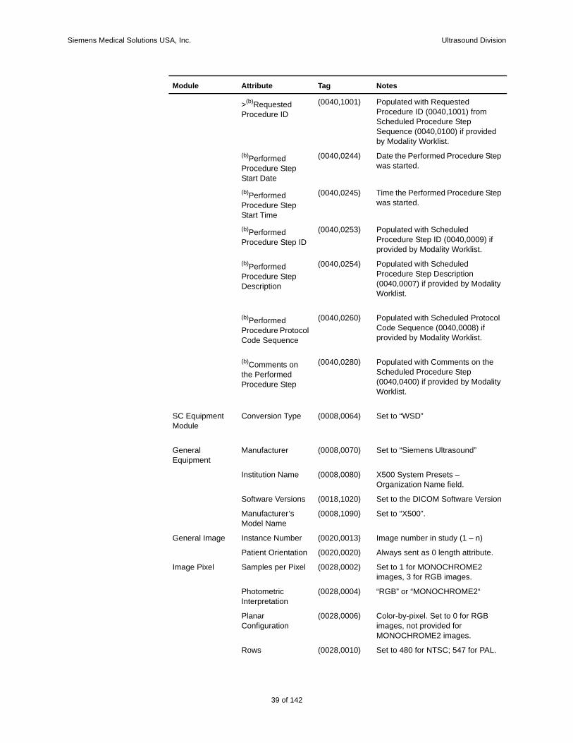
>(b)Requested Procedure ID
(0040,1001) Populated with Requested Procedure ID (0040,1001) from Scheduled Procedure Step Sequence (0040,0100) if provided by Modality Worklist.
(b)Performed Procedure Step Start Date
(0040,0244) Date the Performed Procedure Step was started.
(b)Performed Procedure Step Start Time
(0040,0245) Time the Performed Procedure Step was started.
(b)Performed Procedure Step ID
(0040,0253) Populated with Scheduled Procedure Step ID (0040,0009) if provided by Modality Worklist.
(b)Performed Procedure Step Description
(0040,0254) Populated with Scheduled Procedure Step Description (0040,0007) if provided by Modality Worklist.
(b)Performed Procedure Protocol Code Sequence
(0040,0260) Populated with Scheduled Protocol Code Sequence (0040,0008) if provided by Modality Worklist.
(b)Comments on the Performed Procedure Step
(0040,0280) Populated with Comments on the Scheduled Procedure Step (0040,0400) if provided by Modality Worklist.
General Equipment
Manufacturer (0008,0070) Set to “Siemens Ultrasound”
Institution Name (0008,0080) X500 System Presets – Organization Name field.
Software Versions (0018,1020) Set to the DICOM Software Version
Manufacturer’s Model Name
(0008,1090) Set to “Acuson X500”
General Image Instance Number (0020,0013) Image number in study (1 – n)
Patient Orientation (0020,0020) Always sent as 0 length attribute
Image Pixel Samples per Pixel (0028,0002) Set to 1 for MONOCHROME2 images, 3 for RGB images.
Photometric Interpretation
(0028,0004) Set to “MONOCHROME2“or “RGB“
Planar Configuration
(0028,0006) Color-by-pixel. Set to 0 for RGB images, not provided for MONOCHROME2 images.
Rows (0028,0010) Set to 480 for NTSC; 547 for PAL. For post-processed images and screen captures, this value may be up to 600.
Module Attribute Tag Notes
26 of 142
Siemens Medical Solutions USA, Inc. Ultrasound Division
Columns (0028,0011) Set to 640 for NTSC; 692 for PAL. For post-processed images and screen captures, this value may be up to 800.
Bits Allocated (0028,0100) Set to 8.
Bits Stored (0028,0101) Set to 8.
High Bit (0028,0102) Set to 7.
Pixel Representation
(0028,0103) Set to 0
Pixel Data (7FE0, 0010)
US Image Image Type (0008,0008) Always sent as a 0 length attribute.
Heart Rate (0018,1088) Only provided if heart rate is > 0
Lossy Image Compression
(0028,2110) “00”
SOP Common SOP Class UID (0008,0016) 1.2.840.10008.5.1.4.1.1.6.1 or1.2.840.10008.5.1.4.1.1.6
SOP Instance UID (0008,0018) Generated by X500
Specific Character Set
(0008,0005) Set to values as defined in Section 8.4 of this document
Image Plane Pixel Spacing (0028,0030) Pixel Spacing information is only provided for single, full screen, 2D image types (2D image types are B-mode, B-mode with color, B-mode with power).
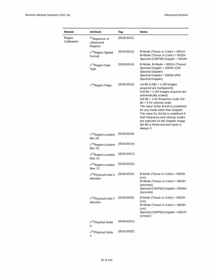
Region Calibration
(c)Sequence of Ultrasound Regions
(0018,6011)
>(c)Region Spatial Format
(0018,6012) B-Mode (Tissue or Color) = 0001HM-Mode (Tissue or Color) = 0002HSpectral (CW/PW) Doppler = 0003H
>(c)Region Data Type
(0018,6014) B-Mode, M-Mode = 0001H (Tissue) Spectral Doppler = 0004H (CW Spectral Doppler) Spectral Doppler = 0003H (PW Spectral Doppler)
>(c)Region Flags (0018,6016) 1st Bit (LSB) = 1 (All images acquired are transparent) 2nd Bit = 1 (All images acquired are automatically scaled) 3rd Bit = 1 for frequency scale 3rd Bit = 0 for velocity scale.The value of the 3rd bit is undefined for any mode other than Doppler. The value for 3rd bit is undefined if both frequency and velocity scales are selected on the Doppler image. 4th Bit is Reserved and value is always 0
Module Attribute Tag Notes
27 of 142
142
Siemens Medical Solutions USA, Inc. Ultrasound Division
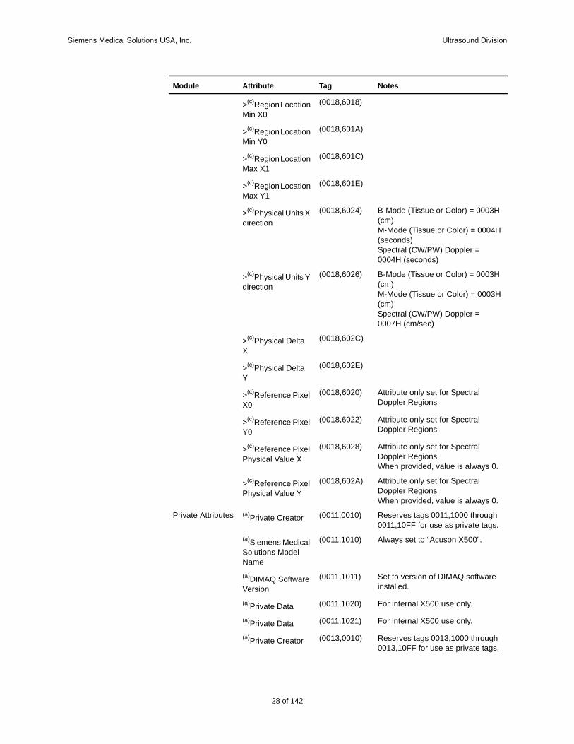
>(c)Region Location Min X0
(0018,6018)
>(c)Region Location Min Y0
(0018,601A)
>(c)Region Location Max X1
(0018,601C)
>(c)Region Location Max Y1
(0018,601E)
>(c)Physical Units X direction
(0018,6024) B-Mode (Tissue or Color) = 0003H (cm)M-Mode (Tissue or Color) = 0004H (seconds)Spectral (CW/PW) Doppler = 0004H (seconds)
>(c)Physical Units Y direction
(0018,6026) B-Mode (Tissue or Color) = 0003H (cm)M-Mode (Tissue or Color) = 0003H (cm)Spectral (CW/PW) Doppler = 0007H (cm/sec)
>(c)Physical Delta X
(0018,602C)
>(c)Physical Delta Y
(0018,602E)
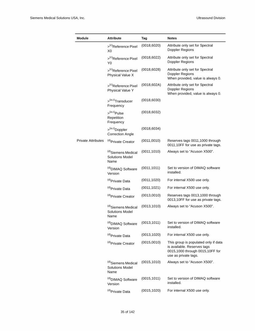
>(c)Reference Pixel X0
(0018,6020) Attribute only set for Spectral Doppler Regions
>(c)Reference Pixel Y0
(0018,6022) Attribute only set for Spectral Doppler Regions
>(c)Reference Pixel Physical Value X
(0018,6028) Attribute only set for Spectral Doppler RegionsWhen provided, value is always 0.
>(c)Reference Pixel Physical Value Y
(0018,602A) Attribute only set for Spectral Doppler RegionsWhen provided, value is always 0.
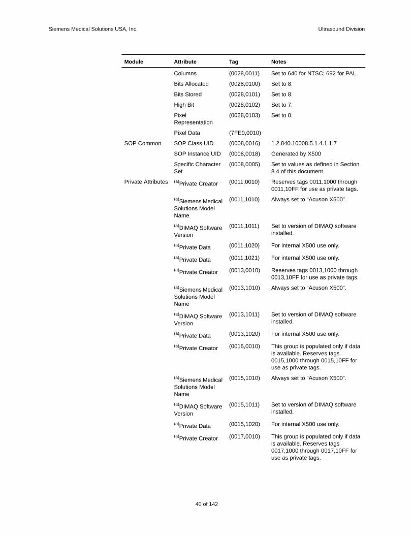
Private Attributes (a)Private Creator (0011,0010) Reserves tags 0011,1000 through 0011,10FF for use as private tags.
(a)Siemens Medical Solutions Model Name
(0011,1010) Always set to “Acuson X500”.
(a)DIMAQ Software Version
(0011,1011) Set to version of DIMAQ software installed.
(a)Private Data (0011,1020) For internal X500 use only.
(a)Private Data (0011,1021) For internal X500 use only.
(a)Private Creator (0013,0010) Reserves tags 0013,1000 through 0013,10FF for use as private tags.
Module Attribute Tag Notes
28 of 142
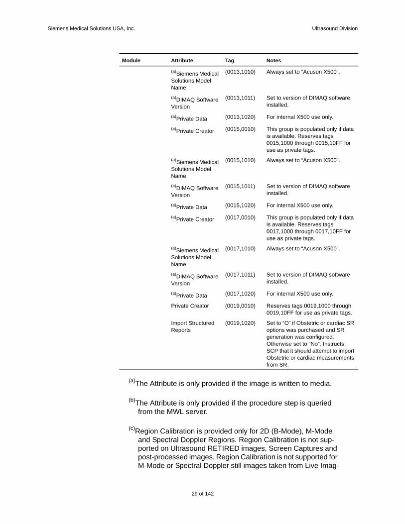
Siemens Medical Solutions USA, Inc. Ultrasound Division
(a)The Attribute is only provided if the image is written to media.
(b)The Attribute is only provided if the procedure step is queried from the MWL server.
(c)Region Calibration is provided only for 2D (B-Mode), M-Mode and Spectral Doppler Regions. Region Calibration is not sup-ported on Ultrasound RETIRED images, Screen Captures and post-processed images. Region Calibration is not supported for M-Mode or Spectral Doppler still images taken from Live Imag-
(a)Siemens Medical Solutions Model Name
(0013,1010) Always set to “Acuson X500”.
(a)DIMAQ Software Version
(0013,1011) Set to version of DIMAQ software installed.
(a)Private Data (0013,1020) For internal X500 use only.
(a)Private Creator (0015,0010) This group is populated only if data is available. Reserves tags 0015,1000 through 0015,10FF for use as private tags.
(a)Siemens Medical Solutions Model Name
(0015,1010) Always set to “Acuson X500”.
(a)DIMAQ Software Version
(0015,1011) Set to version of DIMAQ software installed.
(a)Private Data (0015,1020) For internal X500 use only.
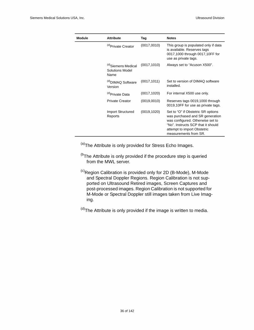
(a)Private Creator (0017,0010) This group is populated only if data is available. Reserves tags 0017,1000 through 0017,10FF for use as private tags.
(a)Siemens Medical Solutions Model Name
(0017,1010) Always set to “Acuson X500”.
(a)DIMAQ Software Version
(0017,1011) Set to version of DIMAQ software installed.
(a)Private Data (0017,1020) For internal X500 use only.
Private Creator (0019,0010) Reserves tags 0019,1000 through 0019,10FF for use as private tags.
Import Structured Reports
(0019,1020) Set to “O” if Obstetric or cardiac SR options was purchased and SR generation was configured. Otherwise set to “No”. Instructs SCP that it should attempt to import Obstetric or cardiac measurements from SR.
Module Attribute Tag Notes
29 of 142
142
Siemens Medical Solutions USA, Inc. Ultrasound Division
ing.
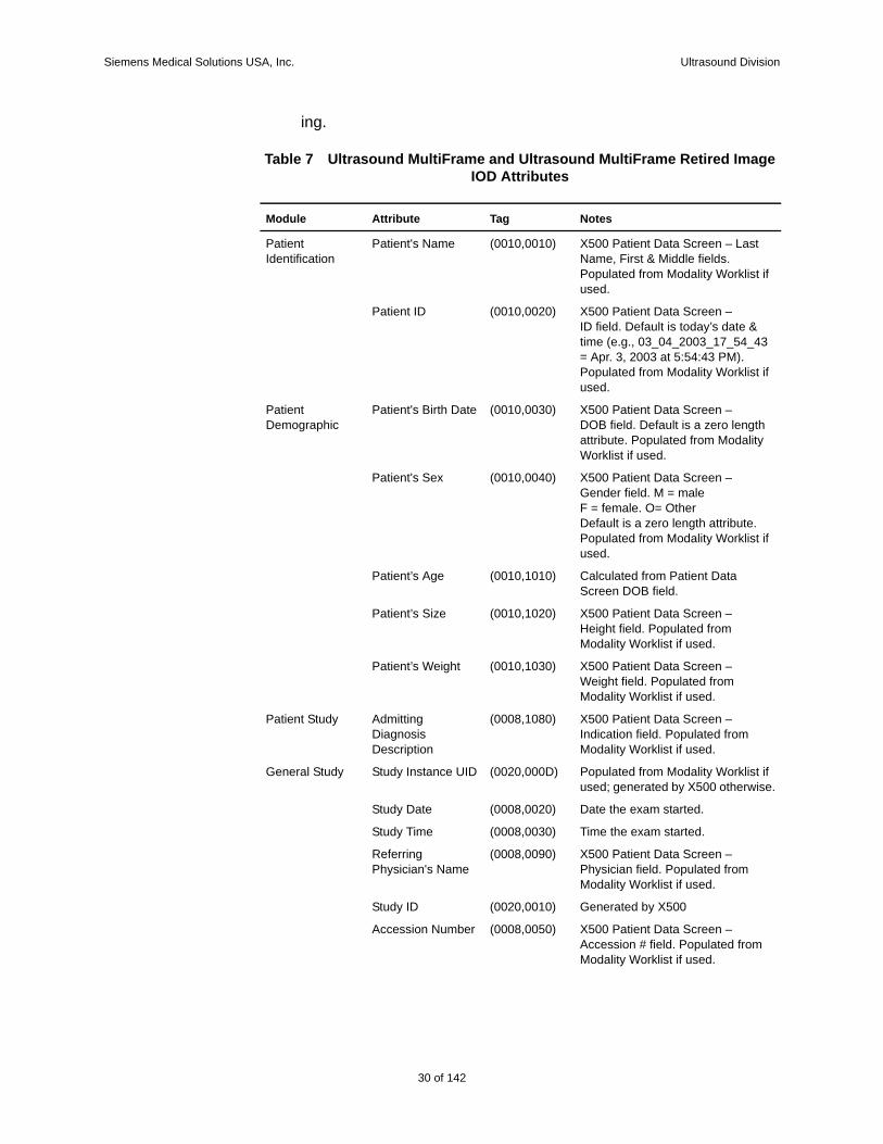
Table 7 Ultrasound MultiFrame and Ultrasound MultiFrame Retired Image IOD Attributes
Module Attribute Tag Notes
Patient Identification
Patient's Name (0010,0010) X500 Patient Data Screen – Last Name, First & Middle fields. Populated from Modality Worklist if used.
Patient ID (0010,0020) X500 Patient Data Screen –ID field. Default is today’s date & time (e.g., 03_04_2003_17_54_43 = Apr. 3, 2003 at 5:54:43 PM). Populated from Modality Worklist if used.
Patient Demographic
Patient's Birth Date (0010,0030) X500 Patient Data Screen –DOB field. Default is a zero length attribute. Populated from Modality Worklist if used.
Patient's Sex (0010,0040) X500 Patient Data Screen –Gender field. M = maleF = female. O= Other Default is a zero length attribute. Populated from Modality Worklist if used.
Patient’s Age (0010,1010) Calculated from Patient Data Screen DOB field.
Patient’s Size (0010,1020) X500 Patient Data Screen –Height field. Populated from Modality Worklist if used.
Patient’s Weight (0010,1030) X500 Patient Data Screen –Weight field. Populated from Modality Worklist if used.
Patient Study Admitting Diagnosis Description
(0008,1080) X500 Patient Data Screen – Indication field. Populated from Modality Worklist if used.
General Study Study Instance UID (0020,000D) Populated from Modality Worklist if used; generated by X500 otherwise.
Study Date (0008,0020) Date the exam started.
Study Time (0008,0030) Time the exam started.
Referring Physician's Name
(0008,0090) X500 Patient Data Screen – Physician field. Populated from Modality Worklist if used.
Study ID (0020,0010) Generated by X500
Accession Number (0008,0050) X500 Patient Data Screen – Accession # field. Populated from Modality Worklist if used.
30 of 142
Siemens Medical Solutions USA, Inc. Ultrasound Division
Study Description (0008,1030) Populated with the first attribute from Modality Worklist in this list that contains a valid value: Study Description (0008,1030), Scheduled Procedure Step Description (0040,0007), Requested Procedure Description (0032,1060). If Modality Worklist was not used or none of the attributes contains a valid value X500 Patient Data Screen – Indication field is used.
General Series Modality (0008,0060) Always set to “US”
Series Instance UID
(0020,000E) Generated by X500
Series Number (0020,0011) Series Number in study (1-n).
Laterality (0020,0060) Always sent as 0 length attribute
(b)Series Date (0008,0021) Date the series started.
(b)Series Time (0008,0031) Time the series started.
(b)Series Description
(0008,103E) Populated with Scheduled Procedure Step Description if a value was provided by Modality Worklist.
(b)Protocol Name (0018,1030) The exam type of the most recent image stored in a particular series. If no images are stored for a series then the value is set to “Ultrasound”.
(b)Request Attributes Sequence
(0040,0275) Populated with Scheduled Procedure Step Sequence (0040,0100) if provided by Modality Worklist.
>(b)Scheduled Procedure Step Description
(0040,0007) Populated with Scheduled Procedure Step Description (0040,0007) from Scheduled Procedure Step Sequence (0040,0100) if provided by Modality Worklist.
>(b)Scheduled Protocol Code Sequence
(0040,0008) Populated with Scheduled Protocol Code Sequence (0040,0008) from Scheduled Procedure Step Sequence (0040,0100) if provided by Modality Worklist.
>(b)Scheduled Procedure Step ID
(0040,0009) Populated with Scheduled Procedure Step ID (0040,0009) from Scheduled Procedure Step Sequence (0040,0100) if provided by Modality Worklist.
Module Attribute Tag Notes
31 of 142
142
Siemens Medical Solutions USA, Inc. Ultrasound Division
>(b)Requested Procedure ID
(0040,1001) Populated with Requested Procedure ID (0040,1001) from Scheduled Procedure Step Sequence (0040,0100) if provided by Modality Worklist.
(b)Performed Procedure Step Start Date
(0040,0244) Date the Performed Procedure Step was started.
(b)Performed Procedure Step Start Time
(0040,0245) Time the Performed Procedure Step was started.
(b)Performed Procedure Step ID
(0040,0253) Populated with Scheduled Procedure Step ID (0040,0009) if provided by Modality Worklist.
(b)Performed Procedure Step Description
(0040,0254) Populated with Scheduled Procedure Step Description (0040,0007) if provided by Modality Worklist.
(b)Performed Procedure Protocol Code Sequence
(0040,0260) Populated with Scheduled Protocol Code Sequence (0040,0008) if provided by Modality Worklist.
(b)Comments on the Performed Procedure Step
(0040,0280) Populated with Comments on the Scheduled Procedure Step (0040,0400) if provided by Modality Worklist.
General Equipment
Manufacturer (0008,0070) Set to “Siemens Ultrasound”
Institution Name (0008,0080) X500 System Presets – Organization Name field.
Software Versions (0018,1020) Set to the DICOM Software Version
Manufacturer’s Model Name
(0008,1090) Set to “Acuson X500”.
General Image Instance Number (0020,0013) Image number in study (1 - n)
Patient Orientation (0020,0020) Always sent as 0 length attribute
Image Pixel Samples per Pixel (0028,0002) Set to 1 for MONOCHROME2 images, 3 for RGB and YBR_FULL_422 images.
Photometric Interpretation
(0028,0004) “YBR_FULL_422” if sent compressed, “RGB” or “MONOCHROME2“if sent uncompressed.
Planar Configuration
(0028,0006) Color-by-pixel. Set to 0 for RGB and YBR_FULL_422 images, not provided for MONOCHROME2 images.
Module Attribute Tag Notes
32 of 142

Siemens Medical Solutions USA, Inc. Ultrasound Division
Rows (0028,0010) Set to 480 for NTSC; 547 for PAL. For Stress Echo clips this value may be from 228 to 288.
Columns (0028,0011) Set to 640 for NTSC; 692 for PAL. For Stress Echo clips this value may be from 288 to 384.
Bits Allocated (0028,0100) Set to 8.
Bits Stored (0028,0101) Set to 8.
High Bit (0028,0102) Set to 7.
Pixel Representation
(0028,0103) Set to 0.
Pixel Data (7FE0, 0010)
US Image Image Type (0008,0008) Sent as a 0 length attribute.
(a)View List (0009,212A) Private attribute
>(a)View Name (0009,2120) Private attribute
(a)Stage Name (0008,2120)
(a)Stage Number (0008,2122)
(a)Number of Stages
(0008,2124)
(a)View Name (0008,2127)
(a)View Number (0008,2128)
(a)Number of Views in Stage
(0008,212A)
(a)Trigger Time (0018,1060)
(a)Nominal Interval (0018,1062)
Heart Rate (0018,1088) Only provided if heart rate is > 0
Lossy Image Compression
(0028,2110) Always set to “01”
SOP Common SOP Class UID (0008,0016) 1.2.840.10008.5.1.4.1.1.3.1 or1.2.840.10008.5.1.4.1.1.3
SOP Instance UID (0008,0018) Generated by X500
Specific Character Set
(0008,0005) Set to values as defined in Section 8.4 of this document
Image Plane Pixel Spacing (0028,0030) Pixel Spacing information is only provided for single, full screen, 2D image types (2D image types are B-Mode, B-Mode Color, B-Mode with power).
Cine Frame Time (0018,1063)
Multi-Frame Number of Frames (0028,0008)
Frame Increment Pointer
(0028,0009) 00181063H
Module Attribute Tag Notes
33 of 142
142
Siemens Medical Solutions USA, Inc. Ultrasound Division
Region Calibration
(c)Sequence of Ultrasound Regions
(0018,6011)
>(c)Region Spatial Format
(0018,6012) B-Mode (Tissue or Color) = 0001HM-Mode (Tissue or Color) = 0002HSpectral (CW/PW) Doppler = 0003H
>(c)Region Data Type
(0018,6014) B-Mode, M-Mode = 0001H (Tissue) Spectral Doppler = 0004H (CW Spectral Doppler) Spectral Doppler = 0003H (PW Spectral Doppler)
>(c)Region Flags (0018,6016) 1st Bit (LSB) = 1 (All images acquired are transparent) 2nd Bit = 1 (All images acquired are automatically scaled) 3rd Bit = 1 for frequency scale 3rd Bit = 0 for velocity scale.The value of the 3rd bit is undefined for any mode other than Doppler. The value for 3rd bit is undefined if both frequency and velocity scales are selected on the Doppler image. 4th Bit is Reserved and value is always 0
>(c)Region Location Min X0
(0018,6018)
>(c)Region Location Min Y0
(0018,601A)
>(c)Region Location Max X1
(0018,601C)
>(c)Region Location Max Y1
(0018,601E)
>(c)Physical Units X direction
(0018,6024) B-Mode (Tissue or Color) = 0003H (cm)M-Mode (Tissue or Color) = 0004H (seconds)Spectral (CW/PW) Doppler = 0004H (seconds)
>(c)Physical Units Y direction
(0018,6026) B-Mode (Tissue or Color) = 0003H (cm)M-Mode (Tissue or Color) = 0003H (cm)Spectral (CW/PW) Doppler = 0007H (cm/sec)
>(c)Physical Delta X
(0018,602C)
>(c)Physical Delta Y
(0018,602E)
Module Attribute Tag Notes
34 of 142
Siemens Medical Solutions USA, Inc. Ultrasound Division
>(c)Reference Pixel X0
(0018,6020) Attribute only set for Spectral Doppler Regions
>(c)Reference Pixel Y0
(0018,6022) Attribute only set for Spectral Doppler Regions
>(c)Reference Pixel Physical Value X
(0018,6028) Attribute only set for Spectral Doppler RegionsWhen provided, value is always 0.
>(c)Reference Pixel Physical Value Y
(0018,602A) Attribute only set for Spectral Doppler RegionsWhen provided, value is always 0.
>(a,c)Transducer Frequency
(0018,6030)
>(a,c)Pulse Repetition Frequency
(0018,6032)
>(a,c)Doppler Correction Angle
(0018,6034)
Private Attributes (d)Private Creator (0011,0010) Reserves tags 0011,1000 through 0011,10FF for use as private tags.
(d)Siemens Medical Solutions Model Name
(0011,1010) Always set to “Acuson X500”.
(d)DIMAQ Software Version
(0011,1011) Set to version of DIMAQ software installed.
(d)Private Data (0011,1020) For internal X500 use only.
(d)Private Data (0011,1021) For internal X500 use only.
(d)Private Creator (0013,0010) Reserves tags 0013,1000 through 0013,10FF for use as private tags.
(d)Siemens Medical Solutions Model Name
(0013,1010) Always set to “Acuson X500”.
(d)DIMAQ Software Version
(0013,1011) Set to version of DIMAQ software installed.
(d)Private Data (0013,1020) For internal X500 use only.
(d)Private Creator (0015,0010) This group is populated only if data is available. Reserves tags 0015,1000 through 0015,10FF for use as private tags.
(d)Siemens Medical Solutions Model Name
(0015,1010) Always set to “Acuson X500”.
(d)DIMAQ Software Version
(0015,1011) Set to version of DIMAQ software installed.
(d)Private Data (0015,1020) For internal X500 use only.
Module Attribute Tag Notes
35 of 142
142
Siemens Medical Solutions USA, Inc. Ultrasound Division
(a)The Attribute is only provided for Stress Echo Images.
(bThe Attribute is only provided if the procedure step is queried from the MWL server.
(c)Region Calibration is provided only for 2D (B-Mode), M-Mode and Spectral Doppler Regions. Region Calibration is not sup-ported on Ultrasound Retired images, Screen Captures and post-processed images. Region Calibration is not supported for M-Mode or Spectral Doppler still images taken from Live Imag-ing.
(d)The Attribute is only provided if the image is written to media.
(d)Private Creator (0017,0010) This group is populated only if data is available. Reserves tags 0017,1000 through 0017,10FF for use as private tags.
(d)Siemens Medical Solutions Model Name
(0017,1010) Always set to “Acuson X500”.
(d)DIMAQ Software Version
(0017,1011) Set to version of DIMAQ software installed.
(d)Private Data (0017,1020) For internal X500 use only.
Private Creator (0019,0010) Reserves tags 0019,1000 through 0019,10FF for use as private tags.
Import Structured Reports
(0019,1020) Set to “O” if Obstetric SR options was purchased and SR generation was configured. Otherwise set to “No”. Instructs SCP that it should attempt to import Obstetric measurements from SR.
Module Attribute Tag Notes
36 of 142
Siemens Medical Solutions USA, Inc. Ultrasound Division
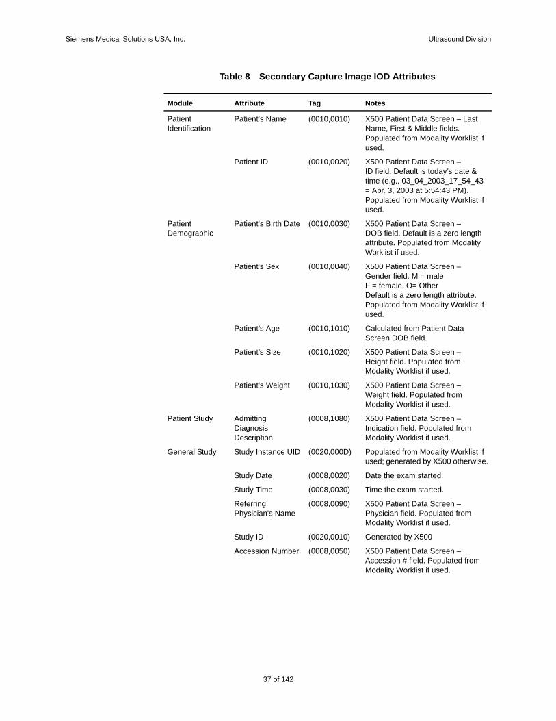
Table 8 Secondary Capture Image IOD Attributes
Module Attribute Tag Notes
Patient Identification
Patient's Name (0010,0010) X500 Patient Data Screen – Last Name, First & Middle fields. Populated from Modality Worklist if used.
Patient ID (0010,0020) X500 Patient Data Screen –ID field. Default is today’s date & time (e.g., 03_04_2003_17_54_43 = Apr. 3, 2003 at 5:54:43 PM). Populated from Modality Worklist if used.
Patient Demographic
Patient's Birth Date (0010,0030) X500 Patient Data Screen –DOB field. Default is a zero length attribute. Populated from Modality Worklist if used.
Patient's Sex (0010,0040) X500 Patient Data Screen –Gender field. M = maleF = female. O= Other Default is a zero length attribute. Populated from Modality Worklist if used.
Patient’s Age (0010,1010) Calculated from Patient Data Screen DOB field.
Patient’s Size (0010,1020) X500 Patient Data Screen –Height field. Populated from Modality Worklist if used.
Patient’s Weight (0010,1030) X500 Patient Data Screen –Weight field. Populated from Modality Worklist if used.
Patient Study Admitting Diagnosis Description
(0008,1080) X500 Patient Data Screen – Indication field. Populated from Modality Worklist if used.
General Study Study Instance UID (0020,000D) Populated from Modality Worklist if used; generated by X500 otherwise.
Study Date (0008,0020) Date the exam started.
Study Time (0008,0030) Time the exam started.
Referring Physician's Name
(0008,0090) X500 Patient Data Screen – Physician field. Populated from Modality Worklist if used.
Study ID (0020,0010) Generated by X500
Accession Number (0008,0050) X500 Patient Data Screen – Accession # field. Populated from Modality Worklist if used.
37 of 142
142

Siemens Medical Solutions USA, Inc. Ultrasound Division
Study Description (0008,1030) Populated with the first attribute from Modality Worklist in this list that contains a valid value: Study Description (0008,1030), Scheduled Procedure Step Description (0040,0007), Requested Procedure Description (0032,1060). If Modality Worklist was not used or none of the attributes contains a valid value X500 Patient Data Screen – Indication field is used.
General Series Modality (0008,0060) Always set to “US”
Series Instance UID
(0020,000E) Generated by X500
Series Number (0020,0011) Series Number in study (1-n).
Laterality (0020,0060) Always sent as 0 length attribute
(b)Series Date (0008,0021) Date the series started.
(b)Series Time (0008,0031) Time the series started.
(b)Series Description
(0008,103E) Populated with Scheduled Procedure Step Description if a value was provided by Modality Worklist.
(b)Protocol Name (0018,1030) The exam type of the most recent image stored in a particular series. If no images are stored for a series then the value is set to “Ultrasound”.
(b)Request Attributes Sequence
(0040,0275) Populated with Scheduled Procedure Step Sequence (0040,0100) if provided by Modality Worklist.
>(b)Scheduled Procedure Step Description
(0040,0007) Populated with Scheduled Procedure Step Description (0040,0007) from Scheduled Procedure Step Sequence (0040,0100) if provided by Modality Worklist.
>(b)Scheduled Protocol Code Sequence
(0040,0008) Populated with Scheduled Protocol Code Sequence (0040,0008) from Scheduled Procedure Step Sequence (0040,0100) if provided by Modality Worklist.
>(b)Scheduled Procedure Step ID
(0040,0009) Populated with Scheduled Procedure Step ID (0040,0009) from Scheduled Procedure Step Sequence (0040,0100) if provided by Modality Worklist.
Module Attribute Tag Notes
38 of 142
Siemens Medical Solutions USA, Inc. Ultrasound Division
>(b)Requested Procedure ID
(0040,1001) Populated with Requested Procedure ID (0040,1001) from Scheduled Procedure Step Sequence (0040,0100) if provided by Modality Worklist.
(b)Performed Procedure Step Start Date
(0040,0244) Date the Performed Procedure Step was started.
(b)Performed Procedure Step Start Time
(0040,0245) Time the Performed Procedure Step was started.
(b)Performed Procedure Step ID
(0040,0253) Populated with Scheduled Procedure Step ID (0040,0009) if provided by Modality Worklist.
(b)Performed Procedure Step Description
(0040,0254) Populated with Scheduled Procedure Step Description (0040,0007) if provided by Modality Worklist.
(b)Performed Procedure Protocol Code Sequence
(0040,0260) Populated with Scheduled Protocol Code Sequence (0040,0008) if provided by Modality Worklist.
(b)Comments on the Performed Procedure Step
(0040,0280) Populated with Comments on the Scheduled Procedure Step (0040,0400) if provided by Modality Worklist.
SC Equipment Module
Conversion Type (0008,0064) Set to “WSD”
General Equipment
Manufacturer (0008,0070) Set to “Siemens Ultrasound”
Institution Name (0008,0080) X500 System Presets – Organization Name field.
Software Versions (0018,1020) Set to the DICOM Software Version
Manufacturer’s Model Name
(0008,1090) Set to “X500”.
General Image Instance Number (0020,0013) Image number in study (1 – n)
Patient Orientation (0020,0020) Always sent as 0 length attribute.
Image Pixel Samples per Pixel (0028,0002) Set to 1 for MONOCHROME2 images, 3 for RGB images.
Photometric Interpretation
(0028,0004) “RGB” or “MONOCHROME2“
Planar Configuration
(0028,0006) Color-by-pixel. Set to 0 for RGB images, not provided for MONOCHROME2 images.
Rows (0028,0010) Set to 480 for NTSC; 547 for PAL.
Module Attribute Tag Notes
39 of 142
142
Siemens Medical Solutions USA, Inc. Ultrasound Division
Columns (0028,0011) Set to 640 for NTSC; 692 for PAL.
Bits Allocated (0028,0100) Set to 8.
Bits Stored (0028,0101) Set to 8.
High Bit (0028,0102) Set to 7.
Pixel Representation
(0028,0103) Set to 0.
Pixel Data (7FE0,0010)
SOP Common SOP Class UID (0008,0016) 1.2.840.10008.5.1.4.1.1.7
SOP Instance UID (0008,0018) Generated by X500
Specific Character Set
(0008,0005) Set to values as defined in Section 8.4 of this document
Private Attributes (a)Private Creator (0011,0010) Reserves tags 0011,1000 through 0011,10FF for use as private tags.
(a)Siemens Medical Solutions Model Name
(0011,1010) Always set to “Acuson X500”.
(a)DIMAQ Software Version
(0011,1011) Set to version of DIMAQ software installed.
(a)Private Data (0011,1020) For internal X500 use only.
(a)Private Data (0011,1021) For internal X500 use only.
(a)Private Creator (0013,0010) Reserves tags 0013,1000 through 0013,10FF for use as private tags.
(a)Siemens Medical Solutions Model Name
(0013,1010) Always set to “Acuson X500”.
(a)DIMAQ Software Version
(0013,1011) Set to version of DIMAQ software installed.
(a)Private Data (0013,1020) For internal X500 use only.
(a)Private Creator (0015,0010) This group is populated only if data is available. Reserves tags 0015,1000 through 0015,10FF for use as private tags.
(a)Siemens Medical Solutions Model Name
(0015,1010) Always set to “Acuson X500”.
(a)DIMAQ Software Version
(0015,1011) Set to version of DIMAQ software installed.
(a)Private Data (0015,1020) For internal X500 use only.
(a)Private Creator (0017,0010) This group is populated only if data is available. Reserves tags 0017,1000 through 0017,10FF for use as private tags.
Module Attribute Tag Notes
40 of 142
Siemens Medical Solutions USA, Inc. Ultrasound Division
(a)The Attribute is only provided if the image is written to media.
(b)The Attribute is only provided if the procedure step is queried from the MWL server.
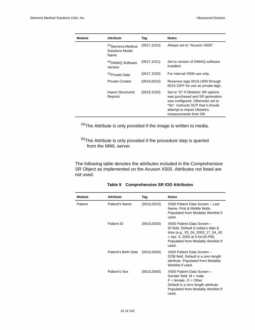
The following table denotes the attributes included in the Comprehensive SR Object as implemented on the Acuson X500. Attributes not listed are not used.
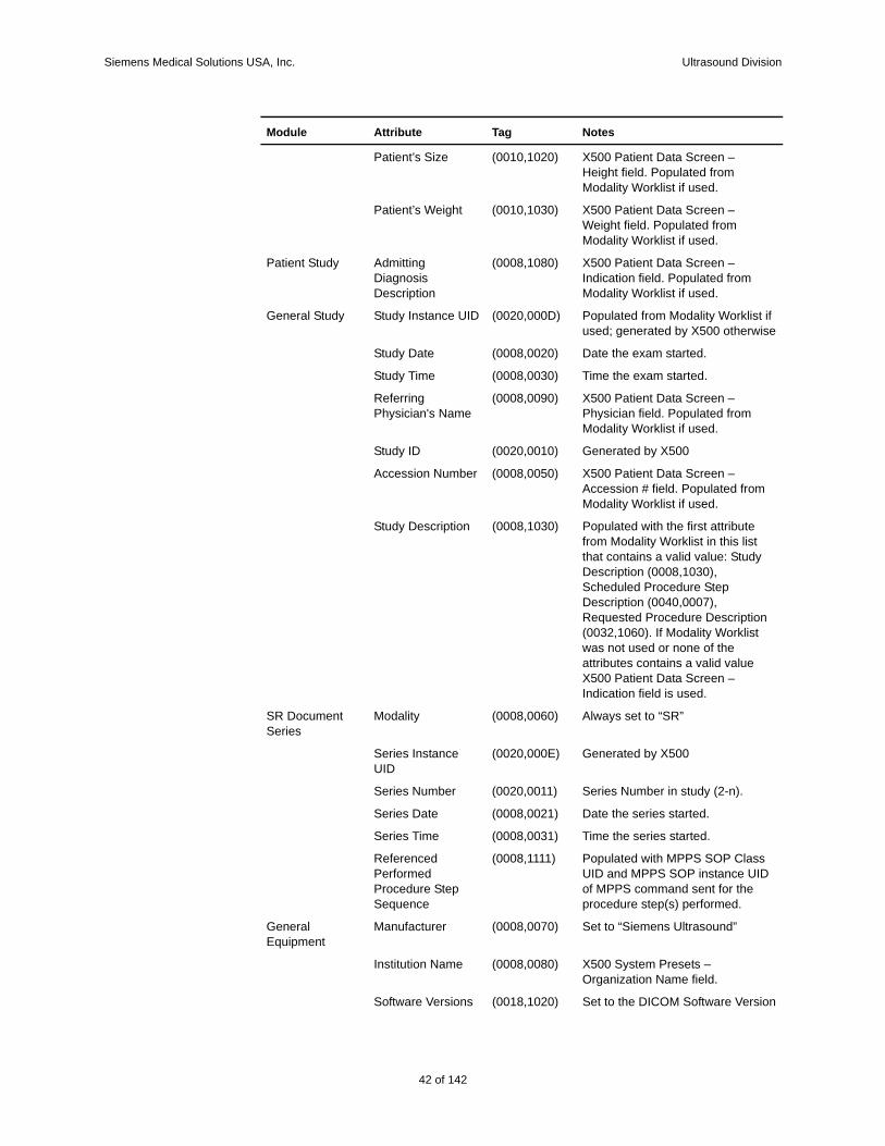
Table 9 Comprehensive SR IOD Attributes
(a)Siemens Medical Solutions Model Name
(0017,1010) Always set to “Acuson X500”.
(a)DIMAQ Software Version
(0017,1011) Set to version of DIMAQ software installed.
(a)Private Data (0017,1020) For internal X500 use only.
Private Creator (0019,0010) Reserves tags 0019,1000 through 0019,10FF for use as private tags.
Import Structured Reports
(0019,1020) Set to “O” if Obstetric SR options was purchased and SR generation was configured. Otherwise set to “No”. Instructs SCP that it should attempt to import Obstetric measurements from SR.
Module Attribute Tag Notes
Patient Patient's Name (0010,0010) X500 Patient Data Screen – Last Name, First & Middle fields. Populated from Modality Worklist if used.
Patient ID (0010,0020) X500 Patient Data Screen –ID field. Default is today’s date & time (e.g., 03_04_2003_17_54_43 = Apr. 3, 2003 at 5:54:43 PM). Populated from Modality Worklist if used.
Patient's Birth Date (0010,0030) X500 Patient Data Screen –DOB field. Default is a zero length attribute. Populated from Modality Worklist if used.
Patient's Sex (0010,0040) X500 Patient Data Screen –Gender field. M = maleF = female. O = Other Default is a zero length attribute. Populated from Modality Worklist if used.
Module Attribute Tag Notes
41 of 142
142
Siemens Medical Solutions USA, Inc. Ultrasound Division
Patient’s Size (0010,1020) X500 Patient Data Screen –Height field. Populated from Modality Worklist if used.
Patient’s Weight (0010,1030) X500 Patient Data Screen –Weight field. Populated from Modality Worklist if used.
Patient Study Admitting Diagnosis Description
(0008,1080) X500 Patient Data Screen – Indication field. Populated from Modality Worklist if used.
General Study Study Instance UID (0020,000D) Populated from Modality Worklist if used; generated by X500 otherwise
Study Date (0008,0020) Date the exam started.
Study Time (0008,0030) Time the exam started.
Referring Physician's Name
(0008,0090) X500 Patient Data Screen – Physician field. Populated from Modality Worklist if used.
Study ID (0020,0010) Generated by X500
Accession Number (0008,0050) X500 Patient Data Screen – Accession # field. Populated from Modality Worklist if used.
Study Description (0008,1030) Populated with the first attribute from Modality Worklist in this list that contains a valid value: Study Description (0008,1030), Scheduled Procedure Step Description (0040,0007), Requested Procedure Description (0032,1060). If Modality Worklist was not used or none of the attributes contains a valid value X500 Patient Data Screen – Indication field is used.
SR Document Series
Modality (0008,0060) Always set to “SR”
Series Instance UID
(0020,000E) Generated by X500
Series Number (0020,0011) Series Number in study (2-n).
Series Date (0008,0021) Date the series started.
Series Time (0008,0031) Time the series started.
Referenced Performed Procedure Step Sequence
(0008,1111) Populated with MPPS SOP Class UID and MPPS SOP instance UID of MPPS command sent for the procedure step(s) performed.
General Equipment
Manufacturer (0008,0070) Set to “Siemens Ultrasound”
Institution Name (0008,0080) X500 System Presets – Organization Name field.
Software Versions (0018,1020) Set to the DICOM Software Version
Module Attribute Tag Notes
42 of 142
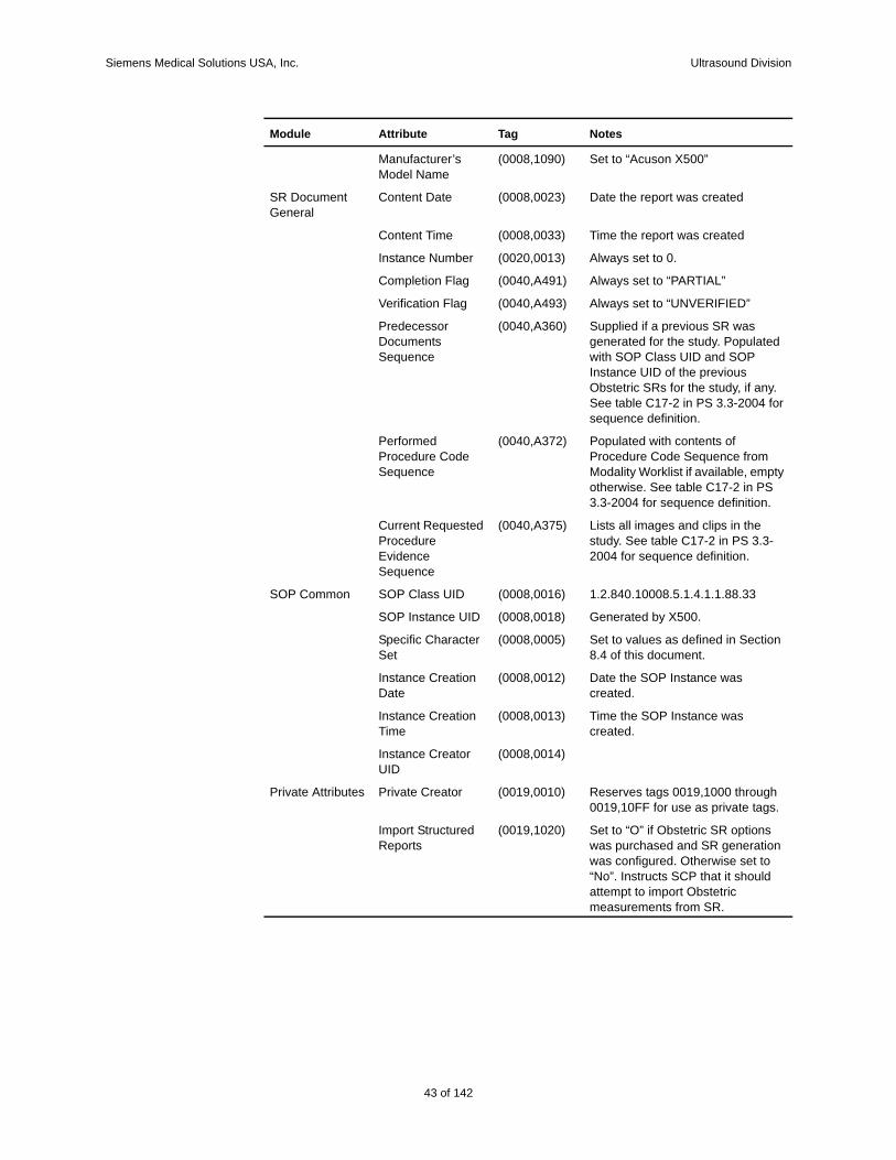
Siemens Medical Solutions USA, Inc. Ultrasound Division
Manufacturer’s Model Name
(0008,1090) Set to “Acuson X500”
SR Document General
Content Date (0008,0023) Date the report was created
Content Time (0008,0033) Time the report was created
Instance Number (0020,0013) Always set to 0.
Completion Flag (0040,A491) Always set to “PARTIAL”
Verification Flag (0040,A493) Always set to “UNVERIFIED”
Predecessor Documents Sequence
(0040,A360) Supplied if a previous SR was generated for the study. Populated with SOP Class UID and SOP Instance UID of the previous Obstetric SRs for the study, if any. See table C17-2 in PS 3.3-2004 for sequence definition.
Performed Procedure Code Sequence
(0040,A372) Populated with contents of Procedure Code Sequence from Modality Worklist if available, empty otherwise. See table C17-2 in PS 3.3-2004 for sequence definition.
Current Requested Procedure Evidence Sequence
(0040,A375) Lists all images and clips in the study. See table C17-2 in PS 3.3-2004 for sequence definition.
SOP Common SOP Class UID (0008,0016) 1.2.840.10008.5.1.4.1.1.88.33
SOP Instance UID (0008,0018) Generated by X500.
Specific Character Set
(0008,0005) Set to values as defined in Section 8.4 of this document.
Instance Creation Date
(0008,0012) Date the SOP Instance was created.
Instance Creation Time
(0008,0013) Time the SOP Instance was created.
Instance Creator UID
(0008,0014)
Private Attributes Private Creator (0019,0010) Reserves tags 0019,1000 through 0019,10FF for use as private tags.
Import Structured Reports
(0019,1020) Set to “O” if Obstetric SR options was purchased and SR generation was configured. Otherwise set to “No”. Instructs SCP that it should attempt to import Obstetric measurements from SR.
Module Attribute Tag Notes
43 of 142
142
Siemens Medical Solutions USA, Inc. Ultrasound Division
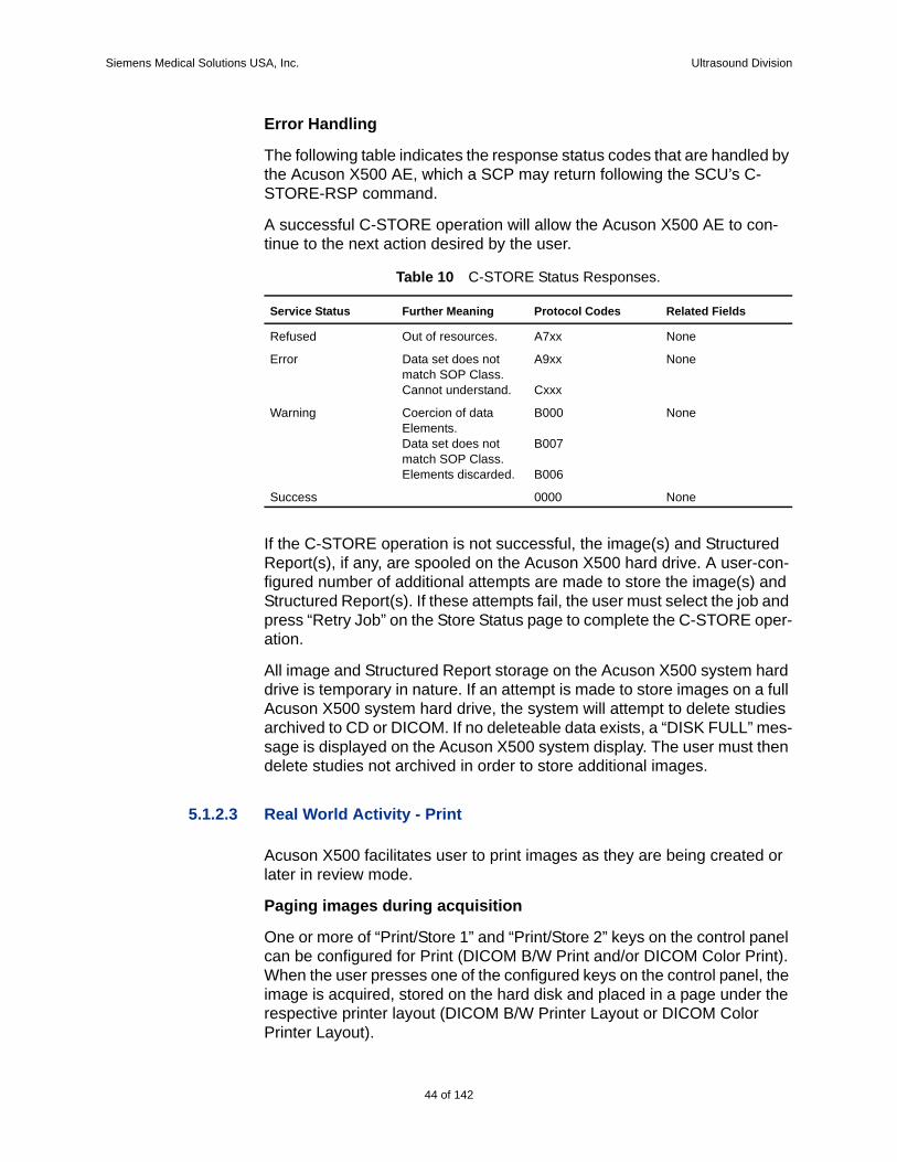
Error Handling
The following table indicates the response status codes that are handled by the Acuson X500 AE, which a SCP may return following the SCU’s C-STORE-RSP command.
A successful C-STORE operation will allow the Acuson X500 AE to con-tinue to the next action desired by the user.
If the C-STORE operation is not successful, the image(s) and Structured Report(s), if any, are spooled on the Acuson X500 hard drive. A user-con-figured number of additional attempts are made to store the image(s) and Structured Report(s). If these attempts fail, the user must select the job and press “Retry Job” on the Store Status page to complete the C-STORE oper-ation.
All image and Structured Report storage on the Acuson X500 system hard drive is temporary in nature. If an attempt is made to store images on a full Acuson X500 system hard drive, the system will attempt to delete studies archived to CD or DICOM. If no deleteable data exists, a “DISK FULL” mes-sage is displayed on the Acuson X500 system display. The user must then delete studies not archived in order to store additional images.
5.1.2.3 Real World Activity - Print
Acuson X500 facilitates user to print images as they are being created or later in review mode.
Paging images during acquisition
One or more of “Print/Store 1” and “Print/Store 2” keys on the control panel can be configured for Print (DICOM B/W Print and/or DICOM Color Print). When the user presses one of the configured keys on the control panel, the image is acquired, stored on the hard disk and placed in a page under the respective printer layout (DICOM B/W Printer Layout or DICOM Color Printer Layout).
Table 10 C-STORE Status Responses.
Service Status Further Meaning Protocol Codes Related Fields
Refused Out of resources. A7xx None
Error Data set does not match SOP Class.Cannot understand.
A9xx
Cxxx
None
Warning Coercion of data Elements.Data set does not match SOP Class.Elements discarded.
B000
B007
B006
None
Success 0000 None
44 of 142
Siemens Medical Solutions USA, Inc. Ultrasound Division
Paging images in Review mode
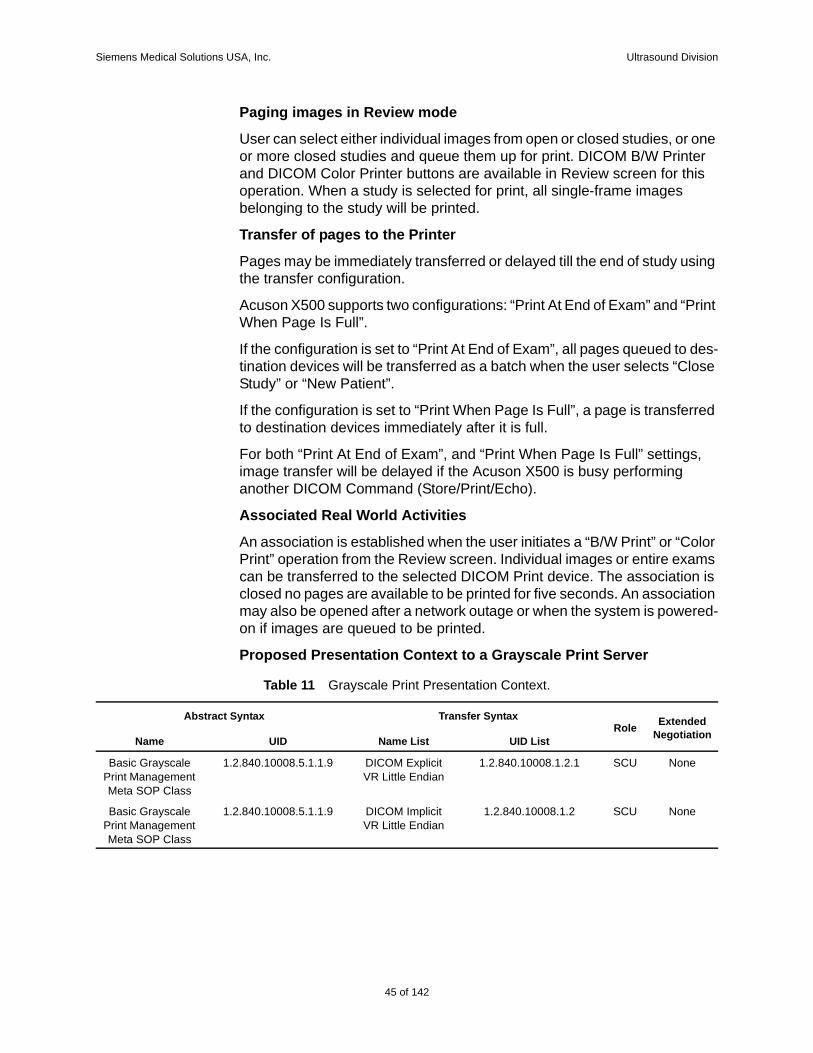
User can select either individual images from open or closed studies, or one or more closed studies and queue them up for print. DICOM B/W Printer and DICOM Color Printer buttons are available in Review screen for this operation. When a study is selected for print, all single-frame images belonging to the study will be printed.
Transfer of pages to the Printer
Pages may be immediately transferred or delayed till the end of study using the transfer configuration.
Acuson X500 supports two configurations: “Print At End of Exam” and “Print When Page Is Full”.
If the configuration is set to “Print At End of Exam”, all pages queued to des-tination devices will be transferred as a batch when the user selects “Close Study” or “New Patient”.
If the configuration is set to “Print When Page Is Full”, a page is transferred to destination devices immediately after it is full.
For both “Print At End of Exam”, and “Print When Page Is Full” settings, image transfer will be delayed if the Acuson X500 is busy performing another DICOM Command (Store/Print/Echo).
Associated Real World Activities
An association is established when the user initiates a “B/W Print” or “Color Print” operation from the Review screen. Individual images or entire exams can be transferred to the selected DICOM Print device. The association is closed no pages are available to be printed for five seconds. An association may also be opened after a network outage or when the system is powered-on if images are queued to be printed.
Proposed Presentation Context to a Grayscale Print Server
Table 11 Grayscale Print Presentation Context.
Abstract Syntax Transfer SyntaxRole Extended
NegotiationName UID Name List UID List
Basic Grayscale Print Management Meta SOP Class
1.2.840.10008.5.1.1.9 DICOM Explicit VR Little Endian
1.2.840.10008.1.2.1 SCU None
Basic Grayscale Print Management Meta SOP Class
1.2.840.10008.5.1.1.9 DICOM Implicit VR Little Endian
1.2.840.10008.1.2 SCU None
45 of 142
142
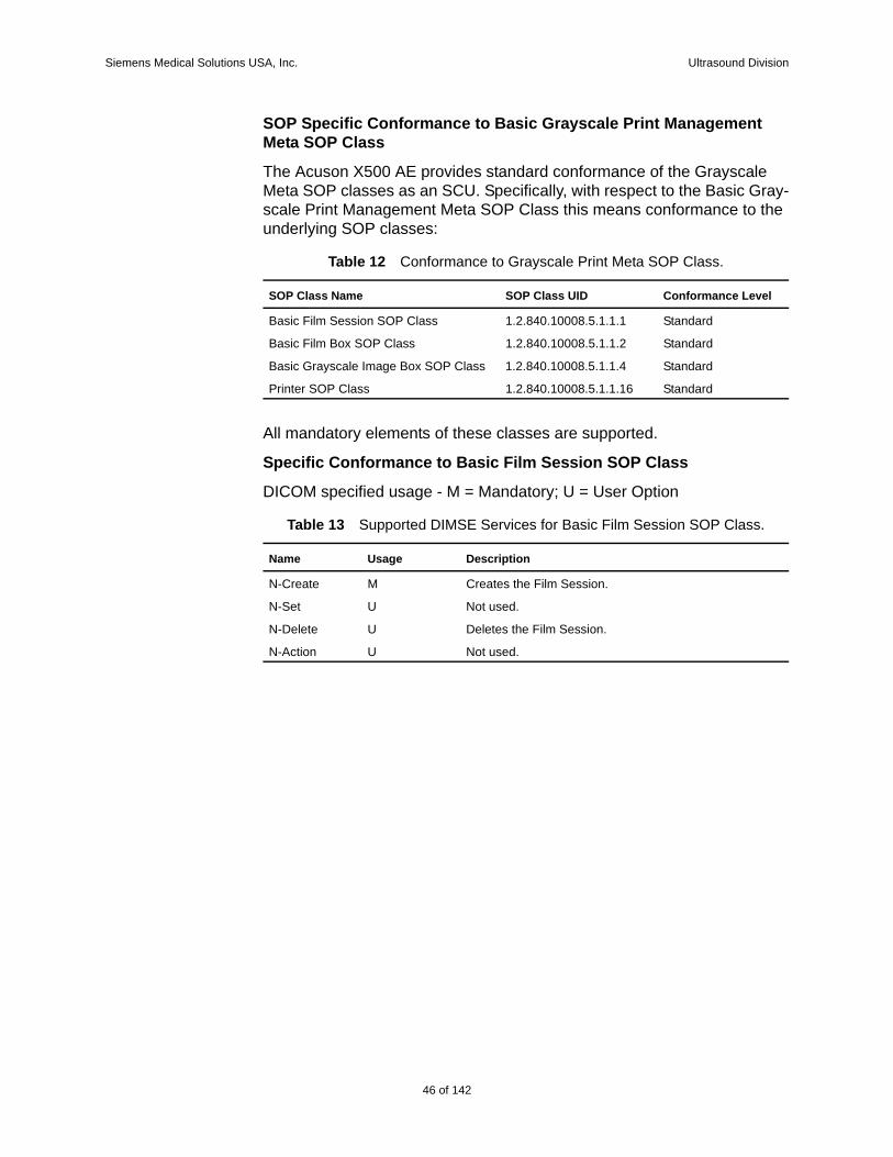
Siemens Medical Solutions USA, Inc. Ultrasound Division
SOP Specific Conformance to Basic Grayscale Print Management Meta SOP Class
The Acuson X500 AE provides standard conformance of the Grayscale Meta SOP classes as an SCU. Specifically, with respect to the Basic Gray-scale Print Management Meta SOP Class this means conformance to the underlying SOP classes:
All mandatory elements of these classes are supported.
Specific Conformance to Basic Film Session SOP Class
DICOM specified usage - M = Mandatory; U = User Option
Table 12 Conformance to Grayscale Print Meta SOP Class.
SOP Class Name SOP Class UID Conformance Level
Basic Film Session SOP Class 1.2.840.10008.5.1.1.1 Standard
Basic Film Box SOP Class 1.2.840.10008.5.1.1.2 Standard
Basic Grayscale Image Box SOP Class 1.2.840.10008.5.1.1.4 Standard
Printer SOP Class 1.2.840.10008.5.1.1.16 Standard
Table 13 Supported DIMSE Services for Basic Film Session SOP Class.
Name Usage Description
N-Create M Creates the Film Session.
N-Set U Not used.
N-Delete U Deletes the Film Session.
N-Action U Not used.
46 of 142
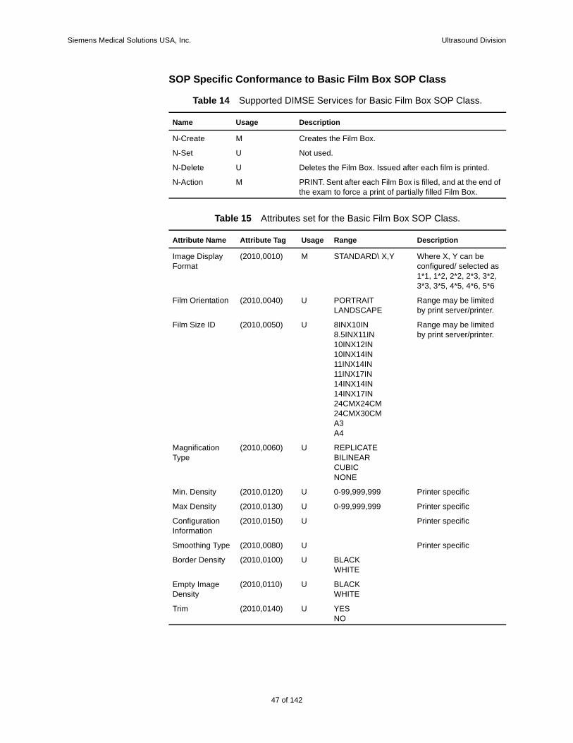
Siemens Medical Solutions USA, Inc. Ultrasound Division
SOP Specific Conformance to Basic Film Box SOP Class
Table 14 Supported DIMSE Services for Basic Film Box SOP Class.
Name Usage Description
N-Create M Creates the Film Box.
N-Set U Not used.
N-Delete U Deletes the Film Box. Issued after each film is printed.
N-Action M PRINT. Sent after each Film Box is filled, and at the end of the exam to force a print of partially filled Film Box.
Table 15 Attributes set for the Basic Film Box SOP Class.
Attribute Name Attribute Tag Usage Range Description
Image Display Format
(2010,0010) M STANDARD\ X,Y Where X, Y can be configured/ selected as 1*1, 1*2, 2*2, 2*3, 3*2, 3*3, 3*5, 4*5, 4*6, 5*6
Film Orientation (2010,0040) U PORTRAITLANDSCAPE
Range may be limited by print server/printer.
Film Size ID (2010,0050) U 8INX10IN8.5INX11IN10INX12IN10INX14IN11INX14IN11INX17IN14INX14IN14INX17IN24CMX24CM24CMX30CMA3A4
Range may be limited by print server/printer.
Magnification Type
(2010,0060) U REPLICATE BILINEARCUBICNONE
Min. Density (2010,0120) U 0-99,999,999 Printer specific
Max Density (2010,0130) U 0-99,999,999 Printer specific
Configuration Information
(2010,0150) U Printer specific
Smoothing Type (2010,0080) U Printer specific
Border Density (2010,0100) U BLACKWHITE
Empty Image Density
(2010,0110) U BLACKWHITE
Trim (2010,0140) U YESNO
47 of 142
142
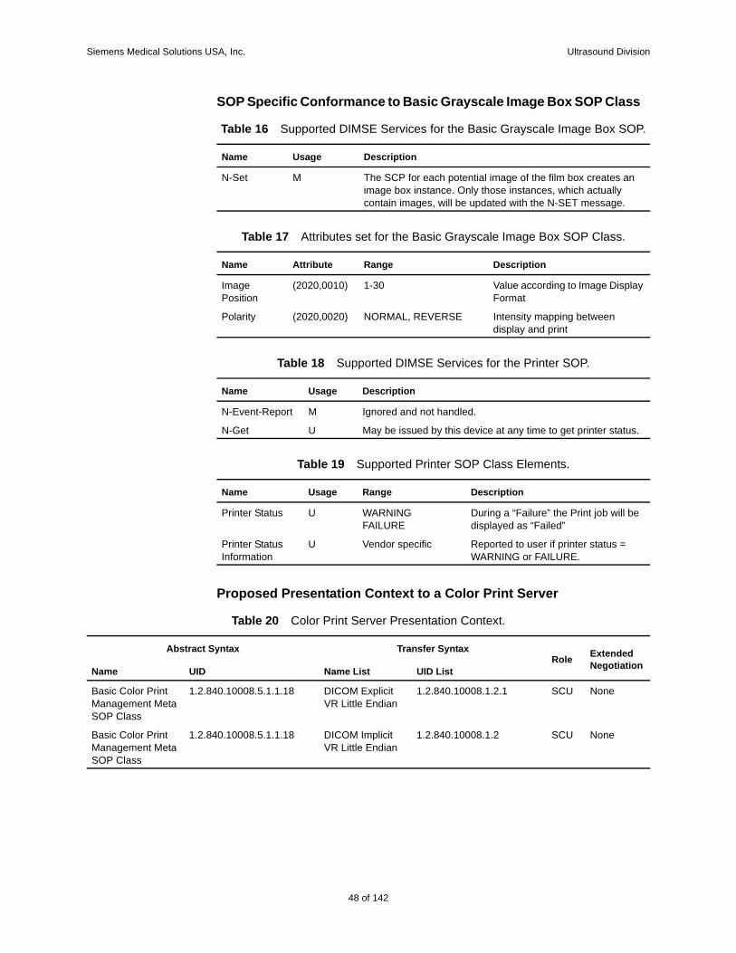
Siemens Medical Solutions USA, Inc. Ultrasound Division
SOP Specific Conformance to Basic Grayscale Image Box SOP Class
Proposed Presentation Context to a Color Print Server
Table 16 Supported DIMSE Services for the Basic Grayscale Image Box SOP.
Name Usage Description
N-Set M The SCP for each potential image of the film box creates an image box instance. Only those instances, which actually contain images, will be updated with the N-SET message.
Table 17 Attributes set for the Basic Grayscale Image Box SOP Class.
Name Attribute Range Description
Image Position
(2020,0010) 1-30 Value according to Image Display Format
Polarity (2020,0020) NORMAL, REVERSE Intensity mapping between display and print
Table 18 Supported DIMSE Services for the Printer SOP.
Name Usage Description
N-Event-Report M Ignored and not handled.
N-Get U May be issued by this device at any time to get printer status.
Table 19 Supported Printer SOP Class Elements.
Name Usage Range Description
Printer Status U WARNINGFAILURE
During a “Failure” the Print job will be displayed as “Failed”
Printer Status Information
U Vendor specific Reported to user if printer status = WARNING or FAILURE.
Table 20 Color Print Server Presentation Context.
Abstract Syntax Transfer SyntaxRole Extended
NegotiationName UID Name List UID List
Basic Color Print Management Meta SOP Class
1.2.840.10008.5.1.1.18 DICOM Explicit VR Little Endian
1.2.840.10008.1.2.1 SCU None
Basic Color Print Management Meta SOP Class
1.2.840.10008.5.1.1.18 DICOM Implicit VR Little Endian
1.2.840.10008.1.2 SCU None
48 of 142
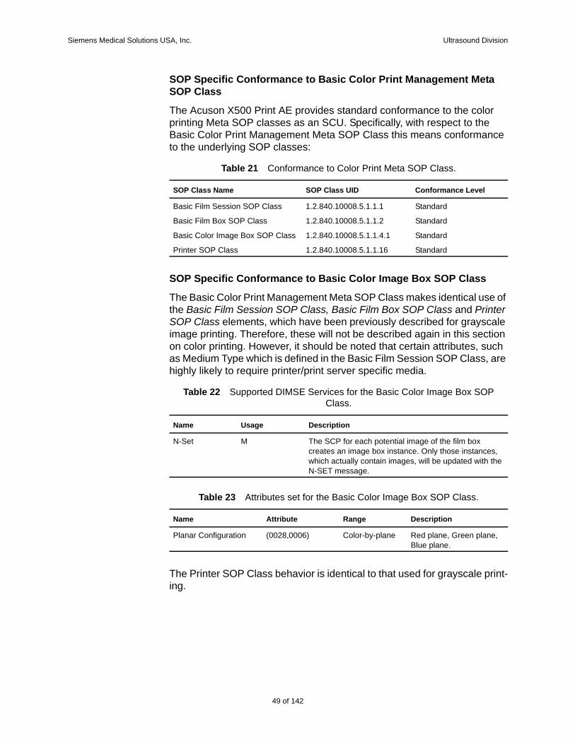
Siemens Medical Solutions USA, Inc. Ultrasound Division
SOP Specific Conformance to Basic Color Print Management Meta SOP Class
The Acuson X500 Print AE provides standard conformance to the color printing Meta SOP classes as an SCU. Specifically, with respect to the Basic Color Print Management Meta SOP Class this means conformance to the underlying SOP classes:
SOP Specific Conformance to Basic Color Image Box SOP Class
The Basic Color Print Management Meta SOP Class makes identical use of the Basic Film Session SOP Class, Basic Film Box SOP Class and Printer SOP Class elements, which have been previously described for grayscale image printing. Therefore, these will not be described again in this section on color printing. However, it should be noted that certain attributes, such as Medium Type which is defined in the Basic Film Session SOP Class, are highly likely to require printer/print server specific media.
The Printer SOP Class behavior is identical to that used for grayscale print-ing.
Table 21 Conformance to Color Print Meta SOP Class.
SOP Class Name SOP Class UID Conformance Level
Basic Film Session SOP Class 1.2.840.10008.5.1.1.1 Standard
Basic Film Box SOP Class 1.2.840.10008.5.1.1.2 Standard
Basic Color Image Box SOP Class 1.2.840.10008.5.1.1.4.1 Standard
Printer SOP Class 1.2.840.10008.5.1.1.16 Standard
Table 22 Supported DIMSE Services for the Basic Color Image Box SOP Class.
Name Usage Description
N-Set M The SCP for each potential image of the film box creates an image box instance. Only those instances, which actually contain images, will be updated with the N-SET message.
Table 23 Attributes set for the Basic Color Image Box SOP Class.
Name Attribute Range Description
Planar Configuration (0028,0006) Color-by-plane Red plane, Green plane, Blue plane.
49 of 142
142
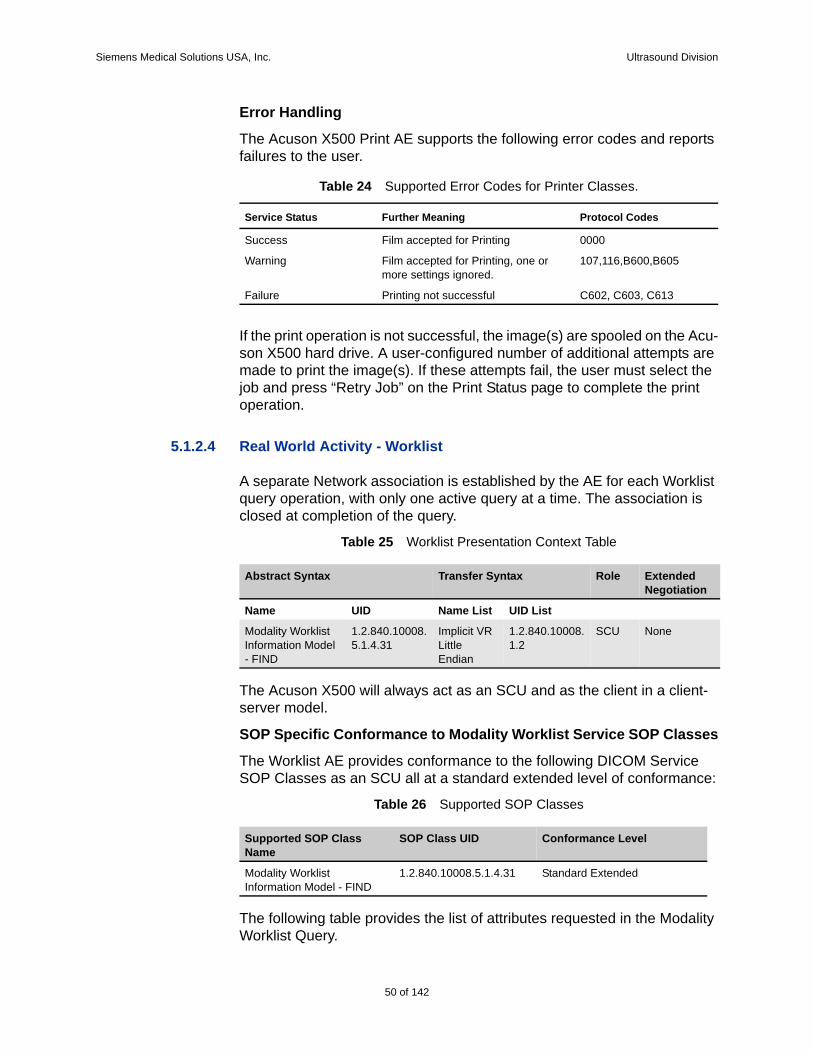
Siemens Medical Solutions USA, Inc. Ultrasound Division
Error Handling
The Acuson X500 Print AE supports the following error codes and reports failures to the user.
If the print operation is not successful, the image(s) are spooled on the Acu-son X500 hard drive. A user-configured number of additional attempts are made to print the image(s). If these attempts fail, the user must select the job and press “Retry Job” on the Print Status page to complete the print operation.
5.1.2.4 Real World Activity - Worklist
A separate Network association is established by the AE for each Worklist query operation, with only one active query at a time. The association is closed at completion of the query.
Table 25 Worklist Presentation Context Table
The Acuson X500 will always act as an SCU and as the client in a client-server model.
SOP Specific Conformance to Modality Worklist Service SOP Classes
The Worklist AE provides conformance to the following DICOM Service SOP Classes as an SCU all at a standard extended level of conformance:
Table 26 Supported SOP Classes
The following table provides the list of attributes requested in the Modality Worklist Query.
Table 24 Supported Error Codes for Printer Classes.
Service Status Further Meaning Protocol Codes
Success Film accepted for Printing 0000
Warning Film accepted for Printing, one or more settings ignored.
107,116,B600,B605
Failure Printing not successful C602, C603, C613
Abstract Syntax Transfer Syntax Role Extended Negotiation
Name UID Name List UID List
Modality Worklist Information Model - FIND
1.2.840.10008.5.1.4.31
Implicit VR Little Endian
1.2.840.10008.1.2
SCU None
Supported SOP Class Name
SOP Class UID Conformance Level
Modality Worklist Information Model - FIND
1.2.840.10008.5.1.4.31 Standard Extended
50 of 142
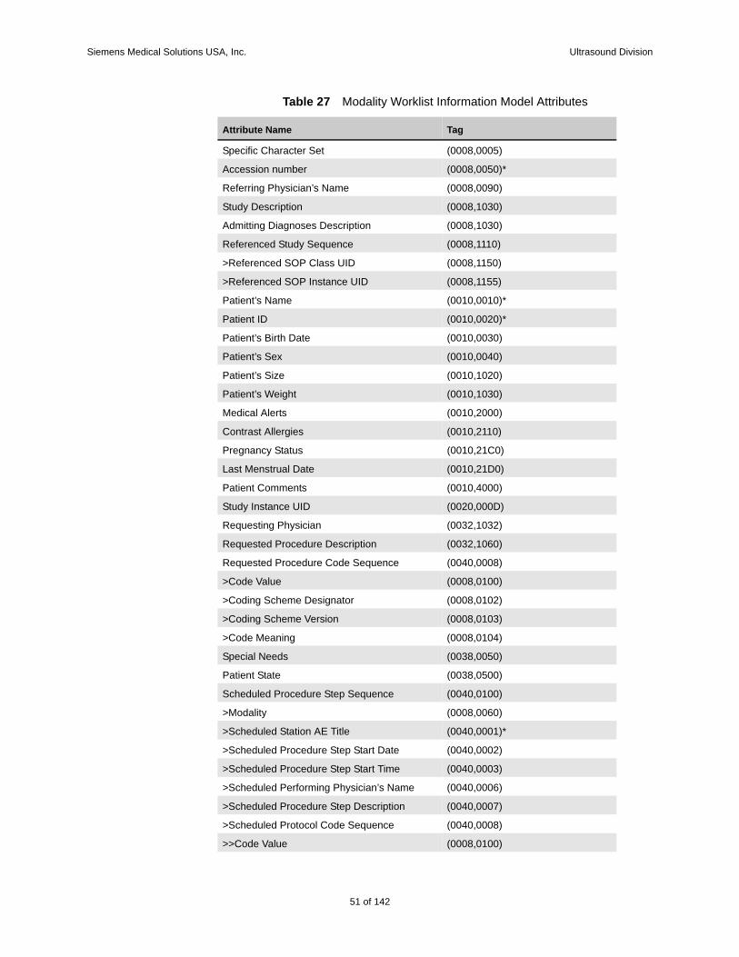
Siemens Medical Solutions USA, Inc. Ultrasound Division
Table 27 Modality Worklist Information Model Attributes
Attribute Name Tag
Specific Character Set (0008,0005)
Accession number (0008,0050)*
Referring Physician’s Name (0008,0090)
Study Description (0008,1030)
Admitting Diagnoses Description (0008,1030)
Referenced Study Sequence (0008,1110)
>Referenced SOP Class UID (0008,1150)
>Referenced SOP Instance UID (0008,1155)
Patient’s Name (0010,0010)*
Patient ID (0010,0020)*
Patient’s Birth Date (0010,0030)
Patient’s Sex (0010,0040)
Patient’s Size (0010,1020)
Patient’s Weight (0010,1030)
Medical Alerts (0010,2000)
Contrast Allergies (0010,2110)
Pregnancy Status (0010,21C0)
Last Menstrual Date (0010,21D0)
Patient Comments (0010,4000)
Study Instance UID (0020,000D)
Requesting Physician (0032,1032)
Requested Procedure Description (0032,1060)
Requested Procedure Code Sequence (0040,0008)
>Code Value (0008,0100)
>Coding Scheme Designator (0008,0102)
>Coding Scheme Version (0008,0103)
>Code Meaning (0008,0104)
Special Needs (0038,0050)
Patient State (0038,0500)
Scheduled Procedure Step Sequence (0040,0100)
>Modality (0008,0060)
>Scheduled Station AE Title (0040,0001)*
>Scheduled Procedure Step Start Date (0040,0002)
>Scheduled Procedure Step Start Time (0040,0003)
>Scheduled Performing Physician’s Name (0040,0006)
>Scheduled Procedure Step Description (0040,0007)
>Scheduled Protocol Code Sequence (0040,0008)
>>Code Value (0008,0100)
51 of 142
142
Siemens Medical Solutions USA, Inc. Ultrasound Division
5.1.2.5 Real World Activity - Modality Performed Procedure Step
This operation allows the AE to create an instance of the Modality Per-formed Procedure Step SOP Class (MPPS) and provide information about a specific real world Performed Procedure Step that is under control of the SCU. This operation is invoked through the DIMSE N-CREATE and N-SET services.
Only the IHE (refer to IHE Rev 5.5) Simple and Abandoned Cases for the relationship between Scheduled Procedure Steps and Performed Proce-dure Steps is supported. Both cases specify that a 1-to-1 relationship must exist between Scheduled Procedure Step and Performed Procedure Step. In the Simple Case the Performed Procedure Step is completed success-fully. In the Abandoned Case the Performed Procedure Step is abandoned before being completed.
A list of scheduled procedures and procedure steps will be accessible from the Worklist and Procedure screens. The Performed Procedure Step User Interface allows the operator to set the status of the performed procedure step. The system shall establish an association for N-CREATE and N-SET, if another N-CREATE or N-SET is available within 5 seconds, it will be sent using the same association.
Starting a Performed Procedure Step
When the user depresses the ‘OK’ button on the New Patient Screen a per-formed procedure SOP Class instance will be created using the N-CREATE DIMSE service for the selected scheduled procedure.
Ending a Performed Procedure Step
When the user selects ‘Completed’ or ‘Discontinued’ from the MPPS User Interface, the performed procedure step will be closed using the N-SET DIMSE service.
New Patient Request
>>Coding Scheme Designator (0008,0102)
>>Coding Scheme Version (0008,0103)
>>Code Meaning (0008,0104)
>Scheduled Procedure Step ID (0040,0009)
>Comments on the Scheduled Procedure Step (0040,0400)
Requested Procedure ID (0040,1001)*
Reason for the Requested Procedure (0040,1002)
*Indicates parameter may be populated for query.
Attribute Name Tag
52 of 142
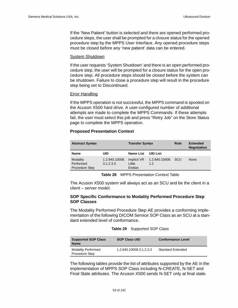
Siemens Medical Solutions USA, Inc. Ultrasound Division
If the ‘New Patient’ button is selected and there are opened performed pro-cedure steps, the user shall be prompted for a closure status for the opened procedure step by the MPPS User Interface. Any opened procedure steps must be closed before any ‘new patient’ data can be entered.
System Shutdown
If the user requests ‘System Shutdown’ and there is an open performed pro-cedure step, the user will be prompted for a closure status for the open pro-cedure step. All procedure steps should be closed before the system can be shutdown. Failure to close a procedure step will result in the procedure step being set to Discontinued.
Error Handling
If the MPPS operation is not successful, the MPPS command is spooled on the Acuson X500 hard drive. A user-configured number of additional attempts are made to complete the MPPS Commands. If these attempts fail, the user must select this job and press “Retry Job” on the Store Status page to complete the MPPS operation.
Proposed Presentation Context
Table 28 MPPS Presentation Context Table
The Acuson X500 system will always act as an SCU and be the client in a client – server model.
SOP Specific Conformance to Modality Performed Procedure Step SOP Classes
The Modality Performed Procedure Step AE provides a conforming imple-mentation of the following DICOM Service SOP Class as an SCU at a stan-dard extended level of conformance.
Table 29 Supported SOP Class
The following tables provide the list of attributes supported by the AE in the implementation of MPPS SOP Class including N-CREATE, N-SET and Final State attributes. The Acuson X500 sends N-SET only at final state.
Abstract Syntax Transfer Syntax Role Extended Negotiation
Name UID Name List UID List
Modality Performed Procedure Step
1.2.840.10008.3.1.2.3.3
Implicit VR Little Endian
1.2.840.10008.1.2
SCU None
Supported SOP Class Name
SOP Class UID Conformance Level
Modality Performed Procedure Step
1.2.840.10008.3.1.2.3.3 Standard Extended
53 of 142
142
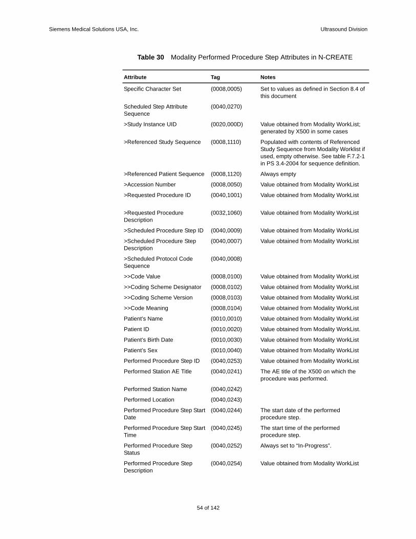
Siemens Medical Solutions USA, Inc. Ultrasound Division
Table 30 Modality Performed Procedure Step Attributes in N-CREATE
Attribute Tag Notes
Specific Character Set (0008,0005) Set to values as defined in Section 8.4 of this document
Scheduled Step Attribute Sequence
(0040,0270)
>Study Instance UID (0020,000D) Value obtained from Modality WorkList; generated by X500 in some cases
>Referenced Study Sequence (0008,1110) Populated with contents of Referenced Study Sequence from Modality Worklist if used, empty otherwise. See table F.7.2-1 in PS 3.4-2004 for sequence definition.
>Referenced Patient Sequence (0008,1120) Always empty
>Accession Number (0008,0050) Value obtained from Modality WorkList
>Requested Procedure ID (0040,1001) Value obtained from Modality WorkList
>Requested Procedure Description
(0032,1060) Value obtained from Modality WorkList
>Scheduled Procedure Step ID (0040,0009) Value obtained from Modality WorkList
>Scheduled Procedure Step Description
(0040,0007) Value obtained from Modality WorkList
>Scheduled Protocol Code Sequence
(0040,0008)
>>Code Value (0008,0100) Value obtained from Modality WorkList
>>Coding Scheme Designator (0008,0102) Value obtained from Modality WorkList
>>Coding Scheme Version (0008,0103) Value obtained from Modality WorkList
>>Code Meaning (0008,0104) Value obtained from Modality WorkList
Patient’s Name (0010,0010) Value obtained from Modality WorkList
Patient ID (0010,0020) Value obtained from Modality WorkList.
Patient’s Birth Date (0010,0030) Value obtained from Modality WorkList
Patient’s Sex (0010,0040) Value obtained from Modality WorkList
Performed Procedure Step ID (0040,0253) Value obtained from Modality WorkList
Performed Station AE Title (0040,0241) The AE title of the X500 on which the procedure was performed.
Performed Station Name (0040,0242)
Performed Location (0040,0243)
Performed Procedure Step Start Date
(0040,0244) The start date of the performed procedure step.
Performed Procedure Step Start Time
(0040,0245) The start time of the performed procedure step.
Performed Procedure Step Status
(0040,0252) Always set to “In-Progress”.
Performed Procedure Step Description
(0040,0254) Value obtained from Modality WorkList
54 of 142
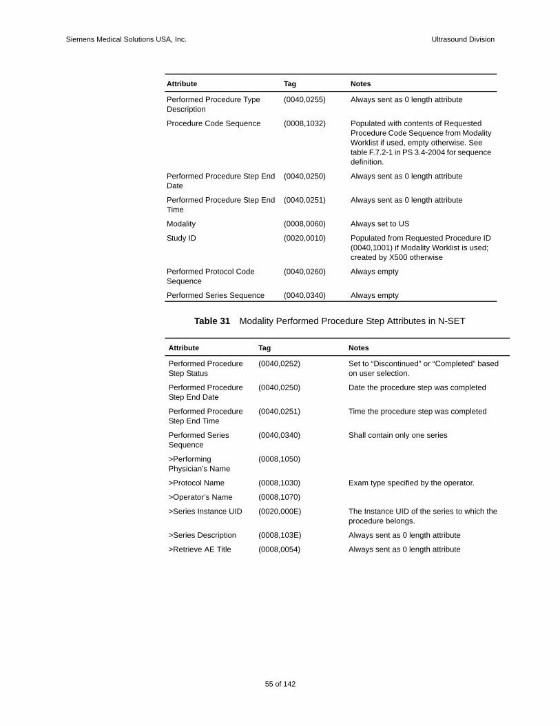
Siemens Medical Solutions USA, Inc. Ultrasound Division
Table 31 Modality Performed Procedure Step Attributes in N-SET
Performed Procedure Type Description
(0040,0255) Always sent as 0 length attribute
Procedure Code Sequence (0008,1032) Populated with contents of Requested Procedure Code Sequence from Modality Worklist if used, empty otherwise. See table F.7.2-1 in PS 3.4-2004 for sequence definition.
Performed Procedure Step End Date
(0040,0250) Always sent as 0 length attribute
Performed Procedure Step End Time
(0040,0251) Always sent as 0 length attribute
Modality (0008,0060) Always set to US
Study ID (0020,0010) Populated from Requested Procedure ID (0040,1001) if Modality Worklist is used; created by X500 otherwise
Performed Protocol Code Sequence
(0040,0260) Always empty
Performed Series Sequence (0040,0340) Always empty
Attribute Tag Notes
Performed Procedure Step Status
(0040,0252) Set to “Discontinued” or “Completed” based on user selection.
Performed Procedure Step End Date
(0040,0250) Date the procedure step was completed
Performed Procedure Step End Time
(0040,0251) Time the procedure step was completed
Performed Series Sequence
(0040,0340) Shall contain only one series
>Performing Physician’s Name
(0008,1050)
>Protocol Name (0008,1030) Exam type specified by the operator.
>Operator’s Name (0008,1070)
>Series Instance UID (0020,000E) The Instance UID of the series to which the procedure belongs.
>Series Description (0008,103E) Always sent as 0 length attribute
>Retrieve AE Title (0008,0054) Always sent as 0 length attribute
Attribute Tag Notes
55 of 142
142
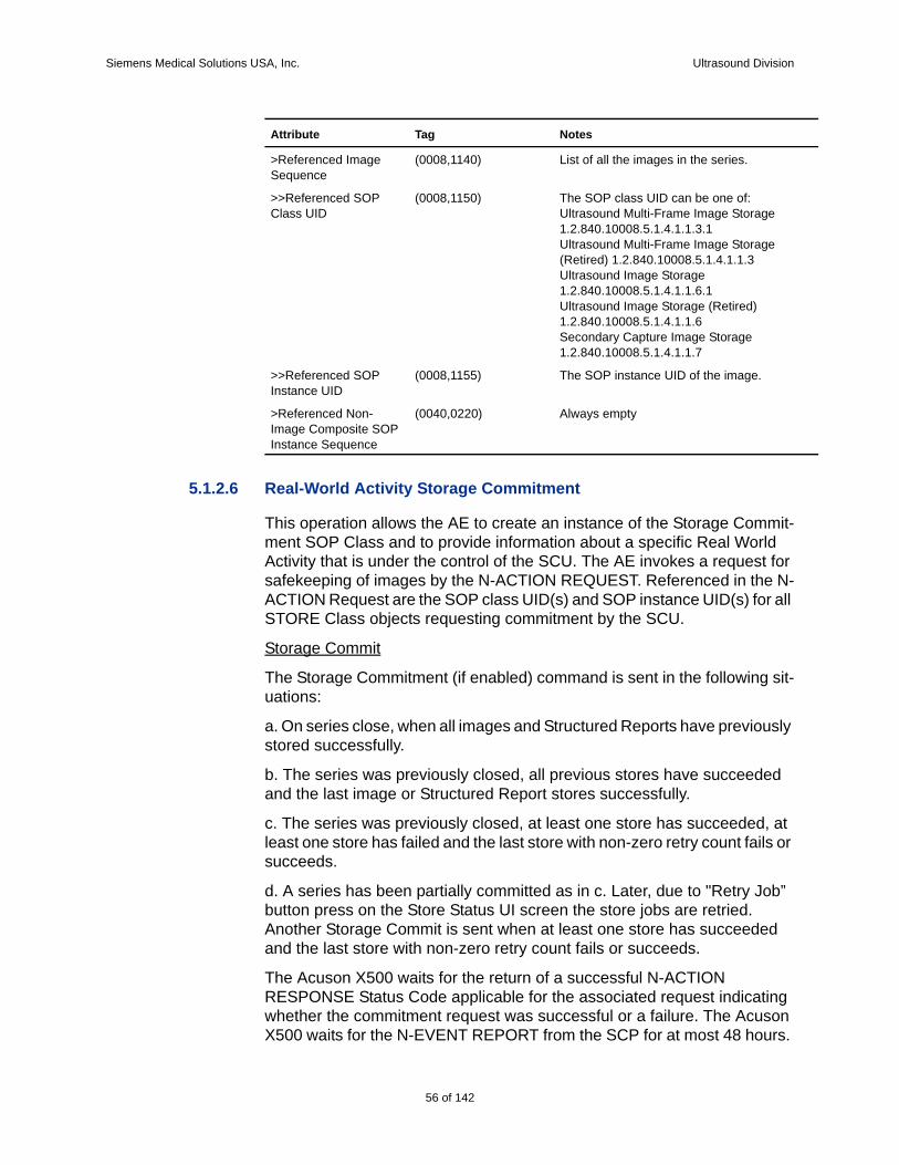
Siemens Medical Solutions USA, Inc. Ultrasound Division
5.1.2.6 Real-World Activity Storage Commitment
This operation allows the AE to create an instance of the Storage Commit-ment SOP Class and to provide information about a specific Real World Activity that is under the control of the SCU. The AE invokes a request for safekeeping of images by the N-ACTION REQUEST. Referenced in the N-ACTION Request are the SOP class UID(s) and SOP instance UID(s) for all STORE Class objects requesting commitment by the SCU.
Storage Commit
The Storage Commitment (if enabled) command is sent in the following sit-uations:
a. On series close, when all images and Structured Reports have previously stored successfully.
b. The series was previously closed, all previous stores have succeeded and the last image or Structured Report stores successfully.
c. The series was previously closed, at least one store has succeeded, at least one store has failed and the last store with non-zero retry count fails or succeeds.
d. A series has been partially committed as in c. Later, due to "Retry Job” button press on the Store Status UI screen the store jobs are retried. Another Storage Commit is sent when at least one store has succeeded and the last store with non-zero retry count fails or succeeds.
The Acuson X500 waits for the return of a successful N-ACTION RESPONSE Status Code applicable for the associated request indicating whether the commitment request was successful or a failure. The Acuson X500 waits for the N-EVENT REPORT from the SCP for at most 48 hours.
>Referenced Image Sequence
(0008,1140) List of all the images in the series.
>>Referenced SOP Class UID
(0008,1150) The SOP class UID can be one of:Ultrasound Multi-Frame Image Storage1.2.840.10008.5.1.4.1.1.3.1Ultrasound Multi-Frame Image Storage (Retired) 1.2.840.10008.5.1.4.1.1.3Ultrasound Image Storage1.2.840.10008.5.1.4.1.1.6.1Ultrasound Image Storage (Retired)1.2.840.10008.5.1.4.1.1.6Secondary Capture Image Storage1.2.840.10008.5.1.4.1.1.7
>>Referenced SOP Instance UID
(0008,1155) The SOP instance UID of the image.
>Referenced Non-Image Composite SOP Instance Sequence
(0040,0220) Always empty
Attribute Tag Notes
56 of 142
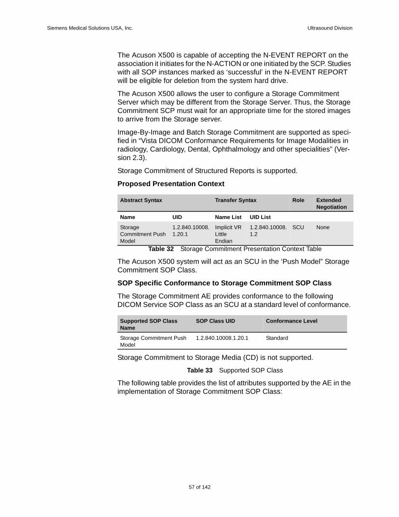
Siemens Medical Solutions USA, Inc. Ultrasound Division
The Acuson X500 is capable of accepting the N-EVENT REPORT on the association it initiates for the N-ACTION or one initiated by the SCP. Studies with all SOP instances marked as ‘successful’ in the N-EVENT REPORT will be eligible for deletion from the system hard drive.
The Acuson X500 allows the user to configure a Storage Commitment Server which may be different from the Storage Server. Thus, the Storage Commitment SCP must wait for an appropriate time for the stored images to arrive from the Storage server.
Image-By-Image and Batch Storage Commitment are supported as speci-fied in “Vista DICOM Conformance Requirements for Image Modalities in radiology, Cardiology, Dental, Ophthalmology and other specialities” (Ver-sion 2.3).
Storage Commitment of Structured Reports is supported.
Proposed Presentation Context
Table 32 Storage Commitment Presentation Context Table
The Acuson X500 system will act as an SCU in the ‘Push Model” Storage Commitment SOP Class.
SOP Specific Conformance to Storage Commitment SOP Class
The Storage Commitment AE provides conformance to the following DICOM Service SOP Class as an SCU at a standard level of conformance.
Storage Commitment to Storage Media (CD) is not supported.
Table 33 Supported SOP Class
The following table provides the list of attributes supported by the AE in the implementation of Storage Commitment SOP Class:
Abstract Syntax Transfer Syntax Role Extended Negotiation
Name UID Name List UID List
Storage Commitment Push Model
1.2.840.10008.1.20.1
Implicit VR Little Endian
1.2.840.10008.1.2
SCU None
Supported SOP Class Name
SOP Class UID Conformance Level
Storage Commitment Push Model
1.2.840.10008.1.20.1 Standard
57 of 142
142
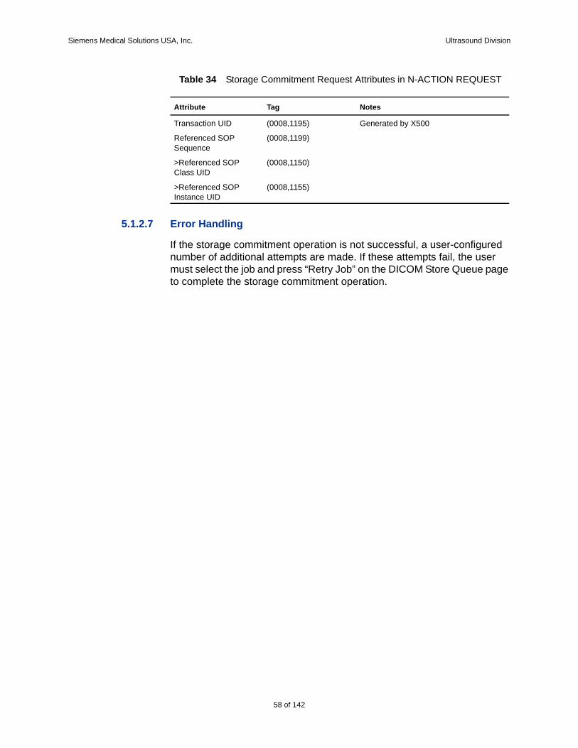
Siemens Medical Solutions USA, Inc. Ultrasound Division
Table 34 Storage Commitment Request Attributes in N-ACTION REQUEST
5.1.2.7 Error Handling
If the storage commitment operation is not successful, a user-configured number of additional attempts are made. If these attempts fail, the user must select the job and press “Retry Job” on the DICOM Store Queue page to complete the storage commitment operation.
Attribute Tag Notes
Transaction UID (0008,1195) Generated by X500
Referenced SOP Sequence
(0008,1199)
>Referenced SOP Class UID
(0008,1150)
>Referenced SOP Instance UID
(0008,1155)
58 of 142
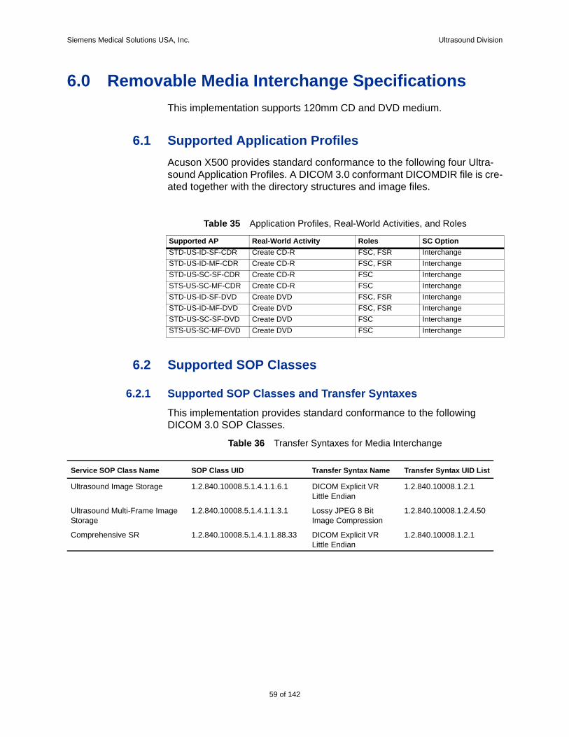
Siemens Medical Solutions USA, Inc. Ultrasound Division
6.0 Removable Media Interchange SpecificationsThis implementation supports 120mm CD and DVD medium.
6.1 Supported Application ProfilesAcuson X500 provides standard conformance to the following four Ultra-sound Application Profiles. A DICOM 3.0 conformant DICOMDIR file is cre-ated together with the directory structures and image files.
Table 35 Application Profiles, Real-World Activities, and Roles
6.2 Supported SOP Classes
6.2.1 Supported SOP Classes and Transfer SyntaxesThis implementation provides standard conformance to the following DICOM 3.0 SOP Classes.
Table 36 Transfer Syntaxes for Media Interchange
Supported AP Real-World Activity Roles SC OptionSTD-US-ID-SF-CDR Create CD-R FSC, FSR InterchangeSTD-US-ID-MF-CDR Create CD-R FSC, FSR InterchangeSTD-US-SC-SF-CDR Create CD-R FSC InterchangeSTS-US-SC-MF-CDR Create CD-R FSC InterchangeSTD-US-ID-SF-DVD Create DVD FSC, FSR InterchangeSTD-US-ID-MF-DVD Create DVD FSC, FSR InterchangeSTD-US-SC-SF-DVD Create DVD FSC InterchangeSTS-US-SC-MF-DVD Create DVD FSC Interchange
Service SOP Class Name SOP Class UID Transfer Syntax Name Transfer Syntax UID List
Ultrasound Image Storage 1.2.840.10008.5.1.4.1.1.6.1 DICOM Explicit VR Little Endian
1.2.840.10008.1.2.1
Ultrasound Multi-Frame Image Storage
1.2.840.10008.5.1.4.1.1.3.1 Lossy JPEG 8 Bit Image Compression
1.2.840.10008.1.2.4.50
Comprehensive SR 1.2.840.10008.5.1.4.1.1.88.33 DICOM Explicit VR Little Endian
1.2.840.10008.1.2.1
59 of 142
142
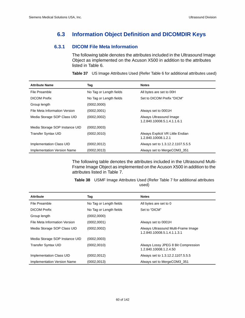
Siemens Medical Solutions USA, Inc. Ultrasound Division
6.3 Information Object Definition and DICOMDIR Keys
6.3.1 DICOM File Meta InformationThe following table denotes the attributes included in the Ultrasound Image Object as implemented on the Acuson X500 in addition to the attributes listed in Table 6.
Table 37 US Image Attributes Used (Refer Table 6 for additional attributes used)
The following table denotes the attributes included in the Ultrasound Multi-Frame Image Object as implemented on the Acuson X500 in addition to the attributes listed in Table 7.
Table 38 USMF Image Attributes Used (Refer Table 7 for additional attributes used)
Attribute Name Tag Notes
File Preamble No Tag or Length fields All bytes are set to 00H
DICOM Prefix No Tag or Length fields Set to DICOM Prefix “DICM”
Group length (0002,0000)
File Meta Information Version (0002,0001) Always set to 0001H
Media Storage SOP Class UID (0002,0002) Always Ultrasound Image1.2.840.10008.5.1.4.1.1.6.1
Media Storage SOP Instance UID (0002,0003)
Transfer Syntax UID (0002,0010) Always Explicit VR Little Endian1.2.840.10008.1.2.1
Implementation Class UID (0002,0012) Always set to 1.3.12.2.1107.5.5.5
Implementation Version Name (0002,0013) Always set to MergeCOM3_351
Attribute Tag Notes
File Preamble No Tag or Length fields All bytes are set to 0
DICOM Prefix No Tag or Length fields Set to “DICM”
Group length (0002,0000)
File Meta Information Version (0002,0001) Always set to 0001H
Media Storage SOP Class UID (0002,0002) Always Ultrasound Multi-Frame Image1.2.840.10008.5.1.4.1.1.3.1
Media Storage SOP Instance UID (0002,0003)
Transfer Syntax UID (0002,0010) Always Lossy JPEG 8 Bit Compression1.2.840.10008.1.2.4.50
Implementation Class UID (0002,0012) Always set to 1.3.12.2.1107.5.5.5
Implementation Version Name (0002,0013) Always set to MergeCOM3_351
60 of 142
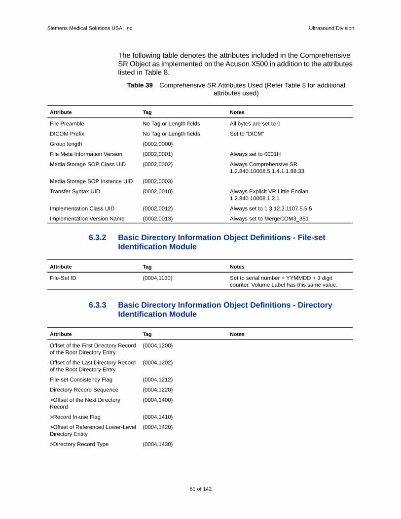
Siemens Medical Solutions USA, Inc. Ultrasound Division
The following table denotes the attributes included in the Comprehensive SR Object as implemented on the Acuson X500 in addition to the attributes listed in Table 8.
Table 39 Comprehensive SR Attributes Used (Refer Table 8 for additional attributes used)
6.3.2 Basic Directory Information Object Definitions - File-set Identification Module
6.3.3 Basic Directory Information Object Definitions - Directory Identification Module
Attribute Tag Notes
File Preamble No Tag or Length fields All bytes are set to 0
DICOM Prefix No Tag or Length fields Set to “DICM”
Group length (0002,0000)
File Meta Information Version (0002,0001) Always set to 0001H
Media Storage SOP Class UID (0002,0002) Always Comprehensive SR1.2.840.10008.5.1.4.1.1.88.33
Media Storage SOP Instance UID (0002,0003)
Transfer Syntax UID (0002,0010) Always Explicit VR Little Endian1.2.840.10008.1.2.1
Implementation Class UID (0002,0012) Always set to 1.3.12.2.1107.5.5.5
Implementation Version Name (0002,0013) Always set to MergeCOM3_351
Attribute Tag Notes
File-Set ID (0004,1130) Set to serial number + YYMMDD + 3 digit counter. Volume Label has this same value.
Attribute Tag Notes
Offset of the First Directory Record of the Root Directory Entry
(0004,1200)
Offset of the Last Directory Record of the Root Directory Entry
(0004,1202)
File-set Consistency Flag (0004,1212)
Directory Record Sequence (0004,1220)
>Offset of the Next Directory Record
(0004,1400)
>Record In-use Flag (0004,1410)
>Offset of Referenced Lower-Level Directory Entity
(0004,1420)
>Directory Record Type (0004,1430)
61 of 142
142
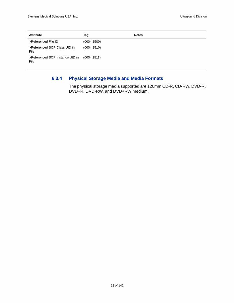
Siemens Medical Solutions USA, Inc. Ultrasound Division
6.3.4 Physical Storage Media and Media FormatsThe physical storage media supported are 120mm CD-R, CD-RW, DVD-R, DVD+R, DVD-RW, and DVD+RW medium.
>Referenced File ID (0004,1500)
>Referenced SOP Class UID in File
(0004,1510)
>Referenced SOP Instance UID in File
(0004,1511)
Attribute Tag Notes
62 of 142
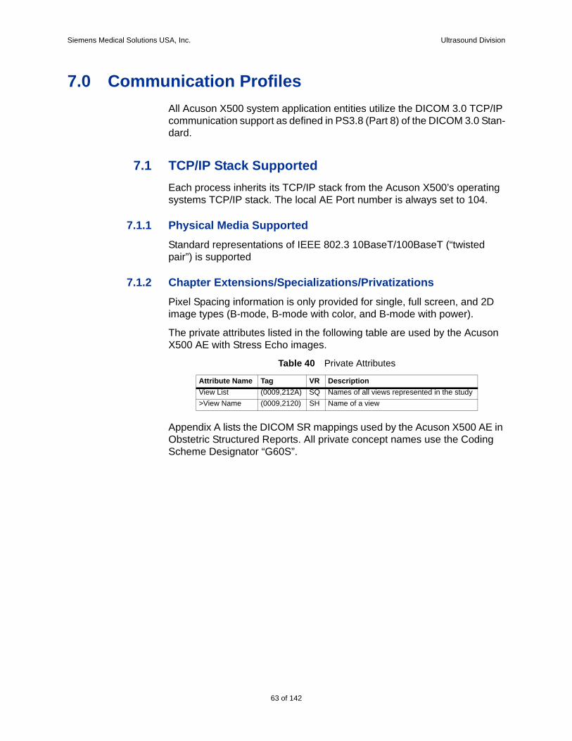
Siemens Medical Solutions USA, Inc. Ultrasound Division
7.0 Communication ProfilesAll Acuson X500 system application entities utilize the DICOM 3.0 TCP/IP communication support as defined in PS3.8 (Part 8) of the DICOM 3.0 Stan-dard.
7.1 TCP/IP Stack SupportedEach process inherits its TCP/IP stack from the Acuson X500’s operating systems TCP/IP stack. The local AE Port number is always set to 104.
7.1.1 Physical Media SupportedStandard representations of IEEE 802.3 10BaseT/100BaseT (“twisted pair”) is supported
7.1.2 Chapter Extensions/Specializations/PrivatizationsPixel Spacing information is only provided for single, full screen, and 2D image types (B-mode, B-mode with color, and B-mode with power).
The private attributes listed in the following table are used by the Acuson X500 AE with Stress Echo images.
Table 40 Private Attributes
Appendix A lists the DICOM SR mappings used by the Acuson X500 AE in Obstetric Structured Reports. All private concept names use the Coding Scheme Designator “G60S”.
Attribute Name Tag VR DescriptionView List (0009,212A) SQ Names of all views represented in the study>View Name (0009,2120) SH Name of a view
63 of 142
142
Siemens Medical Solutions USA, Inc. Ultrasound Division
8.0 ConfigurationAcuson X500 Networking and DICOM parameters can be configured through the Acuson X500 System Presets Menu screens. The following configuration is supported:
General system
Network (local and remote)
DICOM Store
DICOM Print
DICOM Modality Worklist
DICOM Storage Commitment
DICOM Modality Performed Procedure Step
8.1 General System ConfigurationThe following system parameter can be configured via the Acuson X500 System Presets Basic Menu screens. This parameter is mapped to a DICOM image attribute:
Hospital Name
8.1.1 Hospital Name
The user can enter the organization (i.e. hospital, clinic, etc.) as a text string in the Hospital Name field of the System Presets - General menu. The Organization Name field is transferred to DICOM devices as Institution Name - DICOM data element (0008, 0080).
8.2 DICOM Network ConfigurationDICOM and networking parameters can be configured for both the local Acuson X500 device and remote DICOM service class providers through the System Presets DICOM Menu.
8.2.1 Local
The Acuson X500 local network parameters are configurable. The following network parameters can be configured for a Acuson X500 device:
Host Name
IP address
Subnet IP mask
Default Gateway
64 of 142
Siemens Medical Solutions USA, Inc. Ultrasound Division
DICOM Application Entity Title
8.2.2 RemoteMultiple DICOM service class providers can be configured through the sys-tem presets. The following network parameters can be configured for each remote device:
DICOM Device Application Entity Title
IP address
Port Number
8.2.2.1 DICOM Store Configuration
Several configuration settings are provided in addition to those described in Section 8.2.2.
The Image Format setting provides control over the Presentation Contexts proposed during Association negotiation. This is documented in Section 5.1.1.2.
Configuration options are provided to control the Photometric Interpretation of grayscale images stored to a DICOM Store SCP. Grayscale images can be stored as RGB, YBR-FULL-422 or MONOCHROME2.
Many Acuson X500 B-Mode and M-Mode images contain no significant color, the only color is in the Acuson ‘a’ transducer position marker and the ECG trace, if used. When “Store grayscale images as Monochrome” is selected, single-frame images with no significant color content will be stored as MONOCHROME2. When “No Monochrome conversion” is selected, all multi-frame and single frame images are stored as RGB or YBR-FULL-422. When “Store grayscale images and clips as Monochrome” is selected, B-Mode and M-Mode single- and multi-frame images with no significant color content will be stored as MONOCHROME2. When “Store all images as Monochrome” is selected then all single frame images (with or without significant color) are stored as Monochrome2. When “Store all images and clips as Monochrome” is selected then all single and multi frame images (with or without any significant color) are stored as Monochrome2.
8.2.2.2 DICOM Storage Commitment Configuration
Configuration of DICOM Storage Commitment remote devices must be per-formed separately from DICOM Store Configuration. The Acuson X500 supports Storage Commitment to the same remote device as Store or to a different device.
8.2.2.3 DICOM Modality Worklist Configuration
Configuration of DICOM Modality Worklist remote devices
65 of 142
142
Siemens Medical Solutions USA, Inc. Ultrasound Division
8.2.2.4 DICOM Modality Performed Procedure Step Configuration
Configuration of DICOM Modality Performed Procedure Step remote devices must be performed separately from DICOM Modality Worklist Con-figuration. The Acuson X500 supports MPPS to the same remote device as Modality Worklist or to a different device.
The “Store Image Format” setting controls the Referenced SOP Class UID (0008,1150) in the Referenced Image Sequence (0008,1140) of the MPPS N-SET sent by the Acuson X500. Due to the Acuson X500’s ability to select from multiple Presentation Contexts during Association Negotiation, it is necessary to use this setting.
In the majority of installations the “Store Image Format” should be left at the default setting of “New Ultrasound”. There are two cases when the “Store Image Format” must be set to “Old Ultrasound” or “Secondary Capture”:
1. When the active Storage Server “Image Format” is set to “Old Ultra-sound” or “Secondary Capture”.
2. When the active Storage Server “Image Format” is set to “Auto-matic”, but the Storage Server does not support US Image and US Multi-frame Image.
In both cases the correct setting can be determined by reviewing the DICOM Conformance Statement of the Storage Server and following the instructions below. DICOM Conformance Statements are usually available on the manufacturer’s Web site.
If at least one of US Image and US Multi-frame Image are listed in the DICOM Conformance Statement and the active Storage Server “Image For-mat” is set to “Automatic” then “New Ultrasound” is the correct setting for “Store Image Format”.
If the above is not true and at least one of US Image (Retired) and US Multi-frame Image (Retired) are listed in the DICOM Conformance Statement then “Old Ultrasound” is the correct setting for “Store Image Format”.
If neither of the above are true then “Secondary Capture” is the correct set-ting for “Store Image Format”.
66 of 142
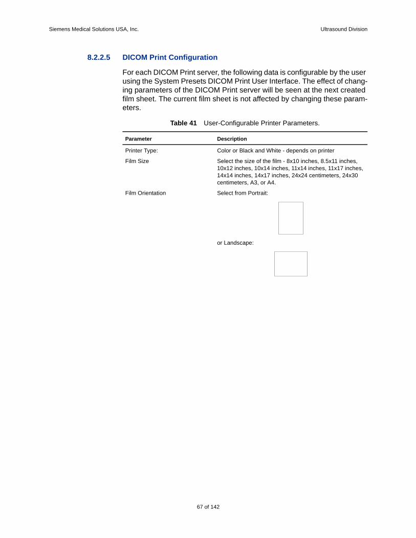
Siemens Medical Solutions USA, Inc. Ultrasound Division
8.2.2.5 DICOM Print Configuration
For each DICOM Print server, the following data is configurable by the user using the System Presets DICOM Print User Interface. The effect of chang-ing parameters of the DICOM Print server will be seen at the next created film sheet. The current film sheet is not affected by changing these param-eters.
Table 41 User-Configurable Printer Parameters.
Parameter Description
Printer Type: Color or Black and White - depends on printer
Film Size Select the size of the film - 8x10 inches, 8.5x11 inches, 10x12 inches, 10x14 inches, 11x14 inches, 11x17 inches, 14x14 inches, 14x17 inches, 24x24 centimeters, 24x30 centimeters, A3, or A4.
Film Orientation Select from Portrait:
or Landscape:
67 of 142
142
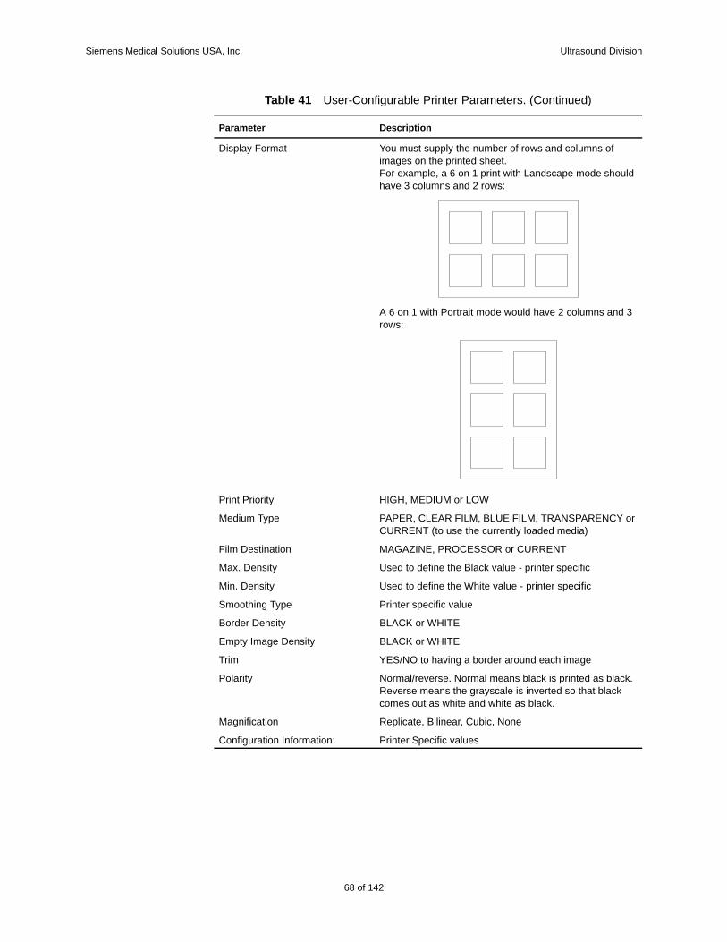
Siemens Medical Solutions USA, Inc. Ultrasound Division
Display Format You must supply the number of rows and columns of images on the printed sheet.For example, a 6 on 1 print with Landscape mode should have 3 columns and 2 rows:
A 6 on 1 with Portrait mode would have 2 columns and 3 rows:
Print Priority HIGH, MEDIUM or LOW
Medium Type PAPER, CLEAR FILM, BLUE FILM, TRANSPARENCY or CURRENT (to use the currently loaded media)
Film Destination MAGAZINE, PROCESSOR or CURRENT
Max. Density Used to define the Black value - printer specific
Min. Density Used to define the White value - printer specific
Smoothing Type Printer specific value
Border Density BLACK or WHITE
Empty Image Density BLACK or WHITE
Trim YES/NO to having a border around each image
Polarity Normal/reverse. Normal means black is printed as black. Reverse means the grayscale is inverted so that black comes out as white and white as black.
Magnification Replicate, Bilinear, Cubic, None
Configuration Information: Printer Specific values
Table 41 User-Configurable Printer Parameters. (Continued)
Parameter Description
68 of 142
Siemens Medical Solutions USA, Inc. Ultrasound Division
8.3 External Equipment ConfigurationThe Acuson X500 user can configure “Hard Key” to “Output Device” map-ping through the System Presets - Customize Keys. Print images are acquired and sent to the assigned device when the user presses the asso-ciated key. The following key assignments are supported:
Clip Store – This key can be assigned to Multi-frame Store Cap-ture, Cine Store or Disk Store
Print/Store 1 – This key can be assigned to any configured DICOM Printer, DICOM Store or OEM printer device.
Print/Store 2 – This key can be assigned to any configured DICOM Printer, DICOM Store or OEM printer device.
8.4 Support of Extended Character SetsThe “ISO-IR 100”, "GB18030", "ISO_IR 144" character sets are supported by the Acuson X500 system based on the following language selections:
English, French, Italian, German, Spanish: "ISO_IR 100"Chinese: "GB18030"
Russian: "ISO_IR 144"
69 of 142
142
Siemens Medical Solutions USA, Inc. Ultrasound Division
9.0 Security
9.1 Security ProfilesNone supported.
9.2 Association Level SecurityNone supported.
9.3 Application Level SecurityNone supported.
9.4 Virus ProtectionThe Acuson X500 computer system's networking has been configured to signifi-cantly reduce the possibility of virus and hacking vulnerabilities. On the X500 computer system, all ingress TCP and UDP ports are closed and/or absent of any type of server. The only exception to this is due to the necessity of a DICOM server available at ingress TCP port 104. Additionally, all non-essential computer services and components are disabled to minimize X500 egress network footprint.
Outside of some minimal network exchanges required by the X500's commercial computer operating system, the only network connections initiated by the X500 are for DICOM connectivity and network-share export function.
70 of 142
Siemens Medical Solutions USA, Inc. Ultrasound Division
rties
10.0 Appendix A: Mapping for Obstetric DICOM SRThe following table lists the DICOM Structured Report mappings used by the Acuson X500 1.0 in Obstetric Structured Reports. All private concept names use the Coding Scheme Designator “G60S”.
Table 42 DICOM SR Mappings for OB Structured Reports
Platform Name Base Measurement Concept Name
Template Section Inferred From Modifiers/Prope
PATIENT_NAME
PATIENT_ID
SEX
REFERRING_MD
BIRTH_DATE
PATIENT_AGE
HEIGHT LN 8302-2, "Patient Height"
TID 5001: Patient Characteristics
WEIGHT LN 29463-7, "Patient Weight"
TID 5001: Patient Characteristics
LMP LN 11955-2, "LMP" TID 5002: OB-GYN Procedure SummaryCID 12003: OB-GYN Dates
DIAGNOSIS_DESCRIPTION
DCM, 121106, "Com-ment"
TID 5001: Patient Characteristics
GRAVIDA LN 11996-6, "Gravida" TID 5001: Patient Characteristics
PARA LN 11977-6, "Para" TID 5001: Patient Characteristics
AB LN 11612-9, "Aborta" TID 5001: Patient Characteristics
ECTOPIC LN 33065-4, "Ectopic Pregnancies"
TID 5001: Patient Characteristics
FETAL_AGE LN 18185-9, "Gesta-tional Age"
TID 5003: OB-GYN Procedure Fetus SummaryCID 12019: OB-GYN Fetus Summary
71 of 142
142
Siemens Medical Solutions USA, Inc. Ultrasound Division
e of
tion tors)
e of
tion tors)
e of
tion tors)
LMP_ESTIMATE_FETAL_AGE
LN 11885-1, "Gesta-tional Age by LMP"
TID 5003: OB-GYN Procedure Fetus SummaryCID 12019: OB-GYN Fetus Summary
IVF_DATE_TIME No match in DICOM Spec
TID 5001: Patient Characteristics
Private Code: IVF_Date_Time_G60S
IVF_ESTIMATE_FETAL_AGE
No match in DICOM Spec
TID 5001: Patient Characteristics
Private Code: IVF_Est_Fetal_Age_G60S
BPD LN 11820-8, "Bipari-etal Diameter"
TID 5005: Fetal Biometry SectionTID 5008: Fetal Biometry GroupCID 12005: Fetal Biometry Measure-ments
MA_BPD_HADLOCK LN 18185-9, "Gesta-tional Age"
TID 5005: Fetal Biometry SectionTID 5008: Fetal Biometry Group
LN 11902-4, BPD, Hadlock 1984(CID 12013: Gesta-tional Age Equations and Tables)
MA_BPD_HADLOCK_P2SD
LN 18185-9, "Gesta-tional Age"
TID 5005: Fetal Biometry SectionTID 5008: Fetal Biometry Group
LN 11902-4, BPD, Hadlock 1984(CID 12013: Gesta-tional Age Equations and Tables)
SRT R-00387, 2 Sigma Upper Valupopulation(CID 226, PopulaStatistical Descrip
MA_BPD_HADLOCK_M2SD
LN 18185-9, "Gesta-tional Age"
TID 5005: Fetal Biometry SectionTID 5008: Fetal Biometry Group
LN 11902-4, BPD, Hadlock 1984(CID 12013: Gesta-tional Age Equations and Tables)
SRT R-00388, 2 Sigma Lower Valupopulation(CID 226, PopulaStatistical Descrip
MA_BPD_MERZ LN 18185-9, "Gesta-tional Age"
TID 5005: Fetal Biometry SectionTID 5008: Fetal Biometry Group
Private Code: MA_BPD_Merz_G60S(Equation)
MA_BPD_MERZ_P2SD
LN 18185-9, "Gesta-tional Age"
TID 5005: Fetal Biometry SectionTID 5008: Fetal Biometry Group
Private Code: MA_BPD_Merz_G60S(Equation)
SRT R-00387, 2 Sigma Upper Valupopulation(CID 226, PopulaStatistical Descrip
72 of 142
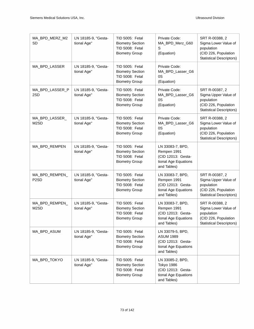
Siemens Medical Solutions USA, Inc. Ultrasound Division
e of
tion tors)
e of
tion tors)
e of
tion tors)
e of
tion tors)
e of
tion tors)
MA_BPD_MERZ_M2SD
LN 18185-9, "Gesta-tional Age"
TID 5005: Fetal Biometry SectionTID 5008: Fetal Biometry Group
Private Code: MA_BPD_Merz_G60S(Equation)
SRT R-00388, 2 Sigma Lower Valupopulation(CID 226, PopulaStatistical Descrip
MA_BPD_LASSER LN 18185-9, "Gesta-tional Age"
TID 5005: Fetal Biometry SectionTID 5008: Fetal Biometry Group
Private Code: MA_BPD_Lasser_G60S(Equation)
MA_BPD_LASSER_P2SD
LN 18185-9, "Gesta-tional Age"
TID 5005: Fetal Biometry SectionTID 5008: Fetal Biometry Group
Private Code: MA_BPD_Lasser_G60S(Equation)
SRT R-00387, 2 Sigma Upper Valupopulation(CID 226, PopulaStatistical Descrip
MA_BPD_LASSER_M2SD
LN 18185-9, "Gesta-tional Age"
TID 5005: Fetal Biometry SectionTID 5008: Fetal Biometry Group
Private Code: MA_BPD_Lasser_G60S(Equation)
SRT R-00388, 2 Sigma Lower Valupopulation(CID 226, PopulaStatistical Descrip
MA_BPD_REMPEN LN 18185-9, "Gesta-tional Age"
TID 5005: Fetal Biometry SectionTID 5008: Fetal Biometry Group
LN 33083-7, BPD, Rempen 1991(CID 12013: Gesta-tional Age Equations and Tables)
MA_BPD_REMPEN_P2SD
LN 18185-9, "Gesta-tional Age"
TID 5005: Fetal Biometry SectionTID 5008: Fetal Biometry Group
LN 33083-7, BPD, Rempen 1991(CID 12013: Gesta-tional Age Equations and Tables)
SRT R-00387, 2 Sigma Upper Valupopulation(CID 226, PopulaStatistical Descrip
MA_BPD_REMPEN_M2SD
LN 18185-9, "Gesta-tional Age"
TID 5005: Fetal Biometry SectionTID 5008: Fetal Biometry Group
LN 33083-7, BPD, Rempen 1991(CID 12013: Gesta-tional Age Equations and Tables)
SRT R-00388, 2 Sigma Lower Valupopulation(CID 226, PopulaStatistical Descrip
MA_BPD_ASUM LN 18185-9, "Gesta-tional Age"
TID 5005: Fetal Biometry SectionTID 5008: Fetal Biometry Group
LN 33079-5, BPD, ASUM 1989(CID 12013: Gesta-tional Age Equations and Tables)
MA_BPD_TOKYO LN 18185-9, "Gesta-tional Age"
TID 5005: Fetal Biometry SectionTID 5008: Fetal Biometry Group
LN 33085-2, BPD, Tokyo 1986(CID 12013: Gesta-tional Age Equations and Tables)
73 of 142
142
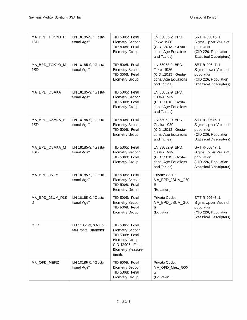
Siemens Medical Solutions USA, Inc. Ultrasound Division
e of
tion tors)
e of
tion tors)
e of
tion tors)
e of
tion tors)
e of
tion tors)
MA_BPD_TOKYO_P1SD
LN 18185-9, "Gesta-tional Age"
TID 5005: Fetal Biometry SectionTID 5008: Fetal Biometry Group
LN 33085-2, BPD, Tokyo 1986(CID 12013: Gesta-tional Age Equations and Tables)
SRT R-00346, 1 Sigma Upper Valupopulation(CID 226, PopulaStatistical Descrip
MA_BPD_TOKYO_M1SD
LN 18185-9, "Gesta-tional Age"
TID 5005: Fetal Biometry SectionTID 5008: Fetal Biometry Group
LN 33085-2, BPD, Tokyo 1986(CID 12013: Gesta-tional Age Equations and Tables)
SRT R-00347, 1 Sigma Lower Valupopulation(CID 226, PopulaStatistical Descrip
MA_BPD_OSAKA LN 18185-9, "Gesta-tional Age"
TID 5005: Fetal Biometry SectionTID 5008: Fetal Biometry Group
LN 33082-9, BPD, Osaka 1989(CID 12013: Gesta-tional Age Equations and Tables)
MA_BPD_OSAKA_P1SD
LN 18185-9, "Gesta-tional Age"
TID 5005: Fetal Biometry SectionTID 5008: Fetal Biometry Group
LN 33082-9, BPD, Osaka 1989(CID 12013: Gesta-tional Age Equations and Tables)
SRT R-00346, 1 Sigma Upper Valupopulation(CID 226, PopulaStatistical Descrip
MA_BPD_OSAKA_M1SD
LN 18185-9, "Gesta-tional Age"
TID 5005: Fetal Biometry SectionTID 5008: Fetal Biometry Group
LN 33082-9, BPD, Osaka 1989(CID 12013: Gesta-tional Age Equations and Tables)
SRT R-00347, 1 Sigma Lower Valupopulation(CID 226, PopulaStatistical Descrip
MA_BPD_JSUM LN 18185-9, "Gesta-tional Age"
TID 5005: Fetal Biometry SectionTID 5008: Fetal Biometry Group
Private Code: MA_BPD_JSUM_G60S(Equation)
MA_BPD_JSUM_P1SD
LN 18185-9, "Gesta-tional Age"
TID 5005: Fetal Biometry SectionTID 5008: Fetal Biometry Group
Private Code: MA_BPD_JSUM_G60S(Equation)
SRT R-00346, 1 Sigma Upper Valupopulation(CID 226, PopulaStatistical Descrip
OFD LN 11851-3, "Occipi-tal-Frontal Diameter"
TID 5005: Fetal Biometry SectionTID 5008: Fetal Biometry GroupCID 12005: Fetal Biometry Measure-ments
MA_OFD_MERZ LN 18185-9, "Gesta-tional Age"
TID 5005: Fetal Biometry SectionTID 5008: Fetal Biometry Group
Private Code: MA_OFD_Merz_G60S(Equation)
74 of 142
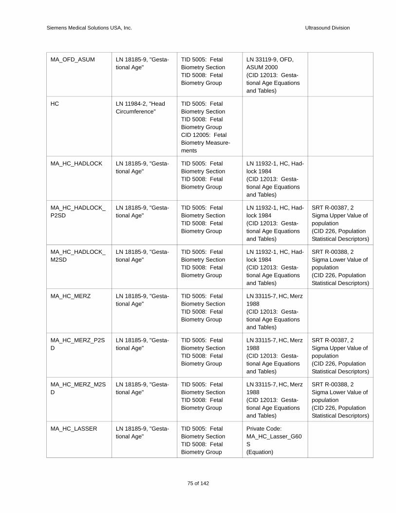
Siemens Medical Solutions USA, Inc. Ultrasound Division
e of
tion tors)
e of
tion tors)
e of
tion tors)
e of
tion tors)
MA_OFD_ASUM LN 18185-9, "Gesta-tional Age"
TID 5005: Fetal Biometry SectionTID 5008: Fetal Biometry Group
LN 33119-9, OFD, ASUM 2000(CID 12013: Gesta-tional Age Equations and Tables)
HC LN 11984-2, "Head Circumference"
TID 5005: Fetal Biometry SectionTID 5008: Fetal Biometry GroupCID 12005: Fetal Biometry Measure-ments
MA_HC_HADLOCK LN 18185-9, "Gesta-tional Age"
TID 5005: Fetal Biometry SectionTID 5008: Fetal Biometry Group
LN 11932-1, HC, Had-lock 1984(CID 12013: Gesta-tional Age Equations and Tables)
MA_HC_HADLOCK_P2SD
LN 18185-9, "Gesta-tional Age"
TID 5005: Fetal Biometry SectionTID 5008: Fetal Biometry Group
LN 11932-1, HC, Had-lock 1984(CID 12013: Gesta-tional Age Equations and Tables)
SRT R-00387, 2 Sigma Upper Valupopulation(CID 226, PopulaStatistical Descrip
MA_HC_HADLOCK_M2SD
LN 18185-9, "Gesta-tional Age"
TID 5005: Fetal Biometry SectionTID 5008: Fetal Biometry Group
LN 11932-1, HC, Had-lock 1984(CID 12013: Gesta-tional Age Equations and Tables)
SRT R-00388, 2 Sigma Lower Valupopulation(CID 226, PopulaStatistical Descrip
MA_HC_MERZ LN 18185-9, "Gesta-tional Age"
TID 5005: Fetal Biometry SectionTID 5008: Fetal Biometry Group
LN 33115-7, HC, Merz 1988(CID 12013: Gesta-tional Age Equations and Tables)
MA_HC_MERZ_P2SD
LN 18185-9, "Gesta-tional Age"
TID 5005: Fetal Biometry SectionTID 5008: Fetal Biometry Group
LN 33115-7, HC, Merz 1988(CID 12013: Gesta-tional Age Equations and Tables)
SRT R-00387, 2 Sigma Upper Valupopulation(CID 226, PopulaStatistical Descrip
MA_HC_MERZ_M2SD
LN 18185-9, "Gesta-tional Age"
TID 5005: Fetal Biometry SectionTID 5008: Fetal Biometry Group
LN 33115-7, HC, Merz 1988(CID 12013: Gesta-tional Age Equations and Tables)
SRT R-00388, 2 Sigma Lower Valupopulation(CID 226, PopulaStatistical Descrip
MA_HC_LASSER LN 18185-9, "Gesta-tional Age"
TID 5005: Fetal Biometry SectionTID 5008: Fetal Biometry Group
Private Code: MA_HC_Lasser_G60S(Equation)
75 of 142
142
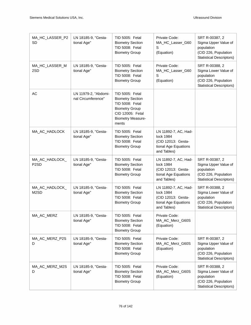
Siemens Medical Solutions USA, Inc. Ultrasound Division
e of
tion tors)
e of
tion tors)
e of
tion tors)
e of
tion tors)
e of
tion tors)
e of
tion tors)
MA_HC_LASSER_P2SD
LN 18185-9, "Gesta-tional Age"
TID 5005: Fetal Biometry SectionTID 5008: Fetal Biometry Group
Private Code: MA_HC_Lasser_G60S(Equation)
SRT R-00387, 2 Sigma Upper Valupopulation(CID 226, PopulaStatistical Descrip
MA_HC_LASSER_M2SD
LN 18185-9, "Gesta-tional Age"
TID 5005: Fetal Biometry SectionTID 5008: Fetal Biometry Group
Private Code: MA_HC_Lasser_G60S(Equation)
SRT R-00388, 2 Sigma Lower Valupopulation(CID 226, PopulaStatistical Descrip
AC LN 11979-2, "Abdomi-nal Circumference"
TID 5005: Fetal Biometry SectionTID 5008: Fetal Biometry GroupCID 12005: Fetal Biometry Measure-ments
MA_AC_HADLOCK LN 18185-9, "Gesta-tional Age"
TID 5005: Fetal Biometry SectionTID 5008: Fetal Biometry Group
LN 11892-7, AC, Had-lock 1984(CID 12013: Gesta-tional Age Equations and Tables)
MA_AC_HADLOCK_P2SD
LN 18185-9, "Gesta-tional Age"
TID 5005: Fetal Biometry SectionTID 5008: Fetal Biometry Group
LN 11892-7, AC, Had-lock 1984(CID 12013: Gesta-tional Age Equations and Tables)
SRT R-00387, 2 Sigma Upper Valupopulation(CID 226, PopulaStatistical Descrip
MA_AC_HADLOCK_M2SD
LN 18185-9, "Gesta-tional Age"
TID 5005: Fetal Biometry SectionTID 5008: Fetal Biometry Group
LN 11892-7, AC, Had-lock 1984(CID 12013: Gesta-tional Age Equations and Tables)
SRT R-00388, 2 Sigma Lower Valupopulation(CID 226, PopulaStatistical Descrip
MA_AC_MERZ LN 18185-9, "Gesta-tional Age"
TID 5005: Fetal Biometry SectionTID 5008: Fetal Biometry Group
Private Code: MA_AC_Merz_G60S(Equation)
MA_AC_MERZ_P2SD
LN 18185-9, "Gesta-tional Age"
TID 5005: Fetal Biometry SectionTID 5008: Fetal Biometry Group
Private Code: MA_AC_Merz_G60S(Equation)
SRT R-00387, 2 Sigma Upper Valupopulation(CID 226, PopulaStatistical Descrip
MA_AC_MERZ_M2SD
LN 18185-9, "Gesta-tional Age"
TID 5005: Fetal Biometry SectionTID 5008: Fetal Biometry Group
Private Code: MA_AC_Merz_G60S(Equation)
SRT R-00388, 2 Sigma Lower Valupopulation(CID 226, PopulaStatistical Descrip
76 of 142
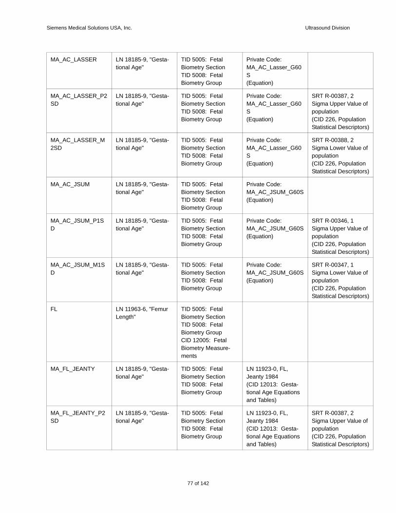
Siemens Medical Solutions USA, Inc. Ultrasound Division
e of
tion tors)
e of
tion tors)
e of
tion tors)
e of
tion tors)
e of
tion tors)
MA_AC_LASSER LN 18185-9, "Gesta-tional Age"
TID 5005: Fetal Biometry SectionTID 5008: Fetal Biometry Group
Private Code: MA_AC_Lasser_G60S(Equation)
MA_AC_LASSER_P2SD
LN 18185-9, "Gesta-tional Age"
TID 5005: Fetal Biometry SectionTID 5008: Fetal Biometry Group
Private Code: MA_AC_Lasser_G60S(Equation)
SRT R-00387, 2 Sigma Upper Valupopulation(CID 226, PopulaStatistical Descrip
MA_AC_LASSER_M2SD
LN 18185-9, "Gesta-tional Age"
TID 5005: Fetal Biometry SectionTID 5008: Fetal Biometry Group
Private Code: MA_AC_Lasser_G60S(Equation)
SRT R-00388, 2 Sigma Lower Valupopulation(CID 226, PopulaStatistical Descrip
MA_AC_JSUM LN 18185-9, "Gesta-tional Age"
TID 5005: Fetal Biometry SectionTID 5008: Fetal Biometry Group
Private Code: MA_AC_JSUM_G60S(Equation)
MA_AC_JSUM_P1SD
LN 18185-9, "Gesta-tional Age"
TID 5005: Fetal Biometry SectionTID 5008: Fetal Biometry Group
Private Code: MA_AC_JSUM_G60S(Equation)
SRT R-00346, 1 Sigma Upper Valupopulation(CID 226, PopulaStatistical Descrip
MA_AC_JSUM_M1SD
LN 18185-9, "Gesta-tional Age"
TID 5005: Fetal Biometry SectionTID 5008: Fetal Biometry Group
Private Code: MA_AC_JSUM_G60S(Equation)
SRT R-00347, 1 Sigma Lower Valupopulation(CID 226, PopulaStatistical Descrip
FL LN 11963-6, "Femur Length"
TID 5005: Fetal Biometry SectionTID 5008: Fetal Biometry GroupCID 12005: Fetal Biometry Measure-ments
MA_FL_JEANTY LN 18185-9, "Gesta-tional Age"
TID 5005: Fetal Biometry SectionTID 5008: Fetal Biometry Group
LN 11923-0, FL, Jeanty 1984(CID 12013: Gesta-tional Age Equations and Tables)
MA_FL_JEANTY_P2SD
LN 18185-9, "Gesta-tional Age"
TID 5005: Fetal Biometry SectionTID 5008: Fetal Biometry Group
LN 11923-0, FL, Jeanty 1984(CID 12013: Gesta-tional Age Equations and Tables)
SRT R-00387, 2 Sigma Upper Valupopulation(CID 226, PopulaStatistical Descrip
77 of 142
142
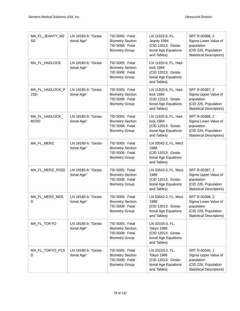
Siemens Medical Solutions USA, Inc. Ultrasound Division
e of
tion tors)
e of
tion tors)
e of
tion tors)
e of
tion tors)
e of
tion tors)
e of
tion tors)
MA_FL_JEANTY_M2SD
LN 18185-9, "Gesta-tional Age"
TID 5005: Fetal Biometry SectionTID 5008: Fetal Biometry Group
LN 11923-0, FL, Jeanty 1984(CID 12013: Gesta-tional Age Equations and Tables)
SRT R-00388, 2 Sigma Lower Valupopulation(CID 226, PopulaStatistical Descrip
MA_FL_HADLOCK LN 18185-9, "Gesta-tional Age"
TID 5005: Fetal Biometry SectionTID 5008: Fetal Biometry Group
LN 11920-6, FL, Had-lock 1984(CID 12013: Gesta-tional Age Equations and Tables)
MA_FL_HADLOCK_P2SD
LN 18185-9, "Gesta-tional Age"
TID 5005: Fetal Biometry SectionTID 5008: Fetal Biometry Group
LN 11920-6, FL, Had-lock 1984(CID 12013: Gesta-tional Age Equations and Tables)
SRT R-00387, 2 Sigma Upper Valupopulation(CID 226, PopulaStatistical Descrip
MA_FL_HADLOCK_M2SD
LN 18185-9, "Gesta-tional Age"
TID 5005: Fetal Biometry SectionTID 5008: Fetal Biometry Group
LN 11920-6, FL, Had-lock 1984(CID 12013: Gesta-tional Age Equations and Tables)
SRT R-00388, 2 Sigma Lower Valupopulation(CID 226, PopulaStatistical Descrip
MA_FL_MERZ LN 18185-9, "Gesta-tional Age"
TID 5005: Fetal Biometry SectionTID 5008: Fetal Biometry Group
LN 33542-2, FL, Merz 1988(CID 12013: Gesta-tional Age Equations and Tables)
MA_FL_MERZ_P2SD LN 18185-9, "Gesta-tional Age"
TID 5005: Fetal Biometry SectionTID 5008: Fetal Biometry Group
LN 33542-2, FL, Merz 1988(CID 12013: Gesta-tional Age Equations and Tables)
SRT R-00387, 2 Sigma Upper Valupopulation(CID 226, PopulaStatistical Descrip
MA_FL_MERZ_M2SD
LN 18185-9, "Gesta-tional Age"
TID 5005: Fetal Biometry SectionTID 5008: Fetal Biometry Group
LN 33542-2, FL, Merz 1988(CID 12013: Gesta-tional Age Equations and Tables)
SRT R-00388, 2 Sigma Lower Valupopulation(CID 226, PopulaStatistical Descrip
MA_FL_TOKYO LN 18185-9, "Gesta-tional Age"
TID 5005: Fetal Biometry SectionTID 5008: Fetal Biometry Group
LN 33103-3, FL, Tokyo 1986(CID 12013: Gesta-tional Age Equations and Tables)
MA_FL_TOKYO_P1SD
LN 18185-9, "Gesta-tional Age"
TID 5005: Fetal Biometry SectionTID 5008: Fetal Biometry Group
LN 33103-3, FL, Tokyo 1986(CID 12013: Gesta-tional Age Equations and Tables)
SRT R-00346, 1 Sigma Upper Valupopulation(CID 226, PopulaStatistical Descrip
78 of 142
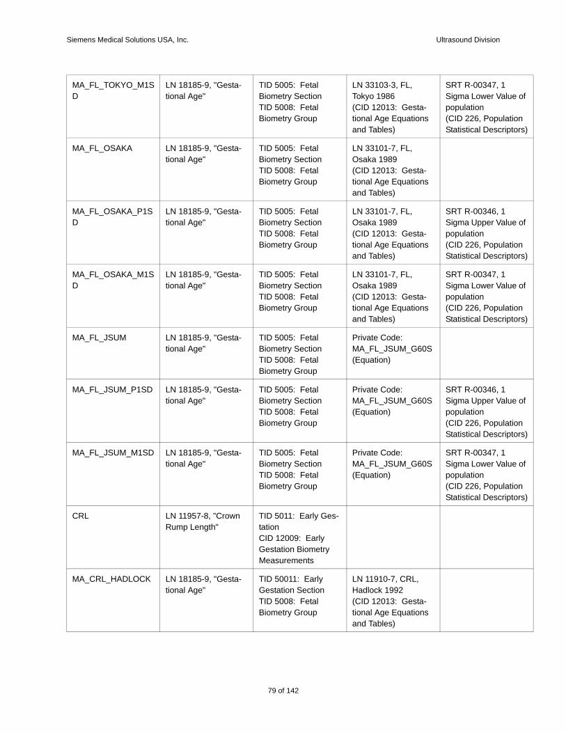
Siemens Medical Solutions USA, Inc. Ultrasound Division
e of
tion tors)
e of
tion tors)
e of
tion tors)
e of
tion tors)
e of
tion tors)
MA_FL_TOKYO_M1SD
LN 18185-9, "Gesta-tional Age"
TID 5005: Fetal Biometry SectionTID 5008: Fetal Biometry Group
LN 33103-3, FL, Tokyo 1986(CID 12013: Gesta-tional Age Equations and Tables)
SRT R-00347, 1 Sigma Lower Valupopulation(CID 226, PopulaStatistical Descrip
MA_FL_OSAKA LN 18185-9, "Gesta-tional Age"
TID 5005: Fetal Biometry SectionTID 5008: Fetal Biometry Group
LN 33101-7, FL, Osaka 1989(CID 12013: Gesta-tional Age Equations and Tables)
MA_FL_OSAKA_P1SD
LN 18185-9, "Gesta-tional Age"
TID 5005: Fetal Biometry SectionTID 5008: Fetal Biometry Group
LN 33101-7, FL, Osaka 1989(CID 12013: Gesta-tional Age Equations and Tables)
SRT R-00346, 1 Sigma Upper Valupopulation(CID 226, PopulaStatistical Descrip
MA_FL_OSAKA_M1SD
LN 18185-9, "Gesta-tional Age"
TID 5005: Fetal Biometry SectionTID 5008: Fetal Biometry Group
LN 33101-7, FL, Osaka 1989(CID 12013: Gesta-tional Age Equations and Tables)
SRT R-00347, 1 Sigma Lower Valupopulation(CID 226, PopulaStatistical Descrip
MA_FL_JSUM LN 18185-9, "Gesta-tional Age"
TID 5005: Fetal Biometry SectionTID 5008: Fetal Biometry Group
Private Code: MA_FL_JSUM_G60S(Equation)
MA_FL_JSUM_P1SD LN 18185-9, "Gesta-tional Age"
TID 5005: Fetal Biometry SectionTID 5008: Fetal Biometry Group
Private Code: MA_FL_JSUM_G60S(Equation)
SRT R-00346, 1 Sigma Upper Valupopulation(CID 226, PopulaStatistical Descrip
MA_FL_JSUM_M1SD LN 18185-9, "Gesta-tional Age"
TID 5005: Fetal Biometry SectionTID 5008: Fetal Biometry Group
Private Code: MA_FL_JSUM_G60S(Equation)
SRT R-00347, 1 Sigma Lower Valupopulation(CID 226, PopulaStatistical Descrip
CRL LN 11957-8, "Crown Rump Length"
TID 5011: Early Ges-tationCID 12009: Early Gestation Biometry Measurements
MA_CRL_HADLOCK LN 18185-9, "Gesta-tional Age"
TID 50011: Early Gestation SectionTID 5008: Fetal Biometry Group
LN 11910-7, CRL, Hadlock 1992(CID 12013: Gesta-tional Age Equations and Tables)
79 of 142
142
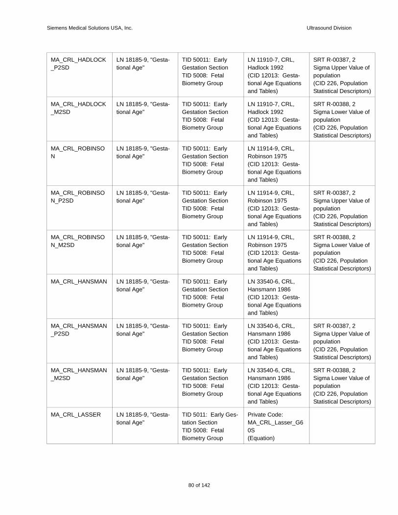
Siemens Medical Solutions USA, Inc. Ultrasound Division
e of
tion tors)
e of
tion tors)
e of
tion tors)
e of
tion tors)
e of
tion tors)
e of
tion tors)
MA_CRL_HADLOCK_P2SD
LN 18185-9, "Gesta-tional Age"
TID 50011: Early Gestation SectionTID 5008: Fetal Biometry Group
LN 11910-7, CRL, Hadlock 1992(CID 12013: Gesta-tional Age Equations and Tables)
SRT R-00387, 2 Sigma Upper Valupopulation(CID 226, PopulaStatistical Descrip
MA_CRL_HADLOCK_M2SD
LN 18185-9, "Gesta-tional Age"
TID 50011: Early Gestation SectionTID 5008: Fetal Biometry Group
LN 11910-7, CRL, Hadlock 1992(CID 12013: Gesta-tional Age Equations and Tables)
SRT R-00388, 2 Sigma Lower Valupopulation(CID 226, PopulaStatistical Descrip
MA_CRL_ROBINSON
LN 18185-9, "Gesta-tional Age"
TID 50011: Early Gestation SectionTID 5008: Fetal Biometry Group
LN 11914-9, CRL, Robinson 1975(CID 12013: Gesta-tional Age Equations and Tables)
MA_CRL_ROBINSON_P2SD
LN 18185-9, "Gesta-tional Age"
TID 50011: Early Gestation SectionTID 5008: Fetal Biometry Group
LN 11914-9, CRL, Robinson 1975(CID 12013: Gesta-tional Age Equations and Tables)
SRT R-00387, 2 Sigma Upper Valupopulation(CID 226, PopulaStatistical Descrip
MA_CRL_ROBINSON_M2SD
LN 18185-9, "Gesta-tional Age"
TID 50011: Early Gestation SectionTID 5008: Fetal Biometry Group
LN 11914-9, CRL, Robinson 1975(CID 12013: Gesta-tional Age Equations and Tables)
SRT R-00388, 2 Sigma Lower Valupopulation(CID 226, PopulaStatistical Descrip
MA_CRL_HANSMAN LN 18185-9, "Gesta-tional Age"
TID 50011: Early Gestation SectionTID 5008: Fetal Biometry Group
LN 33540-6, CRL, Hansmann 1986(CID 12013: Gesta-tional Age Equations and Tables)
MA_CRL_HANSMAN_P2SD
LN 18185-9, "Gesta-tional Age"
TID 50011: Early Gestation SectionTID 5008: Fetal Biometry Group
LN 33540-6, CRL, Hansmann 1986(CID 12013: Gesta-tional Age Equations and Tables)
SRT R-00387, 2 Sigma Upper Valupopulation(CID 226, PopulaStatistical Descrip
MA_CRL_HANSMAN_M2SD
LN 18185-9, "Gesta-tional Age"
TID 50011: Early Gestation SectionTID 5008: Fetal Biometry Group
LN 33540-6, CRL, Hansmann 1986(CID 12013: Gesta-tional Age Equations and Tables)
SRT R-00388, 2 Sigma Lower Valupopulation(CID 226, PopulaStatistical Descrip
MA_CRL_LASSER LN 18185-9, "Gesta-tional Age"
TID 5011: Early Ges-tation SectionTID 5008: Fetal Biometry Group
Private Code: MA_CRL_Lasser_G60S(Equation)
80 of 142
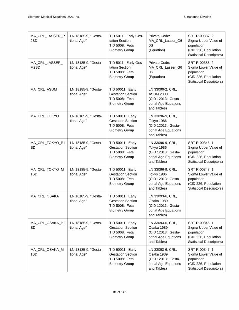
Siemens Medical Solutions USA, Inc. Ultrasound Division
e of
tion tors)
e of
tion tors)
e of
tion tors)
e of
tion tors)
e of
tion tors)
e of
tion tors)
MA_CRL_LASSER_P2SD
LN 18185-9, "Gesta-tional Age"
TID 5011: Early Ges-tation SectionTID 5008: Fetal Biometry Group
Private Code: MA_CRL_Lasser_G60S(Equation)
SRT R-00387, 2 Sigma Upper Valupopulation(CID 226, PopulaStatistical Descrip
MA_CRL_LASSER_M2SD
LN 18185-9, "Gesta-tional Age"
TID 5011: Early Ges-tation SectionTID 5008: Fetal Biometry Group
Private Code: MA_CRL_Lasser_G60S(Equation)
SRT R-00388, 2 Sigma Lower Valupopulation(CID 226, PopulaStatistical Descrip
MA_CRL_ASUM LN 18185-9, "Gesta-tional Age"
TID 50011: Early Gestation SectionTID 5008: Fetal Biometry Group
LN 33090-2, CRL, ASUM 2000(CID 12013: Gesta-tional Age Equations and Tables)
MA_CRL_TOKYO LN 18185-9, "Gesta-tional Age"
TID 50011: Early Gestation SectionTID 5008: Fetal Biometry Group
LN 33096-9, CRL, Tokyo 1986(CID 12013: Gesta-tional Age Equations and Tables)
MA_CRL_TOKYO_P1SD
LN 18185-9, "Gesta-tional Age"
TID 50011: Early Gestation SectionTID 5008: Fetal Biometry Group
LN 33096-9, CRL, Tokyo 1986(CID 12013: Gesta-tional Age Equations and Tables)
SRT R-00346, 1 Sigma Upper Valupopulation(CID 226, PopulaStatistical Descrip
MA_CRL_TOKYO_M1SD
LN 18185-9, "Gesta-tional Age"
TID 50011: Early Gestation SectionTID 5008: Fetal Biometry Group
LN 33096-9, CRL, Tokyo 1986(CID 12013: Gesta-tional Age Equations and Tables)
SRT R-00347, 1 Sigma Lower Valupopulation(CID 226, PopulaStatistical Descrip
MA_CRL_OSAKA LN 18185-9, "Gesta-tional Age"
TID 50011: Early Gestation SectionTID 5008: Fetal Biometry Group
LN 33093-6, CRL, Osaka 1989(CID 12013: Gesta-tional Age Equations and Tables)
MA_CRL_OSAKA_P1SD
LN 18185-9, "Gesta-tional Age"
TID 50011: Early Gestation SectionTID 5008: Fetal Biometry Group
LN 33093-6, CRL, Osaka 1989(CID 12013: Gesta-tional Age Equations and Tables)
SRT R-00346, 1 Sigma Upper Valupopulation(CID 226, PopulaStatistical Descrip
MA_CRL_OSAKA_M1SD
LN 18185-9, "Gesta-tional Age"
TID 50011: Early Gestation SectionTID 5008: Fetal Biometry Group
LN 33093-6, CRL, Osaka 1989(CID 12013: Gesta-tional Age Equations and Tables)
SRT R-00347, 1 Sigma Lower Valupopulation(CID 226, PopulaStatistical Descrip
81 of 142
142
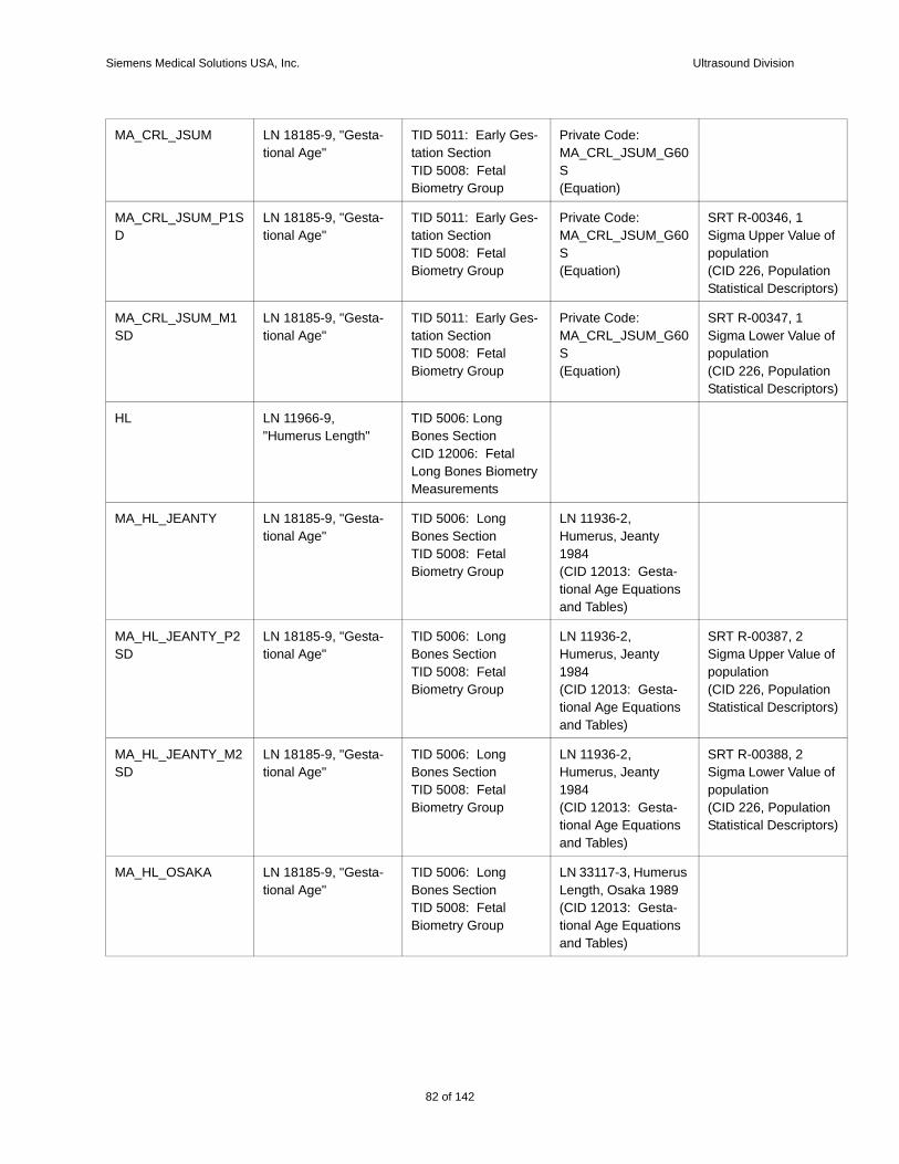
Siemens Medical Solutions USA, Inc. Ultrasound Division
e of
tion tors)
e of
tion tors)
e of
tion tors)
e of
tion tors)
MA_CRL_JSUM LN 18185-9, "Gesta-tional Age"
TID 5011: Early Ges-tation SectionTID 5008: Fetal Biometry Group
Private Code: MA_CRL_JSUM_G60S(Equation)
MA_CRL_JSUM_P1SD
LN 18185-9, "Gesta-tional Age"
TID 5011: Early Ges-tation SectionTID 5008: Fetal Biometry Group
Private Code: MA_CRL_JSUM_G60S(Equation)
SRT R-00346, 1 Sigma Upper Valupopulation(CID 226, PopulaStatistical Descrip
MA_CRL_JSUM_M1SD
LN 18185-9, "Gesta-tional Age"
TID 5011: Early Ges-tation SectionTID 5008: Fetal Biometry Group
Private Code: MA_CRL_JSUM_G60S(Equation)
SRT R-00347, 1 Sigma Lower Valupopulation(CID 226, PopulaStatistical Descrip
HL LN 11966-9, "Humerus Length"
TID 5006: Long Bones SectionCID 12006: Fetal Long Bones Biometry Measurements
MA_HL_JEANTY LN 18185-9, "Gesta-tional Age"
TID 5006: Long Bones SectionTID 5008: Fetal Biometry Group
LN 11936-2, Humerus, Jeanty 1984(CID 12013: Gesta-tional Age Equations and Tables)
MA_HL_JEANTY_P2SD
LN 18185-9, "Gesta-tional Age"
TID 5006: Long Bones SectionTID 5008: Fetal Biometry Group
LN 11936-2, Humerus, Jeanty 1984(CID 12013: Gesta-tional Age Equations and Tables)
SRT R-00387, 2 Sigma Upper Valupopulation(CID 226, PopulaStatistical Descrip
MA_HL_JEANTY_M2SD
LN 18185-9, "Gesta-tional Age"
TID 5006: Long Bones SectionTID 5008: Fetal Biometry Group
LN 11936-2, Humerus, Jeanty 1984(CID 12013: Gesta-tional Age Equations and Tables)
SRT R-00388, 2 Sigma Lower Valupopulation(CID 226, PopulaStatistical Descrip
MA_HL_OSAKA LN 18185-9, "Gesta-tional Age"
TID 5006: Long Bones SectionTID 5008: Fetal Biometry Group
LN 33117-3, Humerus Length, Osaka 1989(CID 12013: Gesta-tional Age Equations and Tables)
82 of 142
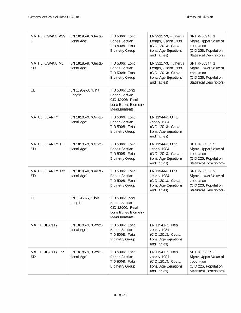
Siemens Medical Solutions USA, Inc. Ultrasound Division
e of
tion tors)
e of
tion tors)
e of
tion tors)
e of
tion tors)
e of
tion tors)
MA_HL_OSAKA_P1SD
LN 18185-9, "Gesta-tional Age"
TID 5006: Long Bones SectionTID 5008: Fetal Biometry Group
LN 33117-3, Humerus Length, Osaka 1989(CID 12013: Gesta-tional Age Equations and Tables)
SRT R-00346, 1 Sigma Upper Valupopulation(CID 226, PopulaStatistical Descrip
MA_HL_OSAKA_M1SD
LN 18185-9, "Gesta-tional Age"
TID 5006: Long Bones SectionTID 5008: Fetal Biometry Group
LN 33117-3, Humerus Length, Osaka 1989(CID 12013: Gesta-tional Age Equations and Tables)
SRT R-00347, 1 Sigma Lower Valupopulation(CID 226, PopulaStatistical Descrip
UL LN 11969-3, "Ulna Length"
TID 5006: Long Bones SectionCID 12006: Fetal Long Bones Biometry Measurements
MA_UL_JEANTY LN 18185-9, "Gesta-tional Age"
TID 5006: Long Bones SectionTID 5008: Fetal Biometry Group
LN 11944-6, Ulna, Jeanty 1984(CID 12013: Gesta-tional Age Equations and Tables)
MA_UL_JEANTY_P2SD
LN 18185-9, "Gesta-tional Age"
TID 5006: Long Bones SectionTID 5008: Fetal Biometry Group
LN 11944-6, Ulna, Jeanty 1984(CID 12013: Gesta-tional Age Equations and Tables)
SRT R-00387, 2 Sigma Upper Valupopulation(CID 226, PopulaStatistical Descrip
MA_UL_JEANTY_M2SD
LN 18185-9, "Gesta-tional Age"
TID 5006: Long Bones SectionTID 5008: Fetal Biometry Group
LN 11944-6, Ulna, Jeanty 1984(CID 12013: Gesta-tional Age Equations and Tables)
SRT R-00388, 2 Sigma Lower Valupopulation(CID 226, PopulaStatistical Descrip
TL LN 11968-5, "Tibia Length"
TID 5006: Long Bones SectionCID 12006: Fetal Long Bones Biometry Measurements
MA_TL_JEANTY LN 18185-9, "Gesta-tional Age"
TID 5006: Long Bones SectionTID 5008: Fetal Biometry Group
LN 11941-2, Tibia, Jeanty 1984(CID 12013: Gesta-tional Age Equations and Tables)
MA_TL_JEANTY_P2SD
LN 18185-9, "Gesta-tional Age"
TID 5006: Long Bones SectionTID 5008: Fetal Biometry Group
LN 11941-2, Tibia, Jeanty 1984(CID 12013: Gesta-tional Age Equations and Tables)
SRT R-00387, 2 Sigma Upper Valupopulation(CID 226, PopulaStatistical Descrip
83 of 142
142
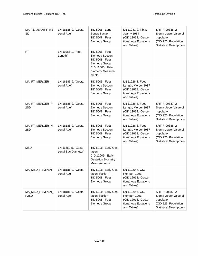
Siemens Medical Solutions USA, Inc. Ultrasound Division
e of
tion tors)
e of
tion tors)
e of
tion tors)
e of
tion tors)
MA_TL_JEANTY_M2SD
LN 18185-9, "Gesta-tional Age"
TID 5006: Long Bones SectionTID 5008: Fetal Biometry Group
LN 11941-2, Tibia, Jeanty 1984(CID 12013: Gesta-tional Age Equations and Tables)
SRT R-00388, 2 Sigma Lower Valupopulation(CID 226, PopulaStatistical Descrip
FT LN 11965-1, "Foot Length"
TID 5005: Fetal Biometry SectionTID 5008: Fetal Biometry GroupCID 12005: Fetal Biometry Measure-ments
MA_FT_MERCER LN 18185-9, "Gesta-tional Age"
TID 5005: Fetal Biometry SectionTID 5008: Fetal Biometry Group
LN 11926-3, Foot Length, Mercer 1987(CID 12013: Gesta-tional Age Equations and Tables)
MA_FT_MERCER_P2SD
LN 18185-9, "Gesta-tional Age"
TID 5005: Fetal Biometry SectionTID 5008: Fetal Biometry Group
LN 11926-3, Foot Length, Mercer 1987(CID 12013: Gesta-tional Age Equations and Tables)
SRT R-00387, 2 Sigma Upper Valupopulation(CID 226, PopulaStatistical Descrip
MA_FT_MERCER_M2SD
LN 18185-9, "Gesta-tional Age"
TID 5005: Fetal Biometry SectionTID 5008: Fetal Biometry Group
LN 11926-3, Foot Length, Mercer 1987(CID 12013: Gesta-tional Age Equations and Tables)
SRT R-00388, 2 Sigma Lower Valupopulation(CID 226, PopulaStatistical Descrip
MSD LN 11850-5, "Gesta-tional Sac Diameter"
TID 5011: Early Ges-tationCID 12009: Early Gestation Biometry Measurements
MA_MSD_REMPEN LN 18185-9, "Gesta-tional Age"
TID 5011: Early Ges-tation SectionTID 5008: Fetal Biometry Group
LN 11929-7, GS, Rempen 1991(CID 12013: Gesta-tional Age Equations and Tables)
MA_MSD_REMPEN_P2SD
LN 18185-9, "Gesta-tional Age"
TID 5011: Early Ges-tation SectionTID 5008: Fetal Biometry Group
LN 11929-7, GS, Rempen 1991(CID 12013: Gesta-tional Age Equations and Tables)
SRT R-00387, 2 Sigma Upper Valupopulation(CID 226, PopulaStatistical Descrip
84 of 142
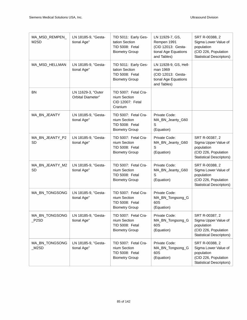
Siemens Medical Solutions USA, Inc. Ultrasound Division
e of
tion tors)
e of
tion tors)
e of
tion tors)
e of
tion tors)
e of
tion tors)
MA_MSD_REMPEN_M2SD
LN 18185-9, "Gesta-tional Age"
TID 5011: Early Ges-tation SectionTID 5008: Fetal Biometry Group
LN 11929-7, GS, Rempen 1991(CID 12013: Gesta-tional Age Equations and Tables)
SRT R-00388, 2 Sigma Lower Valupopulation(CID 226, PopulaStatistical Descrip
MA_MSD_HELLMAN LN 18185-9, "Gesta-tional Age"
TID 5011: Early Ges-tation SectionTID 5008: Fetal Biometry Group
LN 11928-9, GS, Hell-man 1969(CID 12013: Gesta-tional Age Equations and Tables)
BN LN 11629-3, "Outer Orbital Diameter"
TID 5007: Fetal Cra-nium SectionCID 12007: Fetal Cranium
MA_BN_JEANTY LN 18185-9, "Gesta-tional Age"
TID 5007: Fetal Cra-nium SectionTID 5008: Fetal Biometry Group
Private Code: MA_BN_Jeanty_G60S(Equation)
MA_BN_JEANTY_P2SD
LN 18185-9, "Gesta-tional Age"
TID 5007: Fetal Cra-nium SectionTID 5008: Fetal Biometry Group
Private Code: MA_BN_Jeanty_G60S(Equation)
SRT R-00387, 2 Sigma Upper Valupopulation(CID 226, PopulaStatistical Descrip
MA_BN_JEANTY_M2SD
LN 18185-9, "Gesta-tional Age"
TID 5007: Fetal Cra-nium SectionTID 5008: Fetal Biometry Group
Private Code: MA_BN_Jeanty_G60S(Equation)
SRT R-00388, 2 Sigma Lower Valupopulation(CID 226, PopulaStatistical Descrip
MA_BN_TONGSONG LN 18185-9, "Gesta-tional Age"
TID 5007: Fetal Cra-nium SectionTID 5008: Fetal Biometry Group
Private Code: MA_BN_Tongsong_G60S(Equation)
MA_BN_TONGSONG_P2SD
LN 18185-9, "Gesta-tional Age"
TID 5007: Fetal Cra-nium SectionTID 5008: Fetal Biometry Group
Private Code: MA_BN_Tongsong_G60S(Equation)
SRT R-00387, 2 Sigma Upper Valupopulation(CID 226, PopulaStatistical Descrip
MA_BN_TONGSONG_M2SD
LN 18185-9, "Gesta-tional Age"
TID 5007: Fetal Cra-nium SectionTID 5008: Fetal Biometry Group
Private Code: MA_BN_Tongsong_G60S(Equation)
SRT R-00388, 2 Sigma Lower Valupopulation(CID 226, PopulaStatistical Descrip
85 of 142
142

Siemens Medical Solutions USA, Inc. Ultrasound Division
e of
tion tors)
e of
tion tors)
ASD LN 11818-2, "Anterior-Posterior Abdominal Diameter"
TID 5005: Fetal Biometry SectionTID 5008: Fetal Biometry GroupCID 12005: Fetal Biometry Measure-ments
MA_ASD_MERZ LN 18185-9, "Gesta-tional Age"
TID 5005: Fetal Biometry SectionTID 5008: Fetal Biometry Group
Private Code: MA_ASD_Merz_G60S(Equation)
ATD LN 11862-0, "Tran-verse Abdominal Diameter"
TID 5005: Fetal Biometry SectionTID 5008: Fetal Biometry GroupCID 12005: Fetal Biometry Measure-ments
MA_ATD_MERZ LN 18185-9, "Gesta-tional Age"
TID 5005: Fetal Biometry SectionTID 5008: Fetal Biometry Group
Private Code: MA_ATD_Merz_G60S(Equation)
GS LN 11850-5, "Gesta-tional Sac Diameter"
TID 5011: Early Ges-tational SectionTID 5008: Fetal Biometry Group
MA_GS_TOKYO LN 18185-9, "Gesta-tional Age"
TID 5011: Early Ges-tational SectionTID 5008: Fetal Biometry Group
LN 33108-2, GS, Tokyo 1986
MA_GS_TOKYO_P1SD
LN 18185-9, "Gesta-tional Age"
TID 5011: Early Ges-tational SectionTID 5008: Fetal Biometry Group
LN 33108-2, GS, Tokyo 1986
SRT R-00346, 1 Sigma Upper Valupopulation(CID 226, PopulaStatistical Descrip
MA_GS_TOKYO_M1SD
LN 18185-9, "Gesta-tional Age"
TID 5011: Early Ges-tational SectionTID 5008: Fetal Biometry Group
LN 33108-2, GS, Tokyo 1986
SRT R-00347, 1 Sigma Lower Valupopulation(CID 226, PopulaStatistical Descrip
86 of 142
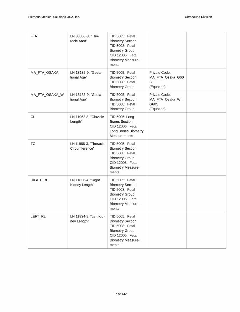
Siemens Medical Solutions USA, Inc. Ultrasound Division
FTA LN 33068-8, "Tho-racic Area"
TID 5005: Fetal Biometry SectionTID 5008: Fetal Biometry GroupCID 12005: Fetal Biometry Measure-ments
MA_FTA_OSAKA LN 18185-9, "Gesta-tional Age"
TID 5005: Fetal Biometry SectionTID 5008: Fetal Biometry Group
Private Code: MA_FTA_Osaka_G60S(Equation)
MA_FTA_OSAKA_W LN 18185-9, "Gesta-tional Age"
TID 5005: Fetal Biometry SectionTID 5008: Fetal Biometry Group
Private Code: MA_FTA_Osaka_W_G60S(Equation)
CL LN 11962-8, "Clavicle Length"
TID 5006: Long Bones SectionCID 12006: Fetal Long Bones Biometry Measurements
TC LN 11988-3, "Thoracic Circumference"
TID 5005: Fetal Biometry SectionTID 5008: Fetal Biometry GroupCID 12005: Fetal Biometry Measure-ments
RIGHT_RL LN 11836-4, "Right Kidney Length"
TID 5005: Fetal Biometry SectionTID 5008: Fetal Biometry GroupCID 12005: Fetal Biometry Measure-ments
LEFT_RL LN 11834-9, "Left Kid-ney Length"
TID 5005: Fetal Biometry SectionTID 5008: Fetal Biometry GroupCID 12005: Fetal Biometry Measure-ments
87 of 142
142
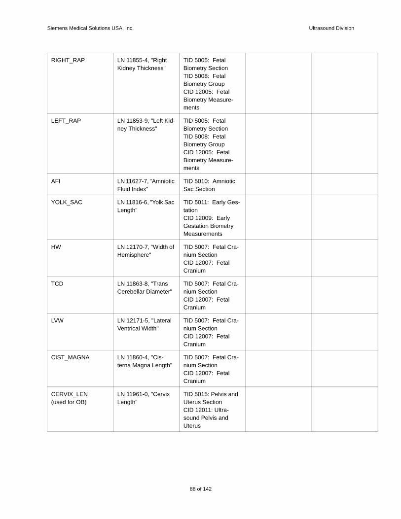
Siemens Medical Solutions USA, Inc. Ultrasound Division
RIGHT_RAP LN 11855-4, "Right Kidney Thickness"
TID 5005: Fetal Biometry SectionTID 5008: Fetal Biometry GroupCID 12005: Fetal Biometry Measure-ments
LEFT_RAP LN 11853-9, "Left Kid-ney Thickness"
TID 5005: Fetal Biometry SectionTID 5008: Fetal Biometry GroupCID 12005: Fetal Biometry Measure-ments
AFI LN 11627-7, "Amniotic Fluid Index"
TID 5010: Amniotic Sac Section
YOLK_SAC LN 11816-6, "Yolk Sac Length"
TID 5011: Early Ges-tationCID 12009: Early Gestation Biometry Measurements
HW LN 12170-7, "Width of Hemisphere"
TID 5007: Fetal Cra-nium SectionCID 12007: Fetal Cranium
TCD LN 11863-8, "Trans Cerebellar Diameter"
TID 5007: Fetal Cra-nium SectionCID 12007: Fetal Cranium
LVW LN 12171-5, "Lateral Ventrical Width"
TID 5007: Fetal Cra-nium SectionCID 12007: Fetal Cranium
CIST_MAGNA LN 11860-4, "Cis-terna Magna Length"
TID 5007: Fetal Cra-nium SectionCID 12007: Fetal Cranium
CERVIX_LEN(used for OB)
LN 11961-0, "Cervix Length"
TID 5015: Pelvis and Uterus SectionCID 12011: Ultra-sound Pelvis and Uterus
88 of 142
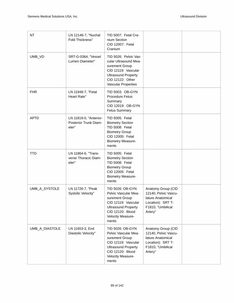
Siemens Medical Solutions USA, Inc. Ultrasound Division
NT LN 12146-7, "Nuchal Fold Thickness"
TID 5007: Fetal Cra-nium SectionCID 12007: Fetal Cranium
UMB_VD SRT-G-0364, "Vessel Lumen Diameter"
TID 5026: Pelvic Vas-cular Ultrasound Mea-surement GroupCID 12119: Vascular Ultrasound PropertyCID 12122: Other Vascular Properties
FHR LN 11948-7, "Fetal Heart Rate"
TID 5003: OB-GYN Procedure Fetus SummaryCID 12019: OB-GYN Fetus Summary
APTD LN 11819-0, "Anterior-Posterior Trunk Diam-eter"
TID 5005: Fetal Biometry SectionTID 5008: Fetal Biometry GroupCID 12005: Fetal Biometry Measure-ments
TTD LN 11864-6, "Trans-verse Thoracic Diam-eter"
TID 5005: Fetal Biometry SectionTID 5008: Fetal Biometry GroupCID 12005: Fetal Biometry Measure-ments
UMB_A_SYSTOLE LN 11726-7, "Peak Systolic Velocity"
TID 5026: OB-GYN Pelvic Vascular Mea-surement GroupCID 12119: Vascular Ultrasound PropertyCID 12120: Blood Velocity Measure-ments
Anatomy Group (CID 12140, Pelvic Vascu-lature Anatomical Location): SRT T-F1810, "Umbilical Artery"
UMB_A_DIASTOLE LN 11653-3, End Diastolic Velocity"
TID 5026: OB-GYN Pelvic Vascular Mea-surement GroupCID 12119: Vascular Ultrasound PropertyCID 12120: Blood Velocity Measure-ments
Anatomy Group (CID 12140, Pelvic Vascu-lature Anatomical Location): SRT T-F1810, "Umbilical Artery"
89 of 142
142
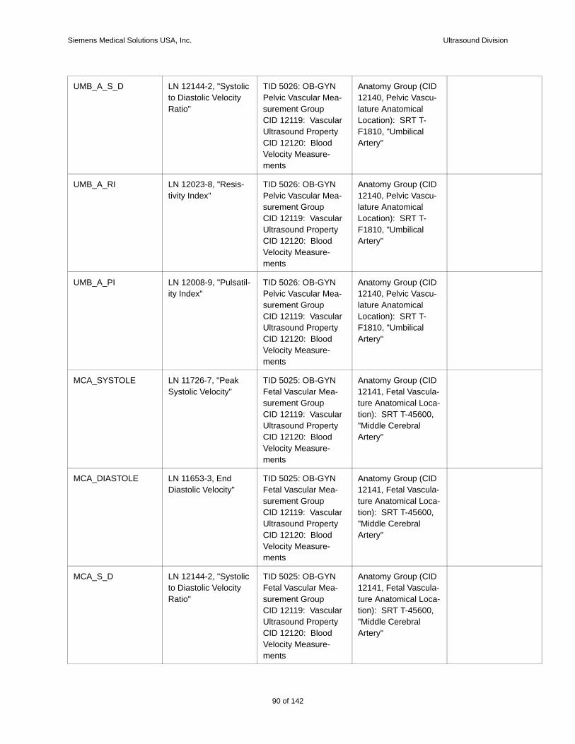
Siemens Medical Solutions USA, Inc. Ultrasound Division
UMB_A_S_D LN 12144-2, "Systolic to Diastolic Velocity Ratio"
TID 5026: OB-GYN Pelvic Vascular Mea-surement GroupCID 12119: Vascular Ultrasound PropertyCID 12120: Blood Velocity Measure-ments
Anatomy Group (CID 12140, Pelvic Vascu-lature Anatomical Location): SRT T-F1810, "Umbilical Artery"
UMB_A_RI LN 12023-8, "Resis-tivity Index"
TID 5026: OB-GYN Pelvic Vascular Mea-surement GroupCID 12119: Vascular Ultrasound PropertyCID 12120: Blood Velocity Measure-ments
Anatomy Group (CID 12140, Pelvic Vascu-lature Anatomical Location): SRT T-F1810, "Umbilical Artery"
UMB_A_PI LN 12008-9, "Pulsatil-ity Index"
TID 5026: OB-GYN Pelvic Vascular Mea-surement GroupCID 12119: Vascular Ultrasound PropertyCID 12120: Blood Velocity Measure-ments
Anatomy Group (CID 12140, Pelvic Vascu-lature Anatomical Location): SRT T-F1810, "Umbilical Artery"
MCA_SYSTOLE LN 11726-7, "Peak Systolic Velocity"
TID 5025: OB-GYN Fetal Vascular Mea-surement GroupCID 12119: Vascular Ultrasound PropertyCID 12120: Blood Velocity Measure-ments
Anatomy Group (CID 12141, Fetal Vascula-ture Anatomical Loca-tion): SRT T-45600, "Middle Cerebral Artery"
MCA_DIASTOLE LN 11653-3, End Diastolic Velocity"
TID 5025: OB-GYN Fetal Vascular Mea-surement GroupCID 12119: Vascular Ultrasound PropertyCID 12120: Blood Velocity Measure-ments
Anatomy Group (CID 12141, Fetal Vascula-ture Anatomical Loca-tion): SRT T-45600, "Middle Cerebral Artery"
MCA_S_D LN 12144-2, "Systolic to Diastolic Velocity Ratio"
TID 5025: OB-GYN Fetal Vascular Mea-surement GroupCID 12119: Vascular Ultrasound PropertyCID 12120: Blood Velocity Measure-ments
Anatomy Group (CID 12141, Fetal Vascula-ture Anatomical Loca-tion): SRT T-45600, "Middle Cerebral Artery"
90 of 142
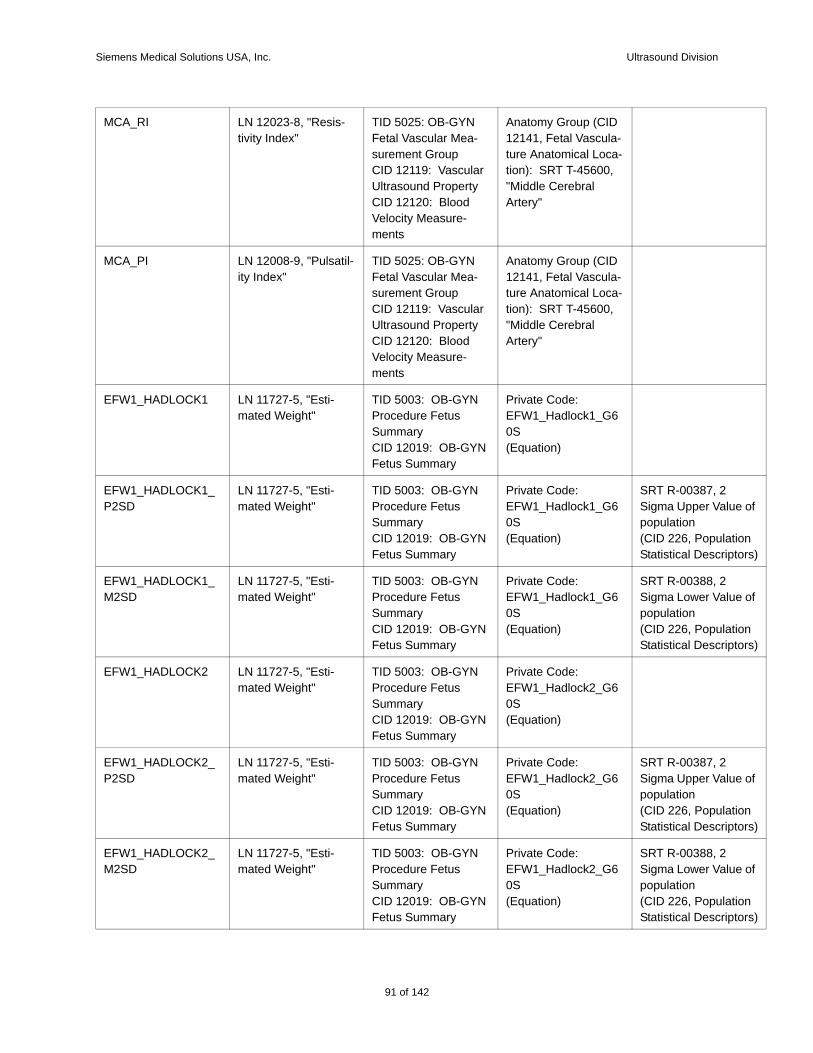
Siemens Medical Solutions USA, Inc. Ultrasound Division
e of
tion tors)
e of
tion tors)
e of
tion tors)
e of
tion tors)
MCA_RI LN 12023-8, "Resis-tivity Index"
TID 5025: OB-GYN Fetal Vascular Mea-surement GroupCID 12119: Vascular Ultrasound PropertyCID 12120: Blood Velocity Measure-ments
Anatomy Group (CID 12141, Fetal Vascula-ture Anatomical Loca-tion): SRT T-45600, "Middle Cerebral Artery"
MCA_PI LN 12008-9, "Pulsatil-ity Index"
TID 5025: OB-GYN Fetal Vascular Mea-surement GroupCID 12119: Vascular Ultrasound PropertyCID 12120: Blood Velocity Measure-ments
Anatomy Group (CID 12141, Fetal Vascula-ture Anatomical Loca-tion): SRT T-45600, "Middle Cerebral Artery"
EFW1_HADLOCK1 LN 11727-5, "Esti-mated Weight"
TID 5003: OB-GYN Procedure Fetus SummaryCID 12019: OB-GYN Fetus Summary
Private Code: EFW1_Hadlock1_G60S(Equation)
EFW1_HADLOCK1_P2SD
LN 11727-5, "Esti-mated Weight"
TID 5003: OB-GYN Procedure Fetus SummaryCID 12019: OB-GYN Fetus Summary
Private Code: EFW1_Hadlock1_G60S(Equation)
SRT R-00387, 2 Sigma Upper Valupopulation(CID 226, PopulaStatistical Descrip
EFW1_HADLOCK1_M2SD
LN 11727-5, "Esti-mated Weight"
TID 5003: OB-GYN Procedure Fetus SummaryCID 12019: OB-GYN Fetus Summary
Private Code: EFW1_Hadlock1_G60S(Equation)
SRT R-00388, 2 Sigma Lower Valupopulation(CID 226, PopulaStatistical Descrip
EFW1_HADLOCK2 LN 11727-5, "Esti-mated Weight"
TID 5003: OB-GYN Procedure Fetus SummaryCID 12019: OB-GYN Fetus Summary
Private Code: EFW1_Hadlock2_G60S(Equation)
EFW1_HADLOCK2_P2SD
LN 11727-5, "Esti-mated Weight"
TID 5003: OB-GYN Procedure Fetus SummaryCID 12019: OB-GYN Fetus Summary
Private Code: EFW1_Hadlock2_G60S(Equation)
SRT R-00387, 2 Sigma Upper Valupopulation(CID 226, PopulaStatistical Descrip
EFW1_HADLOCK2_M2SD
LN 11727-5, "Esti-mated Weight"
TID 5003: OB-GYN Procedure Fetus SummaryCID 12019: OB-GYN Fetus Summary
Private Code: EFW1_Hadlock2_G60S(Equation)
SRT R-00388, 2 Sigma Lower Valupopulation(CID 226, PopulaStatistical Descrip
91 of 142
142
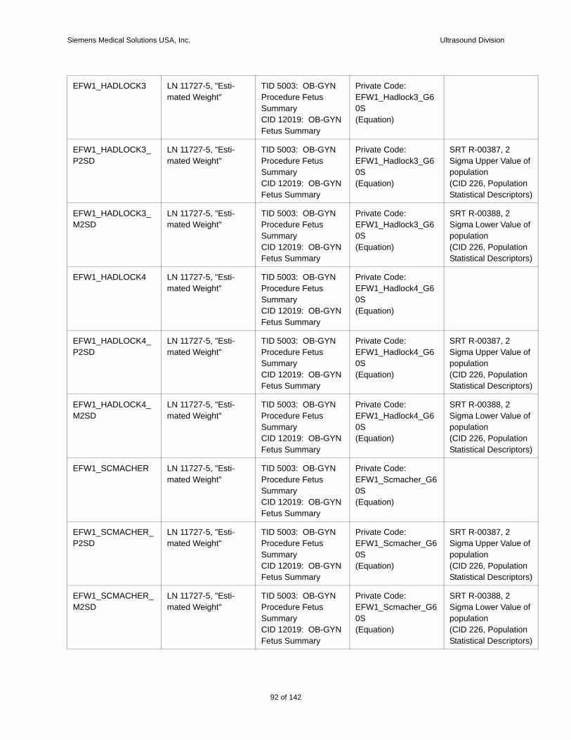
Siemens Medical Solutions USA, Inc. Ultrasound Division
e of
tion tors)
e of
tion tors)
e of
tion tors)
e of
tion tors)
e of
tion tors)
e of
tion tors)
EFW1_HADLOCK3 LN 11727-5, "Esti-mated Weight"
TID 5003: OB-GYN Procedure Fetus SummaryCID 12019: OB-GYN Fetus Summary
Private Code: EFW1_Hadlock3_G60S(Equation)
EFW1_HADLOCK3_P2SD
LN 11727-5, "Esti-mated Weight"
TID 5003: OB-GYN Procedure Fetus SummaryCID 12019: OB-GYN Fetus Summary
Private Code: EFW1_Hadlock3_G60S(Equation)
SRT R-00387, 2 Sigma Upper Valupopulation(CID 226, PopulaStatistical Descrip
EFW1_HADLOCK3_M2SD
LN 11727-5, "Esti-mated Weight"
TID 5003: OB-GYN Procedure Fetus SummaryCID 12019: OB-GYN Fetus Summary
Private Code: EFW1_Hadlock3_G60S(Equation)
SRT R-00388, 2 Sigma Lower Valupopulation(CID 226, PopulaStatistical Descrip
EFW1_HADLOCK4 LN 11727-5, "Esti-mated Weight"
TID 5003: OB-GYN Procedure Fetus SummaryCID 12019: OB-GYN Fetus Summary
Private Code: EFW1_Hadlock4_G60S(Equation)
EFW1_HADLOCK4_P2SD
LN 11727-5, "Esti-mated Weight"
TID 5003: OB-GYN Procedure Fetus SummaryCID 12019: OB-GYN Fetus Summary
Private Code: EFW1_Hadlock4_G60S(Equation)
SRT R-00387, 2 Sigma Upper Valupopulation(CID 226, PopulaStatistical Descrip
EFW1_HADLOCK4_M2SD
LN 11727-5, "Esti-mated Weight"
TID 5003: OB-GYN Procedure Fetus SummaryCID 12019: OB-GYN Fetus Summary
Private Code: EFW1_Hadlock4_G60S(Equation)
SRT R-00388, 2 Sigma Lower Valupopulation(CID 226, PopulaStatistical Descrip
EFW1_SCMACHER LN 11727-5, "Esti-mated Weight"
TID 5003: OB-GYN Procedure Fetus SummaryCID 12019: OB-GYN Fetus Summary
Private Code: EFW1_Scmacher_G60S(Equation)
EFW1_SCMACHER_P2SD
LN 11727-5, "Esti-mated Weight"
TID 5003: OB-GYN Procedure Fetus SummaryCID 12019: OB-GYN Fetus Summary
Private Code: EFW1_Scmacher_G60S(Equation)
SRT R-00387, 2 Sigma Upper Valupopulation(CID 226, PopulaStatistical Descrip
EFW1_SCMACHER_M2SD
LN 11727-5, "Esti-mated Weight"
TID 5003: OB-GYN Procedure Fetus SummaryCID 12019: OB-GYN Fetus Summary
Private Code: EFW1_Scmacher_G60S(Equation)
SRT R-00388, 2 Sigma Lower Valupopulation(CID 226, PopulaStatistical Descrip
92 of 142
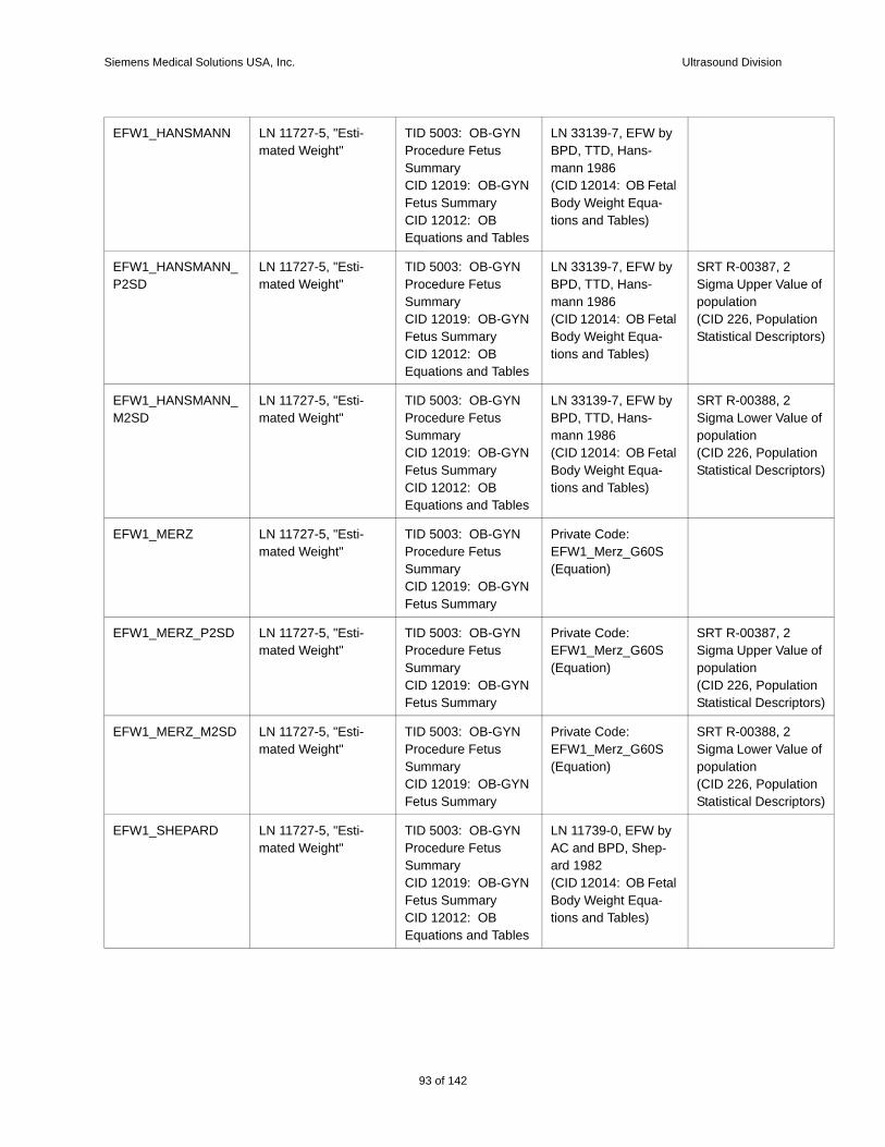
Siemens Medical Solutions USA, Inc. Ultrasound Division
e of
tion tors)
e of
tion tors)
e of
tion tors)
e of
tion tors)
EFW1_HANSMANN LN 11727-5, "Esti-mated Weight"
TID 5003: OB-GYN Procedure Fetus SummaryCID 12019: OB-GYN Fetus SummaryCID 12012: OB Equations and Tables
LN 33139-7, EFW by BPD, TTD, Hans-mann 1986(CID 12014: OB Fetal Body Weight Equa-tions and Tables)
EFW1_HANSMANN_P2SD
LN 11727-5, "Esti-mated Weight"
TID 5003: OB-GYN Procedure Fetus SummaryCID 12019: OB-GYN Fetus SummaryCID 12012: OB Equations and Tables
LN 33139-7, EFW by BPD, TTD, Hans-mann 1986(CID 12014: OB Fetal Body Weight Equa-tions and Tables)
SRT R-00387, 2 Sigma Upper Valupopulation(CID 226, PopulaStatistical Descrip
EFW1_HANSMANN_M2SD
LN 11727-5, "Esti-mated Weight"
TID 5003: OB-GYN Procedure Fetus SummaryCID 12019: OB-GYN Fetus SummaryCID 12012: OB Equations and Tables
LN 33139-7, EFW by BPD, TTD, Hans-mann 1986(CID 12014: OB Fetal Body Weight Equa-tions and Tables)
SRT R-00388, 2 Sigma Lower Valupopulation(CID 226, PopulaStatistical Descrip
EFW1_MERZ LN 11727-5, "Esti-mated Weight"
TID 5003: OB-GYN Procedure Fetus SummaryCID 12019: OB-GYN Fetus Summary
Private Code: EFW1_Merz_G60S(Equation)
EFW1_MERZ_P2SD LN 11727-5, "Esti-mated Weight"
TID 5003: OB-GYN Procedure Fetus SummaryCID 12019: OB-GYN Fetus Summary
Private Code: EFW1_Merz_G60S(Equation)
SRT R-00387, 2 Sigma Upper Valupopulation(CID 226, PopulaStatistical Descrip
EFW1_MERZ_M2SD LN 11727-5, "Esti-mated Weight"
TID 5003: OB-GYN Procedure Fetus SummaryCID 12019: OB-GYN Fetus Summary
Private Code: EFW1_Merz_G60S(Equation)
SRT R-00388, 2 Sigma Lower Valupopulation(CID 226, PopulaStatistical Descrip
EFW1_SHEPARD LN 11727-5, "Esti-mated Weight"
TID 5003: OB-GYN Procedure Fetus SummaryCID 12019: OB-GYN Fetus SummaryCID 12012: OB Equations and Tables
LN 11739-0, EFW by AC and BPD, Shep-ard 1982(CID 12014: OB Fetal Body Weight Equa-tions and Tables)
93 of 142
142
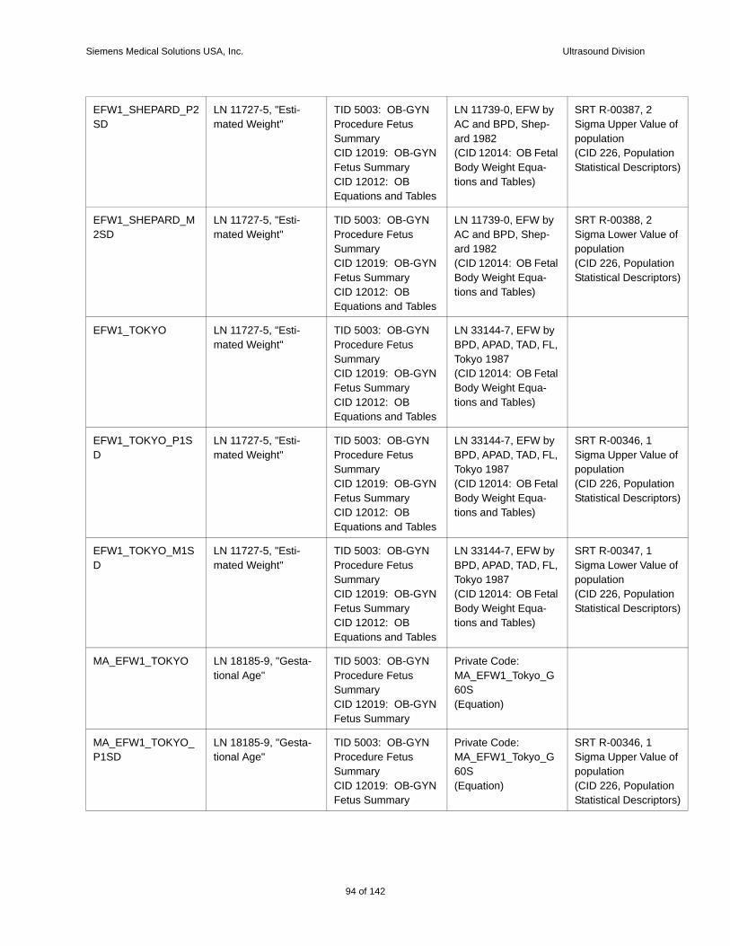
Siemens Medical Solutions USA, Inc. Ultrasound Division
e of
tion tors)
e of
tion tors)
e of
tion tors)
e of
tion tors)
e of
tion tors)
EFW1_SHEPARD_P2SD
LN 11727-5, "Esti-mated Weight"
TID 5003: OB-GYN Procedure Fetus SummaryCID 12019: OB-GYN Fetus SummaryCID 12012: OB Equations and Tables
LN 11739-0, EFW by AC and BPD, Shep-ard 1982(CID 12014: OB Fetal Body Weight Equa-tions and Tables)
SRT R-00387, 2 Sigma Upper Valupopulation(CID 226, PopulaStatistical Descrip
EFW1_SHEPARD_M2SD
LN 11727-5, "Esti-mated Weight"
TID 5003: OB-GYN Procedure Fetus SummaryCID 12019: OB-GYN Fetus SummaryCID 12012: OB Equations and Tables
LN 11739-0, EFW by AC and BPD, Shep-ard 1982(CID 12014: OB Fetal Body Weight Equa-tions and Tables)
SRT R-00388, 2 Sigma Lower Valupopulation(CID 226, PopulaStatistical Descrip
EFW1_TOKYO LN 11727-5, "Esti-mated Weight"
TID 5003: OB-GYN Procedure Fetus SummaryCID 12019: OB-GYN Fetus SummaryCID 12012: OB Equations and Tables
LN 33144-7, EFW by BPD, APAD, TAD, FL, Tokyo 1987(CID 12014: OB Fetal Body Weight Equa-tions and Tables)
EFW1_TOKYO_P1SD
LN 11727-5, "Esti-mated Weight"
TID 5003: OB-GYN Procedure Fetus SummaryCID 12019: OB-GYN Fetus SummaryCID 12012: OB Equations and Tables
LN 33144-7, EFW by BPD, APAD, TAD, FL, Tokyo 1987(CID 12014: OB Fetal Body Weight Equa-tions and Tables)
SRT R-00346, 1 Sigma Upper Valupopulation(CID 226, PopulaStatistical Descrip
EFW1_TOKYO_M1SD
LN 11727-5, "Esti-mated Weight"
TID 5003: OB-GYN Procedure Fetus SummaryCID 12019: OB-GYN Fetus SummaryCID 12012: OB Equations and Tables
LN 33144-7, EFW by BPD, APAD, TAD, FL, Tokyo 1987(CID 12014: OB Fetal Body Weight Equa-tions and Tables)
SRT R-00347, 1 Sigma Lower Valupopulation(CID 226, PopulaStatistical Descrip
MA_EFW1_TOKYO LN 18185-9, "Gesta-tional Age"
TID 5003: OB-GYN Procedure Fetus SummaryCID 12019: OB-GYN Fetus Summary
Private Code: MA_EFW1_Tokyo_G60S(Equation)
MA_EFW1_TOKYO_P1SD
LN 18185-9, "Gesta-tional Age"
TID 5003: OB-GYN Procedure Fetus SummaryCID 12019: OB-GYN Fetus Summary
Private Code: MA_EFW1_Tokyo_G60S(Equation)
SRT R-00346, 1 Sigma Upper Valupopulation(CID 226, PopulaStatistical Descrip
94 of 142
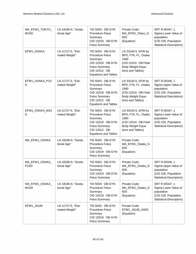
Siemens Medical Solutions USA, Inc. Ultrasound Division
e of
tion tors)
e of
tion tors)
e of
tion tors)
e of
tion tors)
e of
tion tors)
MA_EFW1_TOKYO_M1SD
LN 18185-9, "Gesta-tional Age"
TID 5003: OB-GYN Procedure Fetus SummaryCID 12019: OB-GYN Fetus Summary
Private Code: MA_EFW1_Tokyo_G60S(Equation)
SRT R-00347, 1 Sigma Lower Valupopulation(CID 226, PopulaStatistical Descrip
EFW1_OSAKA LN 11727-5, "Esti-mated Weight"
TID 5003: OB-GYN Procedure Fetus SummaryCID 12019: OB-GYN Fetus SummaryCID 12012: OB Equations and Tables
LN 33140-5, EFW by BPD, FTA, FL, Osaka 1990(CID 12014: OB Fetal Body Weight Equa-tions and Tables)
EFW1_OSAKA_P1SD
LN 11727-5, "Esti-mated Weight"
TID 5003: OB-GYN Procedure Fetus SummaryCID 12019: OB-GYN Fetus SummaryCID 12012: OB Equations and Tables
LN 33140-5, EFW by BPD, FTA, FL, Osaka 1990(CID 12014: OB Fetal Body Weight Equa-tions and Tables)
SRT R-00346, 1 Sigma Upper Valupopulation(CID 226, PopulaStatistical Descrip
EFW1_OSAKA_M1SD
LN 11727-5, "Esti-mated Weight"
TID 5003: OB-GYN Procedure Fetus SummaryCID 12019: OB-GYN Fetus SummaryCID 12012: OB Equations and Tables
LN 33140-5, EFW by BPD, FTA, FL, Osaka 1990(CID 12014: OB Fetal Body Weight Equa-tions and Tables)
SRT R-00347, 1 Sigma Lower Valupopulation(CID 226, PopulaStatistical Descrip
MA_EFW1_OSAKA LN 18185-9, "Gesta-tional Age"
TID 5003: OB-GYN Procedure Fetus SummaryCID 12019: OB-GYN Fetus Summary
Private Code: MA_EFW1_Osaka_G60S(Equation)
MA_EFW1_OSAKA_P1SD
LN 18185-9, "Gesta-tional Age"
TID 5003: OB-GYN Procedure Fetus SummaryCID 12019: OB-GYN Fetus Summary
Private Code: MA_EFW1_Osaka_G60S(Equation)
SRT R-00346, 1 Sigma Upper Valupopulation(CID 226, PopulaStatistical Descrip
MA_EFW1_OSAKA_M1SD
LN 18185-9, "Gesta-tional Age"
TID 5003: OB-GYN Procedure Fetus SummaryCID 12019: OB-GYN Fetus Summary
Private Code: MA_EFW1_Osaka_G60S(Equation)
SRT R-00347, 1 Sigma Lower Valupopulation(CID 226, PopulaStatistical Descrip
EFW1_JSUM LN 11727-5, "Esti-mated Weight"
TID 5003: OB-GYN Procedure Fetus SummaryCID 12019: OB-GYN Fetus Summary
Private Code: EFW1_JSUM_G60S(Equation)
95 of 142
142
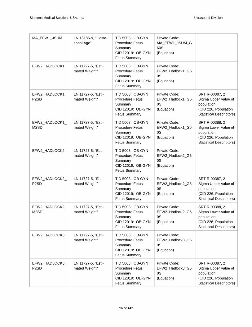
Siemens Medical Solutions USA, Inc. Ultrasound Division
e of
tion tors)
e of
tion tors)
e of
tion tors)
e of
tion tors)
e of
tion tors)
MA_EFW1_JSUM LN 18185-9, "Gesta-tional Age"
TID 5003: OB-GYN Procedure Fetus SummaryCID 12019: OB-GYN Fetus Summary
Private Code: MA_EFW1_JSUM_G60S(Equation)
EFW2_HADLOCK1 LN 11727-5, "Esti-mated Weight"
TID 5003: OB-GYN Procedure Fetus SummaryCID 12019: OB-GYN Fetus Summary
Private Code: EFW2_Hadlock1_G60S(Equation)
EFW2_HADLOCK1_P2SD
LN 11727-5, "Esti-mated Weight"
TID 5003: OB-GYN Procedure Fetus SummaryCID 12019: OB-GYN Fetus Summary
Private Code: EFW2_Hadlock1_G60S(Equation)
SRT R-00387, 2 Sigma Upper Valupopulation(CID 226, PopulaStatistical Descrip
EFW2_HADLOCK1_M2SD
LN 11727-5, "Esti-mated Weight"
TID 5003: OB-GYN Procedure Fetus SummaryCID 12019: OB-GYN Fetus Summary
Private Code: EFW2_Hadlock1_G60S(Equation)
SRT R-00388, 2 Sigma Lower Valupopulation(CID 226, PopulaStatistical Descrip
EFW2_HADLOCK2 LN 11727-5, "Esti-mated Weight"
TID 5003: OB-GYN Procedure Fetus SummaryCID 12019: OB-GYN Fetus Summary
Private Code: EFW2_Hadlock2_G60S(Equation)
EFW2_HADLOCK2_P2SD
LN 11727-5, "Esti-mated Weight"
TID 5003: OB-GYN Procedure Fetus SummaryCID 12019: OB-GYN Fetus Summary
Private Code: EFW2_Hadlock2_G60S(Equation)
SRT R-00387, 2 Sigma Upper Valupopulation(CID 226, PopulaStatistical Descrip
EFW2_HADLOCK2_M2SD
LN 11727-5, "Esti-mated Weight"
TID 5003: OB-GYN Procedure Fetus SummaryCID 12019: OB-GYN Fetus Summary
Private Code: EFW2_Hadlock2_G60S(Equation)
SRT R-00388, 2 Sigma Lower Valupopulation(CID 226, PopulaStatistical Descrip
EFW2_HADLOCK3 LN 11727-5, "Esti-mated Weight"
TID 5003: OB-GYN Procedure Fetus SummaryCID 12019: OB-GYN Fetus Summary
Private Code: EFW2_Hadlock3_G60S(Equation)
EFW2_HADLOCK3_P2SD
LN 11727-5, "Esti-mated Weight"
TID 5003: OB-GYN Procedure Fetus SummaryCID 12019: OB-GYN Fetus Summary
Private Code: EFW2_Hadlock3_G60S(Equation)
SRT R-00387, 2 Sigma Upper Valupopulation(CID 226, PopulaStatistical Descrip
96 of 142
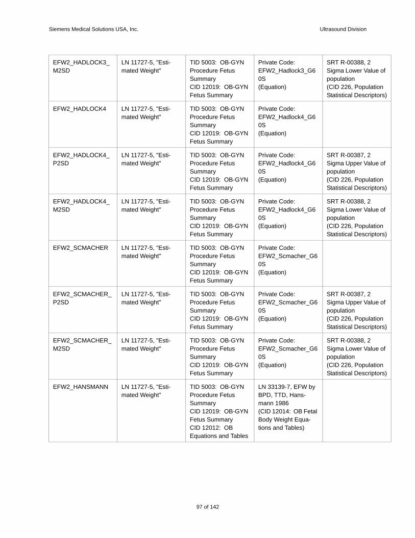
Siemens Medical Solutions USA, Inc. Ultrasound Division
e of
tion tors)
e of
tion tors)
e of
tion tors)
e of
tion tors)
e of
tion tors)
EFW2_HADLOCK3_M2SD
LN 11727-5, "Esti-mated Weight"
TID 5003: OB-GYN Procedure Fetus SummaryCID 12019: OB-GYN Fetus Summary
Private Code: EFW2_Hadlock3_G60S(Equation)
SRT R-00388, 2 Sigma Lower Valupopulation(CID 226, PopulaStatistical Descrip
EFW2_HADLOCK4 LN 11727-5, "Esti-mated Weight"
TID 5003: OB-GYN Procedure Fetus SummaryCID 12019: OB-GYN Fetus Summary
Private Code: EFW2_Hadlock4_G60S(Equation)
EFW2_HADLOCK4_P2SD
LN 11727-5, "Esti-mated Weight"
TID 5003: OB-GYN Procedure Fetus SummaryCID 12019: OB-GYN Fetus Summary
Private Code: EFW2_Hadlock4_G60S(Equation)
SRT R-00387, 2 Sigma Upper Valupopulation(CID 226, PopulaStatistical Descrip
EFW2_HADLOCK4_M2SD
LN 11727-5, "Esti-mated Weight"
TID 5003: OB-GYN Procedure Fetus SummaryCID 12019: OB-GYN Fetus Summary
Private Code: EFW2_Hadlock4_G60S(Equation)
SRT R-00388, 2 Sigma Lower Valupopulation(CID 226, PopulaStatistical Descrip
EFW2_SCMACHER LN 11727-5, "Esti-mated Weight"
TID 5003: OB-GYN Procedure Fetus SummaryCID 12019: OB-GYN Fetus Summary
Private Code: EFW2_Scmacher_G60S(Equation)
EFW2_SCMACHER_P2SD
LN 11727-5, "Esti-mated Weight"
TID 5003: OB-GYN Procedure Fetus SummaryCID 12019: OB-GYN Fetus Summary
Private Code: EFW2_Scmacher_G60S(Equation)
SRT R-00387, 2 Sigma Upper Valupopulation(CID 226, PopulaStatistical Descrip
EFW2_SCMACHER_M2SD
LN 11727-5, "Esti-mated Weight"
TID 5003: OB-GYN Procedure Fetus SummaryCID 12019: OB-GYN Fetus Summary
Private Code: EFW2_Scmacher_G60S(Equation)
SRT R-00388, 2 Sigma Lower Valupopulation(CID 226, PopulaStatistical Descrip
EFW2_HANSMANN LN 11727-5, "Esti-mated Weight"
TID 5003: OB-GYN Procedure Fetus SummaryCID 12019: OB-GYN Fetus SummaryCID 12012: OB Equations and Tables
LN 33139-7, EFW by BPD, TTD, Hans-mann 1986(CID 12014: OB Fetal Body Weight Equa-tions and Tables)
97 of 142
142
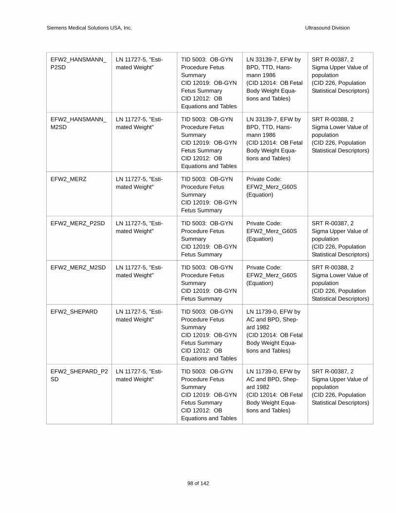
Siemens Medical Solutions USA, Inc. Ultrasound Division
e of
tion tors)
e of
tion tors)
e of
tion tors)
e of
tion tors)
e of
tion tors)
EFW2_HANSMANN_P2SD
LN 11727-5, "Esti-mated Weight"
TID 5003: OB-GYN Procedure Fetus SummaryCID 12019: OB-GYN Fetus SummaryCID 12012: OB Equations and Tables
LN 33139-7, EFW by BPD, TTD, Hans-mann 1986(CID 12014: OB Fetal Body Weight Equa-tions and Tables)
SRT R-00387, 2 Sigma Upper Valupopulation(CID 226, PopulaStatistical Descrip
EFW2_HANSMANN_M2SD
LN 11727-5, "Esti-mated Weight"
TID 5003: OB-GYN Procedure Fetus SummaryCID 12019: OB-GYN Fetus SummaryCID 12012: OB Equations and Tables
LN 33139-7, EFW by BPD, TTD, Hans-mann 1986(CID 12014: OB Fetal Body Weight Equa-tions and Tables)
SRT R-00388, 2 Sigma Lower Valupopulation(CID 226, PopulaStatistical Descrip
EFW2_MERZ LN 11727-5, "Esti-mated Weight"
TID 5003: OB-GYN Procedure Fetus SummaryCID 12019: OB-GYN Fetus Summary
Private Code: EFW2_Merz_G60S(Equation)
EFW2_MERZ_P2SD LN 11727-5, "Esti-mated Weight"
TID 5003: OB-GYN Procedure Fetus SummaryCID 12019: OB-GYN Fetus Summary
Private Code: EFW2_Merz_G60S(Equation)
SRT R-00387, 2 Sigma Upper Valupopulation(CID 226, PopulaStatistical Descrip
EFW2_MERZ_M2SD LN 11727-5, "Esti-mated Weight"
TID 5003: OB-GYN Procedure Fetus SummaryCID 12019: OB-GYN Fetus Summary
Private Code: EFW2_Merz_G60S(Equation)
SRT R-00388, 2 Sigma Lower Valupopulation(CID 226, PopulaStatistical Descrip
EFW2_SHEPARD LN 11727-5, "Esti-mated Weight"
TID 5003: OB-GYN Procedure Fetus SummaryCID 12019: OB-GYN Fetus SummaryCID 12012: OB Equations and Tables
LN 11739-0, EFW by AC and BPD, Shep-ard 1982(CID 12014: OB Fetal Body Weight Equa-tions and Tables)
EFW2_SHEPARD_P2SD
LN 11727-5, "Esti-mated Weight"
TID 5003: OB-GYN Procedure Fetus SummaryCID 12019: OB-GYN Fetus SummaryCID 12012: OB Equations and Tables
LN 11739-0, EFW by AC and BPD, Shep-ard 1982(CID 12014: OB Fetal Body Weight Equa-tions and Tables)
SRT R-00387, 2 Sigma Upper Valupopulation(CID 226, PopulaStatistical Descrip
98 of 142
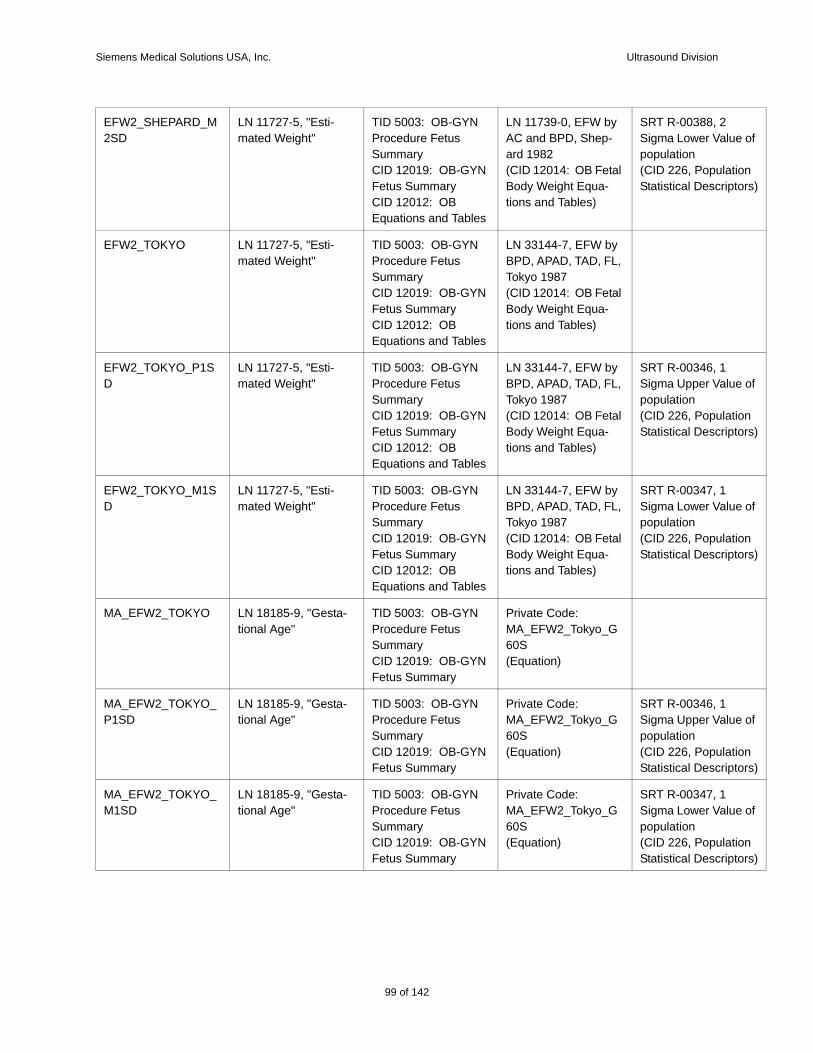
Siemens Medical Solutions USA, Inc. Ultrasound Division
e of
tion tors)
e of
tion tors)
e of
tion tors)
e of
tion tors)
e of
tion tors)
EFW2_SHEPARD_M2SD
LN 11727-5, "Esti-mated Weight"
TID 5003: OB-GYN Procedure Fetus SummaryCID 12019: OB-GYN Fetus SummaryCID 12012: OB Equations and Tables
LN 11739-0, EFW by AC and BPD, Shep-ard 1982(CID 12014: OB Fetal Body Weight Equa-tions and Tables)
SRT R-00388, 2 Sigma Lower Valupopulation(CID 226, PopulaStatistical Descrip
EFW2_TOKYO LN 11727-5, "Esti-mated Weight"
TID 5003: OB-GYN Procedure Fetus SummaryCID 12019: OB-GYN Fetus SummaryCID 12012: OB Equations and Tables
LN 33144-7, EFW by BPD, APAD, TAD, FL, Tokyo 1987(CID 12014: OB Fetal Body Weight Equa-tions and Tables)
EFW2_TOKYO_P1SD
LN 11727-5, "Esti-mated Weight"
TID 5003: OB-GYN Procedure Fetus SummaryCID 12019: OB-GYN Fetus SummaryCID 12012: OB Equations and Tables
LN 33144-7, EFW by BPD, APAD, TAD, FL, Tokyo 1987(CID 12014: OB Fetal Body Weight Equa-tions and Tables)
SRT R-00346, 1 Sigma Upper Valupopulation(CID 226, PopulaStatistical Descrip
EFW2_TOKYO_M1SD
LN 11727-5, "Esti-mated Weight"
TID 5003: OB-GYN Procedure Fetus SummaryCID 12019: OB-GYN Fetus SummaryCID 12012: OB Equations and Tables
LN 33144-7, EFW by BPD, APAD, TAD, FL, Tokyo 1987(CID 12014: OB Fetal Body Weight Equa-tions and Tables)
SRT R-00347, 1 Sigma Lower Valupopulation(CID 226, PopulaStatistical Descrip
MA_EFW2_TOKYO LN 18185-9, "Gesta-tional Age"
TID 5003: OB-GYN Procedure Fetus SummaryCID 12019: OB-GYN Fetus Summary
Private Code: MA_EFW2_Tokyo_G60S(Equation)
MA_EFW2_TOKYO_P1SD
LN 18185-9, "Gesta-tional Age"
TID 5003: OB-GYN Procedure Fetus SummaryCID 12019: OB-GYN Fetus Summary
Private Code: MA_EFW2_Tokyo_G60S(Equation)
SRT R-00346, 1 Sigma Upper Valupopulation(CID 226, PopulaStatistical Descrip
MA_EFW2_TOKYO_M1SD
LN 18185-9, "Gesta-tional Age"
TID 5003: OB-GYN Procedure Fetus SummaryCID 12019: OB-GYN Fetus Summary
Private Code: MA_EFW2_Tokyo_G60S(Equation)
SRT R-00347, 1 Sigma Lower Valupopulation(CID 226, PopulaStatistical Descrip
99 of 142
142
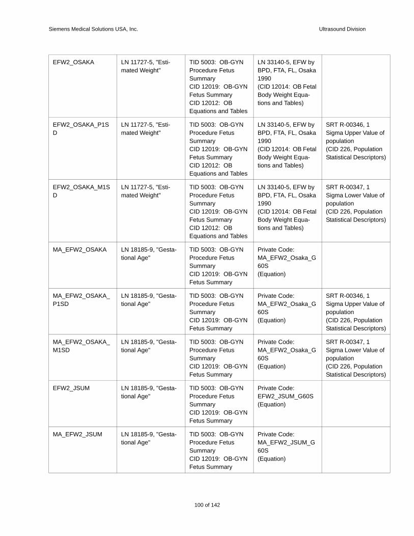
Siemens Medical Solutions USA, Inc. Ultrasound Division
e of
tion tors)
e of
tion tors)
e of
tion tors)
e of
tion tors)
EFW2_OSAKA LN 11727-5, "Esti-mated Weight"
TID 5003: OB-GYN Procedure Fetus SummaryCID 12019: OB-GYN Fetus SummaryCID 12012: OB Equations and Tables
LN 33140-5, EFW by BPD, FTA, FL, Osaka 1990(CID 12014: OB Fetal Body Weight Equa-tions and Tables)
EFW2_OSAKA_P1SD
LN 11727-5, "Esti-mated Weight"
TID 5003: OB-GYN Procedure Fetus SummaryCID 12019: OB-GYN Fetus SummaryCID 12012: OB Equations and Tables
LN 33140-5, EFW by BPD, FTA, FL, Osaka 1990(CID 12014: OB Fetal Body Weight Equa-tions and Tables)
SRT R-00346, 1 Sigma Upper Valupopulation(CID 226, PopulaStatistical Descrip
EFW2_OSAKA_M1SD
LN 11727-5, "Esti-mated Weight"
TID 5003: OB-GYN Procedure Fetus SummaryCID 12019: OB-GYN Fetus SummaryCID 12012: OB Equations and Tables
LN 33140-5, EFW by BPD, FTA, FL, Osaka 1990(CID 12014: OB Fetal Body Weight Equa-tions and Tables)
SRT R-00347, 1 Sigma Lower Valupopulation(CID 226, PopulaStatistical Descrip
MA_EFW2_OSAKA LN 18185-9, "Gesta-tional Age"
TID 5003: OB-GYN Procedure Fetus SummaryCID 12019: OB-GYN Fetus Summary
Private Code: MA_EFW2_Osaka_G60S(Equation)
MA_EFW2_OSAKA_P1SD
LN 18185-9, "Gesta-tional Age"
TID 5003: OB-GYN Procedure Fetus SummaryCID 12019: OB-GYN Fetus Summary
Private Code: MA_EFW2_Osaka_G60S(Equation)
SRT R-00346, 1 Sigma Upper Valupopulation(CID 226, PopulaStatistical Descrip
MA_EFW2_OSAKA_M1SD
LN 18185-9, "Gesta-tional Age"
TID 5003: OB-GYN Procedure Fetus SummaryCID 12019: OB-GYN Fetus Summary
Private Code: MA_EFW2_Osaka_G60S(Equation)
SRT R-00347, 1 Sigma Lower Valupopulation(CID 226, PopulaStatistical Descrip
EFW2_JSUM LN 18185-9, "Gesta-tional Age"
TID 5003: OB-GYN Procedure Fetus SummaryCID 12019: OB-GYN Fetus Summary
Private Code: EFW2_JSUM_G60S(Equation)
MA_EFW2_JSUM LN 18185-9, "Gesta-tional Age"
TID 5003: OB-GYN Procedure Fetus SummaryCID 12019: OB-GYN Fetus Summary
Private Code: MA_EFW2_JSUM_G60S(Equation)
100 of 142

Siemens Medical Solutions USA, Inc. Ultrasound Division
r-
rmal it
r-
rmal it
r-
rmal it
r-
RATIO_HC_AC_CAMPBELL
No match in DICOM Spec
TID 5004: Fetal Biom-etry Ratio SectionCID 12004: Fetal Biometry Ratios
Private Code: RATIO_HC_AC_Campbell_G60S(Equation)
RATIO_HC_AC_CAMPBELL_UPPER
No match in DICOM Spec
TID 5004: Fetal Biom-etry Ratio SectionCID 12004: Fetal Biometry Ratios
Private Code: RATIO_HC_AC_Campbell_G60S(Equation)
SRT R-0038B, Nomal Range UpperLimit
RATIO_HC_AC_CAMPBELL_LOWER
No match in DICOM Spec
TID 5004: Fetal Biom-etry Ratio SectionCID 12004: Fetal Biometry Ratios
Private Code: RATIO_HC_AC_Campbell_G60S(Equation)
SRT R-10041, NoRange Lower Lim
RATIO_FL_AC_HADLOCK
No match in DICOM Spec
TID 5004: Fetal Biom-etry Ratio SectionCID 12004: Fetal Biometry Ratios
Private Code: RATIO_FL_AC_Hadlock_G60S(Equation)
RATIO_FL_AC_HADLOCK_UPPER
No match in DICOM Spec
TID 5004: Fetal Biom-etry Ratio SectionCID 12004: Fetal Biometry Ratios
Private Code: RATIO_FL_AC_Hadlock_G60S(Equation)
SRT R-0038B, Nomal Range UpperLimit
RATIO_FL_AC_HADLOCK_LOWER
No match in DICOM Spec
TID 5004: Fetal Biom-etry Ratio SectionCID 12004: Fetal Biometry Ratios
Private Code: RATIO_FL_AC_Hadlock_G60S(Equation)
SRT R-10041, NoRange Lower Lim
RATIO_FL_BPD_HOHLER
No match in DICOM Spec
TID 5004: Fetal Biom-etry Ratio SectionCID 12004: Fetal Biometry Ratios
Private Code: RATIO_FL_BPD_Hohler_G60S(Equation)
RATIO_FL_BPD_HOHLER_UPPER
No match in DICOM Spec
TID 5004: Fetal Biom-etry Ratio SectionCID 12004: Fetal Biometry Ratios
Private Code: RATIO_FL_BPD_Hohler_G60S(Equation)
SRT R-0038B, Nomal Range UpperLimit
RATIO_FL_BPD_HOHLER_LOWER
No match in DICOM Spec
TID 5004: Fetal Biom-etry Ratio SectionCID 12004: Fetal Biometry Ratios
Private Code: RATIO_FL_BPD_Hohler_G60S(Equation)
SRT R-10041, NoRange Lower Lim
RATIO_TCD_AC_MEYER
No match in DICOM Spec
TID 5004: Fetal Biom-etry Ratio SectionCID 12004: Fetal Biometry Ratios
Private Code: RATIO_TCD_AC_Meyer_G60S(Equation)
RATIO_TCD_AC_MEYER_UPPER
No match in DICOM Spec
TID 5004: Fetal Biom-etry Ratio SectionCID 12004: Fetal Biometry Ratios
Private Code: RATIO_TCD_AC_Meyer_G60S(Equation)
SRT R-0038B, Nomal Range UpperLimit
101 of 142
142
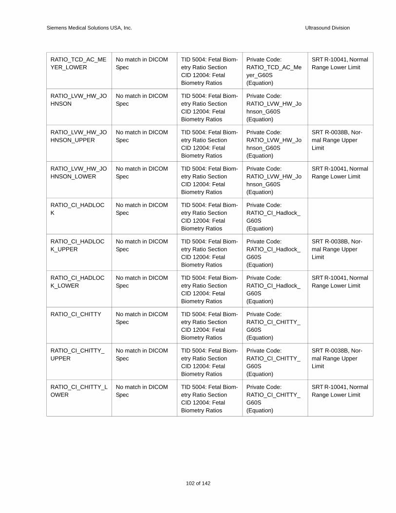
Siemens Medical Solutions USA, Inc. Ultrasound Division
rmal it
r-
rmal it
r-
rmal it
r-
rmal it
RATIO_TCD_AC_MEYER_LOWER
No match in DICOM Spec
TID 5004: Fetal Biom-etry Ratio SectionCID 12004: Fetal Biometry Ratios
Private Code: RATIO_TCD_AC_Meyer_G60S(Equation)
SRT R-10041, NoRange Lower Lim
RATIO_LVW_HW_JOHNSON
No match in DICOM Spec
TID 5004: Fetal Biom-etry Ratio SectionCID 12004: Fetal Biometry Ratios
Private Code: RATIO_LVW_HW_Johnson_G60S(Equation)
RATIO_LVW_HW_JOHNSON_UPPER
No match in DICOM Spec
TID 5004: Fetal Biom-etry Ratio SectionCID 12004: Fetal Biometry Ratios
Private Code: RATIO_LVW_HW_Johnson_G60S(Equation)
SRT R-0038B, Nomal Range UpperLimit
RATIO_LVW_HW_JOHNSON_LOWER
No match in DICOM Spec
TID 5004: Fetal Biom-etry Ratio SectionCID 12004: Fetal Biometry Ratios
Private Code: RATIO_LVW_HW_Johnson_G60S(Equation)
SRT R-10041, NoRange Lower Lim
RATIO_CI_HADLOCK
No match in DICOM Spec
TID 5004: Fetal Biom-etry Ratio SectionCID 12004: Fetal Biometry Ratios
Private Code: RATIO_CI_Hadlock_G60S(Equation)
RATIO_CI_HADLOCK_UPPER
No match in DICOM Spec
TID 5004: Fetal Biom-etry Ratio SectionCID 12004: Fetal Biometry Ratios
Private Code: RATIO_CI_Hadlock_G60S(Equation)
SRT R-0038B, Nomal Range UpperLimit
RATIO_CI_HADLOCK_LOWER
No match in DICOM Spec
TID 5004: Fetal Biom-etry Ratio SectionCID 12004: Fetal Biometry Ratios
Private Code: RATIO_CI_Hadlock_G60S(Equation)
SRT R-10041, NoRange Lower Lim
RATIO_CI_CHITTY No match in DICOM Spec
TID 5004: Fetal Biom-etry Ratio SectionCID 12004: Fetal Biometry Ratios
Private Code: RATIO_CI_CHITTY_G60S(Equation)
RATIO_CI_CHITTY_UPPER
No match in DICOM Spec
TID 5004: Fetal Biom-etry Ratio SectionCID 12004: Fetal Biometry Ratios
Private Code: RATIO_CI_CHITTY_G60S(Equation)
SRT R-0038B, Nomal Range UpperLimit
RATIO_CI_CHITTY_LOWER
No match in DICOM Spec
TID 5004: Fetal Biom-etry Ratio SectionCID 12004: Fetal Biometry Ratios
Private Code: RATIO_CI_CHITTY_G60S(Equation)
SRT R-10041, NoRange Lower Lim
102 of 142
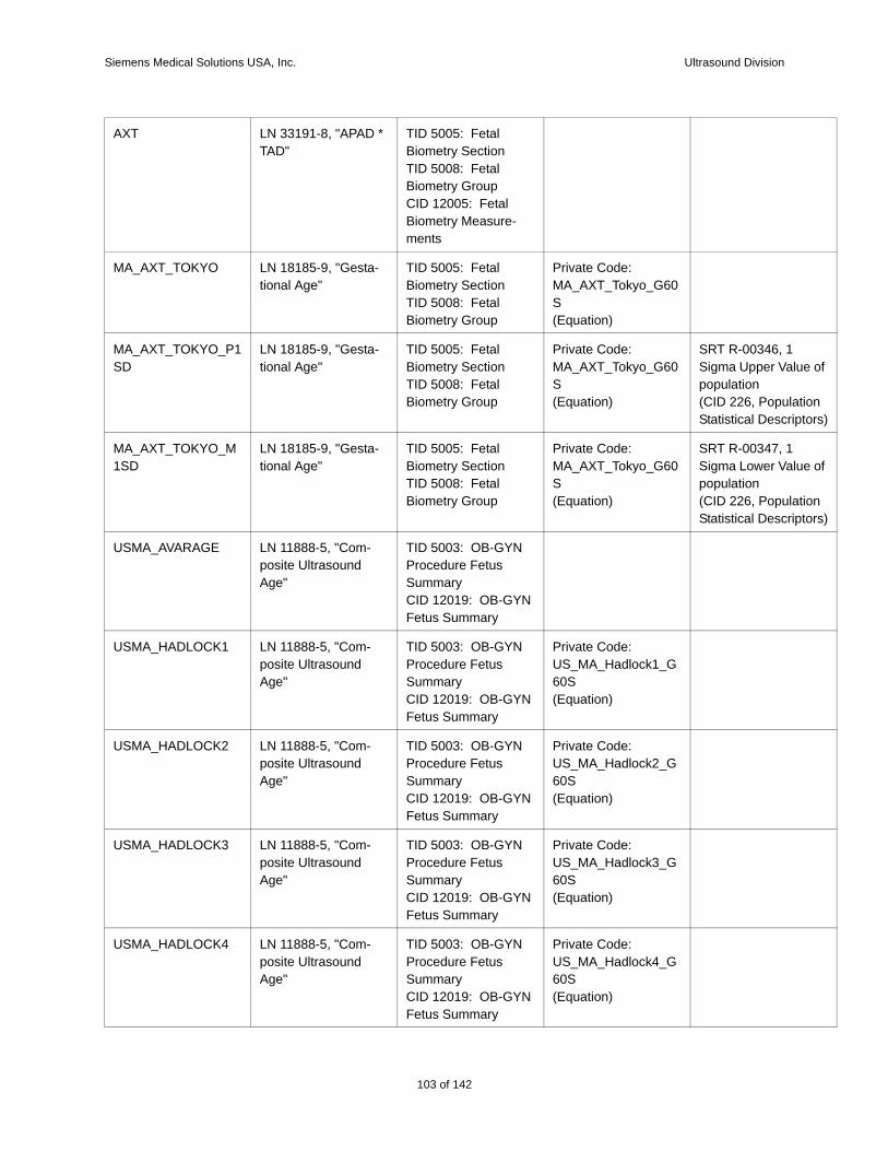
Siemens Medical Solutions USA, Inc. Ultrasound Division
e of
tion tors)
e of
tion tors)
AXT LN 33191-8, "APAD * TAD"
TID 5005: Fetal Biometry SectionTID 5008: Fetal Biometry GroupCID 12005: Fetal Biometry Measure-ments
MA_AXT_TOKYO LN 18185-9, "Gesta-tional Age"
TID 5005: Fetal Biometry SectionTID 5008: Fetal Biometry Group
Private Code: MA_AXT_Tokyo_G60S(Equation)
MA_AXT_TOKYO_P1SD
LN 18185-9, "Gesta-tional Age"
TID 5005: Fetal Biometry SectionTID 5008: Fetal Biometry Group
Private Code: MA_AXT_Tokyo_G60S(Equation)
SRT R-00346, 1 Sigma Upper Valupopulation(CID 226, PopulaStatistical Descrip
MA_AXT_TOKYO_M1SD
LN 18185-9, "Gesta-tional Age"
TID 5005: Fetal Biometry SectionTID 5008: Fetal Biometry Group
Private Code: MA_AXT_Tokyo_G60S(Equation)
SRT R-00347, 1 Sigma Lower Valupopulation(CID 226, PopulaStatistical Descrip
USMA_AVARAGE LN 11888-5, "Com-posite Ultrasound Age"
TID 5003: OB-GYN Procedure Fetus SummaryCID 12019: OB-GYN Fetus Summary
USMA_HADLOCK1 LN 11888-5, "Com-posite Ultrasound Age"
TID 5003: OB-GYN Procedure Fetus SummaryCID 12019: OB-GYN Fetus Summary
Private Code: US_MA_Hadlock1_G60S(Equation)
USMA_HADLOCK2 LN 11888-5, "Com-posite Ultrasound Age"
TID 5003: OB-GYN Procedure Fetus SummaryCID 12019: OB-GYN Fetus Summary
Private Code: US_MA_Hadlock2_G60S(Equation)
USMA_HADLOCK3 LN 11888-5, "Com-posite Ultrasound Age"
TID 5003: OB-GYN Procedure Fetus SummaryCID 12019: OB-GYN Fetus Summary
Private Code: US_MA_Hadlock3_G60S(Equation)
USMA_HADLOCK4 LN 11888-5, "Com-posite Ultrasound Age"
TID 5003: OB-GYN Procedure Fetus SummaryCID 12019: OB-GYN Fetus Summary
Private Code: US_MA_Hadlock4_G60S(Equation)
103 of 142
142
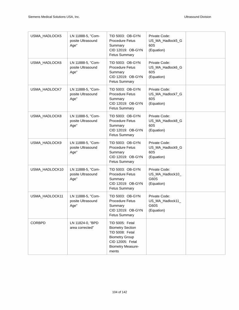
Siemens Medical Solutions USA, Inc. Ultrasound Division
USMA_HADLOCK5 LN 11888-5, "Com-posite Ultrasound Age"
TID 5003: OB-GYN Procedure Fetus SummaryCID 12019: OB-GYN Fetus Summary
Private Code: US_MA_Hadlock5_G60S(Equation)
USMA_HADLOCK6 LN 11888-5, "Com-posite Ultrasound Age"
TID 5003: OB-GYN Procedure Fetus SummaryCID 12019: OB-GYN Fetus Summary
Private Code: US_MA_Hadlock6_G60S(Equation)
USMA_HADLOCK7 LN 11888-5, "Com-posite Ultrasound Age"
TID 5003: OB-GYN Procedure Fetus SummaryCID 12019: OB-GYN Fetus Summary
Private Code: US_MA_Hadlock7_G60S(Equation)
USMA_HADLOCK8 LN 11888-5, "Com-posite Ultrasound Age"
TID 5003: OB-GYN Procedure Fetus SummaryCID 12019: OB-GYN Fetus Summary
Private Code: US_MA_Hadlock8_G60S(Equation)
USMA_HADLOCK9 LN 11888-5, "Com-posite Ultrasound Age"
TID 5003: OB-GYN Procedure Fetus SummaryCID 12019: OB-GYN Fetus Summary
Private Code: US_MA_Hadlock9_G60S(Equation)
USMA_HADLOCK10 LN 11888-5, "Com-posite Ultrasound Age"
TID 5003: OB-GYN Procedure Fetus SummaryCID 12019: OB-GYN Fetus Summary
Private Code: US_MA_Hadlock10_G60S(Equation)
USMA_HADLOCK11 LN 11888-5, "Com-posite Ultrasound Age"
TID 5003: OB-GYN Procedure Fetus SummaryCID 12019: OB-GYN Fetus Summary
Private Code: US_MA_Hadlock11_G60S(Equation)
CORBPD LN 11824-0, "BPD area corrected"
TID 5005: Fetal Biometry SectionTID 5008: Fetal Biometry GroupCID 12005: Fetal Biometry Measure-ments
104 of 142
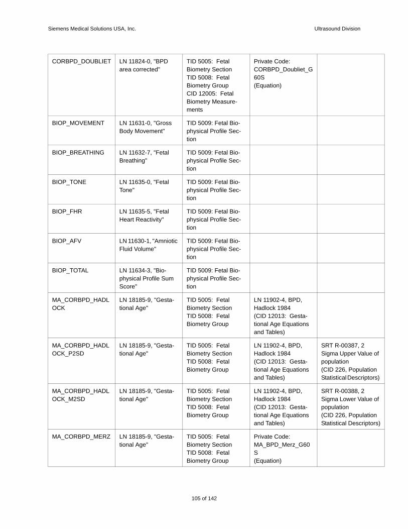
Siemens Medical Solutions USA, Inc. Ultrasound Division
e of
tion tors)
e of
tion tors)
CORBPD_DOUBLIET LN 11824-0, "BPD area corrected"
TID 5005: Fetal Biometry SectionTID 5008: Fetal Biometry GroupCID 12005: Fetal Biometry Measure-ments
Private Code: CORBPD_Doubliet_G60S(Equation)
BIOP_MOVEMENT LN 11631-0, "Gross Body Movement"
TID 5009: Fetal Bio-physical Profile Sec-tion
BIOP_BREATHING LN 11632-7, "Fetal Breathing"
TID 5009: Fetal Bio-physical Profile Sec-tion
BIOP_TONE LN 11635-0, "Fetal Tone"
TID 5009: Fetal Bio-physical Profile Sec-tion
BIOP_FHR LN 11635-5, "Fetal Heart Reactivity"
TID 5009: Fetal Bio-physical Profile Sec-tion
BIOP_AFV LN 11630-1, "Amniotic Fluid Volume"
TID 5009: Fetal Bio-physical Profile Sec-tion
BIOP_TOTAL LN 11634-3, "Bio-physical Profile Sum Score"
TID 5009: Fetal Bio-physical Profile Sec-tion
MA_CORBPD_HADLOCK
LN 18185-9, "Gesta-tional Age"
TID 5005: Fetal Biometry SectionTID 5008: Fetal Biometry Group
LN 11902-4, BPD, Hadlock 1984(CID 12013: Gesta-tional Age Equations and Tables)
MA_CORBPD_HADLOCK_P2SD
LN 18185-9, "Gesta-tional Age"
TID 5005: Fetal Biometry SectionTID 5008: Fetal Biometry Group
LN 11902-4, BPD, Hadlock 1984(CID 12013: Gesta-tional Age Equations and Tables)
SRT R-00387, 2 Sigma Upper Valupopulation(CID 226, PopulaStatistical Descrip
MA_CORBPD_HADLOCK_M2SD
LN 18185-9, "Gesta-tional Age"
TID 5005: Fetal Biometry SectionTID 5008: Fetal Biometry Group
LN 11902-4, BPD, Hadlock 1984(CID 12013: Gesta-tional Age Equations and Tables)
SRT R-00388, 2 Sigma Lower Valupopulation(CID 226, PopulaStatistical Descrip
MA_CORBPD_MERZ LN 18185-9, "Gesta-tional Age"
TID 5005: Fetal Biometry SectionTID 5008: Fetal Biometry Group
Private Code: MA_BPD_Merz_G60S(Equation)
105 of 142
142
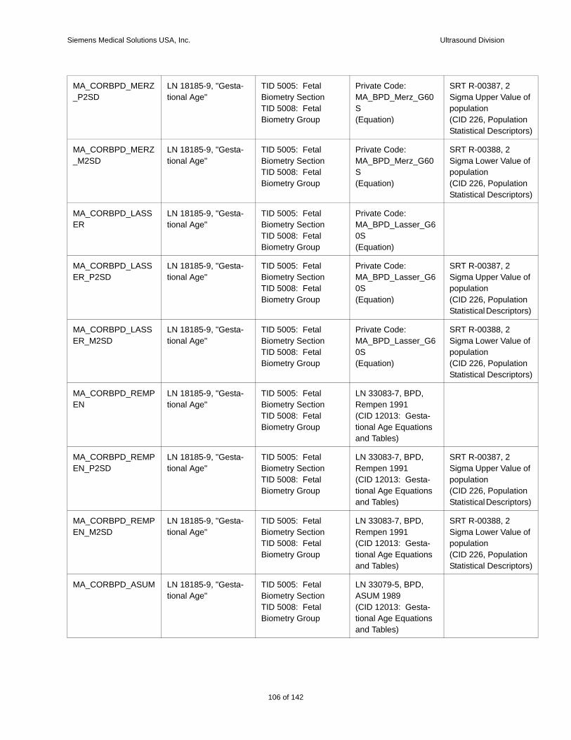
Siemens Medical Solutions USA, Inc. Ultrasound Division
e of
tion tors)
e of
tion tors)
e of
tion tors)
e of
tion tors)
e of
tion tors)
e of
tion tors)
MA_CORBPD_MERZ_P2SD
LN 18185-9, "Gesta-tional Age"
TID 5005: Fetal Biometry SectionTID 5008: Fetal Biometry Group
Private Code: MA_BPD_Merz_G60S(Equation)
SRT R-00387, 2 Sigma Upper Valupopulation(CID 226, PopulaStatistical Descrip
MA_CORBPD_MERZ_M2SD
LN 18185-9, "Gesta-tional Age"
TID 5005: Fetal Biometry SectionTID 5008: Fetal Biometry Group
Private Code: MA_BPD_Merz_G60S(Equation)
SRT R-00388, 2 Sigma Lower Valupopulation(CID 226, PopulaStatistical Descrip
MA_CORBPD_LASSER
LN 18185-9, "Gesta-tional Age"
TID 5005: Fetal Biometry SectionTID 5008: Fetal Biometry Group
Private Code: MA_BPD_Lasser_G60S(Equation)
MA_CORBPD_LASSER_P2SD
LN 18185-9, "Gesta-tional Age"
TID 5005: Fetal Biometry SectionTID 5008: Fetal Biometry Group
Private Code: MA_BPD_Lasser_G60S(Equation)
SRT R-00387, 2 Sigma Upper Valupopulation(CID 226, PopulaStatistical Descrip
MA_CORBPD_LASSER_M2SD
LN 18185-9, "Gesta-tional Age"
TID 5005: Fetal Biometry SectionTID 5008: Fetal Biometry Group
Private Code: MA_BPD_Lasser_G60S(Equation)
SRT R-00388, 2 Sigma Lower Valupopulation(CID 226, PopulaStatistical Descrip
MA_CORBPD_REMPEN
LN 18185-9, "Gesta-tional Age"
TID 5005: Fetal Biometry SectionTID 5008: Fetal Biometry Group
LN 33083-7, BPD, Rempen 1991(CID 12013: Gesta-tional Age Equations and Tables)
MA_CORBPD_REMPEN_P2SD
LN 18185-9, "Gesta-tional Age"
TID 5005: Fetal Biometry SectionTID 5008: Fetal Biometry Group
LN 33083-7, BPD, Rempen 1991(CID 12013: Gesta-tional Age Equations and Tables)
SRT R-00387, 2 Sigma Upper Valupopulation(CID 226, PopulaStatistical Descrip
MA_CORBPD_REMPEN_M2SD
LN 18185-9, "Gesta-tional Age"
TID 5005: Fetal Biometry SectionTID 5008: Fetal Biometry Group
LN 33083-7, BPD, Rempen 1991(CID 12013: Gesta-tional Age Equations and Tables)
SRT R-00388, 2 Sigma Lower Valupopulation(CID 226, PopulaStatistical Descrip
MA_CORBPD_ASUM LN 18185-9, "Gesta-tional Age"
TID 5005: Fetal Biometry SectionTID 5008: Fetal Biometry Group
LN 33079-5, BPD, ASUM 1989(CID 12013: Gesta-tional Age Equations and Tables)
106 of 142
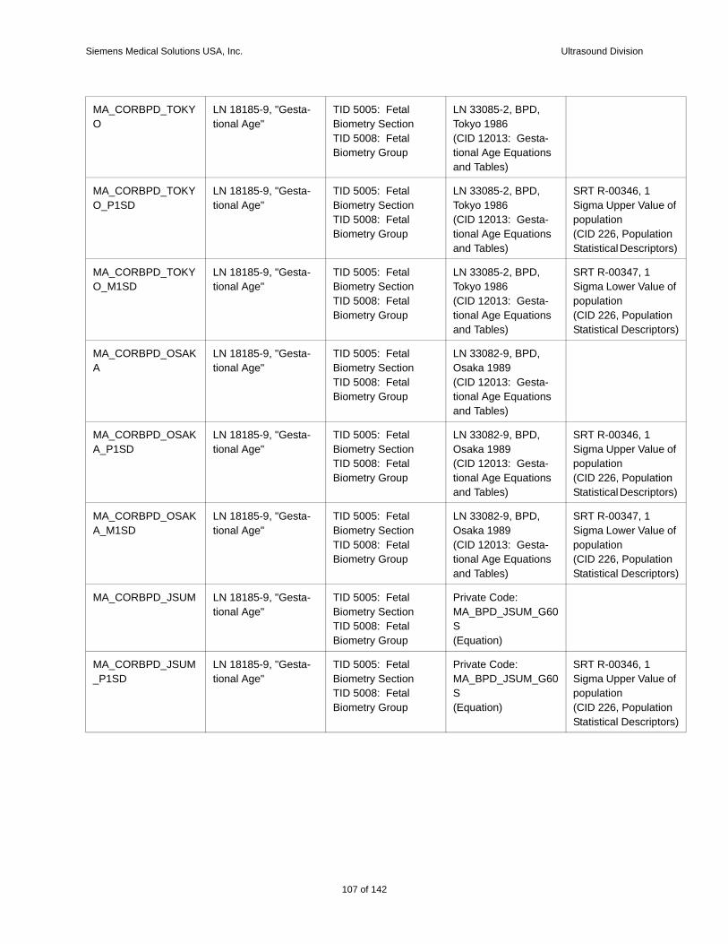
Siemens Medical Solutions USA, Inc. Ultrasound Division
e of
tion tors)
e of
tion tors)
e of
tion tors)
e of
tion tors)
e of
tion tors)
MA_CORBPD_TOKYO
LN 18185-9, "Gesta-tional Age"
TID 5005: Fetal Biometry SectionTID 5008: Fetal Biometry Group
LN 33085-2, BPD, Tokyo 1986(CID 12013: Gesta-tional Age Equations and Tables)
MA_CORBPD_TOKYO_P1SD
LN 18185-9, "Gesta-tional Age"
TID 5005: Fetal Biometry SectionTID 5008: Fetal Biometry Group
LN 33085-2, BPD, Tokyo 1986(CID 12013: Gesta-tional Age Equations and Tables)
SRT R-00346, 1 Sigma Upper Valupopulation(CID 226, PopulaStatistical Descrip
MA_CORBPD_TOKYO_M1SD
LN 18185-9, "Gesta-tional Age"
TID 5005: Fetal Biometry SectionTID 5008: Fetal Biometry Group
LN 33085-2, BPD, Tokyo 1986(CID 12013: Gesta-tional Age Equations and Tables)
SRT R-00347, 1 Sigma Lower Valupopulation(CID 226, PopulaStatistical Descrip
MA_CORBPD_OSAKA
LN 18185-9, "Gesta-tional Age"
TID 5005: Fetal Biometry SectionTID 5008: Fetal Biometry Group
LN 33082-9, BPD, Osaka 1989(CID 12013: Gesta-tional Age Equations and Tables)
MA_CORBPD_OSAKA_P1SD
LN 18185-9, "Gesta-tional Age"
TID 5005: Fetal Biometry SectionTID 5008: Fetal Biometry Group
LN 33082-9, BPD, Osaka 1989(CID 12013: Gesta-tional Age Equations and Tables)
SRT R-00346, 1 Sigma Upper Valupopulation(CID 226, PopulaStatistical Descrip
MA_CORBPD_OSAKA_M1SD
LN 18185-9, "Gesta-tional Age"
TID 5005: Fetal Biometry SectionTID 5008: Fetal Biometry Group
LN 33082-9, BPD, Osaka 1989(CID 12013: Gesta-tional Age Equations and Tables)
SRT R-00347, 1 Sigma Lower Valupopulation(CID 226, PopulaStatistical Descrip
MA_CORBPD_JSUM LN 18185-9, "Gesta-tional Age"
TID 5005: Fetal Biometry SectionTID 5008: Fetal Biometry Group
Private Code: MA_BPD_JSUM_G60S(Equation)
MA_CORBPD_JSUM_P1SD
LN 18185-9, "Gesta-tional Age"
TID 5005: Fetal Biometry SectionTID 5008: Fetal Biometry Group
Private Code: MA_BPD_JSUM_G60S(Equation)
SRT R-00346, 1 Sigma Upper Valupopulation(CID 226, PopulaStatistical Descrip
107 of 142
142
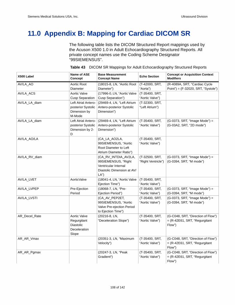
Siemens Medical Solutions USA, Inc. Ultrasound Division
X500 t
AV/L le”)
AV/L
AV/L
AV/L =
AV/L
AV/L =
AV/L
AV/L =
AV/L =
AR_D w”)
AR_A w”)
AR_A w”)
11.0 Appendix B: Mapping for Cardiac DICOM SRThe following table lists the DICOM Structured Report mappings used by the Acuson X500 1.0 in Adult Echocardiography Structured Reports. All private concept names use the Coding Scheme Designator “99SIEMENSUS”.
Table 43 DICOM SR Mappings for Adult Echocardiography Structured Reports
Label Name of ASE Concept
Base Measurement Concept Name Echo Section Concept or Acquisition Contex
Modifiers
A_AO Aortic Root Diameter
(18015-8, LN, “Aortic Root Diameter”)
(T-42000, SRT, “Aorta”)
(R-4089A, SRT, “Cardiac CyclePoint”) = (F-32020, SRT, “Systo
A_ACS Aortic Valve Cusp Separation
(17996-0, LN, “Aortic Valve Cusp Separation”)
(T-35400, SRT, “Aortic Valve”)
A_LA_diam Left Atrial Antero-posterior Systolic Dimension by M-Mode
(29469-4, LN, “Left Atrium Antero-posterior Systolic Dimension”)
(T-32300, SRT, “Left Atrium”)
A_LA_diam Left Atrial Antero-posterior Systolic Dimension by 2-D
(29469-4, LN, “Left Atrium Antero-posterior Systolic Dimension”)
(T-35400, SRT, “Aortic Valve”)
(G-0373, SRT, “Image Mode”) (G-03A2, SRT, “2D mode”)
A_AO/LA (CA_LA_AO2LA, 99SIEMENSUS, “Aortic Root Diameter to Left Atrium Diameter Ratio”)
(T-35400, SRT, “Aortic Valve”)
A_RV_diam (CA_RV_INTDIA_AV2LA, 99SIEMENSUS, “Right Ventricular Internal Diastolic Dimension at AV/LA”)
(T-32500, SRT,“Right Ventricle”)
(G-0373, SRT, “Image Mode”) (G-0394, SRT, “M mode”)
A_LVET AorticValve (18041-4, LN, “Aortic Valve Ejection Time”)
(T-35400, SRT, “Aortic Valve”)
A_LVPEP Pre-Ejection Period
(18068-7, LN, “Pre-Ejection Period”)
(T-35400, SRT, “Aortic Valve”)
(G-0373, SRT, “Image Mode”) (G-0394, SRT, “M mode”)
A_LVSTI (CA_AV_PEP2ET, 99SIEMENSUS, “Aortic Valve Pre-ejection Period to Ejection Time”)
(T-35400, SRT, “Aortic Valve”)
(G-0373, SRT, “Image Mode”) (G-0394, SRT, “M mode”)
ecel_Rate Aortic Valve Regurgitant Diastolic Deceleration Slope
(20216-8, LN, “Deceleration Slope“)
(T-35400, SRT, “Aortic Valve”)
(G-C048, SRT, “Direction of Flo= (R-42E61, SRT, “RegurgitantFlow”)
R_Vmax (20351-3, LN, “Maximum Velocity“)
(T-35400, SRT, “Aortic Valve”)
(G-C048, SRT, “Direction of Flo= (R-42E61, SRT, “RegurgitantFlow”)
R_Pgmax (20247-3, LN, “Peak Gradient“)
(T-35400, SRT, “Aortic Valve”)
(G-C048, SRT, “Direction of Flo= (R-42E61, SRT, “RegurgitantFlow”)
108 of 142

Siemens Medical Solutions USA, Inc. Ultrasound Division
AR_A w”)
AR_A
AR_A w”)
AR_A w”) t
AR_A
AR_A
AR_A w”)
Cube =
Cube =
Cube =
Cube =
Cube =
Cube =
X500 t
R_V_ed (11653-3, LN, “EndDiastolic Velocity“)
(T-35400, SRT, “Aortic Valve”)
(G-C048, SRT, “Direction of Flo= (R-42E61, SRT, “RegurgitantFlow”)
R_Time (17998-6, LN, “Aortic Valve Regurgitant Diastolic Deceleration Time”)
(T-35400, SRT, “Aortic Valve”)
I_Vmax (20351-3, LN, “Maximum Velocity“)
(T-35400, SRT, “Aortic Valve”)
(G-C048, SRT, “Direction of Flo= (R-42E61, SRT, “RegurgitantFlow”)
I_Pgmax (20247-3, LN, “Peak Gradient“)
(T-35400, SRT, “Aortic Valve”)
(G-C048, SRT, “Direction of Flo= (R-42E61, SRT, “RegurgitanFlow”)
I_PHT Aortic Valve RegurgitantDiastolic Pressure Half-time
(20280-4, LN,“Pressure Half-Time”)
(T-35400, SRT, “Aortic Valve”)
(G-C048, SRT, “Direction of Flow”) = (R-42E61,SRT, “Regurgitant Flow”)
I_Dec_Time (20217-6, LN, “Deceleration Time”)
(T-35400, SRT, “Aortic Valve”)
(G-C048, SRT, “Direction of Flow”) = (R-42E61,SRT, “Regurgitant Flow”)
I_Dec_Slope (20216-8, LN, “Deceleration Slope“)
(T-35400, SRT, “Aortic Valve”)
(G-C048, SRT, “Direction of Flo= (R-42E61, SRT, “RegurgitantFlow”)
d(2D)_LVIDd Left Ventricular Internal End Diastolic Dimension by 2-D
(29436-3, LN, “Left Ventricle Internal End Diastolic Dimension”)
(T-32600, SRT, “Left Ventricle”)
(G-0373, SRT, “Image Mode”) (G-03A2, SRT, “2D mode”)
d(2D)_LVIDs Left Ventricular Internal Systolic Dimension by 2-D
(29438-9, LN, “Left Ventricle Internal Systolic Dimension”)
(T-32600, SRT, “Left Ventricle”)
(G-0373, SRT, “Image Mode”) (G-03A2, SRT, “2D mode”)
d(2D)_FS Left Ventricular Fractional Shortening by 2-D
(18051-3, LN, “Left Ventricular Fractional Shortening”)
(T-32600, SRT, “Left Ventricle”)
(G-0373, SRT, “Image Mode”) (G-03A2, SRT, “2D mode”)
d(2D)_IVSd Interventricular Septum Diastolic Thickness by 2-D
(18154-5, LN, “Interventricular Septum Diastolic Thickness”)
(T-32600, SRT, “Left Ventricle”)
(G-0373, SRT, “Image Mode”) (G-03A2, SRT, “2D mode”)
d(2D)_IVSs Interventricular Septum Systolic Thickness by 2-D
(18158-6, LN, “Interventricular Septum Systolic Thickness”)
(T-32600, SRT, “Left Ventricle”)
(G-0373, SRT, “Image Mode”) (G-03A2, SRT, “2D mode”)
d(2D)_LVPWd Left Ventricular Posterior Wall Diastolic Thickness by 2-D
(18152-9, LN, “Left Ventricle Posterior Wall Diastolic Thickness”)
(T-32600, SRT, “Left Ventricle”)
(G-0373, SRT, “Image Mode”) (G-03A2, SRT, “2D mode”)
Label Name of ASE Concept
Base Measurement Concept Name Echo Section Concept or Acquisition Contex
Modifiers
109 of 142
142
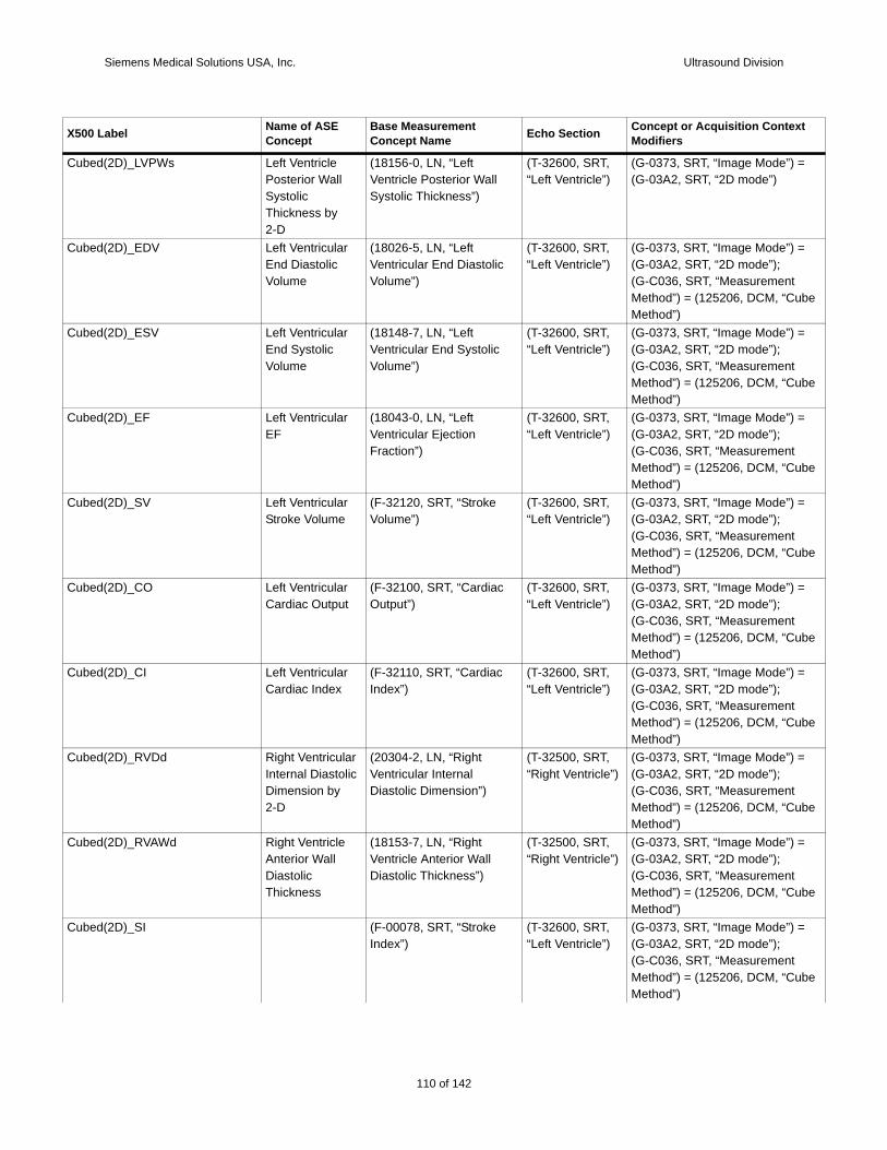
Siemens Medical Solutions USA, Inc. Ultrasound Division
Cube =
Cube =
be
Cube =
be
Cube =
be
Cube =
be
Cube =
be
Cube =
be
Cube =
be
Cube =
be
Cube =
be
X500 t
d(2D)_LVPWs Left Ventricle Posterior Wall Systolic Thickness by 2-D
(18156-0, LN, “Left Ventricle Posterior Wall Systolic Thickness”)
(T-32600, SRT, “Left Ventricle”)
(G-0373, SRT, “Image Mode”) (G-03A2, SRT, “2D mode”)
d(2D)_EDV Left Ventricular End Diastolic Volume
(18026-5, LN, “Left Ventricular End Diastolic Volume”)
(T-32600, SRT, “Left Ventricle”)
(G-0373, SRT, “Image Mode”) (G-03A2, SRT, “2D mode”);(G-C036, SRT, “Measurement Method”) = (125206, DCM, “CuMethod”)
d(2D)_ESV Left Ventricular End Systolic Volume
(18148-7, LN, “Left Ventricular End Systolic Volume”)
(T-32600, SRT, “Left Ventricle”)
(G-0373, SRT, “Image Mode”) (G-03A2, SRT, “2D mode”);(G-C036, SRT, “Measurement Method”) = (125206, DCM, “CuMethod”)
d(2D)_EF Left Ventricular EF
(18043-0, LN, “Left Ventricular Ejection Fraction”)
(T-32600, SRT, “Left Ventricle”)
(G-0373, SRT, “Image Mode”) (G-03A2, SRT, “2D mode”);(G-C036, SRT, “Measurement Method”) = (125206, DCM, “CuMethod”)
d(2D)_SV Left Ventricular Stroke Volume
(F-32120, SRT, “Stroke Volume”)
(T-32600, SRT, “Left Ventricle”)
(G-0373, SRT, “Image Mode”) (G-03A2, SRT, “2D mode”);(G-C036, SRT, “Measurement Method”) = (125206, DCM, “CuMethod”)
d(2D)_CO Left Ventricular Cardiac Output
(F-32100, SRT, “Cardiac Output”)
(T-32600, SRT, “Left Ventricle”)
(G-0373, SRT, “Image Mode”) (G-03A2, SRT, “2D mode”);(G-C036, SRT, “Measurement Method”) = (125206, DCM, “CuMethod”)
d(2D)_CI Left Ventricular Cardiac Index
(F-32110, SRT, “Cardiac Index”)
(T-32600, SRT, “Left Ventricle”)
(G-0373, SRT, “Image Mode”) (G-03A2, SRT, “2D mode”);(G-C036, SRT, “Measurement Method”) = (125206, DCM, “CuMethod”)
d(2D)_RVDd Right Ventricular Internal Diastolic Dimension by 2-D
(20304-2, LN, “Right Ventricular Internal Diastolic Dimension”)
(T-32500, SRT,“Right Ventricle”)
(G-0373, SRT, “Image Mode”) (G-03A2, SRT, “2D mode”);(G-C036, SRT, “Measurement Method”) = (125206, DCM, “CuMethod”)
d(2D)_RVAWd Right Ventricle Anterior Wall Diastolic Thickness
(18153-7, LN, “Right Ventricle Anterior Wall Diastolic Thickness”)
(T-32500, SRT,“Right Ventricle”)
(G-0373, SRT, “Image Mode”) (G-03A2, SRT, “2D mode”);(G-C036, SRT, “Measurement Method”) = (125206, DCM, “CuMethod”)
d(2D)_SI (F-00078, SRT, “Stroke Index”)
(T-32600, SRT, “Left Ventricle”)
(G-0373, SRT, “Image Mode”) (G-03A2, SRT, “2D mode”);(G-C036, SRT, “Measurement Method”) = (125206, DCM, “CuMethod”)
Label Name of ASE Concept
Base Measurement Concept Name Echo Section Concept or Acquisition Contex
Modifiers
110 of 142
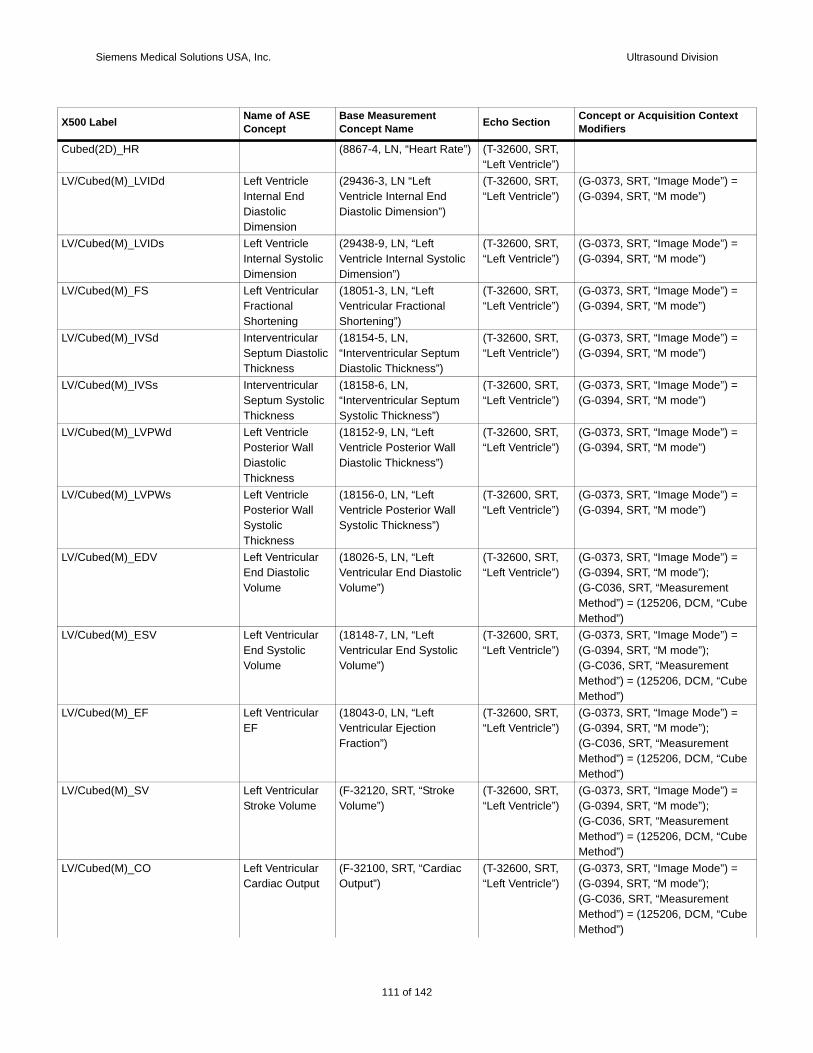
Siemens Medical Solutions USA, Inc. Ultrasound Division
Cube
LV/C =
LV/C =
LV/C =
LV/C =
LV/C =
LV/C =
LV/C =
LV/C =
be
LV/C =
be
LV/C =
be
LV/C =
be
LV/C =
be
X500 t
d(2D)_HR (8867-4, LN, “Heart Rate”) (T-32600, SRT, “Left Ventricle”)
ubed(M)_LVIDd Left Ventricle Internal End Diastolic Dimension
(29436-3, LN “Left Ventricle Internal End Diastolic Dimension”)
(T-32600, SRT, “Left Ventricle”)
(G-0373, SRT, “Image Mode”) (G-0394, SRT, “M mode”)
ubed(M)_LVIDs Left Ventricle Internal Systolic Dimension
(29438-9, LN, “Left Ventricle Internal Systolic Dimension”)
(T-32600, SRT, “Left Ventricle”)
(G-0373, SRT, “Image Mode”) (G-0394, SRT, “M mode”)
ubed(M)_FS Left Ventricular Fractional Shortening
(18051-3, LN, “Left Ventricular Fractional Shortening”)
(T-32600, SRT, “Left Ventricle”)
(G-0373, SRT, “Image Mode”) (G-0394, SRT, “M mode”)
ubed(M)_IVSd Interventricular Septum Diastolic Thickness
(18154-5, LN, “Interventricular Septum Diastolic Thickness”)
(T-32600, SRT, “Left Ventricle”)
(G-0373, SRT, “Image Mode”) (G-0394, SRT, “M mode”)
ubed(M)_IVSs Interventricular Septum Systolic Thickness
(18158-6, LN, “Interventricular Septum Systolic Thickness”)
(T-32600, SRT, “Left Ventricle”)
(G-0373, SRT, “Image Mode”) (G-0394, SRT, “M mode”)
ubed(M)_LVPWd Left Ventricle Posterior Wall Diastolic Thickness
(18152-9, LN, “Left Ventricle Posterior Wall Diastolic Thickness”)
(T-32600, SRT, “Left Ventricle”)
(G-0373, SRT, “Image Mode”) (G-0394, SRT, “M mode”)
ubed(M)_LVPWs Left Ventricle Posterior Wall Systolic Thickness
(18156-0, LN, “Left Ventricle Posterior Wall Systolic Thickness”)
(T-32600, SRT, “Left Ventricle”)
(G-0373, SRT, “Image Mode”) (G-0394, SRT, “M mode”)
ubed(M)_EDV Left Ventricular End Diastolic Volume
(18026-5, LN, “Left Ventricular End Diastolic Volume”)
(T-32600, SRT, “Left Ventricle”)
(G-0373, SRT, “Image Mode”) (G-0394, SRT, “M mode”);(G-C036, SRT, “Measurement Method”) = (125206, DCM, “CuMethod”)
ubed(M)_ESV Left Ventricular End Systolic Volume
(18148-7, LN, “Left Ventricular End Systolic Volume”)
(T-32600, SRT, “Left Ventricle”)
(G-0373, SRT, “Image Mode”) (G-0394, SRT, “M mode”);(G-C036, SRT, “Measurement Method”) = (125206, DCM, “CuMethod”)
ubed(M)_EF Left Ventricular EF
(18043-0, LN, “Left Ventricular Ejection Fraction”)
(T-32600, SRT, “Left Ventricle”)
(G-0373, SRT, “Image Mode”) (G-0394, SRT, “M mode”);(G-C036, SRT, “Measurement Method”) = (125206, DCM, “CuMethod”)
ubed(M)_SV Left Ventricular Stroke Volume
(F-32120, SRT, “Stroke Volume”)
(T-32600, SRT, “Left Ventricle”)
(G-0373, SRT, “Image Mode”) (G-0394, SRT, “M mode”);(G-C036, SRT, “Measurement Method”) = (125206, DCM, “CuMethod”)
ubed(M)_CO Left Ventricular Cardiac Output
(F-32100, SRT, “Cardiac Output”)
(T-32600, SRT, “Left Ventricle”)
(G-0373, SRT, “Image Mode”) (G-0394, SRT, “M mode”);(G-C036, SRT, “Measurement Method”) = (125206, DCM, “CuMethod”)
Label Name of ASE Concept
Base Measurement Concept Name Echo Section Concept or Acquisition Contex
Modifiers
111 of 142
142

Siemens Medical Solutions USA, Inc. Ultrasound Division
LV/C =
be
LV/C =
be
LV/C =
be
LV/C 7-6,
=
be
LV/C =
be
LV/C
LV/C =
LV/C =
be
LV/C =
be
Teich =
Teich =
X500 t
ubed(M)_CI Left Ventricular Cardiac Index
(F-32110, SRT, “Cardiac Index”)
(T-32600, SRT, “Left Ventricle”)
(G-0373, SRT, “Image Mode”) (G-0394, SRT, “M mode”);(G-C036, SRT, “Measurement Method”) = (125206, DCM, “CuMethod”)
ubed(M)_LV_Mass Left Ventricular Mass by M-Mode
(18087-7, LN, “Left Ventricle Mass”)
(T-32600, SRT, “Left Ventricle”)
(G-0373, SRT, “Image Mode”) (G-0394, SRT, “M mode”);(G-C036, SRT, “Measurement Method”) = (125206, DCM, “CuMethod”)
ubed(M)_LV_Mass-c (CA_LV_MASSc, 99SIEMENSUS, “Left Ventricle Mass corrected”)
(T-32600, SRT, “Left Ventricle”)
(G-0373, SRT, “Image Mode”) (G-0394, SRT, “M mode”);(G-C036, SRT, “Measurement Method”) = (125206, DCM, “CuMethod”)
ubed(M)_LV_Mass-I (18087-7, LN, “Left Ventricle Mass”)
(T-32600, SRT, “Left Ventricle”)
(121425, DCM, “Index”) = (827LN, “Body Surface Area”);(G-0373, SRT, “Image Mode”) (G-0394, SRT, “M mode”);(G-C036, SRT, “Measurement Method”) = (125206, DCM, “CuMethod”)
ubed(M)_RVDd Right Ventricular Internal Diastolic Dimension by M-Mode
(20304-2, LN, “Right Ventricular Internal Diastolic Dimension”)
(T-32500, SRT,“Right Ventricle”)
(G-0373, SRT, “Image Mode”) (G-0394, SRT, “M mode”);(G-C036, SRT, “Measurement Method”) = (125206, DCM, “CuMethod”)
ubed(M)_LVET (18041-4, LN, “Aortic Valve Ejection Time”)
(T-32600, SRT, “Left Ventricle”)
ubed(M)_HR (8867-4, LN, “Heart Rate”) (T-32600, SRT, “Left Ventricle”)
(G-0373, SRT, “Image Mode”) (G-0394, SRT, “M mode”)
ubed(M)_SI (F-00078, SRT, “Stroke Index”)
(T-32600, SRT, “Left Ventricle”)
(G-0373, SRT, “Image Mode”) (G-0394, SRT, “M mode”);(G-C036, SRT, “Measurement Method”) = (125206, DCM, “CuMethod”)
ubed(M)_mVcf (CA_LF_VCF, 99SIEMENSUS, “systolic index”)
(T-32600, SRT, “Left Ventricle”)
(G-0373, SRT, “Image Mode”) (G-0394, SRT, “M mode”);(G-C036, SRT, “Measurement Method”) = (125206, DCM, “CuMethod”)
holz(2D)_LVIDd Left Ventricular Internal End Diastolic Dimension by 2-D
(29436-3, LN, “Left Ventricle Internal End Diastolic Dimension”)
(T-32600, SRT, “Left Ventricle”)
(G-0373, SRT, “Image Mode”) (G-03A2, SRT, “2D mode”)
holz(2D)_LVIDs Left Ventricular Internal Systolic Dimension by 2-D
(29438-9, LN, “Left Ventricle Internal Systolic Dimension”)
(T-32600, SRT, “Left Ventricle”)
(G-0373, SRT, “Image Mode”) (G-03A2, SRT, “2D mode”)
Label Name of ASE Concept
Base Measurement Concept Name Echo Section Concept or Acquisition Contex
Modifiers
112 of 142
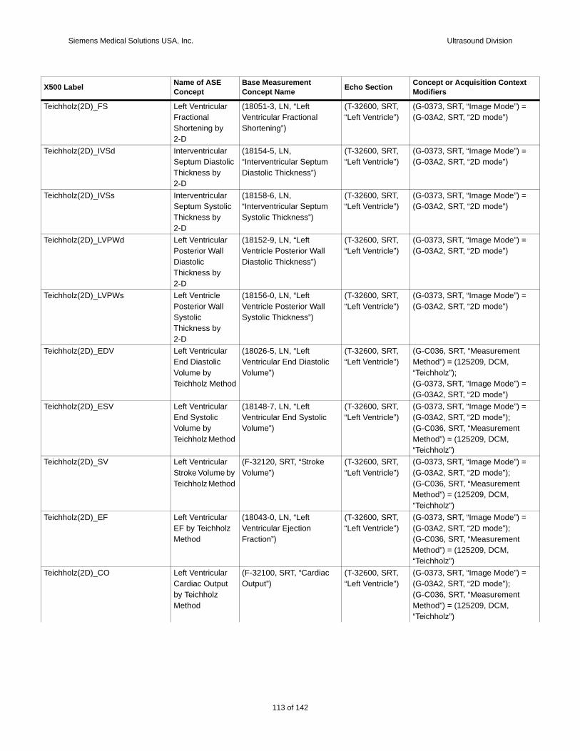
Siemens Medical Solutions USA, Inc. Ultrasound Division
Teich =
Teich =
Teich =
Teich =
Teich =
Teich
=
Teich =
Teich =
Teich =
Teich =
X500 t
holz(2D)_FS Left Ventricular Fractional Shortening by 2-D
(18051-3, LN, “Left Ventricular Fractional Shortening”)
(T-32600, SRT, “Left Ventricle”)
(G-0373, SRT, “Image Mode”) (G-03A2, SRT, “2D mode”)
holz(2D)_IVSd Interventricular Septum Diastolic Thickness by 2-D
(18154-5, LN, “Interventricular Septum Diastolic Thickness”)
(T-32600, SRT, “Left Ventricle”)
(G-0373, SRT, “Image Mode”) (G-03A2, SRT, “2D mode”)
holz(2D)_IVSs Interventricular Septum Systolic Thickness by 2-D
(18158-6, LN, “Interventricular Septum Systolic Thickness”)
(T-32600, SRT, “Left Ventricle”)
(G-0373, SRT, “Image Mode”) (G-03A2, SRT, “2D mode”)
holz(2D)_LVPWd Left Ventricular Posterior Wall Diastolic Thickness by 2-D
(18152-9, LN, “Left Ventricle Posterior Wall Diastolic Thickness”)
(T-32600, SRT, “Left Ventricle”)
(G-0373, SRT, “Image Mode”) (G-03A2, SRT, “2D mode”)
holz(2D)_LVPWs Left Ventricle Posterior Wall Systolic Thickness by 2-D
(18156-0, LN, “Left Ventricle Posterior Wall Systolic Thickness”)
(T-32600, SRT, “Left Ventricle”)
(G-0373, SRT, “Image Mode”) (G-03A2, SRT, “2D mode”)
holz(2D)_EDV Left Ventricular End Diastolic Volume by Teichholz Method
(18026-5, LN, “Left Ventricular End Diastolic Volume”)
(T-32600, SRT, “Left Ventricle”)
(G-C036, SRT, “Measurement Method”) = (125209, DCM, “Teichholz”);(G-0373, SRT, “Image Mode”) (G-03A2, SRT, “2D mode”)
holz(2D)_ESV Left Ventricular End Systolic Volume by Teichholz Method
(18148-7, LN, “Left Ventricular End Systolic Volume”)
(T-32600, SRT, “Left Ventricle”)
(G-0373, SRT, “Image Mode”) (G-03A2, SRT, “2D mode”);(G-C036, SRT, “Measurement Method”) = (125209, DCM, “Teichholz”)
holz(2D)_SV Left Ventricular Stroke Volume by Teichholz Method
(F-32120, SRT, “Stroke Volume”)
(T-32600, SRT, “Left Ventricle”)
(G-0373, SRT, “Image Mode”) (G-03A2, SRT, “2D mode”);(G-C036, SRT, “Measurement Method”) = (125209, DCM, “Teichholz”)
holz(2D)_EF Left Ventricular EF by Teichholz Method
(18043-0, LN, “Left Ventricular Ejection Fraction”)
(T-32600, SRT, “Left Ventricle”)
(G-0373, SRT, “Image Mode”) (G-03A2, SRT, “2D mode”);(G-C036, SRT, “Measurement Method”) = (125209, DCM, “Teichholz”)
holz(2D)_CO Left Ventricular Cardiac Output by Teichholz Method
(F-32100, SRT, “Cardiac Output”)
(T-32600, SRT, “Left Ventricle”)
(G-0373, SRT, “Image Mode”) (G-03A2, SRT, “2D mode”);(G-C036, SRT, “Measurement Method”) = (125209, DCM, “Teichholz”)
Label Name of ASE Concept
Base Measurement Concept Name Echo Section Concept or Acquisition Contex
Modifiers
113 of 142
142
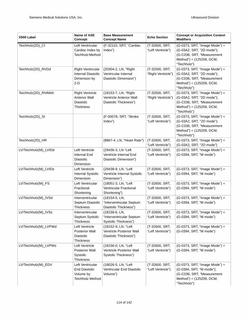
Siemens Medical Solutions USA, Inc. Ultrasound Division
Teich =
Teich =
Teich =
Teich =
Teich =
LV/Te =
LV/Te =
LV/Te =
LV/Te =
LV/Te =
LV/Te =
LV/Te =
LV/Te =
X500 t
holz(2D)_CI Left Ventricular Cardiac Index by Teichholz Method
(F-32110, SRT, “Cardiac Index”)
(T-32600, SRT, “Left Ventricle”)
(G-0373, SRT, “Image Mode”) (G-03A2, SRT, “2D mode”);(G-C036, SRT, “Measurement Method”) = (125209, DCM, “Teichholz”)
holz(2D)_RVDd Right Ventricular Internal Diastolic Dimension by 2-D
(20304-2, LN, “Right Ventricular Internal Diastolic Dimension”)
(T-32500, SRT,“Right Ventricle”)
(G-0373, SRT, “Image Mode”) (G-03A2, SRT, “2D mode”);(G-C036, SRT, “Measurement Method”) = (125209, DCM, “Teichholz”)
holz(2D)_RVAWd Right Ventricle Anterior Wall Diastolic Thickness
(18153-7, LN, “Right Ventricle Anterior Wall Diastolic Thickness”)
(T-32500, SRT,“Right Ventricle”)
(G-0373, SRT, “Image Mode”) (G-03A2, SRT, “2D mode”);(G-C036, SRT, “Measurement Method”) = (125209, DCM, “Teichholz”)
holz(2D)_SI (F-00078, SRT, “Stroke Index”)
(T-32600, SRT, “Left Ventricle”)
(G-0373, SRT, “Image Mode”) (G-03A2, SRT, “2D mode”);(G-C036, SRT, “Measurement Method”) = (125209, DCM, “Teichholz”)
holz(2D)_HR (8867-4, LN, “Heart Rate”) (T-32600, SRT, “Left Ventricle”)
(G-0373, SRT, “Image Mode”) (G-03A2, SRT, “2D mode”)
ichholz(M)_LVIDd Left Ventricle Internal End Diastolic Dimension
(29436-3, LN “Left Ventricle Internal End Diastolic Dimension”)
(T-32600, SRT, “Left Ventricle”)
(G-0373, SRT, “Image Mode”) (G-0394, SRT, “M mode”)
ichholz(M)_LVIDs Left Ventricle Internal Systolic Dimension
(29438-9, LN, “Left Ventricle Internal Systolic Dimension”)
(T-32600, SRT, “Left Ventricle”)
(G-0373, SRT, “Image Mode”) (G-0394, SRT, “M mode”)
ichholz(M)_FS Left Ventricular Fractional Shortening
(18051-3, LN, “Left Ventricular Fractional Shortening”)
(T-32600, SRT, “Left Ventricle”)
(G-0373, SRT, “Image Mode”) (G-0394, SRT, “M mode”)
ichholz(M)_IVSd Interventricular Septum Diastolic Thickness
(18154-5, LN, “Interventricular Septum Diastolic Thickness”)
(T-32600, SRT, “Left Ventricle”)
(G-0373, SRT, “Image Mode”) (G-0394, SRT, “M mode”)
ichholz(M)_IVSs Interventricular Septum Systolic Thickness
(18158-6, LN, “Interventricular Septum Systolic Thickness”)
(T-32600, SRT, “Left Ventricle”)
(G-0373, SRT, “Image Mode”) (G-0394, SRT, “M mode”)
ichholz(M)_LVPWd Left Ventricle Posterior Wall Diastolic Thickness
(18152-9, LN, “Left Ventricle Posterior Wall Diastolic Thickness”)
(T-32600, SRT, “Left Ventricle”)
(G-0373, SRT, “Image Mode”) (G-0394, SRT, “M mode”)
ichholz(M)_LVPWs Left Ventricle Posterior Wall Systolic Thickness
(18156-0, LN, “Left Ventricle Posterior Wall Systolic Thickness”)
(T-32600, SRT, “Left Ventricle”)
(G-0373, SRT, “Image Mode”) (G-0394, SRT, “M mode”)
ichholz(M)_EDV Left Ventricular End Diastolic Volume by Teichholz Method
(18026-5, LN, “Left Ventricular End Diastolic Volume”)
(T-32600, SRT, “Left Ventricle”)
(G-0373, SRT, “Image Mode”) (G-0394, SRT, “M mode”);(G-C036, SRT, “Measurement Method”) = (125209, DCM, “Teichholz”)
Label Name of ASE Concept
Base Measurement Concept Name Echo Section Concept or Acquisition Contex
Modifiers
114 of 142
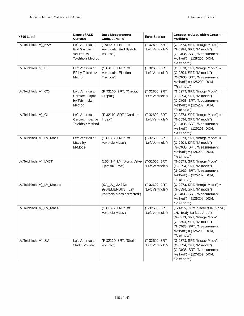
Siemens Medical Solutions USA, Inc. Ultrasound Division
LV/Te =
LV/Te =
LV/Te =
LV/Te =
LV/Te =
LV/Te =
LV/Te =
LV/Te 7-6,
=
LV/Te =
X500 t
ichholz(M)_ESV Left Ventricular End Systolic Volume by Teichholz Method
(18148-7, LN, “Left Ventricular End Systolic Volume”)
(T-32600, SRT, “Left Ventricle”)
(G-0373, SRT, “Image Mode”) (G-0394, SRT, “M mode”);(G-C036, SRT, “Measurement Method”) = (125209, DCM, “Teichholz”)
ichholz(M)_EF Left Ventricular EF by Teichholz Method
(18043-0, LN, “Left Ventricular Ejection Fraction”)
(T-32600, SRT, “Left Ventricle”)
(G-0373, SRT, “Image Mode”) (G-0394, SRT, “M mode”);(G-C036, SRT, “Measurement Method”) = (125209, DCM, “Teichholz”)
ichholz(M)_CO Left Ventricular Cardiac Output by Teichholz Method
(F-32100, SRT, “Cardiac Output”)
(T-32600, SRT, “Left Ventricle”)
(G-0373, SRT, “Image Mode”) (G-0394, SRT, “M mode”);(G-C036, SRT, “Measurement Method”) = (125209, DCM, “Teichholz”)
ichholz(M)_CI Left Ventricular Cardiac Index by Teichholz Method
(F-32110, SRT, “Cardiac Index”)
(T-32600, SRT, “Left Ventricle”)
(G-0373, SRT, “Image Mode”) (G-0394, SRT, “M mode”);(G-C036, SRT, “Measurement Method”) = (125209, DCM, “Teichholz”)
ichholz(M)_LV_Mass Left Ventricular Mass by M-Mode
(18087-7, LN, “Left Ventricle Mass”)
(T-32600, SRT, “Left Ventricle”)
(G-0373, SRT, “Image Mode”) (G-0394, SRT, “M mode”);(G-C036, SRT, “Measurement Method”) = (125209, DCM, “Teichholz”)
ichholz(M)_LVET (18041-4, LN, “Aortic Valve Ejection Time”)
(T-32600, SRT, “Left Ventricle”)
(G-0373, SRT, “Image Mode”) (G-0394, SRT, “M mode”);(G-C036, SRT, “Measurement Method”) = (125209, DCM, “Teichholz”)
ichholz(M)_LV_Mass-c (CA_LV_MASSc, 99SIEMENSUS, “Left Ventricle Mass corrected”)
(T-32600, SRT, “Left Ventricle”)
(G-0373, SRT, “Image Mode”) (G-0394, SRT, “M mode”);(G-C036, SRT, “Measurement Method”) = (125209, DCM, “Teichholz”)
ichholz(M)_LV_Mass-I (18087-7, LN, “Left Ventricle Mass”)
(T-32600, SRT, “Left Ventricle”)
(121425, DCM, “Index”) = (827LN, “Body Surface Area”);(G-0373, SRT, “Image Mode”) (G-0394, SRT, “M mode”);(G-C036, SRT, “Measurement Method”) = (125209, DCM, “Teichholz”)
ichholz(M)_SV Left Ventricular Stroke Volume
(F-32120, SRT, “Stroke Volume”)
(T-32600, SRT, “Left Ventricle”)
(G-0373, SRT, “Image Mode”) (G-0394, SRT, “M mode”);(G-C036, SRT, “Measurement Method”) = (125209, DCM, “Teichholz”)
Label Name of ASE Concept
Base Measurement Concept Name Echo Section Concept or Acquisition Contex
Modifiers
115 of 142
142
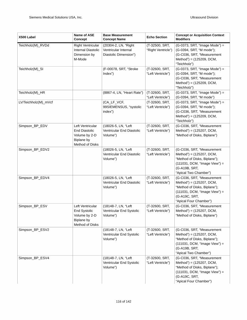
Siemens Medical Solutions USA, Inc. Ultrasound Division
Teich =
Teich =
Teich =
LV/Te =
Simp
Simp
Simp
Simp
Simp
Simp
X500 t
holz(M)_RVDd Right Ventricular Internal Diastolic Dimension by M-Mode
(20304-2, LN, “Right Ventricular Internal Diastolic Dimension”)
(T-32500, SRT,“Right Ventricle”)
(G-0373, SRT, “Image Mode”) (G-0394, SRT, “M mode”);(G-C036, SRT, “Measurement Method”) = (125209, DCM, “Teichholz”)
holz(M)_SI (F-00078, SRT, “Stroke Index”)
(T-32600, SRT, “Left Ventricle”)
(G-0373, SRT, “Image Mode”) (G-0394, SRT, “M mode”);(G-C036, SRT, “Measurement Method”) = (125209, DCM, “Teichholz”)
holz(M)_HR (8867-4, LN, “Heart Rate”) (T-32600, SRT, “Left Ventricle”)
(G-0373, SRT, “Image Mode”) (G-0394, SRT, “M mode”)
ichholz(M)_mVcf (CA_LF_VCF, 99SIEMENSUS, “systolic index”)
(T-32600, SRT, “Left Ventricle”)
(G-0373, SRT, “Image Mode”) (G-0394, SRT, “M mode”);(G-C036, SRT, “Measurement Method”) = (125209, DCM, “Teichholz”)
son_BP_EDV Left Ventricular End Diastolic Volume by 2-D Biplane by Method of Disks
(18026-5, LN, “Left Ventricular End Diastolic Volume”)
(T-32600, SRT, “Left Ventricle”)
(G-C036, SRT, “Measurement Method”) = (125207, DCM, “Method of Disks, Biplane”)
son_BP_EDV2 (18026-5, LN, “Left Ventricular End Diastolic Volume”)
(T-32600, SRT, “Left Ventricle”)
(G-C036, SRT, “Measurement Method”) = (125207, DCM, “Method of Disks, Biplane”);(111031, DCM, “Image View”) =(G-A19B, SRT,“Apical Two Chamber”)
son_BP_EDV4 (18026-5, LN, “Left Ventricular End Diastolic Volume”)
(T-32600, SRT, “Left Ventricle”)
(G-C036, SRT, “Measurement Method”) = (125207, DCM, “Method of Disks, Biplane”);(111031, DCM, “Image View”) =(G-A19C, SRT,“Apical Four Chamber”)
son_BP_ESV Left Ventricular End Systolic Volume by 2-D Biplane by Method of Disks
(18148-7, LN, “Left Ventricular End Systolic Volume”)
(T-32600, SRT, “Left Ventricle”)
(G-C036, SRT, “Measurement Method”) = (125207, DCM, “Method of Disks, Biplane”)
son_BP_ESV2 (18148-7, LN, “Left Ventricular End Systolic Volume”)
(T-32600, SRT, “Left Ventricle”)
(G-C036, SRT, “Measurement Method”) = (125207, DCM, “Method of Disks, Biplane”);(111031, DCM, “Image View”) =(G-A19B, SRT,“Apical Two Chamber”)
son_BP_ESV4 (18148-7, LN, “Left Ventricular End Systolic Volume”)
(T-32600, SRT, “Left Ventricle”)
(G-C036, SRT, “Measurement Method”) = (125207, DCM, “Method of Disks, Biplane”); (111031, DCM, “Image View”) =(G-A19C, SRT,“Apical Four Chamber”)
Label Name of ASE Concept
Base Measurement Concept Name Echo Section Concept or Acquisition Contex
Modifiers
116 of 142
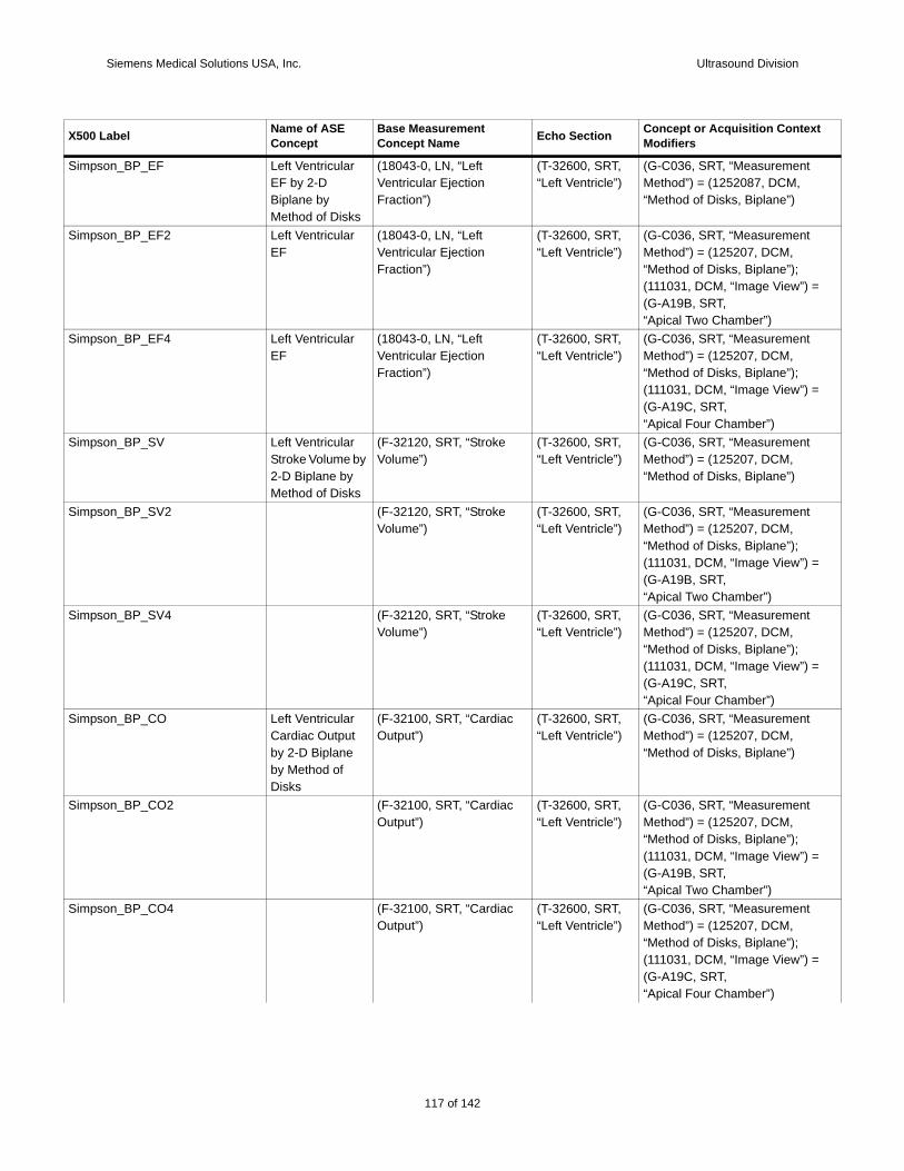
Siemens Medical Solutions USA, Inc. Ultrasound Division
Simp
Simp
Simp
Simp
Simp
Simp
Simp
Simp
Simp
X500 t
son_BP_EF Left Ventricular EF by 2-D Biplane by Method of Disks
(18043-0, LN, “Left Ventricular Ejection Fraction”)
(T-32600, SRT, “Left Ventricle”)
(G-C036, SRT, “Measurement Method”) = (1252087, DCM, “Method of Disks, Biplane”)
son_BP_EF2 Left Ventricular EF
(18043-0, LN, “Left Ventricular Ejection Fraction”)
(T-32600, SRT, “Left Ventricle”)
(G-C036, SRT, “Measurement Method”) = (125207, DCM, “Method of Disks, Biplane”); (111031, DCM, “Image View”) =(G-A19B, SRT,“Apical Two Chamber”)
son_BP_EF4 Left Ventricular EF
(18043-0, LN, “Left Ventricular Ejection Fraction”)
(T-32600, SRT, “Left Ventricle”)
(G-C036, SRT, “Measurement Method”) = (125207, DCM, “Method of Disks, Biplane”);(111031, DCM, “Image View”) =(G-A19C, SRT,“Apical Four Chamber”)
son_BP_SV Left Ventricular Stroke Volume by 2-D Biplane by Method of Disks
(F-32120, SRT, “Stroke Volume”)
(T-32600, SRT, “Left Ventricle”)
(G-C036, SRT, “Measurement Method”) = (125207, DCM, “Method of Disks, Biplane”)
son_BP_SV2 (F-32120, SRT, “Stroke Volume”)
(T-32600, SRT, “Left Ventricle”)
(G-C036, SRT, “Measurement Method”) = (125207, DCM, “Method of Disks, Biplane”);(111031, DCM, “Image View”) =(G-A19B, SRT,“Apical Two Chamber”)
son_BP_SV4 (F-32120, SRT, “Stroke Volume”)
(T-32600, SRT, “Left Ventricle”)
(G-C036, SRT, “Measurement Method”) = (125207, DCM, “Method of Disks, Biplane”);(111031, DCM, “Image View”) =(G-A19C, SRT,“Apical Four Chamber”)
son_BP_CO Left Ventricular Cardiac Output by 2-D Biplane by Method of Disks
(F-32100, SRT, “Cardiac Output”)
(T-32600, SRT, “Left Ventricle”)
(G-C036, SRT, “Measurement Method”) = (125207, DCM, “Method of Disks, Biplane”)
son_BP_CO2 (F-32100, SRT, “Cardiac Output”)
(T-32600, SRT, “Left Ventricle”)
(G-C036, SRT, “Measurement Method”) = (125207, DCM, “Method of Disks, Biplane”);(111031, DCM, “Image View”) =(G-A19B, SRT,“Apical Two Chamber”)
son_BP_CO4 (F-32100, SRT, “Cardiac Output”)
(T-32600, SRT, “Left Ventricle”)
(G-C036, SRT, “Measurement Method”) = (125207, DCM, “Method of Disks, Biplane”);(111031, DCM, “Image View”) =(G-A19C, SRT,“Apical Four Chamber”)
Label Name of ASE Concept
Base Measurement Concept Name Echo Section Concept or Acquisition Contex
Modifiers
117 of 142
142

Siemens Medical Solutions USA, Inc. Ultrasound Division
Simp
Simp
Simp
Simp
Simp
Simp
Simp
Simp
Simp
Simp
X500 t
son_BP_CI Left Ventricular Cardiac Index by 2-D Biplane by Method of Disks
(F-32110, SRT, “Cardiac Index”)
(T-32600, SRT, “Left Ventricle”)
(G-C036, SRT, “Measurement Method”) = (125207, DCM, “Method of Disks, Biplane”)
son_BP_CI2 (F-32110, SRT, “Cardiac Index”)
(T-32600, SRT, “Left Ventricle”)
(G-C036, SRT, “Measurement Method”) = (125207, DCM, “Method of Disks, Biplane”);(111031, DCM, “Image View”) =(G-A19B, SRT,“Apical Two Chamber”)
son_BP_CI4 (F-32110, SRT, “Cardiac Index”)
(T-32600, SRT, “Left Ventricle”)
(G-C036, SRT, “Measurement Method”) = (125207, DCM, “Method of Disks, Biplane”);(111031, DCM, “Image View”) =(G-A19C, SRT,“Apical Four Chamber”)
son_BP_SI (F-00078, SRT, “Stroke Index”)
(T-32600, SRT, “Left Ventricle”)
(G-C036, SRT, “Measurement Method”) = (125207, DCM, “Method of Disks, Biplane”)
son_BP_SI2 (F-00078, SRT, “Stroke Index”)
(T-32600, SRT, “Left Ventricle”)
(G-C036, SRT, “Measurement Method”) = (125207, DCM, “Method of Disks, Biplane”);(111031, DCM, “Image View”) =(G-A19B, SRT,“Apical Two Chamber”)
son_BP_SI4 (F-00078, SRT, “Stroke Index”)
(T-32600, SRT, “Left Ventricle”)
(G-C036, SRT, “Measurement Method”) = (125207, DCM, “Method of Disks, Biplane”);(111031, DCM, “Image View”) =(G-A19C, SRT,“Apical Four Chamber”)
son_BP_HR (8867-4, LN, “Heart Rate”) (T-32600, SRT, “Left Ventricle”)
son_BP_difD(2ch) (CA_LV_DIF, 99SIEMENSUS, LV difference)
(T-32600, SRT, “Left Ventricle”)
(G-C036, SRT, “Measurement Method”) = (125207, DCM, “Method of Disks, Biplane”);(R-4089A, SRT, “Cardiac CyclePoint”) = (F-32011, SRT, “End Diastole”)
son_BP_difD(4ch) (CA_LV_DIF, 99SIEMENSUS, LV difference)
(T-32600, SRT, “Left Ventricle”)
(G-C036, SRT, “Measurement Method”) = (125207, DCM, “Method of Disks, Biplane”);(R-4089A, SRT, “Cardiac CyclePoint”) = (F-32011, SRT, “End Diastole”)
son_BP_difS(2ch) (CA_LV_DIF, 99SIEMENSUS, LV difference)
(T-32600, SRT, “Left Ventricle”)
(G-C036, SRT, “Measurement Method”) = (125207, DCM, “Method of Disks, Biplane”);(109070, DCM, “Cardiac CyclePoint”) = (F-32011, SRT, “End Systole”)
Label Name of ASE Concept
Base Measurement Concept Name Echo Section Concept or Acquisition Contex
Modifiers
118 of 142
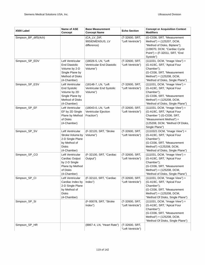
Siemens Medical Solutions USA, Inc. Ultrasound Division
Simp
Simp
”) Simp
”) Simp
ks,
Simp =
”) Simp
”)Simp
”)Simp
”)Simp
X500 t
son_BP_difS(4ch) (CA_LV_DIF, 99SIEMENSUS, LV difference)
(T-32600, SRT, “Left Ventricle”)
(G-C036, SRT, “Measurement Method”) = (125207, DCM, “Method of Disks, Biplane”);(109070, DCM, “Cardiac CyclePoint”) = (F-32011, SRT, “End Systole”)
son_SP_EDV Left Ventricular End Diastolic Volume by 2-D Single Plane by Method of Disks (4-Chamber)
(18026-5, LN, “Left Ventricular End Diastolic Volume”)
(T-32600, SRT, “Left Ventricle”)
(111031, DCM, “Image View”) =(G-A19C, SRT, “Apical Four Chamber”);(G-C036, SRT, “Measurement Method”) = (125208, DCM, “Method of Disks, Single Plane
son_SP_ESV Left Ventricular End Systolic Volume by 2D Single Plane by Method of Disks (4-Chamber)
(18148-7, LN, “Left Ventricular End Systolic Volume”)
(T-32600, SRT, “Left Ventricle”)
(111031, DCM, “Image View”) =(G-A19C, SRT, “Apical Four Chamber”);(G-C036, SRT, “Measurement Method”) = (125208, DCM, “Method of Disks, Single Plane
son_SP_EF Left Ventricular EF by 2D Single Plane by Method of Disks (4-Chamber)
(18043-0, LN, “Left Ventricular Ejection Fraction”)
(T-32600, SRT, “Left Ventricle”)
(111031, DCM, “Image View”) =(G-A19C, SRT, “Apical Four Chamber ”) (G-C036, SRT, “Measurement Method”) = (125208, DCM, “Method Of DisSingle Plane”)
son_SP_SV Left Ventricular Stroke Volume by 2-D Single Plane by Method of Disks (4-Chamber)
(F-32120, SRT, “Stroke Volume”)
(T-32600, SRT, “Left Ventricle”)
(1110321 DCM, “Image View”) (G-A19C, SRT, “Apical Four Chamber”);(G-C036, SRT, “Measurement Method”) =(125208, DCM, “Method of Disks, Single Plane
son_SP_CO Left Ventricular Cardiac Output by 2-D Single Plane by Method of Disks (4-Chamber)
(F-32100, SRT, “Cardiac Output”)
(T-32600, SRT, “Left Ventricle”)
(111031, DCM, “Image View”) =(G-A19C, SRT, “Apical Four Chamber”);(G-C036, SRT, “Measurement Method”) = (125208, DCM, “Method of Disks, Single Plane
son_SP_CI Left Ventricular Cardiac Index by 2-D Single Plane by Method of Disks (4-Chamber)
(F-32110, SRT, “Cardiac Index”)
(T-32600, SRT, “Left Ventricle”)
(111031, DCM, “Image View”) =(G-A19C, SRT, “Apical Four Chamber”);(G-C036, SRT, “Measurement Method”) = (125208, DCM, “Method Of Disks, Single Plane
son_SP_SI (F-00078, SRT, “Stroke Index”)
(T-32600, SRT, “Left Ventricle”)
(111031, DCM, “Image View”) =(G-A19C, SRT, “Apical Four Chamber”);(G-C036, SRT, “Measurement Method”) = (125208, DCM, “Method Of Disks, Single Plane
son_SP_HR (8867-4, LN, “Heart Rate”) (T-32600, SRT, “Left Ventricle”)
Label Name of ASE Concept
Base Measurement Concept Name Echo Section Concept or Acquisition Contex
Modifiers
119 of 142
142
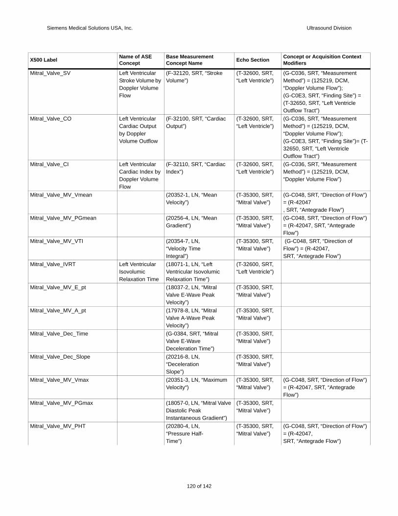
Siemens Medical Solutions USA, Inc. Ultrasound Division
Mitra
=
Mitra
(T-
Mitra
Mitra w”)
Mitra w”)
Mitra
Mitra
Mitra
Mitra
Mitra
Mitra
Mitra w”)
Mitra
Mitra w”)
X500 t
l_Valve_SV Left Ventricular Stroke Volume by Doppler Volume Flow
(F-32120, SRT, “Stroke Volume”)
(T-32600, SRT, “Left Ventricle”)
(G-C036, SRT, “Measurement Method”) = (125219, DCM, “Doppler Volume Flow”);(G-C0E3, SRT, “Finding Site”) (T-32650, SRT, “Left Ventricle Outflow Tract”)
l_Valve_CO Left Ventricular Cardiac Output by Doppler Volume Outflow
(F-32100, SRT, “Cardiac Output”)
(T-32600, SRT, “Left Ventricle”)
(G-C036, SRT, “Measurement Method”) = (125219, DCM, “Doppler Volume Flow”);(G-C0E3, SRT, “Finding Site”)=32650, SRT, “Left Ventricle Outflow Tract”)
l_Valve_CI Left Ventricular Cardiac Index by Doppler Volume Flow
(F-32110, SRT, “Cardiac Index”)
(T-32600, SRT, “Left Ventricle”)
(G-C036, SRT, “Measurement Method”) = (125219, DCM, “Doppler Volume Flow”)
l_Valve_MV_Vmean (20352-1, LN, “Mean Velocity”)
(T-35300, SRT, “Mitral Valve”)
(G-C048, SRT, “Direction of Flo= (R-42047, SRT, “Antegrade Flow”)
l_Valve_MV_PGmean (20256-4, LN, “Mean Gradient”)
(T-35300, SRT, “Mitral Valve”)
(G-C048, SRT, “Direction of Flo= (R-42047, SRT, “Antegrade Flow”)
l_Valve_MV_VTI (20354-7, LN,“Velocity TimeIntegral”)
(T-35300, SRT, “Mitral Valve”)
(G-C048, SRT, “Direction of Flow”) = (R-42047,SRT, “Antegrade Flow”)
l_Valve_IVRT Left Ventricular IsovolumicRelaxation Time
(18071-1, LN, “LeftVentricular IsovolumicRelaxation Time”)
(T-32600, SRT, “Left Ventricle”)
l_Valve_MV_E_pt (18037-2, LN, “MitralValve E-Wave PeakVelocity”)
(T-35300, SRT, “Mitral Valve”)
l_Valve_MV_A_pt (17978-8, LN, “MitralValve A-Wave PeakVelocity”)
(T-35300, SRT, “Mitral Valve”)
l_Valve_Dec_Time (G-0384, SRT, “MitralValve E-WaveDeceleration Time”)
(T-35300, SRT, “Mitral Valve”)
l_Valve_Dec_Slope (20216-8, LN,“DecelerationSlope”)
(T-35300, SRT, “Mitral Valve”)
l_Valve_MV_Vmax (20351-3, LN, “Maximum Velocity“)
(T-35300, SRT, “Mitral Valve”)
(G-C048, SRT, “Direction of Flo= (R-42047, SRT, “Antegrade Flow”)
l_Valve_MV_PGmax (18057-0, LN, “Mitral Valve Diastolic Peak Instantaneous Gradient”)
(T-35300, SRT, “Mitral Valve”)
l_Valve_MV_PHT (20280-4, LN,“Pressure Half-Time”)
(T-35300, SRT, “Mitral Valve”)
(G-C048, SRT, “Direction of Flo= (R-42047,SRT, “Antegrade Flow”)
Label Name of ASE Concept
Base Measurement Concept Name Echo Section Concept or Acquisition Contex
Modifiers
120 of 142
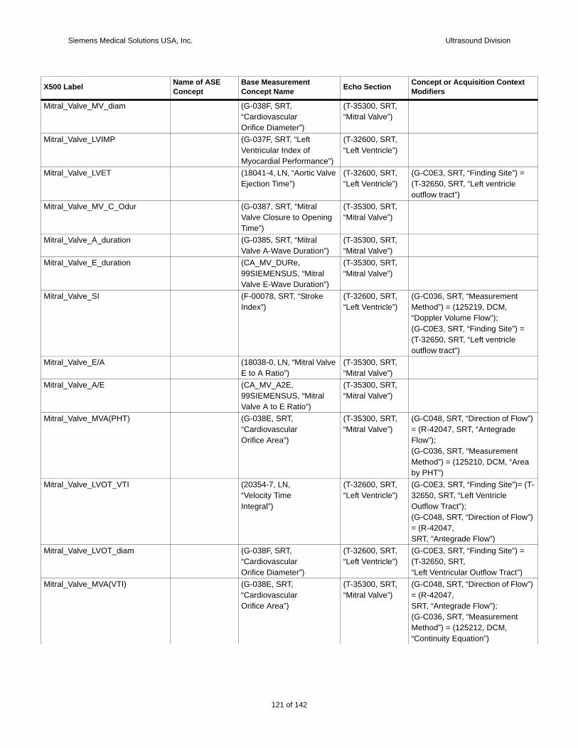
Siemens Medical Solutions USA, Inc. Ultrasound Division
Mitra
Mitra
Mitra =
Mitra
Mitra
Mitra
Mitra
=
Mitra
Mitra
Mitra w”)
ea
Mitra (T-
w”)
Mitra =
)Mitra w”)
X500 t
l_Valve_MV_diam (G-038F, SRT,“CardiovascularOrifice Diameter”)
(T-35300, SRT, “Mitral Valve”)
l_Valve_LVIMP (G-037F, SRT, “Left Ventricular Index of Myocardial Performance”)
(T-32600, SRT, “Left Ventricle”)
l_Valve_LVET (18041-4, LN, “Aortic Valve Ejection Time”)
(T-32600, SRT, “Left Ventricle”)
(G-C0E3, SRT, “Finding Site”) (T-32650, SRT, “Left ventricle outflow tract”)
l_Valve_MV_C_Odur (G-0387, SRT, “Mitral Valve Closure to Opening Time”)
(T-35300, SRT, “Mitral Valve”)
l_Valve_A_duration (G-0385, SRT, “Mitral Valve A-Wave Duration”)
(T-35300, SRT, “Mitral Valve”)
l_Valve_E_duration (CA_MV_DURe, 99SIEMENSUS, “Mitral Valve E-Wave Duration”)
(T-35300, SRT, “Mitral Valve”)
l_Valve_SI (F-00078, SRT, “Stroke Index”)
(T-32600, SRT, “Left Ventricle”)
(G-C036, SRT, “Measurement Method”) = (125219, DCM, “Doppler Volume Flow”);(G-C0E3, SRT, “Finding Site”) (T-32650, SRT, “Left ventricle outflow tract”)
l_Valve_E/A (18038-0, LN, “Mitral Valve E to A Ratio”)
(T-35300, SRT, “Mitral Valve”)
l_Valve_A/E (CA_MV_A2E, 99SIEMENSUS, “Mitral Valve A to E Ratio”)
(T-35300, SRT, “Mitral Valve”)
l_Valve_MVA(PHT) (G-038E, SRT,“CardiovascularOrifice Area”)
(T-35300, SRT, “Mitral Valve”)
(G-C048, SRT, “Direction of Flo= (R-42047, SRT, “Antegrade Flow”);(G-C036, SRT, “Measurement Method”) = (125210, DCM, “Arby PHT”)
l_Valve_LVOT_VTI (20354-7, LN,“Velocity TimeIntegral”)
(T-32600, SRT, “Left Ventricle”)
(G-C0E3, SRT, “Finding Site”)=32650, SRT, “Left Ventricle Outflow Tract”);(G-C048, SRT, “Direction of Flo= (R-42047,SRT, “Antegrade Flow”)
l_Valve_LVOT_diam (G-038F, SRT,“CardiovascularOrifice Diameter”)
(T-32600, SRT, “Left Ventricle”)
(G-C0E3, SRT, “Finding Site”) (T-32650, SRT,“Left Ventricular Outflow Tract”
l_Valve_MVA(VTI) (G-038E, SRT,“CardiovascularOrifice Area”)
(T-35300, SRT, “Mitral Valve”)
(G-C048, SRT, “Direction of Flo= (R-42047,SRT, “Antegrade Flow”);(G-C036, SRT, “Measurement Method”) = (125212, DCM, “Continuity Equation”)
Label Name of ASE Concept
Base Measurement Concept Name Echo Section Concept or Acquisition Contex
Modifiers
121 of 142
142
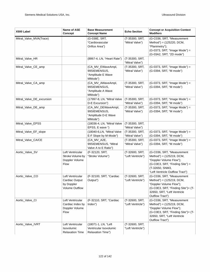
Siemens Medical Solutions USA, Inc. Ultrasound Division
Mitra
=
Mitra
Mitra =
Mitra =
Mitra =
Mitra =
Mitra
Mitra =
Mitra =
Aorti
=
Aorti
(T-
Aorti
(T-
Aorti
X500 t
l_Valve_MVA(Trace) (G-038E, SRT,“CardiovascularOrifice Area”)
(T-35300, SRT, “Mitral Valve”)
(G-C036, SRT, “Measurement Method”) = (125220, DCM, “Planimetry”);(G-0373, SRT, “Image Mode”) (G-03A2, SRT, “2D mode”)
l_Valve_HR (8867-4, LN, “Heart Rate”) (T-35300, SRT, “Mitral Valve”)
l_Valve_CE_amp (CA_MV_EWaveAmpl, 99SIEMENSUS, “Amplitude E Wave MMode”)
(T-35300, SRT, “Mitral Valve”)
(G-0373, SRT, “Image Mode”) (G-0394, SRT, “M mode”)
l_Valve_CA_amp (CA_MV_AWaveAmpl, 99SIEMENSUS, “Amplitude A Wave MMode”)
(T-35300, SRT, “Mitral Valve”)
(G-0373, SRT, “Image Mode”) (G-0394, SRT, “M mode”)
l_Valve_DE_excursion (17997-8, LN, “Mitral Valve D-E Excursion”)
(T-35300, SRT, “Mitral Valve”)
(G-0373, SRT, “Image Mode”) (G-0394, SRT, “M mode”)
l_Valve_DE_amp (CA_MV_DEWaveAmpl, 99SIEMENSUS, “Amplitude D-E Wave MMode”)
(T-35300, SRT, “Mitral Valve”)
(G-0373, SRT, “Image Mode”) (G-0394, SRT, “M mode”)
l_Valve_EPSS (18036-4, LN, “Mitral Valve EPSS, E wave “)
(T-35300, SRT, “Mitral Valve”)
l_Valve_EF_slope (18040-6,LN, “Mitral Valve E-F Slope by M-Mode”)
(T-35300, SRT, “Mitral Valve”)
(G-0373, SRT, “Image Mode”) (G-0394, SRT, “M mode”)
l_Valve_CA/CE (CA_MV_A2E, 99SIEMENSUS, “Mitral Valve A to E Ratio”)
(T-35300, SRT, “Mitral Valve”)
(G-0373, SRT, “Image Mode”) (G-0394, SRT, “M mode”)
c_Valve_SV Left Ventricular Stroke Volume by Doppler Volume Flow
(F-32120, SRT,“Stroke Volume”)
(T-32600, SRT, “Left Ventricle”)
(G-C036, SRT, “Measurement Method”) = (125219, DCM, “Doppler Volume Flow”);(G-C0E3, SRT, “Finding Site”) (T-32650, SNM3,“Left Ventricle Outflow Tract”)
c_Valve_CO Left Ventricular Cardiac Output by Doppler Volume Outflow
(F-32100, SRT, “Cardiac Output”)
(T-32600, SRT, “Left Ventricle”)
(G-C036, SRT, “Measurement Method”) = (125219, DCM, “Doppler Volume Flow”);(G-C0E3, SRT, “Finding Site”)=32650, SRT, “Left Ventricle Outflow Tract”)
c_Valve_CI Left Ventricular Cardiac Index by Doppler Volume Flow
(F-32110, SRT, “Cardiac Index”)
(T-32600, SRT, “Left Ventricle”)
(G-C036, SRT, “Measurement Method”) = (125219, DCM, “Doppler Volume Flow”);(G-C0E3, SRT, “Finding Site”)=32650, SRT, “Left Ventricle Outflow Tract”)
c_Valve_IVRT Left Ventricular IsovolumicRelaxation Time
(18071-1, LN, “LeftVentricular IsovolumicRelaxation Time”)
(T-32600, SRT, “Left Ventricle”)
Label Name of ASE Concept
Base Measurement Concept Name Echo Section Concept or Acquisition Contex
Modifiers
122 of 142
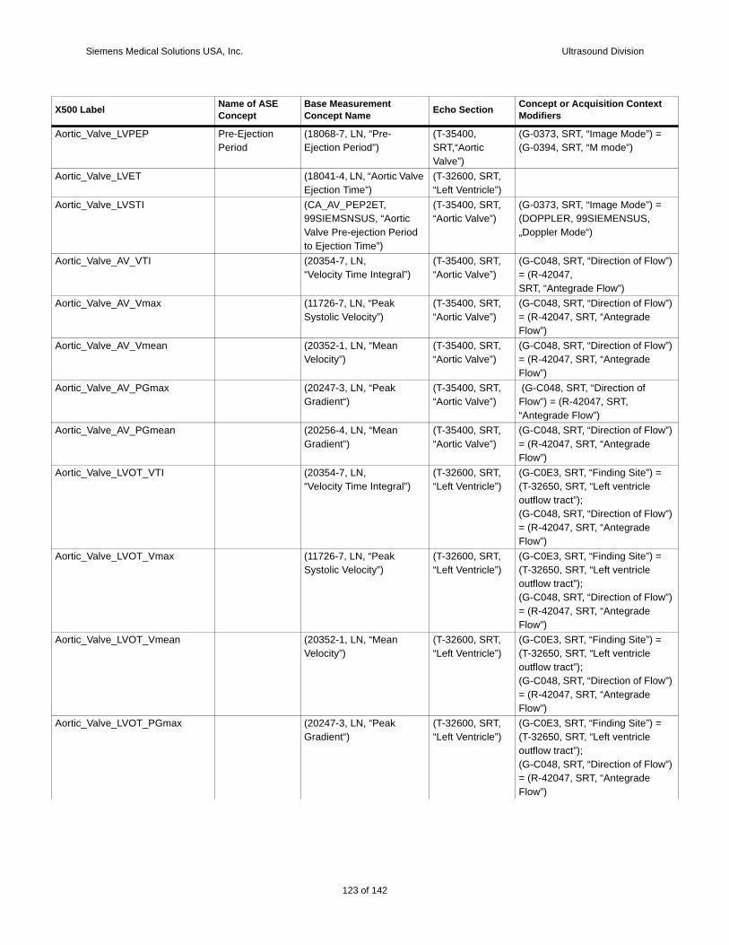
Siemens Medical Solutions USA, Inc. Ultrasound Division
Aorti =
Aorti
Aorti =
Aorti w”)
Aorti w”)
Aorti w”)
Aorti
Aorti w”)
Aorti =
w”)
Aorti =
w”)
Aorti =
w”)
Aorti =
w”)
X500 t
c_Valve_LVPEP Pre-Ejection Period
(18068-7, LN, “Pre-Ejection Period”)
(T-35400, SRT,“Aortic Valve”)
(G-0373, SRT, “Image Mode”) (G-0394, SRT, “M mode”)
c_Valve_LVET (18041-4, LN, “Aortic Valve Ejection Time”)
(T-32600, SRT, “Left Ventricle”)
c_Valve_LVSTI (CA_AV_PEP2ET, 99SIEMSNSUS, “Aortic Valve Pre-ejection Period to Ejection Time”)
(T-35400, SRT, “Aortic Valve”)
(G-0373, SRT, “Image Mode”) (DOPPLER, 99SIEMENSUS, „Doppler Mode“)
c_Valve_AV_VTI (20354-7, LN,“Velocity Time Integral”)
(T-35400, SRT, “Aortic Valve”)
(G-C048, SRT, “Direction of Flo= (R-42047,SRT, “Antegrade Flow”)
c_Valve_AV_Vmax (11726-7, LN, “Peak Systolic Velocity”)
(T-35400, SRT, “Aortic Valve”)
(G-C048, SRT, “Direction of Flo= (R-42047, SRT, “Antegrade Flow”)
c_Valve_AV_Vmean (20352-1, LN, “Mean Velocity”)
(T-35400, SRT, “Aortic Valve”)
(G-C048, SRT, “Direction of Flo= (R-42047, SRT, “Antegrade Flow”)
c_Valve_AV_PGmax (20247-3, LN, “Peak Gradient“)
(T-35400, SRT, “Aortic Valve”)
(G-C048, SRT, “Direction of Flow”) = (R-42047, SRT, “Antegrade Flow”)
c_Valve_AV_PGmean (20256-4, LN, “Mean Gradient”)
(T-35400, SRT, “Aortic Valve”)
(G-C048, SRT, “Direction of Flo= (R-42047, SRT, “Antegrade Flow”)
c_Valve_LVOT_VTI (20354-7, LN,“Velocity Time Integral”)
(T-32600, SRT, “Left Ventricle”)
(G-C0E3, SRT, “Finding Site”) (T-32650, SRT, “Left ventricle outflow tract”);(G-C048, SRT, “Direction of Flo= (R-42047, SRT, “Antegrade Flow”)
c_Valve_LVOT_Vmax (11726-7, LN, “Peak Systolic Velocity”)
(T-32600, SRT, “Left Ventricle”)
(G-C0E3, SRT, “Finding Site”) (T-32650, SRT, “Left ventricle outflow tract”);(G-C048, SRT, “Direction of Flo= (R-42047, SRT, “Antegrade Flow”)
c_Valve_LVOT_Vmean (20352-1, LN, “Mean Velocity”)
(T-32600, SRT, “Left Ventricle”)
(G-C0E3, SRT, “Finding Site”) (T-32650, SRT, “Left ventricle outflow tract”);(G-C048, SRT, “Direction of Flo= (R-42047, SRT, “Antegrade Flow”)
c_Valve_LVOT_PGmax (20247-3, LN, “Peak Gradient“)
(T-32600, SRT, “Left Ventricle”)
(G-C0E3, SRT, “Finding Site”) (T-32650, SRT, “Left ventricle outflow tract”);(G-C048, SRT, “Direction of Flo= (R-42047, SRT, “Antegrade Flow”)
Label Name of ASE Concept
Base Measurement Concept Name Echo Section Concept or Acquisition Contex
Modifiers
123 of 142
142

Siemens Medical Solutions USA, Inc. Ultrasound Division
Aorti =
w”)
Aorti =
)Aorti w”)
y
Aorti
w”)
Aorti
Aorti
=
Aorti
Aorti =
Aorti =
Aorti
Aorti
TR_T w”)
X500 t
c_Valve_LVOT_PGmean (20256-4, LN, “Mean Gradient”)
(T-32600, SRT, “Left Ventricle”)
(G-C0E3, SRT, “Finding Site”) (T-32650, SRT, “Left ventricle outflow tract”);(G-C048, SRT, “Direction of Flo= (R-42047, SRT, “Antegrade Flow”)
c_Valve_LVOT_diam (G-038F, SRT,“CardiovascularOrifice Diameter”)
(T-32600, SRT, “Left Ventricle”)
(G-C0E3, SRT, “Finding Site”) (T-32650, SRT,“Left Ventricular Outflow Tract”
c_Valve_AVA(VTI) (G-038E, SRT,“CardiovascularOrifice Area”)
(T-35400, SRT, “Aortic Valve”);
(G-C048, SRT, “Direction of Flo= (R-42047, SRT, “Antegrade Flow”);(G-C036, SRT, “Measurement Method”) = (125215, DCM, “Continuity Equation by VelocitTime Integral”)
c_Valve_AVA(Vmax) (G-038E, SRT,“CardiovascularOrifice Area”)
(T-35400, SRT, “Aortic Valve”);
(G-C036, SRT, “Measurement Method”) = (125214, DCM, “Continuity Equation by Peak Velocity”);(G-C048, SRT, “Direction of Flo= (R-42047, SRT, “Antegrade Flow”)
c_Valve_AVA(Trace) (G-038E, SRT,“CardiovascularOrifice Area”)
(T-35400, SRT, “Aortic Valve”)
(G-C036, SRT, “Measurement Method”) = (125220, DCM, “Planimetry”)
c_Valve_SI (F-00078, SRT, “Stroke Index”)
(T-32600, SRT, “Left Ventricle”)
(G-C036, SRT, “Measurement Method”) = (125219, DCM, “Doppler Volume Flow”);(G-C0E3, SRT, “Finding Site”) (T-32650, SRT, “Left ventricle outflow tract”);
c_Valve_HR (8867-4, LN, “Heart Rate”) (T-35400, SRT, “Aortic Valve”)
c_Valve_VSD_Vmax (11726-7, LN, “Peak Systolic Velocity”)
(T-32600, SRT, “Left Ventricle”)
(G-C0E3, SRT, “Finding Site”) (D4-31150, SRT,“Ventricular Septal Defect”)
c_Valve_VSD_PGmax (20247-3, LN, “PeakGradient”)
(T-32600, SRT, “Left Ventricle”)
(G-C0E3, SRT, “Finding Site”) (D4-31150, SRT,“Ventricular Septal Defect”)
c_Valve_Qp/Qs (29462-9, LN,“Pulmonary-to-Systemic Shunt Flow Ratio”)
(P5-30031, SRT,“Cardiac Shunt Study”)
(G-C036, SRT, “Measurement Method”) = (125219, DCM, “Doppler Volume Flow”)
c_Valve_Qp-Qs (CA_CS_PF-SF, 99SIEMENSUS, “Pulmonary minus Systemic Shunt Flow”)
(P5-30031, SRT, “Cardiac Shunt Study”)
R_Vmax Tricuspid Valve Regurgitant Peak Velocity
(11726-7, LN, “Peak Systolic Velocity”)
(T-35100, SRT, “Tricuspid valve”)
(G-C048, SRT, “Direction of Flo= (R-42E61, SRT, “RegurgitantFlow”)
Label Name of ASE Concept
Base Measurement Concept Name Echo Section Concept or Acquisition Contex
Modifiers
124 of 142
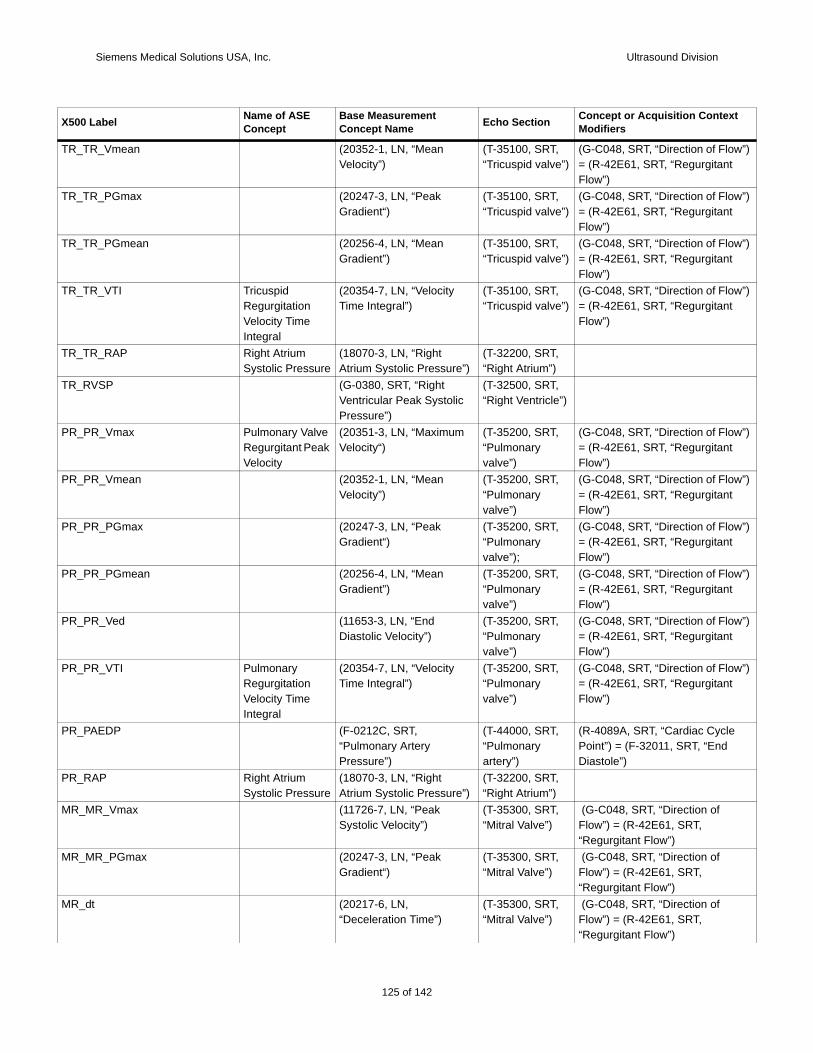
Siemens Medical Solutions USA, Inc. Ultrasound Division
TR_T w”)
TR_T w”)
TR_T w”)
TR_T w”)
TR_T
TR_R
PR_P w”)
PR_P w”)
PR_P w”)
PR_P w”)
PR_P w”)
PR_P w”)
PR_P
PR_R
MR_
MR_
MR_
X500 t
R_Vmean (20352-1, LN, “Mean Velocity”)
(T-35100, SRT, “Tricuspid valve”)
(G-C048, SRT, “Direction of Flo= (R-42E61, SRT, “RegurgitantFlow”)
R_PGmax (20247-3, LN, “Peak Gradient“)
(T-35100, SRT, “Tricuspid valve”)
(G-C048, SRT, “Direction of Flo= (R-42E61, SRT, “RegurgitantFlow”)
R_PGmean (20256-4, LN, “Mean Gradient”)
(T-35100, SRT, “Tricuspid valve”)
(G-C048, SRT, “Direction of Flo= (R-42E61, SRT, “RegurgitantFlow”)
R_VTI Tricuspid Regurgitation Velocity Time Integral
(20354-7, LN, “Velocity Time Integral”)
(T-35100, SRT, “Tricuspid valve”)
(G-C048, SRT, “Direction of Flo= (R-42E61, SRT, “RegurgitantFlow”)
R_RAP Right Atrium Systolic Pressure
(18070-3, LN, “Right Atrium Systolic Pressure”)
(T-32200, SRT,“Right Atrium”)
VSP (G-0380, SRT, “Right Ventricular Peak Systolic Pressure”)
(T-32500, SRT,“Right Ventricle”)
R_Vmax Pulmonary Valve Regurgitant Peak Velocity
(20351-3, LN, “Maximum Velocity“)
(T-35200, SRT, “Pulmonary valve”)
(G-C048, SRT, “Direction of Flo= (R-42E61, SRT, “RegurgitantFlow”)
R_Vmean (20352-1, LN, “Mean Velocity”)
(T-35200, SRT, “Pulmonary valve”)
(G-C048, SRT, “Direction of Flo= (R-42E61, SRT, “RegurgitantFlow”)
R_PGmax (20247-3, LN, “Peak Gradient“)
(T-35200, SRT, “Pulmonary valve”);
(G-C048, SRT, “Direction of Flo= (R-42E61, SRT, “RegurgitantFlow”)
R_PGmean (20256-4, LN, “Mean Gradient”)
(T-35200, SRT, “Pulmonary valve”)
(G-C048, SRT, “Direction of Flo= (R-42E61, SRT, “RegurgitantFlow”)
R_Ved (11653-3, LN, “EndDiastolic Velocity”)
(T-35200, SRT, “Pulmonary valve”)
(G-C048, SRT, “Direction of Flo= (R-42E61, SRT, “RegurgitantFlow”)
R_VTI Pulmonary Regurgitation Velocity Time Integral
(20354-7, LN, “Velocity Time Integral”)
(T-35200, SRT, “Pulmonary valve”)
(G-C048, SRT, “Direction of Flo= (R-42E61, SRT, “RegurgitantFlow”)
AEDP (F-0212C, SRT, “Pulmonary Artery Pressure”)
(T-44000, SRT, “Pulmonary artery”)
(R-4089A, SRT, “Cardiac CyclePoint”) = (F-32011, SRT, “End Diastole”)
AP Right Atrium Systolic Pressure
(18070-3, LN, “Right Atrium Systolic Pressure”)
(T-32200, SRT,“Right Atrium”)
MR_Vmax (11726-7, LN, “Peak Systolic Velocity”)
(T-35300, SRT, “Mitral Valve”)
(G-C048, SRT, “Direction of Flow”) = (R-42E61, SRT, “Regurgitant Flow”)
MR_PGmax (20247-3, LN, “Peak Gradient“)
(T-35300, SRT, “Mitral Valve”)
(G-C048, SRT, “Direction of Flow”) = (R-42E61, SRT, “Regurgitant Flow”)
dt (20217-6, LN, “Deceleration Time”)
(T-35300, SRT, “Mitral Valve”)
(G-C048, SRT, “Direction of Flow”) = (R-42E61, SRT, “Regurgitant Flow”)
Label Name of ASE Concept
Base Measurement Concept Name Echo Section Concept or Acquisition Contex
Modifiers
125 of 142
142
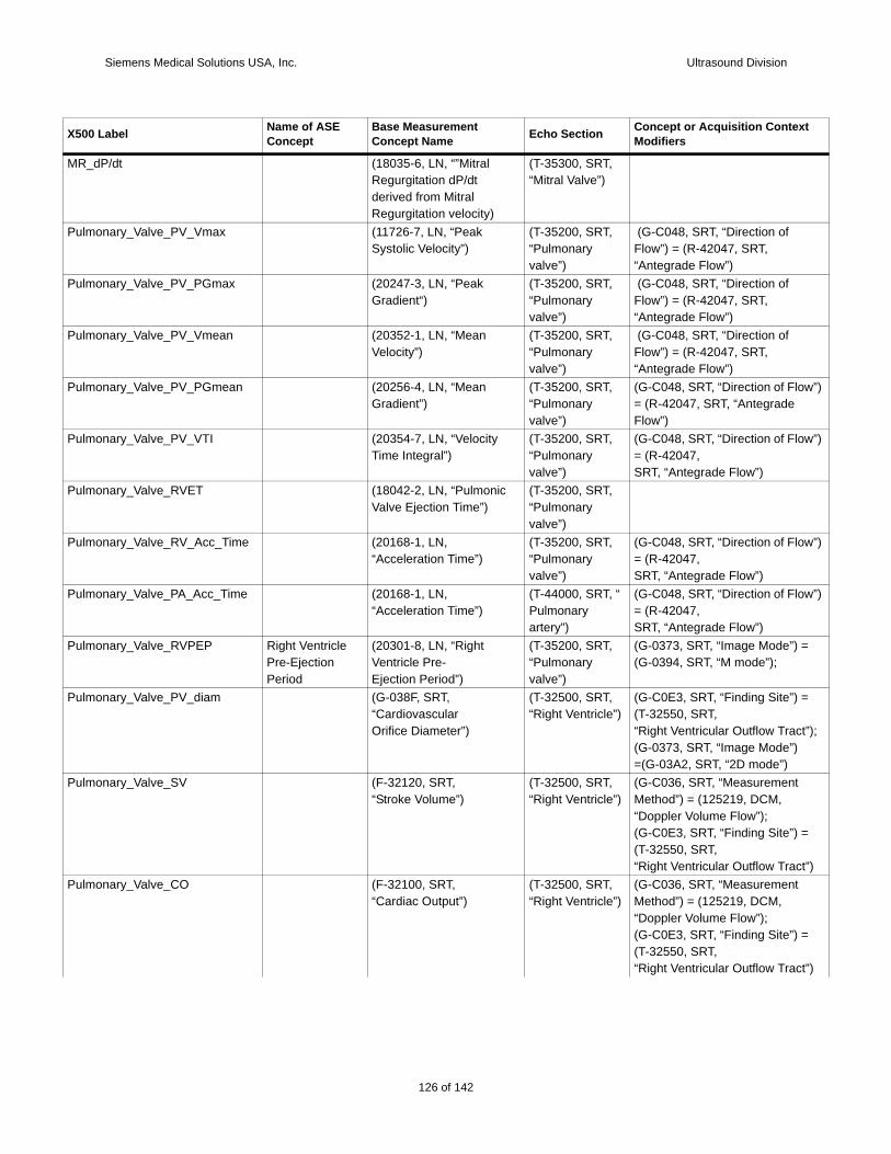
Siemens Medical Solutions USA, Inc. Ultrasound Division
MR_
Pulm
Pulm
Pulm
Pulm w”)
Pulm w”)
Pulm
Pulm w”)
Pulm w”)
Pulm =
Pulm =
t”);
Pulm
=
t”)Pulm
=
t”)
X500 t
dP/dt (18035-6, LN, “”Mitral Regurgitation dP/dt derived from MitralRegurgitation velocity)
(T-35300, SRT, “Mitral Valve”)
onary_Valve_PV_Vmax (11726-7, LN, “Peak Systolic Velocity”)
(T-35200, SRT, “Pulmonary valve”)
(G-C048, SRT, “Direction of Flow”) = (R-42047, SRT, “Antegrade Flow”)
onary_Valve_PV_PGmax (20247-3, LN, “Peak Gradient“)
(T-35200, SRT, “Pulmonary valve”)
(G-C048, SRT, “Direction of Flow”) = (R-42047, SRT, “Antegrade Flow”)
onary_Valve_PV_Vmean (20352-1, LN, “Mean Velocity”)
(T-35200, SRT, “Pulmonary valve”)
(G-C048, SRT, “Direction of Flow”) = (R-42047, SRT, “Antegrade Flow”)
onary_Valve_PV_PGmean (20256-4, LN, “Mean Gradient”)
(T-35200, SRT, “Pulmonary valve”)
(G-C048, SRT, “Direction of Flo= (R-42047, SRT, “Antegrade Flow”)
onary_Valve_PV_VTI (20354-7, LN, “Velocity Time Integral”)
(T-35200, SRT, “Pulmonary valve”)
(G-C048, SRT, “Direction of Flo= (R-42047,SRT, “Antegrade Flow”)
onary_Valve_RVET (18042-2, LN, “Pulmonic Valve Ejection Time”)
(T-35200, SRT, “Pulmonary valve”)
onary_Valve_RV_Acc_Time (20168-1, LN,“Acceleration Time”)
(T-35200, SRT, “Pulmonary valve”)
(G-C048, SRT, “Direction of Flo= (R-42047,SRT, “Antegrade Flow”)
onary_Valve_PA_Acc_Time (20168-1, LN,“Acceleration Time”)
(T-44000, SRT, “ Pulmonary artery”)
(G-C048, SRT, “Direction of Flo= (R-42047,SRT, “Antegrade Flow”)
onary_Valve_RVPEP Right Ventricle Pre-Ejection Period
(20301-8, LN, “Right Ventricle Pre-Ejection Period”)
(T-35200, SRT, “Pulmonary valve”)
(G-0373, SRT, “Image Mode”) (G-0394, SRT, “M mode”);
onary_Valve_PV_diam (G-038F, SRT,“CardiovascularOrifice Diameter”)
(T-32500, SRT,“Right Ventricle”)
(G-C0E3, SRT, “Finding Site”) (T-32550, SRT,“Right Ventricular Outflow Trac(G-0373, SRT, “Image Mode”) =(G-03A2, SRT, “2D mode”)
onary_Valve_SV (F-32120, SRT,“Stroke Volume”)
(T-32500, SRT,“Right Ventricle”)
(G-C036, SRT, “Measurement Method”) = (125219, DCM, “Doppler Volume Flow”);(G-C0E3, SRT, “Finding Site”) (T-32550, SRT,“Right Ventricular Outflow Trac
onary_Valve_CO (F-32100, SRT,“Cardiac Output”)
(T-32500, SRT,“Right Ventricle”)
(G-C036, SRT, “Measurement Method”) = (125219, DCM, “Doppler Volume Flow”);(G-C0E3, SRT, “Finding Site”) (T-32550, SRT,“Right Ventricular Outflow Trac
Label Name of ASE Concept
Base Measurement Concept Name Echo Section Concept or Acquisition Contex
Modifiers
126 of 142
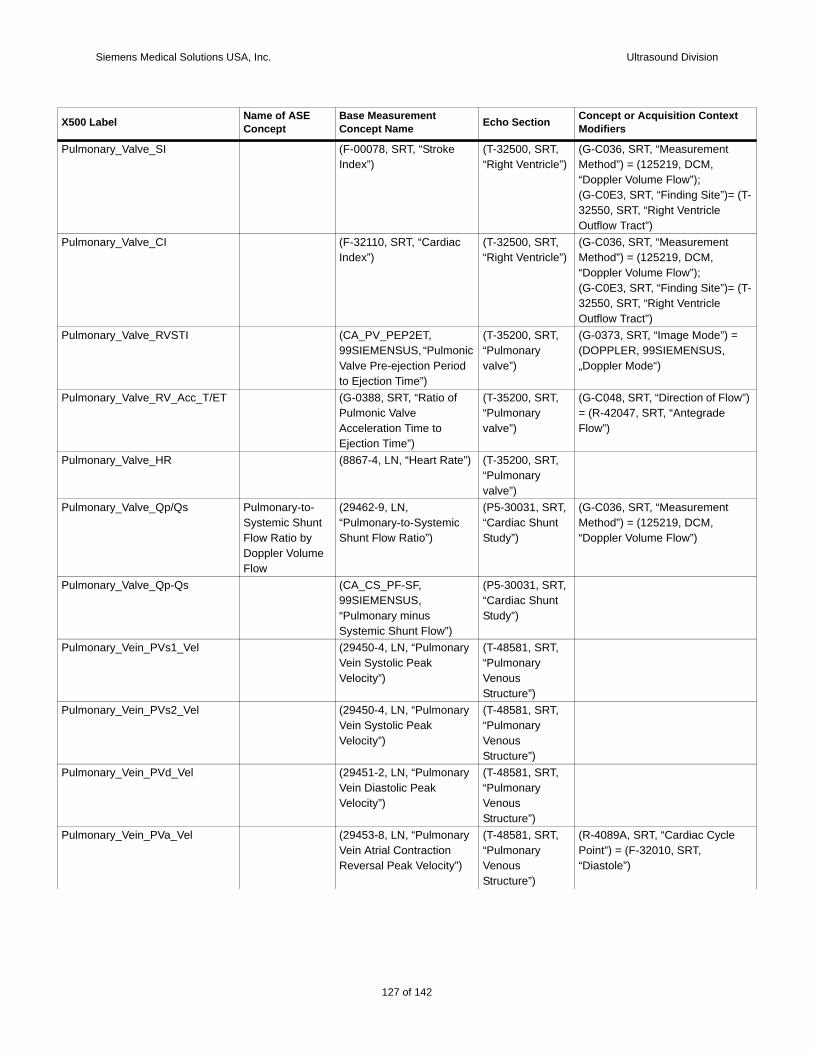
Siemens Medical Solutions USA, Inc. Ultrasound Division
Pulm
(T-
Pulm
(T-
Pulm =
Pulm w”)
Pulm
Pulm
Pulm
Pulm
Pulm
Pulm
Pulm
X500 t
onary_Valve_SI (F-00078, SRT, “Stroke Index”)
(T-32500, SRT,“Right Ventricle”)
(G-C036, SRT, “Measurement Method”) = (125219, DCM, “Doppler Volume Flow”);(G-C0E3, SRT, “Finding Site”)=32550, SRT, “Right Ventricle Outflow Tract”)
onary_Valve_CI (F-32110, SRT, “Cardiac Index”)
(T-32500, SRT,“Right Ventricle”)
(G-C036, SRT, “Measurement Method”) = (125219, DCM, “Doppler Volume Flow”);(G-C0E3, SRT, “Finding Site”)=32550, SRT, “Right Ventricle Outflow Tract”)
onary_Valve_RVSTI (CA_PV_PEP2ET, 99SIEMENSUS, “Pulmonic Valve Pre-ejection Period to Ejection Time”)
(T-35200, SRT, “Pulmonary valve”)
(G-0373, SRT, “Image Mode”) (DOPPLER, 99SIEMENSUS, „Doppler Mode“)
onary_Valve_RV_Acc_T/ET (G-0388, SRT, “Ratio of Pulmonic Valve Acceleration Time toEjection Time”)
(T-35200, SRT, “Pulmonary valve”)
(G-C048, SRT, “Direction of Flo= (R-42047, SRT, “Antegrade Flow”)
onary_Valve_HR (8867-4, LN, “Heart Rate”) (T-35200, SRT, “Pulmonary valve”)
onary_Valve_Qp/Qs Pulmonary-to-Systemic Shunt Flow Ratio by Doppler Volume Flow
(29462-9, LN,“Pulmonary-to-Systemic Shunt Flow Ratio”)
(P5-30031, SRT,“Cardiac Shunt Study”)
(G-C036, SRT, “Measurement Method”) = (125219, DCM, “Doppler Volume Flow”)
onary_Valve_Qp-Qs (CA_CS_PF-SF, 99SIEMENSUS, “Pulmonary minus Systemic Shunt Flow”)
(P5-30031, SRT, “Cardiac Shunt Study”)
onary_Vein_PVs1_Vel (29450-4, LN, “Pulmonary Vein Systolic Peak Velocity”)
(T-48581, SRT,“Pulmonary Venous Structure”)
onary_Vein_PVs2_Vel (29450-4, LN, “Pulmonary Vein Systolic Peak Velocity”)
(T-48581, SRT,“Pulmonary Venous Structure”)
onary_Vein_PVd_Vel (29451-2, LN, “Pulmonary Vein Diastolic PeakVelocity”)
(T-48581, SRT,“Pulmonary Venous Structure”)
onary_Vein_PVa_Vel (29453-8, LN, “Pulmonary Vein Atrial ContractionReversal Peak Velocity”)
(T-48581, SRT,“Pulmonary Venous Structure”)
(R-4089A, SRT, “Cardiac CyclePoint”) = (F-32010, SRT, “Diastole”)
Label Name of ASE Concept
Base Measurement Concept Name Echo Section Concept or Acquisition Contex
Modifiers
127 of 142
142
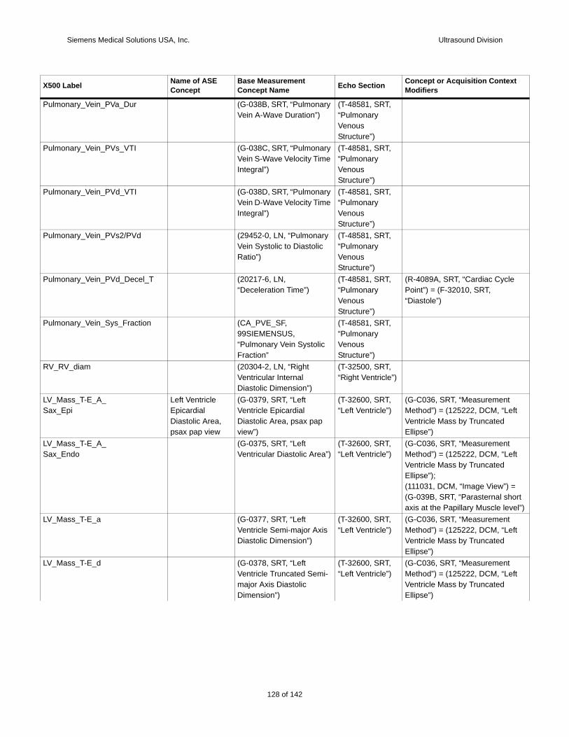
Siemens Medical Solutions USA, Inc. Ultrasound Division
Pulm
Pulm
Pulm
Pulm
Pulm
Pulm
RV_R
LV_MSax_ ft
LV_MSax_ ft
ort el”)
LV_Mft
LV_Mft
X500 t
onary_Vein_PVa_Dur (G-038B, SRT, “Pulmonary Vein A-Wave Duration”)
(T-48581, SRT,“Pulmonary Venous Structure”)
onary_Vein_PVs_VTI (G-038C, SRT, “Pulmonary Vein S-Wave Velocity Time Integral”)
(T-48581, SRT,“Pulmonary Venous Structure”)
onary_Vein_PVd_VTI (G-038D, SRT, “Pulmonary Vein D-Wave Velocity Time Integral”)
(T-48581, SRT,“Pulmonary Venous Structure”)
onary_Vein_PVs2/PVd (29452-0, LN, “Pulmonary Vein Systolic to Diastolic Ratio”)
(T-48581, SRT,“Pulmonary Venous Structure”)
onary_Vein_PVd_Decel_T (20217-6, LN, “Deceleration Time”)
(T-48581, SRT,“Pulmonary Venous Structure”)
(R-4089A, SRT, “Cardiac CyclePoint”) = (F-32010, SRT, “Diastole”)
onary_Vein_Sys_Fraction (CA_PVE_SF, 99SIEMENSUS, “Pulmonary Vein Systolic Fraction”
(T-48581, SRT,“Pulmonary Venous Structure”)
V_diam (20304-2, LN, “Right Ventricular Internal Diastolic Dimension”)
(T-32500, SRT,“Right Ventricle”)
ass_T-E_A_Epi
Left Ventricle Epicardial Diastolic Area, psax pap view
(G-0379, SRT, “Left Ventricle Epicardial Diastolic Area, psax pap view”)
(T-32600, SRT, “Left Ventricle”)
(G-C036, SRT, “Measurement Method”) = (125222, DCM, “LeVentricle Mass by Truncated Ellipse”)
ass_T-E_A_Endo
(G-0375, SRT, “Left Ventricular Diastolic Area”)
(T-32600, SRT, “Left Ventricle”)
(G-C036, SRT, “Measurement Method”) = (125222, DCM, “LeVentricle Mass by Truncated Ellipse”);(111031, DCM, “Image View”) =(G-039B, SRT, “Parasternal shaxis at the Papillary Muscle lev
ass_T-E_a (G-0377, SRT, “Left Ventricle Semi-major Axis Diastolic Dimension”)
(T-32600, SRT, “Left Ventricle”)
(G-C036, SRT, “Measurement Method”) = (125222, DCM, “LeVentricle Mass by Truncated Ellipse”)
ass_T-E_d (G-0378, SRT, “Left Ventricle Truncated Semi-major Axis DiastolicDimension”)
(T-32600, SRT, “Left Ventricle”)
(G-C036, SRT, “Measurement Method”) = (125222, DCM, “LeVentricle Mass by Truncated Ellipse”)
Label Name of ASE Concept
Base Measurement Concept Name Echo Section Concept or Acquisition Contex
Modifiers
128 of 142
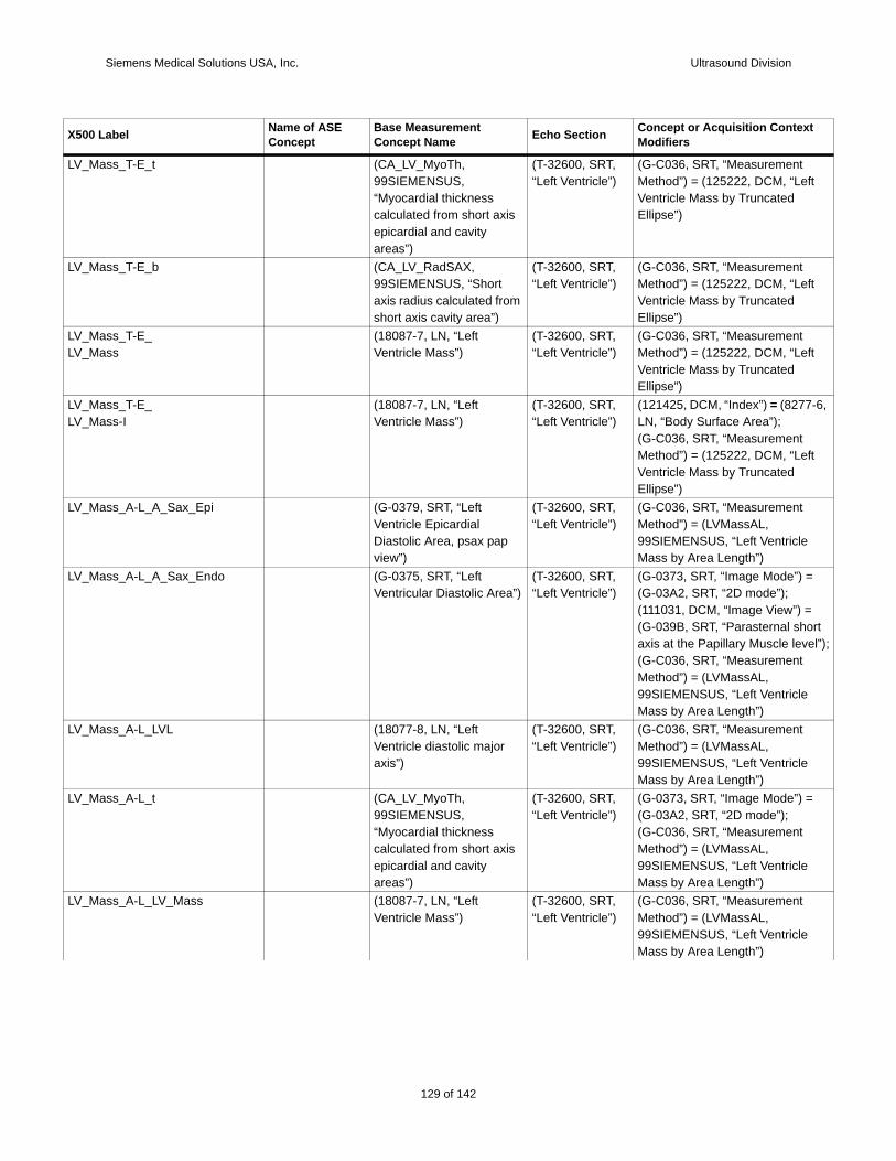
Siemens Medical Solutions USA, Inc. Ultrasound Division
LV_Mft
LV_Mft
LV_MLV_M ft
LV_MLV_M
7-6,
ft
LV_M
LV_M =
ort el”);
LV_M
LV_M =
LV_M
X500 t
ass_T-E_t (CA_LV_MyoTh, 99SIEMENSUS, “Myocardial thickness calculated from short axis epicardial and cavity areas”)
(T-32600, SRT, “Left Ventricle”)
(G-C036, SRT, “Measurement Method”) = (125222, DCM, “LeVentricle Mass by Truncated Ellipse”)
ass_T-E_b (CA_LV_RadSAX, 99SIEMENSUS, “Short axis radius calculated from short axis cavity area”)
(T-32600, SRT, “Left Ventricle”)
(G-C036, SRT, “Measurement Method”) = (125222, DCM, “LeVentricle Mass by Truncated Ellipse”)
ass_T-E_ass
(18087-7, LN, “Left Ventricle Mass”)
(T-32600, SRT, “Left Ventricle”)
(G-C036, SRT, “Measurement Method”) = (125222, DCM, “LeVentricle Mass by Truncated Ellipse”)
ass_T-E_ass-I
(18087-7, LN, “Left Ventricle Mass”)
(T-32600, SRT, “Left Ventricle”)
(121425, DCM, “Index”) = (827LN, “Body Surface Area”);(G-C036, SRT, “Measurement Method”) = (125222, DCM, “LeVentricle Mass by Truncated Ellipse”)
ass_A-L_A_Sax_Epi (G-0379, SRT, “Left Ventricle Epicardial Diastolic Area, psax pap view”)
(T-32600, SRT, “Left Ventricle”)
(G-C036, SRT, “Measurement Method”) = (LVMassAL, 99SIEMENSUS, “Left VentricleMass by Area Length”)
ass_A-L_A_Sax_Endo (G-0375, SRT, “Left Ventricular Diastolic Area”)
(T-32600, SRT, “Left Ventricle”)
(G-0373, SRT, “Image Mode”) (G-03A2, SRT, “2D mode”);(111031, DCM, “Image View”) =(G-039B, SRT, “Parasternal shaxis at the Papillary Muscle lev(G-C036, SRT, “Measurement Method”) = (LVMassAL, 99SIEMENSUS, “Left VentricleMass by Area Length”)
ass_A-L_LVL (18077-8, LN, “Left Ventricle diastolic major axis”)
(T-32600, SRT, “Left Ventricle”)
(G-C036, SRT, “Measurement Method”) = (LVMassAL, 99SIEMENSUS, “Left VentricleMass by Area Length”)
ass_A-L_t (CA_LV_MyoTh, 99SIEMENSUS, “Myocardial thickness calculated from short axis epicardial and cavity areas”)
(T-32600, SRT, “Left Ventricle”)
(G-0373, SRT, “Image Mode”) (G-03A2, SRT, “2D mode”);(G-C036, SRT, “Measurement Method”) = (LVMassAL, 99SIEMENSUS, “Left VentricleMass by Area Length”)
ass_A-L_LV_Mass (18087-7, LN, “Left Ventricle Mass”)
(T-32600, SRT, “Left Ventricle”)
(G-C036, SRT, “Measurement Method”) = (LVMassAL, 99SIEMENSUS, “Left VentricleMass by Area Length”)
Label Name of ASE Concept
Base Measurement Concept Name Echo Section Concept or Acquisition Contex
Modifiers
129 of 142
142
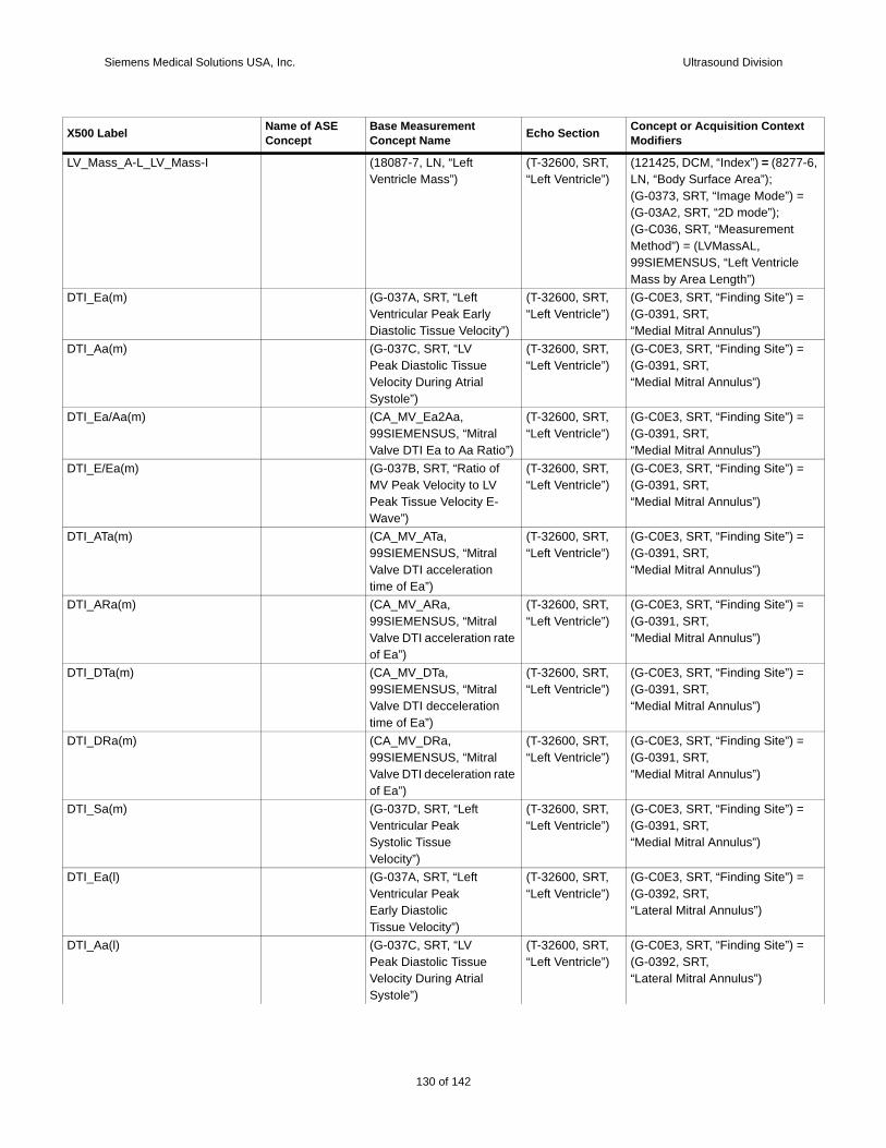
Siemens Medical Solutions USA, Inc. Ultrasound Division
LV_M 7-6,
=
DTI_ =
DTI_ =
DTI_ =
DTI_ =
DTI_ =
DTI_ =
DTI_ =
DTI_ =
DTI_ =
DTI_ =
DTI_ =
X500 t
ass_A-L_LV_Mass-I (18087-7, LN, “Left Ventricle Mass”)
(T-32600, SRT, “Left Ventricle”)
(121425, DCM, “Index”) = (827LN, “Body Surface Area”);(G-0373, SRT, “Image Mode”) (G-03A2, SRT, “2D mode”);(G-C036, SRT, “Measurement Method”) = (LVMassAL, 99SIEMENSUS, “Left VentricleMass by Area Length”)
Ea(m) (G-037A, SRT, “LeftVentricular Peak Early Diastolic Tissue Velocity”)
(T-32600, SRT, “Left Ventricle”)
(G-C0E3, SRT, “Finding Site”) (G-0391, SRT,“Medial Mitral Annulus”)
Aa(m) (G-037C, SRT, “LVPeak Diastolic Tissue Velocity During Atrial Systole”)
(T-32600, SRT, “Left Ventricle”)
(G-C0E3, SRT, “Finding Site”) (G-0391, SRT,“Medial Mitral Annulus”)
Ea/Aa(m) (CA_MV_Ea2Aa, 99SIEMENSUS, “Mitral Valve DTI Ea to Aa Ratio”)
(T-32600, SRT, “Left Ventricle”)
(G-C0E3, SRT, “Finding Site”) (G-0391, SRT,“Medial Mitral Annulus”)
E/Ea(m) (G-037B, SRT, “Ratio of MV Peak Velocity to LV Peak Tissue Velocity E-Wave”)
(T-32600, SRT, “Left Ventricle”)
(G-C0E3, SRT, “Finding Site”) (G-0391, SRT,“Medial Mitral Annulus”)
ATa(m) (CA_MV_ATa, 99SIEMENSUS, “Mitral Valve DTI acceleration time of Ea”)
(T-32600, SRT, “Left Ventricle”)
(G-C0E3, SRT, “Finding Site”) (G-0391, SRT,“Medial Mitral Annulus”)
ARa(m) (CA_MV_ARa, 99SIEMENSUS, “Mitral Valve DTI acceleration rate of Ea”)
(T-32600, SRT, “Left Ventricle”)
(G-C0E3, SRT, “Finding Site”) (G-0391, SRT,“Medial Mitral Annulus”)
DTa(m) (CA_MV_DTa, 99SIEMENSUS, “Mitral Valve DTI decceleration time of Ea”)
(T-32600, SRT, “Left Ventricle”)
(G-C0E3, SRT, “Finding Site”) (G-0391, SRT,“Medial Mitral Annulus”)
DRa(m) (CA_MV_DRa, 99SIEMENSUS, “Mitral Valve DTI deceleration rate of Ea”)
(T-32600, SRT, “Left Ventricle”)
(G-C0E3, SRT, “Finding Site”) (G-0391, SRT,“Medial Mitral Annulus”)
Sa(m) (G-037D, SRT, “LeftVentricular PeakSystolic TissueVelocity”)
(T-32600, SRT, “Left Ventricle”)
(G-C0E3, SRT, “Finding Site”) (G-0391, SRT,“Medial Mitral Annulus”)
Ea(l) (G-037A, SRT, “LeftVentricular PeakEarly DiastolicTissue Velocity”)
(T-32600, SRT, “Left Ventricle”)
(G-C0E3, SRT, “Finding Site”) (G-0392, SRT,“Lateral Mitral Annulus”)
Aa(l) (G-037C, SRT, “LVPeak Diastolic Tissue Velocity During AtrialSystole”)
(T-32600, SRT, “Left Ventricle”)
(G-C0E3, SRT, “Finding Site”) (G-0392, SRT,“Lateral Mitral Annulus”)
Label Name of ASE Concept
Base Measurement Concept Name Echo Section Concept or Acquisition Contex
Modifiers
130 of 142
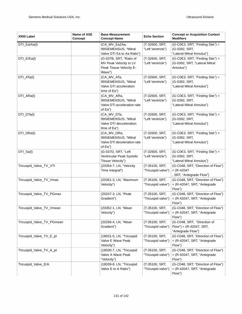
Siemens Medical Solutions USA, Inc. Ultrasound Division
DTI_ =
DTI_ =
DTI_ =
DTI_ =
DTI_ =
DTI_ =
DTI_ =
Tricu w”)
Tricu w”)
Tricu w”)
Tricu w”)
Tricu
Tricu w”)
Tricu w”)
Tricu w”)
X500 t
Ea/Aa(l) (CA_MV_Ea2Aa, 99SIEMENSUS, “Mitral Valve DTI Ea to Aa Ratio”)
(T-32600, SRT, “Left Ventricle”)
(G-C0E3, SRT, “Finding Site”) (G-0392, SRT,“Lateral Mitral Annulus”)
E/Ea(l) (G-037B, SRT, “Ratio of MV Peak Velocity to LV Peak Tissue Velocity E-Wave”)
(T-32600, SRT, “Left Ventricle”)
(G-C0E3, SRT, “Finding Site”) (G-0392, SRT, “Lateral Mitral Annulus”)
ATa(l) (CA_MV_ATa, 99SIEMENSUS, “Mitral Valve DTI acceleration time of Ea”)
(T-32600, SRT, “Left Ventricle”)
(G-C0E3, SRT, “Finding Site”) (G-0392, SRT,“Lateral Mitral Annulus”)
ARa(l) (CA_MV_ARa, 99SIEMENSUS, “Mitral Valve DTI acceleration rate of Ea”)
(T-32600, SRT, “Left Ventricle”)
(G-C0E3, SRT, “Finding Site”) (G-0392, SRT,“Lateral Mitral Annulus”)
DTa(l) (CA_MV_DTa, 99SIEMENSUS, “Mitral Valve DTI decceleration time of Ea”)
(T-32600, SRT, “Left Ventricle”)
(G-C0E3, SRT, “Finding Site”) (G-0392, SRT,“Lateral Mitral Annulus”)
DRa(l) (CA_MV_DRa, 99SIEMENSUS, “Mitral Valve DTI deceleration rate of Ea”)
(T-32600, SRT, “Left Ventricle”)
(G-C0E3, SRT, “Finding Site”) (G-0392, SRT,“Lateral Mitral Annulus”)
Sa(l) (G-037D, SRT, “LeftVentricular Peak Systolic Tissue Velocity”)
(T-32600, SRT, “Left Ventricle”)
(G-C0E3, SRT, “Finding Site”) (G-0392, SRT,“Lateral Mitral Annulus”)
spid_Valve_TV_VTI (20354-7, LN, “Velocity Time Integral”)
(T-35100, SRT, “Tricuspid valve”)
(G-C048, SRT, “Direction of Flo= (R-42047, SRT, “Antegrade Flow”)
spid_Valve_TV_Vmax (20351-3, LN, “Maximum Velocity“)
(T-35100, SRT, “Tricuspid valve”)
(G-C048, SRT, “Direction of Flo= (R-42047, SRT, “Antegrade Flow”)
spid_Valve_TV_PGmax (20247-3, LN, “Peak Gradient“)
(T-35100, SRT, “Tricuspid valve”)
(G-C048, SRT, “Direction of Flo= (R-42047, SRT, “Antegrade Flow”)
spid_Valve_TV_Vmean (20352-1, LN, “Mean Velocity”)
(T-35100, SRT, “Tricuspid valve”)
(G-C048, SRT, “Direction of Flo= (R-42047, SRT, “Antegrade Flow”)
spid_Valve_TV_PGmean (20256-4, LN, “Mean Gradient”)
(T-35100, SRT, “Tricuspid valve”)
(G-C048, SRT, “Direction of Flow”) = (R-42047, SRT, “Antegrade Flow”)
spid_Valve_TV_E_pt (18031-5, LN, “Tricuspid Valve E Wave PeakVelocity”)
(T-35100, SRT, “Tricuspid valve”)
(G-C048, SRT, “Direction of Flo= (R-42047, SRT, “Antegrade Flow”)
spid_Valve_TV_A_pt (18030-7, LN, “Tricuspid Valve A Wave Peak “Velocity”)
(T-35100, SRT, “Tricuspid valve”)
(G-C048, SRT, “Direction of Flo= (R-42047, SRT, “Antegrade Flow”)
spid_Valve_E/A (18039-8, LN, “Tricuspid Valve E to A Ratio”)
(T-35100, SRT, “Tricuspid valve”)
(G-C048, SRT, “Direction of Flo= (R-42047, SRT, “Antegrade Flow”)
Label Name of ASE Concept
Base Measurement Concept Name Echo Section Concept or Acquisition Contex
Modifiers
131 of 142
142
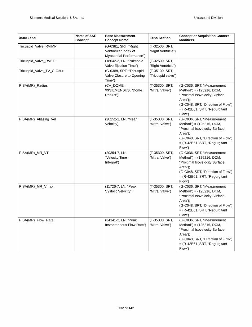
Siemens Medical Solutions USA, Inc. Ultrasound Division
Tricu
Tricu
Tricu
PISA
w”)
PISA
w”)
PISA
w”)
PISA
w”)
PISA
w”)
X500 t
spid_Valve_RVIMP (G-0381, SRT, “Right Ventricular Index of Myocardial Performance”)
(T-32500, SRT,“Right Ventricle”)
spid_Valve_RVET (18042-2, LN, “Pulmonic Valve Ejection Time”)
(T-32500, SRT,“Right Ventricle”)
spid_Valve_TV_C-Odur (G-0389, SRT, “Tricuspid Valve Closure to Opening Time”)
(T-35100, SRT, “Tricuspid valve”)
(MR)_Radius (CA_DOME, 99SIEMENSUS, “Dome Radius”)
(T-35300, SRT, “Mitral Valve”)
(G-C036, SRT, “Measurement Method”) = (125216, DCM, “Proximal Isovelocity Surface Area”);(G-C048, SRT, “Direction of Flo= (R-42E61, SRT, “RegurgitantFlow”)
(MR)_Aliasing_Vel (20252-1, LN, “Mean Velocity)
(T-35300, SRT, “Mitral Valve”)
(G-C036, SRT, “Measurement Method”) = (125216, DCM, “Proximal Isovelocity Surface Area”);(G-C048, SRT, “Direction of Flo= (R-42E61, SRT, “RegurgitantFlow”)
(MR)_MR_VTI (20354-7, LN,“Velocity TimeIntegral”)
(T-35300, SRT, “Mitral Valve”)
(G-C036, SRT, “Measurement Method”) = (125216, DCM, “Proximal Isovelocity Surface Area”);(G-C048, SRT, “Direction of Flo= (R-42E61, SRT, “RegurgitantFlow”)
(MR)_MR_Vmax (11726-7, LN, “Peak Systolic Velocity”)
(T-35300, SRT, “Mitral Valve”)
(G-C036, SRT, “Measurement Method”) = (125216, DCM, “Proximal Isovelocity Surface Area”);(G-C048, SRT, “Direction of Flo= (R-42E61, SRT, “RegurgitantFlow”)
(MR)_Flow_Rate (34141-2, LN, “Peak Instantaneous Flow Rate”)
(T-35300, SRT, “Mitral Valve”)
(G-C036, SRT, “Measurement Method”) = (125216, DCM, “Proximal Isovelocity Surface Area”);(G-C048, SRT, “Direction of Flo= (R-42E61, SRT, “RegurgitantFlow”)
Label Name of ASE Concept
Base Measurement Concept Name Echo Section Concept or Acquisition Contex
Modifiers
132 of 142
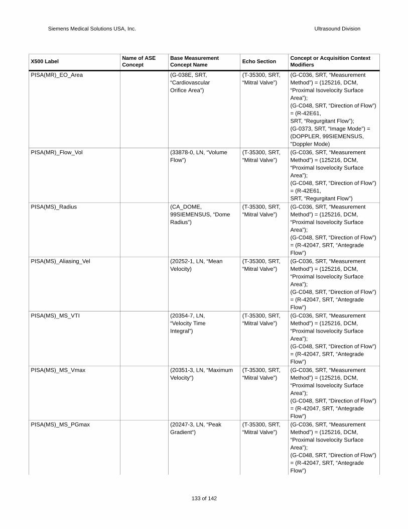
Siemens Medical Solutions USA, Inc. Ultrasound Division
PISA
w”)
=
PISA
w”)
PISA
w”)
PISA
w”)
PISA
w”)
PISA
w”)
PISA
w”)
X500 t
(MR)_EO_Area (G-038E, SRT,“CardiovascularOrifice Area”)
(T-35300, SRT, “Mitral Valve”)
(G-C036, SRT, “Measurement Method”) = (125216, DCM, “Proximal Isovelocity Surface Area”);(G-C048, SRT, “Direction of Flo= (R-42E61,SRT, “Regurgitant Flow”);(G-0373, SRT, “Image Mode”) (DOPPLER, 99SIEMENSUS, "Doppler Mode)
(MR)_Flow_Vol (33878-0, LN, “Volume Flow”)
(T-35300, SRT, “Mitral Valve”)
(G-C036, SRT, “Measurement Method”) = (125216, DCM, “Proximal Isovelocity Surface Area”);(G-C048, SRT, “Direction of Flo= (R-42E61,SRT, “Regurgitant Flow”)
(MS)_Radius (CA_DOME, 99SIEMENSUS, “Dome Radius”)
(T-35300, SRT, “Mitral Valve”)
(G-C036, SRT, “Measurement Method”) = (125216, DCM, “Proximal Isovelocity Surface Area”);(G-C048, SRT, “Direction of Flo= (R-42047, SRT, “Antegrade Flow”)
(MS)_Aliasing_Vel (20252-1, LN, “Mean Velocity)
(T-35300, SRT, “Mitral Valve”)
(G-C036, SRT, “Measurement Method”) = (125216, DCM, “Proximal Isovelocity Surface Area”);(G-C048, SRT, “Direction of Flo= (R-42047, SRT, “Antegrade Flow”)
(MS)_MS_VTI (20354-7, LN,“Velocity TimeIntegral”)
(T-35300, SRT, “Mitral Valve”)
(G-C036, SRT, “Measurement Method”) = (125216, DCM, “Proximal Isovelocity Surface Area”);(G-C048, SRT, “Direction of Flo= (R-42047, SRT, “Antegrade Flow”)
(MS)_MS_Vmax (20351-3, LN, “Maximum Velocity“)
(T-35300, SRT, “Mitral Valve”)
(G-C036, SRT, “Measurement Method”) = (125216, DCM, “Proximal Isovelocity Surface Area”);(G-C048, SRT, “Direction of Flo= (R-42047, SRT, “Antegrade Flow”)
(MS)_MS_PGmax (20247-3, LN, “Peak Gradient“)
(T-35300, SRT, “Mitral Valve”)
(G-C036, SRT, “Measurement Method”) = (125216, DCM, “Proximal Isovelocity Surface Area”);(G-C048, SRT, “Direction of Flo= (R-42047, SRT, “Antegrade Flow”)
Label Name of ASE Concept
Base Measurement Concept Name Echo Section Concept or Acquisition Contex
Modifiers
133 of 142
142
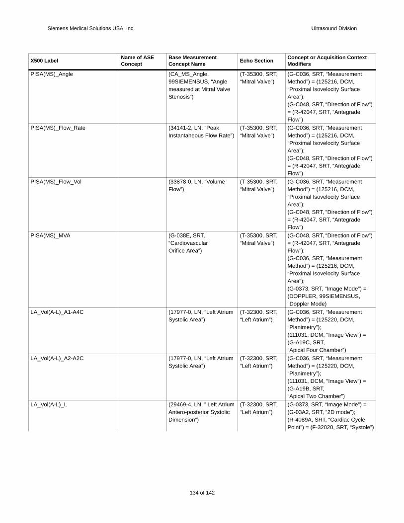
Siemens Medical Solutions USA, Inc. Ultrasound Division
PISA
w”)
PISA
w”)
PISA
w”)
PISA w”)
=
LA_V
LA_V
LA_V =
le”)
X500 t
(MS)_Angle (CA_MS_Angle, 99SIEMENSUS, “Angle measured at Mitral Valve Stenosis”)
(T-35300, SRT, “Mitral Valve”)
(G-C036, SRT, “Measurement Method”) = (125216, DCM, “Proximal Isovelocity Surface Area”);(G-C048, SRT, “Direction of Flo= (R-42047, SRT, “Antegrade Flow”)
(MS)_Flow_Rate (34141-2, LN, “Peak Instantaneous Flow Rate”)
(T-35300, SRT, “Mitral Valve”)
(G-C036, SRT, “Measurement Method”) = (125216, DCM, “Proximal Isovelocity Surface Area”);(G-C048, SRT, “Direction of Flo= (R-42047, SRT, “Antegrade Flow”)
(MS)_Flow_Vol (33878-0, LN, “Volume Flow”)
(T-35300, SRT, “Mitral Valve”)
(G-C036, SRT, “Measurement Method”) = (125216, DCM, “Proximal Isovelocity Surface Area”);(G-C048, SRT, “Direction of Flo= (R-42047, SRT, “Antegrade Flow”)
(MS)_MVA (G-038E, SRT,“CardiovascularOrifice Area”)
(T-35300, SRT, “Mitral Valve”)
(G-C048, SRT, “Direction of Flo= (R-42047, SRT, “Antegrade Flow”);(G-C036, SRT, “Measurement Method”) = (125216, DCM, “Proximal Isovelocity Surface Area”);(G-0373, SRT, “Image Mode”) (DOPPLER, 99SIEMENSUS, "Doppler Mode)
ol(A-L)_A1-A4C (17977-0, LN, “Left Atrium Systolic Area”)
(T-32300, SRT, “Left Atrium”)
(G-C036, SRT, “Measurement Method”) = (125220, DCM, “Planimetry”);(111031, DCM, “Image View”) =(G-A19C, SRT,“Apical Four Chamber”)
ol(A-L)_A2-A2C (17977-0, LN, “Left Atrium Systolic Area”)
(T-32300, SRT, “Left Atrium”)
(G-C036, SRT, “Measurement Method”) = (125220, DCM, “Planimetry”);(111031, DCM, “Image View”) =(G-A19B, SRT,“Apical Two Chamber”)
ol(A-L)_L (29469-4, LN, ” Left Atrium Antero-posterior Systolic Dimension”)
(T-32300, SRT, “Left Atrium”)
(G-0373, SRT, “Image Mode”) (G-03A2, SRT, “2D mode”);(R-4089A, SRT, “Cardiac CyclePoint”) = (F-32020, SRT, “Systo
Label Name of ASE Concept
Base Measurement Concept Name Echo Section Concept or Acquisition Contex
Modifiers
134 of 142

Siemens Medical Solutions USA, Inc. Ultrasound Division
LA_Vea-
=
LA_V 7-6,
ea-
=
LA_V
”);
LA_V
”);
LA_V
LA_V 7-6,
=
RA_V =
RA_V
=
le”)
RA_V =
le”)
X500 t
ol(A-L)_LA_Vol(A-L) (122408, DCM, “Left Atrial ES Volume”)
(T-32300, SRT, “Left Atrium”)
(G-C036, SRT, “Measurement Method”) = (125204, DCM, “ArLength Biplane”);(G-0373, SRT, “Image Mode”) (G-03A2, SRT, “2D mode”)
ol(A-L)_LA_Vol-I (122408, DCM, “Left Atrial ES Volume”)
(T-32300, SRT, “Left Atrium”)
(121425, DCM, “Index”) = (827LN, “Body Surface Area”);(G-C036, SRT, “Measurement Method”) = (125204, DCM, “ArLength Biplane”);(G-0373, SRT, “Image Mode”) (G-03A2, SRT, “2D mode”)
ol(Simp)_4CH (122408, DCM, “Left Atrial ES Volume”)
(T-32300, SRT, “Left Atrium”)
(G-C036, SRT, “Measurement Method”) = (125207, DCM, “Method of Disks, Single Plane(111031, DCM, “Image View”) =(G-A19C, SRT,“Apical Four Chamber”)
ol(Simp)_2CH (122408, DCM, “Left Atrial ES Volume”)
(T-32300, SRT, “Left Atrium”)
(G-C036, SRT, “Measurement Method”) = (125207, DCM, “Method of Disks, Single Plane(111031, DCM, “Image View”) =(G-A19B, SRT,“Apical Two Chamber”)
ol(Simp)_LA_Vol(Simp) (122408, DCM, “Left Atrial ES Volume”)
(T-32300, SRT, “Left Atrium”)
(G-C036, SRT, “Measurement Method”) = (125207, DCM, “Method of Disks, Biplane”);
ol(Simp)_LA_Vol-I (122408, DCM, “Left Atrial ES Volume”)
(T-32300, SRT, “Left Atrium”)
(121425, DCM, “Index”) = (827LN, “Body Surface Area”);(G-C036, SRT, “Measurement Method”) = (125207, DCM, “Method of Disks, Biplane”);(G-0373, SRT, “Image Mode”) (G-03A2, SRT, “2D mode”)
ol(A-L)_A1-A4C (17988-7, LN, “Right Atrium Systolic Area”)
(T-32200, SRT,“Right Atrium”)
(111031, DCM, “Image View”) (G-A19C, SRT,“Apical Four Chamber”)
ol(A-L)_A2-A2C (17988-7, LN, “Right Atrium Systolic Area”)
(T-32200, SRT,“Right Atrium”)
(111031, DCM, “Image View”) =(G-A19B, SRT,“Apical Two Chamber”) ;(G-0373, SRT, “Image Mode”) (G-03A2, SRT, “2D mode”);(R-4089A, SRT, “Cardiac CyclePoint”) = (F-32020, SRT, “Systo
ol(A-L)_L (CA_RA_DIA, 99SIEMENSUS, “Right Atrium Systolic Dimension”)
(T-32200, SRT,“Right Atrium”)
(G-0373, SRT, “Image Mode”) (G-03A2, SRT, “2D mode”);(R-4089A, SRT, “Cardiac CyclePoint”) = (F-32020, SRT, “Systo
Label Name of ASE Concept
Base Measurement Concept Name Echo Section Concept or Acquisition Contex
Modifiers
135 of 142
142
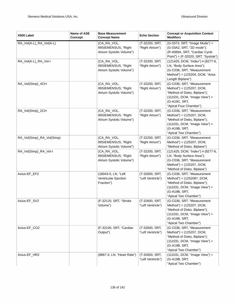
Siemens Medical Solutions USA, Inc. Ultrasound Division
RA_V =
le”)
RA_V 7-6,
ea-
RA_V
RA_V
RA_V
RA_V 7-6,
Axius
Axius
Axius
Axius
X500 t
ol(A-L)_RA_Vol(A-L) (CA_RA_VOL, 99SIEMENSUS, “Right Atrium Systolic Volume”)
(T-32200, SRT,“Right Atrium”)
(G-0373, SRT, “Image Mode”) (G-03A2, SRT, “2D mode”);(R-4089A, SRT, “Cardiac CyclePoint”) = (F-32020, SRT, “Systo
ol(A-L)_RA_Vol-I (CA_RA_VOL, 99SIEMENSUS, “Right Atrium Systolic Volume”)
(T-32200, SRT,“Right Atrium”)
(121425, DCM, “Index”) = (827LN, “Body Surface Area”);(G-C036, SRT, “Measurement Method”) = (125204, DCM, “ArLength Biplane”)
ol(Simp)_4CH (CA_RA_VOL, 99SIEMENSUS, “Right Atrium Systolic Volume”)
(T-32200, SRT,“Right Atrium”)
(G-C036, SRT, “Measurement Method”) = (125207, DCM, “Method of Disks, Biplane”);(111031, DCM, “Image View”) =(G-A19C, SRT,“Apical Four Chamber”)
ol(Simp)_2CH (CA_RA_VOL, 99SIEMENSUS, “Right Atrium Systolic Volume”)
(T-32200, SRT,“Right Atrium”)
(G-C036, SRT, “Measurement Method”) = (125207, DCM, “Method of Disks, Biplane”);(111031, DCM, “Image View”) =(G-A19B, SRT,“Apical Two Chamber”)
ol(Simp)_RA_Vol(Simp) (CA_RA_VOL, 99SIEMENSUS, “Right Atrium Systolic Volume”)
(T-32200, SRT,“Right Atrium”)
(G-C036, SRT, “Measurement Method”) = (125207, DCM, “Method of Disks, Biplane”)
ol(Simp)_RA_Vol-I (CA_RA_VOL, 99SIEMENSUS, “Right Atrium Systolic Volume”)
(T-32200, SRT,“Right Atrium”)
(121425, DCM, “Index”) = (827LN, “Body Surface Area”);(G-C036, SRT, “Measurement Method”) = (125207, DCM, “Method of Disks, Biplane”)
-EF_EF2 (18043-0, LN, “Left Ventricular Ejection Fraction”)
(T-32600, SRT, “Left Ventricle”)
(G-C036, SRT, “Measurement Method”) = (1252087, DCM, “Method of Disks, Biplane”);(111031, DCM, “Image View”) =(G-A19B, SRT,“Apical Two Chamber”)
-EF_SV2 (F-32120, SRT, “Stroke Volume”)
(T-32600, SRT, “Left Ventricle”)
(G-C036, SRT, “Measurement Method”) = (125207, DCM, “Method of Disks, Biplane”);(111031, DCM, “Image View”) =(G-A19B, SRT,“Apical Two Chamber”)
-EF_CO2 (F-32100, SRT, “Cardiac Output”)
(T-32600, SRT, “Left Ventricle”)
(G-C036, SRT, “Measurement Method”) = (125207, DCM, “Method of Disks, Biplane”);(111031, DCM, “Image View”) =(G-A19B, SRT,“Apical Two Chamber”)
-EF_HR2 (8867-4, LN, “Heart Rate”) (T-32600, SRT, “Left Ventricle”)
(111031, DCM, “Image View”) =(G-A19B, SRT,“Apical Two Chamber”)
Label Name of ASE Concept
Base Measurement Concept Name Echo Section Concept or Acquisition Contex
Modifiers
136 of 142
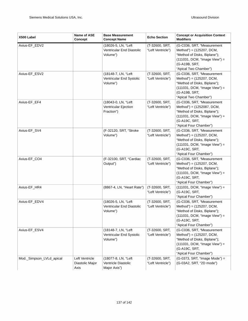
Siemens Medical Solutions USA, Inc. Ultrasound Division
Axius
Axius
Axius
Axius
Axius
Axius
Axius
Axius
Mod. =
X500 t
-EF_EDV2 (18026-5, LN, “Left Ventricular End Diastolic Volume”)
(T-32600, SRT, “Left Ventricle”)
(G-C036, SRT, “Measurement Method”) = (125207, DCM, “Method of Disks, Biplane”);(111031, DCM, “Image View”) =(G-A19B, SRT,“Apical Two Chamber”)
-EF_ESV2 (18148-7, LN, “Left Ventricular End Systolic Volume”)
(T-32600, SRT, “Left Ventricle”)
(G-C036, SRT, “Measurement Method”) = (125207, DCM, “Method of Disks, Biplane”);(111031, DCM, “Image View”) =(G-A19B, SRT,“Apical Two Chamber”)
-EF_EF4 (18043-0, LN, “Left Ventricular Ejection Fraction”)
(T-32600, SRT, “Left Ventricle”)
(G-C036, SRT, “Measurement Method”) = (1252087, DCM, “Method of Disks, Biplane”);(111031, DCM, “Image View”) =(G-A19C, SRT,“Apical Four Chamber”)
-EF_SV4 (F-32120, SRT, “Stroke Volume”)
(T-32600, SRT, “Left Ventricle”)
(G-C036, SRT, “Measurement Method”) = (125207, DCM, “Method of Disks, Biplane”);(111031, DCM, “Image View”) =(G-A19C, SRT,“Apical Four Chamber”)
-EF_CO4 (F-32100, SRT, “Cardiac Output”)
(T-32600, SRT, “Left Ventricle”)
(G-C036, SRT, “Measurement Method”) = (125207, DCM, “Method of Disks, Biplane”);(111031, DCM, “Image View”) =(G-A19C, SRT,“Apical Four Chamber”)
-EF_HR4 (8867-4, LN, “Heart Rate”) (T-32600, SRT, “Left Ventricle”)
(111031, DCM, “Image View”) =(G-A19C, SRT,“Apical Four Chamber”)
-EF_EDV4 (18026-5, LN, “Left Ventricular End Diastolic Volume”)
(T-32600, SRT, “Left Ventricle”)
(G-C036, SRT, “Measurement Method”) = (125207, DCM, “Method of Disks, Biplane”);(111031, DCM, “Image View”) =(G-A19C, SRT,“Apical Four Chamber”)
-EF_ESV4 (18148-7, LN, “Left Ventricular End Systolic Volume”)
(T-32600, SRT, “Left Ventricle”)
(G-C036, SRT, “Measurement Method”) = (125207, DCM, “Method of Disks, Biplane”);(111031, DCM, “Image View”) =(G-A19C, SRT,“Apical Four Chamber”)
_Simpson_LVLd_apical Left Ventricle Diastolic MajorAxis
(18077-8, LN, “LeftVentricle DiastolicMajor Axis”)
(T-32600, SRT, “Left Ventricle”)
(G-0373, SRT, “Image Mode”) (G-03A2, SRT, “2D mode”)
Label Name of ASE Concept
Base Measurement Concept Name Echo Section Concept or Acquisition Contex
Modifiers
137 of 142
142
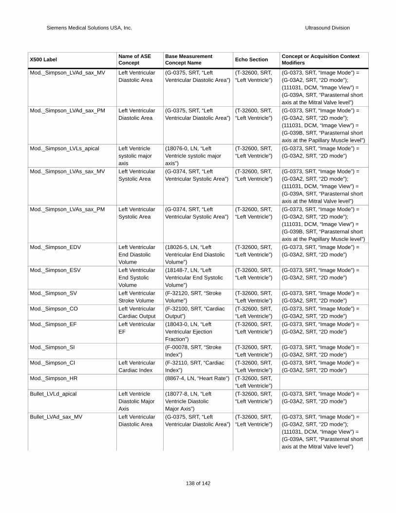
Siemens Medical Solutions USA, Inc. Ultrasound Division
Mod. =
ort
Mod. =
ort el”)
Mod. =
Mod. =
ort
Mod. =
ort el”)
Mod. =
Mod. =
Mod. =
Mod. =
Mod. =
Mod. =
Mod. =
Mod.
Bulle =
Bulle =
ort
X500 t
_Simpson_LVAd_sax_MV Left Ventricular Diastolic Area
(G-0375, SRT, “Left Ventricular Diastolic Area”)
(T-32600, SRT, “Left Ventricle”)
(G-0373, SRT, “Image Mode”) (G-03A2, SRT, “2D mode”);(111031, DCM, “Image View”) =(G-039A, SRT, “Parasternal shaxis at the Mitral Valve level”)
_Simpson_LVAd_sax_PM Left Ventricular Diastolic Area
(G-0375, SRT, “Left Ventricular Diastolic Area”)
(T-32600, SRT, “Left Ventricle”)
(G-0373, SRT, “Image Mode”) (G-03A2, SRT, “2D mode”);(111031, DCM, “Image View”) =(G-039B, SRT, “Parasternal shaxis at the Papillary Muscle lev
_Simpson_LVLs_apical Left Ventricle systolic major axis
(18076-0, LN, “Left Ventricle systolic major axis”)
(T-32600, SRT, “Left Ventricle”)
(G-0373, SRT, “Image Mode”) (G-03A2, SRT, “2D mode”)
_Simpson_LVAs_sax_MV Left Ventricular Systolic Area
(G-0374, SRT, “Left Ventricular Systolic Area”)
(T-32600, SRT, “Left Ventricle”)
(G-0373, SRT, “Image Mode”) (G-03A2, SRT, “2D mode”);(111031, DCM, “Image View”) =(G-039A, SRT, “Parasternal shaxis at the Mitral Valve level”)
_Simpson_LVAs_sax_PM Left Ventricular Systolic Area
(G-0374, SRT, “Left Ventricular Systolic Area”)
(T-32600, SRT, “Left Ventricle”)
(G-0373, SRT, “Image Mode”) (G-03A2, SRT, “2D mode”);(111031, DCM, “Image View”) =(G-039B, SRT, “Parasternal shaxis at the Papillary Muscle lev
_Simpson_EDV Left Ventricular End Diastolic Volume
(18026-5, LN, “Left Ventricular End Diastolic Volume”)
(T-32600, SRT, “Left Ventricle”)
(G-0373, SRT, “Image Mode”) (G-03A2, SRT, “2D mode”)
_Simpson_ESV Left Ventricular End Systolic Volume
(18148-7, LN, “Left Ventricular End Systolic Volume”)
(T-32600, SRT, “Left Ventricle”)
(G-0373, SRT, “Image Mode”) (G-03A2, SRT, “2D mode”)
_Simpson_SV Left Ventricular Stroke Volume
(F-32120, SRT, “Stroke Volume”)
(T-32600, SRT, “Left Ventricle”)
(G-0373, SRT, “Image Mode”) (G-03A2, SRT, “2D mode”)
_Simpson_CO Left Ventricular Cardiac Output
(F-32100, SRT, “Cardiac Output”)
(T-32600, SRT, “Left Ventricle”)
(G-0373, SRT, “Image Mode”) (G-03A2, SRT, “2D mode”)
_Simpson_EF Left Ventricular EF
(18043-0, LN, “Left Ventricular Ejection Fraction”)
(T-32600, SRT, “Left Ventricle”)
(G-0373, SRT, “Image Mode”) (G-03A2, SRT, “2D mode”)
_Simpson_SI (F-00078, SRT, “Stroke Index”)
(T-32600, SRT, “Left Ventricle”)
(G-0373, SRT, “Image Mode”) (G-03A2, SRT, “2D mode”)
_Simpson_CI Left Ventricular Cardiac Index
(F-32110, SRT, “Cardiac Index”)
(T-32600, SRT, “Left Ventricle”)
(G-0373, SRT, “Image Mode”) (G-03A2, SRT, “2D mode”)
_Simpson_HR (8867-4, LN, “Heart Rate”) (T-32600, SRT, “Left Ventricle”)
t_LVLd_apical Left Ventricle Diastolic MajorAxis
(18077-8, LN, “LeftVentricle DiastolicMajor Axis”)
(T-32600, SRT, “Left Ventricle”)
(G-0373, SRT, “Image Mode”) (G-03A2, SRT, “2D mode”)
t_LVAd_sax_MV Left Ventricular Diastolic Area
(G-0375, SRT, “Left Ventricular Diastolic Area”)
(T-32600, SRT, “Left Ventricle”)
(G-0373, SRT, “Image Mode”) (G-03A2, SRT, “2D mode”);(111031, DCM, “Image View”) =(G-039A, SRT, “Parasternal shaxis at the Mitral Valve level”)
Label Name of ASE Concept
Base Measurement Concept Name Echo Section Concept or Acquisition Contex
Modifiers
138 of 142
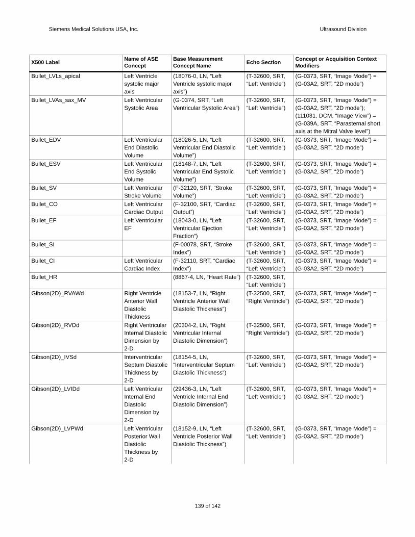
Siemens Medical Solutions USA, Inc. Ultrasound Division
Bulle =
Bulle =
ort
Bulle =
Bulle =
Bulle =
Bulle =
Bulle =
Bulle =
Bulle =
Bulle
Gibs =
Gibs =
Gibs =
Gibs =
Gibs =
X500 t
t_LVLs_apical Left Ventricle systolic major axis
(18076-0, LN, “Left Ventricle systolic major axis”)
(T-32600, SRT, “Left Ventricle”)
(G-0373, SRT, “Image Mode”) (G-03A2, SRT, “2D mode”)
t_LVAs_sax_MV Left Ventricular Systolic Area
(G-0374, SRT, “Left Ventricular Systolic Area”)
(T-32600, SRT, “Left Ventricle”)
(G-0373, SRT, “Image Mode”) (G-03A2, SRT, “2D mode”);(111031, DCM, “Image View”) =(G-039A, SRT, “Parasternal shaxis at the Mitral Valve level”)
t_EDV Left Ventricular End Diastolic Volume
(18026-5, LN, “Left Ventricular End Diastolic Volume”)
(T-32600, SRT, “Left Ventricle”)
(G-0373, SRT, “Image Mode”) (G-03A2, SRT, “2D mode”)
t_ESV Left Ventricular End Systolic Volume
(18148-7, LN, “Left Ventricular End Systolic Volume”)
(T-32600, SRT, “Left Ventricle”)
(G-0373, SRT, “Image Mode”) (G-03A2, SRT, “2D mode”)
t_SV Left Ventricular Stroke Volume
(F-32120, SRT, “Stroke Volume”)
(T-32600, SRT, “Left Ventricle”)
(G-0373, SRT, “Image Mode”) (G-03A2, SRT, “2D mode”)
t_CO Left Ventricular Cardiac Output
(F-32100, SRT, “Cardiac Output”)
(T-32600, SRT, “Left Ventricle”)
(G-0373, SRT, “Image Mode”) (G-03A2, SRT, “2D mode”)
t_EF Left Ventricular EF
(18043-0, LN, “Left Ventricular Ejection Fraction”)
(T-32600, SRT, “Left Ventricle”)
(G-0373, SRT, “Image Mode”) (G-03A2, SRT, “2D mode”)
t_SI (F-00078, SRT, “Stroke Index”)
(T-32600, SRT, “Left Ventricle”)
(G-0373, SRT, “Image Mode”) (G-03A2, SRT, “2D mode”)
t_CI Left Ventricular Cardiac Index
(F-32110, SRT, “Cardiac Index”)
(T-32600, SRT, “Left Ventricle”)
(G-0373, SRT, “Image Mode”) (G-03A2, SRT, “2D mode”)
t_HR (8867-4, LN, “Heart Rate”) (T-32600, SRT, “Left Ventricle”)
on(2D)_RVAWd Right Ventricle Anterior Wall Diastolic Thickness
(18153-7, LN, “Right Ventricle Anterior Wall Diastolic Thickness”)
(T-32500, SRT,“Right Ventricle”)
(G-0373, SRT, “Image Mode”) (G-03A2, SRT, “2D mode”)
on(2D)_RVDd Right Ventricular Internal Diastolic Dimension by 2-D
(20304-2, LN, “Right Ventricular Internal Diastolic Dimension”)
(T-32500, SRT,“Right Ventricle”)
(G-0373, SRT, “Image Mode”) (G-03A2, SRT, “2D mode”)
on(2D)_IVSd Interventricular Septum Diastolic Thickness by 2-D
(18154-5, LN, “Interventricular Septum Diastolic Thickness”)
(T-32600, SRT, “Left Ventricle”)
(G-0373, SRT, “Image Mode”) (G-03A2, SRT, “2D mode”)
on(2D)_LVIDd Left Ventricular Internal End Diastolic Dimension by 2-D
(29436-3, LN, “Left Ventricle Internal End Diastolic Dimension”)
(T-32600, SRT, “Left Ventricle”)
(G-0373, SRT, “Image Mode”) (G-03A2, SRT, “2D mode”)
on(2D)_LVPWd Left Ventricular Posterior Wall Diastolic Thickness by 2-D
(18152-9, LN, “Left Ventricle Posterior Wall Diastolic Thickness”)
(T-32600, SRT, “Left Ventricle”)
(G-0373, SRT, “Image Mode”) (G-03A2, SRT, “2D mode”)
Label Name of ASE Concept
Base Measurement Concept Name Echo Section Concept or Acquisition Contex
Modifiers
139 of 142
142
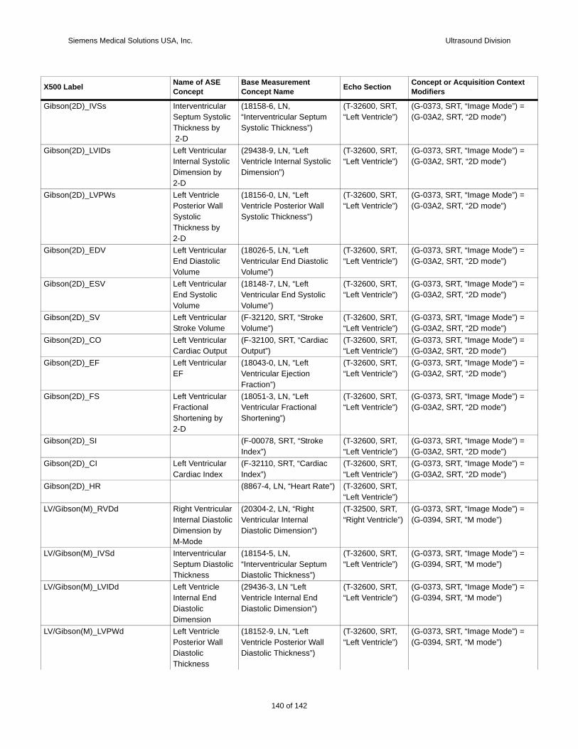
Siemens Medical Solutions USA, Inc. Ultrasound Division
Gibs =
Gibs =
Gibs =
Gibs =
Gibs =
Gibs =
Gibs =
Gibs =
Gibs =
Gibs =
Gibs =
Gibs
LV/G =
LV/G =
LV/G =
LV/G =
X500 t
on(2D)_IVSs Interventricular Septum Systolic Thickness by 2-D
(18158-6, LN, “Interventricular Septum Systolic Thickness”)
(T-32600, SRT, “Left Ventricle”)
(G-0373, SRT, “Image Mode”) (G-03A2, SRT, “2D mode”)
on(2D)_LVIDs Left Ventricular Internal Systolic Dimension by 2-D
(29438-9, LN, “Left Ventricle Internal Systolic Dimension”)
(T-32600, SRT, “Left Ventricle”)
(G-0373, SRT, “Image Mode”) (G-03A2, SRT, “2D mode”)
on(2D)_LVPWs Left Ventricle Posterior Wall Systolic Thickness by2-D
(18156-0, LN, “Left Ventricle Posterior Wall Systolic Thickness”)
(T-32600, SRT, “Left Ventricle”)
(G-0373, SRT, “Image Mode”) (G-03A2, SRT, “2D mode”)
on(2D)_EDV Left Ventricular End Diastolic Volume
(18026-5, LN, “Left Ventricular End Diastolic Volume”)
(T-32600, SRT, “Left Ventricle”)
(G-0373, SRT, “Image Mode”) (G-03A2, SRT, “2D mode”)
on(2D)_ESV Left Ventricular End Systolic Volume
(18148-7, LN, “Left Ventricular End Systolic Volume”)
(T-32600, SRT, “Left Ventricle”)
(G-0373, SRT, “Image Mode”) (G-03A2, SRT, “2D mode”)
on(2D)_SV Left Ventricular Stroke Volume
(F-32120, SRT, “Stroke Volume”)
(T-32600, SRT, “Left Ventricle”)
(G-0373, SRT, “Image Mode”) (G-03A2, SRT, “2D mode”)
on(2D)_CO Left Ventricular Cardiac Output
(F-32100, SRT, “Cardiac Output”)
(T-32600, SRT, “Left Ventricle”)
(G-0373, SRT, “Image Mode”) (G-03A2, SRT, “2D mode”)
on(2D)_EF Left Ventricular EF
(18043-0, LN, “Left Ventricular Ejection Fraction”)
(T-32600, SRT, “Left Ventricle”)
(G-0373, SRT, “Image Mode”) (G-03A2, SRT, “2D mode”)
on(2D)_FS Left Ventricular Fractional Shortening by 2-D
(18051-3, LN, “Left Ventricular Fractional Shortening”)
(T-32600, SRT, “Left Ventricle”)
(G-0373, SRT, “Image Mode”) (G-03A2, SRT, “2D mode”)
on(2D)_SI (F-00078, SRT, “Stroke Index”)
(T-32600, SRT, “Left Ventricle”)
(G-0373, SRT, “Image Mode”) (G-03A2, SRT, “2D mode”)
on(2D)_CI Left Ventricular Cardiac Index
(F-32110, SRT, “Cardiac Index”)
(T-32600, SRT, “Left Ventricle”)
(G-0373, SRT, “Image Mode”) (G-03A2, SRT, “2D mode”)
on(2D)_HR (8867-4, LN, “Heart Rate”) (T-32600, SRT, “Left Ventricle”)
ibson(M)_RVDd Right Ventricular Internal Diastolic Dimension by M-Mode
(20304-2, LN, “Right Ventricular Internal Diastolic Dimension”)
(T-32500, SRT,“Right Ventricle”)
(G-0373, SRT, “Image Mode”) (G-0394, SRT, “M mode”)
ibson(M)_IVSd Interventricular Septum Diastolic Thickness
(18154-5, LN, “Interventricular Septum Diastolic Thickness”)
(T-32600, SRT, “Left Ventricle”)
(G-0373, SRT, “Image Mode”) (G-0394, SRT, “M mode”)
ibson(M)_LVIDd Left Ventricle Internal End Diastolic Dimension
(29436-3, LN “Left Ventricle Internal End Diastolic Dimension”)
(T-32600, SRT, “Left Ventricle”)
(G-0373, SRT, “Image Mode”) (G-0394, SRT, “M mode”)
ibson(M)_LVPWd Left Ventricle Posterior Wall Diastolic Thickness
(18152-9, LN, “Left Ventricle Posterior Wall Diastolic Thickness”)
(T-32600, SRT, “Left Ventricle”)
(G-0373, SRT, “Image Mode”) (G-0394, SRT, “M mode”)
Label Name of ASE Concept
Base Measurement Concept Name Echo Section Concept or Acquisition Contex
Modifiers
140 of 142
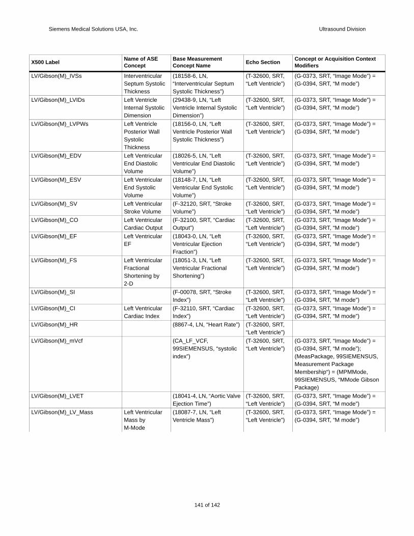
Siemens Medical Solutions USA, Inc. Ultrasound Division
LV/G =
LV/G =
LV/G =
LV/G =
LV/G =
LV/G =
LV/G =
LV/G =
LV/G =
LV/G =
LV/G =
LV/G
LV/G =
S,
on
LV/G =
LV/G =
X500 t
ibson(M)_IVSs Interventricular Septum Systolic Thickness
(18158-6, LN, “Interventricular Septum Systolic Thickness”)
(T-32600, SRT, “Left Ventricle”)
(G-0373, SRT, “Image Mode”) (G-0394, SRT, “M mode”)
ibson(M)_LVIDs Left Ventricle Internal Systolic Dimension
(29438-9, LN, “Left Ventricle Internal Systolic Dimension”)
(T-32600, SRT, “Left Ventricle”)
(G-0373, SRT, “Image Mode”) (G-0394, SRT, “M mode”)
ibson(M)_LVPWs Left Ventricle Posterior Wall Systolic Thickness
(18156-0, LN, “Left Ventricle Posterior Wall Systolic Thickness”)
(T-32600, SRT, “Left Ventricle”)
(G-0373, SRT, “Image Mode”) (G-0394, SRT, “M mode”)
ibson(M)_EDV Left Ventricular End Diastolic Volume
(18026-5, LN, “Left Ventricular End Diastolic Volume”)
(T-32600, SRT, “Left Ventricle”)
(G-0373, SRT, “Image Mode”) (G-0394, SRT, “M mode”)
ibson(M)_ESV Left Ventricular End Systolic Volume
(18148-7, LN, “Left Ventricular End Systolic Volume”)
(T-32600, SRT, “Left Ventricle”)
(G-0373, SRT, “Image Mode”) (G-0394, SRT, “M mode”)
ibson(M)_SV Left Ventricular Stroke Volume
(F-32120, SRT, “Stroke Volume”)
(T-32600, SRT, “Left Ventricle”)
(G-0373, SRT, “Image Mode”) (G-0394, SRT, “M mode”)
ibson(M)_CO Left Ventricular Cardiac Output
(F-32100, SRT, “Cardiac Output”)
(T-32600, SRT, “Left Ventricle”)
(G-0373, SRT, “Image Mode”) (G-0394, SRT, “M mode”)
ibson(M)_EF Left Ventricular EF
(18043-0, LN, “Left Ventricular Ejection Fraction”)
(T-32600, SRT, “Left Ventricle”)
(G-0373, SRT, “Image Mode”) (G-0394, SRT, “M mode”)
ibson(M)_FS Left Ventricular Fractional Shortening by 2-D
(18051-3, LN, “Left Ventricular Fractional Shortening”)
(T-32600, SRT, “Left Ventricle”)
(G-0373, SRT, “Image Mode”) (G-0394, SRT, “M mode”)
ibson(M)_SI (F-00078, SRT, “Stroke Index”)
(T-32600, SRT, “Left Ventricle”)
(G-0373, SRT, “Image Mode”) (G-0394, SRT, “M mode”)
ibson(M)_CI Left Ventricular Cardiac Index
(F-32110, SRT, “Cardiac Index”)
(T-32600, SRT, “Left Ventricle”)
(G-0373, SRT, “Image Mode”) (G-0394, SRT, “M mode”)
ibson(M)_HR (8867-4, LN, “Heart Rate”) (T-32600, SRT, “Left Ventricle”)
ibson(M)_mVcf (CA_LF_VCF, 99SIEMENSUS, “systolic index”)
(T-32600, SRT, “Left Ventricle”)
(G-0373, SRT, “Image Mode”) (G-0394, SRT, “M mode”);(MeasPackage, 99SIEMENSUMeasurement Package Membership“) = (MPMMode, 99SIEMENSUS, “MMode GibsPackage)
ibson(M)_LVET (18041-4, LN, “Aortic Valve Ejection Time”)
(T-32600, SRT, “Left Ventricle”)
(G-0373, SRT, “Image Mode”) (G-0394, SRT, “M mode”)
ibson(M)_LV_Mass Left Ventricular Mass by M-Mode
(18087-7, LN, “Left Ventricle Mass”)
(T-32600, SRT, “Left Ventricle”)
(G-0373, SRT, “Image Mode”) (G-0394, SRT, “M mode”)
Label Name of ASE Concept
Base Measurement Concept Name Echo Section Concept or Acquisition Contex
Modifiers
141 of 142
142
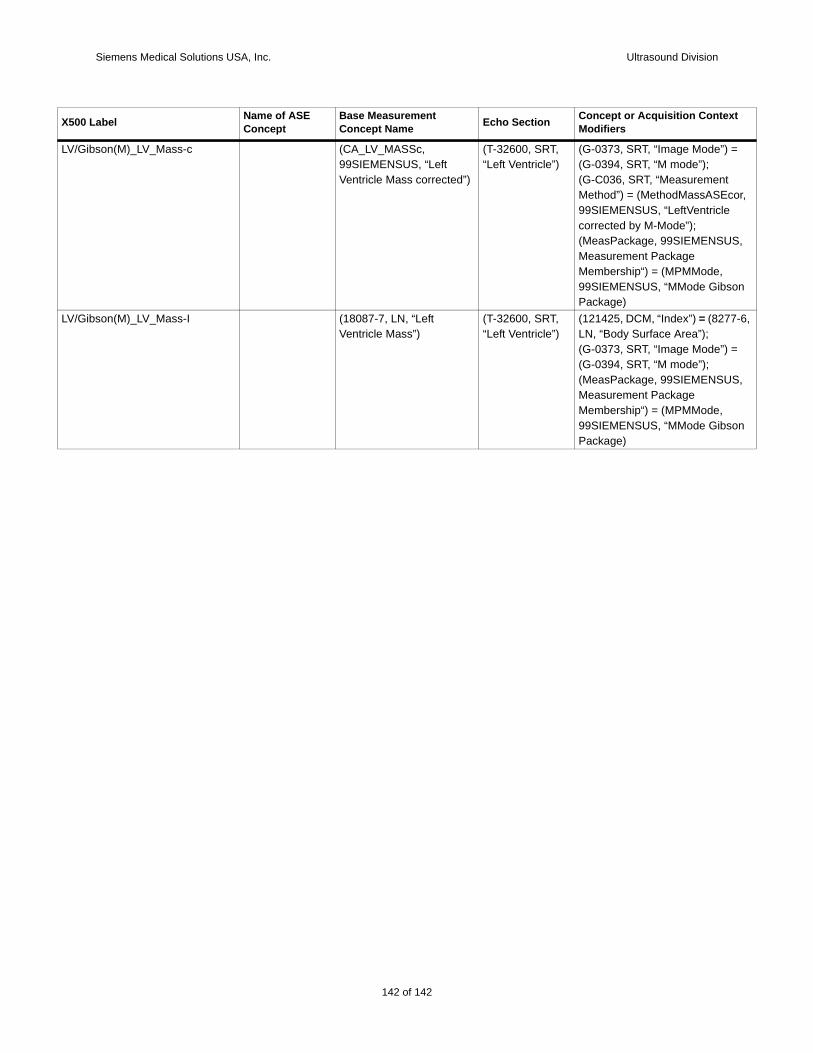
Siemens Medical Solutions USA, Inc. Ultrasound Division
LV/G =
or,
S,
on
LV/G 7-6,
=
S,
on
X500 t
ibson(M)_LV_Mass-c (CA_LV_MASSc, 99SIEMENSUS, “Left Ventricle Mass corrected”)
(T-32600, SRT, “Left Ventricle”)
(G-0373, SRT, “Image Mode”) (G-0394, SRT, “M mode”);(G-C036, SRT, “Measurement Method”) = (MethodMassASEc99SIEMENSUS, “LeftVentricle corrected by M-Mode”);(MeasPackage, 99SIEMENSUMeasurement Package Membership“) = (MPMMode, 99SIEMENSUS, “MMode GibsPackage)
ibson(M)_LV_Mass-I (18087-7, LN, “Left Ventricle Mass”)
(T-32600, SRT, “Left Ventricle”)
(121425, DCM, “Index”) = (827LN, “Body Surface Area”);(G-0373, SRT, “Image Mode”) (G-0394, SRT, “M mode”);(MeasPackage, 99SIEMENSUMeasurement Package Membership“) = (MPMMode, 99SIEMENSUS, “MMode GibsPackage)
Label Name of ASE Concept
Base Measurement Concept Name Echo Section Concept or Acquisition Contex
Modifiers
142 of 142