Activity, Distribution, and Diversity of Sulfate Reducers ...
27
Full Terms & Conditions of access and use can be found at https://www.tandfonline.com/action/journalInformation?journalCode=ugmb20 Geomicrobiology Journal ISSN: 0149-0451 (Print) 1521-0529 (Online) Journal homepage: https://www.tandfonline.com/loi/ugmb20 Activity, Distribution, and Diversity of Sulfate Reducers and Other Bacteria in Sediments above Gas Hydrate (Cascadia Margin, Oregon) Katrin Knittel , Antje Boetius , Andreas Lemke , Heike Eilers , Karin Lochte , Olaf Pfannkuche , Peter Linke & Rudolf Amann To cite this article: Katrin Knittel , Antje Boetius , Andreas Lemke , Heike Eilers , Karin Lochte , Olaf Pfannkuche , Peter Linke & Rudolf Amann (2003) Activity, Distribution, and Diversity of Sulfate Reducers and Other Bacteria in Sediments above Gas Hydrate (Cascadia Margin, Oregon), Geomicrobiology Journal, 20:4, 269-294, DOI: 10.1080/01490450303896 To link to this article: https://doi.org/10.1080/01490450303896 Published online: 10 Nov 2010. Submit your article to this journal Article views: 366 View related articles Citing articles: 202 View citing articles
Transcript of Activity, Distribution, and Diversity of Sulfate Reducers ...

Full Terms & Conditions of access and use can be found athttps://www.tandfonline.com/action/journalInformation?journalCode=ugmb20
Geomicrobiology Journal
ISSN: 0149-0451 (Print) 1521-0529 (Online) Journal homepage: https://www.tandfonline.com/loi/ugmb20
Activity, Distribution, and Diversity of SulfateReducers and Other Bacteria in Sediments aboveGas Hydrate (Cascadia Margin, Oregon)
Katrin Knittel , Antje Boetius , Andreas Lemke , Heike Eilers , Karin Lochte ,Olaf Pfannkuche , Peter Linke & Rudolf Amann
To cite this article: Katrin Knittel , Antje Boetius , Andreas Lemke , Heike Eilers , Karin Lochte ,Olaf Pfannkuche , Peter Linke & Rudolf Amann (2003) Activity, Distribution, and Diversity of SulfateReducers and Other Bacteria in Sediments above Gas Hydrate (Cascadia Margin, Oregon),Geomicrobiology Journal, 20:4, 269-294, DOI: 10.1080/01490450303896
To link to this article: https://doi.org/10.1080/01490450303896
Published online: 10 Nov 2010.
Submit your article to this journal
Article views: 366
View related articles
Citing articles: 202 View citing articles

July 23, 2003 16:21 GMB Tj773-03
Geomicrobiology Journal, 20:269–294, 2003Copyright C© Taylor & Francis Inc.ISSN: 0149-0451 print / 1362-3087 onlineDOI: 10.1080/01490450390241008
Activity, Distribution, and Diversity of SulfateReducers and Other Bacteria in Sediments above
Gas Hydrate (Cascadia Margin, Oregon)
KATRIN KNITTEL
Max Planck Institute for Marine MicrobiologyBremen, Germany
ANTJE BOETIUS
Alfred Wegener Institute for Polar and Marine ResearchBremerhaven, GermanyInternational University BremenBremen, Germany and Max Planck Institute for Marine MicrobiologyBremen, Germany
ANDREAS LEMKEHEIKE EILERS
Max Planck Institute for Marine MicrobiologyBremen, Germany
KARIN LOCHTE
Institut fur Meereskunde an der Universitat KielKiel, Germany
OLAF PFANNKUCHEPETER LINKE
GEOMAR Research Center for Marine GeosciencesKiel, Germany
Received 11 February 2003; accepted 7 April 2003.We thank the officers, crew, and shipboard scientific party for excellent support during RV SONNE cruises
SO143 and SO148. Tina Treude helped with sampling on board, Julia Polansky is acknowledged for assistancewith FISH, Sabine Schafer for total cell counts, Tina Losekann and Heiko Lobner for sulfate reduction rates, DorisSetzkorn for thymidine incorporation, and Dirk Rickert for sulfate and porosity data. We thank Beth Orcutt and thetwo reviewers for their extremely helpful comments on the manuscript. This study was part of the programs MUMM(Mikrobielle UMsatzraten von Methan in gashydrathaltigen Sedimenten, 03G0554A) and TECFLUX I and II(TECtonically induced FLUXes, 03G0148A) supported by the Bundesministerium fur Bildung und Forschung(BMBF, Germany). Further support was provided from the Max-Planck Society, Germany. This is publicationGEOTECH-17 of the program GEOTECHNOLOGIEN of the BMBF and the Deutsche Forschungsgemeinschaft(Germany).
Address correspondence to Katrin Knittel, Max Planck Institute for Marine Microbiology, Department ofMolecular Ecology, Celsiusstrasse 1, 28359 Bremen, Germany. E-mail: [email protected]
269
July 23, 2003 16:21 GMB Tj773-03
270 K. Knittel et al.
RUDOLF AMANN
Max Planck Institute for Marine MicrobiologyBremen, Germany
Cold seep environments such as sediments above outcropping hydrate at Hydrate Ridge(Cascadia margin off Oregon) are characterized by methane venting, high sulfide fluxescaused by the anaerobic oxidation of methane, and the presence of chemosyntheticcommunities. Recent investigations showed that another characteristic feature of coldseeps is the occurrence of methanotrophic archaea, which can be identified by specificbiomarker lipids and 16S rDNA analysis. This investigation deals with the diversityand distribution of sulfate-reducing bacteria, some of which are directly involved inthe anaerobic oxidation of methane as syntrophic partners of the methanotrophic ar-chaea. The composition and activity of the microbial communities at methane vented andnonvented sediments are compared by quantitative methods including total cell counts,fluorescence in situ hybridization (FISH), bacterial production, enzyme activity, and sul-fate reduction rates. Bacteria involved in the degradation of particulate organic carbon(POC) are as active and diverse as at other productive margin sites of similar waterdepths. The availability of methane supports a two orders of magnitude higher microbialbiomass (up to 9.6×10¹⁰ cells cm−3) and sulfate reduction rates (up to 8 µmol cm−3 d−1)in hydrate-bearing sediments, as well as a high bacterial diversity, especially in the groupof δ-proteobacteria including members of the branches Desulfosarcina/Desulfococcus,Desulforhopalus, Desulfobulbus, and Desulfocapsa. Most of the diversity of sulfate-reducing bacteria in hydrate-bearing sediments comprises seep-endemic clades, whichshare only low similarities with previously cultured bacteria.
Keywords anaerobic oxidation of methane, sulfate reduction, sulfate-reducing bacte-ria, bacterial production, extracellular enzymes, syntrophic consortia, bacterial diversity,gas hydrate, Hydrate Ridge, Cascadia margin
Introduction
The greenhouse gas methane is present in huge amounts in marine sediments and existseither as crystalline, solid phase methane hydrates, or as free gas. Methane emission tothe hydrosphere is controlled by a microbial process called anaerobic oxidation of methane(AOM), which involves methane as electron donor and sulfate as electron acceptor (Hinrichsand Boetius 2002, and references therein) and which removes most methane within thesediments (Reeburgh 1996). The metabolic process of AOM is assumed to be a reversalof methane formation involving methanogenic archaea and sulfate-reducing bacteria aspartners (Hoehler et al. 1994; Valentine and Reeburgh 2000). Recent studies have demon-strated that AOM is mediated by a structured consortium of archaea belonging to theorder Methanosarcinales (ANME-2 group), and SRB of the Desulfosarcina/Desulfococcusbranch of the δ-proteobacteria (Boetius et al. 2000a; Orphan et al. 2001b), in the followingreferred to as “ANME-2/DSS aggregates.” These consortia oxidize methane with equimolaramounts of sulfate, yielding carbonate and sulfide, respectively (Nauhaus et al. 2002). Asecond archaeal group (ANME-1), distantly related to Methanosarcinales, has also beenshown to mediate AOM (Michaelis et al. 2002; Orphan et al. 2002). Neither ANME-1 norANME-2 or their sulfate-reducing partners have been isolated yet and the enzymes andbiochemical pathways involved in AOM remain unknown.
This investigation was carried out to quantify microbial activity and distribution ata cold seep environment and to analyze its community composition. At Hydrate Ridge,discrete methane hydrate layers occur at the seafloor, at a water depth of 600–800 m cor-responding to the hydrate stability limit (Suess et al. 1999). The hydrates are located a fewcentimeters beneath the sediment surface and form mounds of several meters in diameter
July 23, 2003 16:21 GMB Tj773-03
SRB and Other Bacteria above Gas Hydrate 271
(Suess et al. 2002). The mounds are covered by sediment and populated by thick mats ofthe sulfur-oxidizing filamentous bacteria Beggiatoa (Sahling et al. 2002; Torres et al. 2002).Another typical chemosynthetic community of Hydrate Ridge is the vesicomyid clams ofCalyptogena forming clam fields of several meters diameter. Both, Beggiatoa and Calypto-gena spp., are indicative of active methane seeping (Tryon et al. 2002). A third communityis formed by the subsurface dwelling bivalves Acharax, which occur farther away from thestrongly methane-vented sites and are here referred to as reference sites. The distributionof chemosynthetic communities at Hydrate Ridge is mainly related to the sulfide flux fromthe surface sediments (Sahling et al. 2002), which is caused by methane-dependent sul-fate reduction (Nauhaus et al. 2002). As a consequence, AOM and sulfate reduction rates(SRR) correlate strongly across the different samples and even within the sediment profile atHydrate Ridge (Treude et al. submitted). The hydrate-bearing sediments of Hydrate Ridgeshow high rates of methane-dependent sulfate reduction and harbor extremely high amountsof AOM mediating ANME-2/DSS aggregate biomass (Boetius et al. 2000a).
This study focused on quantification and comparison of abundance, activity, and di-versity of microbial organisms involved in carbon cycling at methane-vented and methane-depleted sites (Beggiatoa and Calyptogena sites vs. Acharax reference). Earlier investi-gations showed a relatively low diversity of archaea in cold seep sediments compared tothe bacterial diversity in these habitats (Hinrichs et al. 1999; Lanoil et al. 2001; Orphanet al. 2001a, 2002; Teske et al. 2002), despite their quantitative dominance at cold seeps(Boetius et al. 2000a; Michaelis et al. 2002). This study addressed the bacterial diversityand abundance in sediments above gas hydrate, especially regarding the SRB communitystructure.
Materials and Methods
Study Site and Sampling
Sediment samples were obtained during RV SONNE cruises SO143-2 in August 1999(Bohrmann et al. 2000) and SO148-1 in August 2000 (Linke and Suess 2001) at the crest ofsouthern Hydrate Ridge (44◦34′N, 125◦09′W, 780 m water depth) at the Cascadia convergentmargin off the coast of Oregon. Sediment cores of 20–40 cm length were obtained using avideo-guided multiple corer from gas hydrate bearing sediments and from reference sites notenriched in methane in the surface sediments (Table 1). Sediments from above hydrate werecovered by mats of filamentous sulfur-oxidizing bacteria of the genus Beggiatoa (stations105-1 and 19-2; called “Beggiatoa mats”) or by clam fields of Calyptogena spp. (stations185-1 and 38-1; called “Calyptogena fields”).
Geochemical Description of the Study Sites
Sediments from both sites, Beggiatoa mats and Calyptogena fields, were highly sulfidic andgassy. Oxygen had a maximum penetration of a few millimeters. Sulfide concentrations ofup to 28 mM were reached within 3–5 cm depth in the Beggiatoa mats with concurrentdepletion of sulfate down to <2 mM. In Calyptogena fields sulfate penetrated deeper into thesediments due to the bioturbating activity of the clams. Nevertheless, sulfide concentrations>20 mM were reached below 10 cm and sulfate was depleted below. Layers of hydrate werefound in several cores at depths of about 13–20 cm. Strong degassing from decomposinghydrates was observed in several cores from Beggiatoa mats and in a few cores fromCalyptogena fields. Sediments of the southern crest of Hydrate Ridge not covered by eithercommunity but sometimes containing bivalves of the species Acharax contained only tracesof methane and sulfide in the upper 15 cm and are referred to as “reference” site. Upon

July 23, 2003 16:21 GMB Tj773-03
272 K. Knittel et al.
TABLE 1 List of stations sampled during expedition SO143/2 (TECFLUX I) andSO148/1 (TECFLUX II)
Expedition/ Date Depth Types ofStation [d/m/y] [m] Position Site description analysis
143/91-1 03.08.99 850 44◦ 33.880′N,125◦ 08.290′W
Reference (noAcharax)
EEA
143/105-1 05.08.99 780 44◦ 34.140′N,125◦ 08.810′W
MediumBeggiatoa mat
TDR, EEA,TCC, agg.no,FISH
143/114-1 07.08.99 760 44◦ 34.210′N,125◦ 09.850′W
Beggiatoa mat wCalyptogena
TDR, EEA
143/127-1 09.08.99 2321 44◦ 38.500′N,125◦ 14.490′W
Reference (noAcharax)
SRR
143/131-1 10.08.99 780 44◦ 34.180′N,125◦ 08.800′W
Calyptogena field TDR, EEA
143/135-3 11.08.99 783 44◦ 34.196′N,125◦ 08.825′W
Reference (fewAcharax)
EEA
143/139 12.08.99 830 44◦ 34.100′N,125◦ 08.380′W
Reference (noAcharax)
EEA, SRR,TCC, agg.no,FISH
143/151-1 13.08.99 1284 44◦ 33.000′N,125◦ 04.000′W
Reference (noAcharax)
TDR
143/173-1 16.08.99 785 44◦ 34.160′N,125◦ 08.850′W
Beggiatoa mat wCalyptogena
EEA, SRR
143/179-3 17.08.99 785 44◦ 34.210′N,125◦ 08.740′W
Calyptogena field TDR, EEA, TCC
143/185-1 18.08.99 785 44◦ 34.190′N,125◦ 08.830′W
Calyptogena field TCC, agg.no,FISH
143/187-1/4 18.08.99 785 44◦ 36.160′N,125◦ 08.860′W
MediumBeggiatoa mat
TDR, EEA
143/190-1 19.08.99 825 44◦ 34.050′N,125◦ 08.410′W
Reference (fewAcharax)
TDR
143/193-1 20.08.99 685 44◦ 37.509′N,124◦ 55.970′W
Reference (noAcharax)
EEA, TDR
148/19-2 27.07.00 777 44◦ 34.104′N,125◦ 08.807′W
MediumBeggiatoa mat
TCC, agg.no,FISH, 16S
148/38-1 30.07.00 787 44◦ 34.186′N,125◦ 08.847′W
Calyptogena field TCC, agg.no,FISH
148/51-1 01.08.00 775 44◦ 34.198′N,125◦ 08.858′W
Reference (fewAcharax)
TCC, agg.no,FISH
Gas hydrates were recovered from cores with Beggiatoa at sediment depths >13 cm and from Ca-lyptogena fields (>16 cm). Samples from Calyptogena fields contained 1-5 clams per core. “Beggiatoamats” relate to cores covered by a carpet of Beggiatoa extending several mm into the overlying bottomwater. Types of analysis: Bacterial secondary production measured as tymidine incorporation (TDR),total cell counts (TCC), aggregate numbers (agg.no), fluorescence in situ hybridization (FISH), 16SrDNA cloning (16S), sulfate reduction rates (SRR), extracellular enzyme activities (EEA).
July 23, 2003 16:21 GMB Tj773-03
SRB and Other Bacteria above Gas Hydrate 273
recovery sediment cores were immediately transferred to a cold room (4◦C). Samples wereprocessed within 4 h after sampling according to the methods that follow.
DNA Extraction
Cores were sectioned into 1 cm layers and deep frozen (−20◦C) for DNA extraction at thehome laboratory. Total community DNA was directly extracted from the sediment (HydrateRidge, station 19-2, Beggiatoa site) as described by Zhou et al. (1996). Aliquots (ca. 4 g) ofwet sediment of three different sections (depths, 0–1, 2–4, and 8–9 cm) were used for DNAextraction. Crude DNA was purified by dialysis as described previously (Ravenschlag et al.2000).
PCR Amplification and Clone Library Construction
Two universal bacterial primers, GM3F (E. coli 16S rRNA position 0008; Muyzer et al.1995) and EUB1492 (Kane et al. 1993), were used to amplify 16S rRNA genes from theextracted chromosomal DNAs. PCR was performed with a Mastercycler Gradient (Eppen-dorf, Hamburg, Germany) as described previously (Ravenschlag et al. 1999). Products oftwo parallel PCRs were combined and purified with the QiaQuick PCR Purification Kitusing the protocol of the manufacturer (Qiagen, Hilden, Germany). DNA was ligated in thepGEM-T-Easy vector (Promega, Madison, WI, USA), and a clone library was constructedfor each section (0–1, 2–4, and 8–9 cm) as described by Ravenschlag et al. (1999). Thenumber of amplification cycles during PCR was reduced as much as possible to minimizePCR biases, chimera formations, and Taq polymerase error rates; however, 36 cycles wereneeded to yield sufficient product.
ARDRA
A total of 131 clones (01 cm: 34; 24 cm: 56; 89 cm: 41) known to contain the correctlysized insert of 1.5 kb were selected for further analysis. Amplified rDNA restriction analysis(ARDRA) was performed in order to identify clones with different inserts. Digestion withtwo restriction enzymes (HaeIII and RsaI) was used to screen the clones as describedpreviously (Ravenschlag et al. 1999).
Sequencing and Phylogenetic Analysis
Representatives of all ARDRA pattern groups were used for sequencing. Taq Cycle Se-quencing of plasmid DNAs from selected 16S rDNA clones with vector pimers and uni-versal rRNA specific primers was performed by GATC Biotech (Konstanz, Germany) andGAG BioScience (Bremen, Germany). Sequence data were analyzed with the ARB soft-ware package (Strunk et al. 1998). Phylogenetic trees were calculated with sequences fromHydrate Ridge sediments together with sequences, which are available in the databasesEMBL, GenBank, and DDJB, by performing parsimony, neighbor-joining, and maximum-likelihood analysis with different sets of filters. In all cases, general tree topology andclusters were stable. For tree calculation, nearly full length sequences (>1,340 bp) wereconsidered. Partial sequences were inserted into the reconstructed tree by parsimony criteriawith global/local optimization, without allowing changes in the overall tree topology.
July 23, 2003 16:21 GMB Tj773-03
274 K. Knittel et al.
Total Cell Counts, ANME-2/DSS Aggregate Numbers, and Size Measurements
Samples for total cell counts were obtained from 1 cm core slices, fixed with 2% formalde-hyde, and stored cold (4◦C) until analysis at the home laboratory. Total cell counts andthe quantification of aggregates was done via epifluorescence microscopy after staining thesediments with Acridine Orange Direct Counts (AODC) according to the method of Meyer-Reil (1983). Total cell counts were defined as the sum of single cells plus the aggregatedcells in the syntrophic consortia. In sediments from Hydrate Ridge, these ANME2/DSSaggregates can be easily recognized via AODC because of their conspicuous sphericalshape and the surrounding organic matrix. However, the aggregated cells cannot be countedin these spheres. Hence, we used a semidirect method for the quantification of cells asdescribed below. DAPI staining was used to measure ANME2/DSS aggregate sizes viaepifluorescence microscopy of FISH-treated samples.
Five samples were selected for the measurement: Two horizons of the Beggiatoa mat(station 105-1: 1–2 cm, 9–10 cm) and the Calyptogena field (station 185-1: 1–2 cm, 9–10 cm) and one sample of the reference (station 139: 0-1 cm), in which we detected thehighest numbers of ANME2/DSS aggregates. For the upper layer of the Beggiatoa mat andCalyptogena field the diameter of 100 aggregates was measured, that of 50 aggregates forthe other samples. Average diameters were between 3–4 µm in all cases (see Results), witha deviation of about 30% as standard deviation, and 10% as confidence level. We simplyused 3 µm as average aggregate diameter at all stations. Cell numbers in the aggregateswere calculated based on the assumption that both the aggregates and the cells within theaggregates are spheres. The volume of the aggregate sphere (vol 14 µm3) was divided bythe volume of the cell (diameter ca. 0.4 µm for SRB and 0.5 µm for ANME2 cells, i.e.,average 0.043 µm3), resulting in an average cell number of ca. 300 cells (with ca. 200 SRBand 100 archaea (Boetius et al. 2000a).
Microbial biomass in gram carbon was calculated by multiplying cell numbers with acarbon content of 20 fg per cell. This is a standard factor commonly used in aquatic microbialecology and gives similar results to the other commonly used factor of 3 × 10⁻¹³ gC/µm3
biovolume of cells (Børsheim et al. 1991).
Fluorescence In Situ Hybridization (FISH)
For FISH, subsamples of sediment cores were sliced into 1 cm intervals and fixed for 2–3 hwith 3% formaldehyde (final concentration), washed twice with 1 × PBS (10 mM sodiumphosphate; 130 mM NaCl), and finally stored in 1 × PBS/EtOH (1:1) at −20◦C. Fixedsamples were diluted and treated by mild sonication for 20 s with a MS73 probe (SonopulsHD70, Bandelin, Germany) at an amplitude of 42 µm <10 W. An aliquot was filtered on0.2 µm GTTP polycarbonate filters (Millipore, Eschborn, Germany). Hybridization andmicroscopy counts of hybridized and 4′,6′-diamidino-2-phenylindole (DAPI)-stained cellswere performed as described previously (Snaidr et al. 1997). Means were calculated by using10 to 40 randomly chosen fields for each filter section, which corresponded to 800 to 1000DAPI-stained cells. Cy3-monolabeled oligonucleotides were purchased from ThermoHy-baid (Ulm, Germany). Probes and formamide (FA) concentrations used in this study wereas follows: EUB338 I-III targeting Bacteria (Daims et al. 1999; 35% FA), ARCH915 target-ing Archaea (Amann et al. 1990; 35% FA), Non338 (negative control; Wallner et al. 1993;10% FA), DSS658 targeting Desulfosarcina spp./Desulfococcus spp./Desulfofrigus spp. andDesulfofaba spp. (60% FA), DSR651 targeting Desulfosarcina spp. (35% FA), DSV698 tar-geting Desulfovibrio spp. (all in Manz et al. 1998; 35% FA), 221 targeting Desulfobacteriumspp. (35% FA), 660 targeting Desulfobulbus spp. (Devereux et al. 1992; 60% FA), Sva1428targeting Desulfotalea spp./Desulfofustis spp./SEEP-SRB4/Desulfocapsa relatives (Sahm

July 23, 2003 16:21 GMB Tj773-03
SRB and Other Bacteria above Gas Hydrate 275
et al. 1999b; 35% FA), and CF319a targeting the Cytophagal/Flavobacterium cluster (Manzet al. 1996; 35% FA). Background signal of samples, observed with the nonsense probeNON338, was negligible (<0.1%).
Bacterial Secondary Production (Thymidine Incorporation)
Thymidine incorporation was measured in slurries of 1 cm layers of sediment accordingto Boetius et al. (2000b). The procedure for the extraction of the DNA containing the 3H-thymidine and the calculation of thymidine incorporation followed the method of Findlay(1993).
Activity of Extracellular Hydrolytic Enzymes
The extracellular enzymatic activities (EEA) of the hydrolasesα-,β-glucosidase, chitobiase,lipase, sulfatase, phosphatase, butyrase, and aminopeptidase were determined according toBoetius et al. (2000b) at in situ temperature (2◦C) in sediment slurries of 1 cm depthhorizons.
Sulfate Reduction Rates (SRR)
Sediment cores collected from the multiple corer were subsampled with small PVC coresin the cold room (4◦C), and 35SO2−
4 was injected horizontally into the intact sediment coresat 1-cm depth intervals. The cores were incubated for 24 h at in situ temperature before thereaction was stopped by sectioning the sediments at 1 cm intervals and mixing with 20%zinc acetate. The samples were stored frozen until the single-step acidic distillation of theCr-II-reduced sulfur compounds was carried out as described by Fossing and Jørgensen(1989). SRR was calculated from the ratio of radioactive sulfide to the total radioactivesulfate added, multiplied by the porewater sulfate concentration. Sulfate concentrations andporosity data for calculating the rates were kindly provided by D. Rickert (GEOMAR).
Nucleotide Sequence Accession Numbers
The nucleotide sequence data reported in this paper will appear in the EMBL, GenBank,and DDBJ nucleotide sequence database under the accession no. AF535216 to AF535259.
Results
Bacterial Abundance and Secondary Production
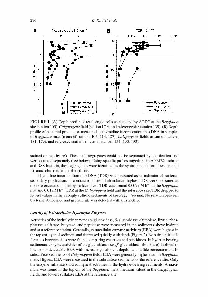
The abundance of single cells was compared among the sulfidic, hydrate-bearing sedimentsrecovered from Beggiatoa mats and Calyptogena fields and the sediments of a referencestation, which was not substantially enriched in methane in the upper 10 cm (Figure 1,Table 2). In the top centimeter layer of methane-rich sediments, numbers of single cellswere 4.5 × 10⁹ cells cm−3 and significantly higher than at the reference site (1 × 10⁹ cellscm−3). Counts of single cells in the strongly sulfidic Beggiatoa mat dropped quickly toaround 1×10⁹ cells cm−3 below sediment surface, and were only slightly higher than at thereference site. However, cell counts in sediments of the Calyptogena field remained highdown to 25 cm. A high number of aggregated cells (>108 aggregates cm−3) was detected inthe sediments above hydrate by the characteristic fluorescent green of nucleic acid stainingwith Acridine Orange (AO). The aggregates were enclosed in an organic matrix, which was
July 23, 2003 16:21 GMB Tj773-03
276 K. Knittel et al.
FIGURE 1 (A) Depth profile of total single cells as detected by AODC at the Beggiatoasite (station 105), Calyptogena field (station 179), and reference site (station 139). (B) Depthprofile of bacterial production measured as thymidine incorporation into DNA in samplesof Beggiatoa mats (mean of stations 105, 114, 187), Calyptogena fields (mean of stations131, 179), and reference stations (mean of stations 151, 190, 193).
stained orange by AO. These cell aggregates could not be separated by sonification andwere counted separately (see below). Using specific probes targeting the ANME2 archaeaand DSS bacteria, these aggregates were identified as the syntrophic consortia responsiblefor anaerobic oxidation of methane.
Thymidine incorporation into DNA (TDR) was measured as an indicator of bacterialsecondary production. In contrast to bacterial abundance, highest TDR were measured atthe reference site. In the top surface layer, TDR was around 0.007 nM h−1 at the Beggiatoamat and 0.01 nM h−1 TDR at the Calyptogena field and the reference site. TDR dropped tolowest values in the strongly sulfidic sediments of the Beggiatoa mat. No relation betweenbacterial abundance and growth rate was detected with this method.
Activity of Extracellular Hydrolytic Enzymes
Activities of the hydrolytic enzymes α-glucosidase, β-glucosidase, chitobiase, lipase, phos-phatase, sulfatase, butyrase, and peptidase were measured in the sediments above hydrateand at a reference station. Generally, extracellular enzyme activities (EEA) were highest inthe top cm layer of sediment and decreased quickly with depth (Figure 2). No substantial dif-ferences between sites were found comparing esterases and peptidases. In hydrate-bearingsediments, enzyme activities of the glucosidases (α-,β-glucosidase, chitobiase) declined tolow or nondetectable EEA with increasing sediment depth, i.e., sulfide concentration. Insubsurface sediments of Calyptogena fields EEA were generally higher than in Beggiatoamats. Highest EEA were measured in the subsurface sediments of the reference site. Onlythe enzyme sulfatase showed highest activities in the hydrate-bearing sediments. A maxi-mum was found in the top cm of the Beggiatoa mats, medium values in the Calyptogenafields, and lowest sulfatase EEA at the reference site.

July 23, 2003 16:21 GMB Tj773-03
TA
BL
E2
Dis
trib
utio
nof
AN
ME
-2/D
SSag
greg
ates
and
tota
lcel
lsin
Hyd
rate
Rid
gese
dim
ents
atdi
ffer
ents
ampl
ing
site
s
Sam
plin
g19
99
Beg
giat
oam
atC
alyp
toge
nafie
ldR
efer
ence
Stat
ion
105-
1St
atio
n18
5-1
Stat
ion
139
No.
No.
No.
Dep
thN
o.ag
g.si
ngle
cells
Tota
lcel
lsC
ells
inag
g.∗
No.
agg.
sing
lece
llsTo
talc
ells
Cel
lsin
agg.
∗N
o.ag
g.si
ngle
cells
Tota
lcel
lsC
ells
inag
g.∗
[cm
][1
07cm
−3]
[109
cm−3
][1
010cm
−3]
[%to
talc
ells
][1
07cm
−3]
[109
cm−3
][1
010cm
−3]
[%to
talc
ells
][1
07cm
−3]
[109
cm−3
][1
010cm
−3]
[%to
talc
ells
]
0.5
13.6
4.6
4.6
9013
.94.
24.
691
1.5
0.9
0.5
841.
58.
31.
72.
794
14.0
5.6
4.8
881.
31.
00.
579
2.5
11.2
2.3
3.6
94n.
d.n.
d.n.
d.n.
d.n.
d.1.
9n.
d.72
3.5
2.9
1.0
1.0
90n.
d.n.
d.n.
d.n.
d.n.
d.1.
2n.
d.n.
d.4.
54.
91.
11.
693
30.5
4.4
9.6
960.
60.
80.
372
5.5
9.7
1.7
3.1
94n.
d.n.
d.n.
d.n.
d.n.
d.0.
8n.
d.n.
d.6.
53.
91.
11.
391
n.d.
n.d.
n.d.
n.d.
n.d.
1.1
n.d.
n.d.
7.5
4.2
1.5
1.4
89n.
d.n.
d.n.
d.n.
d.n.
d.0.
6n.
d.n.
d.8.
5n.
d.n.
d.n.
d.n.
d.n.
d.n.
d.n.
d.n.
d.n.
d.0.
9n.
d.n.
d.9.
54.
41.
31.
491
14.3
2.3
4.5
950.
70.
60.
377
14.5
2.5
0.8
0.8
913.
71.
11.
291
0.3
0.4
0.1
72
Sam
plin
g20
00
Beg
giat
oam
atC
alyp
toge
nafie
ldR
efer
ence
Stat
ion
19-2
Stat
ion
38-1
Stat
ion
51-1
0.5
5.1
2.9
1.8
846.
82.
32.
390
01.
80.
20
1.5
5.1
2.7
1.8
8513
.94.
64.
690
01.
10.
10
2.5
10.0
2.7
3.3
925.
73.
52.
183
0.2
1.3
0.2
363.
512
.73.
94.
291
6.8
4.5
2.5
820
1.1
0.1
04.
57.
62.
62.
590
5.6
3.9
2.1
810.
93.
00.
648
5.5
8.0
2.7
2.7
905.
44.
92.
277
0.9
2.2
0.5
556.
55.
12.
41.
887
6.7
3.0
2.3
870.
31.
40.
239
7.5
3.8
1.6
1.3
888.
93.
83.
188
0.3
1.2
0.2
388.
51.
20.
80.
579
12.7
5.1
4.3
881.
31.
40.
572
9.5
2.1
1.2
0.7
836.
33.
02.
286
0.5
1.6
0.3
4910
.5n.
d.n.
d.n.
d.n.
d.3.
01.
31.
088
n.d.
n.d.
n.d.
n.d.
11.5
n.d.
n.d.
n.d.
n.d.
5.1
2.2
1.7
87n.
d.n.
d.n.
d.n.
d.12
.5n.
d.n.
d.n.
d.n.
d.3.
41.
31.
189
n.d.
n.d.
n.d.
n.d.
∗ Ave
rage
cell
num
bers
per
aggr
egat
e:30
0.
277

July 23, 2003 16:21 GMB Tj773-03
278 K. Knittel et al.
FIGURE 2 Comparison of extracellular hydrolytic activities of different enzymes at dif-ferent study sites (sampling 1999) and sediment depth horizons at Hydrate Ridge. Activitiesare given in nmol cm−3 h−1, Beggiatoa mat: mean of stations 105, 114, 173, 187; Calypto-gena field: mean of stations 114, 131, 135, 173, 179; reference stations: mean of stations91, 139, 193.
Sulfate Reduction Rates
Differences between hydrate-bearing and reference sites were most obvious when compar-ing sulfate reduction rates (SRR, Figure 3). SRR was two orders of magnitude higher abovehydrate than at the reference site (Figure 3C). Also, a difference was noted between SRRin cores from Beggiatoa mats (Figure 3A) and Calyptogena fields (Figure 3B). Generally,samples from Calyptogena fields had a subsurface maximum in SRR whereas Beggiatoamats often showed highest SRR in the top 2 cm of sediments. A considerable variation
FIGURE 3 Depth profiles of sulfate reduction rates (SRR) in three parallel sediment coresof different sampling sites of Hydrate Ridge (sampling 1999). (A) Beggiatoa mat (station173); (B) Calyptogena field (station 173); (C) reference station (station 127).
July 23, 2003 16:21 GMB Tj773-03
SRB and Other Bacteria above Gas Hydrate 279
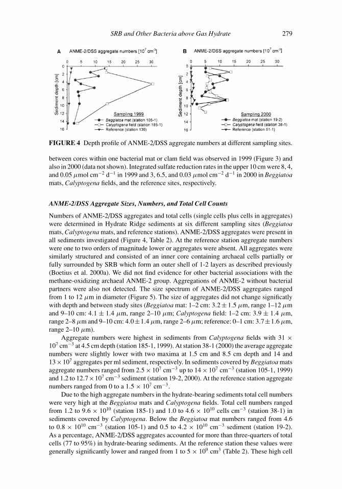
FIGURE 4 Depth profile of ANME-2/DSS aggregate numbers at different sampling sites.
between cores within one bacterial mat or clam field was observed in 1999 (Figure 3) andalso in 2000 (data not shown). Integrated sulfate reduction rates in the upper 10 cm were 8, 4,and 0.05 µmol cm−2 d−1 in 1999 and 3, 6.5, and 0.03 µmol cm−2 d−1 in 2000 in Beggiatoamats, Calyptogena fields, and the reference sites, respectively.
ANME-2/DSS Aggregate Sizes, Numbers, and Total Cell Counts
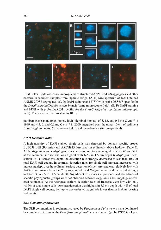
Numbers of ANME-2/DSS aggregates and total cells (single cells plus cells in aggregates)were determined in Hydrate Ridge sediments at six different sampling sites (Beggiatoamats, Calyptogena mats, and reference stations). ANME-2/DSS aggregates were present inall sediments investigated (Figure 4, Table 2). At the reference station aggregate numberswere one to two orders of magnitude lower or aggregates were absent. All aggregates weresimilarly structured and consisted of an inner core containing archaeal cells partially orfully surrounded by SRB which form an outer shell of 1-2 layers as described previously(Boetius et al. 2000a). We did not find evidence for other bacterial associations with themethane-oxidizing archaeal ANME-2 group. Aggregations of ANME-2 without bacterialpartners were also not detected. The size spectrum of ANME-2/DSS aggregates rangedfrom 1 to 12 µm in diameter (Figure 5). The size of aggregates did not change significatlywith depth and between study sites (Beggiatoa mat: 1–2 cm: 3.2 ± 1.5 µm, range 1–12 µmand 9–10 cm: 4.1 ± 1.4 µm, range 2–10 µm; Calyptogena field: 1–2 cm: 3.9 ± 1.4 µm,range 2–8 µm and 9–10 cm: 4.0±1.4 µm, range 2–6 µm; reference: 0–1 cm: 3.7±1.6 µm,range 2–10 µm).
Aggregate numbers were highest in sediments from Calyptogena fields with 31 ×107 cm−3 at 4.5 cm depth (station 185-1, 1999). At station 38-1 (2000) the average aggregatenumbers were slightly lower with two maxima at 1.5 cm and 8.5 cm depth and 14 and13×107 aggregates per ml sediment, respectively. In sediments covered by Beggiatoa matsaggregate numbers ranged from 2.5 × 107 cm−3 up to 14 × 107 cm−3 (station 105-1, 1999)and 1.2 to 12.7×107 cm−3 sediment (station 19-2, 2000). At the reference station aggregatenumbers ranged from 0 to a 1.5 × 107 cm−3.
Due to the high aggregate numbers in the hydrate-bearing sediments total cell numberswere very high at the Beggiatoa mats and Calyptogena fields. Total cell numbers rangedfrom 1.2 to 9.6 × 10¹⁰ (station 185-1) and 1.0 to 4.6 × 10¹⁰ cells cm−3 (station 38-1) insediments covered by Calyptogena. Below the Beggiatoa mat numbers ranged from 4.6to 0.8 × 1010 cm−3 (station 105-1) and 0.5 to 4.2 × 1010 cm−3 sediment (station 19-2).As a percentage, ANME-2/DSS aggregates accounted for more than three-quarters of totalcells (77 to 95%) in hydrate-bearing sediments. At the reference station these values weregenerally significantly lower and ranged from 1 to 5 × 109 cm3 (Table 2). These high cell
July 23, 2003 16:21 GMB Tj773-03
280 K. Knittel et al.
FIGURE 5 Epifluorescence micrographs of structured ANME-2/DSS aggregates and otherbacteria in sediment samples from Hydrate Ridge. (A, B) Size spectrum of DAPI stainedANME-2/DSS aggregates. (C, D) DAPI staining and FISH with probe DSS658 specific forthe Desulfosarcina/Desulfococcus branch (same microscopic field). (E, F) DAPI stainingand FISH with probe DSR651 specific for the Desulforhopalus spp. (same microscopicfield). The scale bar is equivalent to 10 µm.
numbers correspond to extremely high microbial biomass of 5, 13, and 0.8 mg C cm−2 in1999 and 4.5, 6, and 0.6 mg C cm−2 in 2000 integrated over the upper 10 cm of sedimentfrom Beggiatoa mats, Calyptogena fields, and the reference sites, respectively.
FISH Detection Rates
A high quantity of DAPI-stained single cells was detected by domain specific probesEUB338 I-III (Bacteria) and ARCH915 (Archaea) in sediments above hydrate (Table 3).At the Beggiatoa and Calyptogena sites detection of Bacteria ranged between 40 and 52%at the sediment surface and was highest with 62% in 1.5 cm depth (Calyptogena field,station 38-1). Below this depth the detection rate strongly decreased to less than 10% oftotal DAPI cell counts. In contrast, detection rates for single cell Archaea increased withincreasing depth. At the sediment surface detection of such Archaea was relatively low with1–2% in sediments from the Calyptogena field and Beggiatoa mat and increased stronglyto 18–31% in 9.5 to 14.5 cm depth. Significant differences in presence and abundance ofspecific phylogenetic groups were not observed between Beggiatoa and Calyptogena cov-ered sediments. At the reference stations detection rates of Bacteria were low with only<19% of total single cells. Archaea detection was highest in 8.5 cm depth with 4% of totalDAPI single cell counts, i.e., up to one order of magnitude lower than in hydrate-bearingsediments.
SRB Community Structure
The SRB communities in sediments covered by Beggiatoa or Calyptogena were dominatedby complete oxidizers of the Desulfosarcina/Desulfococcus branch (probe DSS658). Up to

July 23, 2003 16:21 GMB Tj773-03
SRB and Other Bacteria above Gas Hydrate 281
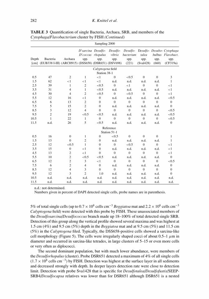
TABLE 3 Quantification of single Bacteria, Archaea, SRB, and members of theCytophaga/Flavobacterium cluster by FISH
Sampling 1999
D′sarcina Desulfo- Desulfo- Desulfo- Desulfo- Desulto- CytophagaD′coccus rhopalus vibrio bacterium talea bulbus Flavobact.
Depth Bacteria Archaea spp. spp. spp. spp. spp. spp. spp.[cm] (EUB338 I-III) (ARCH915) (DSS658) (DSR651) (DSV698) (221) (Sval428) (660) (CF319a)
Beggiatoa matStation 105-1
0.5 52 1 1 4 <1 0 2 <0.5 51.5 22 4 4 2 <1 0 0 <1 42.5 24 2 2 1 <0.5 0 0 <0.5 13.5 20 1 3 <1 0 0 1 0 <0.54.5 21 7 2 <0.5 0 n.d. <1 0 <15.5 20 3 1 <0.5 n.d. <1 2 0 16.5 18 1 2 <0.5 0 0 0 <0.5 17.5 17 5 2 <0.5 0 0 0 0 09.5 12 23 4 0 0 0 0 0 014.5 15 14 1 <0.5 0 0 <1 0 0
Calyptogena fieldStation 185-1
0.5 48 3 3 <1 <0.5 0 <0.5 0 01.5 25 2 2 <1 <0.5 0 <0.5 0 32.5 n.d. n.d. n.d. n.d. n.d. n.d. n.d. n.d. n.d.3.5 n.d. n.d. n.d. n.d. n.d. n.d. n.d. n.d. n.d.4.5 18 23 1 <1 0 0 0 0 <15.5 n.d. n.d. n.d. n.d. n.d. n.d. n.d. n.d. n.d.6.5 n.d. n.d. n.d. n.d. n.d. n.d. n.d. n.d. n.d.7.5 n.d. n.d. n.d. n.d. n.d. n.d. n.d. n.d. n.d.9.5 15 30 5 0 0 0 0 <0.5 <0.514.5 9 28 0 0 0 0 <0.5 <0.5 0
ReferenceStation 139
0.5 17 <1 <1 <1 <1 0 <1 0 <11.5 19 <1 1 <0.5 <1 0 0 0 <12.5 n.d. n.d. n.d. n.d. n.d. n.d. n.d. n.d. n.d.3.5 n.d. n.d. n.d. n.d. n.d. n.d. n.d. n.d. n.d.4.5 11 <0.5 <1 0 0 0 0 0 25.5 n.d. n.d. n.d. n.d. n.d. n.d. n.d. n.d. n.d.6.5 n.d. n.d. n.d. n.d. n.d. n.d. n.d. n.d. n.d.7.5 n.d. n.d. n.d. n.d. n.d. n.d. n.d. n.d. n.d.9.5 n.d. n.d. <0.5 0 n.d. 0 0 0 214.5 n.d. n.d. <0.5 0 0 0 0 0 0
Sampling 2000
Beggiatoa matStation 19-2
0.5 40 1 <1 2 <1 0 0 0 11.5 17 1 2 <0.5 n.d. n.d. n.d. n.d. <12.5 24 3 2 <0.5 0 0 0 0 <0.53.5 15 1 2 <0.5 n.d. n.d. n.d. n.d. <0.54.5 9 3 <1 <0.5 0 0 0 0 <0.55.5 14 4 <1 <0.5 n.d. n.d. n.d. n.d. <0.56.5 7 9 1 0 0 0 0 0 07.5 8 25 1 0 n.d. n.d. n.d. n.d. 08.5 7 22 <1 0 0 0 0 0 09.5 9 18 5 0 n.d. n.d. n.d. n.d. 0
10.5 n.d. n.d. n.d. n.d. n.d. n.d. n.d. n.d. n.d.11.5 n.d. n.d. n.d. n.d. n.d. n.d. n.d. n.d. n.d.
(Continued on next page)
July 23, 2003 16:21 GMB Tj773-03
282 K. Knittel et al.
TABLE 3 Quantification of single Bacteria, Archaea, SRB, and members of theCytophaga/Flavobacterium cluster by FISH (Continued)
Sampling 2000
D′sarcina Desulfo- Desulfo- Desulfo- Desulfo- Desulto- CytophagaD′coccus rhopalus vibrio bacterium talea bulbus Flavobact.
Depth Bacteria Archaea spp. spp. spp. spp. spp. spp. spp.[cm] (EUB338 I-III) (ARCH915) (DSS658) (DSR651) (DSV698) (221) (Sval428) (660) (CF319a)
Calyptogena fieldStation 38-1
0.5 47 2 1 <1 0 <0.5 0 0 31.5 62 <1 <1 <1 n.d. n.d. n.d. n.d. 12.5 39 1 2 <0.5 0 <1 0 0 <13.5 31 4 1 <0.5 n.d. n.d. n.d. n.d. <14.5 30 4 2 <0.5 0 <0.5 0 0 <15.5 12 16 <1 0 n.d. n.d. n.d. n.d. <0.56.5 6 13 2 0 0 0 0 0 07.5 5 15 2 0 n.d. n.d. n.d. n.d. 08.5 3 12 4 0 0 0 0 0 <0.59.5 2 19 <0.5 <0.5 n.d. n.d. n.d. n.d. <0.5
10.5 1 22 1 0 0 0 0 0 <0.511.5 n.d. 26 5 <0.5 n.d. n.d. n.d. n.d. 0
ReferenceStation 51-1
0.5 16 0 1 0 <0.5 0 0 0 11.5 13 0 2 0 n.d. n.d. n.d. n.d. 12.5 12 <0.5 1 0 0 <0.5 0 0 <13.5 15 0 <1 0 n.d. n.d. n.d. n.d. <14.5 13 2 <1 0 0 0 0 0 <15.5 10 2 <0.5 <0.5 n.d. n.d. n.d. n.d. 06.5 12 2 3 <1 0 0 0 0 <0.57.5 6 2 4 0 n.d. n.d. n.d. n.d. 08.5 12 4 3 0 0 0 0 0 09.5 12 3 2 1.0 n.d. n.d. n.d. n.d. 0
10.5 n.d. n.d. n.d. n.d. n.d. n.d. n.d. n.d. n.d.11.5 n.d. n.d. n.d. n.d. n.d. n.d. n.d. n.d. n.d.
n.d.: not determined.Numbers given in percent of DAPI detected single cells, probe names are in parenthesis.
5% of total single cells (up to 0.7 × 10⁸ cells cm−3 Beggiatoa mat and 2.2 × 10⁸ cells cm−3
Calyptogena field) were detected with this probe by FISH. These unassociated members ofthe Desulfosarcina/Desulfococcus branch made up 18–100% of total detected single SRB.Detection of this group along the vertical profile showed several maxima and was highest at1.5 cm (4%) and 9.5 cm (5%) depth in the Beggiatoa mat and at 9.5 cm (5%) and 11.5 cm(5%) in the Calyptogena filed. Typically, the DSS658-positive cells showed a sarcina-likecell morphology (Figure 5). The cells were irregularly shaped cocci of about 0.5–1 µm indiameter and occurred in sarcina-like tetrades, in large clusters of 5–15 or even more cellsor very often as diplococci.
The second dominant population, but with much lower abundance, were members ofthe Desulforhopalus (cluster). Probe DSR651 detected a maximum of 4% of all single cells(1.7 × 10⁸ cells cm−3) by FISH. Detection was highest at the surface layer in all sedimentsand decreased strongly with depth. In deeper layers detection rate were close to detectionlimit. Detection with probe Sva1428 that is specific for Desulfotalea/Desulfofustis/SEEP-SRB4/Desulfocapsa relatives was lower than for DSR651 although DSR651 is a nested

July 23, 2003 16:21 GMB Tj773-03
SRB and Other Bacteria above Gas Hydrate 283
probe for Sva1428. We only found relevant detection rates at one Beggiatoa site (station105-1) with maximum 2% of total single cells at the sediment surface and 2% in 5.5 cmdepth. In sediments covered by Calyptogena fields Sva1428-targeted SRB were detected,but at numbers very close to the detection limit. Probe Sva1428 was originally designedto target Desulfotalea species (Sahm et al. 1999b). However, while databases increases,unspecificity of this probe turned out. The underestimation of the Sva1428-target groupscould be explained by a lack of detectability due to the frequently observed weak probesignals in sediment samples. Cells with a low rRNA content will probably not be detectedby this probe. Desulfovibrio species frequently cultivated in coastal marine sediments couldonly be detected in the upper layers (surface layer to 2.5 cm depth) with low numbers of1% of DAPI cell counts at maximum (0.14×10⁸ cells cm−3). The cell morphology was notvibrio-like as typical for Desulfovibrio species, but short or long thin rods and few coccoidalcells. This can be due to probe unspecificity but there are also rod-shaped Desulfovibriospecies, e.g., Desulfovibrio piger or Desulfovibrio carbinolicus (Widdel and Bak 1992a).Members of the genus Desulfobacterium (probe 221), completely oxidizing bacteria, andDesulfobulbus (probe 660), propionate-oxidizing species, could only be detected in verylow numbers of <1% of total DAPI cell counts.
Cytophaga/Flavobacterium Cluster
Cytophaga/Flavobacterium related clone sequences were abundant in the clone librariesfrom Hydrate Ridge sediments. Therefore, the in situ abundance of this group was quantifiedby FISH as well. In the surface layers a fraction of >1% could be affiliated with theCytophaga/Flavobacterium cluster in the surface sediments at all sites (Table 3). Theirabundance ranged from 5% in the uppermost layers to <0.5% at 10.5 cm (Beggiatoa mat,station 105-1) and generally decreased with depth.
Phylogenetic Diversity
Three 16S rDNA clone libraries have been constructed to study the diversity of SRB andother bacteria in Hydrate Ridge sediments (Beggiatoa site, station 19-2). Different sedimentdepths (0–1 cm, 2–4 cm, and 8–9 cm) were selected according to biogeochemical gradients(concentrations of sulfate and sulfide, abundance of aggregates) for library construction.A total of 131 16S rDNA clones with the correct sized insert of 1.5 kb were screened byARDRA and assigned to specific ARDRA patterns. There was no substantial difference insequence diversity among the clone libraries from the different sediment horizons.
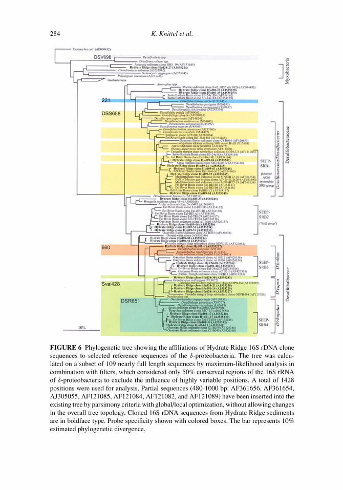
The 16S rDNA clone libraries obtained are phylogenetically diverse and include nu-merous cultivated as well as uncultivated bacterial lineages represented by 44 differentARDRA patterns (Table 4). SRB within the δ-proteobacteria dominated the clone librariesfrom Hydrate Ridge sediments and constituted 53% (n = 69) of all clones (Figure 6). Se-quence similarity to cultivated species was in all cases lower than 95%. However, similarityto sequences from uncultivated organisms was always high and often exceeded 98%. Se-quences related to Desulfosarcina species were the most abundant phylotype (14.5%) withalmost 95% similarity. Together with environmental sequences from the Eel River Basinand the Guaymas Basin they form a clade specific for methane-rich sites with 97.0–99.0%similarity to each other (group SEEP-SRB4). SEEP-SRB4 phylotypes were most often de-tected at the sediment surface and in 2–4 cm depth but not in the deep (8–9 cm) sedimentlayer.
Sequences related to the Desulfosarcina/Desulfococcus branch were the second mostabundant phylotype recovered (12.2%). This group comprises the complete-oxidizing
July 23, 2003 16:21 GMB Tj773-03
284 K. Knittel et al.
FIGURE 6 Phylogenetic tree showing the affiliations of Hydrate Ridge 16S rDNA clonesequences to selected reference sequences of the δ-proteobacteria. The tree was calcu-lated on a subset of 109 nearly full length sequences by maximum-likelihood analysis incombination with filters, which considered only 50% conserved regions of the 16S rRNAof δ-proteobacteria to exclude the influence of highly variable positions. A total of 1428positions were used for analysis. Partial sequences (480-1000 bp: AF361656, AF361654,AJ305055, AF121085, AF121084, AF121082, and AF121089) have been inserted into theexisting tree by parsimony criteria with global/local optimization, without allowing changesin the overall tree topology. Cloned 16S rDNA sequences from Hydrate Ridge sedimentsare in boldface type. Probe specificity shown with colored boxes. The bar represents 10%estimated phylogenetic divergence.

July 23, 2003 16:21 GMB Tj773-03
SRB and Other Bacteria above Gas Hydrate 285
genera Desulfosarcina spp., Desulfococcus spp., Desulfofrigus spp., Desulfofaba sp., andseveral other strains, which have not been further characterized yet. Hydrate Ridge se-quences show a maximum similarity of 93.4% to cultivated species of this group. Theseand other environmental sequences form a seep-specific clade with 90.2–99.2% similarityto each other (group SEEP-SRB1).
Other SRB sequences from Hydrate Ridge group together with two additional seep-specific groups (SEEP-SRB2 and SEEP-SRB3) of environmental sequences from methane-rich sites. SEEP-SRB2 comprises few sequences from Hydrate Ridge (2.3%) and severalsequences from the Eel River Basin and the Guaymas Basin. SEEP-SRB2 contains theuncultivated “Eel-2-group” from Eel River methane seep sediments (Orphan et al. 2001a),and has no close cultivated relatives (87% similarity). Sequence similarity to each otherwas 92.8–99.5% (without the Guaymas Basin clone sequence A2 B025 even higher with96.1–99.5%). In all analysis, they formed an independent phylogenetic cluster among theδ-proteobacteria that could not be assigned to cultivated genera.
SEEP-SRB3 is related to Desulfobulbus species (91% similarity). Sequences of thisgroup have 93.1-99.0% similarity to each other. Again, this group is formed by sequencesfrom the Hydrate Ridge, the Eel River, and Guaymas Basin and also contains a sequencefrom deep sea sediments near Japan (Nankai Trough) (Li et al. 1999b), which is knownto bear methane hydrate (Reed et al. 2002). The fourth group defined in this study namedSEEP-SRB4 is related to Desulforhopalus species. Within this group sequence similarityis quite high with 97.0–99.9%.
In addition to δ-proteobacteria Hydrate Ridge libraries contained a large percentageof sequences affiliated with the Cytophaga/Flavobacterium phylum (34%, n = 45). Se-quence similarity to cultivated species of the Cytophaga/Flavobacterium group was verylow and in the range of 82 to 92%. Two main groups (12 and 15%) were detected, both mostclosely affiliated with Cytophaga marina and Flexibacter maritimus within the Flavobac-teraceae. Many Hydrate Ridge sequences were closely related to sequence CS B009 fromthe hydrothermally active Guaymas Basin sediment (99.6% similarity) (Teske et al. 2002)although recovered from a geographically distant and cold site. These sequences were allretrieved from the 2–4 cm deep sediment layer.
Sequences related to γ -proteobacteria were detected in Hydrate Ridge sediments butcomprised only a relatively small percentage (5%). Clone sequences represented withinthis group included those highly affiliated with the aerobic methane-oxidizing bacteriumMethylobacter luteus (94.4%) and the sulfur-oxidizing bacteria Acidothiobacillus ferroxi-dans (94.9%) and Leucothrix mucor (89.5%).
One phylotype (4 clones) affiliated with ε-proteobacteria was found in the HydrateRidge sediment clone library from 2–4 cm depth. It branches with bacteria associatedwith the hydrothermal vent tubeworm Riftia pachyptila, with epibionts of the hydrothermalvent annelid Alvinella pompejana, and with environmental sequences from uncultivatedδ-proteobacteria found in cold seeps and the deep sea. Clones affiliated with other bacte-rial groups were retrieved in low numbers, including members of the phylum Firmicutes(Clostridiales, Haloanaerobiales) and the recently described Acidobacterium/Holophagadivision. Sequences affiliated with α- and β-proteobacteria were not retrieved.
Discussion
Bacterial Activity and Distribution above Hydrate
There are two sources of energy and carbon at Hydrate Ridge nourishing the benthic micro-bial communities: (1) methane, which rises from below with venting fluids and decomposing
July 23, 2003 16:21 GMB Tj773-03
286 K. Knittel et al.
hydrates, and (2) particulate organic matter (POC), which sinks to the seafloor from the pro-ductive ocean surface. Just as at any other continental margin the POC arriving at the seafloorof Hydrate Ridge fuels heterotrophic benthic communities, including aerobic microbes andmetazoa as well as fermenting microbes and other anaerobic bacteria and archaea. Fromprimary productivity in the water column it was estimated that about 2 mmol C m−2 d−1
settle to the seafloor at a depth of 780 m (Sommer et al. 2002), a typical value for up-per continental margins of productive oceans. It can be assumed that all investigated sitesreceive the same amount of POC. However, there is a considerable difference as to theavailability of electron acceptors and electron donors at the different sites. At the referencestation, nitrate penetrates down to 15 cm and methane and sulfide is very low or absent inthe surface sediments (Bohrmann et al. 2000). In several of the reference cores, bivalves ofthe genus Acharax that harbor sulfide-oxidizing symbionts in their gills dwell deep into thesediments for access to sulfide. The reference site may obtain low fluxes of methane fromgreater depths or by advection from nearby vented sites; however, most of this methane isalready consumed in the subsurface sediments below 15 cm.
At the methane vented Calyptogena fields, nitrate penetrates only a few cm and sulfideincreases considerably to values of >10 mM at 10 cm sediment depth (Torres et al. 2002).Beggiatoa mats receive the highest methane flux and are the most sulfidic, with >10 mMH2S already in the top few cm (Torres et al. 2002). Accordingly, there are differences in themicrobial abundance and activity of all three sites. Typical numbers of cells, production ratesestimated from thymidine uptake into DNA, and extracellular hydrolytic activities for thedegradation of POC were found in the surface sediments of the reference sites a few hundredmeter away from the vented, hydrate-bearing sediments covered by Beggiatoa or Calytogena(for comparison see Boetius et al. 2000b). Similar production rates and hydrolytic enzymeactivities as well as abundances of members of the Cytophaga/Flavobacterium cluster werefound in the surface sediments of all sites, showing the activity of the microbial community inPOC degradation. The hydrolytic enzyme activities found in the Hydrate Ridge sedimentsare comparable to those of other continental margin sediments at similar water depths(Boetius and Damm, 1998; Boetius et al. 2000b). In the subsurface sediments (2–15 cm),integrated production rates estimated from thymidine uptake were higher at the reference sitethan at the hydrate-bearing sites. The most abundant bacteria in hydrate-bearing sedimentsare SRB, which do not incorporate thymidine nor possess extracellular hydrolytic enzymesfor the degradation of particulate matter (Widdel 1988). The only hydrolytic enzyme activity,which was significantly higher in hydrate-bearing, sulfidic sediments was that of sulfatase.Possibly, this enzyme is expressed by some bacteria to acquire sulfate from organic sources.Accordingly, the higher activity of sulfatase could be caused by the relatively high numbersof cells of the Cytophaga/Flavobacterium cluster found in the Beggiatoa surface layers(station 105-1, 185-1). Cytophaga spp. require sulfatases to cover its sulfur needs in theproduction of sulfonated sphingolipids (Godchaux and Leadbetter 1983).
Another substantial difference between the reference site and the hydrate-bearing sed-iments was total cell abundance, concerning single cells as well as cell aggregates. Inhydrate-bearing sediments, total microbial biomass was much higher than at the referencesite. Most of this “excess” microbial biomass was attributed to the methanotrophic com-munity (Table 2) profiting from the much higher availability of methane compared to POC.Methane fluxes across the surface sediments exceed 100 mmol C m−2 d−1 at vented sites(Torres et al. 2002; Boetius and Suess, submitted) compared to a POC input of 2 mmolC m−2 d−1. According to the difference in the source of energy, cell numbers and sulfatereduction rates are also two orders of magnitude higher at Beggiatoa mats and Calytogenafields compared to the reference site. Sulfate reduction rates are around 50 mmol m−2 d−1,matching very well the sulfide fluxes of 60 and 20 mmol m−2 d−1 beneath Beggiatoa mats

July 23, 2003 16:21 GMB Tj773-03
SRB and Other Bacteria above Gas Hydrate 287
and Calyptogena fields, respectively (Sahling et al. 2002). In contrast, sulfate reductionrates of 0.5 mmol m−2 d−1 at the reference site are relatively typical for a margin site at thiswater depth and can be explained solely by the POC input.
Bacterial Diversity above Hydrate
The bacterial diversity in clone libraries from Hydrate Ridge sediments was relatively high.In our study all identified clone sequences were <95% similar to cultivated organisms. Incontrast, in deep subsurface gas hydrate sediments from the same habitat (subduction zonein the Cascadia Margin, ODP leg 146, site 889/890) and in a massive gas hydrate fromthe Gulf of Mexico most of the bacterial clone sequences were >95% similar to cultivatedorganisms (Lanoil et al. 2001; Marchesi et al. 2001). The investigated surface sedimentsfrom above outcropping hydrate are constantly vented by high concentrations of methaneand replenished with electron acceptors from the oxic bottom waters by a variety of mixingprocesses (Sahling et al. 2002; Boetius and Suess, submitted). High biomass and diversesubstrates are provided by the benthic macrofauna inhabiting the sediment surface (Sommeret al. 2002) and microbial activity is very high. This may explain the higher diversity foundin hydrate-bearing surface sediments compared to deep subsurface zones and bulk hydrate.
The relatively high diversity in Hydrate Ridge sediments is in very good agreementwith the findings from surface sediments of other methane-rich habitats. Teske et al. (2002)reported an extremely high bacterial diversity in hydrothermal sediments in the GuaymasBasin with 18 different phylogenetic groups. In hydrate-bearing sediments from NankaiTrough, Reed et al. (2002) reported a diverse microbial community with 22 unique sequencesand 5 phylogenetic phyla; likewise, Lanoil et al. (2001) found 21 different phylotypes insediments associated with hydrates from the Gulf of Mexico.
The phylogenetic analysis of Hydrate Ridge sequences resulted in many overlapswith sequences from other methane-rich sites. Gamma-, delta-, and epsilon-proteobacteria,members of the Cytophaga/Flavobacterium cluster, Actinobacteria, and Firmicutes arewidespread in almost all methane-rich sediments investigated (e.g., Lanoil et al. 2001;Marchesi et al. 2001; Reed et al. 2002; Teske et al. 2002). However, within these phy-logenetic groups no cluster seems to be endemic to methane-rich habitats (exception: δ-proteobacteria). These groups have been typically recovered from marine sediments andare likely involved in the degradation of organic substances other than methane (e.g., Katoet al. 1997; Li et al. 1999a, 1999c; Ravenschlag et al. 1999; Rossello-Mora et al. 1999;Urakawa et al. 1999; Bowman et al. 2000). Members of the Cytophaga/Flavobacteriumcluster were also relatively abundant in clone libraries and were detected by FISH in thesurface sediments. Cytophaga/Flavobacterium are known as ubiquitous degraders of poly-mers in marine habitats (Reichenbach 1991). This group is phylogenetically highly diverseand seems to play an ecological role as hydrolytic fermentative bacteria (Rossello-Moraet al. 1999). Hence, members of this group were relatively abundant at all sites, but ratherlinked to the detritus food chain than to AOM. However, bacteria of this group could alsouse the high biomass of the DSS/ANME-2 aggregates.
One phylotype retrieved from Hydrate Ridge sediment clustered within the Myxobacte-ria. It was related to Chondromyces robustus and most closely affiliated with clone OB3 39from Antarctic sediments (98.9%). Known pure cultures of Myxobacteria are almost allpredators of microorganisms (Reichenbach and Dworkin 1992). Therefore, they might playa role in the control of bacterial abundance.
Phylogenetic diversity within the δ-proteobacterial SRB was very high and the HydrateRidge clone sequences could be affiliated with several genera (Figure 6). Most often thesesequences originate from similar environments such as methane seeps from Eel River Basin

July 23, 2003 16:21 GMB Tj773-03
288 K. Knittel et al.
(Orphan et al. 2001a), the Santa Barbara Basin (Orphan et al. 2001a), the Guaymas Basin(Teske et al. 2002), and the Gulf of Mexico (Lanoil et al. 2001). Four distinct clusters ofhighly related sequences could be detected that appear to be specifically associated withmethane seep communities (see discussion below). In contrast to the relatively high bacterialdiversity, the archaeal diversity in methane-rich environments has always been described asquite low (Bidle et al. 1999; Hinrichs et al. 1999; Lanoil et al. 2001; Marchesi et al. 2001;Orphan et al. 2001a; Reed et al. 2002; Teske et al. 2002; Thomsen et al. 2001). Most oftenonly two phylogenetic groups, ANME-1 and ANME-2, were retrieved from the sediments.However, the phylogenetic diversity of archaea capable of anaerobic oxidation of methaneis still under investigation, and more groups may be found when further clone libraries areexamined.
Another SRB type of interesting function in the highly sulfidic sediments of HydrateRidge are those related to Desulfocapsa sulfexigens, a bacterium capable of inorganic sulfurdisproportionation (Finster et al. 1998). Sequences related to Desulfocapsa were the thirdmost abundant SRB phylotype recovered (11.4%) and closely affiliated with sequencesfrom Cascadia margin deep subsurface sediments (Bidle et al. 1999). These bacteria mayplay an important role in the very rapid sulfur cycling at seeps.
SRB Involved in AOM—A Single Specialist or Diversity of Different SRB?
Previous studies have shown that both archaeal ANME groups and members of the Desul-fosarcina/Desulfococcus branch co-occur in methane-rich sediments (Boetius et al. 2000a;Orphan et al. 2001b, 2002; Michaelis et al. 2002). Studies using FISH and secondary ionmass spectrometry analysis provided direct evidence for their involvement in AOM (Orphanet al. 2001b, 2002). As in most syntrophic consortia, this process does not seem to obli-gatorily require the physical attachment of SRB to methanogenic archaea. Orphan et al.showed that monospecific single archaeal cells and cell aggregates were depleted in δ13Cindicative of methanotrophy, as were multispecific consortia (Orphan et al. 2002). An im-portant open question for understanding the process of AOM still remaining is whether allAOM consortia contain a highly specialized SRB partner (Desulfosarcina/Desulfococcuscells) or whether the role of H2 and/or acetate scavenging syntrophic partners can be filledby a diversity of different SRB, including free-living single cells.
Not only the archaeal groups ANME1 and ANME2 can be detected exclusively inmethane-rich anoxic habitats (Hinrichs et al. 1999; Lanoil et al. 2001; Orphan et al. 2001a;Thomsen et al. 2001; Heijs et al. 2002; Teske et al. 2002), but there are also several groupsof SRB endemic to seep sites. Four such distinct clusters of highly related sequences weredetected: Besides Hydrate Ridge sequences these newly defined δ-proteobacterial groups(SEEP-SRB 1 to SEEP-SRB4) comprise multiple sequences retrieved from various sedi-ment samples from different habitats: the Gulf of Mexico gas hydrates (Lanoil et al. 2001),Eel River and Santa Barbara Basin methane seeps (Orphan et al. 2001a), Mediterraneancarbonate crusts from mud volcano sites (Heijs et al. 2002), Cascadia margin deep sub-surface sediments (Bidle et al. 1999), and surface sediments at hydrothermal vents in theGuaymas Basin (Teske et al. 2002). Sequences within this Desulfosarcina/Desulfococcusbranch (defined as SEEP-SRB1) were very abundant in the clone library in Hydrate Ridgesediments. This group represents the AOM syntrophic SRB group, which lives togetherwith ANME-2 cells in a syntrophic consortium, and that also occurs as partner of ANME-1(Michaelis et al. 2002). The SRB of this group were the most abundant bacteria at HydrateRidge with total numbers of aggregated cells of more than 1010 cm−3 in hydrate-bearingsediments (Table 2). Furthermore, they were also the most abundant single sulfate reducersin Hydrate Ridge sediment samples and accounted for 18% up to 100% of total detected

July 23, 2003 16:21 GMB Tj773-03
SRB and Other Bacteria above Gas Hydrate 289
SRB (Table 3). In contrast, single ANME-2 have barely been detected (<1% of total singlecells). In addition to being a common component of methane-rich habitats, members of theDesulfosarcina/Desulfococcus branch have also been shown to be very abundant in othermarine sediments (Rooney-Varga et al. 1997; Edgcomb et al. 1999; Sahm et al. 1999a;Ravenschlag et al. 2000) and made up to 73% of total SRB in Arctic sediments. Also, theyoccur as symbionts in a marine oligochaete (Dubilier et al. 2001). The high abundancein different habitats also underlines that members of this group seem to be fit to surviveunder various conditions. Members of the Desulfosarcina/Desulfococcus branch includenutritionally versatile SRB that oxidize organic compounds including acetate completely toCO2, and several species can grow autotrophically with CO2, H2, and sulfate (Widdel andHansen 1992b). The low sequence similarity of group SEEP-SRB1 to cultivated species(max. 93%) and within the group itself (90.2–99.2%) points to new, still uncharacterizedspecies or even new genera with unknown physiological properties. This is supported bydata from an accompanying study by Elvert et al. (2003, this issue) who found an enrichmentof unusual fatty acids in Hydrate Ridge sediments (C16:1ω5c, C17:1ω6c, and cyC17:0ω5,6) whichhave the lowest δ13C values throught the sediment core. This specific fatty acid pattern wasdefined as a stable chemotaxonomic marker for SRB of the Desulfosarcina/Desulfococcusbranch represented in ANME-2/DSS aggregates (for detailed discussion see Elvert et al.2003, this issue).
In this study we defined three more SRB groups (i.e., SEEP-SRB2 to SRB4) that are cur-rently associated exclusively with methane-rich, anoxic sites. The presence of these groupssuggests a direct or indirect involvement in AOM by using intermediates or metabolic endproducts of this process, or maybe specific organic substrates provided by seep-characteristicmacrofaunal communities around vent sites (e.g., mats of filamentous sulfur-oxidizing bac-teria Beggiatoa spp., vesicomyid clams Calyptogena spp., solemyid bivalve molluscs ofgenus Acharax [Levin et al. 2000; Olu et al. 1997; Sahling et al. 2002]). However, Nauhauset al. (2002) tested in vitro if the SRB community (including members of the Desulfos-arcina/Desulfococcus branch) of hydrate-bearing sediments from Hydrate Ridge can bedecoupled from methane oxidation by adding various potential SRB substrates (or AOMintermediates). Sulfate reduction was fueled by methane more than by any other electrondonor, pointing to a direct link between methane oxidation and sulfate reduction.
The methane seep-specific groups SEEP-SRB3 and SEEP-SRB4 were located withinthe Desulfobulbaceaea and related to the genera Desulfobulbus and Desulforhopalus, re-spectively. SRB detected by probe DSR651, specific for Desulforhopalus relatives andSEEP-SRB4, were the most abundant group in the clone library and the second abun-dant group as detected by FISH in situ. However, their numbers of up to 1.7 × 10⁸ cellsper ml in the uppermost layer were two orders of magnitude lower than those of theSRB of the Desulfosarcina/Desulfococcus branch represented in ANME-2/DSS aggregates.This once again shows the limitation in making quantitative assumptions based on clonefrequencies.
SEEP-SRB2 (unclear phylogenetic affiliation) and SEEP-SRB3 (related to Desulfob-ulbus species) were not targeted by the set of probes used in this study. New probes haveto be developed to quantify the abundance and will clarify the importance of these groupsfor AOM sites. A first indication of AOM-relevance of these groups was shown by verynegative δ13C-values of specific fatty acids extracted from Hydrate Ridge sediments. Elvertet al. (2003, this issue) reported a significant 12C-enriched carbon isotope value (−80%in 0–19 cm depth in average) for the membrane fatty acid C17:1ω6c, which is specific forDesulfobulbus species (but also found in Desulforhopalus species). In addition the dominantfatty acid C16:1ω5c in Hydrate Ridge sediments, which were formerly assigned to organismswithin the Desulforhopalus/Desulfococcus branch (13C: −90%), has also been detected
July 23, 2003 16:21 GMB Tj773-03
290 K. Knittel et al.
in Desulfobulbus and Desulfosarcina. These findings support the hypothesis that bothDesulforhopalus and Desulfobulbus related AOM-specific groups (SEEP-SRB34) mightbe involved in AOM besides members of the Desulfosarcina/Desulfococcus branch. Ex-tremely 13C-depleted bacterial lipids recovered from other methane-rich sites were alsostructurally diverse and likely originated from multiple bacterial sources (Pancost et al.2000; Orphan et al. 2001a; Thiel et al. 2001; Zhang et al. 2002). It may be that these bac-terial groups form partnerships with other methane-oxidizing archaea unknown at present.In Hydrate Ridge sediments, however, we did not find evidence for other bacterial as-sociations than Desulfosarcina/Desulfococcus cells with the methane-oxidizing archaealANME-2 group, or for other cell associations. In contrast, in Eel River Basin sediment sam-ples Orphan and coworkers found ANME-2 aggregates, which comprised bacterial partnersother than the Desulfosarcina related species (Orphan et al. 2002). Furthermore, Orphanet al. (2001a) could define several “Eel groups” (Eel 1, Eel 3) which were missing at HydrateRidge.
Conclusions
This is the first quantitative analysis of bacterial diversity, distribution, and activity of a coldseep environment. Typical for continental margin sediments of productive ocean regions,an active and diverse bacterial community involved in the degradation of organic matterwas found in the surface sediments. However, highest biomasses, activities, and phyloge-netic diversity occurred at the methane-vented sediments above surficial gas hydrates. Thedominant process in these sediments is the anaerobic oxidation of methane via sulfate re-duction. A high diversity of sulfate-reducing bacteria was found in the gas hydrate-bearingsediments and four seep-specific phylogenetic clusters were recognized. Members of theDesulfosarcina/Desulfococcus group represent the dominant cluster of SRB, and are di-rectly involved in anaerobic oxidation of methane as part of a syntrophic consortium. Mostof the bacterial diversity comprises seep-endemic clades, which share only low similaritieswith previously cultured bacteria.
References
Amann RI, Krumholz L, Stahl DA. 1990. Fluorescent-oligonucleotide probing of whole cells fordeterminative, phylogenetic, and environmental studies in microbiology. J Bacteriol 172:762–770.
Bidle KA, Kastner M, Bartlett DH. 1999. A phylogenetic analysis of microbial communities associatedwith methane hydrate containing marine fluids and sediments in the Cascadia margin (ODP8site 892B). FEMS Microbiol Lett 177:101–108.
Boetius A, Damm E. 1998. Benthic oxygen uptake, hydrolytic potentials and microbial biomass atthe Arctic continental slope. Deep-Sea Res I 45:239–275.
Boetius A, Ferdelmann T, Lochte K. 2000b. Bacterial activity in sediments of the deep Arabian Seain relation to vertical flux. Deep Sea Res II 47:2835–2875.
Boetius A, Ravenschlag K, Schubert C, Rickert D, Widdel F, Gieseke A, Amann R, Jørgensen BB,Witte U, Pfannkuche O. 2000a. A marine microbial consortium apparently mediating anaerobicoxidation of methane. Nature 407:623–626.
Boetius A, Suess E, submitted. Hydrate Ridge: a natural laboratory for the study of microbial lifefueled by methane from near-surface gas hydrates. Chem Geol.
Bohrmann G, Linke P, Suess E, Pfannkuche O, 2000. R.V. SONNE cruise Report SO143: TECFLUX-I(June 29–September 6, 1999; Honolulu—Astoria—San Diego). GEOMAR Report 93:217.
Børsheim KY, Bratbak G, Heldal M. 1991. Enumeration and biomass estimation of planktonic bacteriaand viruses by transmission electron microscopy. Appl Environ Microbiol 56:352–356.
July 23, 2003 16:21 GMB Tj773-03
SRB and Other Bacteria above Gas Hydrate 291
Bowman JP, Rea SM, McCammon SA, McMeekin TA. 2000. Diversity and community structurewithin anoxic sediment from marine salinity meromyctic lakes and a coastal meromictic marinebasin, Vestfold Hills, eastern Antarctica. Environ Microbiol 2:227–237.
Daims H, Bruhl A, Amann R, Schleifer KH, Wagner, M. 1999. The domain-specific probe EUB338 isinsufficient for the detection of all bacteria: development and evaluation of a more comprehensiveprobe set. Syst Appl Microbiol 22:434–444.
Devereux R, Kane MD, Winfrey J, Stahl DA. 1992. Genus- and group-specific hybridization probesfor determinative and environmental studies of sulfate-reducing bacteria. Syst Appl Microbiol15:601–609.
Dubilier N, Mulders C, Ferdelman T, de Beer D, Pernthaler A, Klein M, Wagner M, Erseus C, Thier-mann F, Krieger J, Giere O, Amann R. 2001. Endosymbiotic sulphate-reducing and sulphide-oxidizing bacteria in an oligochaete worm. Nature 411:298–302.
Edgcomb VP, McDonald JH, Devereux R, Smith DW. 1999. Estimation of bacterial cell numbers inhumic acid-rich salt marsh sediments with probes directed to 16S ribosomal DNA. Appl EnvironMicrobiol 65:1516–1523.
Elvert M, Boetius A, Knittel K, Jørgensen BB. 2003. Characterization of specific membrane fattyacids as chemotaxonomic markers for sulfate-reducing bacteria involved in anaerobic oxidationof methane. Geomicrobiol J 20:403–419.
Findlay S. 1993. Thymidine incorporation into DNA as an estimate of sediment bacterial produc-tion. In: PF Kemp, editor. Handbook of Methods in Aquatic Microbial Ecology. Florida: LewisPublishers. p 505–508.
Finster K, Liesack W, Thamdrup B. 1998. Elemental sulfur and thiosulfate disproportionation byDesulfocapsa sulfoexigens sp. nov., a new anaerobic bacterium isolated from marine surfacesediments. Appl Environ Microbiol 64:119–125.
Fossing H, Jørgensen BB. 1989. Measurements of bacterial sulfate reduction in sediments: evaluationof a single-step chromium reduction method. Biogeochemistry 8:205–222.
Godchaux W 3rd, Leadbetter ER. 1983. Unusual sulfonolipids are characteristic of the Cytophaga-Flexibacter group. J Bacteriol 153:1238–1246.
Hansen LB, Finster K, Fossing H, Iversen N. 1998. Anaerobic methane oxidation in sulfate depletedsediments: effects of sulfate and molybdate additions. Aquat Microb Ecol 14:195–204.
Heijs SK, Aloisi G, Bouloubassi I, Forney LJ, Pancost RD, Pierre C, Sinninghe Damste JS, GottschalJC. 2002. Microbial community structure of three deep sea carbonate crusts: novel Archaeainvolved in anaerobic methane oxidation? Database release: AF361645–AF361680.
Hinrichs KU, Hayes JM, Sylva SP, Brewer PG, DeLong EF. 1999. Methane-consuming archaebacteriain marine sediments. Nature 398:802–805.
Hinrichs K-U, Boetius A. 2002. The anaerobic oxidation of methane: new insights in microbialecology and biogeochemistry. In: Wefer G, Billett D, Hebbeln D, Jørgensen BB, Schluter M, T.VW, editors. Ocean Margin Systems. Berlin Heidelberg: Springer-Verlag. p 457–477.
Hoehler TM, Alperin MJ, Albert DB, Martens CS. 1994. Field and laboratory studies of methaneoxidation in an anoxic marine sediment: evidence for a methanogen-sulfate reducer consortium.Glob Biogeochem Cycles 8:451–463.
Kane MD, Poulsen LK, Stahl DA. 1993. Monitoring the enrichment and isolation of sulfate-reducingbacteria by using oligonucleotide hybridization probes designed from environmentally derived16S rRNA sequences. Appl Environ Microbiol 59:682–686.
Kato C, Li L, Tamaoka J, Horikoshi K. 1997. Molecular analyses of the sediment of the 11000-mdeep Mariana Trench. Extremophiles 1:117–123.
Lanoil BD, Sassen R, La Duc MT, Sweet ST, Nealson KH. 2001. Bacteria and Archaea physicallyassociated with Gulf of Mexico Gas Hydrates. Appl Environ Microbiol 67:5143–5153.
Levin LA, James DW, Martin CM, Rathburn AE, Harris LH, Michener RH. 2000. Do methane seepssupport distinct macrofaunal assemblages? Observations on community structure and nutritionfrom the northern California slope and shelf. Mar Ecol Prog Ser 208:21–39.
Li L, Kato C, Horikoshi K. 1999a. Bacterial diversity in deep-sea sediments from different depths.Biodivers Conserv 8:659–677.
Li L, Guezennec J, Nichols P, Henry P, Yanagibayashi M, Kato C. 1999b. Microbial diversity inNankai Trough sediments at a depth of 3843 m. J Oceanogr 55:635–642.

July 23, 2003 16:21 GMB Tj773-03
292 K. Knittel et al.
Li L, Kato C, Horikoshi K. 1999c. Microbial diversity in sediments collected from the deepest cold-seep area, the Japan trench. Mar Biotechnol 1:391–400.
Linke P, Suess E. 2001. R.V. SONNE cruise Report SO148 TECFLUX-II-2000 (Victoria–Victoria;July 20–August 12, 2000). GEOMAR Report 98:122.
Manz W, Amann R, Ludwig W, Vancanneyt M, Schleifer K-H. 1996. Application of a suite of 16SrRNA-specific oligonucleotide probes designed to investigate bacteria of the phylum cytophaga-flavobacter-bacteroides in the natural environment. Microbiology 142:1097–1106.
Manz W, Eisenbrecher M, Neu TR, Szewzyk U. 1998. Abundance and spatial organization of gramnegative sulfate-reducing bacteria in activated sludge investigated by in situ probing with specific16S rRNA targeted oligonucleotides. FEMS Microbiol Ecol 25:43–61.
Marchesi JR, Weightman AJ, Cragg BA, Parkes RJ, Fry JC. 2001. Methanogen and bacterial diversityand distribution in deep gas hydrate sediments from the Cascadia Margin as revealed by 16SrRNA molecular analysis. FEMS Microbiol Ecol 34:221–228.
Meyer-Reil, LA 1983. Benthic response to sedimentation events during autumn to spring at a shallowwater station in the western Kiel Bight. Mar Biol 77:247–256.
Michaelis W, Seifert R, Nauhaus K, Treude T, Thiel V, Blumenberg M, Knittel K, Gieseke A,Peterkencht K, Pape T, Boetius A, Amann R, Jørgensen BB, Widdel F, Peckmann J, PimenovNV, Gulin MB. 2002. Microbial reefs in the Black Sea fueled by anaerobic oxidation of methane.Science 297:1013–1015.
Muyzer G, Teske A, Wirsen CO, Jannasch HW. 1995. Phylogenetic relationships of Thiomicrospiraspecies and their identification in deep-sea hydrothermal vent samples by denaturing gradientgel electrophoresis of 16S rDNA fragments. Arch Microbiol 164:165–172.
Nauhaus K, Boetius A, Kruger M, Widdel F. 2002. In vitro demonstration of anaerobic oxidationof methane coupled to sulphate reduction in sediment from a marine gas hydrate area. EnvironMicrobiol 4:296–305.
Olu K, Lance S, Sibuet M, Henry P, Fiala-Medioni A, Dinet A. 1997. Cold seep communities as indi-cators of fluid expulsion patterns through mud vulcanoes seaward of the Barbardos accretionaryprisms. Deep-Sea Res 44:811–841.
Orphan VJ, Hinrichs K-U, Ussler III W, Paull CK, Taylor LT, Sylva SP, Hayes JM, DeLong EF.2001a. Comparative analysis of methane-oxidizing archaea and sulfate-reducing bacteria inanoxic marine sediments. Appl Environ Microbiol 67:1922–1934.
Orphan VJ, House CH, Hinrichs K-U, McKeegan KD, DeLong EF. 2001b. Methane-consumingarchaea revealed by directly coupled isotopic and phylogenetic analysis. Science 293:484–487.
Orphan VJ, House CH, Hinrichs K-U, McKeegan KD, DeLong EF. 2002. Multiple archaeal groupsmediate methane oxidation in anoxic cold seep sediments. Proc Natl Acad Sci 99:7663–7668.
Pace NR. 1997. A molecular view of microbial diversity and the biosphere. Science 276:734–740.Pancost RD, Sinninghe Damste SJ, de Lint S, van der Maarel MJEC, Gottschal JC, the Medinaut
Shipboard Scientific Party. 2000. Biomarker evidence for widespread anaerobic methane oxida-tion in Mediterranean sediments by a consortium of methanogenic archaea and bacteria. ApplEnviron Microbiol 66:1126–1132.
Ravenschlag K, Sahm K, Pernthaler J, Amann R. 1999. High bacterial diversity in permanently coldmarine sediments. Appl Environ Microbiol 65:3982–3989.
Ravenschlag K, Sahm K, Knoblauch C, Jørgensen BB, Amann R. 2000. Community structure, cellularrRNA content and activity of sulfate-reducing bacteria in marine Arctic sediments. Appl EnvironMicrobiol 66:3592–3602.
Ravenschlag K, Sahm K, Amann R. 2001. Quantitative molecular analysis of the microbial communityin marine Arctic sediments (Svalbard). Appl Environ Microbiol 67:387–395.
Reeburgh WS. 1996. “Soft spots” in the global methane budget. In: Lidstrom ME, Tabita FR, editors.Microbial Growth on C1 Compounds. Dordrecht: Kluwer Academic Publishers. p 334–342.
Reed DW, Fujita Y, Delwiche ME, Blackwelder DB, Sheridan PP, Uchida T, Colwell FS. 2002.Microbial communities from methane hydrate-bearing deep marine sediments in a forearc basin.Appl Environ Microbiol 68:3759–3770.
Reichenbach H. 1991. The order Cytophagales. In: Balows A, Truper HG, Dworkin M, Harder W,Schleifer K-H, editors. The Prokaryotes. Berlin, Germany: Springer-Verlag. p 3631–3675.
July 23, 2003 16:21 GMB Tj773-03
SRB and Other Bacteria above Gas Hydrate 293
Reichenbach H, Dworkin M. 1992. The myxobacteria. In: Balows A, Truper HG, Dworkin M, HarderW, Schleifer K-H, editors. The Prokaryotes. New York: Springer-Verlag. p 3416–3487.
Rooney-Varga JN, Devereux R, Evans RS, Hines ME. 1997. Seasonal changes in the relative abun-dance of uncultivated sulfate-reducing bacteria in a salt marsh sediment and in the rhizosphereof Spartina alterniflora. Appl Environ Microbiol 63:3895–3901.
Rossello-Mora R, Thamdrup B, Schaefer H, Weller R, Amann R. 1999. The response of the microbialcommunity of marine sediments to organic carbon input under anaerobic conditions. Syst ApplMicrobiol 22:237–248.
Sahling H, Rickert D, Lee RW, Linke P, Suess E. 2002. Macrofaunal community structure and sulfideflux at gas hydrate deposits from the Cascadia convergent margin, NE Pacific. Mar Ecol ProgSer 231:121–138.
Sahm K, MacGregor BJ, Jørgensen BB, Stahl DA. 1999a. Sulfate reduction and vertical distribution ofsulfate-reducing bacteria quantified by rRNA slot-blot hybridization in a coastal marine sediment.Environ Microbiol 1:65–74.
Sahm K, Knoblauch C, Amann R. 1999b. Phylogenetic affiliation and quantification of psychrophilicsulfate-reducing isolates in marine arctic sediments. Appl Environ Microbiol 65:3976–3981.
Snaidr J, Amann R, Huber I, Ludwig W, Schleifer KH. 1997. Phylogenetic analysis and in situidentification of bacteria in activated sludge. Appl Environ Microbiol 63:2884–2896.
Sommer S, Pfannkuche O, Rickert D, Kahler A, 2002. Surficial marine gas hydrates and their eco-logical implications for the small-sized benthic biota at the Hydrate Ridge. Mar Ecol Prog Ser243:25–38.
Strunk O, Gross O, Reichel B, May M, Hermann S, Stuckman N, Nonhoff B, Lenke M, Ginhart A,Vilbig A, Ludwig T, Bode A, Schleifer K-H, Ludwig W. 1998. ARB: a software environment forsequence data. http://www.mikro.biologie.tumuenchen.de. Department of Microbiology Tech-nische Universitat Munchen, Munich, Germany.
Suess E, Torres ME, Bohrmann G, Collier RW, Greinert J, Linke P, Rehder G, Trehu A, WallmannK, Winckler G, Zuleger E. 1999. Gas hydrate destabilization: enhanced dewatering, benthicmaterial turnover and large methane plumes at the Cascadia convergent margin. Earth Planet SciLett 170:1–15.
Suess E, Bohrmann G, Rickert D, Kuhs W, Torres ME, Trehu A, Linke P, 2002. Properties and fabricof near-surface methane hydrates at Hydrate Ridge, Cascadia margin. Proc 4th Intl Conf GasHydrates, Yokohama: 740–744.
Teske A, Hinrichs K-U, Edgcomb V, de Vera Gomez A, Kysela D, Sylva SP, Sogin ML, JannaschHW. 2002. Microbial diversity of hydrothermal sediments in the Guaymas Basin: evidence foranaerobic methanotrophic communities. Appl Environ Microbiol 68:1994–2007.
Thiel V, Peckmann J, Richnow HH, Luth U, Reitner J, Michaelis W. 2001. Molecular signals foranaerobic methane oxidation in Black Sea seep carbonates and a microbial mat. Mar Chem73:97–112.
Thomsen TR, Finster K, Ramsing NB. 2001. Biogeochemical and molecular signatures of anaerobicmethane oxidation in a marine sediment. Appl Environ Microbiol 67:1646–1656.
Torres ME, McManus J, Hammond D, de Angelis MA, Heeschen K, Colbert S, Tryon MD, BrownKM, Suess E. 2002. Fluid and chemical fluxes in and out of sediments hosting methane hydratedeposits on Hydrate Ridge, OR, I: Hydrological Provinces, Earth Planet Sci Lett 201:525–540.
Treude T, Boctius A, Knittel K, Wallmann K, Jørgensen BB. Anaerobic oxidation of methane abovegas hydrates (Hydrate Ridge, OR). submitted. Aquat Microbiol Ecol.
Tryon MD, Brown KM, Torres ME. 2002. Fluid and chemical flux in and out of sediments hostingmethane hydrate deposits on Hydrate Ridge, OR, II: Hydrological processes. Earth Plan Sci Lett201:541–557.
Urakawa H, Kita-Tsukamota K, Ohwada K. 1999. Microbial diversity in marine sediments fromSagami Bay and Tokyo Bay, Japan, as determined by 16S rRNA gene analysis. Microbiology145:3305–3315.
Valentine DL, Reeburgh WS. 2000. New perspectives on anaerobic methane oxidation. EnvironMicrobiol 2:477–484.
July 23, 2003 16:21 GMB Tj773-03
294 K. Knittel et al.
Wallner G, Amann R, Beisker W. 1993. Optimizing fluorescent in situ hybridization with rRNA-targeted oligonucleotide probes for flow cytometric identification of microorganism. Cytometry14:136–143.
Widdel F, 1988. Microbiology and ecology of sulfate- and sulfur reducing bacteria. In: A. Zehnder,editor. Biology of Anaerobic Microorganisms. New York: Wiley. p 469–585.
Widdel F, Bak F. 1992a. Gram-negative mesophilic sulfate-reducing bacteria. In: Balows A, TruperHG, Dworkin M, Harder W, Schleifer K-H, editors. The Prokaryotes. 2 ed. New York: Springer-Verlag, p 3352–3378.
Widdel F, Hansen T. 1992b. The dissimilatory sulfate- and sulfur-reducing bacteria. In: Balows A,Truper HG, Dworkin M, Harder W, Schleifer K-H, editors. The Prokaryotes. 2 ed. New York:Springer-Verlag. p 583–624.
Zhang CLYL, Wall JD, Larsen L, Sassen R, Huang Y, Wang Y, Peacock A, White DC, Horita J,Cole DR. 2002. Lipid and carbon isotopic evidence of methane-oxidizing and sulfate-reducingbacteria in association with gas hydrates from the Gulf of Mexico. Geology 30:239–242.
Zhou J, Brunns MA, Tiedje JM. 1996. DNA recovery from soils of diverse composition. Appl EnvironMicrobiol 62:316–322.