Mines Douai U Lille Université UNivERsrrfiDe MINES Lille 1 ...

- 1 -
ACTIVATION OF THE PRO-DRUG ETHIONAMIDE IS
REGULATED IN MYCOBACTERIA✝
Alain R. Baulard1, Joanna C. Betts2, Jean Engohang-Ndong1, Selwyn Quan2, Patrick J.
Brennan3, Camille Locht1, Gurdyal S. Besra4*
1 From the INSERM U447, Institut de Biologie de Lille, Institut Pasteur de Lille,
59019 Lille, France; 2Glaxo Wellcome Research and Development, Gunnels Wood
Road, Stevenage, SG1 2NY, England; 3Department of Microbiology, Colorado State
University, Fort Collins, Colorado, CO 80523-1677, USA; 4Department of
Microbiology & Immunology, University of Newcastle upon Tyne, Newcastle upon
Tyne, NE2 4HH, England.
Running title: Ethionamide resistance in M. tuberculosis
*To whom correspondence should be addressed. Telephone: (0191) 222 5412; FAX
(0191) 222 7736; E-mail: [email protected]
Copyright 2000 by The American Society for Biochemistry and Molecular Biology, Inc.
JBC Papers in Press. Published on June 26, 2000 as Manuscript M003744200 by guest on July 5, 2018
http://ww
w.jbc.org/
Dow
nloaded from
- 2 -
ABSTRACT
The anti-tuberculosis drug ethionamide (ETH), which is a structural analog of
isoniazid (INH), is known to strongly inhibit mycolic acid synthesis in
Mycobacterium tuberculosis. While several targets have been identified for INH, only
speculative information is available concerning ETH. Mutations within the promoter
and the coding region of enoyl-ACP reductase (InhA) were found to confer resistance
to both drugs, thus leading to the impression that INH and ETH may share a common
mode of action. However, a notable distinction between the two drugs lies in the lack
of cross-resistance in clinical isolates. This may be attributed in part to the fact that
the pro-drug INH must be activated via KatG, and no activation step for ETH has yet
been described. Here we report the identification of an activator for ETH. The ETH
activator (Rv3854c), which we have termed EthA, was found to be homologous to
various monooxygenases and induced ETH sensitivity when over-expressed in
mycobacteria. Interestingly, the neighbouring open reading frame (Rv3855), which
was found homologous to transcriptional repressors of the tetR family, led to ETH
resistance when over-expressed. In addition, chromosomal inactivation of this gene by
transposition led to ETH hyper-sensitivity. These data strongly suggest that Rv3855,
which we have termed EthR, regulates the production of EthA which subsequently
activates the pro-drug ETH. This study opens up new avenues of research relating to
ETH activation in mycobacteria, - possibly leading to an improved efficacy of ETH
and to the generation of new anti-mycobacterial agents.
by guest on July 5, 2018http://w
ww
.jbc.org/D
ownloaded from

- 3 -
INTRODUCTION
The treatment of tuberculosis involves extremely lengthy and specialized
chemotherapy regimens (1). The molecular composition and structural features of the
mycobacterial cell envelope are thought to confer low permeability and thereby a
basal resistance to most hydrophilic drugs (2). As a consequence, the lack of potency
and protracted duration of drug administration are a major cause of rampant
mutational drug resistance of Mycobacterium tuberculosis (3). An essential step in
developing novel therapies for the treatment of M. tuberculosis infections is to
determine why multi-drug resistant strains of M. tuberculosis are resistant to many
existing anti-mycobacterial agents. Possible mechanisms of resistance include:
alteration of the target enzyme that has become resistant to antibiotics; increased
expression of the gene encoding the target enzyme; mutations causing impermeability
of the mycobacterial cell to the antibiotic, and/or alterations of an activation
mechanism. Thus, the urgent need to develop new therapies for treating M .
tuberculosis infections requires the definition of the failures of existing treatments and
the discovery of new drug targets.
The bacterial cell wall has been an effective target for many drugs (4). Many
anti-tuberculosis agents including ethambutol, cycloserine, isoniazid (INH),
ethionamide (ETH), thiocarlide and the thiosemicarbazones are known to inhibit cell
wall biosynthesis. INH (Fig. 1.), which is one of the most efficient and the most
by guest on July 5, 2018http://w
ww
.jbc.org/D
ownloaded from

- 4 -
widely used anti-tuberculosis drugs, has been the subject of intensive research during
the past decade (5-7). Both M. tuberculosis and Mycobacterium bovis BCG are
extremely susceptible to INH (minimum inhibitory concentrations [MIC], 0.02-0.2
µg/ml), and earlier evidence suggested that INH specifically inhibits the synthesis of
mycolic acids in M. tuberculosis and M. bovis BCG (8-11). ETH (Fig. 1), a structural
analog of INH, is a useful second-line anti-tuberculosis drug. The two drugs have
almost identical effects in that both strongly inhibit the synthesis of mycolic acids (12,
13). Banerjee and colleagues demonstrated that a single mutation in the inhA gene
(NADH-specific, 2-trans-enoyl ACP reductase) confers resistance to both INH and
ETH (14), leading to the impression that the mode of action of both drugs is identical.
In addition, mutations within katG, encoding a catalase-peroxidase lead to the
majority of INH-resistant isolates (6), demonstrating that INH is a pro-drug and that
an activated metabolite is responsible for its mode of action (15, 16). However, the
notable distinction between the actions of ETH and INH resides in the lack of cross-
resistance (4, 17). The majority of strains resistant to ETH are sensitive to INH, while
some strains resistant to INH show slightly increased sensitivity to ETH (18). Thus,
there are subtle discrepancies between biochemical and genetic information on the
modes of sensitivity and resistance in the cases of INH and ETH.
Since INH requires activation via KatG, it is tempting to postulate an
activation process for ETH. Inactivation of such an ETH-specific process could
account for the lack of cross-resistance between the two drugs. Interestingly, ETH
by guest on July 5, 2018http://w
ww
.jbc.org/D
ownloaded from

- 5 -
undergoes oxidation by rat liver microsomes to generate a highly reactive S-oxide,
possibly a sulfinate (Fig. 1) which exhibits greater activity in vitro against M.
tuberculosis than ETH itself (19-21). These observations, in addition to the fact that
genetic alterations in katG do not confer resistance to ETH, have led us to the
hypothesis that ETH needs to be activated through a KatG-independent mechanism.
In this report, we describe the cloning and characterization of the gene
Rv3855, which we now term ethR, that confers resistance to ETH, but not to INH
when it is over-expressed in either Mycobacterium smegmatis, M. bovis BCG or M.
tuberculosis on a multycopy vector. Furthermore, a transposon mutant of ethR leads
to ETH hyper-sensitivity in M. bovis BCG. In addition, genetic and biochemical
evidence suggests that ethR encodes a transcriptional regulator that is not directly
implicated in mycolic acid biosynthesis, but rather plays an important role in the
regulation of a second open reading frame (ORF) which is responsible for the
activation of ETH. Analysis of the locus surrounding ethR revealed the presence of an
adjacent gene now termed ethA, which encodes a putative monooxygenase, the
predicted activator of ETH. Over-expression of ethA lead to hyper-sensitivity to ETH
in mycobacteria. Thus, the data presented are compatible with the notion that EthR
represses ethA which encodes the equivalent protein of KatG implicated in the
activation of ETH.
by guest on July 5, 2018http://w
ww
.jbc.org/D
ownloaded from

- 6 -
EXPERIMENTAL PROCEDURES
Bacterial strains and growth conditions.
M. smegmatis mc2155, M. bovis BCG 1173P2 (WHO, Stockholm, Sweden),
M. tuberculosis H37Ra and their transformants were grown on 7H11 agar
supplemented with oleic-albumin-dextrose-catalase enrichment (OADC)(Difco,
Detroit, MI), or on Sauton medium (22) supplemented with 0.001 ‰ ZnSO4 and 0.25
‰ Triton WR1339 (Sigma). M. smegmatis mc2155 is an electroporation-efficient
mutant of mc2 6 (23). M. smegmatis, M. bovis BCG and M. tuberculosis were
transformed as described previously (24). Large-scale cultures of mycobacteria were
grown to mid-log phase (M. bovis BCG, 10-14 days; M. smegmatis mc2155, 36 h; M.
tuberculosis H37Ra, 12-16 days), harvested, washed with PBS and stored at -20°C
until further use. Escherichia coli strains XL1-Blue (Stratagene, La Jolla, CA),
NK5587 and their transformants were grown in Luria-Bertoni LB broth (Gibco BRL,
Life Technologies) and agar medium. E. coli SH305 was grown in 2YT (25)
supplemented with 1% glucose. All strains were incubated at 37°C. Antibiotics
(Sigma) were added to media at the following concentrations: kanamycin at 20 µg/ml,
tetracycline at 10 µg/ml, chloramphenicol at 100 µg/ml, and ampicillin at 100 µg/ml.
by guest on July 5, 2018http://w
ww
.jbc.org/D
ownloaded from

- 7 -
Plasmids and DNA manipulation.
The E. coli-mycobacterial shuttle vector pMV261 containing the hsp60
promoter was used as described previously (26). Restriction enzymes and T4 DNA
ligase were purchased from Boehringer Mannheim (Mannheim, Germany), and Vent
DNA polymerase was purchased from New England Biolabs. All DNA manipulations
were performed using standard protocols, as described by Sambrook et al. (25).
Identification of pETH80 and transposon γδ mutagenesis of
pETH80 in E. coli
A genomic library of M. tuberculosis H37Rv constructed by cloning 35- to 40-
kb Sau3AI fragments of chromosomal DNA into the shuttle cosmid pYUB18 (27),
was the source of cosmid pETH80 conferring ETH resistance to M. smegmatis. The
minimal region of pETH80 involved in ETH resistance was identified by γ δ
transposition in E. coli as described by Guyer (28). Briefly, a suitable E. coli donor
strain NK5587 (F' lacY::Tn9) was transformed with pETH80. One isolated
transformant was subsequently conjugated with E. coli SH305 (recA1 srl::Tn10) with
selection for ampicillin resistant - tetracycline resistant exconjugants. Plasmid DNA
was extracted from 50 double resistant exconjugants, re-transformed into M.
smegmatis, and scored for their ETHR phenotype. Tn1000 (γδ) insertions were
localized by restriction mapping and sequencing using primers near the end of γδ
by guest on July 5, 2018http://w
ww
.jbc.org/D
ownloaded from

- 8 -
(Primer γ δ1: 5'-CAACGAATTATCTCCTT-3'; Primer γ δ2 : 5 ' -
TCAATAAGTTATACCAT-3').
Cloning and expression of Rv3855 and Rv3854c.
Rv3855 was cloned into the mycobacterial over-expression vector pMV261 as
follows. PCR amplification was performed using the upstream primer N°142: 5'-
CCACCTCCGCGGCCAGTCAGG-3' and the downstream primer N°141: 5'-
TTTGGCACTGAGAATTCACCGAGCACCC-3' which contains an EcoRI
restriction site (underlined). The 690-bp PCR product was digested with EcoRI and
cloned into MluNI/EcoRI-restricted pMV261, giving rise to pMV261-ethR, where
ethR is fused in frame with the ATG initiation codon of hsp60. A similar strategy was
used to construct the pMV261-based expression vector for Rv3854c. Rv3854c was
amplif ied by PCR using the upstream primer N°150: 5 '-
AGCACCTCGACGTTGTCATC-3' and the downstream primer N°149: 5'-
ACGGATCCCCGCAAGAGCACCA-3' which contains a BamHI restriction site
(underlined). The 1624-bp fragment was digested by BamHI and cloned into
MluNI/BamHI-restricted pMV261, generating pMV261-ethA. The coding sequences
of all amplified genes were verified by DNA sequencing after their cloning in
pMV261.
by guest on July 5, 2018http://w
ww
.jbc.org/D
ownloaded from
- 9 -
Determination of the in vivo effects of ETH and INH on fatty acid
and mycolic acid synthesis in mycobacteria.
M. smegmatis mc2155, M. bovis BCG and M. tuberculosis were grown to mid-
log phase (A600~0.3). ETH or INH were added at various concentrations followed by
further incubation for 4 h (for M. smegmatis) or 24 h (for M. bovis BCG and M.
tuberculosis). At this point 1,2-[14C]acetate (1 µCi/ml) (Amersham, France) was
added and the cultures further incubated with gentle agitation at 37oC for 4 h (for M.
smegmatis) or 24 h (for M. bovis BCG and M. tuberculosis). The resulting [14C]-
labelled cells from ETH- or INH-treated cultures were harvested, washed twice with
phosphate buffered saline, resuspended into 3 ml of 15% tetrabutylammonium
hydroxide (TBAH) and saponified at 100oC for 15 h. After cooling, dichloromethane
(4 ml), water (2ml) and iodomethane (300 µl) were added, and the entire reaction
mixture agitated for 30 min. After centrifugation, the upper, aqueous phase was
discarded and the lower, organic phase washed twice with water, dried in a sand bath,
extracted twice with diethyl ether (3 ml), and the etheral extracts were dried and
resuspended into 500 µl of dichloromethane for counting of radioactivity. Equal
volumes of this extract, which is composed of fatty acid methyl esters (FAMEs) and
mycolic acid methyl esters (MAMEs), were separated by thin-layer chromatography
(TLC) on silica gel (Merck 5735 silica gel 60F254, Darmstadt, Germany) and
developed once in petroleum ether:acetone (95:5). Subsequent autoradiography
by guest on July 5, 2018http://w
ww
.jbc.org/D
ownloaded from
- 10 -
revealed [14C]-labelled fatty acid and mycolic acid methyl esters after overnight
exposure of the TLC plates to Kodak BioMax MR film.
Characterization of the M. bovis BCG transposon insertions.
Genomic DNA (200-500 ng) of the mutagenized clones was digested with
RsaI (NEB). After heat inactivation of the restriction enzyme at 65° for 20 minutes,
the digested DNA (40-100 ng) was ligated with T4 DNA ligase (NEB) at room
temperature. PCR reactions were carried out in a total volume of 25 µl containing
Amplitaq PCR buffer (Perkin-Elmer), 10% (v/v) DMSO, 0.25 µM dNTPs, 0.4 µM
84L-F (5'-GTCATCCGGCTCATCACCAG-3') and 0.4 µM 84L-R (5'-
AACTGGCGCAGTTCCTCTGG-3') primers, 4-10 ng of template DNA and 0.5 U of
Amplitaq Gold (Perkin-Elmer). Thermal cycling was performed in a Perkin-Elmer
9600 machine with an initial denaturation at 94°C for 10 minutes, followed by 40
cycles of 94°C for 30s, 50°C for 30s, 72°C for 90s and a final extension of 72°C for 7
minutes.
by guest on July 5, 2018http://w
ww
.jbc.org/D
ownloaded from

- 11 -
RESULTS
Identification of cosmids conferring ETH resistance in M.
smegmatis.
The chromosomal region involved in ETH resistance was sought using drug
resistance imparted via gene over-expression by a plasmid vector, as a selection tool.
A genomic library of DNA from M. tuberculosis H37Rv was screened using the ETHS
(MIC of 15 µg/ml) M. smegmatis host strain mc2155, for clones that exhibited an
ETHR phenotype. Electrotransformants were plated in duplicate on medium
containing kanamycin with ETH at 80 µg/ml. Colonies were scored as ETHR if growth
appeared on plates after 4 days of incubation at 37°C. Single ETHR colonies were re-
streaked onto duplicate 7H10/OADC plates containing kanamycin and INH (5 µg/ml).
Specific ETHR-clones were selected that grew in the presence of ETH but remained
fully sensitive to INH. The cosmids which confer ETHR were recovered from the M.
smegmatis transformants by electroduction into E. coli (29). Following analysis, all
clones were found to contain the same cosmid (pETH80) which corresponded to a
minimum of two chromosomal regions, probably associated during the packaging
process (data not shown).
by guest on July 5, 2018http://w
ww
.jbc.org/D
ownloaded from

- 12 -
Identification by transposon γδ (Tn1000) mutagenesis of the
minimal region of pETH80 responsible for ETH resistance.
The minimal region required for ETH resistance in M. smegmatis was
determined by transposon mutagenesis of pETH80. The method, adapted from Guyer
et al. (28) is based on the transposition in E. coli of γδ during F-mediated conjugal
mobilization. Briefly, the mutagenesis technique is based upon the observation that
conjugal transmission of pBR322 by the conjugative plasmid F is dependent upon, or
at least completely correlated with, the transposition of γδ from F to pBR322 (28).
The transposition of γδ has also proved efficient on plasmids other than pBR322 (30).
The cosmid pETH80 identified in this study is an E. coli-mycobacterial shuttle vector
based on colE1 origin of replication. Thus, the mapping process of the minimal region
of pETH80 responsible for ETHR in mycobacteria was achieved in two steps. Firstly,
mutagenesis of pETH80 in E. coli before the re-introduction of the derivatized DNA
into mycobacteria allowed the analysis of insertion mutations. Out of the 50 clones
analyzed, 38 γδ insertions were located in the insert portion of pETH80. Three γδ
insertions were able to abolish ETHR in M. smegmatis. Restriction analysis of these
clones revealed that these γδ insertions were located within 0.6 kb of DNA.
Sequencing of the regions located left and right of γδ (with primers γδ1 and γδ2)
revealed that all 3 insertions disrupted the same ORF annotated Rv3855 in the M .
tuberculosis H37Rv genome database, which was thus termed ethR.
by guest on July 5, 2018http://w
ww
.jbc.org/D
ownloaded from
- 13 -
Cloning and overexpression of ethR (Rv3855) in mycobacteria.
To avoid possible interference of other ORF's of pETH80 in association with
ETHR, the coding region of ethR was amplified by PCR and cloned in frame with
hsp60 into pMV261. The resultant plasmid pMV261-ethR was transformed into M.
smegmatis, M. bovis BCG and M. tuberculosis, and the MIC's of the transformed
bacteria were compared with those of untransformed strains and with those of strains
containing the original pETH80. The MIC’s were determined by plating serial
dilutions onto medium containing kanamycin plus 0-200 µg/ml of ETH in increments
of 10 µg/ml or 0-10 µg/ml of INH in increments of 1 µg/ml. The MIC was defined as
the lowest concentration of ETH that inhibited the growth of 99% of the bacteria.
Table 1 summarizes the results obtained suggesting a direct correlation between the
level of expression of ethR and ETH resistance. All three mycobacterial species
transformed with pMV261-ethR remained sensitive to INH, thus demonstrating that
over-expression of ethR specifically induces ETH resistance. In parallel, susceptibility
assays with isoxyl, thiolactomycin, erythromycin, crystal violet and streptomycin
indicated that resistance was specific for ETH.
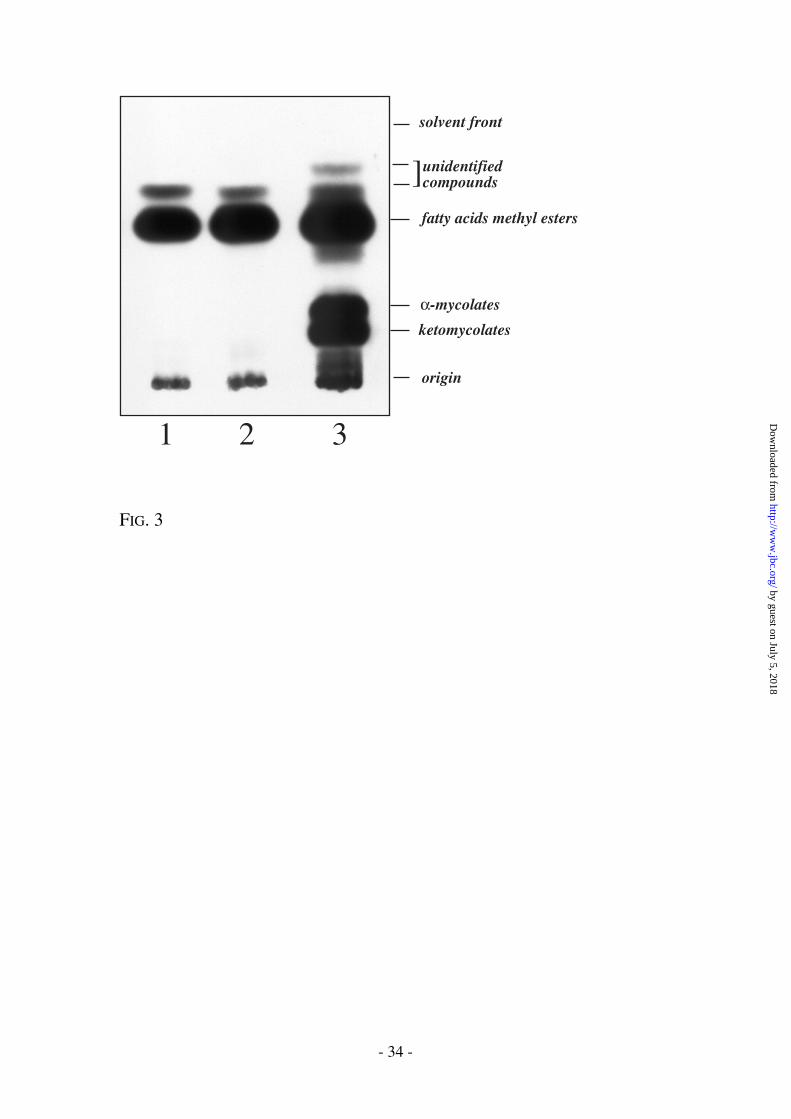
Selective effects of ETH on inhibition of mycolic acid synthesis.
Untransformed and pMV261-ethR transformed M. bovis BCG were grown in
the presence or absence of ETH at various concentrations, following which cultures
were labelled with 1,2-[14C]acetate. Combined MAMEs and FAMEs were extracted,
by guest on July 5, 2018http://w
ww
.jbc.org/D
ownloaded from

- 14 -
resolved and fractionated on TLC plates. Untransformed M. bovis BCG exhibited a
clear decrease in the incorporation of radioactivity into MAMEs in the presence of
ETH (from 0 to 100 µg/ml) (Fig. 2). Examination of the individual classes of
mycolates revealed that the production of all species was specifically inhibited. In
addition, the production of a yet undescribed and unidentified product, possibly a
mycolate specific fatty acid precursor, was progressively inhibited after treatment
with ETH (and with INH) (Fig. 3). The characterization of this product may be crucial
for the identification of the specific target for ETH, but has been hampered by its
relatively low abundance in the mycobacterial cell wall. In contrast, treatment of M.
bovis BCG (pMV261-ethR) with ETH (0 to 100 µg/ml) had no dramatic effect on the
synthesis of MAMEs and FAMEs (Fig. 2). Possible mechanisms related to ETH
resistance may include detoxification of the drug, or repression of an activation
process of ETH leading to its active metabolite, conceivably an S-oxide type
derivative (19). The latter seemed an attractive scenario since EthR is homologous to
bacterial regulatory proteins of the TetR family (see Fig. 4B). Many of the proteins of
the TetR family are repressors. As for EthR (23.725 kDa), they all have similar
molecular weights - from 21 to 25 kDa. The most conserved region between EthR and
the TetR family members is characterized by a helix-turn-helix motif located in the N-
terminal third of the protein. As illustrated in Figure 4A, the signature pattern
(PS01081) starts four residues before the helix-turn-helix motif and ends six residues
after the motif.
by guest on July 5, 2018http://w
ww
.jbc.org/D
ownloaded from

- 15 -
Construction of a M. bovis BCG ethR knock out mutant.
Mutagenesis of M. bovis BCG NCTC 5692 was performed as previously described
using the mycobacteriophage mini-transposon delivery system pJSC84 (31); Jeff Cox,
unpublished results). Individual mutants were isolated, and the transposon insertions
were characterized by inverse PCR. A clone with a transposon insertion between
nucleotides 4327971 and 4327972 in the M. bovis BCG genome was identified (Fig.
5). The insertion disrupted ethR between nucleotide positions 426 and 427, leading to
the production of a truncated polypeptide of 142 amino acids (instead of 648 for the
normal protein). The M. bovis BCG ethR::hyg strain was found to be extremely
sensitive to ETH, with a MIC < 0.6 µg/ml (see Table 1). The MIC for INH remained
identical for M. bovis BCG and M. bovis BCG ethR::hyg, confirming that ethR was
specific for ETH resistance.
Inhibition of mycolic acid synthesis in M. bovis BCG(pMV261-
ethR)
The isolated M. bovis BCG ethR::hyg clone was grown in the presence or
absence of ETH and examined for the relative inhibition of mycolic acid synthesis.
Concentrations as low as 2.5 µg/ml of ETH completely abolished mycolic acid
synthesis in comparison to 50 µg/ml for the wild-type M. bovis BCG strain (Fig. 6)
thus, supporting our hypothesis that ethR is involved in the repression of the
formation of the active metabolite of ETH. Interestingly, in the absence of ETH, the
by guest on July 5, 2018http://w
ww
.jbc.org/D
ownloaded from

- 16 -
FAME and MAME profiles of the mutant were unaffected, confirming the notion that
ethR is not directly implicated in mycolic acid synthesis, but may rather be related to
ETH activation.
Identification of the gene encoding the putative ETH activator.
It is not unusual to find a gene under the control of a transcriptional regulator
encoded by the neighbouring ORF. The intergenic region located between Rv3854c
and Rv3855 (ethR) is 76 base pair long, and the two genes are transcribed in opposite
directions. This situation is consistent with the presence of divergent promoters in this
region (32). For some regions of divergent transcription described so far, one
transcript determines a regulatory molecule and the other determines a non-regulatory
polypeptide. The regulatory molecule may act within the divergent transcript unit to
control transcription of the non-regulatory polypeptides; often it also regulates its own
synthesis (32). Interestingly, Rv3854c, the adjacent ORF to ethR, is homologous to
various monooxygenases (Fig 4C). Thus, Rv3854c could be a candidate to play a role
equivalent to eukaryotic monooxygenases in the generation of active metabolites of
ETH, notably S-oxides (20, 21). To test this hypothesis, Rv3854c was over-expressed
in M. smegmatis (pMV261-ethA) and the recombinant strain was examined for ETH
sensitivity. As anticipated, M. smegmatis (pMV261-ethA) was hyper-sensitive to ETH
(Table 1), and a dramatic inhibition of mycolic acid synthesis was observed at very
low concentrations of ETH in comparison to wild-type M. smegmatis (Fig. 7). These
by guest on July 5, 2018http://w
ww
.jbc.org/D
ownloaded from

- 17 -
results strongly suggest that Rv3854c, which we have now termed ethA, plays a role
equivalent of katG for INH, but in relation to ETH.
by guest on July 5, 2018http://w
ww
.jbc.org/D
ownloaded from

- 18 -
DISCUSSION
INH and ETH are specific antituberculosis drugs, which inhibit mycolic acid
synthesis through InhA as suggested by resistance to both ETH and INH of an inhA
mutant (14). Moreover, by the use of microarray hybridization, Wilson and coworkers
recently demonstrated that ETH treatment of M. tuberculosis induced the same genes
that were induced by INH (33). However, Fattorini and co-workers recently reported
that of 46 INH-resistant strains of M. tuberculosis isolated from Italian patients only 2
were also ETH resistant (17). Before INH exerts its lethal effect it must be converted
to an active form, possibly an isonicotinic acyl anion (34) or an isonicotinic acyl
radical (35) produced by the catalase-peroxidase KatG. ETH and other thioamides are
sulphur-containing compounds, which are known to be substrates for oxidative
catalysts, such as flavin-containing monooxygenases (FMO) and cytochrome P-450
monooxygenases. An NADPH-dependent oxidation of ETH has previously been
demonstrated in rat microsomes (20). More recently, Johnsson et al. (34) suggested
that in vitro oxidation of ETH produces electrophilic intermediates (S-oxides) capable
of undergoing addition reactions to nucleophilic protein side chains (35). During the
1950’s ETH, and subsequently prothionamide were introduced in the treatment of
tuberculosis and were deemed clinically as effective as dapsone in the treatment of
leprosy (36). However, 25% of the patients suffered from various gastrointestinal
symptoms and sometimes jaundice, especially when ETH was combined with
by guest on July 5, 2018http://w
ww
.jbc.org/D
ownloaded from

- 19 -
rifampicin (37). The hepatotoxicity induced by administration of ETH and
thionicotinamide (TNA) was decreased by pre-administration of methimazole (MMI).
Pre-administration of MMI was also shown to decrease the levels of excretion of TNA
S-oxide, indicating that thioamide S-oxidation, mediated by the flavin-containing
monooxygenases, may be linked to the initiation of hepatotoxicity induced by these
thioamides (38).
Here we describe evidence that ETH is activated by the monooxygenase
homolog Rv3854c (EthA). EthA would then be the equivalent of KatG for INH. Thus,
in a similar fashion by which mutations in katG abolish INH sensitivity without
leading to ETH resistance, we would expect that mutations in ethA leads to ETH
resistance without affecting INH sensitivity. Studies are currently in progress
examining clinical isolates for point mutations within ethA. Over-expression of ethA
led to a dramatic increase in ETH sensitivity of M. smegmatis and clearly indicated
that only a small proportion of ETH is activated when mycobacterial cells are grown
under laboratory conditions. Alternatively, when administered to humans, ETH may
be activated by either eukaryotic oxidative processes as mentioned earlier or by EthA
(or perhaps a combination of both). Thus, determining the respective contribution of
the bacterial and the eukaryotic activation of thioamides to their ultimate necrogenic
forms would be crucial to understand the impact of EthA on the efficacy of ETH in
vivo, and to help designing new improved versions of ETH.
by guest on July 5, 2018http://w
ww
.jbc.org/D
ownloaded from

- 20 -
ETH resistance may also be mediated by the overproduction of the putative
repressor EthR. As a regulator, it is logical to assume that the production of EthR is
also regulated, perhaps by signals external to the bacteria. Thus, any agent able to
block EthR, or any physiological condition down-regulating ethR may favour the
production of EthA and lead to the activation of substantial amounts of ETH, thereby
increasing the sensitivity of the bacilli to ETH. Over-expression of ethA dramatically
decreased the MIC of M. smegmatis for ETH to a level comparable to the MIC of M.
tuberculosis (see Table 1). This suggests that in part the innate relative resistance to
ETH of M. smegmatis is associated with a low production of EthA and thus,
indirectly, with EthR. Perhaps this mechanism may also account for the various
susceptibility profiles to ETH displayed by other mycobacterial species.
Interestingly, whilst synthesis of α and α’ mycolates is insensitive to ETH in
M. smegmatis overexpressing ethR, synthesis of epoxymycolates remained highly
sensitive to the drug in this strain (data not shown). Thus, possible additional targets
to InhA exist in relation to ETH and epoxymycolate synthesis, which has been
previously shown to be highly sensitive to ETH (13).
It is interesting to note that EthA and EthR orthologs are also present in M.
avium and M. leprae. The EthAR loci of M. avium and M. tuberculosis share a very
similar architecture. In both cases, the two genes are oriented "head to head" and are
separated by a putative 76 base pair-divergent promoter. In contrast, the two M.
leprae orthologs are not located in the same locus. The initiation codons of the M.
by guest on July 5, 2018http://w
ww
.jbc.org/D
ownloaded from
- 21 -
leprae EthA and EthR are found in the genome in positions 86,698 and 1,235,214,
respectively. The genome of M. leprae seems to be reduced to a minimal pool of
genes essential to the survival of this strictly intracellular pathogen. In contrast to
many genes of M. leprae, EthA and EthR are apparently not truncated and are highly
similar to their orthologs in M. tuberculosis and M. smegmatis. Altogether, these
observations suggest an important role for these genes in vivo. In addition, the M.
tuberculosis genome possibly encodes more than 30 monooxygenases and the relative
role of each enzyme in the oxidation of ETH remains to be assessed in vivo.
The understanding of the mode of action of ETH is valuable for the design of
improved versions of the drug, for the elaboration of potentiating agents and for the
understanding of drug resistance. Finally, an intriguing question, which awaits further
experimentation, concerns the possibility of an equivalent mechanism of regulation of
katG and INH resistance.
by guest on July 5, 2018http://w
ww
.jbc.org/D
ownloaded from
- 22 -
REFERENCES
1. De Cock, K. M., and Chaisson, R. E. (1999) Int. J. Tuberc. Lung Dis. 3, 457-65
2. Liu, J., Barry, C. E. I., and Nikaido, H. (1999) in Mycobacteria : molecular
biology and virulence (Ratledge, C., and Dale, J., eds), Blackwell Science,
Malden, Mass.
3. Espinal, M. (1999) WHO/TB/99.260
4. Winder, F. (1982) in The biology of the mycobacteria. Vol 1, Physiology,
identification and classification. (Ratledge, and Stanford, eds), pp. 353-438,
Academic Press, London
5. Heym, B., Saint-Joanis, B., and Cole, S. T. (1999) Tuber. Lung. Dis. 79, 267-71
6. Barry, C. E., 3rd, Slayden, R. A., and Mdluli, K. (1998) Drug Resistance Updates
1, 128-134
7. Loewen, P. C., Klotz, M. G., and Hassett, D. J. (2000) ASM news 66, 76-82
8. Winder, F. G., and Collins, P. B. (1970) J. Gen. Microbiol. 63, 41-8
9. Takayama, K., Wang, L., and David, H. L. (1972) Antimicrob. Agents
Chemother. 2, 29-35
10. Wang, L., and Takayama, K. (1972) Antimicrob. Agents Chemother. 2, 438-41
11. Takayama, K. (1974) Ann. N. Y. Acad. Sci. 235, 426-38
by guest on July 5, 2018http://w
ww
.jbc.org/D
ownloaded from

- 23 -
12. Winder, F. G., Collins, P. B., and Whelan, D. (1971) J. Gen. Microbiol. 66, 379-
80
13. Quemard, A., Laneelle, G., and Lacave, C. (1992) Antimicrob. Agents
Chemother. 36, 1316-21
14. Banerjee, A., Dubnau, E., Quemard, A., Balasubramanian, V., Um, K. S., Wilson,
T., Collins, D., Delisle, G., and Jacobs, W. R. (1994) Science 263, 227-230
15. Zhang, Y., Heym, B., Allen, B., Young, D., and Cole, S. (1992) Nature (London)
358, 591-593
16. Lei, B., Wei, C. J., and Tu, S. C. (2000) J. Biol. Chem. 275, 2520-6
17. Fattorini, L., Iona, E., Ricci, M. L., Thoresen, O. F., Orru, G., Oggioni, M. R.,
Tortoli, E., Piersimoni, C., Chiaradonna, P., Tronci, M., Pozzi, G., and Orefici,
G. (1999) Microb. Drug Resist. 5, 265-70
18. Rist, N. (1960) Adv. Tuberculosis Res. 10, 69-126
19. Johnsson, K., King, D. S., and Schultz, P. G. (1995) J. Am. Chem. Soc. 117,
5009-5010
20. Ruse, M. J., and Waring, R. H. (1991) Biochem. Soc. Trans. 19, 233S
21. Chieli, E., and Malvaldi, G. (1985) Biochem. Pharmacol. 34, 395-396
22. Sauton, M. B. (1912) C R Acad Sci Paris 155, 860-861
23. Snapper, S. B., Melton, R. E., Mustafa, S., Kieser, T., and Jacobs, W. R., Jr.
(1990) Mol. Microbiol. 4, 1911-9
24. Wards, B. J., and Collins, D. M. (1996) FEMS Microbiol. Lett. 145, 101-105
by guest on July 5, 2018http://w
ww
.jbc.org/D
ownloaded from

- 24 -
25. Sambrook, J., Fritsch, E. F., and Maniatis, T. (1989) Molecular cloning : a
laboratory manual, 2nd Ed., Cold Spring Harbor Laboratory, Cold Spring
Harbor, N.Y.
26. Stover, C. K., de la Cruz, V. F., Fuerst, T. R., Burlein, J. E., Benson, L. A.,
Bennet, L. T., Bansal, G. P., Young, J. F., Lee, M. H., Hatfull, G. F., Snapper,
S. B., Barletta, R. G., Jacobs Jr, W. R., and Bloom, B. R. (1991) Nature
(London) 351, 456-460
27. Jacobs, W. R., Kalpana, G. V., Cirillo, J. D., Pascopella, L., Snapper, S., Udani,
R. A., Jones, W., Barletta, R. G., and Bloom, B. R. (1991) Methods Enzymol.
204, 537-555
28. Guyer, M. S. (1978) J. Mol. Biol. 126, 347-65
29. Baulard, A., Jourdan, C., Mercenier, A., and Locht, C. (1992) Nucleic Acids Res.
20, 4105
30. de Lencastre, H., Chak, K. F., and Piggot, P. J. (1983) J. Gen. Microbiol. 129,
3203-10
31. Bardarov, S., Kriakov, J., Carriere, C., Yu, S. W., Vaamonde, C., McAdam, R.
A., Bloom, B. R., Hatfull, G. F., and Jacobs, W. R. (1997) Proc. Natl. Acad. Sci.
USA 94, 10961-10966
32. Beck, C. F., and Warren, R. A. (1988) Microbiol. Rev. 52, 318-26
33. Wilson, M., DeRisi, J., Kristensen, H. H., Imboden, P., Rane, S., Brown, P. O.,
and Schoolnik, G. K. (1999) Proc. Natl. Acad. Sci. USA 96, 12833-8
by guest on July 5, 2018http://w
ww
.jbc.org/D
ownloaded from

- 25 -
34. Shoeb, H. A., Bowman, B. U., Jr., Ottolenghi, A. C., and Merola, A. J. (1985)
Antimicrob. Agents Chemother. 27, 399-403
35. Johnson, K., and Schultz, P. G. (1994) J. Am. Chem. Soc. 116, 7425-7426
36. Rollier, R., and Rollier, M.-T. (1972) Maroc. Medecine 52, 148-166
37. Waters, M. F. R. (1989) in The Biology of the mycobacteria. Clinical Aspects of
mycobacterial disease (Ratledge, C., Stanford, J., and Grange, J. M., eds) Vol.
3, pp. 405-474, Academic Press, London ; New York
38. Ruse, M. J., and Waring, R. H. (1991) Toxicol. Lett. 58, 37-41
by guest on July 5, 2018http://w
ww
.jbc.org/D
ownloaded from

- 26 -
FOOTNOTES
1The abbreviations used are: ETH, ethionamide; ETHR, ETH-resistant; ETHs,
ETH-susceptible; INH, isoniazid; FAME, fatty acid methyl ester; MAME, mycolic
acid methyl ester, TLC: Thin layer chromatography, ORF: open reading frame,
TBAH: tetra-butyl ammonium hydroxide, hyg: hygromycin resistance gene; MMI,
methimazole; TNA, thionicotinamide; cc, concentration.
✝This work was supported by the Institut Pasteur de Lille, INSERM, the
Alliance Joint Research Programme from the Ministère des Affaires Etrangères in
France and the British Council, The Medical Research Council (UK), Glaxo
Wellcome Research & Development (UK), the National Institute of Allergy and
Infectious Diseases (NIAID), National Institutes of Health (NIH) (AI-18357, AI-
33706), and grant AI-38087 from the National Cooperative Drug Discovery Groups
for the Treatment of Opportunistic Infections (NCDDG-OI), NIAID, NIH. We also
thank the NIAID, NIH Tuberculosis Research Materials and Vaccine Testing Contract
(NO1-AI-75320) for genomic DNA. GSB is currently a Lister Institute-Jenner
Research Fellow. The costs of publication of this article were defrayed in part by the
payment of page charges. This article must therefore be hereby marked
“advertisement” in accordance with 18 U.S.C. Section 1734 solely to indicate this
fact.
by guest on July 5, 2018http://w
ww
.jbc.org/D
ownloaded from

- 27 -
Acknowledgments - We gratefully acknowledge Dr. Françoise Jacob-
Dubuisson for critical reading of the manuscript, W.R. Jacobs (Albert Einstein
College of Medicine) and R.A. McAdam (Glaxo Wellcome) for the provision/usage of
the transposon library.
by guest on July 5, 2018http://w
ww
.jbc.org/D
ownloaded from
- 28 -
FIGURE LEGENDS.
Fig. 1. Structures of INH, ETH and ETH-S-Oxide.
Fig. 2. The dose-response effects of ETH on fatty acid and mycolic acid synthesis in
M. bovis BCG (pMV261) and M. bovis BCG (pMV261-ethR). The inhibitory
effect on the incorporation of 1,2-[14C]acetate was assayed by labeling in the
presence of increasing concentrations of ETH and terminated by addition of
15% TBAH at 100°C overnight. The corresponding FAMEs and MAMEs
were isolated, spotted on aluminium-backed TLC plates, which were
developed once in petroleum ether:acetone (95:5) and exposed overnight to a
Kodak BioMax MS film.
Fig. 3. Effect of INH and ETH on fatty acid and mycolic acid synthesis. The
samples are FAMEs and MAMEs from M. bovis BCG + ETH 40 µg/ml (lane
1), M. bovis BCG + INH 10 µg/ml (lane 2), M. bovis BCG (lane 3).
Fig 4. Locus Rv3854c and Rv3855 of M. tuberculosis H37Rv. A) The tetR family
members are characterized by a helix-turn-helix motif located in the initial
third of the protein. The signature pattern is a conserved domain which starts
by guest on July 5, 2018http://w
ww
.jbc.org/D
ownloaded from
- 29 -
four residues before the helix-turn-helix motif and ends six residues after the
motif. With the exception of Pro41, the Rv3855 (ethR) region from Pro41 to
Val71, fits the "Prosite" signature PDOC00830 (tetR) (http://www.expasy.ch/cgi-
bin/nicedoc.pl?PDOC00830), strongly suggesting that Rv3855 (e t h R) is a
transcriptional regulator. B) The tetR consensus as determined at the Sanger
center is named PF00440 (http://www.sanger.ac.uk/Software/Pfam/). A few
members of the family have been chosen, and their PF00440 domain were
aligned with Rv3855 (ethR). A more detailed and exhaustive alignment is
a v a i l a b l e o n l i n e a t t h e f o l l o w i n g
URL:http://www.sanger.ac.uk/cgibin/Pfam/getalignment.pl?name=tetR&acc=PF00440&ty
pe=full&format=link. C) Rv3854c (ethA) belongs to a large and diverse family
of monooxygenases. The region homologous to monooxygenases is located
in the N-terminal part of the protein.
Fig. 5 Positioning of the transposon insertion in the genome of the clone M. bovis
BCG NCTC 5692 ethR::hyg.
Fig. 6 The dose-response effects of ETH on FAME and MAME profiles in M. bovis
BCG NTC 5692 and M. bovis BCG NTC 5692 ethR::hyg.
by guest on July 5, 2018http://w
ww
.jbc.org/D
ownloaded from

- 30 -
Fig. 7 The dose-response effects of ETH on FAME and MAME profiles in M.
smegmatis (pMV261) and M. smegmatis (pMV261-ethA).
by guest on July 5, 2018http://w
ww
.jbc.org/D
ownloaded from

- 31 -
Table 1. MIC's (µg/ml) of ETH for mycobacteria harbouring ethR, ethA, or ethR KO.
pMV261 pETH80 pMV261-ethR pMV261-ethA ethR::hyg
M. smegmatis (a) 15 80 250 < 1 nt(e)
M. bovis BCG (b) 2 25 35 nt < 0,6(c)
M. tuberculosis (d) 1 20 30 nt nt
(a) strain mc2155; (b) strain Paris 1173P2 except in lane 5; (c) strain NCTC 5692; (d) strain H37Ra; (e)
not tested.
by guest on July 5, 2018http://w
ww
.jbc.org/D
ownloaded from

- 32 -
FIGURES
N
O NHNH2
N
S NH2
CH2CH3 N
OxS NH2+
CH2CH3
FIG. 1
by guest on July 5, 2018http://w
ww
.jbc.org/D
ownloaded from

- 33 -
M. bovis BCG (pMV261) M. bovis BCG (pMV261-ethR)
ETH cc (µg/ml) 0 10 25 50 100 0 10 25 50 100
fatty acidsmethyl esters
α-mycolates
ketomycolates
origin
unidentifiedcompound
solvent front
FIG. 2
by guest on July 5, 2018http://w
ww
.jbc.org/D
ownloaded from
- 34 -
1 2 3
]fatty acids methyl esters
α-mycolates
ketomycolates
origin
unidentifiedcompounds
solvent front
FIG. 3
by guest on July 5, 2018http://w
ww
.jbc.org/D
ownloaded from

- 35 -
I L A T A E N L L E D R X P L A D I S V D D L A K G A G I S R P T F Y F Y F P S K E A V L L T L Rv3855/29-75I L D V A L R L F S Q Q X G V S S T S L G E I A K A A G V T R G A I Y W H F K D K S D L F S E I ACRR_ECOLI/16-62I F S A S L L L F A - E R G F D A T T M P M I A E N A K V G A G T I Y R Y F K N K E S L V N E L BM3R_BACME/11-57L I E T A I A Q F A Q H - G V S K T T L N D I A D A A N V T R G A I Y W H F E N K T Q L F N E M ENVR_ECOLI/16-62L M L A A L E T F Y R - K G I A R T S L N E I A Q A A G V T R G A L Y W H F K N K E D L F D A L MTRR_NEIGO/15-61L V N S A R I L F V - E K G Y Q A V S I D E I S G K A L V T K G A F Y H H F K N K K Q L L S A C TETC_ECOLI/18-64I L E A A T K S F T - Q F G Y K A T T M D L V A K L A N V G K G T I Y T F F K N K E E L F D E I YHGD_BACSU/9-55
Rv3854c (ethA)
Rv3855 (ethR)
l i e a A l e l f n e k e G y e a t T t a k I A k a a g V s k G a l Y w H F k n K e e L f d a l+ + + A l + + + + + + + + + + + A k a g + s + + + + Y + F + + K e + + + + + l
C
A
B PF00440E value: 2.6e-09
Rv3855/29-75ILATAENLLEDRPLADISVDDLAKGAGISRPTFYFYFPSKEAVLLTL ::::::::::::::::::::::::::::::
GLxxxSVxxLxxxxxISxPxFYxYFxSKxxV I TL I LT G LF HV T L V SI V VA A IS F D I M M M QG R VT N M F T SE M Y N S Q H
PROSITE: PS01081 ( tetR )
E H L D V V I V G A G I S G V S A A W H L Q D R . C P T . K S Y A I L E K R E S M G G T W D L F R Y P G I Rv3854c/3.69T P R S Y C V V G G G I S G L T S A Y R L R Q A V . G D D A T I T L F E P A D R L G G V L R T . . E H I G PPOX_MYCTU/1.70T S R S Y C V V G G G I S G L T A A Y R L R V A T . G D D V A I T L F D P G D R L G G V L R T . . E C V G PPOX_MYCLE/1.70. M Y D Y L I V G A G L S G A I F A Y E . A T K R G K K . . . V K V I D K R N H I G G N I Y . . C E N V E O86897/1.74L R R S V A V V G S G V A G L T A A Y I L S G R D . . . R . . V T L Y E A D G R L G G H A H T . . H . . . O53734/1.47. M F K Y I I V G A G L A G S V M A E R I A S Q L G E R . . . V L V V E R R R H V G G N C Y D E V D . D N O26444/1.76A R F D L F V V G S G F F G L T I A E R V A T Q L D K R . . . V L V L E R R P H I G G N A Y S E A E P Q T O06934/1.83N K F D F I V L G A G I S G I V L S H V L A Q H . G K S . . . V L L L E K R N Q L G G N C Y D K L D E T T GLF_MYCPN/13.85N S F D I L I V G A G I S G I V L A N I L A N H . N K R . . . V L I V E K R D H I G G N C Y D K V D S K T GLF_MYCGE/19.81. M Y D Y I I V G S G L F G A V C A N E . L K K L N K K . . . V L V I E K R N H I G G N A Y . . T E D C E GLF_ECOLI/1.71N N K N I M I V G A G F S G V V I A R Q L A E Q . G Y T . . . V K I I D R R D H I G G N S Y D T R D P Q T GLF8_KLEPN/1.79K S K K I L I V G A G F S G A V I G R Q L A E K . G H Q . . . V H I I D Q R D H I G G N S Y D A R D S E T GLF1_KLEPN/1.9
FIG. 4
by guest on July 5, 2018http://w
ww
.jbc.org/D
ownloaded from

- 36 -
CTGTGGTCGACGTTTATG CAGAAGTGGATCGC
L W S T F M Q K W I A
Rv3855 (ethR) : 648 bp
IR IRHygr
...
...
4327971 4327972
aa142 aa143
EthR: 216 aa
genome coordinates
FIG. 5
by guest on July 5, 2018http://w
ww
.jbc.org/D
ownloaded from

- 37 -
origin
ketomycolatesα-mycolates
fatty acidsmethyl esters
solvent front
M. bovis BCG (pMV261) M. bovis BCG ethR::γδ
ETH cc (µg/ml) 0 2.5 5 10 0 2.5 5 10
FIG. 6
by guest on July 5, 2018http://w
ww
.jbc.org/D
ownloaded from

- 38 -
ETH cc (µg/ml) 0 20 40 60 80 0 0.5 1 5 10
M. smegmatis (pMV261-ethA)M. smegmatis (pMV261)
fatty acidsmethyl esters
α-mycolatesα’-mycolates
origin
solvent front
epoxy-mycolates
FIG. 7
by guest on July 5, 2018http://w
ww
.jbc.org/D
ownloaded from
Brennan, Camille Locht and Gurdyal S. BesraAlain R. Baulard, Joanna C. Betts, Jean Engohang-Ndong, Selwyn Quan, Patrick J.
Activation of the pro-drug ethionamide is regulated in mycobacteria
published online June 26, 2000J. Biol. Chem.
10.1074/jbc.M003744200Access the most updated version of this article at doi:
Alerts:
When a correction for this article is posted•
When this article is cited•
to choose from all of JBC's e-mail alertsClick here
by guest on July 5, 2018http://w
ww
.jbc.org/D
ownloaded from


![ABST]RACTS-Universit6 Paul Sabatier, Toulouse,-Laboratoire d'Analyse Num6rique (Universit6 de Lille I),-Universit6 de Valenciennes,-Institut National des Sciences Appliqu6es de Toulouse.-Institut](https://static.fdocuments.in/doc/165x107/60e2a9d5db49b671f43f6185/abstracts-universit6-paul-sabatier-toulouse-laboratoire-danalyse-num6rique.jpg)