Activation of AMP-activated protein kinase sensitizes...
7
Lung Cancer 86 (2014) 151–157 Contents lists available at ScienceDirect Lung Cancer jou rn al hom epage: www.elsevier.com/locate/lungcan Activation of AMP-activated protein kinase sensitizes lung cancer cells and H1299 xenografts to erlotinib Helen J. Hülsmann a,1 , Jana Rolff b,c,1 , Christian Bender d,2 , Mostafa Jarahian e , Ulrike Korf d , Ralf Herwig f , Holger Fröhlich g , Michael Thomas h,i , Johannes Merk j,3 , Iduna Fichtner b , Holger Sültmann a,i,∗ , Ruprecht Kuner a,i a Unit Cancer Genome Research, German Cancer Research Center and National Center for Tumor Diseases, Heidelberg, Germany b Experimental Pharmacology, Max-Delbrück-Center, Berlin, Germany c Experimental Pharmacology & Oncology Berlin-Buch GmbH, Berlin, Germany d Division of Molecular Genome Analysis, German Cancer Research Center, Heidelberg, Germany e Translational Immunology Unit, German Cancer Research Center and National Center for Tumor Diseases, Heidelberg, Germany f Vertebrate Genomics Department, Max Planck Institute for Molecular Genetics, Berlin, Germany g University of Bonn, Bonn-Aachen International Center for IT, Bonn, Germany h Internistische Onkologie der Thoraxtumoren, Thoraxklinik im Universitätsklinikum, Heidelberg, Germany i Translational Lung Research Center Heidelberg (TLRC-H), Member of the German Center for Lung Research, Heidelberg, Germany j Evangelische Lungenklinik, Berlin, Germany a r t i c l e i n f o Article history: Received 15 May 2014 Received in revised form 31 July 2014 Accepted 3 September 2014 Keywords: EGFR AMPK Erlotinib Lung cancer Xenograft models a b s t r a c t Objectives: The therapeutic scheme for non-small cell lung cancer (NSCLC) patients can be improved if adapted to the individual response. For example, 60–70% of adenocarcinoma patients show response to EGFR-tyrosine kinase inhibitors in the presence of mutated EGFR. We searched for additional target molecules involved in the action of the EGFR-tyrosine kinase inhibitor erlotinib in the absence of EGFR mutations, which might be suitable for combinatorial therapy approaches. Materials and Methods: Erlotinib-response associated proteins were investigated in patient-derived NSCLC mouse xenografts by reverse-phase protein array technology (RPPA) and Western blotting. A combinatorial treatment approach was carried out in NSCLC cell lines and H1299 mouse xenografts, and subsequently analyzed for consequences in cell growth and signal transduction. Results: AMP-activated protein kinase (AMPK) expression was increased in erlotinib responders before and after treatment. In a combinatorial approach, activation of AMPK by A-769662 and erlotinib treatment showed a synergistic effect in cell growth reduction and apoptosis activation in H1299 cells compared to the single drugs. AMPK pathway analyses revealed an effective inhibition of mTOR signaling by drug combination. In H1299 xenografts, the tumor size was significantly decreased after combinatorial treat- ment. Conclusion: Our results suggest that AMPK activation status affects response to erlotinib in distinct lung tumor models. © 2014 Elsevier Ireland Ltd. All rights reserved. ∗ Corresponding author at: German Cancer Consortium (DKTK), Unit Cancer Genome Research, Division of Molecular Genetics, German Cancer Research Center and National Center for Tumor Diseases, Im Neuenheimer Feld 460, 69120 Heidel- berg, Germany. Tel.: +49 6221 56 5934; fax: +49 6221 56 5382. E-mail address: [email protected] (H. Sültmann). 1 These authors contributed equally to this work. 2 Present address: TRON, Mainz, Germany. 3 Present address: Sektion Thoraxchirurgie, Klinik für Herz-, Thorax- und Gefäßchirurgie, Universitätsklinikum Ulm, Germany. 1. Introduction Recent efforts in lung cancer research to detect driver muta- tions and novel target sites hold promise to categorize lung cancer patients for suitable therapy regimens. For example, the EGFR-TKI erlotinib represents a potentially effective cancer drug for a sub- set of lung tumors dependent on the mutation status of EGFR and KRAS [1–3]. However, 30–40% of patients do not respond to ther- apy, and most of the Erlotinib responders develop resistance after few months [4]. In the absence of EGFR mutations, 1–8% of patients respond to erlotinib [5–7]. A better stratification of patients for EGFR-TKI is hampered by the fact that drug sensitivity depends http://dx.doi.org/10.1016/j.lungcan.2014.09.001 0169-5002/© 2014 Elsevier Ireland Ltd. All rights reserved.
Transcript of Activation of AMP-activated protein kinase sensitizes...

Ac
HRHa
b
c
d
e
f
g
h
i
j
a
ARRA
KEAELX
Gab
G
h0
Lung Cancer 86 (2014) 151–157
Contents lists available at ScienceDirect
Lung Cancer
jou rn al hom epage: www.elsev ier .com/ locate / lungcan
ctivation of AMP-activated protein kinase sensitizes lung cancerells and H1299 xenografts to erlotinib
elen J. Hülsmanna,1, Jana Rolffb,c,1, Christian Benderd,2, Mostafa Jarahiane, Ulrike Korfd,alf Herwigf, Holger Fröhlichg, Michael Thomash,i, Johannes Merkj,3, Iduna Fichtnerb,olger Sültmanna,i,∗, Ruprecht Kunera,i
Unit Cancer Genome Research, German Cancer Research Center and National Center for Tumor Diseases, Heidelberg, GermanyExperimental Pharmacology, Max-Delbrück-Center, Berlin, GermanyExperimental Pharmacology & Oncology Berlin-Buch GmbH, Berlin, GermanyDivision of Molecular Genome Analysis, German Cancer Research Center, Heidelberg, GermanyTranslational Immunology Unit, German Cancer Research Center and National Center for Tumor Diseases, Heidelberg, GermanyVertebrate Genomics Department, Max Planck Institute for Molecular Genetics, Berlin, GermanyUniversity of Bonn, Bonn-Aachen International Center for IT, Bonn, GermanyInternistische Onkologie der Thoraxtumoren, Thoraxklinik im Universitätsklinikum, Heidelberg, GermanyTranslational Lung Research Center Heidelberg (TLRC-H), Member of the German Center for Lung Research, Heidelberg, GermanyEvangelische Lungenklinik, Berlin, Germany
r t i c l e i n f o
rticle history:eceived 15 May 2014eceived in revised form 31 July 2014ccepted 3 September 2014
eywords:GFRMPKrlotinibung cancerenograft models
a b s t r a c t
Objectives: The therapeutic scheme for non-small cell lung cancer (NSCLC) patients can be improved ifadapted to the individual response. For example, 60–70% of adenocarcinoma patients show responseto EGFR-tyrosine kinase inhibitors in the presence of mutated EGFR. We searched for additional targetmolecules involved in the action of the EGFR-tyrosine kinase inhibitor erlotinib in the absence of EGFRmutations, which might be suitable for combinatorial therapy approaches.Materials and Methods: Erlotinib-response associated proteins were investigated in patient-derivedNSCLC mouse xenografts by reverse-phase protein array technology (RPPA) and Western blotting. Acombinatorial treatment approach was carried out in NSCLC cell lines and H1299 mouse xenografts, andsubsequently analyzed for consequences in cell growth and signal transduction.Results: AMP-activated protein kinase (AMPK) expression was increased in erlotinib responders beforeand after treatment. In a combinatorial approach, activation of AMPK by A-769662 and erlotinib treatmentshowed a synergistic effect in cell growth reduction and apoptosis activation in H1299 cells compared
to the single drugs. AMPK pathway analyses revealed an effective inhibition of mTOR signaling by drugcombination. In H1299 xenografts, the tumor size was significantly decreased after combinatorial treat-ment.Conclusion: Our results suggest that AMPK activation status affects response to erlotinib in distinct lungtumor models.∗ Corresponding author at: German Cancer Consortium (DKTK), Unit Cancerenome Research, Division of Molecular Genetics, German Cancer Research Centernd National Center for Tumor Diseases, Im Neuenheimer Feld 460, 69120 Heidel-erg, Germany. Tel.: +49 6221 56 5934; fax: +49 6221 56 5382.
E-mail address: [email protected] (H. Sültmann).1 These authors contributed equally to this work.2 Present address: TRON, Mainz, Germany.3 Present address: Sektion Thoraxchirurgie, Klinik für Herz-, Thorax- undefäßchirurgie, Universitätsklinikum Ulm, Germany.
ttp://dx.doi.org/10.1016/j.lungcan.2014.09.001169-5002/© 2014 Elsevier Ireland Ltd. All rights reserved.
© 2014 Elsevier Ireland Ltd. All rights reserved.
1. Introduction
Recent efforts in lung cancer research to detect driver muta-tions and novel target sites hold promise to categorize lung cancerpatients for suitable therapy regimens. For example, the EGFR-TKIerlotinib represents a potentially effective cancer drug for a sub-set of lung tumors dependent on the mutation status of EGFR andKRAS [1–3]. However, 30–40% of patients do not respond to ther-
apy, and most of the Erlotinib responders develop resistance afterfew months [4]. In the absence of EGFR mutations, 1–8% of patientsrespond to erlotinib [5–7]. A better stratification of patients forEGFR-TKI is hampered by the fact that drug sensitivity depends
1 ng Can
ovstm
afolFp
uThanTtgiwgpo
2
2
baoait
c(eg7i5Td
2
P2pGS[msTPnbp
52 H.J. Hülsmann et al. / Lu
n the mutation type of the target and subsequently on the acti-ation status of EGFR downstream pathway and potential bypassignaling [8]. Thus, additional predictive biomarkers are neededo improve patient stratification, and to suggest novel targets for
ono- or combination therapy.AMP-activated protein kinase (AMPK) activators and inhibitors
re under investigation as novel cancer drugs [9]. However, the dif-erent functions of AMPK signaling suggest an ambivalent role inncogenic transformation. In general, AMPK is activated by cellu-ar stress like starvation and regulates energy homeostasis [10,11].urthermore, AMPK is an upstream regulator of mTOR signalingroposing a tumor suppressor role [12].
In the present study, tumor specimens from twenty-five individ-al patient-derived lung cancer xenograft models were analyzed.heir drug sensitivity to chemotherapeutics and EGFR inhibitorsas been previously characterized [13]. These NSCLC xenografts,ll EGFR wild-type, were screened for putative biomarkers andovel drug targets associated with therapy response to EGFR-TKIs.he drug response patterns and protein profiles before and afterreatment of NSCLC models suggested AMPK as a putative tar-et protein. Combinatorial treatment including AMPK activatorsn diverse lung cancer cell lines and a H1299 xenograft model
as done to investigate changes in erlotinib sensitivity and tumorrowth. Furthermore, the consequences on signal transductionathways upon single and combinatorial treatment were examinedn the protein level.
. Materials and methods
.1. NSCLC xenograft models
Animals were handled according to regulations establishedy the European Community Council Directive and to protocolspproved by the animal care and use committee of the State Officef Health and Social Affairs Berlin (LAGeSo; permit 04520). Detailsbout drug response data, mutation status and NSCLC histology ofndividual tumors have been previously reported [13]. None of theumor models harbors an active EGFR mutation.
Xenografts of the NSCLC cell line H1299 were established by sub-utaneous implantation of 1 × 10⁷ cells to female NMRI:nu/nu mice6–8 weeks old). At palpable tumor size (75–100 mm3) five miceach were randomly distributed to three treatment and one controlroup. Treatment was initiated at day 7 following the scheme: A-69662 (Selleck Chemicals Co., Ltd., Houston, USA), 30 mg/kg, QD5,
.p. or erlotinib (Hoffmann-LaRoche, Grenzach-Wyhlen, Germany),0 mg/kg, QD5, p.o. as monotherapy and in combination therapy.umor size was measured in two dimensions twice weekly for 34ays.
.2. Reverse Phase Protein Arrays (RPPA)
Xenograft tissues (cell lines) were lysed in T-PER buffer (M-ER buffer) (Thermo Fisher Scientific, Waltham, USA) including
�M staurosporine, 1× PhosStop phosphatase and 1× Mini Com-lete EDTA-free protease inhibitor cocktails (Roche, Mannheim,ermany). Proteins were quantified by BCA assay (Thermo Fishercientific). RPPA spotting was carried out as previously described14]. After blocking, slides were incubated for 2 h at RT with pri-
ary antibodies (Supplementary Table S1). Antibody-mediatedignal amplification (AMSA) was used for signal detection [15].he average intensity of each spot was determined using GenePix
ro 5.0 software (Molecular Devices, Ismaning, Germany). Dataormalization was done using total protein intensity determinedy Fast green FCF staining. Data were quality checked relying onrotein lysate dilution curve data and protein signal intensities.cer 86 (2014) 151–157
Spearman’s rank correlation and correlation test statistics was usedto compare protein expression with the drug response patterns ofthe xenografts (T/C values). Proteins were ranked according to thep-value without adjustment (p < 0.05).
2.3. Western blotting
20 �g protein was separated by 6–12% SDS-PAGE gels andblotted to PVDF membranes using the Trans-Blot SD Semi-DryTransfer Cell (Bio-Rad, Hercules, CA, USA). After blocking, the blotswere incubated with primary antibodies (Supplementary Table S1)overnight at 4 ◦C. Conjugated secondary goat anti-mouse (anti-rabbit) IgG DyLightTM680 (800) antibodies were incubated for 2 h.Signals were detected using the Odyssey® Infrared Imaging System(LI-COR Biosciences).
2.4. Cell culture and drug treatment
The human NSCLC cell lines H1299, A549 and H1650 were pur-chased from ATCC (Manassas, VA). The cell lines were authenticatedand single mutations were confirmed in A549 (KRAS, G12S), H1299(NRAS, Q61K), and H1650 (EGFR, E746 A750del) using MassARRAYtechnology (OncoCarta v1.0 Genotyping, Sequenom, San Diego, CA;Supplementary Table S2). H1299 and H1650 cells were culturedin RPMI-1640 medium, A549 cells in Ham’s F-12 K medium, with10% FBS (Life Technologies, Carlsbad, CA). The cells were seeded on96-well plates (6-well plates) at densities between 1 and 2 × 10³
(0.5 and 2 × 10⁵) cells per well. Cells were treated with erlotinib(Tarceva, Roche), A-769662 (Selleck Chemicals Co., Ltd., Hous-ton, USA), metformin (Enzo Life Sciences, Farmingdale, USA) andAICAR (Cayman Chemical Company, USA) at different concentra-tions.
2.5. Cell viability (WST-1 assay, cell counting)
Cell viability was analyzed using the “Cell Proliferation Reagent”(WST-1, Roche). Absorbance was measured using a microplatereader (Infinite M200, TECAN, Männedorf, Switzerland). All treat-ments were carried out in two independent experiments, eachwith six technical replicates. Cells were counted using an improvedNeubauer hemocytometer.
2.6. FACS analysis (FACS-PI)
Cells were harvested by Accutase (Life Technologies) treat-ment, washed with PBS, fixed in 71% ethanol for at least 24 h at−20 ◦C, treated with RNase A and stained with propidium iodide.DNA fragmentation was determined using FACS-PI (Propidiumiodide). The subG1 DNA content was determined to define apop-totic cells. Cytofluorometric analyses were performed using theBD FACSCantoTM II flow cytometer with BD FACSDivaTM softwarev. 6.1.3 (Becton Dickinson, Heidelberg, Germany). FACS analyseswere carried out in two independent experiments each with twotechnical replicates 72 and 120 h after treatment.
3. Results
3.1. Identification of proteins associated with erlotinib therapyresponse
Reverse-phase protein analyses were conducted from tumorprotein lysates of 25 untreated xenografts. Raw data from four
samples were excluded from further analysis for quality reasons.Normalized RPPA data are summarized in Supplementary TableS3. Spearman’s correlation between normalized protein expres-sion values and erlotinib (or cetuximab) response indicated a
H.J. Hülsmann et al. / Lung Cancer 86 (2014) 151–157 153
Table 1Protein expression associated with Erlotinib response in xenograft models.
Protein Detected Phospho-Site Symbol Erlotinib response Cetuximab response
rsa p-value rs p-value
RAF1 None RAF1 −0.6980 0.0004 ns nsP-AMPK alpha Thr172 PRKAA1, PRKAA2 −0.6290 0.0022 −0.5500 0.0099P-c-RAF S289, S296, S301 RAF1 −0.5970 0.0043 ns nsS6K-alpha-3 None RPS6KA3 −0.5590 0.0084 ns nsP-c-RAF S259 RAF1 −0.5530 0.0093 ns nsPDK1 None PDK1 −0.5330 0.0129 ns nsTGFB None TGFB1 −0.5200 0.0158 ns nsp21 None CDKN1A 0.5160 0.0167 ns nsSTAT5 None STAT5 −0.5080 0.0187 ns nsSTAT1 None STAT1 −0.4840 0.0262 ns nsRRM1 None RRM1 −0.4750 0.0294 ns nsSTK4 None STK4 −0.4750 0.0294 ns nsSTAT3 None STAT3 −0.4570 0.0371 ns nsP70S6K1 None RPS6KB1 −0.4510 0.0400 ns nsP-mTOR Ser2448 MTOR ns ns −0.4840 0.0262ERbB4 None ERBB4 ns ns −0.4510 0.0400
a y usina
pdAt(wtbPoe(tmtn
FettA
Protein expression was compared with tumor response pattern (T/C value) bccording to the unadjusted p-value (ns ≥ 0.05).
otential association for 16 out of 77 analyzed proteins in thisataset (Table 1). Higher expression of phosphorylated (Thr172)MP-activated protein kinase (AMPK) alpha correlated with a bet-
er response to erlotinib (rs = −0.63) and cetuximab (rs = −0.55)Supplementary Fig. S1). The expression of AMPK and P-AMPKas further investigated by Western blotting in eight xenograft
umors with and without erlotinib-treatment (Fig. 1). In the threeest responders (T/C value 26–31), the expression of AMPK and-AMPK was increased after erlotinib treatment, which was notbvious in erlotinib non-responders (T/C value 76–97). Weakxpression of the proteins was shown for two non-responders7747, 7433). One non-responder (7187) had equal target pro-ein expression with and without erlotinib exposure. In one
odel (7166) AMPK expression strongly decreased upon erlotinibreatment indicating a change in the signaling cascade of thison-responder.
ig. 1. Western blot analysis of Erlotinib-treated and control xenograft mod-ls. Xenograft models are characterized by erlotinib response pattern (Responderop, Non-Responder bottom) based on T/C values. All eight xenograft models werereated with erlotinib and normal saline (control). The abundance of the proteinsMPK and P-AMPK was determined; �-Actin staining was used as loading control.
g Spearman’s correlation (rs) and correlation test statistics. Proteins are ranked
3.2. A-769662 treatment activates AMPK in H1299 cells in adose-dependent manner
Three NSCLC cell lines were treated with different concentra-tions of A-769662, a known activator of AMPK signaling. First,protein expression changes were screened by RPPA analysis. InH1299 cells, RPPA data indicated P-AMPK and PRKCA increaseupon A-769662 treatment. In A549 cells, Caveolin-1 and P-Acetyl-CoA (ACC) showed a higher abundance. In H1650 cells, E2F2 wasincreased, and P-RB and P-S6 were decreased upon A-769662treatment (Supplementary Table S4). For P-AMPK, lowest endoge-nous protein levels were observed in H1299 cells based on RPPAand Western blotting (Fig. 2). Increased P-AMPK protein levelsdependent on elevated A-769662 concentrations were detectablein H1299 cells. H1650 cells showed a non-linear P-AMPK increaseafter A-769662 treatment, A549 cells did not show any changes.
3.3. Combinatorial erlotinib and A-769662 treatment decreasescell viability and activates apoptosis in H1299 cells
The sensitivity of three NSCLC cell lines was determined forA-769662 and erlotinib. For A-769662, drug sensitivity (IC50:150–400 �M) decreased from H1299, H1650 to A549 cell line(Fig. 3A). For erlotinib, drug sensitivity was highest in H1650 cellscompared to A549 and H1299, as previously reported [16]. Com-binatorial treatment was most effective in H1299 (50–65% cellviability reduction), marginally effective in A549 (10–25%), and notbeneficial in H1650 cells (Fig. 3B). Cell counting in the same experi-mental setting confirmed the viability decrease in H1299 and A549cells after combinatorial treatment (Supplementary Fig. S2).
H1299 cells were treated with 5, 7.5 or 10 �M erlotinib aloneor in combination with 15, 45 or 75 �M A-769662. Cell viabilitywas constantly decreased in H1299 cells after combined A-769662and erlotinib treatment (72 h) in comparison with single drugs andDMSO controls (Fig. 3C). A-769662 alone had a marginal effect oncell viability (0–10%), erlotinib alone slightly decreased cell viability(7–28%) compared with DMSO control (100%). In contrast, combi-natorial treatment using A-769662 and erlotinib strongly inhibitedcell viability (52–76%), with the strongest inhibitory effect at the
highest concentrations (75 �M A-769662 and 10 �M erlotinib).H1299 cell growth between 0 and 72 h supported the additivecombinatorial effects of the A-769662 and erlotinib combination(Supplementary Fig. S3).
154 H.J. Hülsmann et al. / Lung Cancer 86 (2014) 151–157
Fig. 2. AMPK activation was determined after A-769662 treatment in lung cancer cell lines. Three different lung cancer cell lines were treated with AMPK activatorA este
1w(sT7
pvtim
Fvaiw
-769662 (0, 10, 50 and 100 �M). Proteins were measured by RPPA (P-AMPK) and W
SubG1 FACS detection (PI-staining) of H1299 cells upon 72 h and20 h combinatorial treatment (10 �M erlotinib, 75 �M A-769662)as carried out. We detected the strongest increase of apoptotic
subG1) cells in cultures treated with both drugs (21%) in compari-on to single drugs (4–7%) and DMSO solvent control (3%) (Fig. 3D).his indicated that combinatorial treatment with erlotinib and A-69662 activates apoptosis in H1299 cells.
To further corroborate the combinatorial effect, treatment waserformed in H1299 cells with 10 �M erlotinib and the AMPK acti-ators metformin (1 mM) or AICAR (50 �M). AMPK activation by
hese compounds also had a positive effect on erlotinib sensitiv-ty in H1299 cells: we observed residual cell viability of 56% foretformin and of 71% for AICAR (Supplementary Fig. S4).
ig. 3. Combinatorial treatment using AMPK activator A-769662 and erlotinib inhibits ceiability changes upon treatment with increasing A-769662 concentrations (72 h) in differnd A-769662 (15–75 �M) treatment compared to erlotinib alone. (C) Decreased cell viabin diverse concentrations compared to single drugs. (D) Induction of apoptosis in H1299
as detected by subG1 FACS detection (PI-staining).
rn blotting (P-AMPK and �-Actin loading control).
3.4. Combinatorial A-769662 and erlotinib treatment of H1299cells inhibits mTOR signaling
To identify the molecular consequences of AMPK activation anderlotinib treatment on cellular signaling, Western blot analysisof H1299 protein lysates after single and combinatorial treat-ment was carried out (Fig. 4). P-AMPK levels differed in particulartreatment settings between both time points. After 24 h, AMPK,P-AMPK and downstream P-ACC levels increased after combina-torial treatment indicating activated AMPK signaling. P-mTOR did
not differ between single and combinatorial treatment. In contrast,P-Raptor, whose phosphorylation inhibits the mTOR complex 1(mTORC1), increased in the presence of AMPK activator A-769662.ll growth in diverse NSCLC cell lines and activates apoptosis in H1299 cells. (A) Cellent NSCLC cell lines. (B) Cell growth inhibition after combinatorial erlotinib (10 �M)lity was observed in H1299 after combined A-769662 and erlotinib treatment (72 h)
after 72 h and 120 h combinatorial treatment (10 �M erlotinib, 75 �M A-769662)
H.J. Hülsmann et al. / Lung Can
Fig. 4. Combinatorial treatment of H1299 cells with A-769662 and erlotinibstrongly inhibited signaling proteins like p70s6K and S6. Proteins associated withA7c
TpcP
F(sm
MPK and mTOR signaling were analyzed 6 and 24 h after treatment (DMSO, A-69662, erlotinib, and combination) of H1299 cells. �-Actin was used as loadingontrol.
he strongest differences were detected for the mTOR downstreamroteins P-P70S6K and P-S6: their levels strongly decreased in theells treated with both compounds only (6 and 24 h). Inhibition of-EIF4B, a downstream protein of P70S6K, was most evident after
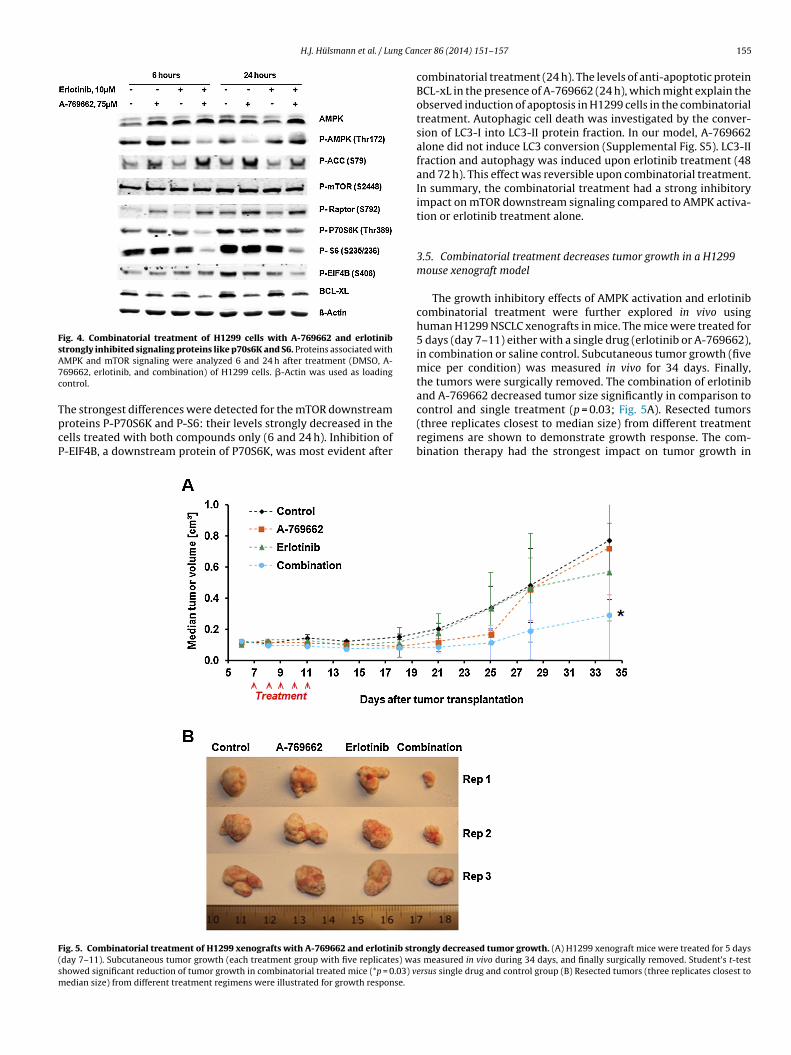
ig. 5. Combinatorial treatment of H1299 xenografts with A-769662 and erlotinib strday 7–11). Subcutaneous tumor growth (each treatment group with five replicates) wahowed significant reduction of tumor growth in combinatorial treated mice (*p = 0.03) vedian size) from different treatment regimens were illustrated for growth response.
cer 86 (2014) 151–157 155
combinatorial treatment (24 h). The levels of anti-apoptotic proteinBCL-xL in the presence of A-769662 (24 h), which might explain theobserved induction of apoptosis in H1299 cells in the combinatorialtreatment. Autophagic cell death was investigated by the conver-sion of LC3-I into LC3-II protein fraction. In our model, A-769662alone did not induce LC3 conversion (Supplemental Fig. S5). LC3-IIfraction and autophagy was induced upon erlotinib treatment (48and 72 h). This effect was reversible upon combinatorial treatment.In summary, the combinatorial treatment had a strong inhibitoryimpact on mTOR downstream signaling compared to AMPK activa-tion or erlotinib treatment alone.
3.5. Combinatorial treatment decreases tumor growth in a H1299mouse xenograft model
The growth inhibitory effects of AMPK activation and erlotinibcombinatorial treatment were further explored in vivo usinghuman H1299 NSCLC xenografts in mice. The mice were treated for5 days (day 7–11) either with a single drug (erlotinib or A-769662),in combination or saline control. Subcutaneous tumor growth (fivemice per condition) was measured in vivo for 34 days. Finally,the tumors were surgically removed. The combination of erlotiniband A-769662 decreased tumor size significantly in comparison to
control and single treatment (p = 0.03; Fig. 5A). Resected tumors(three replicates closest to median size) from different treatmentregimens are shown to demonstrate growth response. The com-bination therapy had the strongest impact on tumor growth inongly decreased tumor growth. (A) H1299 xenograft mice were treated for 5 dayss measured in vivo during 34 days, and finally surgically removed. Student’s t-testersus single drug and control group (B) Resected tumors (three replicates closest to

1 ng Can
Hofia
4
miiamn[r2mmtpa
mtesAmAdntbdLdt
cwdtutopctrAo
labattB[htii
56 H.J. Hülsmann et al. / Lu
1299 xenografts. No significant differences in tumor size werebserved for the other experimental conditions (Fig. 5B). Thesendings suggested that AMPK activation enhances the antitumorction of erlotinib in vivo.
. Discussion
EGFR mutations and the EML4-ALK gene fusion represent twoolecular alterations in about 15% of lung adenocarcinoma patients
n Western countries, and affect target sites for which drugs aren clinical use [17]. The benefit for patients treated with erlotinibnd gefitinib was only significant when stratified according to EGFRutation status [18,19]. However, about 30–40% of patients do
ot respond to EGFR inhibitors using the current selection criteria1,20]. About 1–8% of patients without relevant EGFR mutations areesponders (Mok, NEJM, 2009; Janne, JCO, 2012; Garassino, Lancet,013). The complexity of genomic alterations in lung tumorsay determine these limitations. For example, the role of a KRASutation as negative predictor for EGFR-targeted therapy is con-
roversially discussed [21]. It is likely that the activity of additionalroteins affects the sensitivity against EGFR-targeted therapies inn individual patient.
In this study, NSCLC xenograft models were screened to identifyolecules associated with therapy response to the EGFR-targeted
herapeutics, erlotinib and cetuximab. As a novel finding, P-AMPKxpression was elevated in several NSCLC xenograft models whichubsequently respond better to erlotinib or cetuximab. In addition,MPK and P-AMPK expression was increased after erlotinib treat-ent in the three best responders. These findings suggested thatMPK activation status impacts on EGFR targeted drug response inistinct lung cancer models. In lung cancer, AMPK signaling is con-ected with important oncogenic pathways, and associated withumorigenesis and progression. It was shown that metformin, aiguanide anti-diabetic drug and activator of the AMPK signaling,ecreases the risk for cancer in diabetes patients [22]. Furthermore,KB1, a known upstream activator of AMPK signaling, has beenescribed as tumor suppressor in lung cancer cell lines and mouseumors [23,24].
In our study, AMPK activation using A-769662 sensitizes lungancer cells to erlotinib treatment. The reduction of cell growthas most convincing in EGFR wild-type H1299 cells, which mayue be to the low endogenous AMPK levels, its strongest induc-ion via A-769662, and insensitivity to erlotinib. AMPK activationsing alternative compounds like metformin or AICAR confirmedhe increased erlotinib sensitivity. Several studies in NSCLC andther cancers have shown that response to EGFR-targeted thera-eutics depends on the activation status of mTOR signaling, andan be increased by mTOR-inhibiting agents [25]. An example ishe synergistic effect of combinatorial treatment with erlotinib andapamycin [26]. Moreover, AMPK has an impact on mTOR signaling:MPK can directly phosphorylate Raptor leading to the inhibitionf the mTOR complex 1 and cell cycle arrest [12].
Solely administration of A-769662 in H1299 raised the proteinevels of downstream effectors P-ACC and P-Raptor, but did notffect mTOR downstream targets and cell growth. In contrast, com-ination of erlotinib and A-769662 strongly decreased cell viabilitynd activated apoptosis in H1299. Apoptosis induction may be dueo inhibition of the anti-apoptotic protein BCL-xL upon combina-orial treatment. It has been described that AMPK is required forCL-xL mRNA destabilization and initiation of intrinsic apoptosis27]. The observed autophagy-linked LC3 conversion upon erlotinib
as been previously reported [28]. However, autophagy is likely nothe dominant effect on cell viability upon combinatorial treatmentn our study. In contrast to single treatments, combination resultedn a strong reduction of mTOR downstream targets P-P70S6K,cer 86 (2014) 151–157
P-S6 and P-EIF4B, which likely induced cell growth inhibition. Thesefindings might be caused by a concerted action of both agentstargeting mTOR signaling. In diverse tumor entities EGFR muta-tional status was associated with phosphorylation of mTOR and S6[29,30]. In our NSCLC model, inhibition of mTOR downstream genesindependent of P-mTOR suggests an alternative signal transductionmode via phosphorylation of Raptor.
From three tested NSCLC cell lines, the strongest effects of treat-ment combination were shown in H1299 cells. The impact on tumorgrowth was confirmed in H1299 xenograft models. Possibly, spe-cific mutations influence the sensitivity to this treatment concept.NRAS mutations were found in about 1% of lung tumors, and celllines like H1299 harboring NRAS mutation behaved insensitive toEGFR-TKI [31]. In NRAS-mutant melanoma cells treatment withAMPK activator AICAR inhibited cell growth [32]. In A549 cells, P-AMPK was hardly activated by A-769662 that is probably due to aloss of functional LKB1 [33]. The detailed crosstalk between EGFRand AMPK signaling concerning dual action on mTOR downstreampathway in the context of specific tumor genome alterations has tobe further elucidated.
In conclusion, we observed that AMPK activation using A-769662 sensitizes distinct lung cancer cell lines and H1299xenograft models to erlotinib. The activation status of AMPKsignaling in a subset of lung tumors may affect the sensitivity toEGFR-targeted therapies.
Role of the funding source
This study was supported by the German Federal Ministryof Education and Research BMBF including the grants PREDICT(0315428C), and the BMBF grant German Center for Lung Research(82DZL00404).
Conflict of interest statement
The authors declare no conflict of interest.
Acknowledgments
We thank Jennifer Metzger, Sabrina Gerhardt and Nadine Mohrfor their expert technical assistance.
Appendix A. Supplementary data
Supplementary material related to this article can be found,in the online version, at http://dx.doi.org/10.1016/j.lungcan.2014.09.001.
References
[1] Eberhard DA, Johnson BE, Amler LC, Goddard AD, Heldens SL, Herbst RS, et al.Mutations in the epidermal growth factor receptor and in KRAS are predic-tive and prognostic indicators in patients with non-small-cell lung cancertreated with chemotherapy alone and in combination with erlotinib. J ClinOncol 2005;23:5900–9.
[2] Miller VA, Riely GJ, Zakowski MF, Li AR, Patel JD, Heelan RT, et al. Molecularcharacteristics of bronchioloalveolar carcinoma and adenocarcinoma, bron-chioloalveolar carcinoma subtype, predict response to erlotinib. J Clin Oncol2008;26:1472–8.
[3] Zhou C, Wu YL, Chen G, Feng J, Liu XQ, Wang C, et al. Erlotinib versus chemother-apy as first-line treatment for patients with advanced EGFR mutation-positivenon-small-cell lung cancer (OPTIMAL, CTONG-0802): a multicentre, open-label, randomised, phase 3 study. Lancet Oncol 2011;12:735–42.
[4] Ettinger DS, Akerley W, Borghaei H, Chang AC, Cheney RT, Chirieac LR,et al. Non-small cell lung cancer. J Natl Comprehensive Cancer Netw: JNCCN
2012;10:1236–71.[5] Garassino MC, Martelli O, Broggini M, Farina G, Veronese S, Rulli E, et al. Erlotinibversus docetaxel as second-line treatment of patients with advanced non-small-cell lung cancer and wild-type EGFR tumours (TAILOR): a randomisedcontrolled trial. Lancet Oncol 2013;14:981–8.

ng Can
[
[
[
[
[
[
[
[
[
[
[
[
[
[
[
[
[
[
[
[
[
[
[
H.J. Hülsmann et al. / Lu
[6] Janne PA, Wang X, Socinski MA, Crawford J, Stinchcombe TE, Gu L, et al. Random-ized phase II trial of erlotinib alone or with carboplatin and paclitaxel in patientswho were never or light former smokers with advanced lung adenocarcinoma:CALGB 30406 trial. J Clin Oncol 2012;30:2063–9.
[7] Mok TS, Wu YL, Thongprasert S, Yang CH, Chu DT, Saijo N, et al. Gefitinibor carboplatin-paclitaxel in pulmonary adenocarcinoma. New Engl J Med2009;361:947–57.
[8] Martini M, Vecchione L, Siena S, Tejpar S, Bardelli A. Targeted therapies: howpersonal should we go? Nat Rev Clin Oncol 2012;9:87–97.
[9] Liang J, Mills GB. AMPK: a contextual oncogene or tumor suppressor? CancerRes 2013;73:2929–35.
10] Faubert B, Boily G, Izreig S, Griss T, Samborska B, Dong Z, et al. AMPK is a nega-tive regulator of the Warburg effect and suppresses tumor growth in vivo. CellMetabol 2013;17:113–24.
11] Kato K, Ogura T, Kishimoto A, Minegishi Y, Nakajima N, Miyazaki M, et al. Criticalroles of AMP-activated protein kinase in constitutive tolerance of cancer cellsto nutrient deprivation and tumor formation. Oncogene 2002;21:6082–90.
12] Gwinn DM, Shackelford DB, Egan DF, Mihaylova MM, Mery A, Vasquez DS, et al.AMPK phosphorylation of raptor mediates a metabolic checkpoint. Mol Cell2008;30:214–26.
13] Fichtner I, Rolff J, Soong R, Hoffmann J, Hammer S, Sommer A, et al. Establish-ment of patient-derived non-small cell lung cancer xenografts as models forthe identification of predictive biomarkers. Clin Cancer Res 2008;14:6456–68.
14] Loebke C, Sueltmann H, Schmidt C, Henjes F, Wiemann S, Poustka A, et al.Infrared-based protein detection arrays for quantitative proteomics. Pro-teomics 2007;7:558–64.
15] Brase JC, Mannsperger H, Frohlich H, Gade S, Schmidt C, Wiemann S, et al.Increasing the sensitivity of reverse phase protein arrays by antibody-mediatedsignal amplification. Proteome Sci 2010;8:36.
16] Yauch RL, Januario T, Eberhard DA, Cavet G, Zhu W, Fu L, et al. Epithelialversus mesenchymal phenotype determines in vitro sensitivity and predictsclinical activity of erlotinib in lung cancer patients. Clin Cancer Res 2005;11:8686–98.
17] Pao W, Chmielecki J. Rational, biologically based treatment of EGFR-mutantnon-small-cell lung cancer. Nat Rev Cancer 2010;10:760–74.
18] Paez JG, Janne PA, Lee JC, Tracy S, Greulich H, Gabriel S, et al. EGFR mutations
in lung cancer: correlation with clinical response to gefitinib therapy. Science2004;304:1497–500.19] Tsao MS, Sakurada A, Cutz JC, Zhu CQ, Kamel-Reid S, Squire J, et al. Erlotinibin lung cancer - molecular and clinical predictors of outcome. N Engl J Med2005;353:133–44.
[
cer 86 (2014) 151–157 157
20] Sequist LV, Martins RG, Spigel D, Grunberg SM, Spira A, Janne PA, et al. First-line gefitinib in patients with advanced non-small-cell lung cancer harboringsomatic EGFR mutations. J Clin Oncol 2008;26:2442–9.
21] Roberts PJ, Stinchcombe TE, Der CJ, Socinski MA. Personalized medicinein non-small-cell lung cancer: is KRAS a useful marker in selectingpatients for epidermal growth factor receptor-targeted therapy? J Clin Oncol2010;28:4769–77.
22] Evans JM, Donnelly LA, Emslie-Smith AM, Alessi DR, Morris AD. Metformin andreduced risk of cancer in diabetic patients. BMJ 2005;330:1304–5.
23] Ji H, Ramsey MR, Hayes DN, Fan C, McNamara K, Kozlowski P, et al. LKB1 mod-ulates lung cancer differentiation and metastasis. Nature 2007;448:807–10.
24] Gill RK, Yang SH, Meerzaman D, Mechanic LE, Bowman ED, Jeon HS, et al. Fre-quent homozygous deletion of the LKB1/STK11 gene in non-small cell lungcancer. Oncogene 2011;30:3784–91.
25] Bliesath J, Huser N, Omori M, Bunag D, Proffitt C, Streiner N, et al. Combined inhi-bition of EGFR and CK2 augments the attenuation of PI3K-Akt-mTOR signalingand the killing of cancer cells. Cancer Lett 2012;322:113–8.
26] Buck E, Eyzaguirre A, Brown E, Petti F, McCormack S, Haley JD, et al. Rapamycinsynergizes with the epidermal growth factor receptor inhibitor erlotinib in non-small-cell lung, pancreatic, colon, and breast tumors. Mol Cancer Therapeut2006;5:2676–84.
27] Day RM, Lee YH, Han L, Kim YC, Feng YH. Angiotensin II activates AMPK for exe-cution of apoptosis through energy-dependent and -independent mechanisms.Am J Physiol - Lung Cell Mol Physiol 2011;301:L772–81.
28] Li YY, Lam SK, Mak JC, Zheng CY, Ho JC. Erlotinib-induced autophagy in epider-mal growth factor receptor mutated non-small cell lung cancer. Lung Cancer2013;81:354–61.
29] Dobashi Y, Suzuki S, Kimura M, Matsubara H, Tsubochi H, Imoto I, et al.Paradigm of kinase-driven pathway downstream of epidermal growth factorreceptor/Akt in human lung carcinomas. Hum Pathol 2011;42:214–26.
30] Fan QW, Cheng C, Knight ZA, Haas-Kogan D, Stokoe D, James CD, et al. EGFRsignals to mTOR through PKC and independently of Akt in glioma. Sci Signal2009;2:ra4.
31] Ohashi K, Sequis LV, Arcila ME, Lovly CM, Chen X, Rudin CM, et al. Characteristicsof Lung Cancers Harboring NRAS Mutations. Clin Cancer Res 2013.
32] Petti C, Vegetti C, Molla A, Bersani I, Cleris L, Mustard KJ, et al. AMPK acti-
vators inhibit the proliferation of human melanomas bearing the activatedMAPK pathway. Melanoma Res 2012;22:341–50.33] Sanchez-Cespedes M, Parrella P, Esteller M, Nomoto S, Trink B, Engles JM, et al.Inactivation of LKB1/STK11 is a common event in adenocarcinomas of the lung.Cancer Res 2002;62:3659–62.