Action potential broadening in - Proceedings of the ... · Proc. Nati. Acad. Sci. USA Vol. 88, pp....
5
Proc. Nati. Acad. Sci. USA Vol. 88, pp. 380-384, January 1991 Neurobiology Action potential broadening and frequency-dependent facilitation of calcium signals in pituitary nerve terminals (neurohypophysis/stlmulus-ecretion coupling/neuropeptide/hormone secretion/synaptic plasticity) MEYER B. JACKSON*, ARTHUR KONNERTHtt, AND GEORGE J. AUGUSTINE§ tMax-Planck-Institut fur Biophysikalische Chemie, D-3400 Gottingen, Federal Republic of Germany; *Department of Physiology, University of Wisconsin, Madison, WI 53706; and §Neurobiology Section, Department of Biological Sciences, University of Southern California, Los Angeles, CA 90089-2520 Communicated by Erwin Neher, October 11, 1990 (received for review July 19, 1990) ABSTRACT Hormone release from nerve terminals in the neurohypophysis Is a sensitive function of action potential frequency. We have investigated the cellular mechanisms re- sponsible for this frequency-dependent facilitation by combin- ing patch clamp and fluorimetric Ca2+ measurements in single neurosecretory terminals in thin slices of the rat posterior pituitary. In these terminals both action potential-induced changes in the intracellular Ca2+ concentration ([Ca2+]J) and action potential duration were enhanced by high-frequency stimuli, all with a frequency dependence similar to that of hormone release. Furthermore, brief voltage clamp pulses inactivated a K+ current with a very similar frequency depen- dence. These results support a model for frequency-dependent facilitation in which the inactivation of a K+ current broadens action potentials, leading to an enhancement of [Ca2+]J signals. Further experiments tested for a causal relationship between action potential broadening and facilitation of [Ca2+J1 changes. First, increasing the duration of depolarization, either by broadening action potentials with the K+-channel blocker tetraethylammonium or by applying longer depolarizing volt- age damp steps, increased [Ca2+J1 changes. Second, eliminat- ing frequency-dependent changes in duration, by voltage clamping the terminal with constant duration pulses, substan- tially reduced the frequency-dependent enhancement of [Ca2+], changes. These results indicate that action potential broaden- ing contributes to frequency-dependent facilitation of [Ca2+]J changes. However, the small residual frequency dependence of [Ca2+J1 changes seen with constant duration stimulation sug- gests that a second process, distinct from action potential broadening, also contributes to facilitation. These two frequen- cy-dependent mechanisms may also contribute to activity- dependent plasticity in synaptic terminals. Little is known about the mechanisms that regulate secretion from nerve terminals, despite the importance of these mech- anisms to many physiological processes. For example, a presynaptic site has been proposed as an important locus for change in several forms of use-dependent synaptic plasticity, including facilitation and posttetanic potentiation at neuro- muscular synapses (1) and long-term potentiation in the mammalian hippocampus (2, 3). However, in these and most other systems one can do little more than infer that some presynaptic change occurs, because technical difficulties preclude a direct physiological characterization of the nerve terminals. Although a direct physiological characterization is possible in some invertebrate systems (4, 5), until recently there was no comparable vertebrate system for the study of presynaptic physiology. In an effort to bridge this technical gap, some investigators have made electrical and optical recordings from the nerve terminals of the vertebrate neurohypophysis (6-12). These terminals release the neuropeptides vasopressin and oxy- tocin and have received much attention as a model system for the study of secretion (6-12). Peptide secretion from these terminals also exhibits a striking form of use-dependent plasticity: secretion is a sensitive function of action potential frequency (11, 13-17). This frequency dependence is essen- tial to the input-output properties of the hypothalamic- hypophyseal axis (18) and has been proposed to be due to frequency-dependent modulation of action potential duration (10, 11, 13, 14, 19-23). In the present study, we use patch clamp techniques and fluorimetric calcium indicators to test this hypothesis in individual nerve terminals in pituitary slices. A preliminary account of this work has appeared (24). METHODS Neurohypophysial Slices. The neurointermediate lobe ofthe pituitary was removed from male rats with ages ranging from 2 to 4 months and placed in carbogen (95% 02/5% C02)- saturated rat Ringer's solution (125 mM NaCl, 2.5 mM KCl, 1.25 mM NaH2PO4, 26 mM NaHCO3, 2 mM CaCl2, 1 mM MgCl2, and 20 mM glucose) at 0WC for at least 1 min. Slices 70-80 j&m thick were then cut with an FTB vibratome, with continued bathing in 0WC rat Ringer's solution. Slices were either kept in a 34WC bath or transferred to a recording chamber at room temperature (20-220C) in rat Ringer's solution identical to that described above, except that the KCl concentration was 4 mM. Slices were suitable for recording immediately and were viable for up to 4 hr. Patch clamp recordings from these slices were made at room temperature (20-220C) following published methods (25), except that no cleaning of tissue or enzyme treatment was necessary. Patch electrodes with resistances between 2.5 and 6 Mfl were fabricated from borosilicate glass (i.d. = 1.4 mm; o.d. = 2.0 mm). Tight-seal intracellular recordings (26) were achieved with series resistances ranging from 4.5 to 15 MW. For experiments in which [Ca2+], was not simultane- ously measured, patch pipettes were filled with a solution consisting of 140 mM KCl, 10 mM EGTA, 4 mM ATP, 4 mM MgCI2, and 10 mM Hepes at pH 7.3. Signals were recorded with an EPC-7 patch clamp amplifier and digitized and stored on an Atari computer. A computer program written by Michael Pusch (University of Genoa) was used to acquire data. Intracellular Ca2l Measurements. For intracellular Ca2` concentration ([Ca2+]o) measurements from single terminals, the fluorescent calcium indicator fura-2 (27) (100 or 300 ,uM) was added to a patch pipette filling solution with the com- position 135 mM KCl, 5 mM NaCl, 0.2 mM EGTA, 4 mM ATP, 4 mM MgCl2, and 10 mM Hepes at pH 7.3. The ratiometric technique used to measure [Ca2+], was identical to that described by Neher (28). In brief, terminals were Abbreviation: TEA, tetraethylammonium. tTo whom reprint requests should be addressed. 380 The publication costs of this article were defrayed in part by page charge payment. This article must therefore be hereby marked "advertisement" in accordance with 18 U.S.C. §1734 solely to indicate this fact.
-
Upload
nguyenthien -
Category
Documents
-
view
216 -
download
0
Transcript of Action potential broadening in - Proceedings of the ... · Proc. Nati. Acad. Sci. USA Vol. 88, pp....
Proc. Nati. Acad. Sci. USAVol. 88, pp. 380-384, January 1991Neurobiology
Action potential broadening and frequency-dependent facilitation ofcalcium signals in pituitary nerve terminals
(neurohypophysis/stlmulus-ecretion coupling/neuropeptide/hormone secretion/synaptic plasticity)
MEYER B. JACKSON*, ARTHUR KONNERTHtt, AND GEORGE J. AUGUSTINE§tMax-Planck-Institut fur Biophysikalische Chemie, D-3400 Gottingen, Federal Republic of Germany; *Department of Physiology, University of Wisconsin,Madison, WI 53706; and §Neurobiology Section, Department of Biological Sciences, University of Southern California, Los Angeles, CA 90089-2520
Communicated by Erwin Neher, October 11, 1990 (receivedfor review July 19, 1990)
ABSTRACT Hormone release from nerve terminals in theneurohypophysis Is a sensitive function of action potentialfrequency. We have investigated the cellular mechanisms re-sponsible for this frequency-dependent facilitation by combin-ing patch clamp and fluorimetric Ca2+ measurements in singleneurosecretory terminals in thin slices of the rat posteriorpituitary. In these terminals both action potential-inducedchanges in the intracellular Ca2+ concentration ([Ca2+]J) andaction potential duration were enhanced by high-frequencystimuli, all with a frequency dependence similar to that ofhormone release. Furthermore, brief voltage clamp pulsesinactivated a K+ current with a very similar frequency depen-dence. These results support a model for frequency-dependentfacilitation in which the inactivation of a K+ current broadensaction potentials, leading to an enhancement of [Ca2+]J signals.Further experiments tested for a causal relationship betweenaction potential broadening and facilitation of [Ca2+J1 changes.First, increasing the duration of depolarization, either bybroadening action potentials with the K+-channel blockertetraethylammonium or by applying longer depolarizing volt-age damp steps, increased [Ca2+J1 changes. Second, eliminat-ing frequency-dependent changes in duration, by voltageclamping the terminal with constant duration pulses, substan-tially reduced the frequency-dependent enhancement of [Ca2+],changes. These results indicate that action potential broaden-ing contributes to frequency-dependent facilitation of [Ca2+]Jchanges. However, the small residual frequency dependence of[Ca2+J1 changes seen with constant duration stimulation sug-gests that a second process, distinct from action potentialbroadening, also contributes to facilitation. These two frequen-cy-dependent mechanisms may also contribute to activity-dependent plasticity in synaptic terminals.
Little is known about the mechanisms that regulate secretionfrom nerve terminals, despite the importance of these mech-anisms to many physiological processes. For example, apresynaptic site has been proposed as an important locus forchange in several forms ofuse-dependent synaptic plasticity,including facilitation and posttetanic potentiation at neuro-muscular synapses (1) and long-term potentiation in themammalian hippocampus (2, 3). However, in these and mostother systems one can do little more than infer that somepresynaptic change occurs, because technical difficultiespreclude a direct physiological characterization of the nerveterminals. Although a direct physiological characterization ispossible in some invertebrate systems (4, 5), until recentlythere was no comparable vertebrate system for the study ofpresynaptic physiology.
In an effort to bridge this technical gap, some investigatorshave made electrical and optical recordings from the nerveterminals of the vertebrate neurohypophysis (6-12). These
terminals release the neuropeptides vasopressin and oxy-tocin and have received much attention as a model system forthe study of secretion (6-12). Peptide secretion from theseterminals also exhibits a striking form of use-dependentplasticity: secretion is a sensitive function of action potentialfrequency (11, 13-17). This frequency dependence is essen-tial to the input-output properties of the hypothalamic-hypophyseal axis (18) and has been proposed to be due tofrequency-dependent modulation of action potential duration(10, 11, 13, 14, 19-23). In the present study, we use patchclamp techniques and fluorimetric calcium indicators to testthis hypothesis in individual nerve terminals in pituitaryslices. A preliminary account of this work has appeared (24).
METHODSNeurohypophysial Slices. The neurointermediate lobe ofthe
pituitary was removed from male rats with ages ranging from2 to 4 months and placed in carbogen (95% 02/5% C02)-saturated rat Ringer's solution (125 mM NaCl, 2.5 mM KCl,1.25 mM NaH2PO4, 26 mM NaHCO3, 2 mM CaCl2, 1 mMMgCl2, and 20 mM glucose) at 0WC for at least 1 min. Slices70-80 j&m thick were then cut with an FTB vibratome, withcontinued bathing in 0WC rat Ringer's solution. Slices wereeither kept in a 34WC bath or transferred to a recordingchamber at room temperature (20-220C) in rat Ringer'ssolution identical to that described above, except that theKCl concentration was 4 mM. Slices were suitable forrecording immediately and were viable for up to 4 hr.Patch clamp recordings from these slices were made at
room temperature (20-220C) following published methods(25), except that no cleaning of tissue or enzyme treatmentwas necessary. Patch electrodes with resistances between 2.5and 6 Mfl were fabricated from borosilicate glass (i.d. = 1.4mm; o.d. = 2.0 mm). Tight-seal intracellular recordings (26)were achieved with series resistances ranging from 4.5 to 15MW. For experiments in which [Ca2+], was not simultane-ously measured, patch pipettes were filled with a solutionconsisting of 140 mM KCl, 10mM EGTA, 4 mM ATP, 4 mMMgCI2, and 10 mM Hepes at pH 7.3. Signals were recordedwith an EPC-7 patch clamp amplifier and digitized and storedon an Atari computer. A computer program written byMichael Pusch (University of Genoa) was used to acquiredata.
Intracellular Ca2l Measurements. For intracellular Ca2`concentration ([Ca2+]o) measurements from single terminals,the fluorescent calcium indicator fura-2 (27) (100 or 300 ,uM)was added to a patch pipette filling solution with the com-position 135 mM KCl, 5 mM NaCl, 0.2 mM EGTA, 4 mMATP, 4 mM MgCl2, and 10 mM Hepes at pH 7.3. Theratiometric technique used to measure [Ca2+], was identicalto that described by Neher (28). In brief, terminals were
Abbreviation: TEA, tetraethylammonium.tTo whom reprint requests should be addressed.
380
The publication costs of this article were defrayed in part by page chargepayment. This article must therefore be hereby marked "advertisement"in accordance with 18 U.S.C. §1734 solely to indicate this fact.

Proc. Natl. Acad. Sci. USA 88 (1991) 381
alternately excited with UV light, filtered at 360 or 390 nmwith a wheel rotating at 5 Hz, and the emitted fluorescencewas measured with a Hamamatsu photomultiplier tube (mod-el R928). [Ca2+], was then computed from the ratio ofemission at the two wavelengths. Such measurements of[Ca2+]i could be made from single terminals for up to 30 min.
RESULTSNerve terminals were visible in thin slices of the neurohy-pophysis when viewed under Nomarski optics (Fig. 1). Theseterminals are round in appearance, with diameters rangingfrom I to 20 gm. The identity of these structures as terminalshas been established (S. A. DeRiemer, R. Schneggenburger,M.B.J., and A.K., unpublished results) by the followingcriteria: (i) absence ofnuclei, asjudged by the lack ofstainingwith a nuclear dye; (ii) orthograde transport of dye fromhypothalamic neurons; and (iii) axonal stimulation generatesaction potentials. Furthermore, as shown below, electricalstimulation leads to increases in [Ca2+li in these structures.The terminals observed on the upper surface of a slice wereoften exposed and accessible to patch pipettes. An exampleof such a terminal, filled with the fluorescent dye Luciferyellow, is shown in Fig. 1.Calcium Signaling in Single Nerve Terminals. Loading of
the fluorescent Ca2" indicator fura-2 (27) from the patchpipette solution was accomplished in 2 min or less. Theresting [Ca2+], in these terminals was 110 ± 10 nM (mean ±SEM; n = 16), which is comparable to the resting [Ca2+]i ofmany cell types (29). The value of 350 nM reported forsecretosomes prepared from the posterior pituitary is con-siderably higher (30), but this discrepancy can be explainedby leakage of dye from secretosomes.
Since secretion from the nerve terminals of the posteriorpituitary is triggered by Ca2" entry (8, 9, 31), we were
interested in asking whether the frequency dependence ofsecretion was a result of the frequency dependence of [Ca2"Jchanges. Repetitive stimulation ofcurrent-clamped terminalswith current pulses large enough to evoke action potentialscaused transient increases in [Ca2+Ji (Fig. 2). These increaseswere rapid, with a rate of rise that depended on the durationand frequency of stimulation. The peak in [Ca2+]i was fol-lowed by an exponential decay back to the resting level. Thehalf-time for decay was in the range of 10 to 20 s. Ca2' bufferssuch as fura-2 are known to slow [Ca2+]j recovery (ref. 32;G.J.A. and E. Neher, unpublished results), so that the fura-2may have slowed the decay of [Ca2+], signals. However,lowering the fura-2 concentration from 300 to 100 /iM hadlittle effect on the time course of recovery, which suggeststhat such effects were relatively minor in our experiments.
Stimulation with action potentials at high frequencies in-creased [Ca2+]J much more than stimulation with actionpotentials at low frequencies (Fig. 2). At 1 Hz the change in[Ca2+], induced by 100 action potentials was barely detect-able, but following 100 action potentials at frequencies of 10Hz or higher, [Ca2+]i increased to 300 nM or more (Fig. 2A).High-frequency stimulation also enhanced the rate of in-crease of [Ca2+]i during the stimulus train. The frequencydependence of action potential-evoked increases in [Ca2+]J isshown in Fig. 2B. This frequency dependence resembles thatreported for vasopressin release from isolated neurointerme-diate lobes (13, 14). Both signals are maximal at =10 Hz,although the frequency of the half-maximal response isroughly 3 Hz higher for vasopressin release than for [Ca2+],signals (Fig. 2B). This difference could be due to a nonlinearrelationship between [Ca2+]i and the rate of secretion (33-35)or to minor differences in experimental conditions (vasopres-sin release was measured in a similar saline but with a bovineserum albumin concentration of 1.5 mg/ml at 37°C). None-theless, the similar frequency dependences of [Ca2+]i and
FIG. 1. Nerve terminals on the surface of a thin pituitary slice. A photomicrograph with simultaneous Nomarski and fluorescence opticsshows a Lucifer yellow-filled nerve terminal. The patch electrode, filled with Lucifer yellow (0.2 mg/ml) in our normal patch-electrode fillingsolution, is also visible. (Bar = 20 iam.)
Neurobiology: Jackson et al.

382 Neurobiology: Jackson et al.
A20Hz'- 0.20.1 [COal
I 040 s
5 Hz
1 Hz
B .- 1.,
u
<0.
0
N
-a 0.1Eo
Z 0.1.0 10 20 30
Frequency (Hz)40
FIG. 2. (A) [Ca2]i in a single nerve terminal. A terminal filledwith fura-2 was stimulated with 100 current pulses (3 ms, 200 pA) atthe indicated frequencies. The period of stimulation is indicated bythe solid line. The holding current was adjusted to produce amembrane potential of -70 mV. Under these conditions of stimu-lation, each current pulse produced an action potential. (B) Fre-quency dependence of [Ca2+]i changes (i) determined from data suchas those shown in A. The frequency dependence of vasopressin (VP)release (o) was replotted from ref. 13 for comparison. The datarepresent the arithmetic mean SEM of six experiments, normal-ized to the maximal values.
hormone secretion, together with the established require-ment for Ca2+ in the triggering of secretion (8, 9, 31, 36),suggest that the facilitation of Ca2` signals by high-frequencystimulation contributes to the frequency dependence of hor-mone release. We therefore investigated the mechanismsunderlying the frequency dependence offacilitation of[Ca2+]signals.Frequency-Dependent Action Potentials and K+ Currents. It
has been proposed that action potential broadening plays arole in the frequency dependence of secretion from theneurohypophysis (10, 11, 14-16, 18-22). As reported previ-ously (14, 23), action potentials of pituitary terminals broad-ened during high-frequency stimulation (Fig. 3A). After 't20action potentials at 10 Hz, the width at half-height reached a
F 20 (Frequency (Hz)
maximum that was 37% larger than that of a control actionpotential. The frequency dependence of these changes inaction potential duration (Fig. 3B) was also similar to thatobserved for [Ca2+]i increases and vasopressin release (Fig.2B).Because K+ currents are critical in determining action
potential duration (37), these currents were examined withthe voltage clamp technique to evaluate their role in frequen-cy-dependent changes in action potential duration. Depolar-ization activated a complex pattern of outward current (Fig.3C). On the basis of reversal potential, ion substitutionexperiments, and block by tetraethylammonium (TEA) and4-aminopyridine, we concluded that this outward current wascarried by K+ (data not shown). Prolonged depolarizationpartially inactivated the K+ current and allowed this currentto be separated into rapidly and slowly inactivating compo-nents (Fig. 3C). These components have different kinetic andpharmacological properties and presumably are generated bydifferent populations of K+ channels (M.B.J. and A.K.,unpublished results).To determine how these K+ currents change during a
facilitating stimulus, trains of 30 brief (3-ms duration) pulseswere applied at various frequencies. This conditioning regi-men selectively reduced the rapidly inactivating componentof K+ current (Fig. 3C). The inactivation induced by theseconditioning pulse trains was a sensitive function of pulsefrequency (Fig. 3D), producing a relationship essentiallyidentical to the frequency dependence of action potentialduration (Fig. 3B). The similarity between these two plotssuggests that action potential broadening by high-frequencystimulation is caused by K+-current inactivation.
Tests of the Action Potential Broadening Model of Facilita-tion. The similar frequency dependence of hormone release,[Ca2W]i enhancement, action potential broadening, and K+-current inactivation supports a model for frequency-dependent facilitation in which the depolarizations of manyaction potentials in rapid succession inactivate a K+ current.As the K+ current is inactivated, action potentials repolarizemore slowly, leading to increased Ca2+ entry, enhancementof [Ca2+]j, and a pursuant increase in hormone release. Wenext performed two types of experiments to test the hypoth-esis that action potential broadening is the basis for frequen-cy-dependent enhancement of [Ca2+]1.
Ifaction potential broadening contributes to the increase in[Ca2+]i signals, then the [Ca2]1i change should depend on theduration of a depolarizing stimulus. This was first examinedby voltage clamping the terminals and applying brief, spike-like, depolarizing pulses ofvarying duration. Depolarizationswere made to +10 mV to produce maximum signals (see
2.8
,, FIG. 3. Broadening of action potentials by high-- 2A4 E frequency stimulation. (A) A control action potential* (elicited following at least 5 s of inactivity) is displayed* together with the last action potential of trains of 30 at2 both 1 and 10 Hz. (B) Dependence of action potential
(AP) duration (width at half-height) on stimulus fre-< quency. The data represent the arithmetic mean +
SEM for three cells. (C) Voltage clamp measurements- 1.6 of K+ currents elicited by pulses from -80 mV to 500.8 mV. The small brief inward current at the very begin-
ning of the pulses is a tetrodotoxin-sensitive Na+_ 0.7 - current. The largest K+ current was observed with no
. prior conditioning. Conditioning stimuli consisted of0.6- trains of 30 pulses (3-ms duration) to 50 mV applied
a° immediately before the test pulse. This conditionin0.5 - reduced the peak current. CT, control; CD, condition-
ing; T, test. (D) Reduction in the ratio of the final K+- 0.4 current (Ifin) to the peak K+ current (Ip,) versus
40 frequency of stimulation. The data represent the arith-metic mean + SEM for three cells.
12
8L S
two *A[Ca],
T ~~~~~0VP release
Proc. Natl. Acad. Sci. USA 88 (1991)
Proc. Natl. Acad. Sci. USA 88 (1991) 383
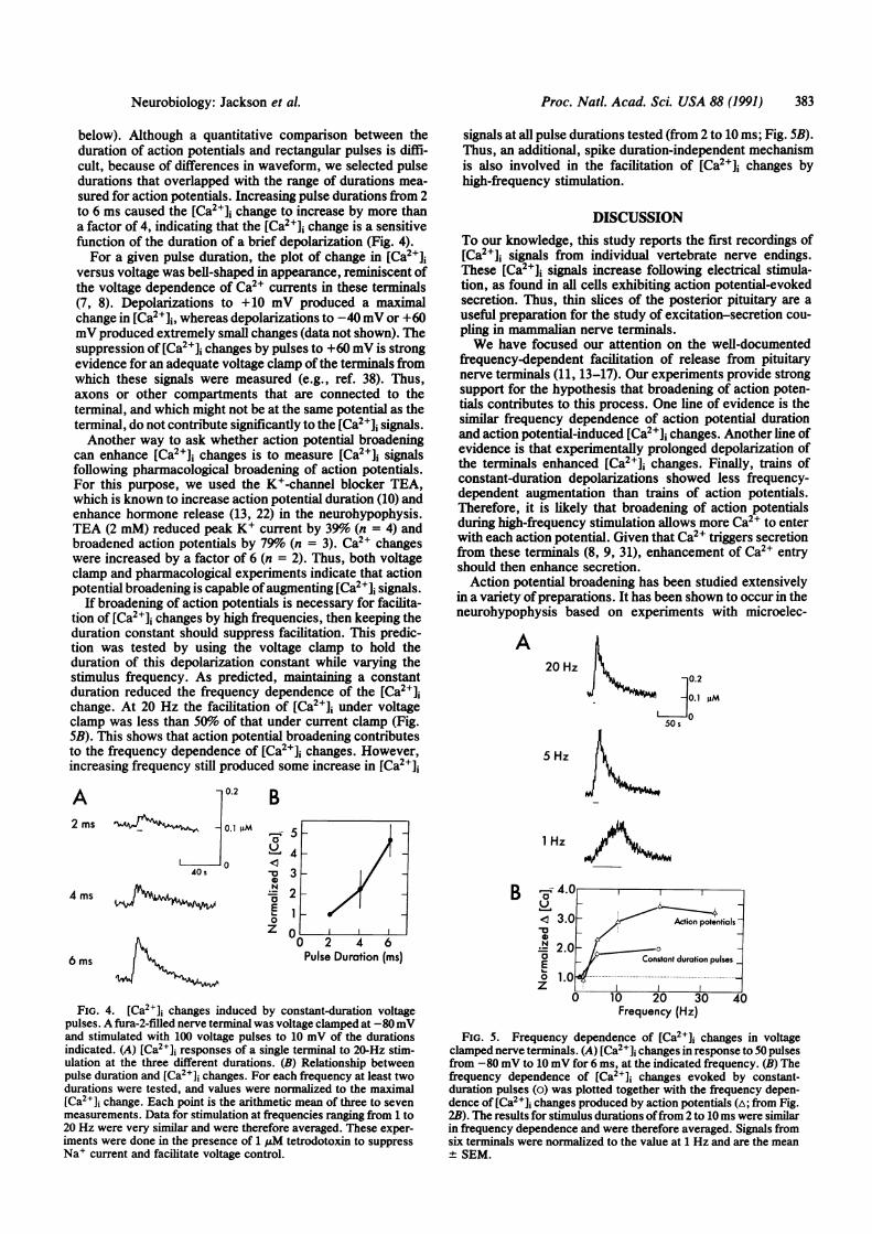
below). Although a quantitative comparison between theduration of action potentials and rectangular pulses is diffi-cult, because of differences in waveform, we selected pulsedurations that overlapped with the range of durations mea-sured for action potentials. Increasing pulse durations from 2to 6 ms caused the [Ca2+]i change to increase by more thana factor of 4, indicating that the [Ca2+]j change is a sensitivefunction of the duration of a brief depolarization (Fig. 4).For a given pulse duration, the plot of change in [Ca21],
versus voltage was bell-shaped in appearance, reminiscent ofthe voltage dependence of Ca2+ currents in these terminals(7, 8). Depolarizations to +10 mV produced a maximalchange in [Ca2+]1, whereas depolarizations to -40 mV or +60mV produced extremely small changes (data not shown). Thesuppression of [Ca2+], changes by pulses to +60 mV is strongevidence for an adequate voltage clamp of the terminals fromwhich these signals were measured (e.g., ref. 38). Thus,axons or other compartments that are connected to theterminal, and which might not be at the same potential as theterminal, do not contribute significantly to the [Ca2+]j signals.Another way to ask whether action potential broadening
can enhance [Ca2+], changes is to measure [Ca2+]i signalsfollowing pharmacological broadening of action potentials.For this purpose, we used the K+-channel blocker TEA,which is known to increase action potential duration (10) andenhance hormone release (13, 22) in the neurohypophysis.TEA (2 mM) reduced peak K+ current by 39%o (n = 4) andbroadened action potentials by 79% (n = 3). Ca2+ changeswere increased by a factor of 6 (n = 2). Thus, both voltageclamp and pharmacological experiments indicate that actionpotential broadening is capable ofaugmenting [Ca2+]j signals.
If broadening of action potentials is necessary for facilita-tion of [Ca2+]j changes by high frequencies, then keeping theduration constant should suppress facilitation. This predic-tion was tested by using the voltage clamp to hold theduration of this depolarization constant while varying thestimulus frequency. As predicted, maintaining a constantduration reduced the frequency dependence of the [Ca2+],change. At 20 Hz the facilitation of [Ca2+]1 under voltageclamp was less than 50% of that under current clamp (Fig.5B). This shows that action potential broadening contributesto the frequency dependence of [Ca2+], changes. However,increasing frequency still produced some increase in [Ca2+]J
signals at all pulse durations tested (from 2 to 10 ms; Fig. 5B).Thus, an additional, spike duration-independent mechanismis also involved in the facilitation of [Ca2+]i changes byhigh-frequency stimulation.
DISCUSSIONTo our knowledge, this study reports the first recordings of[Ca2+]i signals from individual vertebrate nerve endings.These [Ca2+]i signals increase following electrical stimula-tion, as found in all cells exhibiting action potential-evokedsecretion. Thus, thin slices of the posterior pituitary are auseful preparation for the study of excitation-secretion cou-pling in mammalian nerve terminals.We have focused our attention on the well-documented
frequency-dependent facilitation of release from pituitarynerve terminals (11, 13-17). Our experiments provide strongsupport for the hypothesis that broadening of action poten-tials contributes to this process. One line of evidence is thesimilar frequency dependence of action potential durationand action potential-induced [Ca2+]i changes. Another line ofevidence is that experimentally prolonged depolarization ofthe terminals enhanced [Ca2+]j changes. Finally, trains ofconstant-duration depolarizations showed less frequency-dependent augmentation than trains of action potentials.Therefore, it is likely that broadening of action potentialsduring high-frequency stimulation allows more Ca21 to enterwith each action potential. Given that Ca2' triggers secretionfrom these terminals (8, 9, 31), enhancement of Ca2' entryshould then enhance secretion.Action potential broadening has been studied extensively
in a variety ofpreparations. It has been shown to occur in theneurohypophysis based on experiments with microelec-
A20 Hz
5 Hz
0.2
0 _ 0.1 AM
5050 s
A 0.2
2ms --A1 0.1hM
I 040s
4 ms
6 ms
B
1 Hza.1
-U0NaE0z
B 4.< 3.0N 2.aEo 1.z
Pulse Duration (ms)
FIG. 4. [Ca2+], changes induced by constant-duration voltagepulses. A fura-2-filled nerve terminal was voltage clamped at -80mVand stimulated with 100 voltage pulses to 10 mV of the durationsindicated. (A) [Ca2+]i responses of a single terminal to 20-Hz stim-ulation at the three different durations. (B) Relationship betweenpulse duration and [Ca2+]1 changes. For each frequency at least twodurations were tested, and values were normalized to the maximal[Ca2+]i change. Each point is the arithmetic mean of three to sevenmeasurements. Data for stimulation at frequencies ranging from 1 to20 Hz were very similar and were therefore averaged. These exper-iments were done in the presence of 1 uM tetrodotoxin to suppressNa+ current and facilitate voltage control.
.0
.0 Action potentials]
.0- _Constant duration pulses _
.nl ... ---0 10 20 30
Frequency (Hz)
"I40
FIG. 5. Frequency dependence of [Ca2+]i changes in voltageclamped nerve terminals. (A) [Ca2+]i changes in response to 50 pulsesfrom -80 mV to 10 mV for 6 ms, at the indicated frequency. (B) Thefrequency dependence of [Ca2+]j changes evoked by constant-duration pulses (o) was plotted together with the frequency depen-dence of [Ca2+]i changes produced by action potentials (A; from Fig.2B). The results for stimulus durations offrom 2 to 10 ms were similarin frequency dependence and were therefore averaged. Signals fromsix terminals were normalized to the value at 1 Hz and are the mean+ SEM.
Neurobiology: Jackson et al.
.V17-

384 Neurobiology: Jackson et al.
trodes (23) and voltage-sensitive dyes (14). It has also beendemonstrated in the hypothalamic cell bodies from which thenerve endings of the posterior pituitary originate (19, 20). Themechanism of action potential broadening has been espe-cially well studied in molluscan neurons (39) and can becaused by K+-current inactivation. We tested the role ofK+-current inactivation in broadening of pituitary actionpotentials by using trains of brief, spike-like voltage pulses.Such trains inactivated K+ current in a frequency-dependentmanner similar to that of action potential broadening. Thebroadening of action potentials by treatment with 2mM TEA,which reduces the rapidly inactivating component of K+current, is additional support for the involvement of thiscomponent of the K+ current in action potential repolariza-tion. Thus, reduction of the K+ current by inactivation,which occurs during high-frequency stimulation (Fig. 3),would be expected to broaden action potentials.For action potential broadening to be effective in enhanc-
ing secretion, stimulus-induced [Ca2+], increases must be asensitive function of the duration of depolarization. We havefound this to be the case for both action potentials and voltageclamp pulses. It has been proposed that a high sensitivity ofCa2+ flux to stimulus duration is caused by the delayedactivation of voltage-gated Ca2+ channels (40). This delay inCa2+-current activation has been described in squid terminals(4, 38) and chromaffin cells (41, 42) and may be a general andfunctionally significant feature of Ca2+ channels that triggersecretion.
In addition to demonstrating a role for action potentialbroadening in the frequency dependence of secretion fromthe nerve endings of the neurohypophysis, this study showedthat not all of the frequency dependence of Ca2l signaling canbe attributed to action potential broadening. When the du-ration of a stimulus is kept fixed, high frequencies stillenhanced [Ca2+], changes to some extent (Fig. SB). Becausethe [Ca2+]j changes relax slowly, over seconds, the mostlikely source of this other component is temporal summationof the successive [Ca2W]i increases caused by single depolar-izations. Although Ca2+-channel facilitation could provide anadditional frequency-dependent mechanism (41, 42), facili-tation of capacitance changes in secretosomes from theneurohypophysis has been reported to take place withoutincreases in Ca2+ current (8). Duration-independent facilita-tion of Ca2+ signals could also involve the use-dependentgeneration of another, unknown second messenger.There are many examples of use-dependent enhancement
of neurotransmitter secretion. These include facilitation,posttetanic potentiation, and long-term potentiation. In noinstance has an underlying presynaptic mechanism beenelucidated. The two mechanisms that we have identified inpituitary nerve terminals could play roles in these other formsof synaptic plasticity. While action potential broadening isnot necessary for synaptic facilitation in squid nerve termi-nals (43, 44), it may be involved in other forms of use-dependent plasticity. A role for action potential broadening inheterosynaptic facilitation is well documented (45, 46). Like-wise, our proposal of temporal summation of [Ca2W]i signalsis reminiscent of the residual calcium models for synapticfacilitation (36) and posttetanic potentiation (1).
We thank Frauke Friedlein for technical assistance, Michael Puschfor providing and modifying computer programs, and Erwin Neherfor suggestions and encouragement. We thank Ana Lia Obaid andBrian Salzberg for comments on the manuscript. This research wassupported by grant NS01149 to M.B.J. and grant NS21624 to G.J.A.from the National Institutes of Health and by grants from theDeutsche Forschungsgemeinschaft and the Bundesministerium fOrForschungs und Technologie to A.K. M.B.J. and G.J.A. also grate-fully acknowledge support from the Alexander von Humboldt Foun-dation.
1. Magleby, K. L. & Zengel, J. E. (1975) J. Physiol. (London) 245,163-182.
2. Nicoll, R. A., Kauer, J. A. & Malenka, R. C. (1988) Neuron 1,97-103.
3. Bliss, T. V. P. & Lynch, M. A. (1988) in Long-Term Potentiation:From Biophysics to Behavior, eds. Landsfield, P. W. & Deadwyler,S. A. (Liss, New York), pp. 3-72.
4. Llinas, R. (1984) Curr. Top. Membr. Transp. 22, 519-571.5. Augustine, G. J., Charlton, M. P. & Smith, S. J. (1987) Annu. Rev.
Neurosci. 10, 633-693.6. Mason, W. T. & Dyball, R. E. J. (1986) Brain Res. 283, 279-286.7. Lemos, J. R. & Nowycky, M. C. (1989) Neuron 2, 1419-1426.8. Fidler, N. H., Nowycky, M. C. & Bookman, R. J. (1990) Nature
(London) 344, 449-451.9. Cazalis, M., Dayanithi, G. & Nordmann, J. J. (1987) J. Physiol.
(London) 390, 55-70.10. Obaid, A. L., Orkand, R. K., Gainer, H. & Salzberg, B. M. (1985)
J. Gen. Physiol. 85, 481-489.11. Salzberg, B. M. & Obaid, A. L. (1988) J. Exp. Biol. 139, 195-231.12. Salzberg, B. M., Obaid, A. L., Senseman, D. M. & Gainer, H.
(1983) Nature (London) 306, 36-40.13. Bondy, C. A., Gainer, H. & Russell, J. T. (1987) Neuroendocrin-
ology 46, 258-267.14. Gainer, H., Wolfe, S. A., Obaid, A. L. & Salzberg, B. M. (1986)
Neuroendocrinology 43, 557-563.15. Dutton, A. & Dyball, R. E. J. (1979) J. Physiol. (London) 290,
433-440.16. Dreifuss, J. J., Kalnins, I., Kelly, J. S. & Ruf, K. B. (1971) J.
Physiol. (London) 215, 805-817.17. Gainer, H. (1978) in Comparative Neuroendocrinology, eds. Gail-
lard, P. J. & Boer, H. H. (Elsevier/North Holland, Amsterdam),pp. 293-304.
18. Poulain, D. A. & Wakerly, J. B. (1982) Neuroscience 7, 773-808.19. Andrew, R. D. & Dudek, F. E. (1985) Brain Res. 334, 176-179.20. Bourque, C. W. & Renaud, L. P. (1985) J. Physiol. (London) 363,
429-439.21. Nordmann, J. J. & Stuenkel, E. L. (1986) J. Physiol. (London) 380,
521-539.22. Hobbach, H. P., Hurth, S., Jost, D. & Racke, K. (1988) J. Physiol.
(London) 397, 539-554.23. Bourque, C. W. (1990) J. Physiol. (London) 421, 247-262.24. Jackson, M. B., Augustine, G. J. & Konnerth, A. (1990) Soc.
Neurosci. Abstr. 16, 41%.25. Edwards, F. A., Konnerth, A., Sakmann, B. & Takahashi, T. (1989)
Pflagers Arch. 414, 600-612.26. Hamill, 0. P., Marty, A., Neher, E., Sakmann, B. & Sigworth, F. J.
(1981) Pflugers Arch. 391, 85-100.27. Grynkiewicz, G., Poenie, M. & Tsien, R. Y. (1985) J. Biol. Chem.
260, 3440-3450.28. Neher, E. (1989) in Neuromuscular Junction, eds. Sellin, L. C.,
Libelius, R. & Thesleff, S. (Elsevier, Amsterdam), pp. 65-76.29. Tsien, R. Y. (1989) Annu. Rev. Neurosci. 12, 227-253.30. Brethes, D., Dayanithi, G., Letellier, L. & Nordmann, J. J. (1987)
Proc. Natl. Acad. Sci. USA 84, 1439-1443.31. Douglas, W. W. & Poisner, A. M. (1964) J. Physiol. (London) 172,
1-18.32. Baylor, S. M. & Hollingsworth, S. (1988) J. Physiol. (London) 403,
151-192.33. Dodge, F. A. & Rahamimoff, R. (1967) J. Physiol. (London) 193,
419-432.34. Knight, D. E. & Baker, P. F. (1982) J. Membr. Biol. 68, 107-140.35. Augustine, G. J. & Charlton, M. P. (1986) J. Physiol. (London) 381,
619-640.36. Katz, B. & Miledi, R. (1967) J. Physiol. (London) 192, 407-436.37. Hodgkin, A. L. & Huxley, A. F. (1952) J. Physiol. (London) 117,
500-544.38. Augustine, G. J., Charlton, M. P. & Smith, S. J. (1985) J. Physiol.
(London) 367, 143-162.39. Aldrich, R. W., Getting, P. A. & Thompson, S. H. (1979) J. Phys-
iol. (London) 291, 531-544.40. Augustine, G. J. (1990) J. Physiol. (London), in press.41. Fenwick, E. M., Marty, A. & Neher, E. (1982) J. Physiol. (London)
331, 499-535.42. Hoshi, T., Rothlein, J. & Smith, S. J. (1984) Proc. Nati. Acad. Sci.
USA 81, 5871-5875.43. Charlton, M. P. & Bittner, G. D. (1978) J. Gen. Physiol. 72,
471-486.44. Charlton, M. P., Smith, S. J. & Zucker, R. S. (1982) J. Physiol.
(London) 323, 173-193.45. Hochner, B., Klein, M., Schacher, S. & Kandel, E. R. (1986) Proc.
Nati. Acad. Sci. USA 83, 8410-8414.46. Kandel, E. R. & Schwartz, J. H. (1982) Science 229, 433-443.
Proc. Natl. Acad. Sci. USA 88 (1991)