
Action expertise reduces brain activity for …avanzini/downloads/paper/petrini_neurim11.pdf ·...
13
Action expertise reduces brain activity for audiovisual matching actions: An fMRI study with expert drummers Karin Petrini a, ⁎, Frank E. Pollick a , Sofia Dahl b , Phil McAleer a , Lawrie McKay c , Davide Rocchesso d , Carl Haakon Waadeland e , Scott Love a , Federico Avanzini f , Aina Puce g a Department of Psychology, University of Glasgow, Glasgow, Scotland, UK b Department of Media Technology, Aalborg University Copenhagen, Copenhagen, Denmark c Netherlands Institute for Neuroscience (NIN), Amsterdam, The Netherlands d Department of Art and Industrial Design, IUAV University of Venice, Venice, Italy e Department of Music, Norwegian University of Science and Technology, Trondheim, Norway f Department of Information Engineering, University of Padua, Padua, Italy g Department of Psychological and Brain Sciences, Indiana University, Bloomington, USA abstract article info Article history: Received 29 September 2010 Revised 2 March 2011 Accepted 3 March 2011 Available online 11 March 2011 Keywords: Drumming Biological motion fMRI Audiovisual synchrony Action–sound representation Action expertise When we observe someone perform a familiar action, we can usually predict what kind of sound that action will produce. Musical actions are over-experienced by musicians and not by non-musicians, and thus offer a unique way to examine how action expertise affects brain processes when the predictability of the produced sound is manipulated. We used functional magnetic resonance imaging to scan 11 drummers and 11 age- and gender-matched novices who made judgments on point-light drumming movements presented with sound. In Experiment 1, sound was synchronized or desynchronized with drumming strikes, while in Experiment 2 sound was always synchronized, but the natural covariation between sound intensity and velocity of the drumming strike was maintained or eliminated. Prior to MRI scanning, each participant completed psychophysical testing to identify personal levels of synchronous and asynchronous timing to be used in the two fMRI activation tasks. In both experiments, the drummers' brain activation was reduced in motor and action representation brain regions when sound matched the observed movements, and was similar to that of novices when sound was mismatched. This reduction in neural activity occurred bilaterally in the cerebellum and left parahippocampal gyrus in Experiment 1, and in the right inferior parietal lobule, inferior temporal gyrus, middle frontal gyrus and precentral gyrus in Experiment 2. Our results indicate that brain functions in action-sound representation areas are modulated by multimodal action expertise. © 2011 Elsevier Inc. All rights reserved. Introduction Music and dance have formed a significant part of human culture throughout history. Listening to music performed by a group of musicians, as well as dancing with others, relies on the ability to integrate sight with sound, as well as predicting what sound an action will produce and when. This is something that we can do with relatively little effort in a social setting. As non-expert observers, we also have the capacity to appreciate the performances of virtuoso musicians and dancers from their audiovisual output — when these individuals have spent years perfecting and honing their motor skills. Indeed, the majority of human actions are multisensory in nature, and our ability to predict the auditory consequence of others' actions relies on acquired knowledge gained from performing these actions ourselves. That our brain produces similar neural activity for seen and per- formed actions is now well-known (e.g. Gallese et al., 1996; Rizzolatti et al., 1996; Decety and Grèzes, 1999; Nishitani and Hari, 2000; Buccino et al., 2001, 2004; Iacoboni, 2005; Haslinger et al., 2005; Calvo-Merino et al., 2005, 2006). These so-called ‘mirror neurons’ are not triggered only by visual stimuli, but also by auditory stimuli (Kohler et al., 2002; Keysers et al., 2003; Wilson et al., 2004). Based on these findings, the existence of a cross-modal neural system for action recognition has been suggested (Kohler et al., 2002; Keysers et al., 2003). A recent study by Lahav et al. (2007) examined the underlying mechanisms linking motor and auditory actions, and showed that frontoparietal regions were activated when listening to pieces of music listeners had both learned and played, but not when listening to other familiar music pieces. These data are consistent with findings in the visuomotor domain, where regions such as the dorsal premotor cortex, inferior parietal sulcus and cerebellum are activated when participants view actions that form part of their existing motor repertoire (Calvo-Merino et al., 2005, 2006). Relatively few studies have investigated how visual and auditory information interact when recognizing the actions of others and how NeuroImage 56 (2011) 1480–1492 ⁎ Corresponding author. E-mail address: [email protected] (K. Petrini). 1053-8119/$ – see front matter © 2011 Elsevier Inc. All rights reserved. doi:10.1016/j.neuroimage.2011.03.009 Contents lists available at ScienceDirect NeuroImage journal homepage: www.elsevier.com/locate/ynimg
Transcript of Action expertise reduces brain activity for …avanzini/downloads/paper/petrini_neurim11.pdf ·...

NeuroImage 56 (2011) 1480–1492
Contents lists available at ScienceDirect
NeuroImage
j ourna l homepage: www.e lsev ie r.com/ locate /yn img
Action expertise reduces brain activity for audiovisual matching actions: An fMRIstudy with expert drummers
Karin Petrini a,⁎, Frank E. Pollick a, Sofia Dahl b, Phil McAleer a, Lawrie McKay c, Davide Rocchesso d,Carl Haakon Waadeland e, Scott Love a, Federico Avanzini f, Aina Puce g
a Department of Psychology, University of Glasgow, Glasgow, Scotland, UKb Department of Media Technology, Aalborg University Copenhagen, Copenhagen, Denmarkc Netherlands Institute for Neuroscience (NIN), Amsterdam, The Netherlandsd Department of Art and Industrial Design, IUAV University of Venice, Venice, Italye Department of Music, Norwegian University of Science and Technology, Trondheim, Norwayf Department of Information Engineering, University of Padua, Padua, Italyg Department of Psychological and Brain Sciences, Indiana University, Bloomington, USA
⁎ Corresponding author.E-mail address: [email protected] (K. Petrini).
1053-8119/$ – see front matter © 2011 Elsevier Inc. Aldoi:10.1016/j.neuroimage.2011.03.009
a b s t r a c t
a r t i c l e i n f oArticle history:Received 29 September 2010Revised 2 March 2011Accepted 3 March 2011Available online 11 March 2011
Keywords:DrummingBiological motionfMRIAudiovisual synchronyAction–sound representationAction expertise
When we observe someone perform a familiar action, we can usually predict what kind of sound that actionwill produce. Musical actions are over-experienced by musicians and not by non-musicians, and thus offer aunique way to examine how action expertise affects brain processes when the predictability of the producedsound is manipulated. We used functional magnetic resonance imaging to scan 11 drummers and 11 age- andgender-matched novices who made judgments on point-light drumming movements presented with sound.In Experiment 1, sound was synchronized or desynchronized with drumming strikes, while in Experiment 2sound was always synchronized, but the natural covariation between sound intensity and velocity ofthe drumming strike was maintained or eliminated. Prior to MRI scanning, each participant completedpsychophysical testing to identify personal levels of synchronous and asynchronous timing to be used in thetwo fMRI activation tasks. In both experiments, the drummers' brain activation was reduced in motor andaction representation brain regions when soundmatched the observed movements, and was similar to that ofnovices when sound was mismatched. This reduction in neural activity occurred bilaterally in the cerebellumand left parahippocampal gyrus in Experiment 1, and in the right inferior parietal lobule, inferior temporalgyrus, middle frontal gyrus and precentral gyrus in Experiment 2. Our results indicate that brain functions inaction-sound representation areas are modulated by multimodal action expertise.
l rights reserved.
© 2011 Elsevier Inc. All rights reserved.
Introduction
Music and dance have formed a significant part of human culturethroughout history. Listening to music performed by a group ofmusicians, as well as dancing with others, relies on the ability tointegrate sight with sound, as well as predicting what sound an actionwill produce and when. This is something that we can do withrelatively little effort in a social setting. As non-expert observers, wealso have the capacity to appreciate the performances of virtuosomusicians and dancers from their audiovisual output — when theseindividuals have spent years perfecting and honing their motor skills.Indeed, the majority of human actions are multisensory in nature,and our ability to predict the auditory consequence of others' actionsrelies on acquired knowledge gained from performing these actionsourselves.
That our brain produces similar neural activity for seen and per-formed actions is now well-known (e.g. Gallese et al., 1996; Rizzolattiet al., 1996; Decety and Grèzes, 1999; Nishitani and Hari, 2000; Buccinoet al., 2001, 2004; Iacoboni, 2005; Haslinger et al., 2005; Calvo-Merinoet al., 2005, 2006). These so-called ‘mirror neurons’ are not triggeredonly by visual stimuli, but also by auditory stimuli (Kohler et al., 2002;Keysers et al., 2003; Wilson et al., 2004). Based on these findings, theexistence of a cross-modal neural system for action recognition hasbeen suggested (Kohler et al., 2002; Keysers et al., 2003). A recent studyby Lahav et al. (2007) examined the underlying mechanisms linkingmotor andauditory actions, and showed that frontoparietal regionswereactivated when listening to pieces of music listeners had both learnedand played, but not when listening to other familiar music pieces. Thesedata are consistent with findings in the visuomotor domain, whereregions such as the dorsal premotor cortex, inferior parietal sulcus andcerebellum are activated when participants view actions that formpart of their existingmotor repertoire (Calvo-Merino et al., 2005, 2006).
Relatively few studies have investigated how visual and auditoryinformation interact when recognizing the actions of others and how
1481K. Petrini et al. / NeuroImage 56 (2011) 1480–1492
expertise with the represented actions influences the underlying brainprocesses (e.g. Musacchia et al., 2007; Haslinger et al., 2005; Hodgeset al., 2005). Here, we asked the question of whether expertisefor represented audiovisual actions will modulate brain activationin action-sound representation areas when the natural relationbetween sight and sound of familiar actions is disrupted. We thusaim at determining the basis of the higher perceptual and motorexpertise, respectively, of drummers as compared to novices. To thisend, we developed two different experiments, the first of whichaimed to examine the role of perceptual and motor expertise whenprocessing human actions for which the natural synchronizationbetween movements and sounds was eliminated. In line with this,the second experiment examined the role of perceptual and motorexpertise when processing human actions for which the naturalcovariation between movements' velocity and sounds' intensity waseliminated.
For a number of reasons, we chose to use the biological motionof drumming actions which were highly familiar to expert drummersand unfamiliar to non-musicians. Our first premise was that in musicperformance each visual action produces a sound (Zatorre et al.,2007). Drummers can better anticipate when the sound will occurbased on the viewed action, compared with novices (Petrini et al.,2009b). Consequently, this makes drumming actions ideal stimuli forstudying interactions between seeing and hearing when perceivingothers' actions. Secondly, drumming actions are over-experiencedby drummers, but not by non-musicians, allowing the study of brainactivation differences which are driven by differences in actionexpertise (Calvo-Merino et al., 2005, 2006). Thirdly, music has beenextensively used to study visuomotor (e.g. Stewart et al., 2003;Buccino et al., 2004; Vogt et al., 2007) and audiomotor (e.g. Parsonset al., 2005; Baumann et al., 2005, 2007; Bangert et al., 2006; Lahavet al., 2007) brain processes involved in action representation. Finally,point-light biological motion (Johansson, 1973) has been previouslyused to focus on brain processes driven by visual motion cues ofactions (Saygin et al., 2004; Saygin, 2007), as well as by audiovisualintegration (Brooks et al., 2007; Klin et al., 2009).
We therefore ran two event-related fMRI experiments in 11drummers and 11 novices matched for age and gender. In Experiment1, they evaluated animated displays where the correspondencebetween drumming action and resulting sound was eliminated bydesynchronizing the two signals. In Experiment 2, the same par-ticipants were scanned while viewing animated displays where thetemporal correspondence between the signals was maintained, butcovariation between the drummer's movements and the resultingsound was eliminated (i.e. sound intensity did not covary with thevelocity). We hypothesized that acquiring perceptual and motorexpertise through practice with the portrayed audiovisual actionwould alter brain processes, and that this would result in differentnetworks of brain areas being activated in Experiments 1 and 2, sincethe difference between matching and mismatching displays in thetwo experiments differentiates between sensory synchrony andsensory congruency.
Materials and methods
Participants
Eleven right-handed drummers (all males; age 35±12 years)were studied. Seven of the 11 were presently either professionalmusicians and/or music teachers. The average number of years ofdrumming experience was 24±11 years (range 13 to 45 years). Alldrummers were right-handed, as assessed by the Edinburgh Hand-edness Questionnaire (Oldfield, 1971).
We gender- and age-matched our novice participant group,relative to our expert group, to study 11 males (age 35±11 years)with no previous drumming experience and, importantly, little or no
previous musical training. Eight of the novices were right-handed, asassessed by the Edinburgh Handedness Questionnaire (Oldfield,1971).
Stimulus creation
Audiovisual stimuli were created in three distinct steps from initialmotion capture data of a professional jazz drummer playing a swinggroove beat (Petrini et al., 2009a,b), as briefly described below.
Step 1 consisted of converting the 3D movement coordinates ofthe drummer into point-light displays of drumming actions, usingcomputer graphics. Step 2 consisted of converting the times andvelocities of stick impact into a realistic stream of sound by using anaturalistic sound generation algorithm. Step 3 consisted of combin-ing the visual point-light displays and the audio stream of drum beatsby using video editing software.
Motion-capture data
Data were recorded from the movements of a professional jazzdrummer playing a swing groove at 120 beats per minute, with theaccent on the second beat (Waadeland, 2006). Markers were placed atsix locations, including the tip of the drumstick, the level of the grip ofthe drumstick, and at the drummer's shoulder, elbow, wrist and hand.
The 3D coordinates of these marker locations were sampled at240 Hz using a Proreflex 3D motion capture system. The sampled3D motion capture data were first downsampled to 60 Hz. Theywere then converted into a series of 2D images using Matlab andPsychtoolbox routines (Brainard, 1997; Pelli, 1997) running underOSX and utilizing OpenGL graphics with anti-aliasing enabled. Whitedisks (luminance: 85 cd/m2; diameter: 2 mm) on a black background(luminance: 0.12 cd/m2) represented the drummer's arm anddrumstick (Fig. 1). The drum head was represented using a thickwhite line (Fig. 1), orientated 25° from horizontal (width: 2.2 cm;height: 2 mm; luminance: 85 cd/m2). The image sequences weresaved as video using an AVI file format at a 60 Hz frame rate.
Auditory stimuli
The synthetic drumming sounds were obtained by an algorithmthat took as input the times and velocities of a series of strikes, andoutputted the simulated audio signal (Fontana et al., 2004). To obtaina very natural sound, the algorithm simulated the first 25 modes of acircular membrane. Both the time and impact velocity of a strikewere derived by plotting the displacement and velocity of thedrumstick tip marker against the time of the drummer performance,and selecting, for each impact, the frame at which the drumstick tipvelocity changed from negative to positive (Dahl, 2004, pp. 765). Touse only displacement and velocity perpendicular to the drumhead(Dahl, 2004, pp. 765), data were rotated to a coordinate frame, wherehorizontal was parallel to the drumhead and vertical perpendicular tothe drumhead. These operations were performed on the 240 Hz,unfiltered displacements and velocities.
The resulting sounds were saved as audio files (WAV format)with a 15-second duration. The audio files were created to eithercorrespond to the original movement data recordings, or to notcorrespond (see Fig. 1). The corresponding audio files were obtainedby taking the impact times and impact velocities found in the originalrecorded movement and using these as input to the algorithm. Forthe non-corresponding audio files, the identical impact times wereused, but the set of velocities was randomly scrambled. Thus, for thematching displays, there was a natural covariation between theoriginal movements of the drummer and the resulting sound (e.g. faststrikes, loud sounds; slow strikes, soft sounds), while for themismatching displays the timing of the strikes was identical, but
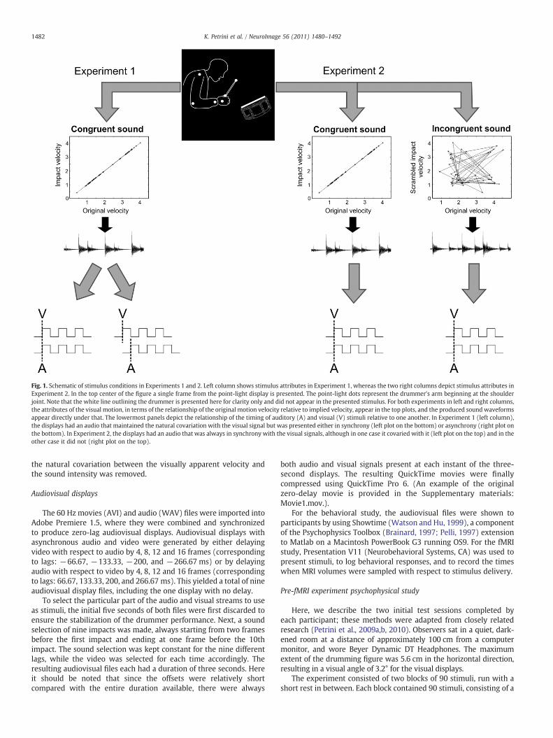
Fig. 1. Schematic of stimulus conditions in Experiments 1 and 2. Left column shows stimulus attributes in Experiment 1, whereas the two right columns depict stimulus attributes inExperiment 2. In the top center of the figure a single frame from the point-light display is presented. The point-light dots represent the drummer's arm beginning at the shoulderjoint. Note that the white line outlining the drummer is presented here for clarity only and did not appear in the presented stimulus. For both experiments in left and right columns,the attributes of the visual motion, in terms of the relationship of the original motion velocity relative to implied velocity, appear in the top plots, and the produced sound waveformsappear directly under that. The lowermost panels depict the relationship of the timing of auditory (A) and visual (V) stimuli relative to one another. In Experiment 1 (left column),the displays had an audio that maintained the natural covariation with the visual signal but was presented either in synchrony (left plot on the bottom) or asynchrony (right plot onthe bottom). In Experiment 2, the displays had an audio that was always in synchrony with the visual signals, although in one case it covaried with it (left plot on the top) and in theother case it did not (right plot on the top).
1482 K. Petrini et al. / NeuroImage 56 (2011) 1480–1492
the natural covariation between the visually apparent velocity andthe sound intensity was removed.
Audiovisual displays
The 60 Hzmovies (AVI) and audio (WAV) files were imported intoAdobe Premiere 1.5, where they were combined and synchronizedto produce zero-lag audiovisual displays. Audiovisual displays withasynchronous audio and video were generated by either delayingvideo with respect to audio by 4, 8, 12 and 16 frames (correspondingto lags: −66.67, −133.33, −200, and −266.67 ms) or by delayingaudio with respect to video by 4, 8, 12 and 16 frames (correspondingto lags: 66.67, 133.33, 200, and 266.67 ms). This yielded a total of nineaudiovisual display files, including the one display with no delay.
To select the particular part of the audio and visual streams to useas stimuli, the initial five seconds of both files were first discarded toensure the stabilization of the drummer performance. Next, a soundselection of nine impacts was made, always starting from two framesbefore the first impact and ending at one frame before the 10thimpact. The sound selection was kept constant for the nine differentlags, while the video was selected for each time accordingly. Theresulting audiovisual files each had a duration of three seconds. Hereit should be noted that since the offsets were relatively shortcompared with the entire duration available, there were always
both audio and visual signals present at each instant of the three-second displays. The resulting QuickTime movies were finallycompressed using QuickTime Pro 6. (An example of the originalzero-delay movie is provided in the Supplementary materials:Movie1.mov.).
For the behavioral study, the audiovisual files were shown toparticipants by using Showtime (Watson and Hu, 1999), a componentof the Psychophysics Toolbox (Brainard, 1997; Pelli, 1997) extensionto Matlab on a Macintosh PowerBook G3 running OS9. For the fMRIstudy, Presentation V11 (Neurobehavioral Systems, CA) was used topresent stimuli, to log behavioral responses, and to record the timeswhen MRI volumes were sampled with respect to stimulus delivery.
Pre-fMRI experiment psychophysical study
Here, we describe the two initial test sessions completed byeach participant; these methods were adapted from closely relatedresearch (Petrini et al., 2009a,b, 2010). Observers sat in a quiet, dark-ened room at a distance of approximately 100 cm from a computermonitor, and wore Beyer Dynamic DT Headphones. The maximumextent of the drumming figure was 5.6 cm in the horizontal direction,resulting in a visual angle of 3.2° for the visual displays.
The experiment consisted of two blocks of 90 stimuli, run with ashort rest in between. Each block contained 90 stimuli, consisting of a
1483K. Petrini et al. / NeuroImage 56 (2011) 1480–1492
random ordering of the two audiovisual conditions (natural covaria-tion maintained, natural covariation eliminated) with nine time lags(−266.67,−200,−133.33,−66.67, 0, 66.67, 133.33, 200, 266.67 ms)and five repetitions of each item. Participants were instructed to press‘1’ on the keypad if the drummer's movements were perceived to bein synchrony with the sound, or press ‘3’ if they were perceived asbeing asynchronous. After three training trials, the experimenterleft the participant alone to perform the experiment.
The behavioral experiment produced 90 synchronization responsesfor both the displays presenting the natural covariation and thedisplays presenting the unnatural covariation, which were distributedas 10 repetitions at each of the nine audiovisual timings — from soundpreceding video by 266 ms, to sound lagging video by 266 ms. The datawere plotted as numbers of synchrony responses for each of the ninetimings, and this data was fit with a Gaussian function. The peak ofthe Gaussian fit revealed the best perceived audiovisual synchrony forboth naturally covarying and unnaturally covarying stimuli for eachparticipant, and the tail of the distribution furthest from the peakrevealed the best perceived audiovisual asynchrony. The displayswith the best perceived synchrony and best perceived asynchronywere selected as stimuli for Experiments 1 and 2 of the fMRI studyas appropriate.
fMRI experiments: stimulus delivery
A Windows PC (Dell Precision 690) running Presentation V11.1(Neurobehavioral Systems, CA) presented stimuli and logged behav-ioral responses and MRI data acquisition pulses for each acquiredbrain volume. The first MRI scanner pulse initiated the presentationscript, which presented an initial eight-second cue to the participantsthat the imaging run was beginning, during which a total of four MRIexcitations were performed without MRI data acquisition to achievesteady-state magnetization. Visual clips were projected through theMRI scanner's control roomwindow onto a screen at the participant'sfeet using a video projector (NEC Corporation, LT10 DLP). The par-ticipant lay supine in the MRI scanner and viewed the display througha mirror mounted on the quadrature head coil. The stimulus sizewas adjusted so that the visual angle of the drummer was identical tothe 3.2° of visual arc used in the initial behavioral experiments. Audiowas presented through a high-quality sound card interface (CDX01,Digital Audio). A sound mixer (1642VLZ pro mixer, Mackie Inc.)and commercially available MR-compatible electrostatic ear buds(STAX SRS-005 Earspeaker system; Stax LTD., Gardena, CA) wornunder sound-attenuating ear muffs were used to deliver sound to theparticipant's ear.
In both experiments, a trial began with the three-second audio-visual clip, and participants responded with a button press beforevideo clip offset. After offset the trial ended with a blank (black)screen. The blank screenwas presented for an average of four seconds,with this duration varying randomly between two seconds and sixseconds. A total of 50 trials were presented in randomized orderduring an imaging run which lasted 6 min and 40 s. Experiment 1contained three runs and Experiment 2 contained two runs.
In fMRI Experiment 1, there were three stimulus conditions:Synchronous (Matching), Asynchronous (Mismatching), and a nullcondition containing just fixation. Participants indicated with a two-button forced choice response whether sound and video wereappropriately matched or mismatched. The synchronous and asyn-chronous items were obtained from the pre-scan psychophysicalstudy.
In fMRI Experiment 2, there were two stimulus conditions:naturally covarying (matching), unnaturally covarying (mismatch-ing). We did not have a null condition in Experiment 2 becausewe needed to strike a balance between reducing the overall durationof the experiment and including an optimal number of conditions.Indeed, if the activation differences of Experiments 1 and 2 had
overlapped, then the additional null condition in Experiment 1 wouldhave allowed us to carry out the analysis for this second experimentwithin the region of interest (ROI) obtained from Experiment 1. If,on the other hand, such overlap did not occur, we would havebeen able to eliminate the null condition from the main analysis ofExperiment 1 in order to carry out the same mixed factorial analysisfor both experiments. Participants indicated with a two-button forcedchoice response whether sound and video were appropriatelymatched or mismatched. Since the display with the best perceivedsynchrony for the naturally covarying condition had been used inExperiment 1 with the same participants, we created a new displaywith the same timing but taken from a non-overlapping section ofthe original motion capture of the drumming performance.
fMRI data acquisition
Functional imagesDuring each fMRI experiment for each participant, we acquired
200 volumes per run of blood oxygen-level dependent (BOLD) activityin a near-whole brain acquisition, using a gradient echo spiral in-outsequence (36) on a 3 Tesla Horizon HD MRI scanner (General ElectricMedical Systems, Inc.). A total of 600 volumes were acquired forExperiment 1, whereas 400 volumes were acquired for Experiment 2.Functional images used in subsequent analyses consisted of averagedimages of spiral-in and spiral-out trajectories, reconstructed offlineusing routines written in C (courtesy of G. Glover, Stanford University,CA) and running under Linux (Fedora Core Release 5, Raleigh, NC).The averaged spiral in-out trajectories optimized sampling from brainregions prone to susceptibility artifacts and MR signal drop-out. Atotal of 22 axial slices beginning from the vertex (4 mm thickwith 1 mmgap)were acquiredwith the followingparameters:matrix=128×128, FOV=240 mm, (in plane resolution=1.875 mm), band-width=125, and TE/TR=35/2000ms.
Anatomical imagesFor each participant, we acquired a near whole-brain T1-weighted
anatomical volume with identical slice prescription to functionalimages, and a whole-brain high-resolution anatomical SPGR volume(1.5 mm×0.9375 mm×0.9375 mm, FOV=240, matrix 256×256,124 slices).
fMRI data analysis
The functional and anatomical images were analyzed using BrainVoyager QX 1.9.10 (Brain Innovation, Maastricht, The Netherlands).
Pre-processing of functional dataFunctional imaging data (ANALYZE format) were pre-processed by
performing a slice scan time correction. Slice scan time correction wasperformed using sinc interpolation based on information aboutthe TR (2000 ms) and the order of slice scanning (ascending, inter-leaved). 3D motion correction (6 df) was performed to detect andcorrect for small head movements by spatial alignment of all volumesof a subject to the first volume by rigid body transformations.Estimated translation and rotation parameters were inspected andnever exceeded 3 mm or 2°. A linear trend removal and temporalhigh-pass filtering were then applied to remove low-frequency non-linear drifts of three or fewer cycles (0.0063 Hz) per time course. ThefunctionalMR imageswere smoothed using a Gaussian filter with full-width at half-maximum (FWHM) equal to 8 mm.
Pre-processing of anatomical dataThe anatomical data (ANALYZE format) of each subject was loaded
and converted into BrainVoyager's internal ‘VMR’ data format. Thedata were then resampled to 1 mm resolution and transformed intoanterior commissure–posterior commissure (AC–PC) and Talairach

Fig. 2. The response time of drummers (right panels) and novices (left panels) is displayedfor Experiment 1 (top panels) and Experiment 2 (bottom panels) as a function of experi-mental runs.
1484 K. Petrini et al. / NeuroImage 56 (2011) 1480–1492
standard space. The three spatial transformations were combined andapplied backward in one step to avoid quality loss due to successivedata sampling. The two affine transformations, iso-voxel scalingand AC–PC transformation, were concatenated to form a single 4×4transformation matrix. For each voxel coordinates in the target(Talairach) space a piecewise affine ‘Un-Talairach’ step was per-formed, followed by application of the inverted spatial transforma-tion of the aforementioned matrix. The computed coordinates wereused to sample the data points in the original 3D space using sincinterpolation.
Normalization of functional dataTo transform the functional data into Talairach space, the
functional time series data of each subject was first coregisteredwith the subject's 3D anatomical dataset, followed by the applicationof the same transformation steps as performed for the 3D anatomicaldataset (see above). This step results in normalized 4D volumetime course (‘VTC’) data. In order to avoid quality loss due to suc-cessive data sampling, normalization was performed in a single stepcombining a functional–anatomical affine transformation matrix, arigid-body AC–PC transformation matrix, and a piecewise affineTalairach grid scaling step. As described for the anatomical normal-ization procedure, these steps were performed backward, startingwith a voxel in Talairach space and sampling the corresponding datain the original functional space. The functional slices were coregis-tered to the anatomical volume using manual alignment to obtainoptimal fit and reduce as much as possible the geometrical distortionsof the echo-planar images. The necessary scaling adjustment wasdone interactively, using appropriate transformation and visualiza-tion tools of BrainVoyager QX.
Analysis
First level analysisAnalyses were performed on the data of individual participants
using multiple linear regression of the BOLD-response time course ineach voxel using three predictors (Matching, Mismatching and Blank)in Experiment 1, and two predictors (Matching, Mismatching) inExperiment 2. For each run of each participant's event-related data, aBrainVoyager protocol file (PRT) was derived, representing the onsetand duration of the events for the different conditions. Predictors'time courses were adjusted for the hemodynamic response delay byconvolution with a hemodynamic response function.
Second level analysisStatistical evaluation of group data was based on a second-level
GLM random effects analysis. For Experiment 1 we carried out a 2(expertise: drummers and novices)×2 (audiovisual synchrony:matching and mismatching) analysis of variance, with expertise asthe between-participants factor and audiovisual synchrony as thewithin-participants factor. Similarly, for Experiment 2 we carried outa 2 (expertise: drummers and novices)×2 (audiovisual covariation:matching and mismatching) analysis of variance, with expertise asthe between-participants factor and audiovisual covariation as thewithin-participants factor. In both experiments the statistical mapswere corrected for multiple comparisons using cluster-size thresh-olding (Forman et al., 1995; Goebel et al., 2006). In this method, foreach statistical map the uncorrected voxel-level threshold was set atPb0.001, and then was submitted to a whole-brain correctioncriterion based on the estimate of the map's spatial smoothness andon an iterative procedure (Monte Carlo simulation) for estimatingcluster-level false-positive rates (i.e. the theoretical number of “false”positive voxels that are activated in each random map). After 1000iterations the minimum cluster-size that yielded a cluster-level false-positive rate of 5% was used to threshold the statistical map. The
minimum cluster-size for Pb0.05 is reported according to the originaltable (in voxels) and the interpolated table (in mm3).
Results
In the Results and Discussion sections we focus on activationdifferences between experts and novices, as drummers are moreexperienced with matching audiovisual drumming actions.
Experiment 1
Behavioral data acquired during the fMRI scanning sessionIn Experiment 1, behavioral data acquired during MRI scanning
sessions indicate that both novices and experienced drummersclassified the audiovisual synchrony and asynchrony displays above90% correctly. Comparing performance between novices and drum-mers failed to show significant differences between the two groups'abilities to discriminate the synchronous display as matching andthe asynchronous one as mismatching (independent samples t-test:t(20)=0.167, p=0.869, two-tailed), and this is not surprisinggiven that each participant was tested with individualized stimulibased on previous psychophysical testing. This is important, asdifferences in brain activity between the groups cannot beattributed to novices finding the task more difficult than drummers.As well as comparing the classification performances, we also com-pared the response time of the two groups. To this end we carriedout a 2 (expertise: drummers, novices)×2 (stimuli: matching, mis-matching)×3 (experimental run) mixed model ANOVA, withexpertise as the between-subjects factor and stimuli and run aswithin-subjects factors. This analysis showed that only the mainfactor ‘stimuli’ (F(1, 20)=6.694, p=0.018) and the interactionbetween ‘stimuli’ and ‘expertise’ (F(1, 20)=13.085, p=0.002)affected significantly the response time (Fig. 2, top panels). Pairedsamples t-test, corrected for multiple comparisons, showed thatwhilst drummers differed in the response time for matchingand mismatching displays (t(10)=3.756, p=0.008), novices did
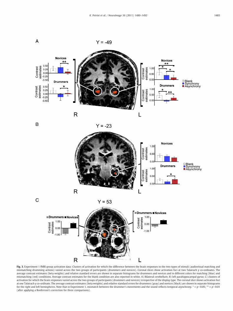
Fig. 3. Experiment 1 fMRI group activation data: Clusters of activation for which the difference between the brain responses to the two types of stimuli (audiovisual matching andmismatching drumming actions) varied across the two groups of participants (drummers and novices). Coronal slices show activation foci at two Talairach y co-ordinates. Theaverage contrast estimates (beta weights) and relative standard errors are shown in separate histograms for drummers and novices and in different colors for matching (blue) andmismatching (red) conditions. Average contrast estimates for the blank condition are also reported in white. A) Bilateral cerebellum; B) left parahippocampal gyrus; C) clusters ofactivation for which the brain responses varied across the two groups of participants (drummers and novices) irrespective of the display type. The coronal slice shows activation fociat one Talairach y co-ordinate. The average contrast estimates (beta weights) and relative standard errors for drummers (gray) and novices (black) are shown in separate histogramsfor the right and left hemispheres. Note that in Experiment 1, mismatch between the drummer's movements and the sound reflects temporal asynchrony. *=pb0.05; **=pb0.01(after applying a Bonferroni's correction for three comparisons).
1485K. Petrini et al. / NeuroImage 56 (2011) 1480–1492

Table 1Experiment 1 clusters of activation from a 2×2 ANOVA with ‘expertise’ as a between-participants factor and ‘audiovisual synchrony’ as a within-participants factor. Legend:BA — Brodmann's area; MFG = middle frontal gyrus.
1486 K. Petrini et al. / NeuroImage 56 (2011) 1480–1492
not (t(10)=−0.914, p=0.382). Specifically, drummers were foundto be slower when responding to matching displays thanmismatching displays (Fig. 2, right top panel).
Anatomicalregion
Hemisphere Talairachcoordinate(x,y,z)
Numberof voxels
Effect sizea BA
F(1, 20) P
Expertise (novicesNexperts)MFG Right 10, 54, −12 539 18.77 0.0004 10MFG Left −19, 54, 3 108 16.29 0.0006 10
Expertise×SynchronyCerebellum Right 30, −43, −25 1442 16.35 0.0006Cerebellum Left −17, −58,
−211608 17.83 0.0005
Parahippocampalgyrus
Left −26, −23,−29
191 16.95 0.0005 36
a Effect size = average F value for all voxels in the ROI.
fMRI activation dataA two-way ANOVA1 (participant group×audiovisual synchrony)
revealed a main effect of expertise bilaterally in the MFG (middlefrontal gyrus), no main effect of synchrony, and a significant inter-action between expertise and synchrony bilaterally in the cerebellum(Fig. 3A) and left parahippocampal gyrus (Fig. 3B). Examination of thecontrast parameters in these areas for the three left-handed and eightright-handed novices indicated that handedness did not affect thefound differences or lack of such between matching and mismatchingstimuli within this group (Supplemental Fig. 1).
The anatomical location and details of the activated foci are listedin Table 1. To control for multiple comparisons, each map wasadjusted to an initial P value of b0.001 (uncorrected) and then sub-mitted to a volume-based cluster-threshold algorithm yielding anew map thresholded at Pb0.05 (corrected) with a minimum clusterthreshold. The minimum cluster threshold that yielded a cluster-levelfalse-positive rate of 5% was k=4, 80 mm3 for the map assessed forexpertise×audiovisual synchrony, and k=3, 81 mm3 for the mapassessed for expertise (Goebel et al., 2006). Average activation (i.e.averaged beta weights) indicated that the main effect of expertisewas attributable to a lower level of activation in drummers relative tonovices in the left and right MFG. An interaction effect in the bilateralcerebellum (Figs. 3A and 4A) and in the parahippocampal gyrus(Fig. 3B) was also attributable to reduction in activity in these areasfor drummers, but not for novices, when viewing synchronousaudiovisual displays. Activity in these areas during the blank con-dition (Fig. 3) suggested that the significant difference in activationwas produced by negative BOLD responses for drummers. Forexample, Fig. 3 shows a reduction in activity in the bilateral cere-bellum and parahippocampal gyrus when drummers viewed audio-visual matching displays (in blue) relative to blank displays (inwhite).
Fig. 4A shows how the reduction in activity we found bilaterallyin the cerebellum, when going from an average of zero years ofdrumming experience for the novices to an average of 24 years for thedrummers, overlaps with the reduction in activity found by Lehericyet al. (2005) in their motor sequence learning study. A regressionanalysis confirmed this observation, indicating that the brain activityin the cerebellum negatively correlated with the difference betweennovices' and drummers' years of experience (right cerebellum: r=−0.436, p=0.042; left cerebellum: r=−0.413, p=0.056). We didnot find, however, any significant correlation between the brainactivity (betas weights) for each experimental condition (matching,mismatching) and the years of experience within our drummersgroup in either the right (matching: r=0.052, p=0.880; mismatch-ing: r=0.028, p=0.935) or left cerebellum (matching: r=−0.121,p=0.724; mismatching: r=−0.195, p=0.565). Neither was asignificant correlation found between brain activity and drummers'experience in the left parahippocampal gyrus (matching: r=−0.073,p=0.830; mismatching: r=0.071, p=0.835). This further analysissuggests that there was no further reduction in activity in these areas
1 We did not include the blank condition in the ANOVA to be consistent with theanalysis carried out in Experiment 2, thus allowing an easier comparison of the results.Nevertheless, including the blank condition and carrying out a 2 (participants: novices,drummers)×3 (stimulus condition: matching, mismatching, and blank) repeatedmeasure ANOVA with expertise as the between factor and stimulus condition as thewithin factor did not change the main findings. For the right cerebellum (F(2, 19)=14.492, pb0.001), the left cerebellum (F(2, 19)=10.342, p=0.001), and the leftparahippocampal gyrus (F(2, 19)=5.465, p=0.013) the higher order interaction wasstill significant. The same holds true for the rightMFG (F(1, 20)=29.462, pb0.001), andthe left MFG (F(1, 20)=18.033, pb0.001), for which the between factor was stillsignificant.
after 13 years of drumming experience (i.e. the bottom range of yearsof experience in our drummers group).
Since we found that drummers were slower than novices (Fig. 2)when they had to respond to matching displays than mismatchingdisplays, we further investigated whether this correlated with thecontrast parameters in the regions of interest assessed in Experi-ment 1. A regression analysis showed a significant negative corre-lation between the contrast parameters obtained from the leftcerebellum and the response time (r=−0.480, p=0.024; Fig. 5, topleft panel), as well as between the contrast parameters obtainedfrom the right cerebellum and the response time (r=−0.460,p=0.031; Fig. 5, top right panel). No significant correlation wasfound between the contrast parameters of right and left cerebellumand response time for the mismatching displays (right: r=−0.158,p=0.482; left: r=−0.406, p=0.061; Fig. 5, bottom panels), andbetween the contrast parameters of the left parahippocampal gyrusand the response time for the matching (r=−0.183, p=0.416) andmismatching displays (r=−0.253, p=0.257). The top panels ofFig. 5 show that the differences between novices and expertscontribute to this regression. Indeed, the lower brain activity ofthe drummers group in the bilateral cerebellum corresponded to alonger response time to the matching displays; in contrast, thegreater brain activity of the novices group in the bilateral cere-bellum corresponded to a shorter response time to the matchingdisplays.
Experiment 2
Behavioral data acquired during the fMRI scanning sessionIn Experiment 2, participants' judgments of whether or not the
sight and soundmatched showed a significant difference (independentsamples t-test: t(20)=3.902, p=0.001, two-tailed) between thedrummers (80% correct judgments) and novices (50% correct judg-ments), with the novices not being significantly different from chance(one sample t-test: t(10)=0.111, mean=50.54, two-tailed, StandardError=4.90). Similarly toExperiment1,we also compared the responsetime of the two groups. To this end we carried out a 2 (expertise:drummers, novices)×2 (stimuli: matching, mismatching)×2 (experi-mental run) mixed model ANOVA with expertise as the between-subjects factor and stimuli and run as within-subjects factors. Thisanalysis showed that only the main factor ‘run’ (F(1, 20)=10.680,p=0.004) affected significantly the response time, because there was adecrease in response timewhengoing fromrun1 to run2 (Fig. 2, bottompanels).
fMRI activation dataA two-way ANOVA (participant group×audiovisual covariation)
revealed no main effect of expertise. However, an extended network
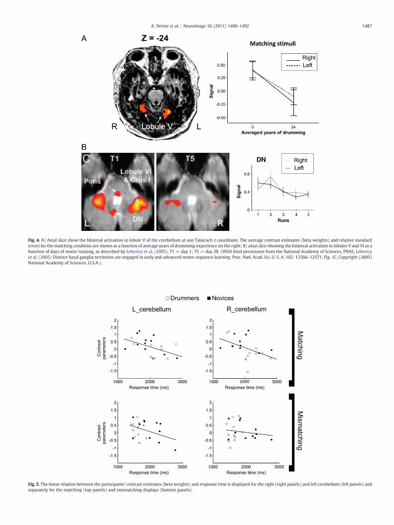
Fig. 4. A) Axial slice show the bilateral activation in lobule V of the cerebellum at one Talairach z coordinate. The average contrast estimates (beta weights) and relative standarderrors for thematching condition are shown as a function of average years of drumming experience on the right; B) axial slice showing the bilateral activation in lobules V and VI as afunction of days of motor training, as described by Lehericy et al. (2005). T1 = day 1; T5 = day 28. (With kind permission from the National Academy of Sciences, PNAS, Lehericyet al. (2005) Distinct basal ganglia territories are engaged in early and advanced motor sequence learning. Proc. Natl. Acad. Sci. U. S. A. 102: 12566–12571, Fig. 1C, Copyright (2005)National Academy of Sciences, U.S.A.).
Fig. 5. The linear relation between the participants' contrast estimates (beta weights) and response time is displayed for the right (right panels) and left cerebellum (left panels) andseparately for the matching (top panels) and mismatching displays (bottom panels).
1487K. Petrini et al. / NeuroImage 56 (2011) 1480–1492

Fig. 6. Experiment 2 fMRI group activation data: Clusters of activation for which the difference between the brain responses to the two types of stimuli (matched and mismatcheddrumming actions) varied across the two groups of participants (drummers and novices). Axial slices show activation foci at four Talairach z coordinates. The average contrastestimates (beta weights) and relative standard errors are shown in separate diagrams for drummers and novices and in different colors for the matching (green) and mismatching(yellow) conditions. A) Right inferior parietal lobule; B) right inferior temporal gyrus; C) right middle frontal gyrus; D) right precentral gyrus. Note that in Experiment 2 themismatch between the drummer's movements and sound reflects the lack of correspondence between strike velocities and sound intensities, while the temporal synchrony betweenthe signals is maintained. *=pb0.05; **=pb0.01.
1488 K. Petrini et al. / NeuroImage 56 (2011) 1480–1492
of significant areas for themain effect of audiovisual covariation, and asignificant interaction between expertise and covariation in the rightIPL (inferior parietal lobule: Fig. 6A), right ITG (inferior temporalgyrus: Fig. 6B), right MFG (Fig. 6C), and right precentral gyrus(Fig. 6D) were observed. Examination of the contrast parameters inthese areas for the three left-handed and eight right-handed novicesindicated that handedness did not affect the found differences or lackof such between matching and mismatching stimuli within this group(Supplemental Fig. 2). The anatomical location and details of theactivated foci are listed in Table 2. To control for multiple com-parisons, each map was adjusted to an initial P value of b0.001
Table 2Experiment 2 clusters of activation from a 2×2 ANOVA with ‘expertise’ as a between-participants factor and ‘audiovisual covariation’ as a within-participants factor. Legend:BA — Brodmann's area; MFG = middle frontal gyrus; MOG = middle occipital gyrus;IFG = inferior frontal gyrus; SFG = superior frontal gyrus; STG = superior temporalgyrus; IPL = inferior parietal lobule; ITG = inferior temporal gyrus.
Anatomicalregion
Hemisphere Talairachcoordinate(x,y,z)
Numberof voxels
Effect sizea BA
F(1, 20) P
Audiovisual covariation (matchingNmismatching)MOG Right 45, −76, 3 2294 18.69 0.0005 19Cerebellum Right 33, −71, −19 459 17.27 0.0005Lingual Gyrus Right 34, −60, 9 849 16.56 0.0006 19Cerebellum Right 31, −53, −20 462 17.35 0.0005Cerebellum Right 35, −36, −21 337 16.92 0.0006PosteriorCingulate
Left −3, −62, 14 7084 19.43 0.0004 23
Cerebellum Left −23, −72, −24 1985 17.21 0.0005
Audiovisual covariation (mismatchingNmatching)Insula Left −43, 13, 3 6083 20.52 0.0003 13STG Left −53, −43, 16 1985 19.12 0.0004 13IFG Right 48, 21, 18 3695 18.01 0.0004 45IFG Right 34, 21, −9 1047 21.59 0.0003 47SFG Left 0, 16, 51 4802 21.50 0.0002 8
Expertise×CovariationIPL Right 67, −33, 33 496 19.94 0.0003 40ITG Right 60, −50, −13 633 16.37 0.0006 20MFG Right 49, 11, 39 334 16.64 0.0006 8PrecentralGyrus
Right 44, −4, 50 275 18.24 0.0004 6
a Effect size = average F value for all voxels in the ROI.
(uncorrected) and then submitted to a volume-based cluster-threshold algorithm yielding a new map thresholded at Pb0.05(corrected) with a minimum cluster threshold. The minimum clusterthreshold that yielded a cluster-level false-positive rate of 5%was k=3, 77 mm3 for the map assessed for expertise×audiovisualcovariation, and k=4, 82 mm3 for the map assessed for audiovisualcovariation (Goebel et al., 2006).
The effect of interaction, similarly to Experiment 1, was due to areduction in activity in these areas for the drummers, but not forthe novices, when viewing the audiovisual matching displays (Fig. 4in green). We did not find any significant correlation betweenthe brain activity (betas weights) for each experimental conditions(matching, mismatching) and the years of drumming experiencein either right IPL, right ITG, right MFG, or right precentral gyrus.
Discussion
Here we used novel audiovisual biological motion stimuli toinvestigate how action expertise modulates processing of matchingand mismatching audiovisual actions. Although the effect of actionexpertise has been previously studied by using dance (Calvo-Merinoet al., 2005; 2006) and music performance (Haslinger et al., 2005;Hodges et al., 2005; Musacchia et al., 2007; Lahav et al., 2007; Vogtet al., 2007), the way perceptual and motor expertise alters audio-visual processes is still poorly understood. Thus in Experiments 1and 2 we examined the role of perceptual and motor expertise whenprocessing audiovisual human actions.
Experiment 1
The results of Experiment 1 show that the brain's responses toaudiovisually matching (synchronous) and mismatching (asynchro-nous) stimuli varied with action expertise. Specifically, we found areduction of activity in the drummers group when viewing matchingdisplays bilaterally in the lobule V of the cerebellum and left para-hippocampal gyrus. A reduction in neural activity in response toaudiovisual training has recently been reported (Powers et al., 2010)and can be related to their earlier findings of increased sensitivity toaudiovisual asynchrony (Powers et al., 2009). Since in our previousbehavioral studies we repeatedly found that drummers were muchmore sensitive to audiovisual asynchrony than novices (Petrini et al.,

1489K. Petrini et al. / NeuroImage 56 (2011) 1480–1492
2009a,b, 2010), we believe that the reduction in brain activationfor drummers when viewing over-learned drumming actions mayreflect this acquired higher sensitivity.
CerebellumThe bilateral activation of the cerebellum was mainly found at the
level of the primary fissure (hemisphere lobule V; see Schmahmannet al., 1999, for a detailed atlas of the cerebellum) and was causedby a reduction in activity in drummers in these areas when viewingthe matched audiovisual displays. An area very similar in peakcoordinates (Talairach: x=−20, y=−56, z=−28; x=−16, y=−54, z=−20) to our left cerebellum was found in a PET study bySadato et al. (1997) when comparing either tapping movements orbimanual abduction–adduction movements of the index finger to therest condition (where no movements were performed by partici-pants). A more recent fMRI study by Lehericy et al. (2005) showedthat a motor sequence learning task decreased brain activationbilaterally in lobules V and VI of the cerebellum asmovement learningprogressed (Flament et al., 1996; Doyon et al., 2002; Imamizu et al.,2000). Finally, Buccino et al. (2004) reported bilateral activationof the cerebellum, at very similar Talairach coordinates to ours,when musical novices imitated observed chords on guitar frets, andwhen they played chords of their choice. Our results thus agree withprevious findings implicating lobule V of the cerebellum as one of theareas pertaining to a brain network that subtends action-soundrepresentation and learning of new patterns of movement (Lehericyet al., 2005; Buccino et al., 2004). Interestingly, drummers were foundto be slower than novices when judging the matching displays, andthis behavioral measure was found to correlate with the differencein brain activity shown by the two groups. Thus, a reduction in brainactivity of the cerebellum might affect the timing of the motorresponse. An alternative explanation by which drummers wereslower because they found the task more difficult than novices isnot plausible, as demonstrated by the behavioral data during the scanand also by the behavioral data obtained from previous behavioralstudies (e.g. Petrini et al., 2009a).
Additionally, our results support the idea of the cerebellum beingimportant for controlling motor timing (Chen et al., 2006; Zatorreet al., 2007) and for computing predictive models of movementthat would include movement timing (Bastian, 2006; Ohyama et al.,2003). Indeed, Jantzen et al. (2004) showed that an area of the rightcerebellum with peak activity (Talairach: x=28, y=−43, z=−28)resembling ours activated more for synchronized to sound tappingthan asynchronized to sound tapping. This result is in line with whatwe found for the novices group, which showed higher activationin lobule V of the cerebellum when viewing matching (synchrony)displays than when viewing mismatching (asynchrony) displays.
Parahippocampal gyrusBesides the cerebellum, a significant effect of expertise for viewing
matching and mismatching displays was found in the left parahippo-campal gyrus. Similarly to the cerebellum, the parahippocampal gyrusshowed a reduction in activity in drummers when viewing matchingdisplays, while no difference in activation was observed betweenmatching and mismatching displays in novices. The left parahippo-campal gyrus (BA 36) is known to exhibit interaction effects be-tween motor experience and self-ratings of ability to perform dancemovements (Cross et al., 2006), and when contrasting activationelicited by meaningful actions versus meaningless actions for theexplicit purpose of subsequent recognition (Decety et al., 1997).Neuropsychological studies indicate that lesions of the hippocampalregion impair the ability to rapidly acquire and store new informationabout facts and events (Squire and Knowlton, 1995; Ungerleider,1995). These data are supported by subsequent studies showingthat left parahippocampal gyrus activation correlates with thenumber of unrelated words recalled (Alkire et al., 1998), and
decreases in patients with Alzheimer's disease compared withcontrols (Rombouts et al., 2000). Thus the reduction in activity inthe left parahippocampal gyrus for the drummers when presentedwith the matching displays might indicate a decrease in brain activityused by this group when recalling over-learned information aboutdrumming actions.
Experiment 2
The reduced level of activation we showed in Experiment 1 fordrummers when viewing matching actions was replicated inExperiment 2. However, the interaction effects indicate that thebrain's responses to audiovisual matching (naturally covarying) andmismatching (unnaturally covarying) varied with action expertise incompletely different areas within the right hemisphere. These areaswere the IPL (BA 40), ITG (BA 20), MFG (BA 8), and precentralgyrus (BA 6). The absence of the cerebellum from the areas found inthis second experiment adds further evidence to the idea that thecerebellummainly responds to discrepancies in temporal occurrence ofthe represented action and resulting sound, while other brain regionsrespond to discrepancies in temporal congruency. In other words,since themovements of the drummers and the drumming soundwerealways synchronous in this second experiment, the cerebellummaintained the same level of activation for both groups when viewingthe matching and mismatching displays.
An alternative explanation of why we found such different resultsin Experiments 1 and 2 might be that the task in Experiment 2 wasmore difficult and required the participation of frontal areas (e.g.right precentral gyrus and right MFG). This would explain also whyin Experiment 2, but not in Experiment 1, we found two groups ofactivated brain areas independent of expertise level, one group moreactivated for the matching than the mismatching condition, andanother more activated for the mismatching than the matchingcondition (Table 2). Furthermore, this idea is supported by the factthat the novices could not correctly classify matching and mismatch-ing displays in Experiment 2 (indeed their performance was at chancelevel), and by the fact that drummers' ability to correctly classify thetwo kinds of display decreased when going from Experiment 1 to 2(although this group accuracy remained significantly above chancelevel). However, this alternative explanation fails to explain why wedid not also find a difference across groups in the cerebellum andparahippocampal gyrus when comparing matching and mismatchingcondition, if the only difference between Experiments 1 and 2 was intask difficulty.
Inferior parietal gyrus, inferior temporal gyrus, medial frontal gyrus andprecentral gyrus
Similar areas to our IPL, ITG, and precentral gyrus were found toactivate more for observation of meaningless versus meaningfulactions in the study of Decety et al. (1997), while the MFG activatedfor the opposite contrast. All or some of these four areas are active inaudiomotor (Bangert et al., 2006; Baumann et al., 2007; Lahav et al.,2007), visuomotor (Grèzes et al., 1999, 2003; Perani et al., 2001;Gallagher and Frith, 2004; Buccino et al., 2004) and audiovisualrepresentation studies (Haslinger et al., 2005). Hence, we regard theseregions as being crucial for action observation/listening and imitation,as well as for action expertise.
The activation we observed in the right precentral gyrus includedboth the premotor ventral (PMv) and premotor dorsal (PMd) cortices,if we follow the convention proposed by Rizzolatti et al. (2002) thatin humans the border between the ventral and the dorsal premotorcortex lies at the level of the upper border of the frontal eye field,approximately at Talairach coordinate Z=50. A similar cluster ofactivation at the border between PMv and PMd has been also found byBuccino et al. (2004) during observation and imitation of guitarchords. While PMv appears to be involved in direct visuomotor
1490 K. Petrini et al. / NeuroImage 56 (2011) 1480–1492
transformations, PMd appears to be involved in indirect transforma-tions (Hoshi and Tanji, 2006; Zatorre et al., 2007). Direct transforma-tions from sound to motor actions are very relevant for musicperformance and have been shown to engage PMv (Bangert et al.,2006; Baumann et al., 2007; Lahav et al., 2007), indicating that thisportion of premotor cortex may activate when hearing a sound forwhich one has an acquired model in his/her own repertoire. On theother hand, PMd may have a more indirect role in sensory-motortransformation by retrieving and integrating sensory information toplan and execute an action (Hoshi and Tanji, 2004, 2006; Zatorre et al.,2007). In our experiment, both the activation of an acquired motorprogram and the integration of different sensory cues linked to therepresented actions may have been essential for judging correctcorrespondence between drummers' movements and sound infor-mation, with consequent activation of both portions of the premotorcortex. Furthermore, the idea that PMd is involved in higher orderaspects of movements, such as when increasing metrical saliency(Chen et al., 2006), and when increasing rhythmic complexity ofmovements (Chen et al., 2008), would explain why this area wasaffected by expertise level only in Experiment 2, where a morecomplex feature of the drumming action-sound representation wasmanipulated. Music, much more than visual stimuli, has a remarkableability to drive rhythmic, metrically organized motor behavior(Zatorre et al., 2007). In Experiment 1, the sound was a selection ofthree repeated cycles of drumming swing groove (nine impacts) witha regular accent on the second beat, whereas in Experiment 2 thesound (nine impacts with the same impact times of those inExperiment 1) did not contain any regular rhythmic pattern becausethe occurrence of the accented beat was randomized. The drummer'svisually presented movement, in contrast, maintained always aregular swing groove rhythmic pattern with the accent on the secondbeat, even when desynchronized from the sound in Experiment 1(although the starting beat and final beat were different from thoseof the synchronized condition). When considering the action-soundrepresentation, the mismatching condition in Experiment 2 wasmetrically more complex, thus possibly requiring the involvement ofPMd.
Finally, similar areas to our right PMv (BA 6) and right IPL/supramarginal gyrus (BA 40) are known to activate when contrastingobserved piano playing with and without sound with a rest condi-tion (Haslinger et al., 2005). While PMv may be important for therecovering of acquired motor programs, the inferior parietal cortexmay be important for cross-modal processing and integration ofaudiovisual information (e.g. Calvert et al., 2000, 2001; Calvert, 2001).The fact that in Haslinger et al. (2005) the movements werealways synchronized with the sound would support the participationof the IPL/supramarginal gyrus in audiovisual integration of observedactions only as long as the temporal co-occurrence of the signals ismaintained, as was the case in our Experiment 2, where bothmatching andmismatching displayswere audiovisually synchronized.
LimitationsAs indicated in theMaterials andmethods and Discussion sections,
although we did match the level of perceived synchrony andasynchrony for each individual prior to Experiment 1, we did notmatch the level of perceived congruency and incongruency for eachindividual prior to Experiment 2. This introduces some interpreta-tional ambiguities for the results of Experiment 2, which could havebeen a consequence of differences in task difficulty between the twogroups as much as differences in action expertise. Our design cannotdisentangle this possibility from our main interpretation of Experi-ment 2, and further experiments will be needed to draw strongconclusions on this point. Nevertheless the consistency of findingsbetween Experiments 1 and 2 showing a reduction in brain activityfor drummers and not for novices for matching displays is verycompelling, especially when considering that drummers were really
good in discriminating the matching and mismatching displaysin both experiments (Experiment 1: 92% correct; Experiment 2: 80%correct). A further study limitation concerns the relatively smallnumber of drummers and novices included. We had the potentialto enroll more participants than the number we reported, but foundthat gender imbalance would be a problem in data interpretation. Wechose to study expert subjects from one gender who had decadesof experience (mean 24 years). Hence, our sample number in thishomogeneous expert group was 11, to which we additionally age-matched 11 novice controls. Indeed, finding agematched novices withminimal musical experience posed an additional challenge. Futurestudies comparing a greater number of musicians and non-musicianswould strengthen our conclusions. These interpretational limitations,however, do not diminish the novel finding reported here, that brainactivation in action-sound representation areas is reduced by long-term acquired familiarity with audiovisual represented action.
Conclusion
The results from both experiments provide converging evidencethat expertise with a certain audiovisual action reduces activation inbrain areas crucial for action-sound representation and audiovisualintegration. Because both temporal synchrony and temporal congru-ency of the produced sound may be easily predicted from theobserved movements using acquired internal models of that action, areduction in activation may result as a consequence of perceptual andmotor expertise (McKiernan et al., 2003; Parsons et al., 2005).However, which network of areas will reduce their activation inresponse to over-learned and familiar audiovisual actions will dependon the level of stimulus complexity and on the temporal correspon-dence between the visual action and sound information. Our findingsindicate that there are two separate networks processing the soundand sight of others' actions: one network (including cerebellum andparahippocampal gyrus) would be responsible for processing thesensory synchrony of the auditory and visual information, whileanother (including fronto-temporal–parietal regions) would beresponsible for processing the sensory congruency of the two sensoryinformation. This is a novel finding that underlines the complexity offunctional reorganization dictated by perceptual and motor expertise.
Several neuroimaging studies have observed that musicians showlower levels of activity in motor regions than non-musicians duringthe performance of simple motor tasks (Hund-Georgiadis and VonCramon, 1999; Jäncke et al., 2000; Koeneke et al., 2004), and here weshow that this is also the case when musicians are only observing andnot performing an audiovisual action. Even though it is still not clearwhat this reduction in activity entails, we believe that it may explainwhy musicians have been repeatedly found to outperform non-musicians in a variety of tasks that have little to do with music(Schmithorst and Holland, 2004; Brochard et al., 2004; Magne et al.,2006; Musacchia et al., 2007). Indeed, if the brain, through thisreduction in activity, is able to conserve perceptual and motorresources, then these might be reallocated to other brain areas toincrease the musicians' efficiency in other everyday tasks, assuggested by some studies where the reduced activity found incertain brain areas of musicians was accompanied by an increase ofactivity in other areas (Hund-Georgiadis and Von Cramon, 1999;Schmithorst and Holland, 2004). Connectivity studies might give adefinitive answer to this point by examining whether the reduction inactivity of certain areas is followed by an increase in activity of otherbrain areas when musicians perform musical and non-musical tasks.
Finally, we still need to understand what are the mechanisms andwhat is the trajectory for developing a stable and maximal reductionin activity through musical practice, as this could have practicalimplications for the principles of musical training. The lack ofcorrelation between years of drumming experience and reduction inbrain activity in the present study suggests that after 13 years of

1491K. Petrini et al. / NeuroImage 56 (2011) 1480–1492
drumming there is no additional reduction in activation. However,from our findings we cannot derive when a stable and maximalreduction of activity is achieved, and future studies should examinewhen this happens by either comparing musicians with one to10 years of experience or by longitudinally studying the effect ofmusic practice on musicians' performance and brain activity. Thesekinds of investigations will also add valuable information about thedevelopment of brain behavior during learning.
Supplementarymaterials related to this article can be found onlineat doi:10.1016/j.neuroimage.2011.03.009.
Acknowledgments
Wewould like to acknowledge the support of the British Academy(LRG-42455) and the ESRC (RES-060-25-0010). We thank all thedrummers for their assistance, and in particular Robert Davis II andPaul Evans for their helpful discussions. We thank Mary Pettit foradministrative assistance.
References
Alkire, M.T., Haier, R., Fallon, J.H., Cahill, L., 1998. Hippocampal, but not amygdala,activity at encoding correlates with long-term, free recall of non-emotionalinformation. Proc. Natl Acad. Sci. 95, 14506–14510.
Bangert, M., Peschel, T., Schlaug, G., Rotte, M., Drescher, D., Hinrichs, H., Heinze, H.J.,Altenmuller, E., 2006. Shared networks for auditory and motor processing inprofessional pianists: evidence from fMRI conjunction. Neuroimage 30, 917–926.
Bastian, A.J., 2006. Learning to predict the future: the cerebellum adapts feedforwardmovement control. Curr. Opin. Neurobiol. 16, 645–649.
Baumann, S., Koeneke, S., Meyer, M., Lutz, K., Jancke, L., 2005. A network for sensory-motor integration: what happens in the auditory cortex during piano playingwithout acoustic feedback? Ann. NY Acad. Sci. 1060, 186–188.
Baumann, S., Koeneke, S., Schmidt, C.F., Meyer, M., Lutz, K., Jancke, L., 2007. A networkfor audio-motor coordination in skilled pianists and nonmusicians. Brain Res. 1161,65–78.
Brainard, D.H., 1997. The psychophysics toolbox. Spat. Vis. 10, 433–436.Brochard, R., Dufour, A., Després, O., 2004. Effect of musical expertise on visuospatial
abilities: evidence from reaction times and mental imagery. Brain Cogn. 54,103–109.
Brooks, A., van der Zwan, R., Billard, A., Petreska, B., Clarke, S., Blanke, O., 2007. Auditorymotion affects visual biological motion processing. Neuropsychologia 45, 523–530.
Buccino, G., Binkofski, F., Fink, G.R., Fadiga, L., Fogassi, L., Gallese, V., Seitz, R.J., Zilles, K.,Rizzolatti, G., Freund, H.J., 2001. Action observation activates premotor and parietalareas in a somatotopic manner: an fMRI study. Eur. J. Neurosci. 13, 400–404.
Buccino, G., Vogt, S., Ritzl, A., Fink, G.R., Zilles, K., Freund, H.J., Rizzolatti, G., 2004. Neuralcircuits underlying imitation learning of hand actions: an event-related fMRI study.Neuron 42, 323–334.
Calvert, G.A., 2001. Crossmodal processing in the human brain: insights from functionalneuroimaging studies. Cereb. Cortex 11, 1110–1123.
Calvert, G.A., Campbell, R., Brammer, M.J., 2000. Evidence from functional magneticresonance imaging of crossmodal binding in the human heteromodal cortex. Curr.Biol. 10, 649–657.
Calvert, G.A., Hansen, P.C., Iversen, S.D., Brammer, M.J., 2001. Detection of multisensoryintegration sites by application of electrophysiological criteria to the BOLDresponse. Neuroimage 14, 427–438.
Calvo-Merino, B., Glaser, D.E., Grèzes, J., Passingham, R.E., Haggard, P., 2005. Actionobservation and acquired motor skills: an fMRI study with expert dancers. Cereb.Cortex 15, 1243–1249.
Calvo-Merino, B., Grèzes, J., Glaser, D.E., Passingham, R.E., Haggard, P., 2006. Seeing ordoing? Influence of visual andmotor familiarity in action observation. Curr. Biol. 16,1905–1910.
Chen, J.L., Zatorre, R.J., Penhune, V.B., 2006. Interactions between auditory and dorsalpremotor cortex during synchronization to musical rhythms. Neuroimage 32,1771–1781.
Chen, J.L., Penhune, V.B., Zatorre, R.J., 2008. Listening to musical rhythms recruits motorregions of the brain. Cereb. Cortex 18, 2844–2854.
Cross, E.S., Hamilton, A.F.D.C., Grafton, S.T., 2006. Building a motor simulation de novo:observation of dance by dancers. Neuroimage 31, 1257–1267.
Dahl, S., 2004. Playing the accent — comparing striking velocity and timing in anostinato rhythm performed by four drummers. Acta Acustica United Acustica 90,762–776.
Decety, J., Grèzes, J., 1999. Neural mechanisms subserving the perception of humanactions. Trends Cogn. Sci. 3, 172–178.
Decety, J., Grèzes, J., Costes, N., Perani, D., Jeannerod, M., Procyk, E., Grassi, F., Fazio, F.,1997. Brain activity during observation of actions. Influence of action content andsubject's strategy. Brain 120, 1763–1777.
Doyon, J., Song, A.W., Karni, A., Lalonde, F., Adams, M.M., Ungerleider, L.G., 2002.Experience-dependent changes in cerebellar contributions to motor sequencelearning. Proc. Natl Acad. Sci. U.S.A. 99, 1017–1022.
Flament, D., Ellermann, J.M., Kim, S.G., Ugurbil, K., Ebner, T.J., 1996. Functional magneticresonance imaging of cerebellar activation during the learning of a visuomotordissociation task. Hum. Brain Mapp. 4, 210–226.
Fontana, F., Rocchesso, D., Avanzini, F., 2004. Computation of nonlinear filter networkscontaining delay-free paths. 7th International Conference on Digital Audio Effects(DAFX-04), pp. 113–118. Naples, Italy.
Forman, S.D., Cohen, J.D., Fitzgerald, M., Eddy, W.F., Mintun, M.A., Noll, D.C., 1995.Improved assessment of significant activation in functional magnetic resonanceimaging (fMRI): use of a clustersize threshold. Magn. Reson. Med. 33, 636–647.
Gallagher, H.L., Frith, C.D., 2004. Dissociable neural pathways for the perception andrecognition of expressive and instrumental gestures. Neuropsychologia 42,1725–1736.
Gallese, V., Fadiga, L., Fogassi, L., Rizzolatti, G., 1996. Action recognition in the premotorcortex. Brain 119, 593–609.
Goebel, R., Esposito, F., Formisano, E., 2006. Analysis of functional image analysiscontest (FIAC) data with brainvoyager QX: from single-subject to cortically alignedgroup general linear model analysis and self-organizing group independentcomponent analysis. Hum. Brain Mapp. 27, 392–401.
Grèzes, J., Costes, N., Decety, J., 1999. The effects of learning and intention on the neuralnetwork involved in the perception of meaningless actions. Brain 122, 1875–1887.
Grèzes, J., Armony, J.L., Rowe, J., Passingham, R.E., 2003. Activations related to ‘mirror’and ‘canonical’ neurones in the human brain: an fMRI study. Neuroimage 18,928–937.
Haslinger, B., Erhard, P., Altenmuller, E., Schroeder, U., Boecker, H., Ceballos-Baumann,A.O., 2005. Transmodal sensorimotor networks during action observation inprofessional pianists. J. Cogn. Neurosci. 17, 282–293.
Hodges, D.A., Hairston, W.D., Burdette, J.H., 2005. Aspects of multisensory perception:the integration of visual and auditory information in musical experiences. Ann. NYAcad. Sci. 1060, 175–185.
Hoshi, E., Tanji, J., 2004. Functional specialization in dorsal and ventral premotor areas.Prog. Brain Res. 143, 507–511.
Hoshi, E., Tanji, J., 2006. Differential involvement of neurons in the dorsal and ventralpremotor cortex during processing of visual signals for action planning.J. Neurophysiol. 95, 3596–3616.
Hund-Georgiadis, M., Von Cramon, D.Y., 1999. Motor-learning-related changes in pianoplayers and nonmusicians revealed by functional magnetic-resonance signals. Exp.Brain Res. 125, 417–425.
Iacoboni, M., 2005. Neural mechanisms of imitation. Curr. Opin. Neurobiol. 15, 632–637.Imamizu, H., Miyauchi, S., Tamada, T., Sasaki, Y., Takino, R., Putz, B., Yoshioka, T.,
Kawato, M., 2000. Human cerebellar activity reflecting an acquired internal modelof a new tool. Nature 403, 192–195.
Jäncke, L., Shah, N.J., Peters, M., 2000. Cortical activations in primary and secondarymotor areas for complex bimanual movements in professional pianists. Brain Res.Cogn. Brain Res. 10, 177–183.
Jantzen, K.J., Steinberg, F.L., Kelso, J.A.S., 2004. Brain networks underlying human timingbehavior are influenced by prior context. Proc. Natl Acad. Sci. 101, 6815–6820.
Johansson, G., 1973. Visual perception of biological motion and model for its analysis.Percept. Psychophys. 14, 201–211.
Keysers, C., Kohler, E., Umilta, M.A., Nanetti, L., Fogassi, L., Gallese, V., 2003. Audiovisualmirror neurons and action recognition. Exp. Brain Res. 153, 628–636.
Klin, A., Lin, D., Gorrindo, P., Ramsay, G., Jones, W., 2009. Two-year-olds with autismorient to non-social contingencies rather than biological motion. Nature 459(7244), 257–261.
Koeneke, S., Lutz, K., Wustenberg, T., Jäncke, L., 2004. Long-term training affectscerebellar processing in skilled keyboard players. Neuroreport 15, 1279–1282.
Kohler, E., Keysers, C., Umilta, M.A., Fogassi, L., Gallese, V., Rizzolatti, G., 2002. Hearingsounds, understanding actions: action representation in mirror neurons. Science297, 846–848.
Lahav, A., Saltzman, E., Schlaug, G., 2007. Action representation of sound: audiomotorrecognition network while listening to newly acquired actions. J. Neurosci. 27,308–314.
Lehericy, S., Benali, H., Van de Moortele, P.F., Pelegrini-Isaac, M., Waechter, T., Ugerbil,K., Doyon, J., 2005. Distinct basal ganglia territories are engaged in early andadvanced motor sequence learning. Proc. Natl Acad. Sci. U.S.A. 102, 12566–12571.
Magne, C., Schon, D., Besson, M., 2006. Musician children detect pitch violations in bothmusic and language better than non-musician children: behavioral and electro-physiological approaches. J. Cogn. Neurosci. 18, 199–211.
McKiernan, K.A., Kaufman, J.N., Kucera-Thompson, J., Binder, J.R., 2003. A parametricmanipulation of factors affecting task-induced deactivation in functional neuroi-maging. J. Cogn. Neurosci. 15, 394–408.
Musacchia, G., Sams, M., Skoe, E., Kraus, N., 2007. Musicians have enhanced subcorticalauditory and audiovisual processing of speech and music. Proc. Natl Acad. Sci. 104,15894–15898.
Nishitani, N., Hari, R., 2000. Temporal dynamics of cortical representation for action.Proc. Natl Acad. Sci. U.S.A. 97, 913–918.
Ohyama, T., Nores,W.L., Murphy, M., Mauk, M.D., 2003.What the cerebellum computes.Trends Neurosci. 26, 222–227.
Oldfield, R.C., 1971. Assessment and analysis of handedness — Edinburgh inventory.Neuropsychologia 9, 97–113.
Parsons, L.M., Sergent, J., Hodges, D.A., Fox, P.T., 2005. The brain basis of pianoperformance. Neuropsychologia 43, 199–215.
Pelli, D.G., 1997. The VideoToolbox software for visual psychophysics: transformingnumbers into movies. Spat. Vis. 10, 437–442.
Perani, D., Fazio, F., Borghese, N.A., Tettamanti, M., Ferrari, S., Decety, J., Gilardi, M.C.,2001. Different brain correlates for watching real and virtual hand actions.Neuroimage 14, 749–758.

1492 K. Petrini et al. / NeuroImage 56 (2011) 1480–1492
Petrini, K., Dahl, S., Rocchesso, D., Waadeland, D.H., Avanzini, F., Puce, A., Pollick, F.,2009a. Multisensory integration of drumming actions: musical expertise affectsperceived audiovisual asynchrony. Exp. Brain Res. 198, 339–352.
Petrini, K., Russell, M., Pollick, F., 2009b. When knowing can replace seeing inaudiovisual integration of actions. Cognition 110, 432–439.
Petrini, K.,Holt, P.S., Pollick, F., 2010. Expertisewithmultisensoryeventseliminates the effectof biological motion rotation on audiovisual synchrony perception. J. Vis. 10 (5), 1–14.
Powers, A.R., Hillock, A.R., Wallace, M.T., 2009. Perceptual training narrows thetemporal window of multisensory binding. J. Neurosci. 29, 12265–12274.
Powers, A.R., Hevey, M., Wallace, M.T., 2010. The neural correlates of multisensoryperceptual training. Abstract in IMRF2010 Conference Proceedings, Liverpool, UK.
Rizzolatti, G., Fadiga, L., Gallese, V., Fogassi, L., 1996. Premotor cortex and therecognition of motor actions. Brain Res. Cogn. Brain Res. 3, 131–141.
Rizzolatti, G., Fogassi, L., Gallese, V., 2002. Motor and cognitive functions of the ventralpremotor cortex. Curr. Opin. Neurobiol. 12, 149–154.
Rombouts, S.A.R.B., Barkhof, F., Veltman, D.J., Machielsen, W.C.M., Witter, M.P., Bierlaagh,M.A., Lazeron, R.H.C., Valk, J., Scheltens, P., 2000. FunctionalMR imaging inAlzheimer'sdisease during memory encoding. AJNR Am. J. Neuroradiol. 21, 1869–1875.
Sadato, N., Yonekura, Y., Waki, A., Yamada, H., Ishii, Y., 1997. Role of the supplementarymotor area and the right premotor cortex in the coordination of bimanual fingermovements. J. Neurosci. 17, 9667–9674.
Saygin, A.P., 2007. Superior temporal and premotor brain areas necessary for biologicalmotion perception. Brain 130, 2452–2461.
Saygin, A.P., Wilson, S.M., Hagler Jr., D.J., Bates, E., Sereno, M.I., 2004. Point-light biologicalmotion perception activates human premotor cortex. J. Neurosci. 24, 6181–6188.
Schmahmann, J.D., Doyon, J., McDonald, D., Holmes, C., Lavoie, K., Hurwitz, A.S., Kabani,N., Toga, A., Evans, A., Petrides, M., 1999. Three-dimensional MRI atlas of the humancerebellum in proportional stereotaxic space. Neuroimage 10, 233–260.
Schmithorst, V.J., Holland, S.K., 2004. The effect of musical training on the neuralcorrelates of math processing: a functional magnetic resonance imaging study inhumans. Neurosci. Lett. 354, 193–196.
Squire, L., Knowlton, B., 1995. Memory, hippocampus and brain systems. In: Gazzaniga,M. (Ed.), The Cognitive Neurosciences. MIT Press, Cambridge, MA, pp. 825–838.
Stewart, L., Henson, R., Kampe, K., Walsh, V., Turner, R., Frith, U., 2003. Brain changesafter learning to read and play music. Neuroimage 20, 71–83.
Ungerleider, L.G., 1995. Functional brain imaging studies of cortical mechanisms formemory. Science 270, 769–775.
Vogt, S., Buccino, G., Wohlschlager, A.M., Canessa, N., Shah, N.J., Zilles, K., Eickhoff, S.B.,Freund, H.J., Rizzolatti, G., Fink, G.R., 2007. Prefrontal involvement in imitationlearning of hand actions: effects of practice and expertise. Neuroimage 37,1371–1383.
Waadeland, C.H., 2006. Strategies in empirical studies of swing groove. StudiaMusicologica Norvegica 32, 169–191.
Watson, A.B., Hu, J., 1999. ShowTime: a QuickTime-based infrastructure for visionresearch displays. Perception 28 (ECVP Abstract Supplement): 45b. .
Wilson, S.M., Saygin, A.P., Sereno, M.I., Iacoboni, M., 2004. Listening to speech activatesmotor areas involved in speech production. Nat. Neurosci. 7, 701–702.
Zatorre, R.J., Chen, J.L., Penhune, V.B., 2007. When the brain plays music: auditory–motor interactions in music perception and production. Nat. Rev. Neurosci. 8,547–558.


![Unilateral Focused Ultrasound-Induced Blood-Brain Barrier Opening Reduces … · 2019-07-21 · Specifically in Alzheimer’s disease, antibodies against Αβ [31,35] have been mediated](https://static.fdocuments.in/doc/165x107/5f865cb573d58477ee3f1ad2/unilateral-focused-ultrasound-induced-blood-brain-barrier-opening-reduces-2019-07-21.jpg)


