Acta Biomaterialia -...
15
Research on an Mg–Zn alloy as a degradable biomaterial Shaoxiang Zhang a , Xiaonong Zhang a,c, * , Changli Zhao a , Jianan Li a , Yang Song a , Chaoying Xie a , Hairong Tao b , Yan Zhang b , Yaohua He b , Yao Jiang b , Yujun Bian c a State Key Laboratory of Metal Matrix Composites, School of Materials Science and Engineering, Shanghai Jiao Tong University, Shanghai 200240, People’s Republic of China b Orthopaedic Department of the 6th People’s Hospital, Shanghai Jiao Tong University, Shanghai 200233, People’s Republic of China c Shanghai Origin Material and Medical Technology Co. Ltd., Shanghai 200240, People’s Republic of China article info Article history: Received 17 December 2008 Received in revised form 1 June 2009 Accepted 11 June 2009 Available online 21 June 2009 Keywords: Mg–Zn alloy Degradation Mechanical properties Cytotoxicity In vivo biocompatibility abstract In this study a binary Mg–Zn magnesium alloy was researched as a degradable biomedical material. An Mg–Zn alloy fabricated with high-purity raw materials and using a clean melting process had very low levels of impurities. After solid solution treatment and hot working the grain size of the Mg–Zn alloy was finer and a uniform single phase was gained. The mechanical properties of this Mg–Zn alloy were suitable for implant applications, i.e. the tensile strength and elongation achieved were 279.5 MPa and 18.8%, respectively. The results of in vitro degradation experiments including electrochemical measurements and immer- sion tests revealed that the zinc could elevate the corrosion potential of Mg in simulated body fluid (SBF) and reduce the degradation rate. The corrosion products on the surface of Mg–Zn were hydroxyapatite (HA) and other Mg/Ca phosphates in SBF. In addition, the influence caused by in vitro degradation on mechanical properties was studied, and the results showed that the bending strength of Mg–Zn alloy dropped sharply in the earlier stage of degradation, while smoothly during the later period. The in vitro cytotoxicity of Mg–Zn was examined. The result 0–1 grade revealed that the Mg–Zn alloy was harmless to L-929 cells. For in vivo experiments, Mg–Zn rods were implanted into the femoral shaft of rabbits. The radiographs illustrated that the magnesium alloy could be gradually absorbed in vivo at about 2.32 mm/yr degradation rate obtained by weight loss method. Hematoxylin and eosin (HE) stained section around Mg–Zn rods suggested that there were newly formed bone surrounding the implant. HE stained tissue (containing heart, liver, kidney and spleen tissues) and the biochemical measure- ments, including serum magnesium, serum creatinine (CREA), blood urea nitrogen (BUN), glutamic-pyru- vic transaminase (GPT) and creatine kinase (CK) proved that the in vivo degradation of Mg–Zn did not harm the important organs. Moreover, no adverse effects of hydrogen generated by degradation had been observed and also no negative effects caused by the release of zinc were detected. These results suggested that the novel Mg–Zn binary alloy had good biocompatibility in vivo. Ó 2009 Acta Materialia Inc. Published by Elsevier Ltd. All rights reserved. 1. Introduction Due to their very low corrosion potential, magnesium and its alloys are susceptible to dissolution in aqueous solutions, particu- larly in those containing chloride ion electrolytes [1,2]. Making use of the corrodible properties of magnesium alloys, in recent years biomaterials engineers have become interested in developing mag- nesium-based biodegradable medical devices [3–18]. As shown in Table 1, various magnesium alloys have been re- searched as biodegradable materials and some of them have shown biocompatibility. For example, compared with poly-96L/ 4D-lactide, the magnesium alloys AZ31 and AZ91 enhanced the osteogenesis response and increase newly formed bone [4]. Heub- lein et al. reported that the alloy AE21 gradually degraded in pig ar- tery and might be a promising cardiovascular implant material [3]. A more encouraging indication is that a biodegradable magnesium stent has been used in clinical experiments [18]. However, it should be noted that most of the reported biomed- ical magnesium alloys contain aluminum and/or rare earth (RE) elements. It is well known that Al is harmful to neurons [19] and osteoblasts [20] and also associated with dementia and Alzhei- mer’s disease [19]. The administration of RE (Pr, Ce, Y, etc.) could lead to hepatotoxicity [21]. Excessive yttrium ions (Y 3+ ) have been shown to change the expression of some rat genes and to have ad- verse effects on DNA transcription factors [22]. Consequently, Al and RE are unsuitable alloying elements for biomedical magnesium materials, particularly when they are above certain levels. This has 1742-7061/$ - see front matter Ó 2009 Acta Materialia Inc. Published by Elsevier Ltd. All rights reserved. doi:10.1016/j.actbio.2009.06.028 * Corresponding author. Address: State Key Laboratory of Metal Matrix Compos- ites, School of Materials Science and Engineering, Shanghai Jiao Tong University, No. 800 DongChun Road, Shanghai 200240, People’s Republic of China. Tel./fax: +86 21 3420 2759. E-mail address: [email protected] (X. Zhang). Acta Biomaterialia 6 (2010) 626–640 Contents lists available at ScienceDirect Acta Biomaterialia journal homepage: www.elsevier.com/locate/actabiomat
Transcript of Acta Biomaterialia -...

Acta Biomaterialia 6 (2010) 626–640
Contents lists available at ScienceDirect
Acta Biomaterialia
journal homepage: www.elsevier .com/locate /ac tabiomat
Research on an Mg–Zn alloy as a degradable biomaterial
Shaoxiang Zhang a, Xiaonong Zhang a,c,*, Changli Zhao a, Jianan Li a, Yang Song a, Chaoying Xie a,Hairong Tao b, Yan Zhang b, Yaohua He b, Yao Jiang b, Yujun Bian c
a State Key Laboratory of Metal Matrix Composites, School of Materials Science and Engineering, Shanghai Jiao Tong University, Shanghai 200240, People’s Republic of Chinab Orthopaedic Department of the 6th People’s Hospital, Shanghai Jiao Tong University, Shanghai 200233, People’s Republic of Chinac Shanghai Origin Material and Medical Technology Co. Ltd., Shanghai 200240, People’s Republic of China
a r t i c l e i n f o a b s t r a c t
Article history:Received 17 December 2008Received in revised form 1 June 2009Accepted 11 June 2009Available online 21 June 2009
Keywords:Mg–Zn alloyDegradationMechanical propertiesCytotoxicityIn vivo biocompatibility
1742-7061/$ - see front matter � 2009 Acta Materialdoi:10.1016/j.actbio.2009.06.028
* Corresponding author. Address: State Key Laboratites, School of Materials Science and Engineering, Shan800 DongChun Road, Shanghai 200240, People’s Repu3420 2759.
E-mail address: [email protected] (X. Zhang).
In this study a binary Mg–Zn magnesium alloy was researched as a degradable biomedical material. AnMg–Zn alloy fabricated with high-purity raw materials and using a clean melting process had very lowlevels of impurities. After solid solution treatment and hot working the grain size of the Mg–Zn alloywas finer and a uniform single phase was gained. The mechanical properties of this Mg–Zn alloy weresuitable for implant applications, i.e. the tensile strength and elongation achieved were �279.5 MPaand 18.8%, respectively.
The results of in vitro degradation experiments including electrochemical measurements and immer-sion tests revealed that the zinc could elevate the corrosion potential of Mg in simulated body fluid (SBF)and reduce the degradation rate. The corrosion products on the surface of Mg–Zn were hydroxyapatite(HA) and other Mg/Ca phosphates in SBF. In addition, the influence caused by in vitro degradation onmechanical properties was studied, and the results showed that the bending strength of Mg–Zn alloydropped sharply in the earlier stage of degradation, while smoothly during the later period.
The in vitro cytotoxicity of Mg–Zn was examined. The result 0–1 grade revealed that the Mg–Zn alloywas harmless to L-929 cells. For in vivo experiments, Mg–Zn rods were implanted into the femoral shaftof rabbits. The radiographs illustrated that the magnesium alloy could be gradually absorbed in vivo atabout 2.32 mm/yr degradation rate obtained by weight loss method. Hematoxylin and eosin (HE) stainedsection around Mg–Zn rods suggested that there were newly formed bone surrounding the implant.
HE stained tissue (containing heart, liver, kidney and spleen tissues) and the biochemical measure-ments, including serum magnesium, serum creatinine (CREA), blood urea nitrogen (BUN), glutamic-pyru-vic transaminase (GPT) and creatine kinase (CK) proved that the in vivo degradation of Mg–Zn did notharm the important organs. Moreover, no adverse effects of hydrogen generated by degradation had beenobserved and also no negative effects caused by the release of zinc were detected. These results suggestedthat the novel Mg–Zn binary alloy had good biocompatibility in vivo.
� 2009 Acta Materialia Inc. Published by Elsevier Ltd. All rights reserved.
1. Introduction
Due to their very low corrosion potential, magnesium and itsalloys are susceptible to dissolution in aqueous solutions, particu-larly in those containing chloride ion electrolytes [1,2]. Making useof the corrodible properties of magnesium alloys, in recent yearsbiomaterials engineers have become interested in developing mag-nesium-based biodegradable medical devices [3–18].
As shown in Table 1, various magnesium alloys have been re-searched as biodegradable materials and some of them haveshown biocompatibility. For example, compared with poly-96L/
ia Inc. Published by Elsevier Ltd. A
ory of Metal Matrix Compos-ghai Jiao Tong University, No.blic of China. Tel./fax: +86 21
4D-lactide, the magnesium alloys AZ31 and AZ91 enhanced theosteogenesis response and increase newly formed bone [4]. Heub-lein et al. reported that the alloy AE21 gradually degraded in pig ar-tery and might be a promising cardiovascular implant material [3].A more encouraging indication is that a biodegradable magnesiumstent has been used in clinical experiments [18].
However, it should be noted that most of the reported biomed-ical magnesium alloys contain aluminum and/or rare earth (RE)elements. It is well known that Al is harmful to neurons [19] andosteoblasts [20] and also associated with dementia and Alzhei-mer’s disease [19]. The administration of RE (Pr, Ce, Y, etc.) couldlead to hepatotoxicity [21]. Excessive yttrium ions (Y3+) have beenshown to change the expression of some rat genes and to have ad-verse effects on DNA transcription factors [22]. Consequently, Aland RE are unsuitable alloying elements for biomedical magnesiummaterials, particularly when they are above certain levels. This has
ll rights reserved.

Table 1The nominal chemical compositions of Mg alloys researched as degradable biomaterials.
Alloy type Aluminum (wt.%) Rare earth element Zinc (wt.%) Other References
AE21 2 1 wt.% (Ce, Pr, Nd) [3]AZ31 3 1 [4]AZ91 9 1 [4,5,8,10]WE43 3 wt.% (Nd, Ce, Dy), 4 wt.% (Y) [4]LAE442 4 2 wt.% (Ce, La, Nd, Pr) 4 wt.% Li [4,5]AM60B 6 0.07 0.33 wt.% Mn [14]Mg–Ca <2 wt.% Ca [6,7,17]Mg–Zn–Y �0.36–1.54 wt.% Y 2 [11]Mg–Mn–Zn 1 1.2 wt.% Mn [16]
S. Zhang et al. / Acta Biomaterialia 6 (2010) 626–640 627
led to a demand for the development of a novel biodegradablemagnesium alloy without Al, RE or other harmful elements.
With the purpose of searching for suitable alloying elements forbiomedical magnesium alloys, Song [12] explored in vitro corro-sion rates of several magnesium alloys, pointing out that Ca, Mnand Zn could be appropriate candidates. Further investigationsdemonstrated that Mg–Ca [6] and Mg–Mn–Zn [16] alloys graduallydegraded within bone and had good biocompatibility both in vitroand in vivo. Moreover, the degradation process did not raise theserum Mg2+ level and no kidney disorders were observed.
Zinc is one of the most abundant nutritionally essential ele-ments in the human body [23], and has basic safety for biomedicalapplications. Furthermore, zinc can improve the corrosion resis-tance and mechanical properties of magnesium alloys. For exam-ple, the corrosion rate of magnesium could be reduced byincreasing the mass fraction of zinc in magnesium [24]. Moreover,zinc can effectively strengthen magnesium [12,25] through a solidsolution hardening mechanism [25].
According to the Mg–Zn binary phase diagram [26] the maximumsolubility of Zn in Mg is 6.2 wt.% (i.e. 2.5 at.%) at 325 �C. In this paper apatented Mg–6 wt.% Zn alloy (Mg–6Zn) [27] was developed with theaim of making a novel magnesium alloy with good biocompatibility,moderate degradation rate and good mechanical properties. Analy-sis and evaluation of the in vitro and in vivo degradation of this Mg–6Zn alloy are also presented in this paper.
2. Materials and methods
2.1. Materials
Magnesium alloys with a nominal composition of Mg–6 wt.% Znwere prepared using high-purity magnesium (P99.99%) and zinc(P99.9999%) ingots. Melting was carried out at �700–750 �C in ahigh-purity graphite crucible. After about 30 min holding and stir-ring, the melt was cast into a steel mold at about 700 �C. A protec-tive cover gas (99.99% purity argon) was employed throughout themelting and casting processes. The as cast ingots of Mg–6Zn alloywere solid solution treated at about 350 �C for 2 h, followed byquenching in water. Finally, the heat-treated alloy sample washot extruded at about 250 �C with an extrusion ratio of 8:1.
Disk samples for the in vitro degradation and cytotoxicityexperiments, having a diameter of 11.3 mm (so that the cross-sec-tion area was 1 cm2) and a height of 2.0 mm, were machined fromthe extruded Mg–6Zn rods. Rectangular samples with the dimen-sions 5 � 5 � 30 mm were made and used for three-point bendingtesting. Cylindrical rods with a diameter of 4.5 mm and a length of10 mm were used in the animal implant experiments. All sampleswere ground with SiC paper up to 1200 grit and polished with1 lm diamond paste, followed by ultrasonic cleaning in acetone,ethanol and distilled water. Before the in vitro cytotoxicity andin vivo degradation experiments all samples were sterilized with29 kGy of 60Co radiation.
2.2. Composition and microstructure characterization
The chemical compositions of the Mg–6Zn alloy was deter-mined by inductively coupled plasma-atomic emission spectrome-try (ICP-AES) (Iris Advantage 1000). The microstructures of the ascast, heat-treated and extruded samples were characterized byoptical microscopy. X-ray diffraction analysis (XRD) (RigakuD/MAX255) was used to examine the phase of the as cast, heat-treated and extruded Mg–6Zn.
2.3. Mechanical properties
Tension and compression tests were carried out with a MTS810.22 universal testing machine, according to ASTM E8-04 andASTM E9-89a (2000) [28,29]. The tensile samples had a gauge lengthof 35 mm and the compressive samples had diameter of 6 mm and aheight of 9 mm. A crosshead speed of 0.5 mm min�1 was used.
2.4. In vitro degradation tests
In order to evaluate the in vitro degradation properties, electro-chemical measurements and immersion tests were performed in asimulated body fluid (SBF), containing 6.800 g l�1 NaCl, 0.200 g l�1
CaCl2, 0.400 g l�1 KCl, 0.100 g l�1 MgSO4, 2.200 g l�1 NaHCO3,0.126 g l�1 Na2HPO4 and 0.026 g l�1 NaH2PO4 [30]. The pH valueof SBF was adjusted with HCl and NaOH solution to 7.44 and thetemperature was maintained at 37 ± 0.5 �C. Pure Mg (>99.99%)was also tested as a counterpart.
2.4.1. Electrochemical measurementsElectrochemical measurements were performed using a three
electrode system (PARSTAT� 2273). A saturated calomel electrode(SCE) and a platinum mesh were used as the reference and counterelectrodes, respectively. Potentiodynamic polarization curves weremeasured at a scan rate of 1 mV s�1. The corrosion rates of thesamples were calculated by extrapolating the polarization curveaccording to ASTM-G102-89 [31]. Electrochemical impedancespectroscopy (EIS) analysis was also performed at open circuit po-tential with a perturbing signal of 5 mV. The frequency variedfrom100 to 1 MHz. All of the EIS results were fitted and analyzedusing ZsimpWin 3.10 Echem software.
2.4.2. Immersion testsImmersion tests were carried out in conformation with ASTM-
G31-72 [32] (the ratio of surface area to solution volume was1 cm2:20 ml). Samples were removed after 3 and 30 days ofimmersion, rinsed with distilled water and dried at room temper-ature. Then the surface morphology after immersion was observedusing scanning electron microscopy (SEM) with an energy disper-sive spectrometer (EDS). XRD (Rigaku, D/MAX255) was used todetermine the phase of corrosion products on the Mg–6Zn surface.Lastly, the samples were cleaned with 180 g l�1 chromic acid to re-move the corrosion products and the degradation rates (in units of

628 S. Zhang et al. / Acta Biomaterialia 6 (2010) 626–640
mm year�1) were obtained according to ASTM-G31-72. The corro-sion rate is given by Eq. (1) [32]:
Corrosion rate ¼ ðK �WÞ � ðA� T � DÞ ð1Þ
where the coefficient K = 8.76 � 104, W is the weight loss (g), A isthe sample area exposed to solution (cm2), T is the exposure time(h) and D is the density of the material (g cm–3).
The pH value of the solution was also recorded during theimmersion tests (PHS-3C pH meter, Lei-ci, Shanghai).
2.5. In vitro loss of mechanical integrity
Magnesium alloyshave shown very good properties for orthopedicapplications [13]. However, the loss of mechanical integrity duringdegradation may inhibit clinical realization [33]. To evaluate the influ-ence of in vitro degradation on the mechanical properties rectangularsamples with the dimensions 5 � 5 � 30 mm were weighted to anaccuracy of 0.1 mg and then immersed in physiological saline [0.9%NaCl, Baxter Healthcare (Shanghai) Co. Ltd.]. The samples were takenout, cleaned with ethanol and distilled water, dried in warm air andweighed each day to monitor the mass change. The immersion testscontinued until the weight loss percentage [weight loss percent-age = (Wbefore immersion �Wafter immersion) � Wbefore immersion, where Wmeans weight] reached about 5%, 10% and 20%, respectively. Finallythree-point bending tests with a support span of 25 mm were carriedout with a MTS 810.22 universal testing machine to observe the influ-ence of weight loss on mechanical integrity. The morphology of thefracture surface was examined by SEM (JEOL JSM 6460).
2.6. Cytotoxicity assessments
L-929 cells were cultured in Dulbecco’s modified Eagle’s med-ium (DMEM) (Gibco), supplemented with 10% fetal bovine serum(FBS) in a humidified incubator at 95% relative humidity and 5%CO2 at 37 �C. Cytotoxicity was determined by indirect contact. Ex-tracts were prepared according to ISO 10993-5: 1999 [34]. Theextraction media were serially diluted to 50 and 10% concentrationafter 72 h incubation in a humidified atmosphere with 5% CO2 at37 �C. The DMEM medium acted as a negative control while DMEMmedium containing 0.64% phenol as a positive control. Cells wereincubated in 96-well flat bottomed cell culture plates at2.5 � 104 cells ml�1 medium in each well and incubated for 24 hto allow cell attachment. Then the medium was replaced by100 ll extraction medium. After incubation in a humidified atmo-sphere for 2, 4 and 7 days cell morphology was observed by opticalmicroscopy (Nikon ELWD 0.3 inverted microscope). 3-(4,5-Dimethylthiazol-2-yl)-2,5-diphenyltetrazolium bromide (MTT)(Sigma) was then dissolved in phosphate-buffered saline (PBS) ata concentration of 5 mg ml�1. After that 20 ll MTT was added toeach well and the samples were incubated for 4 h (37 �C, 5% CO2,95% relative humidity). Subsequently, 150 ml formazan solutionwas added to each well and optical density (OD) measurementswere conducted at 490 nm using a Wellscan MK3 spectrophotom-eter (Labsystem). The cell relative growth rate (RGR) was calcu-lated according to the following formula:
RGR ¼ ODtest=ODnegative � 100% ð2Þ
2.7. In vivo degradation and biocompatibility
2.7.1. SurgeryAnimal tests were approved by the Ethnics Committee of the
6th Hospital of Shanghai Jiao Tong University. The in vivo degrada-tion experiments were performed in the animal laboratory of the
hospital. A total of 12 adult New Zealand rabbits (6 females) witha body weight of �2.0–2.5 kg were used. The rabbits were ran-domly divided into two groups. In the experimental group oneMg–6Zn rod was implanted into the femoral shaft of each rabbit.In the control group each rabbit had a hole drilled in the femoralshaft, without any implantation. Before implantation a dose of30 mg kg�1 sodium pentobarbital (Shanghai Xinya pharmaceuticsLtd.) was administered by intravenous injection. The femoral re-gion was cleaned with physiological saline and 75% ethanol. Allanimals received a subcutaneous injection of penicillin as ananti-inflammatory.
2.7.2. Biochemical testsDuring the experiments 5 ml blood samples were taken from
the helix vessel of the rabbits before operation and at 3, 15, 21and 28 days after surgery. The blood biochemical tests, includingfor serum magnesium, serum creatinine (CREA), blood urea nitro-gen (BUN), glutamic-pyruvic transaminase (GPT) and creatinekinase (CK), were performed with a Hitachi 7600-020 automaticbiochemical analyzer.
2.7.3. Radiographic and histological evaluationRadiographs were used to observe the in vivo degradation
process during the experiments. Six and 18 weeks post-implanta-tion bone samples around the Mg–6Zn rod were fixed in 4%formaldehyde and then embedded in methyl methacrylate. Histo-logical evaluation was performed on hematoxylin and eosin (HE)stained sections. The HE stained method was also used to ob-serve the tissues around the hydrogen gas bubble 3 weeks afterimplantation.
In addition, tetracycline labeling hard tissue fluorescence wasused to observe the interaction between bone and implants. Mean-while, heart, kidney, spleen and liver tissue from the rabbits werealso inspected with HE staining to verify whether degradation ofthe magnesium alloy harmed these important visceral organs.
2.7.4. In vivo degradation characterizationThe rabbits were killed 14 weeks post-implantation. The
implanted rods were retrieved and weighed and the weight losscorrosion rate calculated according to ASTM-G31-72. The surfacemorphology of the implants was also observed using SEM and EDS.
2.8. Statistical analysis
The two samples t-test was used to determine whether any sig-nificant difference existed in the cytotoxicity and blood biochemi-cal experiments. The statistical significance was defined as P < 0.05.
3. Results
3.1. Microstructure and mechanical properties
Fig. 1 illustrates the optical microstructure and XRD results forthe as cast, heat-treated and extruded Mg–6Zn alloys. The micro-structure of extruded pure Mg is also displayed in Fig. 1d.
It can be seen from Fig. 1a that there were two main phases inthe as cast samples, i.e. the matrix a phase and the second phase c-MgZn, precipitating along the grain boundary [26]. After solid solu-tion treatment the c phase disappeared (Fig. 1b) and the alloy hada supersaturated single phase microstructure. With such a singlephase microstructure the material was much easier to process byhot working. The grain size of the extruded samples (Fig. 1c) wasmuch finer than that of the as cast and heat-treated ones, indicat-ing that hot working had refined the microstructure and thusshould improve the mechanical properties. Neither precipitates

Fig. 1. Microstructures of (a) as cast, (b) heat-treated, (c) extruded Mg–6Zn alloy and (d) extruded Mg and (e) X-ray diffraction results for the Mg–6Zn alloy.
Table 2Chemical composition of the Mg–6Zn alloy.
Material Chemical composition (wt.%)
Fe Si Ni Cu Al Mn Zn Mg
Mg–6Zn 0.0038 0.0016 0.0005 0.0005 0.0085 0.0004 5.6210 Balance
S. Zhang et al. / Acta Biomaterialia 6 (2010) 626–640 629
nor bulk impurities were observed, suggesting a uniform micro-structure in the extruded samples (Fig. 1c). In addition, the grainsize of the extruded pure Mg was similar to that of the extrudedMg–6Zn alloy (Fig. 1d). The XRD results in Fig. 1e also demonstratethat the c-MgZn peaks can be clearly identified in the as cast sam-
ple, while after heat treatment and extrusion the c-MgZn peaksdisappeared in the XRD results, which is consistent with the opticalmicrostructures in Fig. 1a–c.
The chemical composition of the Mg–6Zn alloy obtained by ICP-AES is listed in Table 2. The impurity content of the Mg–6Zn alloy
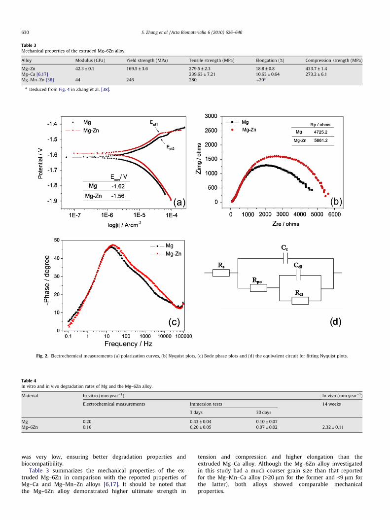
Table 3Mechanical properties of the extruded Mg–6Zn alloy.
Alloy Modulus (GPa) Yield strength (MPa) Tensile strength (MPa) Elongation (%) Compression strength (MPa)
Mg–Zn 42.3 ± 0.1 169.5 ± 3.6 279.5 ± 2.3 18.8 ± 0.8 433.7 ± 1.4Mg–Ca [6,17] 239.63 ± 7.21 10.63 ± 0.64 273.2 ± 6.1Mg–Mn–Zn [38] 44 246 280 �20a
a Deduced from Fig. 4 in Zhang et al. [38].
Fig. 2. Electrochemical measurements (a) polarization curves, (b) Nyquist plots, (c) Bode phase plots and (d) the equivalent circuit for fitting Nyquist plots.
Table 4In vitro and in vivo degradation rates of Mg and the Mg–6Zn alloy.
Material In vitro (mm year�1) In vivo (mm year�1)
Electrochemical measurements Immersion tests 14 weeks
3 days 30 days
Mg 0.20 0.43 ± 0.04 0.10 ± 0.07Mg–6Zn 0.16 0.20 ± 0.05 0.07 ± 0.02 2.32 ± 0.11
630 S. Zhang et al. / Acta Biomaterialia 6 (2010) 626–640
was very low, ensuring better degradation properties andbiocompatibility.
Table 3 summarizes the mechanical properties of the ex-truded Mg–6Zn in comparison with the reported properties ofMg–Ca and Mg–Mn–Zn alloys [6,17]. It should be noted thatthe Mg–6Zn alloy demonstrated higher ultimate strength in
tension and compression and higher elongation than theextruded Mg–Ca alloy. Although the Mg–6Zn alloy investigatedin this study had a much coarser grain size than that reportedfor the Mg–Mn–Ca alloy (>20 lm for the former and <9 lm forthe latter), both alloys showed comparable mechanicalproperties.
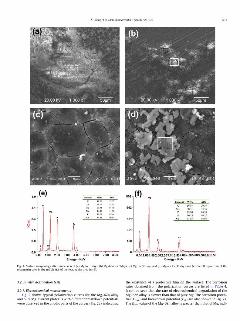
Fig. 3. Surface morphology after immersion of (a) Mg for 3 days, (b) Mg–6Zn for 3 days, (c) Mg for 30 days and (d) Mg–Zn for 30 days and (e) the EDS spectrum of therectangular area in (b) and (f) EDS of the rectangular area in (d).
S. Zhang et al. / Acta Biomaterialia 6 (2010) 626–640 631
3.2. In vitro degradation tests
3.2.1. Electrochemical measurementsFig. 2 shows typical polarization curves for the Mg–6Zn alloy
and pure Mg. Current plateaus with different breakdown potentialswere observed in the anodic parts of the curves (Fig. 2a), indicating
the existence of a protective film on the surface. The corrosionrates obtained from the polarization curves are listed in Table 4.It can be seen that the rate of electrochemical degradation of theMg–6Zn alloy is slower than that of pure Mg. The corrosion poten-tial (Ecorr) and breakdown potential (Ept) are also shown in Fig. 2a.The Ecorr value of the Mg–6Zn alloy is greater than that of Mg, indi-

Fig. 5. The pH of SBF during 72 h immersion.
Fig. 6. The deterioration in bending strength during in vitro degradation.
632 S. Zhang et al. / Acta Biomaterialia 6 (2010) 626–640
cating that the Mg–6Zn alloy is less susceptible to corrosion. Theaddition of Zn also increased the Ept value (Fig. 2a, Ept1 > Ept2), sug-gesting that the corrosion layers on the Mg–6Zn sample were moreprotective than those on the pure Mg sample.
Fig. 2b and c presents Nyquist plots and Bode phase plots,respectively. It can be deduced from the Bode phase plots that anequivalent circuit with two time constants is reasonable. Theequivalent circuit is shown in Fig. 2d, and the polarization resis-tance Rp (Rp = Rpo + Rct, the higher the Rp, the lower the corrosionrate [35]) of Mg and Mg–6Zn are shown in Fig. 2b. The Rp of Mg–6Zn is higher than that of Mg, which is in consistent with the Ecorr
data and electrochemical rates.
3.2.2. Immersion experimentsThe degradation rates of the pure Mg and the Mg–6Zn alloy after 3
and 30 days immersion are listed in Table 4. The Mg–6Zn alloy de-graded more slowly than pure Mg, which is in good agreement withthe electrochemical results. Fig. 3a–d illustrates the surface mor-phologies and Fig. 3e–f shows the EDS results for the surface corro-sion products on the Mg–6Zn alloy. As shown in Fig. 3a and b, bothpure Mg and the Mg–6Zn alloy sample experienced pitting corrosionand were covered with partially protective corrosion products.
A number of cracks were observed on the surface of the samplesafter 30 days immersion (Fig. 3c and d). The EDS results (Fig. 3e andf) reveal that the surface corrosion products (rectangular area inFig. 3b and d) were rich in O, Mg, P and Ca. The XRD results suggestthat magnesium hydroxide [Mg(OH)2] and hydroxyapatite (HA)were precipitated on the Mg–6Zn surface (Fig. 4). Furthermore,Kuwahara et al. [36] pointed out that the corrosion products onthe surface of Mg immersed in Hank’s solution might be amor-phous (Ca0.86Mg0.14)10(PO4)6(OH)2, a rather complicated com-pound. In view of the similar ion concentrations in the SBF usedin this study to those in Hank’s solution, there might be someamorphous phosphates containing magnesium/calcium, as Kuwa-hara indicated. In fact, a strong background and broadened peakscan be observed in Fig. 4, which might be due to the presence ofamorphous corrosion products.
Fig. 5 illustrates the pH variation over the first 72 h of theimmersion tests. It can be seen that the pH rose rapidly, i.e. from7.44 to 8 in 2 h. After 20 h the pH had stabilized. At the end ofthe immersion tests (after 30 days) the pH was 9.22 for pure Mgand 9.32 for the Mg–6Zn alloy.
Hydrogen evolution showed a similar trend to the pH value. Inthe early stage of immersion both pure Mg and the Mg–6Zn alloy
Fig. 4. X-ray diffraction pattern of Mg–6Zn immersed in SBF for 30 days.
Fig. 7. The corrosion surface and corrosion holes formed on the samples with 12%weight loss.
reacted with SBF acutely and a rapid generation of bubbles was ob-served, indicating a fast rate of hydrogen evolution. After 48 himmersion, however, fewer bubbles appeared, suggesting that

Fig. 8. Morphology of the fracture section after a three-point bending test: (a) extruded Mg–6Zn alloy; (b) after 5.8% weight loss; (c) after 11.9% weight loss; and (d) after 18%weight loss.
S. Zhang et al. / Acta Biomaterialia 6 (2010) 626–640 633
the reaction had slowed down and that the rate of hydrogen evo-lution had decreased.
It should be noted that the degradation rate measured after30 days immersion was lower than that measured after 3 days(Table 4), because the corrosion films, including HA and other phos-phates, had a protective effect and hence retarded furtherdegradation.
3.3. In vitro loss of mechanical integrity
Fig. 6 demonstrates the influence of in vitro degradation on thebending strength of the Mg–6Zn alloy. As shown in Fig. 6, thebending strength decreased rapidly in the early stage of degrada-tion. After that the bending strength deteriorated continuously asthe percentage weight loss increased. This may be due to surfacedefects such as corrosion holes formed during degradation, asshown in Fig. 7.
Some dimples were observed on the fracture surface (Fig. 8),which indicates ductile fracture characteristics of the Mg–6Znalloy during the degradation process. The bending tests showedthat degradation undermined the bending strength, although thefracture mode was still ductile during degradation.
3.4. Cytotoxicity assessments
Fig. 9 shows the morphologies of L-929 cells cultured in differ-ent extracts after 7 days incubation. Fig. 10 shows the RGR of L-929cells after 2, 4 and 7 days incubation. It can be seen from Fig. 9 thatthe cell morphologies in different extracts were normal and
healthy, similar to that of the negative control. There was no signif-icant difference between the RGR of cells in the extracts and thosein the negative control. According to ISO 10993-5: 1999 [34] thecytotoxicity of these extracts was Grade 0–1. In other words, theMg–6Zn alloy has a level of biosafety suitable for in cellularapplications.
3.5. In vivo degradation and biocompatibility
3.5.1. Biochemical testsAfter implantation of the Mg–6Zn rods no rabbits displayed
inflammation and there were no unexpected deaths. The variationsin serum magnesium, CREA, BUN, GPT and CK are shown in Fig. 11.There were no significant differences (P > 0.05) in these biochemi-cal indicators before and after operation, which indicated that deg-radation of the Mg–6Zn implants did not raise serum Mg2+ levelsand did not affect kidney and liver function either. The tests indi-cated good biocompatibility of the Mg–6Zn alloy in vivo.
3.5.2. Viscera histologyFig. 12 shows HE stained slices of heart, liver, kidney and spleen.
It can be seen that the tissues were normal, which is in good agree-ment of the biomechanical tests (Fig. 11) and suggests good bio-compatibility of the Mg–6Zn alloy in vivo.
3.5.3. Radiographic evaluationFig. 13a and b shows the radiographs 3 and 12 weeks post-oper-
ation. The Mg–6Zn implant started to degrade in the first 3 weeks,as is evident from the observation that the edge of the implant be-
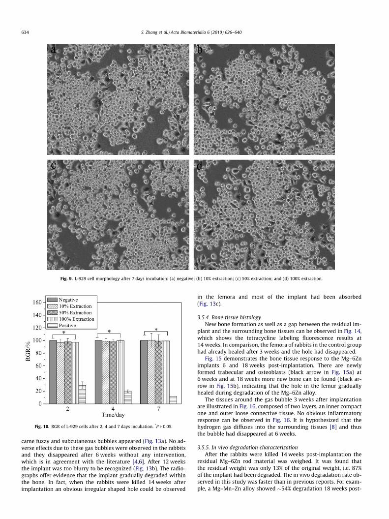
Fig. 9. L-929 cell morphology after 7 days incubation: (a) negative; (b) 10% extraction; (c) 50% extraction; and (d) 100% extraction.
Fig. 10. RGR of L-929 cells after 2, 4 and 7 days incubation. *P > 0.05.
634 S. Zhang et al. / Acta Biomaterialia 6 (2010) 626–640
came fuzzy and subcutaneous bubbles appeared (Fig. 13a). No ad-verse effects due to these gas bubbles were observed in the rabbitsand they disappeared after 6 weeks without any intervention,which is in agreement with the literature [4,6]. After 12 weeksthe implant was too blurry to be recognized (Fig. 13b). The radio-graphs offer evidence that the implant gradually degraded withinthe bone. In fact, when the rabbits were killed 14 weeks afterimplantation an obvious irregular shaped hole could be observed
in the femora and most of the implant had been absorbed(Fig. 13c).
3.5.4. Bone tissue histologyNew bone formation as well as a gap between the residual im-
plant and the surrounding bone tissues can be observed in Fig. 14,which shows the tetracycline labeling fluorescence results at14 weeks. In comparison, the femora of rabbits in the control grouphad already healed after 3 weeks and the hole had disappeared.
Fig. 15 demonstrates the bone tissue response to the Mg–6Znimplants 6 and 18 weeks post-implantation. There are newlyformed trabecular and osteoblasts (black arrow in Fig. 15a) at6 weeks and at 18 weeks more new bone can be found (black ar-row in Fig. 15b), indicating that the hole in the femur graduallyhealed during degradation of the Mg–6Zn alloy.
The tissues around the gas bubble 3 weeks after implantationare illustrated in Fig. 16, composed of two layers, an inner compactone and outer loose connective tissue. No obvious inflammatoryresponse can be observed in Fig. 16. It is hypothesized that thehydrogen gas diffuses into the surrounding tissues [8] and thusthe bubble had disappeared at 6 weeks.
3.5.5. In vivo degradation characterizationAfter the rabbits were killed 14 weeks post-implantation the
residual Mg–6Zn rod material was weighed. It was found thatthe residual weight was only 13% of the original weight, i.e. 87%of the implant had been degraded. The in vivo degradation rate ob-served in this study was faster than in previous reports. For exam-ple, a Mg–Mn–Zn alloy showed �54% degradation 18 weeks post-
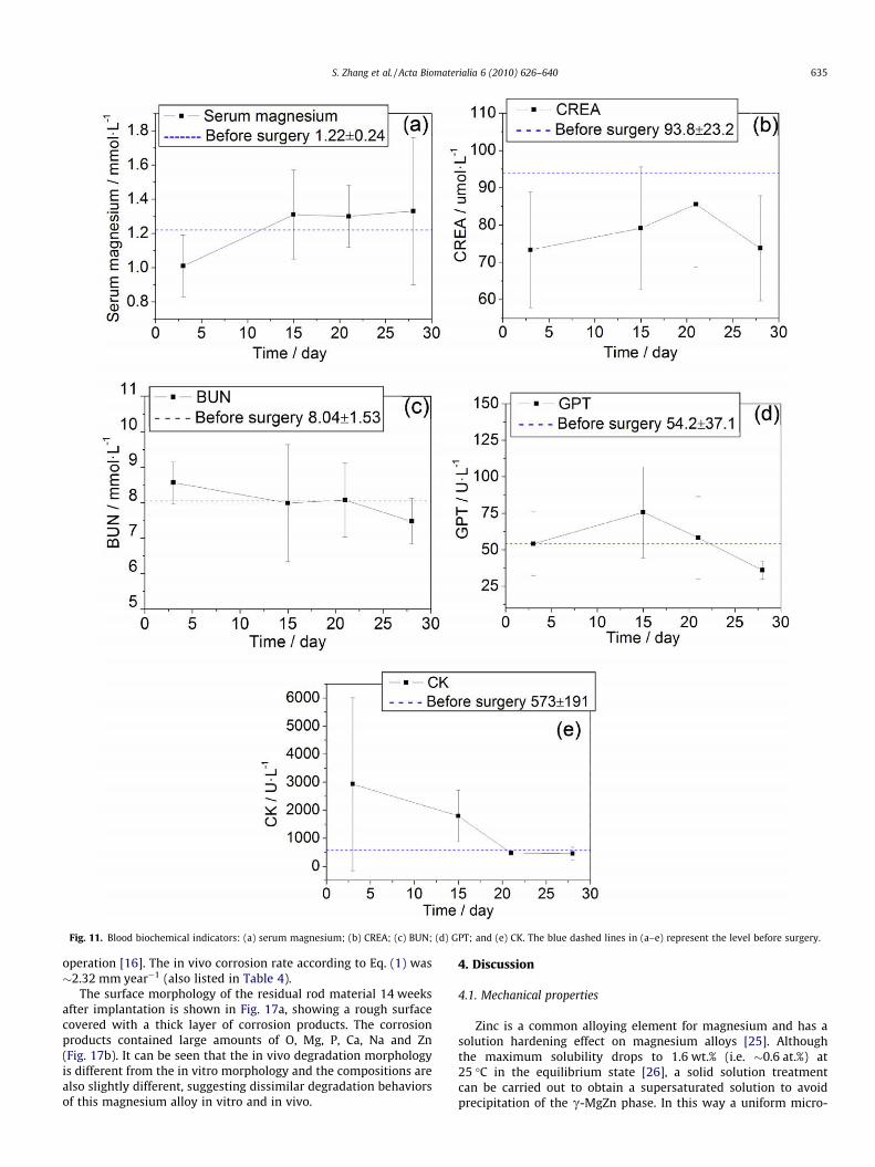
Fig. 11. Blood biochemical indicators: (a) serum magnesium; (b) CREA; (c) BUN; (d) GPT; and (e) CK. The blue dashed lines in (a–e) represent the level before surgery.
S. Zhang et al. / Acta Biomaterialia 6 (2010) 626–640 635
operation [16]. The in vivo corrosion rate according to Eq. (1) was�2.32 mm year�1 (also listed in Table 4).
The surface morphology of the residual rod material 14 weeksafter implantation is shown in Fig. 17a, showing a rough surfacecovered with a thick layer of corrosion products. The corrosionproducts contained large amounts of O, Mg, P, Ca, Na and Zn(Fig. 17b). It can be seen that the in vivo degradation morphologyis different from the in vitro morphology and the compositions arealso slightly different, suggesting dissimilar degradation behaviorsof this magnesium alloy in vitro and in vivo.
4. Discussion
4.1. Mechanical properties
Zinc is a common alloying element for magnesium and has asolution hardening effect on magnesium alloys [25]. Althoughthe maximum solubility drops to 1.6 wt.% (i.e. �0.6 at.%) at25 �C in the equilibrium state [26], a solid solution treatmentcan be carried out to obtain a supersaturated solution to avoidprecipitation of the c-MgZn phase. In this way a uniform micro-
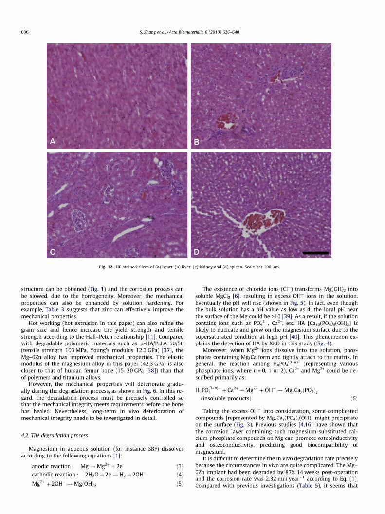
Fig. 12. HE stained slices of (a) heart, (b) liver, (c) kidney and (d) spleen. Scale bar 100 lm.
636 S. Zhang et al. / Acta Biomaterialia 6 (2010) 626–640
structure can be obtained (Fig. 1) and the corrosion process canbe slowed, due to the homogeneity. Moreover, the mechanicalproperties can also be enhanced by solution hardening. Forexample, Table 3 suggests that zinc can effectively improve themechanical properties.
Hot working (hot extrusion in this paper) can also refine thegrain size and hence increase the yield strength and tensilestrength according to the Hall–Petch relationship [11]. Comparedwith degradable polymeric materials such as l-HA/PLLA 50/50(tensile strength 103 MPa, Young’s modulus 12.3 GPa) [37], theMg–6Zn alloy has improved mechanical properties. The elasticmodulus of the magnesium alloy in this paper (42.3 GPa) is alsocloser to that of human femur bone (15–20 GPa [38]) than thatof polymers and titanium alloys.
However, the mechanical properties will deteriorate gradu-ally during the degradation process, as shown in Fig. 6. In this re-gard, the degradation process must be precisely controlled sothat the mechanical integrity meets requirements before the bonehas healed. Nevertheless, long-term in vivo deterioration ofmechanical integrity needs to be investigated in detail.
4.2. The degradation process
Magnesium in aqueous solution (for instance SBF) dissolvesaccording to the following equations [1]:
anodic reaction : Mg!Mg2þ þ 2e ð3Þcathodic reaction : 2H2Oþ 2e! H2 þ 2OH� ð4ÞMg2þ þ 2OH� !MgðOHÞ2 ð5Þ
The existence of chloride ions (Cl�) transforms Mg(OH)2 intosoluble MgCl2 [6], resulting in excess OH� ions in the solution.Eventually the pH will rise (shown in Fig. 5). In fact, even thoughthe bulk solution has a pH value as low as 4, the local pH nearthe surface of the Mg could be >10 [39]. As a result, if the solutioncontains ions such as PO4
3�, Ca2+, etc. HA [Ca10(PO4)6(OH)2] islikely to nucleate and grow on the magnesium surface due to thesupersaturated condition at high pH [40]. This phenomenon ex-plains the detection of HA by XRD in this study (Fig. 4).
Moreover, when Mg2+ ions dissolve into the solution, phos-phates containing Mg/Ca form and tightly attach to the matrix. Ingeneral, the reaction among HnPO4
(3–n)– (representing variousphosphate ions, where n = 0, 1 or 2), Ca2+ and Mg2+ could be de-scribed primarily as:
HnPOð3�nÞ�4 þ Ca2þ þMg2þ þ OH� !MgxCayðPO4Þz
ðinsoluble productsÞ ð6Þ
Taking the excess OH� into consideration, some complicatedcompounds [represented by MgxCay(PO4)z(OH)] might precipitateon the surface (Fig. 3). Previous studies [4,16] have shown thatthe corrosion layer containing such magnesium-substituted cal-cium phosphate compounds on Mg can promote osteoinductivityand osteoconductivity, predicting good biocompatibility ofmagnesium.
It is difficult to determine the in vivo degradation rate preciselybecause the circumstances in vivo are quite complicated. The Mg–6Zn implant had been degraded by 87% 14 weeks post-operationand the corrosion rate was 2.32 mm year�1 according to Eq. (1).Compared with previous investigations (Table 5), it seems that

Fig. 13. Radiographs (a) 3 weeks post-operation, (b) 12 weeks post-operation and (c) macroscopic photograph 14 weeks post-operation, with an irregular shaped hole in thefemur (arrow).
Fig. 14. Tetracycline labeling 14 weeks post-operation.
S. Zhang et al. / Acta Biomaterialia 6 (2010) 626–640 637
the Mg–6Zn in the present study degraded slightly faster. Severalmethods can be used to regulate the degradation rate, for instancecalcium phosphate coating and anodic oxidation.
Zinc is able to elevate the corrosion potential of magnesium alloysand improve the corrosion resistance [41], which is in agreementwith this study (Table 4). In addition, zinc has beneficial effects onthe corrosion film and can reduce the effect of impurities once thetolerance limits have been exceeded [42]. Furthermore, it has beenfound that zinc can also elevate the charge transfer resistance ofmagnesium and thus reduce the corrosion rate [43]. The degradationrates of the Mg–6Zn alloy (0.20 ± 0.05 for 3 days and 0.07 ± 0.02 for30 days) are slower than those of pure Mg (0.43 ± 0.04 for 3 days and0.10 ± 0.07 for 30 days).
For both the Mg–6Zn alloy and pure Mg the degradationrates for 30 days were lower than those for 3 days, owing tothe protective layer on the surface (shown in Fig. 3a–d).During the early stage of immersion in SBF Mg and the Mg–6Zn alloy degraded quickly, accompanied by the rapid formation

Fig. 15. HE stained bone surrounding Mg–Zn rods: 6 weeks (a) and 18 weeks (b) post-implantation. Scale bar 500 lm.
Fig. 16. HE stained tissues around the gas bubble 3 weeks after implantation.
Fig. 17. Surface morphology and composition of the corrosion products of theimplant after 14 weeks degradation: (a) SEM; and (b) EDS of framed area in (a).
638 S. Zhang et al. / Acta Biomaterialia 6 (2010) 626–640
of an insoluble protective corrosion layer, which retardeddegradation.
It should be noted that the compositions and the morphologiesof the in vitro and in vivo degradation products are different. Thisis mainly due to the different corrosive environments. The in vitrotesting solution was SBF, containing inorganic ions such as Cl�,H2PO4
� and Ca2+. Thus, the corrosion products were mineral-likeand the corrosion layer could be clearly observed, which includedHA, Mg(OH)2 and other amorphous magnesium-substituted cal-cium phosphates. However, there were numbers of organic compo-nents in the in vivo environment, such as proteins and cells. Rettiget al. found that albumin influenced the corrosion process of mag-nesium alloys in SBF [44]. As a consequence, the in vivo degrada-tion products are quite complex. The composition of the in vivodegradation products in Fig. 17b shows a high nitrogen level inthe corrosion layer, indicating the possible adsorption and/or adhe-sion of proteins, cells or other tissue fragments to the magnesiumalloy.
The different morphologies and compositions in vitro andin vivo suggest that degradation of the magnesium alloy will begreatly influenced by the surrounding environments. Thus, if themagnesium alloy is used in different parts of the body (e.g. in fe-mur marrow or within muscle) possible different degradation
behaviors must be taken into consideration, otherwise the rate ofdegradation may be estimate incorrectly.
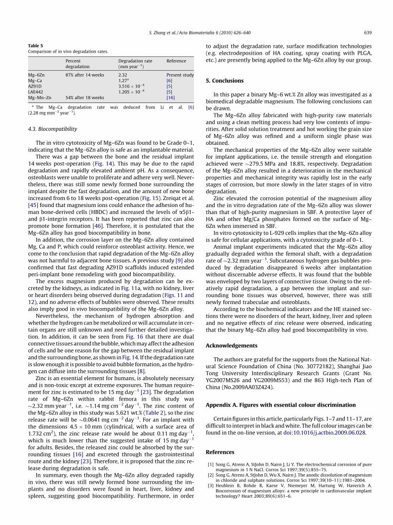
Table 5Comparison of in vivo degradation rates.
Percentdegradation
Degradation rate(mm year�1)
Reference
Mg–6Zn 87% after 14 weeks 2.32 Present studyMg–Ca 1.27a [6]AZ91D 3.516 � 10�4 [5]LAE442 1.205 � 10�4 [5]Mg–Mn–Zn 54% after 18 weeks [16]
a The Mg–Ca degradation rate was deduced from Li et al. [6](2.28 mg mm�2 year�1).
S. Zhang et al. / Acta Biomaterialia 6 (2010) 626–640 639
4.3. Biocompatibility
The in vitro cytotoxicity of Mg–6Zn was found to be Grade 0–1,indicating that the Mg–6Zn alloy is safe as an implantable material.
There was a gap between the bone and the residual implant14 weeks post-operation (Fig. 14). This may be due to the rapiddegradation and rapidly elevated ambient pH. As a consequence,osteoblasts were unable to proliferate and adhere very well. Never-theless, there was still some newly formed bone surrounding theimplant despite the fast degradation, and the amount of new boneincreased from 6 to 18 weeks post-operation (Fig. 15). Zreiqat et al.[45] found that magnesium ions could enhance the adhesion of hu-man bone-derived cells (HBDC) and increased the levels of a5b1-and b1-integrin receptors. It has been reported that zinc can alsopromote bone formation [46]. Therefore, it is postulated that theMg–6Zn alloy has good biocompatibility in bone.
In addition, the corrosion layer on the Mg–6Zn alloy containedMg, Ca and P, which could reinforce osteoblast activity. Hence, wecome to the conclusion that rapid degradation of the Mg–6Zn alloywas not harmful to adjacent bone tissues. A previous study [9] alsoconfirmed that fast degrading AZ91D scaffolds induced extendedperi-implant bone remodeling with good biocompatibility.
The excess magnesium produced by degradation can be ex-creted by the kidneys, as indicated in Fig. 11a, with no kidney, liveror heart disorders being observed during degradation (Figs. 11 and12), and no adverse effects of bubbles were observed. These resultsalso imply good in vivo biocompatibility of the Mg–6Zn alloy.
Nevertheless, the mechanism of hydrogen absorption andwhether the hydrogen can be metabolized or will accumulate in cer-tain organs are still unknown and need further detailed investiga-tion. In addition, it can be seen from Fig. 16 that there are dualconnective tissues around the bubble, which may affect the adhesionof cells and be one reason for the gap between the residual implantand the surrounding bone, as shown in Fig. 14. If the degradation rateis slow enough it is possible to avoid bubble formation, as the hydro-gen can diffuse into the surrounding tissues [8].
Zinc is an essential element for humans, is absolutely necessaryand is non-toxic except at extreme exposures. The human require-ment for zinc is estimated to be 15 mg day�1 [23]. The degradationrate of Mg–6Zn within rabbit femora in this study was�2.32 mm year�1, i.e. �1.14 mg cm�2 day�1. The zinc content ofthe Mg–6Zn alloy in this study was 5.621 wt.% (Table 2), so the zincrelease rate will be �0.0641 mg cm�2 day�1. For an implant withthe dimensions 4.5 � 10 mm (cylindrical, with a surface area of1.732 cm2), the zinc release rate would be about 0.11 mg day�1,which is much lower than the suggested intake of 15 mg day�1
for adults. Besides, the released zinc could be absorbed by the sur-rounding tissues [16] and excreted through the gastrointestinalroute and the kidney [23]. Therefore, it is proposed that the zinc re-lease during degradation is safe.
In summary, even though the Mg–6Zn alloy degraded rapidlyin vivo, there was still newly formed bone surrounding the im-plants and no disorders were found in heart, liver, kidney andspleen, suggesting good biocompatibility. Furthermore, in order
to adjust the degradation rate, surface modification technologies(e.g. electrodeposition of HA coating, spray coating with PLGA,etc.) are presently being applied to the Mg–6Zn alloy by our group.
5. Conclusions
In this paper a binary Mg–6 wt.% Zn alloy was investigated as abiomedical degradable magnesium. The following conclusions canbe drawn.
The Mg–6Zn alloy fabricated with high-purity raw materialsand using a clean melting process had very low contents of impu-rities. After solid solution treatment and hot working the grain sizeof Mg–6Zn alloy was refined and a uniform single phase wasobtained.
The mechanical properties of the Mg–6Zn alloy were suitablefor implant applications, i.e. the tensile strength and elongationachieved were �279.5 MPa and 18.8%, respectively. Degradationof the Mg–6Zn alloy resulted in a deterioration in the mechanicalproperties and mechanical integrity was rapidly lost in the earlystages of corrosion, but more slowly in the later stages of in vitrodegradation.
Zinc elevated the corrosion potential of the magnesium alloyand the in vitro degradation rate of the Mg–6Zn alloy was slowerthan that of high-purity magnesium in SBF. A protective layer ofHA and other Mg/Ca phosphates formed on the surface of Mg–6Zn when immersed in SBF.
In vitro cytotoxicity to L-929 cells implies that the Mg–6Zn alloyis safe for cellular applications, with a cytotoxicity grade of 0–1.
Animal implant experiments indicated that the Mg–6Zn alloygradually degraded within the femoral shaft, with a degradationrate of �2.32 mm year�1. Subcutaneous hydrogen gas bubbles pro-duced by degradation disappeared 6 weeks after implantationwithout discernable adverse effects. It was found that the bubblewas enveloped by two layers of connective tissue. Owing to the rel-atively rapid degradation, a gap between the implant and sur-rounding bone tissues was observed, however, there was stillnewly formed trabeculae and osteoblasts.
According to the biochemical indicators and the HE stained sec-tions there were no disorders of the heart, kidney, liver and spleenand no negative effects of zinc release were observed, indicatingthat the binary Mg–6Zn alloy had good biocompatibility in vivo.
Acknowledgements
The authors are grateful for the supports from the National Nat-ural Science Foundation of China (No. 30772182), Shanghai JiaoTong University Interdisciplinary Research Grants (Grant No.YG2007MS26 and YG2009MS53) and the 863 High-tech Plan ofChina (No.2009AA03Z424).
Appendix A. Figures with essential colour discrimination
Certain figures in this article, particularly Figs. 1–7 and 11–17, aredifficult to interpret in black and white. The full colour images can befound in the on-line version, at doi:10.1016/j.actbio.2009.06.028.
References
[1] Song G, Atrens A, StJohn D, Nairn J, Li Y. The electrochemical corrosion of puremagnesium in 1 N NaCl. Corros Sci 1997;39(5):855–75.
[2] Song G, Atrens A, StJohn D, Wu X, Nairn J. The anodic dissolution of magnesiumin chloride and sulphate solutions. Corros Sci 1997;39(10–11):1981–2004.
[3] Heublein B, Rohde R, Kaese V, Niemeyer M, Hartung W, Haverich A.Biocorrosion of magnesium alloys: a new principle in cardiovascular implanttechnology? Heart 2003;89(6):651–6.
640 S. Zhang et al. / Acta Biomaterialia 6 (2010) 626–640
[4] Witte F, Kaese V, Haferkamp H, Switzer E, Meyer-Lindenberg A, Wirth CJ, et al.In vivo corrosion of four magnesium alloys and the associated bone response.Biomaterials 2005;26(17):3557–63.
[5] Witte F, Fischer J, Nellesen J, Crostack H-A, Kaese V, Pisch A, et al. In vitro andin vivo corrosion measurements of magnesium alloys. Biomaterials2006;27(7):1013–8.
[6] Li Z, Gu X, Lou S, Zheng Y. The development of binary Mg–Ca alloys for use asbiodegradable materials within bone. Biomaterials 2008;29(10):1329–44.
[7] Kim WC, Kim JG, Lee JY, Seok HK. Influence of Ca on the corrosion properties ofmagnesium for biomaterials. Mater Lett 2008;62(25):4146–8.
[8] Witte F, Ulrich H, Rudert M, Willbold E. Biodegradable magnesium scaffolds.Part I. Appropriate inflammatory response. J Biomed Mater Res A2007;81A(3):748–56.
[9] Witte F, Ulrich H, Palm C, Willbold E. Biodegradable magnesium scaffolds. PartII. Peri-implant bone remodeling. J Biomed Mater Res A 2007;81A(3):757–65.
[10] Witte F, Feyerabend F, Maier P, Fischer J, Stormer M, Blawert C, et al.Biodegradable magnesium–hydroxyapatite metal matrix composites.Biomaterials 2007;28(13):2163–74.
[11] Zhang E, He W, Du H, Yang K. Microstructure, mechanical properties andcorrosion properties of Mg–Zn–Y alloys with low Zn content. Mater Sci Eng A2008;488(1–2):102–11.
[12] Song G. Control of biodegradation of biocompatible magnesium alloys. CorrosSci 2007;49(4):1696–701.
[13] Staiger MP, Pietak AM, Huadmai J, Dias G. Magnesium and its alloys asorthopedic biomaterials: a review. Biomaterials 2006;27(9):1728–34.
[14] Levesque J, Hermawan H, Dub D, Mantovani D. Design of a pseudo-physiological test bench specific to the development of biodegradablemetallic biomaterials. Acta Biomater 2008;4(2):284–95.
[15] Song GL, Song SZ. A possible biodegradable magnesium implant material. AdvEng Mater 2007;9(4):298–302.
[16] Xu LP, Yu GN, Zhang E, Pan F, Yang K. In vivo corrosion behavior of Mg–Mn–Znalloy for bone implant application. J Biomed Mater Res A 2007;83A(3):703–11.
[17] Wan Y, Xiong G, Luo H, He F, Huang Y, Zhou X. Preparation andcharacterization of a new biomedical magnesium–calcium alloy. MaterDesign 2008;29(10):2034–7.
[18] Zartner P, Cesnjevar R, Singer H, Weyand M. First successful implantation of abiodegradable metal stent into the left pulmonary artery of a preterm baby.Catheter Cardiovasc Interv 2005;66(4):590–4.
[19] El-Rahman SSA. Neuropathology of aluminum toxicity in rats (glutamate andGABA impairment). Pharmacol Res 2003;47(3):189–94.
[20] Ku C-H, Pioletti DP, Browne M, Gregson PJ. Effect of different Ti–6Al–4V surfacetreatments on osteoblasts behaviour. Biomaterials 2002;23(6):1447–54.
[21] Yumiko N, Yukari T, Yasuhide T, Tadashi S, Yoshio I. Differences in behavioramong the chlorides of seven rare earth elements administered intravenouslyto rats. Fundam Appl Toxicol 1997;37:106–16.
[22] Yang W, Zhang P, Liu J, Xue Y. Effect of long-term intake of Y3+ in drinking water ongene expression in brains of rats. J Rare Earth 2006;24(3):369–73.
[23] Tapiero H, Tew KD. Trace elements in human physiology and pathology: zincand metallothioneins. Biomed Pharmacother 2003;57(9):399–411.
[24] Haferkamp H, Bach F-W, Kaese V, Möhwald K, Niemeyer M, SchreckenbergerH, et al. Magnesium corrosion-processes, protection of anode and cathode. In:Kainer KU, editor. Magnesium-alloys and technology. Weinheim: Wiley-VCH;2003. p. 226–7.
[25] Mordike BL, Lukác P. Physical metallurgy. In: Friedrich HE, Mordike BL, editors.Magnesium technology – metallurgy, design data, applications. Berlin: Springer;2006. p. 76–7.
[26] Okamoto H. Comment on Mg–Zn (magnesium–zinc). J Phase Equilibria Diffus1994;15(1):129–30.
[27] Zhang X. In vivo biodegradable binary Mg–Zn alloy. Chinese Patent no. ZL200510111795.4.
[28] American Society for Testing and Materials. ASTM-E8-04: standard test methodsfor tension testing of metallic materials. In: Annual book of ASTM standards.Philadelphia, PA, USA: American Society for Testing and Materials; 2004.
[29] American Society for Testing and Materials. ASTM E9-89a (2000): standardtest methods of compression testing of metallic materials at roomtemperature. In: Annual Book of ASTM Standards. Philadelphia, PA, USA:American Society for Testing and Materials; 2000.
[30] Standardization Administration of the PRC. GB/T 16886.15-2003: BiologicalEvaluation of Medical Devices. Part 15. Identification and Quantification ofDegradation Products from Metals and Alloys.
[31] American Society for Testing and Materials. ASTM-G102-89: standard practicefor calculation for corrosion rates and related information fromelectrochemical measurements. In: Annual Book of ASTM Standards.Philadelphia, PA, USA: American Society for Testing and Materials; 1999.
[32] American Society for Testing and Materials. ASTM-G31-72: standardpractice for laboratory immersion corrosion testing of metals. In: AnnualBook of ASTM Standards. Philadelphia, PA: American Society for Testing andMaterials; 2004.
[33] Kannan MB, Raman RKS. In vitro degradation and mechanical integrity ofcalcium-containing magnesium alloys in modified-simulated body fluid.Biomaterials 2008;29(15):2306–14.
[34] ANSI/AAMI. ISO 10993-5: 1999. Biological evaluation of medical devices. Part5. Tests for cytotoxicity: in vitro methods. Arlington, VA: ANSI/AAMI.
[35] Rondellia G, Torricellib P, Finib M, Giardinob R. In vitro corrosion study by EISof a nickel-free stainless steel for orthopaedic applications. Biomaterials2005;26:739–44.
[36] Kuwahara H, Al-Abdullat Y, Mazaki N, Tsutsumi S, Aizawa T. Precipitation ofmagnesium apatite on pure magnesium surface during immersing in Hank’ssolution. Mater Trans 2001;42(7):1317–21.
[37] Shikinami Y, Okuno M. Bioresorbable devices made of forged composites ofhydroxyapatite (HA) particles and poly-L-lactide (PLLA). Part I. Basiccharacteristics. Biomaterials 1999;20:859–77.
[38] Zhang E, Yin D, Xu L, Yang L, Yang K. Microstructure, mechanical and corrosionproperties and biocompatibility of Mg–Zn–Mn alloys for biomedicalapplication. Mater Sci Eng C. doi:10.1016/j.msec.2008.08.024.
[39] Simaranov A, Sokolova I, Marshakov A, Mikhailovskii Y. Corrosion-electrochemical behavior of magnesium in acidic media, containingoxidants. Prot Metal 1991;27(3):329–34.
[40] Jonasova L, Muller F, Helebrant A, Strnad J, Greil P. Biomimetic apatiteformation on chemically treated titanium. Biomaterials 2004;25:1187–94.
[41] Shi Z, Song G, Atrens A. Corrosion resistance of anodised single-phase Mgalloys. Surf Coat Technol 2006;201:492–503.
[42] Song GL, Atrens A. Corrosion mechanisms of magnesium alloys. Adv Eng Mater1999;1(1):11–33.
[43] Zhang SX, Li JN, Song Y, Zhao CL, Zhang XN, Xie CY, et al. In vitro degradation,hemolysis and MC3T3-E1 cell adhesion of biodegradable Mg–Zn alloy. MaterSci Eng C 2009. doi:10.1016/j.msec.2009.03.001.
[44] Rettig R, Virtanen S. Time-dependent electrochemical characterization of thecorrosion of a magnesium rare-earth alloy in simulated body fluids. J BiomedMater Res A 2008;85A(1):167–75.
[45] Zreiqat H, Howlett C, Zannettino A, Evans P, Schulze-Tanzil G, Knabe C, et al.Mechanisms of magnesium-stimulated adhesion of osteoblastic cells tocommonly used orthopaedic implants. J Biomed Mater Res A 2002;62:175–84.
[46] Hashizume M, Yamaguchi M. Stimulatory effect of beta-alanyl-L-histidinatozinc on cell proliferation is dependent on protein synthesis in osteoblasticMC3T3–E1 cells. Mol Cell Biochem 1993;122(1):59–64.