Acquired Demyelinating Syndromes Elles...Acquired demyelinating syndromes ... disorders, and...
193
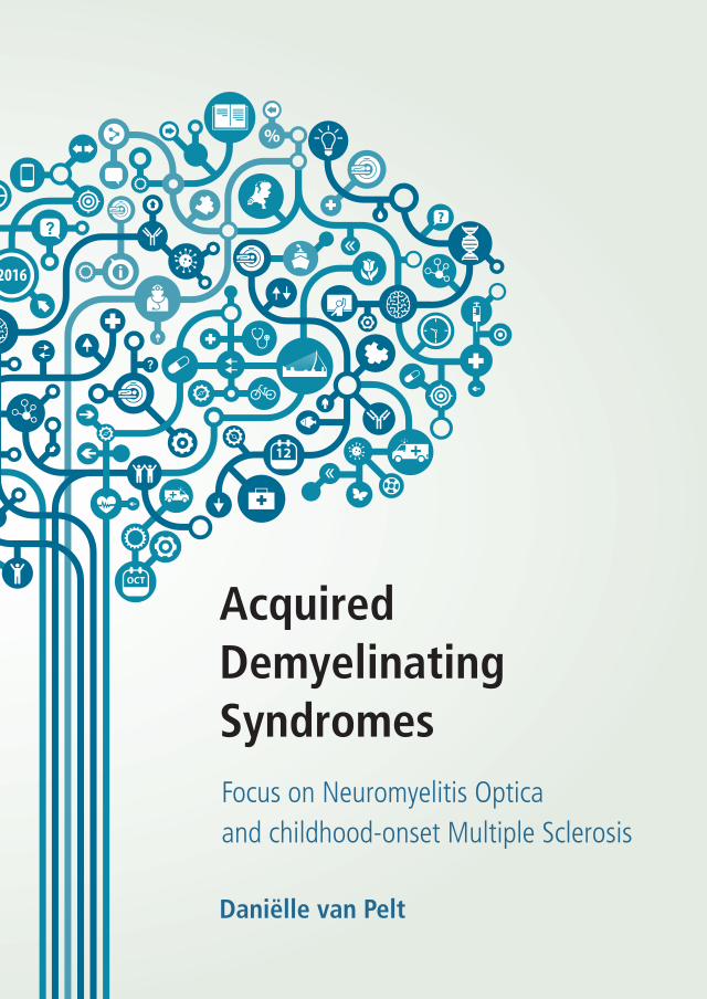
2016 12 OCT Acquired Demyelinating Syndromes Focus on Neuromyelitis Optica and childhood-onset Multiple Sclerosis Daniëlle van Pelt
-
Upload
nguyenthuan -
Category
Documents
-
view
227 -
download
0
Transcript of Acquired Demyelinating Syndromes Elles...Acquired demyelinating syndromes ... disorders, and...
2016
12
OCT
Acquired Demyelinating Syndromes Focus on Neuromyelitis Optica and childhood-onset Multiple Sclerosis
Daniëlle van Pelt
Daniëlle van Pelt
Acquired D
emyelinating Syndrom
es

Acquired Demyelinating Syndromes Focus on Neuromyelitis Optica
and childhood-onset Multiple Sclerosis
Daniëlle van Pelt
AcknowleDgements
The research described in this thesis is supported by the Dutch MS Research Foundation. In addition the studies described in chapter 5 and 7 were funded by direct donations to MS research at the Hospital for Sick Children (Toronto, Canada) and the Canadian MS Sci-entific Research Foundation. Besides, the study reported in chapter 9 was supported by a program grant provided by the EC 7th framework programme of the Marie Curie Initial Training Network, The United Europeans for the Development of Pharmacogenomics in Multiple Sclerosis, the Dutch Organization for Scientific Research (ZON—MW) and the Brain Foundation Netherlands.
Printing of this thesis was financially supported by the Dutch MS Research Foundation, Erasmus University Medical Center Rotterdam, and Sanofi Genzyme.
Acquired demyelinating syndromes: focus on neuromyelitis opticaand childhood-onset multiple sclerosis
ISBN: 978-94-6169-930-5
Lay-out and cover design by Optima Grafische Communicatie, Rotterdam, The Netherlands
Printed by Optima Grafische Communicatie, Rotterdam, The Netherlands
© 2016 E.D. van Pelt, Rotterdam, the Netherlands
Acquired Demyelinating syndromes:Focus on neuromyelitis optica and childhood-onset multiple sclerosis
Verworven demyeliniserende syndromen:focus op neuromyelitis optica en multiple sclerose op de kinderleeftijd
Proefschrift
ter verkrijging van de graad van doctor aan deErasmus Universiteit Rotterdam
op gezag van derector magnificus
Prof.dr. H.A.P. Pols
en volgens besluit van het College voor Promoties.De openbare verdediging zal plaatsvinden op
woensdag 12 oktober 2016 om 9.30 uur
door
elles Daniëlle van Pelt – gravesteijngeboren te Gouda

Promotiecommissie
Promotor: Prof.dr. R.Q. Hintzen
Overige leden: Dr. C.E. Catsman-Berrevoets Dr. A.M.C. van Rossum Prof.dr. M.A. Willemsen
Copromotor: Dr. R.F. Neuteboom
tAble oF contents
Chapter 1 General introduction 7
Part one: neuromyelitis optica spectrum disordersChapter 2 Incidence of AQP4-IgG seropositive neuromyelitis optica
spectrum disorders in the Netherlands: about one in a million33
Chapter 3 Neuromyelitis optica spectrum disorders: comparison of clinical and MRI characteristics of AQP4-IgG versus MOG-IgG seropositive cases in the Netherlands
41
Part two: Acquired demyelinating syndromes in childrenChapter 4 Application of the 2012 revised diagnostic definitions for
paediatric multiple sclerosis and immune-mediated central nervous system demyelination disorders
57
Chapter 5 Validation of MRI predictors of multiple sclerosis diagnosis in children with acute CNS demyelination
69
Chapter 6 Disease course after CIS in children versus adults: a prospective cohort study
83
Chapter 7 Risk genes associated with pediatric-onset MS but not with monophasic acquired CNS demyelination
95
Chapter 8 Anti-MOG antibodies plead against MS diagnosis in an ADS cohort
111
Chapter 9 Gray matter related proteins are associated with childhood-onset multiple sclerosis
125
Chapter 10 General discussion 145
Chapter 11 Summary | Samenvatting 169
Chapter 12 Epilogue 177Dankwoord 179Authors and Affiliations 182About the author 183Publications 184PhD portfolio 187Abbreviations 189


2016
12
OCT
Chapter 1
General introduction

9
General introduction
1AcQuireD DemyelinAting synDromes oF the centrAl nerVous system
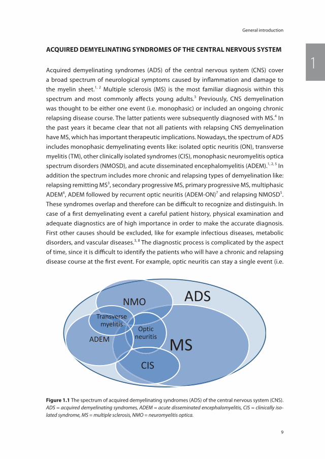
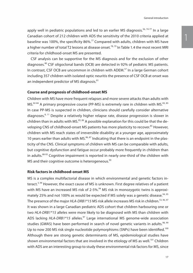
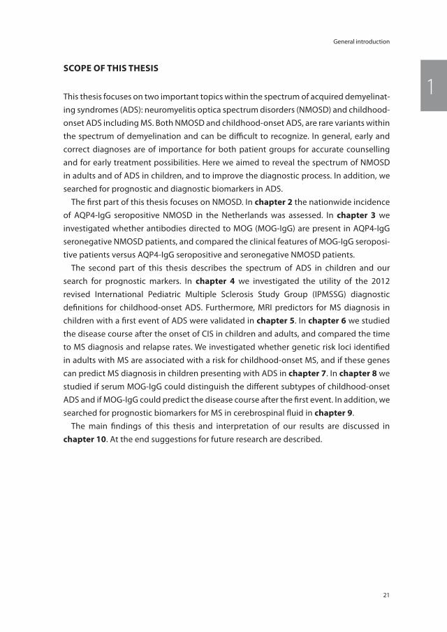
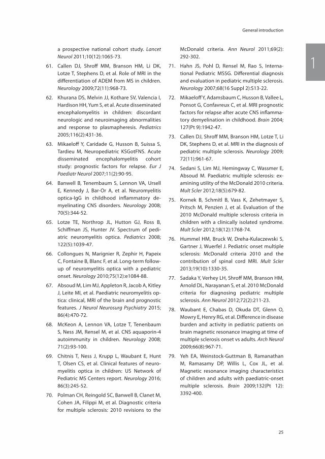
Acquired demyelinating syndromes (ADS) of the central nervous system (CNS) cover a broad spectrum of neurological symptoms caused by infl ammation and damage to the myelin sheet.1, 2 Multiple sclerosis (MS) is the most familiar diagnosis within this spectrum and most commonly aff ects young adults.3 Previously, CNS demyelination was thought to be either one event (i.e. monophasic) or included an ongoing chronic relapsing disease course. The latter patients were subsequently diagnosed with MS.4 In the past years it became clear that not all patients with relapsing CNS demyelination have MS, which has important therapeutic implications. Nowadays, the spectrum of ADS includes monophasic demyelinating events like: isolated optic neuritis (ON), transverse myelitis (TM), other clinically isolated syndromes (CIS), monophasic neuromyelitis optica spectrum disorders (NMOSD), and acute disseminated encephalomyelitis (ADEM).1, 2, 5 In addition the spectrum includes more chronic and relapsing types of demyelination like: relapsing remitting MS3, secondary progressive MS, primary progressive MS, multiphasic ADEM6, ADEM followed by recurrent optic neuritis (ADEM-ON)7 and relapsing NMOSD5. These syndromes overlap and therefore can be diffi cult to recognize and distinguish. In case of a fi rst demyelinating event a careful patient history, physical examination and adequate diagnostics are of high importance in order to make the accurate diagnosis. First other causes should be excluded, like for example infectious diseases, metabolic disorders, and vascular diseases.3, 8 The diagnostic process is complicated by the aspect of time, since it is diffi cult to identify the patients who will have a chronic and relapsing disease course at the fi rst event. For example, optic neuritis can stay a single event (i.e.
MS
NMO
Optic neuritis
CIS
ADEM
Transverse myelitis
ADS
Figure 1.1 The spectrum of acquired demyelinating syndromes (ADS) of the central nervous system (CNS).ADS = acquired demyelinating syndromes, ADEM = acute disseminated encephalomyelitis, CIS = clinically iso-lated syndrome, MS = multiple sclerosis, NMO = neuromyelitis optica.
Chapter 1
10
idiopathic ON) but also can occur in MS, NMOSD, chronic relapsing inflammatory optic neuropathy (CRION)9, or in other autoimmune diseases with CNS involvement.10 In other words, the group of ADS patients is heterogeneous and their clinical characteristics are dynamic. It can take several years, and in rare cases even decades, before a second and NMOSD or MS diagnostic event occurs.5, 11 However, accurate and early diagnoses are of importance for the patient for establishing their prognosis and for the correct treatment initiation. Figure 1.1 illustrates the spectrum of ADS and its overlap.
The first part of this thesis on ADS focuses on NMOSD and describes the incidence and clinical features of NMOSD in the Netherlands. The second part, and main focus of this the-sis, is on ADS in children. The goal of our studies is to reveal the clinical spectrum of ADS and to find diagnostic and prognostic markers, which allow for an early and safe diagnosis.
neuromyelitis oPticA sPectrum DisorDers
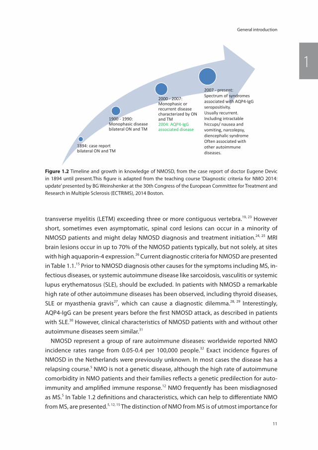
Neuromyelitis optica (NMO), previously known as Devic’s disease, is a rare variant of MS, characterized by optic neuritis and transverse myelitis.5, 12 Classic NMO, as described by doctor Eugene Devic in 1894, was a monophasic illness including coincident bilateral optic neuritis and transverse myelitis.13 However, in current literature there are clues that earlier cases of NMO have been described.14 In the past years the spectrum of NMO has broadened and the nomenclature of Devic’s disease became insufficient and outdated.15 At present the unifying term neuromyelitis optica spectrum disorders (NMOSD) is used.
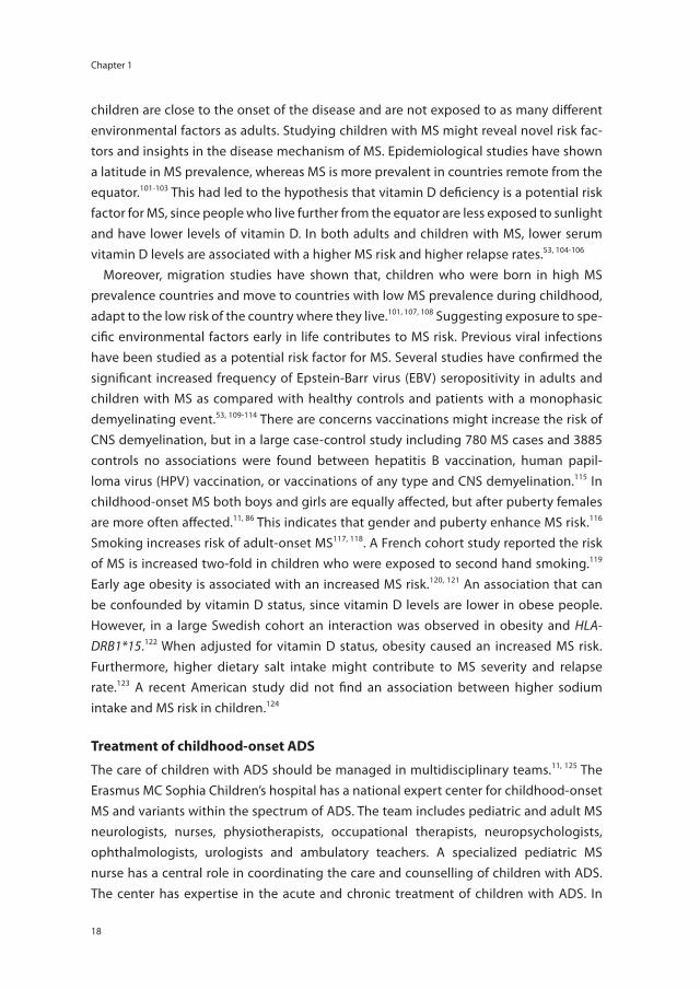
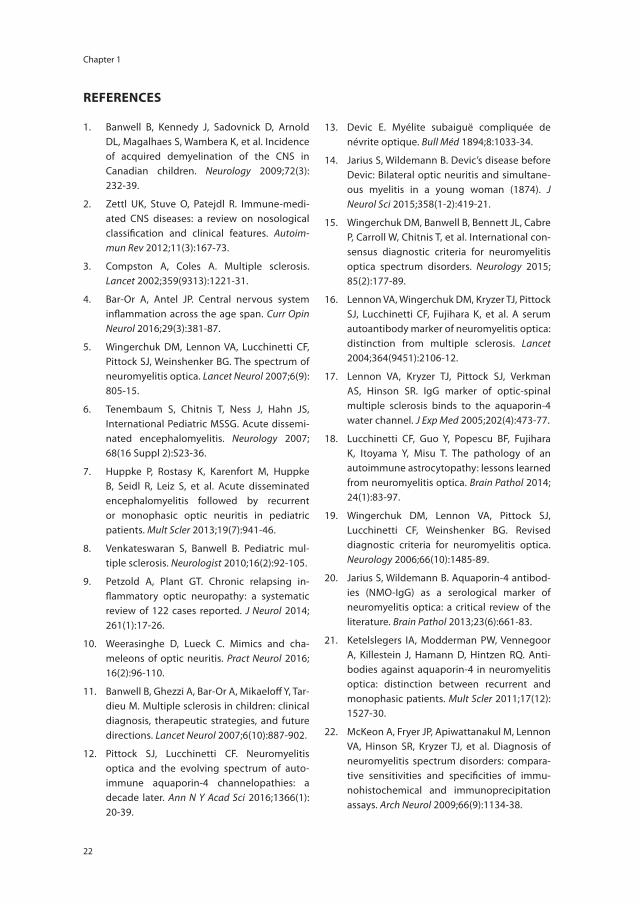
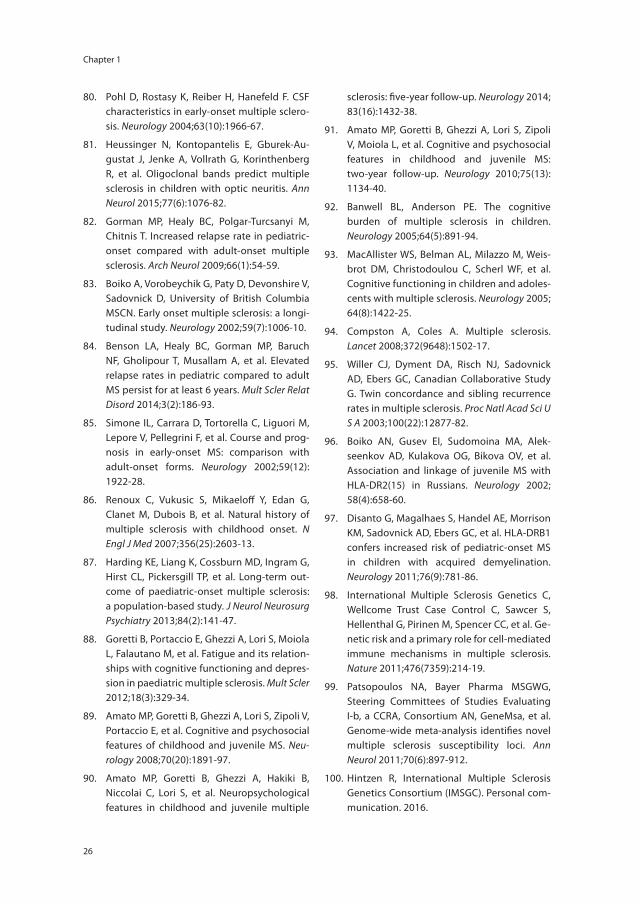
In 2004 an important milestone in the field of NMO was reached by the discovery of specific antibodies directed against aquaporin-4 (AQP4-IgG), which led to the distinc-tion of MS.16, 17 The identification of this specific antibody has led to the understanding of NMO pathophysiology as a B-cell mediated astrocytopathy, whereas the inflamma-tory response causes demyelination as collateral damage.18 The timeline and growth in knowledge of NMOSD are illustrated in Figure 1.2. In 2006 the antibody was incorporated in the diagnostic criteria for NMO.19 Since then diagnostic AQP4-IgG assays have been improved.20 In the Netherlands the highly sensitive diagnostic AQP4-IgG cell-based assay (CBA) is performed at one centralized NMO expert center at Sanquin Diagnostic Services in Amsterdam.21 Aquaporin-4 antibodies are present in the majority of NMOSD patients (±77%).15 Serum AQP4 antibodies are highly specific for the disease.22 The presence of AQP4-IgG allowed for a broadening of the clinical NMO spectrum including limited forms such as isolated or recurrent optic neuritis, transverse myelitis, brainstem syndromes (including area postrema syndrome with intractable nausea, vomiting and hiccups) and cerebral syndromes (including narcolepsy and acute diencephalic clinical syndrome).5, 15 NMOSD diagnosis is based on clinical characteristics supported by AQP4-IgG status and MRI findings.15 Typical MRI findings in NMO are a longitudinally extensive
11
General introduction
1
transverse myelitis (LETM) exceeding three or more contiguous vertebra.19, 23 However short, sometimes even asymptomatic, spinal cord lesions can occur in a minority of NMOSD patients and might delay NMOSD diagnosis and treatment initiation.24, 25 MRI brain lesions occur in up to 70% of the NMOSD patients typically, but not solely, at sites with high aquaporin-4 expression.26 Current diagnostic criteria for NMOSD are presented in Table 1.1.15 Prior to NMOSD diagnosis other causes for the symptoms including MS, in-fectious diseases, or systemic autoimmune disease like sarcoidosis, vasculitis or systemic lupus erythematosus (SLE), should be excluded. In patients with NMOSD a remarkable high rate of other autoimmune diseases has been observed, including thyroid diseases, SLE or myasthenia gravis27, which can cause a diagnostic dilemma.28, 29 Interestingly, AQP4-IgG can be present years before the first NMOSD attack, as described in patients with SLE.30 However, clinical characteristics of NMOSD patients with and without other autoimmune diseases seem similar.31
NMOSD represent a group of rare autoimmune diseases: worldwide reported NMO incidence rates range from 0.05-0.4 per 100,000 people.32 Exact incidence figures of NMOSD in the Netherlands were previously unknown. In most cases the disease has a relapsing course.5 NMO is not a genetic disease, although the high rate of autoimmune comorbidity in NMO patients and their families reflects a genetic predilection for auto-immunity and amplified immune response.12 NMO frequently has been misdiagnosed as MS.5 In Table 1.2 definitions and characteristics, which can help to differentiate NMO from MS, are presented.5, 12, 15 The distinction of NMO from MS is of utmost importance for
1894: case report bilateral ON and TM
1900 - 1990: Monophasic disease bilateral ON and TM
2000 - 2007: Monophasic or recurrent disease characterized by ON and TM 2004: AQP4-IgG associated disease
2007 - present: Spectrum of syndromes associated with AQP4-IgG seropositivity. Usually recurrent. Including intractable hiccups/ nausea and vomiting, narcolepsy, diencephalic syndrome Often associated with other autoimmune diseases.
Figure 1.2 Timeline and growth in knowledge of NMOSD, from the case report of doctor Eugene Devic in 1894 until present.This figure is adapted from the teaching course ‘Diagnostic criteria for NMO 2014: update’ presented by BG Weinshenker at the 30th Congress of the European Committee for Treatment and Research in Multiple Sclerosis (ECTRIMS), 2014 Boston.
Chapter 1
12
establishing the prognosis and initiating the correct treatment. The treatment of attacks and chronic treatment differ significantly between MS and NMOSD. In NMOSD relapses are more severe than in MS and cause deterioration and progression of disability.23, 33, 34 Acute NMOSD relapses should be urgently treated with high dose intravenous methyl-prednisolone in order to minimise disability.35-37 If corticosteroid therapy is insufficient patients can benefit from plasmapheresis. Escalation of the acute therapy improved the outcome of NMOSD patients in a large German cohort and decreased the proportion of non-responders.38 Preferably relapses and thus further deterioration are prevented. Therefore immunosuppressive treatment is indicated in relapsing NMOSD patients.35-37 AQP4-IgG seropositive patients are at high risk for future relapses and therefore chronic treatment is advised in these patients after the first event. MS therapeutics can be po-tentially harmful for NMOSD patients and should be avoided.39-42 Immunosuppressive therapy with azathioprine or mycophenolate mofetil is advised in NMOSD patients.35-37, 43
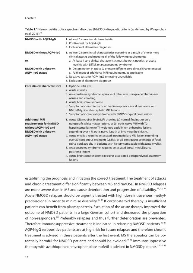
table 1.1 Neuromyelitis optica spectrum disorders (NMOSD) diagnostic criteria (as defined by Wingerchuk et al. 2015).15
nmosD with AQP4-igg 1. At least 1 core clinical characteristic2. Positive test for AQP4-IgG3. Exclusion of alternative diagnoses
nmosD without AQP4-igg
or
nmosD with unknown AQP4-igg status
1. At least 2 core clinical characteristics occurring as a result of one or more clinical attacks and meeting all of the following requirements:
a. At least 1 core clinical characteristic must be optic neuritis, or acute myelitis with LETM, or area postrema syndrome
b. Dissemination in space (2 or more different core clinical characteristics) c. Fulfillment of additional MRI requirements, as applicable2. Negative tests for AQP4-IgG, or testing unavailable3. Exclusion of alternative diagnoses
core clinical characteristics 1. Optic neuritis (ON)2. Acute myelitis3. Area postrema syndrome: episode of otherwise unexplained hiccups or
nausea and vomiting4. Acute brainstem syndrome5. Symptomatic narcolepsy or acute diencephalic clinical syndrome with
NMOSD-typical diencephalic MRI lesions6. Symptomatic cerebral syndrome with NMOSD-typical brain lesions
Additional mri requirements for nmosD without AQP4-igg and nmosD with unknown AQP4-igg status
1. Acute ON: requires brain MRI showing (a) normal findings or only nonspecific white matter lesions, or (b) optic nerve MRI with T2-hyperintense lesion or T1-weighted gadolinium enhancing lesions extending over > ½ optic nerve length or involving the chiasm.
2. Acute myelitis: requires associated intramedullary MRI lesion extending over ≥3 contiguous segments (LETM), or ≥3 contiguous segments of focal spinal cord atrophy in patients with history compatible with acute myelitis
3. Area postrema syndrome: requires associated dorsal medulla/area postrema lesions
4. Acute brainstem syndrome: requires associated periependymal brainstem lesions
13
General introduction
1
In case of failure of the first-line treatments, rituximab should be considered. Future im-munotherapeutic targets include complement proteins, the IL-6 receptor, neutrophils, eosinophils, CD19 and AQP4.12, 36, 44
At the Dutch NMO expert center, which includes Sanquin Diagnostic Services in Amsterdam and the NMO expert clinic at Erasmus MC in Rotterdam, we focus on the accurate diagnosis and treatment of NMOSD patients. The expert center is a referral center for clinicians in the Netherlands for the confirmation of NMOSD diagnosis and treatment advice.
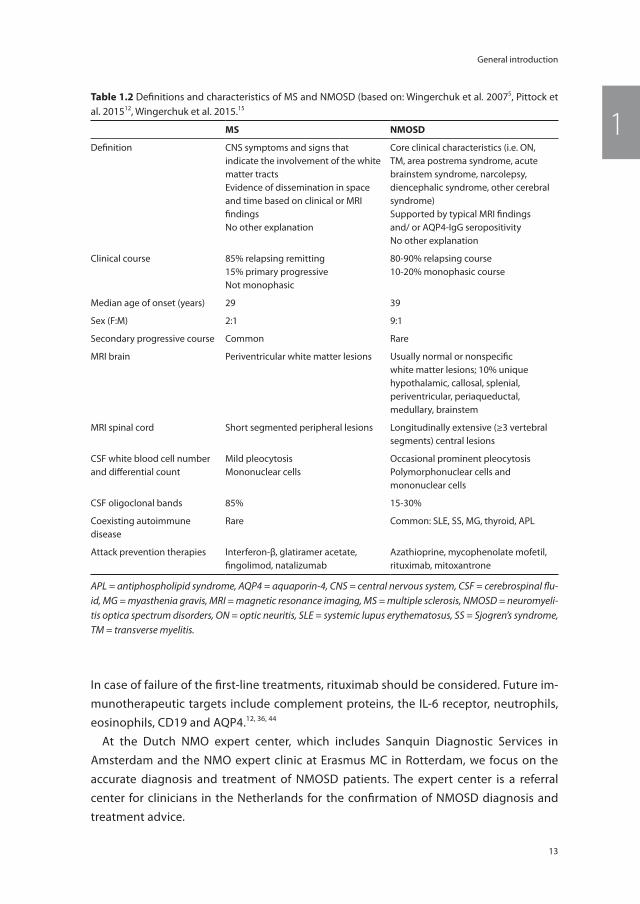
table 1.2 Definitions and characteristics of MS and NMOSD (based on: Wingerchuk et al. 20075, Pittock et al. 201512, Wingerchuk et al. 2015.15
ms nmosD
Definition CNS symptoms and signs that indicate the involvement of the white matter tractsEvidence of dissemination in space and time based on clinical or MRI findingsNo other explanation
Core clinical characteristics (i.e. ON, TM, area postrema syndrome, acute brainstem syndrome, narcolepsy, diencephalic syndrome, other cerebral syndrome)Supported by typical MRI findings and/ or AQP4-IgG seropositivityNo other explanation
Clinical course 85% relapsing remitting15% primary progressiveNot monophasic
80-90% relapsing course10-20% monophasic course
Median age of onset (years) 29 39
Sex (F:M) 2:1 9:1
Secondary progressive course Common Rare
MRI brain Periventricular white matter lesions Usually normal or nonspecific white matter lesions; 10% unique hypothalamic, callosal, splenial, periventricular, periaqueductal, medullary, brainstem
MRI spinal cord Short segmented peripheral lesions Longitudinally extensive (≥3 vertebral segments) central lesions
CSF white blood cell number and differential count
Mild pleocytosisMononuclear cells
Occasional prominent pleocytosisPolymorphonuclear cells and mononuclear cells
CSF oligoclonal bands 85% 15-30%
Coexisting autoimmune disease
Rare Common: SLE, SS, MG, thyroid, APL
Attack prevention therapies Interferon-β, glatiramer acetate, fingolimod, natalizumab
Azathioprine, mycophenolate mofetil, rituximab, mitoxantrone
APL = antiphospholipid syndrome, AQP4 = aquaporin-4, CNS = central nervous system, CSF = cerebrospinal flu-id, MG = myasthenia gravis, MRI = magnetic resonance imaging, MS = multiple sclerosis, NMOSD = neuromyeli-tis optica spectrum disorders, ON = optic neuritis, SLE = systemic lupus erythematosus, SS = Sjogren’s syndrome, TM = transverse myelitis.
Chapter 1
14
AcQuireD DemyelinAting synDromes in chilDhooD
ADS also occur in children and, although rare, can evolve into childhood-onset MS.8, 11 Diagnosing childhood-onset ADS can be challenging due to unfamiliarity of clinicians with these rare diseases. Furthermore, children can be too young to report their symptoms to their caregivers. Symptoms can be mild and usually are self-limiting and therefore a delay can occur before a clinician is consulted. Up to 10% of all MS patients have their first attack during childhood, prior to their 18th birthday.8 In the Netherlands a nationwide prospective study was started in 2007 investigating ADS in children.45 The main goal of the PROUDkids study (PRedicting the OUtcome of a Demyelinating event in children) is to identify prognostic factors, which predict a future MS diagnosis at the first event of ADS. Children with a first event of ADS and their families face their lives with uncertainty about their future.46 For this reason, adequate counselling, early diagnosis and treatment initiation are of utmost importance. Reported incidence rates of pediatric ADS range from 0.66 per 100,000 in the Netherlands to 1.66 per 100,000
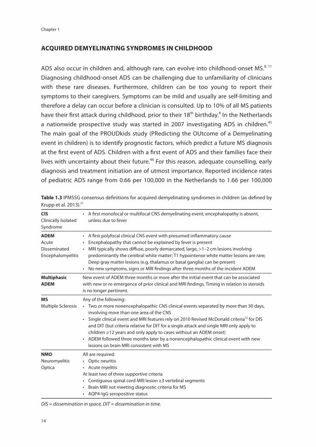
table 1.3 IPMSSG consensus definitions for acquired demyelinating syndromes in children (as defined by Krupp et al. 2013).51
cisClinically Isolated Syndrome
• A first monofocal or multifocal CNS demyelinating event; encephalopathy is absent, unless due to fever
ADemAcute Disseminated Encephalomyelitis
• A first polyfocal clinical CNS event with presumed inflammatory cause• Encephalopathy that cannot be explained by fever is present• MRI typically shows diffuse, poorly demarcated, large, >1–2 cm lesions involving
predominantly the cerebral white matter; T1 hypointense white matter lesions are rare; Deep gray matter lesions (e.g. thalamus or basal ganglia) can be present
• No new symptoms, signs or MRI findings after three months of the incident ADEM
multiphasic ADem
New event of ADEM three months or more after the initial event that can be associated with new or re-emergence of prior clinical and MRI findings. Timing in relation to steroids is no longer pertinent.
msMultiple Sclerosis
Any of the following:• Two or more nonencephalopathic CNS clinical events separated by more than 30 days,
involving more than one area of the CNS• Single clinical event and MRI features rely on 2010 Revised McDonald criteria52 for DIS
and DIT (but criteria relative for DIT for a single attack and single MRI only apply to children ≥12 years and only apply to cases without an ADEM onset)
• ADEM followed three months later by a nonencephalopathic clinical event with new lesions on brain MRI consistent with MS
nmoNeuromyelitis Optica
All are required:• Optic neuritis• Acute myelitisAt least two of three supportive criteria• Contiguous spinal cord MRI lesion ≥3 vertebral segments• Brain MRI not meeting diagnostic criteria for MS• AQP4-IgG seropositive status
DIS = dissemination in space, DIT = dissemination in time.
15
General introduction
1children per year in other cohorts.1, 45, 47-49 Approximately 5 – 10 children are diagnosed with MS per year in the Netherlands. In 2007 the International Pediatric MS Study Group (IPMSSG) first developed diagnostic criteria for childhood-onset ADS.50 In 2012 these cri-teria were revised based on the tremendous growth in research and gain in knowledge of childhood-onset ADS.51 The current IPMSSG definitions of the various ADS subtypes are presented in Table 1.3.
A first demyelinating event in children can present with symptoms caused by a single lesion (monofocal) or by multiple lesions (polyfocal).52 In the Dutch ADS cohort 22% of the children presented with ON, 24% with ADEM and 30% with polyfocal CIS without encephalopathy.45 Any type of ADS can be the first presentation of MS in children. Based on previous cohort studies, it is estimated that 21-32% of children who presented with a first event of ADS will have a future diagnosis of MS.53, 54 MS percentages differ between the various reported pediatric ADS cohorts.53-56 This is partly explained by the introduc-tion and revision of diagnostic criteria, differences in study design and referral bias.52
ADEM is a relatively common subtype of ADS, especially in young children.6 The acute event of ADEM is often preceded by viral infections and characterized by polyfocal neu-rological deficits and encephalopathy. Encephalopathy, defined as behavioural changes and or alterations in consciousness not explained by fever51, is not a typical feature of MS.53 Children with ADEM usually have a good prognosis.57, 58 A minority of children with ADEM have a severe deteriorating disease course, which can lead to ICU admission and sometimes death.58, 59 MRI typically shows large poorly demarcated lesions in the white and gray matter (basal ganglia).60, 61 The symptoms and MRI findings of ADEM can fluctu-ate within three months after the first onset of symptoms.51, 62 Fluctuations within this time period are considered as part of one event.51 ADEM usually is monophasic, but in rare cases a second event of ADEM can occur (i.e. multiphasic ADEM).57, 63 A minority of children with ADEM are diagnosed with MS during follow-up.51, 60 In the Dutch cohort 5 out of 92 children with ADEM (6%) converted to MS during follow-up.58 However, one event of ADEM followed by a second non-ADEM event might still reflect a transient de-myelinating disease. Therefore, children with ADEM have to fulfill strict criteria prior to MS diagnosis: i.e. ADEM should be followed by two non-encephalopathic events, or one new event with the appearance of new MS-specific MRI lesions fulfilling dissemination in time and space.51 In addition, a disease course with isolated relapsing optic neuritis can occur after the first event of ADEM (ADEM-ON).7
NMO occurs approximately in 3% of all children with ADS.45, 64 Children with NMO can have a very diverse clinical presentation with diffuse ADEM-like inflammatory lesions on first brain MRI.64-67 In one study of 88 children with AQP4-IgG seropositive NMOSD, 45% had episodic cerebral manifestations including encephalopathy.68 Since the clini-cal syndromes of ADEM and NMO can overlap in children, AQP4-IgG testing should be considered in children with an ADEM-like event including ON and LETM.51, 64, 65 A recent
Chapter 1
16
American study in 38 children with NMO demonstrated the NMOSD 2015 criteria apply well in the childhood setting (sensitivity 97%) and could diminish treatment delay in children with NMOSD.69
ms diagnosis in childhood
Prognostic factors for a future MS diagnosis in children are: female gender, age at onset ≥ 10 years old, onset of symptoms without encephalopathy, elevated IgG index and/or positive oligoclonal bands in cerebrospinal fluid (CSF), presence of MS-like lesions on MRI.53-55 As in adults, children who had a clinical event of CNS demyelination supported by clinical and/or radiological evidence of dissemination in time and space can be diag-nosed with MS.51, 70 Alternative diagnoses should be excluded. The differential diagnosis of MS in children is much more complex than in adults and includes a long list of other diagnoses like CNS infectious diseases, neoplasms, leukodystrophy, and inflammatory diseases.8, 71 Additional laboratory results and MRI can aid in the diagnostic workup and differentiation of ADS from other diseases. MRI is an important diagnostic tool in childhood-onset ADS by confirming demyelinating whiter matter lesions. In the past years several MRI criteria have been developed for early MS diagnosis and distinction from other ADS subtypes and alternative diagnoses in children.61, 72, 73 MRI in MS patients typically shows T2 lesions in locations characteristic for MS: periventricular, juxtacortical, infratentorial and spinal cord.70 Unique for the current 2010 McDonald diagnostic MRI criteria for adults is the opportunity to confirm MS diagnosis at the incident event. Sev-eral studies have confirmed that the 2010 McDonald MRI criteria, designed for adults,
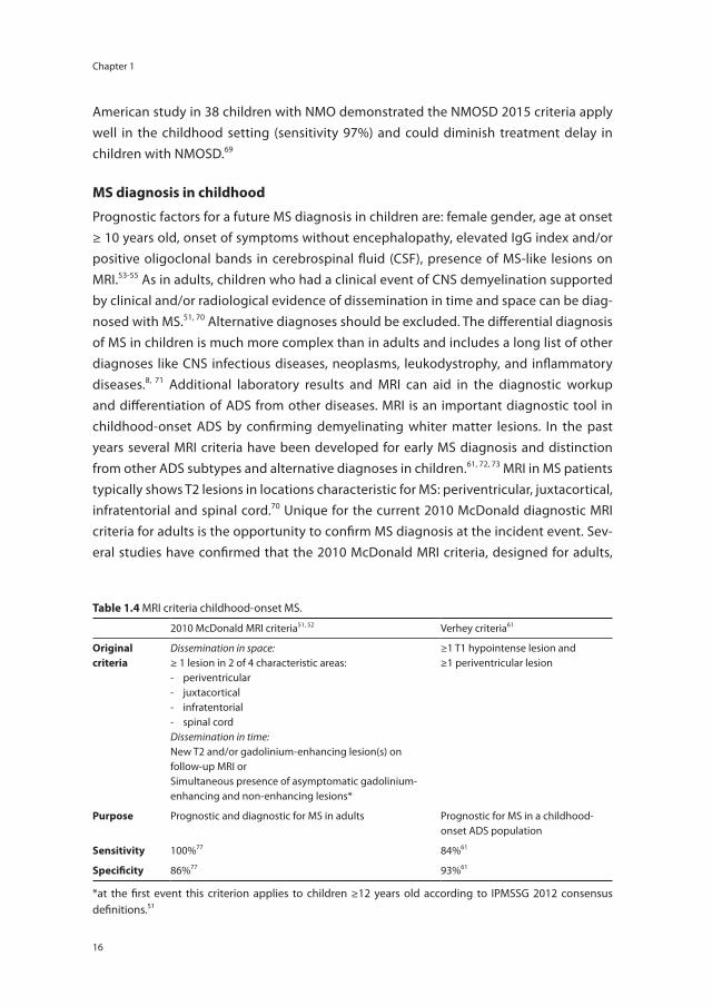
table 1.4 MRI criteria childhood-onset MS.
2010 McDonald MRI criteria51, 52 Verhey criteria61
original criteria
Dissemination in space:≥ 1 lesion in 2 of 4 characteristic areas:- periventricular- juxtacortical- infratentorial- spinal cordDissemination in time:New T2 and/or gadolinium-enhancing lesion(s) on follow-up MRI orSimultaneous presence of asymptomatic gadolinium-enhancing and non-enhancing lesions*
≥1 T1 hypointense lesion and≥1 periventricular lesion
Purpose Prognostic and diagnostic for MS in adults Prognostic for MS in a childhood-onset ADS population
sensitivity 100%77 84%61
specificity 86%77 93%61
*at the first event this criterion applies to children ≥12 years old according to IPMSSG 2012 consensus definitions.51
17
General introduction
1apply well in pediatric populations and led to an earlier MS diagnosis.56, 74-77 In a large Canadian cohort of 212 children with ADS the sensitivity of the 2010 criteria applied at baseline was 100%, the specificity 86%.77 Compared with adults, children with MS have a higher number of total T2 lesions at disease onset.78, 79 In Table 1.4 the most recent MRI criteria for childhood-onset MS are presented.
CSF analysis can be supportive for the MS diagnosis and for the exclusion of other diagnoses.80 CSF oligoclonal bands (OCB) are detected in 92% of pediatric MS patients. In contrast, CSF OCB are uncommon in children with ADEM.57 In a large German cohort including 357 children with isolated optic neuritis the presence of CSF OCB at onset was an independent predictor of MS diagnosis.81
course and prognosis of childhood-onset ms
Children with MS have more frequent relapses and more severe attacks than adults with MS.82-84 A primary progressive course (PP-MS) is extremely rare in children with MS.83, 85 In case PP-MS is suspected in children, clinicians should carefully consider alternative diagnoses.8, 11 Despite a relatively higher relapse rate, disease progression is slower in children than in adults with MS.85, 86 A possible explanation for this could be that the de-veloping CNS of childhood-onset MS patients has more plasticity to recover.85 However, children with MS reach states of irreversible disability at a younger age, approximately 10 years earlier than adults with MS.86, 87 Indicating that there is an endpoint in the plas-ticity of the CNS. Clinical symptoms of children with MS can be comparable with adults, but cognitive dysfunction and fatigue occur probably more frequently in children than in adults.88-93 Cognitive impairment is reported in nearly one-third of the children with MS and their cognitive outcome is heterogeneous.90
risk factors in childhood-onset ms
MS is a complex multifactorial disease in which environmental and genetic factors in-teract.3, 94 However, the exact cause of MS is unknown. First degree relatives of a patient with MS have an increased MS risk of 2-5%.94 MS risk in monozygotic twins is approxi-mately 25% and not 100% as would be expected if MS solely was a genetic disease.94, 95 The presence of the major HLA-DRB1*15 MS risk allele increases MS risk in children.53, 96, 97 It was shown in a large Canadian pediatric ADS cohort that children harbouring one or two HLA-DRB1*15 alleles were more likely to be diagnosed with MS than children with ADS lacking HLA-DRB1*15 alleles.97 Large international MS genome-wide association studies (GWAS) have been performed in search of novel genetic variants in adults.98, 99 Up to now 200 MS risk single nucleotide polymorphisms (SNPs) have been identified.100 Although there are strong genetic determinants of MS, epidemiological studies have shown environmental factors that are involved in the etiology of MS as well.101 Children with ADS are an interesting group to study these environmental risk factors for MS, since
Chapter 1
18
children are close to the onset of the disease and are not exposed to as many different environmental factors as adults. Studying children with MS might reveal novel risk fac-tors and insights in the disease mechanism of MS. Epidemiological studies have shown a latitude in MS prevalence, whereas MS is more prevalent in countries remote from the equator.101-103 This had led to the hypothesis that vitamin D deficiency is a potential risk factor for MS, since people who live further from the equator are less exposed to sunlight and have lower levels of vitamin D. In both adults and children with MS, lower serum vitamin D levels are associated with a higher MS risk and higher relapse rates.53, 104-106
Moreover, migration studies have shown that, children who were born in high MS prevalence countries and move to countries with low MS prevalence during childhood, adapt to the low risk of the country where they live.101, 107, 108 Suggesting exposure to spe-cific environmental factors early in life contributes to MS risk. Previous viral infections have been studied as a potential risk factor for MS. Several studies have confirmed the significant increased frequency of Epstein-Barr virus (EBV) seropositivity in adults and children with MS as compared with healthy controls and patients with a monophasic demyelinating event.53, 109-114 There are concerns vaccinations might increase the risk of CNS demyelination, but in a large case-control study including 780 MS cases and 3885 controls no associations were found between hepatitis B vaccination, human papil-loma virus (HPV) vaccination, or vaccinations of any type and CNS demyelination.115 In childhood-onset MS both boys and girls are equally affected, but after puberty females are more often affected.11, 86 This indicates that gender and puberty enhance MS risk.116 Smoking increases risk of adult-onset MS117, 118. A French cohort study reported the risk of MS is increased two-fold in children who were exposed to second hand smoking.119 Early age obesity is associated with an increased MS risk.120, 121 An association that can be confounded by vitamin D status, since vitamin D levels are lower in obese people. However, in a large Swedish cohort an interaction was observed in obesity and HLA-DRB1*15.122 When adjusted for vitamin D status, obesity caused an increased MS risk. Furthermore, higher dietary salt intake might contribute to MS severity and relapse rate.123 A recent American study did not find an association between higher sodium intake and MS risk in children.124
treatment of childhood-onset ADs
The care of children with ADS should be managed in multidisciplinary teams.11, 125 The Erasmus MC Sophia Children’s hospital has a national expert center for childhood-onset MS and variants within the spectrum of ADS. The team includes pediatric and adult MS neurologists, nurses, physiotherapists, occupational therapists, neuropsychologists, ophthalmologists, urologists and ambulatory teachers. A specialized pediatric MS nurse has a central role in coordinating the care and counselling of children with ADS. The center has expertise in the acute and chronic treatment of children with ADS. In
19
General introduction
1addition, it has an advisory role for pediatricians and neurologists in the Netherlands, who are less frequently consulted with children with ADS. The center has an important role in counselling, increasing the awareness of childhood-onset ADS and educating the school system. For example, via a special website (http://www.kindermscentrum.nl/) and the organisation of an annually informative day, where children with MS and their families can meet and share their experiences. At the specialized outpatient clinic children are guided in the transition from pediatric to adult care.
The acute treatment of children with ADS is with intravenous corticosteroid thera-py.11, 126, 127 The common regimen is 10-30 mg/kg/dose of methylprednisolone intrave-nous for 3 – 5 days. In case children do not respond to the first pulse of corticosteroids, a second pulse could be considered. Intravenous immunoglobulins or plasmapheresis could be considered in the acute phase when children not tolerate or not respond to corticosteroids.
The effect of chronic immunomodulatory treatment on MS is well established in adults128, but is less proven in children.129 The small numbers of patients and more strict ethical concerns challenge therapeutic trials in children with MS.130 First-line treatment with interferon-β or glatiramer acetate have not been formally evaluated in children with MS.129 However, several case series report a reduction in relapse rate and an overall high safety-profile of those first-line therapies in children with MS.129, 131 Most notable side ef-fect of interferon-β is a transient increase of liver transaminases.52, 131 Side effects are less severe in case the dose is titrated. Currently DMT is initiated at 25% of the full dose and titrated gradually. Long- term effects on growth, puberty and adverse effects of immu-nomodulatory therapies for MS in children have yet to be established.127 MS is a chronic and ongoing disease and despite the initiation of DMT relapses can still occur. Switching to more aggressive second-line therapy with more severe side effects requires a care-ful consideration of the potential risks and benefits per individual patient. The IPMSSG provided a definition for inadequate treatment response and recommends switching treatments in case children are compliant on full-dose therapy for at least 6 months and have an increase or no reduction in relapse rate, or new MRI lesions as compared with the pre-treatment period, or in case they had 2 or more relapses within a 12-month period or less.129 Options for switching are, change between first-line therapies, or switch to second-line therapy with natalizumab. An Italian study reported a cohort of 101 children of whom the majority tolerated natalizumab well.132 In this cohort strong MS suppres-sion was observed with only 9 relapses during a mean follow-up of 34.2± 18.3 months. Long-term effects of natalizumab in children are unknown and there are concerns for the risk of progressive multifocal leukoencephalopathy (PML).132, 133 Patients at high risk of PML can be identified by serum anti-JC virus antibodies.134 Children at high risk of PML might switch to oral second-line therapy with fingolimod in the future. A Brazilian study reported no serious adverse events of fingolimod in 17 children with MS.135 The effect
Chapter 1
20
and safety of fi ngolimod is investigated in the ongoing PARADIGMS trial: a double-blind randomized controlled trial which compares the safety and eff ect of fi ngolimod versus intramuscular interferon-β-1a.136 Recently, new oral fi rst-line therapies came available for adults with MS: dimethyl fumarate and terifl unomide.137, 138 First-line treatment with dimethyl fumarate was safe and well tolerated in a small cohort of 13 American children with MS.139 Novel exciting MS therapeutics are currently underway.140
seArch For new biomArkers: myelin oligoDenDrocyte glycoProtein AntiboDies
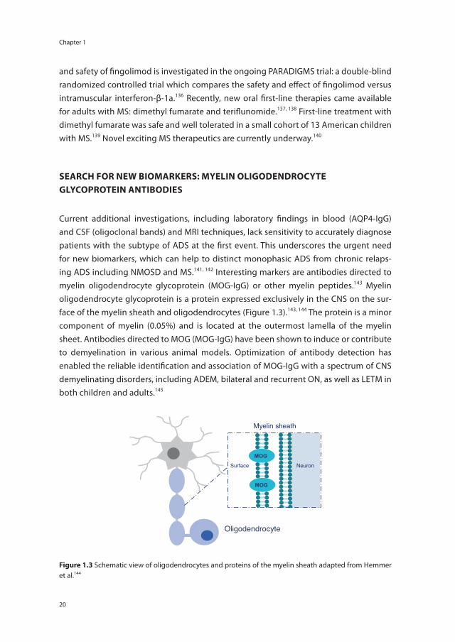
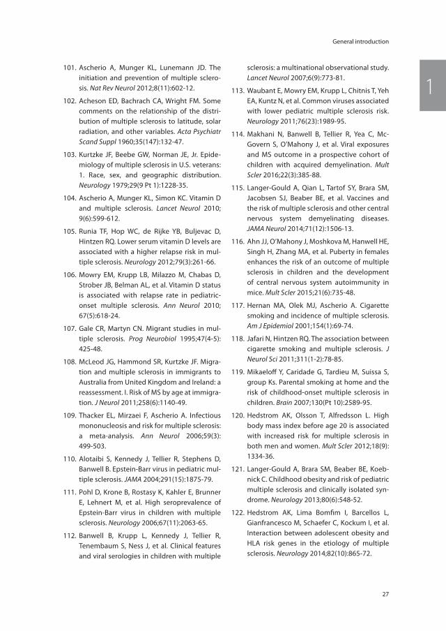
Current additional investigations, including laboratory fi ndings in blood (AQP4-IgG) and CSF (oligoclonal bands) and MRI techniques, lack sensitivity to accurately diagnose patients with the subtype of ADS at the fi rst event. This underscores the urgent need for new biomarkers, which can help to distinct monophasic ADS from chronic relaps-ing ADS including NMOSD and MS.141, 142 Interesting markers are antibodies directed to myelin oligodendrocyte glycoprotein (MOG-IgG) or other myelin peptides.143 Myelin oligodendrocyte glycoprotein is a protein expressed exclusively in the CNS on the sur-face of the myelin sheath and oligodendrocytes (Figure 1.3).143, 144 The protein is a minor component of myelin (0.05%) and is located at the outermost lamella of the myelin sheet. Antibodies directed to MOG (MOG-IgG) have been shown to induce or contribute to demyelination in various animal models. Optimization of antibody detection has enabled the reliable identifi cation and association of MOG-IgG with a spectrum of CNS demyelinating disorders, including ADEM, bilateral and recurrent ON, as well as LETM in both children and adults.145
Oligodendrocyte
Surface
Myelin sheath
Neuron
MOG
MOG
Figure 1.3 Schematic view of oligodendrocytes and proteins of the myelin sheath adapted from Hemmer et al.144
21
General introduction
1scoPe oF this thesis
This thesis focuses on two important topics within the spectrum of acquired demyelinat-ing syndromes (ADS): neuromyelitis optica spectrum disorders (NMOSD) and childhood-onset ADS including MS. Both NMOSD and childhood-onset ADS, are rare variants within the spectrum of demyelination and can be diffi cult to recognize. In general, early and correct diagnoses are of importance for both patient groups for accurate counselling and for early treatment possibilities. Here we aimed to reveal the spectrum of NMOSD in adults and of ADS in children, and to improve the diagnostic process. In addition, we searched for prognostic and diagnostic biomarkers in ADS.
The fi rst part of this thesis focuses on NMOSD. In chapter 2 the nationwide incidence of AQP4-IgG seropositive NMOSD in the Netherlands was assessed. In chapter 3 we investigated whether antibodies directed to MOG (MOG-IgG) are present in AQP4-IgG seronegative NMOSD patients, and compared the clinical features of MOG-IgG seroposi-tive patients versus AQP4-IgG seropositive and seronegative NMOSD patients.
The second part of this thesis describes the spectrum of ADS in children and our search for prognostic markers. In chapter 4 we investigated the utility of the 2012 revised International Pediatric Multiple Sclerosis Study Group (IPMSSG) diagnostic defi nitions for childhood-onset ADS. Furthermore, MRI predictors for MS diagnosis in children with a fi rst event of ADS were validated in chapter 5. In chapter 6 we studied the disease course after the onset of CIS in children and adults, and compared the time to MS diagnosis and relapse rates. We investigated whether genetic risk loci identifi ed in adults with MS are associated with a risk for childhood-onset MS, and if these genes can predict MS diagnosis in children presenting with ADS in chapter 7. In chapter 8 we studied if serum MOG-IgG could distinguish the diff erent subtypes of childhood-onset ADS and if MOG-IgG could predict the disease course after the fi rst event. In addition, we searched for prognostic biomarkers for MS in cerebrospinal fl uid in chapter 9.
The main fi ndings of this thesis and interpretation of our results are discussed in chapter 10. At the end suggestions for future research are described.
Chapter 1
22
reFerences
1. Banwell B, Kennedy J, Sadovnick D, Arnold DL, Magalhaes S, Wambera K, et al. Incidence of acquired demyelination of the CNS in Canadian children. Neurology 2009; 72(3): 232-39.
2. Zettl UK, Stuve O, Patejdl R. Immune-medi-ated CNS diseases: a review on nosological classification and clinical features. Autoim-mun Rev 2012; 11(3): 167-73.
3. Compston A, Coles A. Multiple sclerosis. Lancet 2002; 359(9313): 1221-31.
4. Bar-Or A, Antel JP. Central nervous system inflammation across the age span. Curr Opin Neurol 2016; 29(3): 381-87.
5. Wingerchuk DM, Lennon VA, Lucchinetti CF, Pittock SJ, Weinshenker BG. The spectrum of neuromyelitis optica. Lancet Neurol 2007; 6(9): 805-15.
6. Tenembaum S, Chitnis T, Ness J, Hahn JS, International Pediatric MSSG. Acute dissemi-nated encephalomyelitis. Neurology 2007; 68(16 Suppl 2): S23-36.
7. Huppke P, Rostasy K, Karenfort M, Huppke B, Seidl R, Leiz S, et al. Acute disseminated encephalomyelitis followed by recurrent or monophasic optic neuritis in pediatric patients. Mult Scler 2013; 19(7): 941-46.
8. Venkateswaran S, Banwell B. Pediatric mul-tiple sclerosis. Neurologist 2010; 16(2): 92-105.
9. Petzold A, Plant GT. Chronic relapsing in-flammatory optic neuropathy: a systematic review of 122 cases reported. J Neurol 2014; 261(1): 17-26.
10. Weerasinghe D, Lueck C. Mimics and cha-meleons of optic neuritis. Pract Neurol 2016; 16(2): 96-110.
11. Banwell B, Ghezzi A, Bar-Or A, Mikaeloff Y, Tar-dieu M. Multiple sclerosis in children: clinical diagnosis, therapeutic strategies, and future directions. Lancet Neurol 2007; 6(10): 887-902.
12. Pittock SJ, Lucchinetti CF. Neuromyelitis optica and the evolving spectrum of auto-immune aquaporin-4 channelopathies: a decade later. Ann N Y Acad Sci 2016; 1366(1): 20-39.
13. Devic E. Myélite subaiguë compliquée de névrite optique. Bull Méd 1894; 8: 1033-34.
14. Jarius S, Wildemann B. Devic’s disease before Devic: Bilateral optic neuritis and simultane-ous myelitis in a young woman (1874). J Neurol Sci 2015; 358(1-2): 419-21.
15. Wingerchuk DM, Banwell B, Bennett JL, Cabre P, Carroll W, Chitnis T, et al. International con-sensus diagnostic criteria for neuromyelitis optica spectrum disorders. Neurology 2015; 85(2): 177-89.
16. Lennon VA, Wingerchuk DM, Kryzer TJ, Pittock SJ, Lucchinetti CF, Fujihara K, et al. A serum autoantibody marker of neuromyelitis optica: distinction from multiple sclerosis. Lancet 2004; 364(9451): 2106-12.
17. Lennon VA, Kryzer TJ, Pittock SJ, Verkman AS, Hinson SR. IgG marker of optic-spinal multiple sclerosis binds to the aquaporin-4 water channel. J Exp Med 2005; 202(4): 473-77.
18. Lucchinetti CF, Guo Y, Popescu BF, Fujihara K, Itoyama Y, Misu T. The pathology of an autoimmune astrocytopathy: lessons learned from neuromyelitis optica. Brain Pathol 2014; 24(1): 83-97.
19. Wingerchuk DM, Lennon VA, Pittock SJ, Lucchinetti CF, Weinshenker BG. Revised diagnostic criteria for neuromyelitis optica. Neurology 2006; 66(10): 1485-89.
20. Jarius S, Wildemann B. Aquaporin-4 antibod-ies (NMO-IgG) as a serological marker of neuromyelitis optica: a critical review of the literature. Brain Pathol 2013; 23(6): 661-83.
21. Ketelslegers IA, Modderman PW, Vennegoor A, Killestein J, Hamann D, Hintzen RQ. Anti-bodies against aquaporin-4 in neuromyelitis optica: distinction between recurrent and monophasic patients. Mult Scler 2011; 17(12): 1527-30.
22. McKeon A, Fryer JP, Apiwattanakul M, Lennon VA, Hinson SR, Kryzer TJ, et al. Diagnosis of neuromyelitis spectrum disorders: compara-tive sensitivities and specificities of immu-nohistochemical and immunoprecipitation assays. Arch Neurol 2009; 66(9): 1134-38.
23
General introduction
123. Wingerchuk DM, Hogancamp WF, O’Brien PC,
Weinshenker BG. The clinical course of neuro-myelitis optica (Devic’s syndrome). Neurology 1999; 53(5): 1107-14.
24. Flanagan EP, Weinshenker BG, Krecke KN, Len-non VA, Lucchinetti CF, McKeon A, et al. Short myelitis lesions in aquaporin-4-IgG-positive neuromyelitis optica spectrum disorders. JAMA Neurol 2015; 72(1): 81-87.
25. Flanagan EP, Weinshenker BG, Krecke KN, Pittock SJ. Asymptomatic myelitis in neuro-myelitis optica and autoimmune aquaporin-4 channelopathy. Neurol Clin Pract 2015; 5(2): 175-77.
26. Kim HJ, Paul F, Lana-Peixoto MA, Tenembaum S, Asgari N, Palace J, et al. MRI characteristics of neuromyelitis optica spectrum disorder: an international update. Neurology 2015; 84(11): 1165-73.
27. Leite MI, Coutinho E, Lana-Peixoto M, Aposto-los S, Waters P, Sato D, et al. Myasthenia gravis and neuromyelitis optica spectrum disorder: a multicenter study of 16 patients. Neurology 2012; 78(20): 1601-07.
28. Wingerchuk DM, Weinshenker BG. The emerging relationship between neuromy-elitis optica and systemic rheumatologic autoimmune disease. Mult Scler 2012; 18(1): 5-10.
29. Iyer A, Elsone L, Appleton R, Jacob A. A review of the current literature and a guide to the early diagnosis of autoimmune disorders associated with neuromyelitis optica. Autoim-munity 2014; 47(3): 154-61.
30. Kovacs KT, Kalluri SR, Boza-Serrano A, Deier-borg T, Csepany T, Simo M, et al. Change in autoantibody and cytokine responses during the evolution of neuromyelitis optica in patients with systemic lupus erythematosus: A preliminary study. Mult Scler 2016; 22(9): 1192-201.
31. Zhang B, Zhong Y, Wang Y, Dai Y, Qiu W, Zhang L, et al. Neuromyelitis optica spectrum disor-ders without and with autoimmune diseases. BMC Neurol 2014; 14: 162.
32. Pandit L, Asgari N, Apiwattanakul M, Palace J, Paul F, Leite MI, et al. Demographic and clini-
cal features of neuromyelitis optica: A review. Mult Scler 2015; 21(7): 845-53.
33. Collongues N, Cabre P, Marignier R, Zephir H, Papeix C, Audoin B, et al. A benign form of neuromyelitis optica: does it exist? Arch Neurol 2011; 68(7): 918-24.
34. Collongues N, Marignier R, Zephir H, Papeix C, Blanc F, Ritleng C, et al. Neuromyelitis optica in France: a multicenter study of 125 patients. Neurology 2010; 74(9): 736-42.
35. Sellner J, Boggild M, Clanet M, Hintzen RQ, Illes Z, Montalban X, et al. EFNS guidelines on diagnosis and management of neuromyelitis optica. Eur J Neurol 2010; 17(8): 1019-32.
36. Papadopoulos MC, Bennett JL, Verkman AS. Treatment of neuromyelitis optica: state-of-the-art and emerging therapies. Nat Rev Neurol 2014; 10(9): 493-506.
37. Kessler RA, Mealy MA, Levy M. Treatment of Neuromyelitis Optica Spectrum Disorder: Acute, Preventive, and Symptomatic. Curr Treat Options Neurol 2016; 18(1): 2.
38. Kleiter I, Gahlen A, Borisow N, Fischer K, Wernecke KD, Wegner B, et al. Neuromyelitis optica: Evaluation of 871 attacks and 1,153 treatment courses. Ann Neurol 2016; 79(2): 206-16.
39. Palace J, Leite MI, Nairne A, Vincent A. Inter-feron Beta treatment in neuromyelitis optica: increase in relapses and aquaporin 4 antibody titers. Arch Neurol 2010; 67(8): 1016-17.
40. Harmel J, Ringelstein M, Ingwersen J, Mathys C, Goebels N, Hartung HP, et al. Interferon-beta-related tumefactive brain lesion in a Caucasian patient with neuromyelitis optica and clinical stabilization with tocilizumab. BMC Neurol 2014; 14: 247.
41. Jacob A, Hutchinson M, Elsone L, Kelly S, Ali R, Saukans I, et al. Does natalizumab therapy worsen neuromyelitis optica? Neurology 2012; 79(10): 1065-66.
42. Min JH, Kim BJ, Lee KH. Development of extensive brain lesions following fingolimod (FTY720) treatment in a patient with neuro-myelitis optica spectrum disorder. Mult Scler 2012; 18(1): 113-35.
Chapter 1
24
43. Mealy MA, Wingerchuk DM, Palace J, Green-berg BM, Levy M. Comparison of relapse and treatment failure rates among patients with neuromyelitis optica: multicenter study of treatment efficacy. JAMA Neurol 2014; 71(3): 324-30.
44. Tradtrantip L, Zhang H, Saadoun S, Phuan PW, Lam C, Papadopoulos MC, et al. Anti-aquaporin-4 monoclonal antibody blocker therapy for neuromyelitis optica. Ann Neurol 2012; 71(3): 314-22.
45. Ketelslegers IA, Catsman-Berrevoets CE, Neuteboom RF, Boon M, van Dijk KG, Eikelen-boom MJ, et al. Incidence of acquired de-myelinating syndromes of the CNS in Dutch children: a nationwide study. J Neurol 2012; 259(9): 1929-35.
46. Spiro DB. Early onset multiple sclerosis: a review for nurse practitioners. J Pediatr Health Care 2012; 26(6): 399-408.
47. Absoud M, Lim MJ, Chong WK, De Goede CG, Foster K, Gunny R, et al. Paediatric acquired demyelinating syndromes: incidence, clinical and magnetic resonance imaging features. Mult Scler 2013; 19(1): 76-86.
48. Gudbjornsson BT, Haraldsson A, Einarsdottir H, Thorarensen O. Nationwide Incidence of Acquired Central Nervous System Demy-elination in Icelandic Children. Pediatr Neurol 2015; 53(6): 503-07.
49. Langer-Gould A, Zhang JL, Chung J, Yeung Y, Waubant E, Yao J. Incidence of acquired CNS demyelinating syndromes in a multiethnic cohort of children. Neurology 2011; 77(12): 1143-48.
50. Krupp LB, Banwell B, Tenembaum S, Interna-tional Pediatric MSSG. Consensus definitions proposed for pediatric multiple sclerosis and related disorders. Neurology 2007; 68(16 Suppl 2): S7-12.
51. Krupp LB, Tardieu M, Amato MP, Banwell B, Chitnis T, Dale RC, et al. International Pediatric Multiple Sclerosis Study Group criteria for pediatric multiple sclerosis and immune-me-diated central nervous system demyelinating disorders: revisions to the 2007 definitions. Mult Scler 2013; 19(10): 1261-67.
52. Waldman A, Ghezzi A, Bar-Or A, Mikaeloff Y, Tardieu M, Banwell B. Multiple sclerosis in children: an update on clinical diagnosis, therapeutic strategies, and research. Lancet Neurol 2014; 13(9): 936-48.
53. Banwell B, Bar-Or A, Arnold DL, Sadovnick D, Narayanan S, McGowan M, et al. Clinical, environmental, and genetic determinants of multiple sclerosis in children with acute demyelination: a prospective national cohort study. Lancet Neurol 2011; 10(5): 436-45.
54. Neuteboom RF, Boon M, Catsman-Berrevoets CE, Vles JS, Gooskens RH, Stroink H, et al. Prognostic factors after a first attack of inflammatory CNS demyelination in children. Neurology 2008; 71(13): 967-73.
55. Mikaeloff Y, Suissa S, Vallee L, Lubetzki C, Ponsot G, Confavreux C, et al. First episode of acute CNS inflammatory demyelination in childhood: prognostic factors for multiple sclerosis and disability. J Pediatr 2004; 144(2): 246-52.
56. Tantsis EM, Prelog K, Brilot F, Dale RC. Risk of multiple sclerosis after a first demyelinating syndrome in an Australian Paediatric cohort: clinical, radiological features and application of the McDonald 2010 MRI criteria. Mult Scler 2013; 19(13): 1749-59.
57. Tenembaum S, Chamoles N, Fejerman N. Acute disseminated encephalomyelitis: a long-term follow-up study of 84 pediatric patients. Neurology 2002; 59(8): 1224-31.
58. Ketelslegers IA, Visser IE, Neuteboom RF, Boon M, Catsman-Berrevoets CE, Hintzen RQ. Disease course and outcome of acute dis-seminated encephalomyelitis is more severe in adults than in children. Mult Scler 2011; 17(4): 441-48.
59. Koelman DL, Chahin S, Mar SS, Venkatesan A, Hoganson GM, Yeshokumar AK, et al. Acute disseminated encephalomyelitis in 228 pa-tients: A retrospective, multicenter US study. Neurology 2016; 86(22): 2085-93.
60. Verhey LH, Branson HM, Shroff MM, Callen DJ, Sled JG, Narayanan S, et al. MRI parameters for prediction of multiple sclerosis diagnosis in children with acute CNS demyelination:
25
General introduction
1a prospective national cohort study. Lancet Neurol 2011; 10(12): 1065-73.
61. Callen DJ, Shroff MM, Branson HM, Li DK, Lotze T, Stephens D, et al. Role of MRI in the differentiation of ADEM from MS in children. Neurology 2009; 72(11): 968-73.
62. Khurana DS, Melvin JJ, Kothare SV, Valencia I, Hardison HH, Yum S, et al. Acute disseminated encephalomyelitis in children: discordant neurologic and neuroimaging abnormalities and response to plasmapheresis. Pediatrics 2005; 116(2): 431-36.
63. Mikaeloff Y, Caridade G, Husson B, Suissa S, Tardieu M, Neuropediatric KSGotFNS. Acute disseminated encephalomyelitis cohort study: prognostic factors for relapse. Eur J Paediatr Neurol 2007; 11(2): 90-95.
64. Banwell B, Tenembaum S, Lennon VA, Ursell E, Kennedy J, Bar-Or A, et al. Neuromyelitis optica-IgG in childhood inflammatory de-myelinating CNS disorders. Neurology 2008; 70(5): 344-52.
65. Lotze TE, Northrop JL, Hutton GJ, Ross B, Schiffman JS, Hunter JV. Spectrum of pedi-atric neuromyelitis optica. Pediatrics 2008; 122(5): 1039-47.
66. Collongues N, Marignier R, Zephir H, Papeix C, Fontaine B, Blanc F, et al. Long-term follow-up of neuromyelitis optica with a pediatric onset. Neurology 2010; 75(12): e1084-88.
67. Absoud M, Lim MJ, Appleton R, Jacob A, Kitley J, Leite MI, et al. Paediatric neuromyelitis op-tica: clinical, MRI of the brain and prognostic features. J Neurol Neurosurg Psychiatry 2015; 86(4): 470-72.
68. McKeon A, Lennon VA, Lotze T, Tenenbaum S, Ness JM, Rensel M, et al. CNS aquaporin-4 autoimmunity in children. Neurology 2008; 71(2): 93-100.
69. Chitnis T, Ness J, Krupp L, Waubant E, Hunt T, Olsen CS, et al. Clinical features of neuro-myelitis optica in children: US Network of Pediatric MS Centers report. Neurology 2016; 86(3): 245-52.
70. Polman CH, Reingold SC, Banwell B, Clanet M, Cohen JA, Filippi M, et al. Diagnostic criteria for multiple sclerosis: 2010 revisions to the
McDonald criteria. Ann Neurol 2011; 69(2): 292-302.
71. Hahn JS, Pohl D, Rensel M, Rao S, Interna-tional Pediatric MSSG. Differential diagnosis and evaluation in pediatric multiple sclerosis. Neurology 2007; 68(16 Suppl 2): S13-22.
72. Mikaeloff Y, Adamsbaum C, Husson B, Vallee L, Ponsot G, Confavreux C, et al. MRI prognostic factors for relapse after acute CNS inflamma-tory demyelination in childhood. Brain 2004; 127(Pt 9): 1942-47.
73. Callen DJ, Shroff MM, Branson HM, Lotze T, Li DK, Stephens D, et al. MRI in the diagnosis of pediatric multiple sclerosis. Neurology 2009; 72(11): 961-67.
74. Sedani S, Lim MJ, Hemingway C, Wassmer E, Absoud M. Paediatric multiple sclerosis: ex-amining utility of the McDonald 2010 criteria. Mult Scler 2012; 18(5): 679-82.
75. Kornek B, Schmitl B, Vass K, Zehetmayer S, Pritsch M, Penzien J, et al. Evaluation of the 2010 McDonald multiple sclerosis criteria in children with a clinically isolated syndrome. Mult Scler 2012; 18(12): 1768-74.
76. Hummel HM, Bruck W, Dreha-Kulaczewski S, Gartner J, Wuerfel J. Pediatric onset multiple sclerosis: McDonald criteria 2010 and the contribution of spinal cord MRI. Mult Scler 2013; 19(10): 1330-35.
77. Sadaka Y, Verhey LH, Shroff MM, Branson HM, Arnold DL, Narayanan S, et al. 2010 McDonald criteria for diagnosing pediatric multiple sclerosis. Ann Neurol 2012; 72(2): 211-23.
78. Waubant E, Chabas D, Okuda DT, Glenn O, Mowry E, Henry RG, et al. Difference in disease burden and activity in pediatric patients on brain magnetic resonance imaging at time of multiple sclerosis onset vs adults. Arch Neurol 2009; 66(8): 967-71.
79. Yeh EA, Weinstock-Guttman B, Ramanathan M, Ramasamy DP, Willis L, Cox JL, et al. Magnetic resonance imaging characteristics of children and adults with paediatric-onset multiple sclerosis. Brain 2009; 132(Pt 12): 3392-400.
Chapter 1
26
80. Pohl D, Rostasy K, Reiber H, Hanefeld F. CSF characteristics in early-onset multiple sclero-sis. Neurology 2004; 63(10): 1966-67.
81. Heussinger N, Kontopantelis E, Gburek-Au-gustat J, Jenke A, Vollrath G, Korinthenberg R, et al. Oligoclonal bands predict multiple sclerosis in children with optic neuritis. Ann Neurol 2015; 77(6): 1076-82.
82. Gorman MP, Healy BC, Polgar-Turcsanyi M, Chitnis T. Increased relapse rate in pediatric-onset compared with adult-onset multiple sclerosis. Arch Neurol 2009; 66(1): 54-59.
83. Boiko A, Vorobeychik G, Paty D, Devonshire V, Sadovnick D, University of British Columbia MSCN. Early onset multiple sclerosis: a longi-tudinal study. Neurology 2002; 59(7): 1006-10.
84. Benson LA, Healy BC, Gorman MP, Baruch NF, Gholipour T, Musallam A, et al. Elevated relapse rates in pediatric compared to adult MS persist for at least 6 years. Mult Scler Relat Disord 2014; 3(2): 186-93.
85. Simone IL, Carrara D, Tortorella C, Liguori M, Lepore V, Pellegrini F, et al. Course and prog-nosis in early-onset MS: comparison with adult-onset forms. Neurology 2002; 59(12): 1922-28.
86. Renoux C, Vukusic S, Mikaeloff Y, Edan G, Clanet M, Dubois B, et al. Natural history of multiple sclerosis with childhood onset. N Engl J Med 2007; 356(25): 2603-13.
87. Harding KE, Liang K, Cossburn MD, Ingram G, Hirst CL, Pickersgill TP, et al. Long-term out-come of paediatric-onset multiple sclerosis: a population-based study. J Neurol Neurosurg Psychiatry 2013; 84(2): 141-47.
88. Goretti B, Portaccio E, Ghezzi A, Lori S, Moiola L, Falautano M, et al. Fatigue and its relation-ships with cognitive functioning and depres-sion in paediatric multiple sclerosis. Mult Scler 2012; 18(3): 329-34.
89. Amato MP, Goretti B, Ghezzi A, Lori S, Zipoli V, Portaccio E, et al. Cognitive and psychosocial features of childhood and juvenile MS. Neu-rology 2008; 70(20): 1891-97.
90. Amato MP, Goretti B, Ghezzi A, Hakiki B, Niccolai C, Lori S, et al. Neuropsychological features in childhood and juvenile multiple
sclerosis: five-year follow-up. Neurology 2014; 83(16): 1432-38.
91. Amato MP, Goretti B, Ghezzi A, Lori S, Zipoli V, Moiola L, et al. Cognitive and psychosocial features in childhood and juvenile MS: two-year follow-up. Neurology 2010; 75(13): 1134-40.
92. Banwell BL, Anderson PE. The cognitive burden of multiple sclerosis in children. Neurology 2005; 64(5): 891-94.
93. MacAllister WS, Belman AL, Milazzo M, Weis-brot DM, Christodoulou C, Scherl WF, et al. Cognitive functioning in children and adoles-cents with multiple sclerosis. Neurology 2005; 64(8): 1422-25.
94. Compston A, Coles A. Multiple sclerosis. Lancet 2008; 372(9648): 1502-17.
95. Willer CJ, Dyment DA, Risch NJ, Sadovnick AD, Ebers GC, Canadian Collaborative Study G. Twin concordance and sibling recurrence rates in multiple sclerosis. Proc Natl Acad Sci U S A 2003; 100(22): 12877-82.
96. Boiko AN, Gusev EI, Sudomoina MA, Alek-seenkov AD, Kulakova OG, Bikova OV, et al. Association and linkage of juvenile MS with HLA-DR2(15) in Russians. Neurology 2002; 58(4): 658-60.
97. Disanto G, Magalhaes S, Handel AE, Morrison KM, Sadovnick AD, Ebers GC, et al. HLA-DRB1 confers increased risk of pediatric-onset MS in children with acquired demyelination. Neurology 2011; 76(9): 781-86.
98. International Multiple Sclerosis Genetics C, Wellcome Trust Case Control C, Sawcer S, Hellenthal G, Pirinen M, Spencer CC, et al. Ge-netic risk and a primary role for cell-mediated immune mechanisms in multiple sclerosis. Nature 2011; 476(7359): 214-19.
99. Patsopoulos NA, Bayer Pharma MSGWG, Steering Committees of Studies Evaluating I-b, a CCRA, Consortium AN, GeneMsa, et al. Genome-wide meta-analysis identifies novel multiple sclerosis susceptibility loci. Ann Neurol 2011; 70(6): 897-912.
100. Hintzen R, International Multiple Sclerosis Genetics Consortium (IMSGC). Personal com-munication. 2016.
27
General introduction
1101. Ascherio A, Munger KL, Lunemann JD. The
initiation and prevention of multiple sclero-sis. Nat Rev Neurol 2012; 8(11): 602-12.
102. Acheson ED, Bachrach CA, Wright FM. Some comments on the relationship of the distri-bution of multiple sclerosis to latitude, solar radiation, and other variables. Acta Psychiatr Scand Suppl 1960; 35(147): 132-47.
103. Kurtzke JF, Beebe GW, Norman JE, Jr. Epide-miology of multiple sclerosis in U.S. veterans: 1. Race, sex, and geographic distribution. Neurology 1979; 29(9 Pt 1): 1228-35.
104. Ascherio A, Munger KL, Simon KC. Vitamin D and multiple sclerosis. Lancet Neurol 2010; 9(6): 599-612.
105. Runia TF, Hop WC, de Rijke YB, Buljevac D, Hintzen RQ. Lower serum vitamin D levels are associated with a higher relapse risk in mul-tiple sclerosis. Neurology 2012; 79(3): 261-66.
106. Mowry EM, Krupp LB, Milazzo M, Chabas D, Strober JB, Belman AL, et al. Vitamin D status is associated with relapse rate in pediatric-onset multiple sclerosis. Ann Neurol 2010; 67(5): 618-24.
107. Gale CR, Martyn CN. Migrant studies in mul-tiple sclerosis. Prog Neurobiol 1995; 47(4-5): 425-48.
108. McLeod JG, Hammond SR, Kurtzke JF. Migra-tion and multiple sclerosis in immigrants to Australia from United Kingdom and Ireland: a reassessment. I. Risk of MS by age at immigra-tion. J Neurol 2011; 258(6): 1140-49.
109. Thacker EL, Mirzaei F, Ascherio A. Infectious mononucleosis and risk for multiple sclerosis: a meta-analysis. Ann Neurol 2006; 59(3): 499-503.
110. Alotaibi S, Kennedy J, Tellier R, Stephens D, Banwell B. Epstein-Barr virus in pediatric mul-tiple sclerosis. JAMA 2004; 291(15): 1875-79.
111. Pohl D, Krone B, Rostasy K, Kahler E, Brunner E, Lehnert M, et al. High seroprevalence of Epstein-Barr virus in children with multiple sclerosis. Neurology 2006; 67(11): 2063-65.
112. Banwell B, Krupp L, Kennedy J, Tellier R, Tenembaum S, Ness J, et al. Clinical features and viral serologies in children with multiple
sclerosis: a multinational observational study. Lancet Neurol 2007; 6(9): 773-81.
113. Waubant E, Mowry EM, Krupp L, Chitnis T, Yeh EA, Kuntz N, et al. Common viruses associated with lower pediatric multiple sclerosis risk. Neurology 2011; 76(23): 1989-95.
114. Makhani N, Banwell B, Tellier R, Yea C, Mc-Govern S, O’Mahony J, et al. Viral exposures and MS outcome in a prospective cohort of children with acquired demyelination. Mult Scler 2016; 22(3): 385-88.
115. Langer-Gould A, Qian L, Tartof SY, Brara SM, Jacobsen SJ, Beaber BE, et al. Vaccines and the risk of multiple sclerosis and other central nervous system demyelinating diseases. JAMA Neurol 2014; 71(12): 1506-13.
116. Ahn JJ, O’Mahony J, Moshkova M, Hanwell HE, Singh H, Zhang MA, et al. Puberty in females enhances the risk of an outcome of multiple sclerosis in children and the development of central nervous system autoimmunity in mice. Mult Scler 2015; 21(6): 735-48.
117. Hernan MA, Olek MJ, Ascherio A. Cigarette smoking and incidence of multiple sclerosis. Am J Epidemiol 2001; 154(1): 69-74.
118. Jafari N, Hintzen RQ. The association between cigarette smoking and multiple sclerosis. J Neurol Sci 2011; 311(1-2): 78-85.
119. Mikaeloff Y, Caridade G, Tardieu M, Suissa S, group Ks. Parental smoking at home and the risk of childhood-onset multiple sclerosis in children. Brain 2007; 130(Pt 10): 2589-95.
120. Hedstrom AK, Olsson T, Alfredsson L. High body mass index before age 20 is associated with increased risk for multiple sclerosis in both men and women. Mult Scler 2012; 18(9): 1334-36.
121. Langer-Gould A, Brara SM, Beaber BE, Koeb-nick C. Childhood obesity and risk of pediatric multiple sclerosis and clinically isolated syn-drome. Neurology 2013; 80(6): 548-52.
122. Hedstrom AK, Lima Bomfim I, Barcellos L, Gianfrancesco M, Schaefer C, Kockum I, et al. Interaction between adolescent obesity and HLA risk genes in the etiology of multiple sclerosis. Neurology 2014; 82(10): 865-72.
Chapter 1
28
123. Farez MF, Fiol MP, Gaitan MI, Quintana FJ, Correale J. Sodium intake is associated with increased disease activity in multiple sclero-sis. J Neurol Neurosurg Psychiatry 2015; 86(1): 26-31.
124. McDonald J, Graves J, Waldman A, Lotze T, Schreiner T, Belman A, et al. A case-control study of dietary salt intake in pediatric-onset multiple sclerosis. Mult Scler Relat Disord 2016; 6: 87-92.
125. Boyd JR, MacMillan LJ. Multiple sclerosis in childhood: understanding and caring for children with an “adult” disease. Axone 2000; 22(2): 15-21.
126. Pohl D, Waubant E, Banwell B, Chabas D, Chit-nis T, Weinstock-Guttman B, et al. Treatment of pediatric multiple sclerosis and variants. Neurology 2007; 68(16 Suppl 2): S54-65.
127. Ghezzi A, Banwell B, Boyko A, Amato MP, An-lar B, Blinkenberg M, et al. The management of multiple sclerosis in children: a European view. Mult Scler 2010; 16(10): 1258-67.
128. Gallo P, Van Wijmeersch B, Paradig MSG. Overview of the management of relapsing-remitting multiple sclerosis and practical recommendations. Eur J Neurol 2015; 22 Suppl 2: 14-21.
129. Chitnis T, Tenembaum S, Banwell B, Krupp L, Pohl D, Rostasy K, et al. Consensus statement: evaluation of new and existing therapeutics for pediatric multiple sclerosis. Mult Scler 2012; 18(1): 116-27.
130. Chitnis T, Tardieu M, Amato MP, Banwell B, Bar-Or A, Ghezzi A, et al. International Pedi-atric MS Study Group Clinical Trials Summit: meeting report. Neurology 2013; 80(12): 1161-68.
131. Banwell B, Reder AT, Krupp L, Tenembaum S, Eraksoy M, Alexey B, et al. Safety and tolerabil-ity of interferon beta-1b in pediatric multiple sclerosis. Neurology 2006; 66(4): 472-76.
132. Ghezzi A, Moiola L, Pozzilli C, Brescia-Morra V, Gallo P, Grimaldi LM, et al. Natalizumab in the pediatric MS population: results of the Italian registry. BMC Neurol 2015; 15: 174.
133. Kappos L, Bates D, Edan G, Eraksoy M, Garcia-Merino A, Grigoriadis N, et al. Natalizumab
treatment for multiple sclerosis: updated recommendations for patient selection and monitoring. Lancet Neurol 2011; 10(8): 745-58.
134. Plavina T, Subramanyam M, Bloomgren G, Richman S, Pace A, Lee S, et al. Anti-JC virus antibody levels in serum or plasma further define risk of natalizumab-associated pro-gressive multifocal leukoencephalopathy. Ann Neurol 2014; 76(6): 802-12.
135. Fragoso YD, Alves-Leon SV, Barreira AA, Cal-legaro D, Brito Ferreira ML, Finkelsztejn A, et al. Fingolimod Prescribed for the Treatment of Multiple Sclerosis in Patients Younger Than Age 18 Years. Pediatr Neurol 2015; 53(2): 166-68.
136. Gärtner J, Banwell B, Karlsson G, Karna R, Merschhemke M, Putzki N, et al. Congress Ab-stract. PARADIGMS: Fingolimod in Children and Adolescents with MS. Neuropediatrics 2014; 45: p026.
137. Burness CB, Deeks ED. Dimethyl fumarate: a review of its use in patients with relapsing-remitting multiple sclerosis. CNS Drugs 2014; 28(4): 373-87.
138. Sartori A, Carle D, Freedman MS. Teriflu-nomide: a novel oral treatment for relapsing multiple sclerosis. Expert Opin Pharmacother 2014; 15(7): 1019-27.
139. Makhani N, Schreiner T. Oral Dimethyl Fu-marate in Children With Multiple Sclerosis: A Dual-Center Study. Pediatr Neurol 2016; 57: 101-04.
140. Radick L, Mehr SR. The Latest Innovations in the Drug Pipeline for Multiple Sclerosis. Am Health Drug Benefits 2015; 8(8): 448-53.
141. Melamed E, Levy M, Waters PJ, Sato DK, Ben-nett JL, John GR, et al. Update on biomarkers in neuromyelitis optica. Neurol Neuroimmunol Neuroinflamm 2015; 2(4): e134.
142. Comabella M, Montalban X. Body fluid bio-markers in multiple sclerosis. Lancet Neurol 2014; 13(1): 113-26.
143. Reindl M, Di Pauli F, Rostasy K, Berger T. The spectrum of MOG autoantibody-associated demyelinating diseases. Nat Rev Neurol 2013; 9(8): 455-61.

29
General introduction
1144. Hemmer B, Archelos JJ, Hartung HP. New
concepts in the immunopathogenesis of multiple sclerosis. Nat Rev Neurosci 2002; 3(4): 291-301.
145. Ramanathan S, Dale RC, Brilot F. Anti-MOG antibody: The history, clinical phenotype, and pathogenicity of a serum biomarker for demyelination. Autoimmun Rev 2016; 15(4): 307-24.


2016
12
OCT
Part one
Neuromyelitis optica spectrum disorders

2016
12
OCT
Chapter 2
Incidence of AQP4-IgG seropositive neuromyelitis optica spectrum disorders in the Netherlands: about one in a million
E.D. van Pelt, Y.Y.M. Wong, I.A. Ketelslegers, T.A.M. Siepman, D. Hamann, R.Q. Hintzen.
Multiple Sclerosis Journal - Experimental, Translational and Clinical, 2016
Chapter 2
34
AbstrAct
Neuromyelitis optica (NMO) is a rare autoimmune disease affecting the optic nerves and spinal cord. In the majority of NMO patients anti-aquaporin-4 antibodies (AQP4-IgG) are detected. Here we assessed a nationwide incidence of AQP4-IgG seropositive NMO spectrum disorders (NMOSD) in the Netherlands based on results of one central laboratory. Data were collected since the introduction of the highly sensitive cell based assay for six consecutive years. Samples of 2,795 individual patients have been received, of them 94 (3.4%) were seropositive. Based on the Dutch population with 16,6 million inhabitants the mean incidence of AQP4-IgG seropositive NMOSD was calculated at 0.09 per 100,000 people.
35
Incidence of NMOSD in the Netherlands
2
introDuction
Neuromyelitis optica (NMO) is a rare autoimmune disease classically affecting the optic nerves and spinal cord.1 Exact incidence figures of NMO in the Netherlands are currently unknown. The clinical spectrum of NMO has broadened in the past years and besides Devic’s syndrome it includes limited forms such as isolated or recurrent optic neuritis, transverse myelitis, brainstem syndromes and other cerebral presentations.2, 3 In approximately 77% of the patients with NMO spectrum disorders (NMOSD) specific antibodies against aquaporin-4 (AQP4-IgG) are detected.2 In the Netherlands diagnostic testing of these antibodies is performed in one centralised NMO expert centre. This pro-vides a unique chance to get insight in a nationwide incidence of AQP4-IgG seropositive NMOSD. Epidemiological figures of NMOSD are of interest for patient care and counsel-ing and for the estimation of the socioeconomic burden of the disease. The purpose of this study is to estimate a nationwide incidence of NMOSD in the Netherlands.
methoDs
Patients
This study was conducted at the Dutch national NMO expert centre which includes Sanquin Diagnostic Services in Amsterdam and the NMO expert clinic at the Erasmus university Medical Centre (Erasmus MC) in Rotterdam. We collected demographic data (age and gender) from serum samples sent for routine AQP4-IgG diagnostics. Data were collected since the introduction of the highly sensitive cell based assay (CBA) for AQP4-IgG detection in May 2009 for six consecutive years. Samples sent in from abroad, mainly Belgium and the Dutch Caribbean, were excluded from this study (n=139 patients). Of these foreign patients 8 were AQP4-IgG seropositive. Incidence rates were calculated as the number of AQP4-IgG seropositive patients per year divided by the number of Dutch inhabitants per 100,000 people. Population figures were extracted from Statistics Neth-erlands.4 From the patients known at the Erasmus MC in Rotterdam clinical data were collected. Magnetic resonance images (MRIs) were evaluated for the presence of lesions, longitudinally extensive transverse myelitis (LETM)3 and cerebral NMO-like lesions.5 In five patients known at Erasmus MC the diagnosis of NMOSD was made prior to the time of the AQP4-IgG assay in 2009 based on their clinical characteristics and therefore they were not included in the incidence calculations. This study was approved by the Medical Ethical Committee of the Erasmus MC in Rotterdam. All patients from the Erasmus MC provided informed consent.
Chapter 2
36
AQP4-igg cell based assay
We used a CBA for AQP4-IgG detection as has previously been described.6 In short, patient serum was incubated with HEK293 cells transiently transfected with AQP4-M23 (final serum dilution 1:20). After washing, cells were subsequently incubated with goat anti-human IgG Allophycocyanin (APC) conjugated secondary antibody and analysed after washing using fluorescence-activated cell sorter (FACS). The cut-off was determined in every assay as average deltaMFI + 10 standard deviations of 8 individual negative control sera.
statistical analysis
Statistical analysis was performed using SPSS 21.0. The Chi-Square test and Mann-Whitney U test were used to compare categorical and continuous data respectively.
results
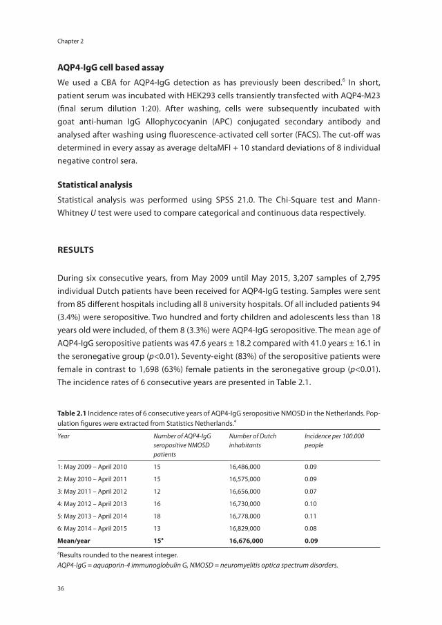
During six consecutive years, from May 2009 until May 2015, 3,207 samples of 2,795 individual Dutch patients have been received for AQP4-IgG testing. Samples were sent from 85 different hospitals including all 8 university hospitals. Of all included patients 94 (3.4%) were seropositive. Two hundred and forty children and adolescents less than 18 years old were included, of them 8 (3.3%) were AQP4-IgG seropositive. The mean age of AQP4-IgG seropositive patients was 47.6 years ± 18.2 compared with 41.0 years ± 16.1 in the seronegative group (p<0.01). Seventy-eight (83%) of the seropositive patients were female in contrast to 1,698 (63%) female patients in the seronegative group (p<0.01). The incidence rates of 6 consecutive years are presented in Table 2.1.
table 2.1 Incidence rates of 6 consecutive years of AQP4-IgG seropositive NMOSD in the Netherlands. Pop-ulation figures were extracted from Statistics Netherlands.4
Year Number of AQP4-IgG seropositive NMOSD patients
Number of Dutch inhabitants
Incidence per 100.000 people
1: May 2009 – April 2010 15 16,486,000 0.09
2: May 2010 – April 2011 15 16,575,000 0.09
3: May 2011 – April 2012 12 16,656,000 0.07
4: May 2012 – April 2013 16 16,730,000 0.10
5: May 2013 – April 2014 18 16,778,000 0.11
6: May 2014 – April 2015 13 16,829,000 0.08
mean/year 15a 16,676,000 0.09
aResults rounded to the nearest integer.AQP4-IgG = aquaporin-4 immunoglobulin G, NMOSD = neuromyelitis optica spectrum disorders.
37
Incidence of NMOSD in the Netherlands
2
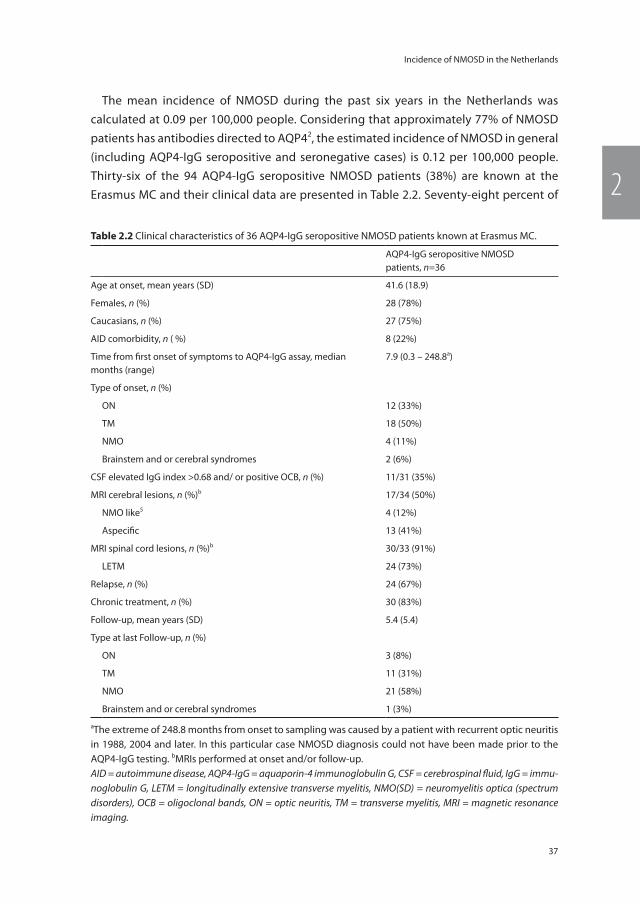
The mean incidence of NMOSD during the past six years in the Netherlands was calculated at 0.09 per 100,000 people. Considering that approximately 77% of NMOSD patients has antibodies directed to AQP42, the estimated incidence of NMOSD in general (including AQP4-IgG seropositive and seronegative cases) is 0.12 per 100,000 people. Thirty-six of the 94 AQP4-IgG seropositive NMOSD patients (38%) are known at the Erasmus MC and their clinical data are presented in Table 2.2. Seventy-eight percent of
table 2.2 Clinical characteristics of 36 AQP4-IgG seropositive NMOSD patients known at Erasmus MC.
AQP4-IgG seropositive NMOSD patients, n=36
Age at onset, mean years (SD) 41.6 (18.9)
Females, n (%) 28 (78%)
Caucasians, n (%) 27 (75%)
AID comorbidity, n ( %) 8 (22%)
Time from first onset of symptoms to AQP4-IgG assay, median months (range)
7.9 (0.3 – 248.8a)
Type of onset, n (%)
ON 12 (33%)
TM 18 (50%)
NMO 4 (11%)
Brainstem and or cerebral syndromes 2 (6%)
CSF elevated IgG index >0.68 and/ or positive OCB, n (%) 11/31 (35%)
MRI cerebral lesions, n (%)b 17/34 (50%)
NMO like5 4 (12%)
Aspecific 13 (41%)
MRI spinal cord lesions, n (%)b 30/33 (91%)
LETM 24 (73%)
Relapse, n (%) 24 (67%)
Chronic treatment, n (%) 30 (83%)
Follow-up, mean years (SD) 5.4 (5.4)
Type at last Follow-up, n (%)
ON 3 (8%)
TM 11 (31%)
NMO 21 (58%)
Brainstem and or cerebral syndromes 1 (3%)
aThe extreme of 248.8 months from onset to sampling was caused by a patient with recurrent optic neuritis in 1988, 2004 and later. In this particular case NMOSD diagnosis could not have been made prior to the AQP4-IgG testing. bMRIs performed at onset and/or follow-up.AID = autoimmune disease, AQP4-IgG = aquaporin-4 immunoglobulin G, CSF = cerebrospinal fluid, IgG = immu-noglobulin G, LETM = longitudinally extensive transverse myelitis, NMO(SD) = neuromyelitis optica (spectrum disorders), OCB = oligoclonal bands, ON = optic neuritis, TM = transverse myelitis, MRI = magnetic resonance imaging.
Chapter 2
38
them were females. Twenty-four patients had LETM at some point during their disease course. Eventually at last follow-up 21 patients (58%) fulfilled classic NMO criteria with optic neuritis and transverse myelitis.3
Discussion
Here we report the incidence of AQP4-IgG seropositive NMOSD in the Netherlands, derived from data of the Dutch national NMO expert centre, is nearly one in a million: 0.09 per 100,000 people. Unique for this study is that we have a nationwide coverage given that the CBA is performed in one central laboratory. Our incidence figure is within the range of previous described incidence rates which range from 0.05 – 0.4 per 100,000 people.7 It has to be considered that epidemiological studies on NMOSD are difficult to compare since they are based on different selection and inclusion criteria. For ex-ample different clinical definitions and AQP4-IgG assays were used. Also the ethnicities of included patients and the geographic coverage differed. Two studies performed in comparable geographic areas in Denmark and the United Kingdom differed essentially from our study, as both studies also included AQP4-IgG seronegative NMOSD patients and did not have nationwide coverage.8, 9
In a comparable Austrian study an incidence of 0.05 was calculated.10 The main dif-ference with our study is that the patients they identified were all Caucasian. However there are indications that some ethnic groups are overrepresented in NMOSD.11 In the Netherlands we estimated the incidence of NMOSD is more than twice as high in non-Caucasians. Based on 25 percent of the patients known at Erasmus MC were non-Caucasian and 11.9 percent of the Dutch inhabitants are non-Caucasian4 we estimated a mean annual incidence rate of NMOSD for non-Caucasians of 0.19 per 100,000 people and for Caucasians of 0.08 per 100,000 people.
We think our findings reflect the real incidence of AQP4-IgG seropositive NMOSD in the Netherlands. However, we cannot exclude that mild cases and forme fruste types of the disease2 have been missed. Fifty-eight percent of the NMOSD patients at the Erasmus MC fulfilled classic NMO criteria.3 Unfortunately we did not have access to the clinical data of all patients and therefore we could not present this figure for all NMOSD patients in the Netherlands. Only the clinical data of patients known at the Erasmus MC are presented, however covering over one third of the study population.
More awareness and better recognition of NMOSD might increase the incidence in the future. Further demographic studies and international collaboration in the NMO field would add to a better understanding of NMOSD.
39
Incidence of NMOSD in the Netherlands
2
reFerences
1. Wingerchuk DM, Lennon VA, Lucchinetti CF, Pittock SJ, Weinshenker BG. The spectrum of neuromyelitis optica. Lancet Neurol 2007; 6(9): 805-15.
2. Wingerchuk DM, Banwell B, Bennett JL, Cabre P, Carroll W, Chitnis T, et al. International con-sensus diagnostic criteria for neuromyelitis optica spectrum disorders. Neurology 2015; 85(2): 177-89.
3. Wingerchuk DM, Lennon VA, Pittock SJ, Lucchinetti CF, Weinshenker BG. Revised diagnostic criteria for neuromyelitis optica. Neurology 2006; 66(10): 1485-89.
4. Statistics Netherlands. Population figures, www.cbs.nl (2015, accessed 10 June 2015).
5. Kim HJ, Paul F, Lana-Peixoto MA, Tenembaum S, Asgari N, Palace J, et al. MRI characteristics of neuromyelitis optica spectrum disorder: an international update. Neurology 2015; 84(11): 1165-73.
6. Ketelslegers IA, Modderman PW, Vennegoor A, Killestein J, Hamann D, Hintzen RQ. Anti-bodies against aquaporin-4 in neuromyelitis optica: distinction between recurrent and monophasic patients. Mult Scler 2011; 17(12): 1527-30.
7. Pandit L, Asgari N, Apiwattanakul M, Palace J, Paul F, Leite MI, et al. Demographic and clini-cal features of neuromyelitis optica: A review. Mult Scler 2015; 21(7): 845-53.
8. Asgari N, Lillevang ST, Skejoe HP, Falah M, Stenager E, Kyvik KO. A population-based study of neuromyelitis optica in Caucasians. Neurology 2011; 76(18): 1589-95.
9. Jacob A, Panicker J, Lythgoe D, Elsone L, Mutch K, Das K, et al. The epidemiology of neuromyelitis optica amongst adults in the Merseyside county of United Kingdom. J Neurol 2013; 260(8): 2134-37.
10. Aboul-Enein F, Seifert-Held T, Mader S, Kuenz B, Lutterotti A, Rauschka H, et al. Neuromyeli-tis optica in Austria in 2011: to bridge the gap between neuroepidemiological research and practice in a study population of 8.4 million people. PLoS One 2013; 8(11): e79649.
11. Mealy MA, Wingerchuk DM, Greenberg BM, Levy M. Epidemiology of neuromyelitis optica in the United States. Arch Neurol 2012; 69(9): 1176-80.

2016
12
OCT
Chapter 3
Neuromyelitis optica spectrum disorders: comparison of clinical and MRI characteristics of AQP4-IgG versus MOG-IgG seropositive cases in the Netherlands
E.D. van Pelt*, Y.Y.M. Wong*, I.A. Ketelslegers, D. Hamann, R.Q. Hintzen.
* shared first authors
European Journal of Neurology, 2016
Chapter 3
42
AbstrAct
background and purpose Neuromyelitis optica spectrum disorders (NMOSD) are a group of rare inflammatory demyelinating disorders of the CNS. The identification of specific antibodies directed to aquaporin 4 (AQP4-IgG) led to the distinction from multiple sclerosis (MS). However, up to 25% of the clinically diagnosed NMO patients are seronegative for AQP4-IgG. A subgroup of these patients might be identified by antibodies directed to myelin oligodendrocyte glycoprotein (MOG-IgG). Our objective was to investigate whether the clinical characteristics of these patients differ.
methods Using a cell based assay, samples of 61 AQP4-IgG seronegative patients and 41 AQP4-IgG seropositive patients with clinically NMOSD were analysed for the presence of MOG-IgG. Clinical characteristics of the AQP4-IgG, MOG-IgG seropositive and double seronegative NMOSD patients were compared.
results Twenty of the 61 AQP4-IgG seronegative patients tested MOG-IgG seropositive (33%). MOG-IgG seropositive patients were more frequently males in contrast to AQP4-IgG seropositive patients (55% versus 15%, p<0.01) and Caucasians (90% versus 63%, p=0.03). They more frequently presented with coincident optic neuritis (ON) and trans-verse myelitis (TM) (40% versus 12%, p=0.02) and have a monophasic disease course (70% vs 29%, p<0.01). AQP4-IgG seropositive patients were 2.4 times more likely to suffer from relapses as compared with MOG-IgG seropositive patients (relative risk 2.4, 95% CI 1.2 – 4.7). AQP4-IgG seropositive patients had higher EDSS levels at last follow-up (p<0.01).
conclusion Antibodies directed to MOG identify a subgroup of AQP4-IgG seronegative NMO patients with generally a favourable monophasic disease course.
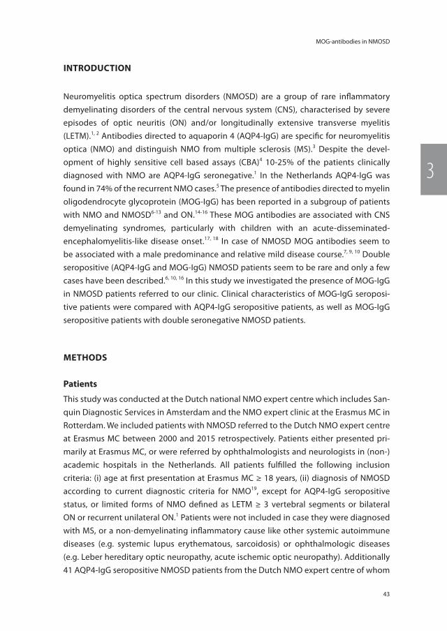
43
MOG-antibodies in NMOSD
3
introDuction
Neuromyelitis optica spectrum disorders (NMOSD) are a group of rare inflammatory demyelinating disorders of the central nervous system (CNS), characterised by severe episodes of optic neuritis (ON) and/or longitudinally extensive transverse myelitis (LETM).1, 2 Antibodies directed to aquaporin 4 (AQP4-IgG) are specific for neuromyelitis optica (NMO) and distinguish NMO from multiple sclerosis (MS).3 Despite the devel-opment of highly sensitive cell based assays (CBA)4 10-25% of the patients clinically diagnosed with NMO are AQP4-IgG seronegative.1 In the Netherlands AQP4-IgG was found in 74% of the recurrent NMO cases.5 The presence of antibodies directed to myelin oligodendrocyte glycoprotein (MOG-IgG) has been reported in a subgroup of patients with NMO and NMOSD6-13 and ON.14-16 These MOG antibodies are associated with CNS demyelinating syndromes, particularly with children with an acute-disseminated-encephalomyelitis-like disease onset.17, 18 In case of NMOSD MOG antibodies seem to be associated with a male predominance and relative mild disease course.7, 9, 10 Double seropositive (AQP4-IgG and MOG-IgG) NMOSD patients seem to be rare and only a few cases have been described.6, 10, 16 In this study we investigated the presence of MOG-IgG in NMOSD patients referred to our clinic. Clinical characteristics of MOG-IgG seroposi-tive patients were compared with AQP4-IgG seropositive patients, as well as MOG-IgG seropositive patients with double seronegative NMOSD patients.
methoDs
Patients
This study was conducted at the Dutch national NMO expert centre which includes San-quin Diagnostic Services in Amsterdam and the NMO expert clinic at the Erasmus MC in Rotterdam. We included patients with NMOSD referred to the Dutch NMO expert centre at Erasmus MC between 2000 and 2015 retrospectively. Patients either presented pri-marily at Erasmus MC, or were referred by ophthalmologists and neurologists in (non-) academic hospitals in the Netherlands. All patients fulfilled the following inclusion criteria: (i) age at first presentation at Erasmus MC ≥ 18 years, (ii) diagnosis of NMOSD according to current diagnostic criteria for NMO19, except for AQP4-IgG seropositive status, or limited forms of NMO defined as LETM ≥ 3 vertebral segments or bilateral ON or recurrent unilateral ON.1 Patients were not included in case they were diagnosed with MS, or a non-demyelinating inflammatory cause like other systemic autoimmune diseases (e.g. systemic lupus erythematous, sarcoidosis) or ophthalmologic diseases (e.g. Leber hereditary optic neuropathy, acute ischemic optic neuropathy). Additionally 41 AQP4-IgG seropositive NMOSD patients from the Dutch NMO expert centre of whom
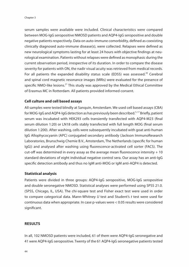
Chapter 3
44
serum samples were available were included. Clinical characteristics were compared between MOG-IgG seropositive NMOSD patients and AQP4-IgG seropositive and double negative patients respectively. Data on auto-immune comorbidity, defined as coexisting clinically diagnosed auto-immune disease(s), were collected. Relapses were defined as new neurological symptoms lasting for at least 24 hours with objective findings at neu-rological examination. Patients without relapses were defined as monophasic during the current observation period, irrespective of its duration. In order to compare the disease severity for patients with ON, the nadir visual acuity was retrieved from medical records. For all patients the expanded disability status scale (EDSS) was assessed.20 Cerebral and spinal cord magnetic resonance images (MRIs) were evaluated for the presence of specific NMO-like lesions.21 This study was approved by the Medical Ethical Committee of Erasmus MC in Rotterdam. All patients provided informed consent.
cell culture and cell based assays
All samples were tested blindly at Sanquin, Amsterdam. We used cell based assays (CBA) for MOG-IgG and AQP4-IgG detection as has previously been described.5, 17 Briefly, patient serum was incubated with HEK293 cells transiently transfected with AQP4-M23 (final serum dilution 1:20) or LN18 cells stably transfected with full length MOG (final serum dilution 1:200). After washing, cells were subsequently incubated with goat anti-human IgG Allophycocyanin (APC) conjugated secondary antibody (Jackson ImmunoResearch Laboratories, Brunschwig Chemie B.V., Amsterdam, The Netherlands (specific for human IgG)) and analysed after washing using fluorescence-activated cell sorter (FACS). The cut-off was determined in every assay as the average mean fluorescence intensity + 10 standard deviations of eight individual negative control sera. Our assay has an anti-IgG specific detection antibody and thus no IgM anti-MOG or IgM anti-AQP4 is detected.
statistical analysis
Patients were divided in three groups: AQP4-IgG seropositive, MOG-IgG seropositive and double seronegative NMOSD. Statistical analyses were performed using SPSS 21.0. (SPSS, Chicago, IL, USA). The chi-square test and Fisher exact test were used in order to compare categorical data. Mann-Whitney U test and Student’s t test were used for continuous data when appropriate. In case p-values were < 0.05 results were considered significant.
results
In all, 102 NMOSD patients were included, 61 of them were AQP4-IgG seronegative and 41 were AQP4-IgG seropositive. Twenty of the 61 AQP4-IgG seronegative patients tested
45
MOG-antibodies in NMOSD
3
MOG-IgG seropositive (33%). In none of the AQP4-IgG seropositive patients MOG-IgG was detected. An overview of the included patients is presented in Figure 3.1. The me-dian time to sampling was 10.7 months (0 – 401.5). Thirteen patients (12.7%) received chronic treatment whilst the sample was collected.
In Table 3.1 clinical characteristics are presented for MOG-IgG seropositive (n=20), AQP4-IgG seropositive (n=41) and double seronegative (n=41) NMOSD patients. Compared with AQP4-IgG seropositive patients, MOG-IgG seropositive patients were more frequently males (55% versus 15%, p<0.01) and Caucasians (90% versus 63%,
Patients with opticospinal phenotype
n = 102
AQP4-IgG seropositive patients n = 41
AQP4-IgG seronegative patients n = 61
MOG-IgG seropositive patients n = 20
Seronegative patients n = 41
Figure 3.1 Overview of included patients.
table 3.1 Clinical characteristics of MOG-IgG seropositive (n=20) and AQP4-IgG seropositive (n=41) and seronegative (n=41) patients with clinically NMOSD.
mog-igg+(n=20)
AQP4-igg+(n=41)
seronegative(n=41)
p-valuea p-valueb
Female, n (%) 9 (45) 35 (85) 25 (61) 0.00 0.24
Mean age at onset, years (SD) 36.2 (14.2) 42.0 (16.1) 40.4 (13.0) 0.23 0.26
Autoimmune comorbidity, n (%) 1 (5) 8 (20) 6 (15) 0.25 0.41
Familial autoimmune disease in fi rst and/or second degree relatives, n (%)
5/18 (28) 19/38 (50) 22/38 (58) 0.12 0.04
Caucasian, n (%) 18 (90) 26 (63) 34 (83) 0.03 0.70
Chapter 3
46
table 3.1 (continued)
mog-igg+(n=20)
AQP4-igg+(n=41)
seronegative(n=41)
p-valuea p-valueb
Follow-up, median,months (range)
23.9 (2.7-463.8) 61.5 (4.3-312.9) 23.9 (5.8-267.6) 0.09 0.49
Monophasic disease course 8.0 (2.7 – 463.8) 11.9 (4.3 – 61.5) 17.8 (5.8 – 202.4) 0.43 0.06
Relapsing disease course 106.4 (30.5 – 234.3) 115.8 (14.3 – 312.9) 34.6 (17.7 – 267.6) 0.81 0.07
Monophasic disease, n (%) 14 (70) 12 (29) 25 (61) 0.00 0.49
Phenotype at onset, n (%)
ON 8 (40) 16 (39) 15 (37) 0.94 0.80
Bilateral ON 6/8 (75) 6/16 (38) 11/15 (73) 0.19 1.00
LETM 4 (20) 20 (49) 15 (37) 0.03 0.19
ON and TM 8 (40) 5 (12) 11 (27) 0.02 0.30
Bilateral ON 7/8 (88) 4/5 (80) 8/11 (73) 1.00 0.60
Phenotype at last follow-up, n (%)
ON 6 (30) 4 (10) 10 (24) 0.07 0.64
LETM 3 (15) 11 (27) 12 (29) 0.35 0.34
ON and TM 11 (55) 26 (63) 19 (46) 0.53 0.53
CSF elevated protein >0.60, n (%) 4/14 (29) 12/32 (38) 9/32 (28) 0.74 1.00
CSF oligoclonal bands, n (%) 3/16 (19) 10/28 (36) 7/35 (20) 0.31 1.00
CSF IgG index >0.68, n (%) 4/13 (31) 13/27 (48) 15/34 (44) 0.30 0.40
EDSS at onset, median (range) 4.0 (2.0-8.0) 4.0 (1.5-9.0) 3.0 (1-9.5) 0.40 0.16
EDSS at best recovery, median (range)
1.0 (0-8.0) 2.0 (0-8.0) 3.0 (1.0-8.0) 0.06 0.01
EDSS at last FU, median (range) 1.0 (0-10) 3.0 (1.0-10.0) 3.0 (1.0-8.0) 0.01 0.02
Nadir visual acuity, median (range)
0.05 (0.003-0.7) 0.003 (0-1.0) 0.05 (0.002-1.0) 0.16 0.83
Chronic treatment, n (%)c 4 (20) 34 (83) 17 (43) 0.00 0.10
Duration, months (range) 40.3 (2.8 -79.1) 30.4 (1.3 – 152.4) 32.3 (3.5 – 213.3) 0.98 1.00
aComparison between MOG-IgG and AQP4-IgG seropositive patients; bcomparison between seropositive MOG-IgG and seronegative patients; c55 patients received chronic therapy: four MOG-IgG seropositive patients (one azathioprine, one mycophenolate, one immunomodulatory treatment, one mycophenolate and rituximab), 34 AQP4-IgG seropositive patients (27 azathioprine, five mycophenolate, two rituximab), 17 seronegative patients (11 azathioprine, four mycophenolate, one low dose oral prednisone, one mitoxan-trone). Significant results are reported in bold.AQP4-IgG = aquaporin-4 immunoglobulin G, CSF = cerebrospinal fluid, EDSS = expanded disability status scale, LETM = longitudinally extensive transverse myelitis, MOG-IgG = myelin oligodendrocyte glycoprotein immuno-globulin G, NMOSD = neuromyelitis optica spectrum disorder, ON = optic neuritis, TM = transverse myelitis.
47
MOG-antibodies in NMOSD
3
p=0.03). They more frequently presented with coincident ON and transverse myelitis (TM) (40% versus 12%, p=0.02). AQP4-IgG seropositive patient more often presented with longitudinally extensive transverse myelitis (LETM) (49% versus 20%, p=0.03). In Table 3.2 MRI features are presented. The cerebral MRI features as recently described in NMOSD patients21 were only found in het AQP4-IgG seropositive patients and not in the MOG-IgG seropositive patients (32% versus 0%, p<0.01). An example of typical NMO dorsal brainstem (a) and midbrain (b) lesions of one of our patients is presented in Figure 3.2. Spinal cord MRI features and cerebrospinal fluid (CSF) findings were similar in the AQP4-IgG versus MOG-IgG seropositive groups. MOG-IgG seropositive patients more frequently had a monophasic disease course (Table 3.1) (70% vs 29%, p<0.01). AQP4-IgG seropositive patients were 2.4 times more likely to suffer from relapses as compared with MOG-IgG seropositive patients (RR 2.4, 95% CI 1.2 – 4.7). EDSS at last follow-up was higher for AQP4-IgG seropositive patients (p<0.01). Presenting phenotype was not predictive for a relapsing disease course. Table 3.3 shows the clinical characteristics of the relapsing patients. TM relapses were more frequently seen in the AQP4-IgG group compared with the MOG-IgG seropositive patients (76% versus 17%, p=0.01).
table 3.2 MRI features of MOG-IgG and AQP4-IgG seropositive patients with NMOSD.
mog-igg+ (n=20) AQP4-igg+ (n=41) p-value
brain
NMO specific brain lesions, n (%) 0 12/38 (32)a 0.01
Aspecific brain lesions, n (%) 4 (20) 12/37 (32) 0.32
spinal cord
Affected spinal cord segments
Cervical 9/12 (75) 31/36 (86) 0.66
Thoracic 10/12 (83) 30/36 (83) 1.00
Lumbar 3/12 (25) 5/36 (14) 0.66
Whole spinal cord 3/12(25) 5/36 (14) 0.66
Central grey matter 10/12 (83) 30/36 (83) 1.00
aMagnetic resonance imaging was reviewed for the presence of NMO specific brain lesions as recently de-scribed.21 Five patients with diencephalic lesions surrounding the third ventricle and cerebral aquaduct, two patients with dorsal brainstem lesions, two patients with both diencephalic and dorsal brainstem lesions, one patient with a dorsal brainstem, diencephalic and periependymal lesion, one patient with a specific lesion of the internal capsule and one patient with an extensive confluent hemispheric lesion were detected.AQP4-IgG = aquaporin-4 immunoglobulin G, MOG-IgG = myelin oligodendrocyte glycoprotein immunoglobulin G, MRI = magnetic resonance imaging, NMO = neuromyelitis optica, NMSOD = neuromyelitis optica spectrum disorder.
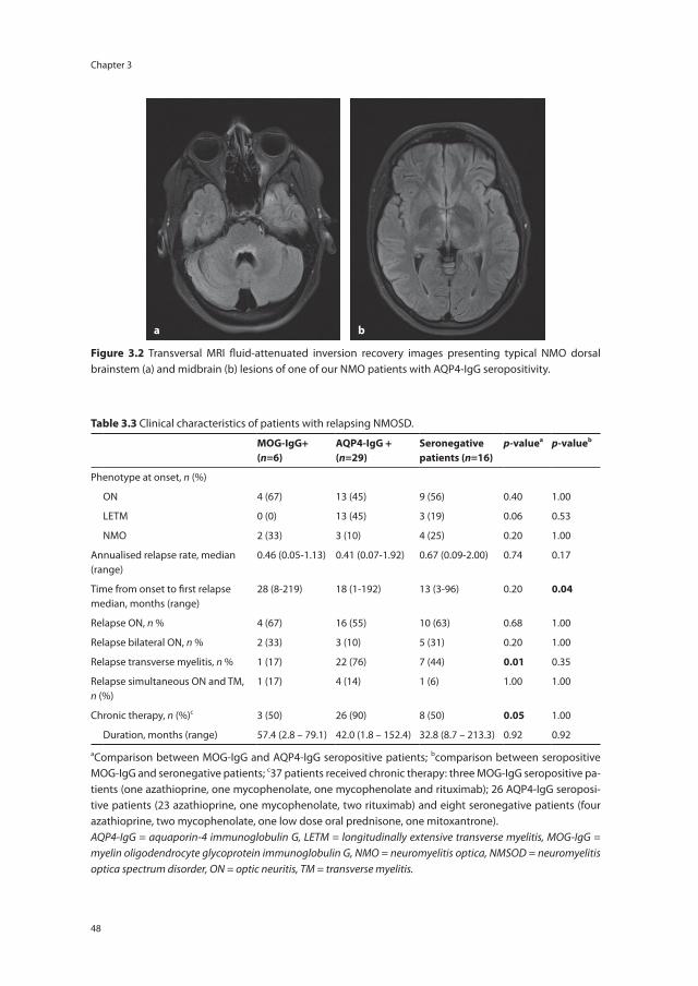
Chapter 3
48
a b
Figure 3.2 Transversal MRI fluid-attenuated inversion recovery images presenting typical NMO dorsal brainstem (a) and midbrain (b) lesions of one of our NMO patients with AQP4-IgG seropositivity.
table 3.3 Clinical characteristics of patients with relapsing NMOSD.
mog-igg+(n=6)
AQP4-igg +(n=29)
seronegativepatients (n=16)
p-valuea p-valueb
Phenotype at onset, n (%)
ON 4 (67) 13 (45) 9 (56) 0.40 1.00
LETM 0 (0) 13 (45) 3 (19) 0.06 0.53
NMO 2 (33) 3 (10) 4 (25) 0.20 1.00
Annualised relapse rate, median (range)
0.46 (0.05-1.13) 0.41 (0.07-1.92) 0.67 (0.09-2.00) 0.74 0.17
Time from onset to first relapse median, months (range)
28 (8-219) 18 (1-192) 13 (3-96) 0.20 0.04
Relapse ON, n % 4 (67) 16 (55) 10 (63) 0.68 1.00
Relapse bilateral ON, n % 2 (33) 3 (10) 5 (31) 0.20 1.00
Relapse transverse myelitis, n % 1 (17) 22 (76) 7 (44) 0.01 0.35
Relapse simultaneous ON and TM, n (%)
1 (17) 4 (14) 1 (6) 1.00 1.00
Chronic therapy, n (%)c 3 (50) 26 (90) 8 (50) 0.05 1.00
Duration, months (range) 57.4 (2.8 – 79.1) 42.0 (1.8 – 152.4) 32.8 (8.7 – 213.3) 0.92 0.92
aComparison between MOG-IgG and AQP4-IgG seropositive patients; bcomparison between seropositive MOG-IgG and seronegative patients; c37 patients received chronic therapy: three MOG-IgG seropositive pa-tients (one azathioprine, one mycophenolate, one mycophenolate and rituximab); 26 AQP4-IgG seroposi-tive patients (23 azathioprine, one mycophenolate, two rituximab) and eight seronegative patients (four azathioprine, two mycophenolate, one low dose oral prednisone, one mitoxantrone).AQP4-IgG = aquaporin-4 immunoglobulin G, LETM = longitudinally extensive transverse myelitis, MOG-IgG = myelin oligodendrocyte glycoprotein immunoglobulin G, NMO = neuromyelitis optica, NMSOD = neuromyelitis optica spectrum disorder, ON = optic neuritis, TM = transverse myelitis.
49
MOG-antibodies in NMOSD
3
Discussion
Antibodies directed to MOG can be detected in a subgroup of children with acquired demyelinating syndromes.17, 18 Recently it has been reported that MOG-IgG also can be found in adults with clinical NMOSD phenotypes and ON.6-16 In this study we confirm that MOG-IgG is present in approximately one third of the AQP4-IgG seronegative NMOSD cases. An overview of previous studies using a CBA for MOG-IgG testing in NMO(SD) and this study is presented in Table 3.4. In line with previous studies we found a male and Caucasian predominance, frequent coincident ON and TM, more often a monophasic disease course and lower EDSS levels at follow-up in MOG-IgG seropositive cases.7, 9, 10 Six out of 8 (75%) MOG-IgG seropositive patients with coincident ON and TM had a mono-phasic disease course as originally described as Devic’s syndrome.1 Follow-up periods between the MOG-IgG and AQP-IgG seropositive patients differed slightly, but not significantly. It cannot be excluded that upon further follow up some of the monophasic MOG-IgG seropositive patients will have relapses. In our study we observed a relatively high rate of a monophasic disease course in the AQP4-IgG seropositive patients, which is a result of our protocol to start immunosuppressive treatment in this group as early as possible, often already after the first attack.
In addition to previous studies, cerebral MRIs from MOG-IgG and AQP4-IgG seroposi-tive patients were assessed for presence of NMO specific brain lesions.21 These lesions were not present in MOG-IgG seropositive NMOSD patients, which might be explained by different underlying disease mechanisms.
AQP4-IgG, MOG-IgG seropositive NMOSD and seronegative NMOSD seem to be simi-lar in their clinical opticospinal phenotype, although there might be pathophysiological differences between these NMOSD subgroups.22, 23 Further neuropathological studies are needed in order to improve criteria for clinical NMOSD phenotype, since MOG-IgG seropositivity might reflect a separate demyelinating syndrome.
The presence of MOG-IgG further expands the spectrum of NMOSD. Although the presence of MOG-IgG is rare, the detection of it seems beneficial in clinical practice to differentiate patients with NMOSD from MS13 and may identify a subgroup of NMOSD patients with favourable outcome with lower EDSS levels at follow-up and less relapses. However it is still important to realise that some MOG IgG seropositive patients experi-ence frequent relapses and have prominent residual neurological deficits.
MOG-IgG was found in 33% of the clinical NMOSD AQP4-IgG seronegative cases. This is higher than has been described in some of the previous studies (Table 3.4).8-13, 15, 16 However, it is difficult to compare these percentages considering the different study protocols and inclusion criteria.
Our relative high percentage of MOG-IgG seropositivity is probably caused by the retrospective character of our study and selection bias of patients who have been
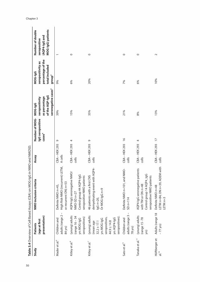
Chapter 3
50
tabl
e 3.
4 O
verv
iew
of C
ell B
ased
Ass
ays
(CBA
) on
MO
G-Ig
G in
NM
O a
nd N
MO
SD.
stud
yPa
tien
ts
(age
at fi
rst
pres
enta
tion
)
nm
o in
clus
ion
crit
eria
Ass
ayn
umbe
r of m
og
-ig
g s
erop
osit
ive
case
sa
mo
g-ig
g
sero
posi
tivi
ty
as p
erce
ntag
e of
the
AQ
P-ig
g
sero
nega
tive
cas
esa
mo
g-ig
g
sero
posi
tivi
ty a
s pe
rcen
tage
of t
he
tota
l inc
lude
d gr
oupa
num
ber o
f dou
ble
sero
posi
tive
(A
QP4
-igg
and
m
og
-igg
) pat
ient
s
Mad
er e
t al.6
Child
ren
and
adul
ts (r
ange
2 –
84
yrs
)
Defi
nite
NM
O n
=45,
Hig
h Ri
sk-N
MO
((re
curr
ent)
LET
M,
or re
curr
ent O
N) n
=53
CBA
– H
EK 2
93
A c
ells
939
%9%
1
Kitle
y et
al.8
(you
ng) a
dults
(r
ange
16
– 34
yr
s M
OG
-IgG
se
ropo
sitiv
es)
AQP4
-IgG
ser
oneg
ativ
e N
MO
/ N
MO
-SD
n=2
7Co
ntro
l gro
up 4
4 AQ
P4-Ig
G
sero
posi
tive
NM
O p
atie
nts
CBA
– H
EK 2
93
cells
415
%6%
0
Kitle
y et
al.7
(you
ng) a
dults
(m
ean
age
32.2
± 1
7.1
yrs
MO
G-Ig
G
sero
posi
tives
, 44
.9 ±
14.
8 yr
s AQ
P4-Ig
G
sero
posi
tives
).
46 p
atie
nts
with
a fi
rst C
NS
dem
yelin
atin
g ev
ent w
ith A
QP4
-Ig
G n
=20,
Or M
OG
-IgG
n=9
CBA
– H
EK 2
93
cells
935
%20
%0
Sato
et a
l.9Ch
ildre
n an
d ad
ults
(ran
ge 3
–
78 y
rs)
Defi
nite
NM
O n
=101
, and
NM
O-
SD n
=114
CBA
– H
EK 2
93
cells
1621
%7%
0
Tana
ka e
t al.11
(you
ng) a
dults
(r
ange
15
-78
yrs)
AQP4
-IgG
ser
oneg
ativ
e pa
tient
s w
ith T
M o
r ON
n=4
8Co
ntro
l gro
up 1
4 AQ
P4_I
gG
sero
posi
tive
NM
O p
atie
nts
CBA
– H
EK 2
93
cells
48%
6%0
Höf
tber
ger e
t al
.10Ad
ults
(ran
ge 1
8 –
77 y
rs)
Defi
nite
NM
O n
=48
LETM
n=8
4, O
N n
=39,
AD
EM w
ith
LETM
n=3
CBA
– H
EK 2
93
cells
1713
%10
%2
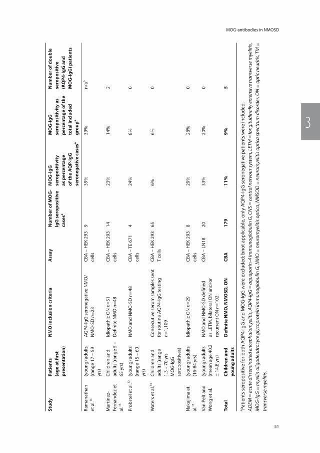
51
MOG-antibodies in NMOSD
3
stud
yPa
tien
ts
(age
at fi
rst
pres
enta
tion
)
nm
o in
clus
ion
crit
eria
Ass
ayn
umbe
r of m
og
-ig
g s
erop
osit
ive
case
sa
mo
g-ig
g
sero
posi
tivi
ty
as p
erce
ntag
e of
the
AQ
P-ig
g
sero
nega
tive
cas
esa
mo
g-ig
g
sero
posi
tivi
ty a
s pe
rcen
tage
of t
he
tota
l inc
lude
d gr
oupa
num
ber o
f dou
ble
sero
posi
tive
(A
QP4
-igg
and
m
og
-igg
) pat
ient
s
Ram
anat
han
et a
l.14(y
oung
) adu
lts
(ran
ge 1
7 –
59
yrs)
AQP4
-IgG
ser
oneg
ativ
e N
MO
/N
MO
-SD
n=2
3CB
A –
HEK
293
ce
lls9
39%
39%
n/ab
Mar
tinez
-Fe
rnan
dez
et
al.16
Child
ren
and
adul
ts (r
ange
5 –
65
yrs
)
Idio
path
ic O
N n
=51
Defi
nite
NM
O n
=48
CBA
– H
EK 2
93
cells
1423
%14
%2
Prob
stel
et a
l.12(y
oung
) adu
lts
(ran
ge 1
5 –
60
yrs)
NM
O a
nd N
MO
-SD
n=4
8CB
A –
TE
671
cells
424
%8%
0
Wat
ers
et a
l.13Ch
ildre
n an
d ad
ults
(ran
ge
1.3
– 70
yrs
M
OG
-IgG
se
ropo
sitiv
es)
Cons
ecut
ive
seru
m s
ampl
es s
ent
for r
outin
e AQ
P4-Ig
G te
stin
g n=
1,10
9
CBA
– H
EK 2
93
T ce
lls65
6%6%
0
Nak
ajim
a et
al
.15(y
oung
) adu
lts
(16-
84 y
rs)
Idio
path
ic O
N n
=29
CBA
– H
EK 2
93
cells
829
%28
%0
Van
Pelt
and
Won
g et
al.
(you
ng) a
dults
(m
ean
age
40.2
±
14.8
yrs
)
NM
O a
nd N
MO
-SD
defi
ned
as L
ETM
, bila
tera
l ON
and
/or
recu
rren
t ON
n=1
02
CBA
– L
N18
2033
%20
%0
tota
lch
ildre
n an
d yo
ung
adul
tsD
efini
te n
mo
, nm
osD
, on
cbA
179
11%
9%5
a Patie
nts
sero
posi
tive
for b
oth
AQP4
-IgG
and
MO
G-Ig
G w
ere
excl
uded
; bno
t app
licab
le, o
nly
AQP4
-IgG
ser
oneg
ativ
e pa
tient
s w
ere
incl
uded
.AD
EM =
acu
te d
isse
min
ated
enc
epha
lom
yelit
is, A
QP4
-IgG
= a
quap
orin
-4 im
mun
oglo
bulin
G, C
NS
= ce
ntra
l ner
vous
syst
em, L
ETM
= lo
ngitu
dina
lly e
xten
sive
tran
sver
se m
yelit
is,
MO
G-Ig
G =
mye
lin o
ligod
endr
ocyt
e gl
ycop
rote
in im
mun
oglo
bulin
G, N
MO
= n
euro
mye
litis
opt
ica,
NM
SOD
= n
euro
mye
litis
opt
ica
spec
trum
dis
orde
r, O
N =
opt
ic n
eurit
is, T
M =
tr
ansv
erse
mye
litis.
Chapter 3
52
referred to our NMO expert centre at Erasmus MC Rotterdam. The scope of the current study was to compare clinical characteristics of MOG-IgG versus AQP4-IgG seropositive NMOSD patients, rather than to determine the prevalence of MOG-IgG seropositive CNS demyelination. Further studies are needed in order to present epidemiological figures of AQP4-IgG and MOG-IgG seropositivity in the Netherlands.
This study confirms MOG-IgG seropositivity in a subgroup of patients with clinically NMOSD in the Netherlands. Limitation of our study is a relative small sample size and therefore we could not make statistical corrections. Also we have not collected sequen-tial samples.
Even though AQP4-IgG and MOG-IgG represent a considerable amount of patients with the clinical profile of NMOSD, there is still a group of patients without antibodies. Future studies are needed to gain more insight in this group of seronegative NMOSD patients in order to possibly detect new autoantibodies, to better customise treatment for individual patients and to predict their prognosis.
53
MOG-antibodies in NMOSD
3
reFerences
1. Wingerchuk DM, Lennon VA, Lucchinetti CF, Pittock SJ, Weinshenker BG. The spectrum of neuromyelitis optica. Lancet Neurol 2007; 6(9): 805-15.
2. McKeon A, Fryer JP, Apiwattanakul M, Lennon VA, Hinson SR, Kryzer TJ, et al. Diagnosis of neuromyelitis spectrum disorders: compara-tive sensitivities and specificities of immu-nohistochemical and immunoprecipitation assays. Arch Neurol 2009; 66(9): 1134-38.
3. Lennon VA, Wingerchuk DM, Kryzer TJ, Pittock SJ, Lucchinetti CF, Fujihara K, et al. A serum autoantibody marker of neuromyelitis optica: distinction from multiple sclerosis. Lancet 2004; 364(9451): 2106-12.
4. Lennon VA, Kryzer TJ, Pittock SJ, Verkman AS, Hinson SR. IgG marker of optic-spinal multiple sclerosis binds to the aquaporin-4 water channel. J Exp Med 2005; 202(4): 473-77.
5. Ketelslegers IA, Modderman PW, Vennegoor A, Killestein J, Hamann D, Hintzen RQ. Anti-bodies against aquaporin-4 in neuromyelitis optica: distinction between recurrent and monophasic patients. Mult Scler 2011; 17(12): 1527-30.
6. Mader S, Gredler V, Schanda K, Rostasy K, Duj-movic I, Pfaller K, et al. Complement activating antibodies to myelin oligodendrocyte glyco-protein in neuromyelitis optica and related disorders. J Neuroinflammation 2011; 8: 184.
7. Kitley J, Waters P, Woodhall M, Leite MI, Murchison A, George J, et al. Neuromyelitis optica spectrum disorders with aquaporin-4 and myelin-oligodendrocyte glycoprotein antibodies: a comparative study. JAMA Neurol 2014; 71(3): 276-83.
8. Kitley J, Woodhall M, Waters P, Leite MI, De-venney E, Craig J, et al. Myelin-oligodendro-cyte glycoprotein antibodies in adults with a neuromyelitis optica phenotype. Neurology 2012; 79(12): 1273-77.
9. Sato DK, Callegaro D, Lana-Peixoto MA, Waters PJ, Jorge FMD, Takahashi T, et al. Distinction between MOG antibody-positive and AQP4 antibody-positive NMO spectrum disorders. Neurology 2014; 82(6): 474-81.
10. Hoftberger R, Sepulveda M, Armangue T, Blanco Y, Rostasy K, Cobo Calvo A, et al. Antibodies to MOG and AQP4 in adults with neuromyelitis optica and suspected limited forms of the disease. Mult Scler 2015; 21(7): 866-74.
11. Tanaka M, Tanaka K. Anti-MOG antibodies in adult patients with demyelinating disorders of the central nervous system. J Neuroim-munol 2014; 270(1-2): 98-99.
12. Probstel AK, Rudolf G, Dornmair K, Collongues N, Chanson JB, Sanderson NS, et al. Anti-MOG antibodies are present in a subgroup of pa-tients with a neuromyelitis optica phenotype. J Neuroinflammation 2015; 12: 46.
13. Waters P, Woodhall M, O’Connor KC, Reindl M, Lang B, Sato DK, et al. MOG cell-based assay detects non-MS patients with inflammatory neurologic disease. Neurol Neuroimmunol Neuroinflamm 2015; 2(3): e89.
14. Ramanathan S, Reddel SW, Henderson A, Par-ratt JD, Barnett M, Gatt PN, et al. Antibodies to myelin oligodendrocyte glycoprotein in bilateral and recurrent optic neuritis. Neurol Neuroimmunol Neuroinflamm 2014; 1(4): e40.
15. Nakajima H, Motomura M, Tanaka K, Fujikawa A, Nakata R, Maeda Y, et al. Antibodies to myelin oligodendrocyte glycoprotein in idiopathic optic neuritis. BMJ Open 2015; 5(4): e007766.
16. Martinez-Hernandez E, Sepulveda M, Rostasy K, Hoftberger R, Graus F, Harvey RJ, et al. An-tibodies to aquaporin 4, myelin-oligodendro-cyte glycoprotein, and the glycine receptor alpha1 subunit in patients with isolated optic neuritis. JAMA Neurol 2015; 72(2): 187-93.
17. Ketelslegers IA, Van Pelt DE, Bryde S, Neute-boom RF, Catsman-Berrevoets CE, Hamann D, et al. Anti-MOG antibodies plead against MS diagnosis in an Acquired Demyelinating Syndromes cohort. Mult Scler 2015; 21(12): 1513-20.
18. Reindl M, Di Pauli F, Rostasy K, Berger T. The spectrum of MOG autoantibody-associated demyelinating diseases. Nat Rev Neurol 2013; 9(8): 455-61.
Chapter 3
54
19. Wingerchuk DM, Banwell B, Bennett JL, Cabre P, Carroll W, Chitnis T, et al. International con-sensus diagnostic criteria for neuromyelitis optica spectrum disorders. Neurology 2015; 85(2): 177-89.
20. Kurtzke JF. Rating neurologic impairment in multiple sclerosis: an expanded disability status scale (EDSS). Neurology 1983; 33(11): 144-52.
21. Kim HJ, Paul F, Lana-Peixoto MA, Tenembaum S, Asgari N, Palace J, et al. MRI characteristics of neuromyelitis optica spectrum disorder: an
international update. Neurology 2015; 84(11): 1165-73.
22. Bernard-Valnet R, Liblau RS, Vukusic S, Marignier R. Neuromyelitis optica: a positive appraisal of seronegative cases. Eur J Neurol 2015; 22(12): 1511-18.
23. Zamvil SS, Slavin AJ. Does MOG Ig-positive AQP4-seronegative opticospinal inflam-matory disease justify a diagnosis of NMO spectrum disorder? Neurol Neuroimmunol Neuroinflamm 2015; 2(1): e62.
2016
12
OCT
Part two
Acquired demyelinating syndromes in children

2016
12
OCT
Chapter 4
Application of the 2012 revised diagnostic definitions for paediatric multiple sclerosis and immune-mediated central nervous system demyelination disorders
E.D. van Pelt, R.F. Neuteboom, I.A. Ketelslegers, M. Boon, C.E. Catsman-Berrevoets, R.Q. Hintzen. On behalf of the Dutch Study Group for Paediatric MS.
Journal of Neurology Neurosurgery and Psychiatry, 2014
Chapter 4
58
AbstrAct
background Recently, the International Paediatric MS Study Group (IPMSSG) definitions for the diagnosis of immune-mediated acquired demyelinating syndromes (ADS) of the central nervous system including paediatric multiple sclerosis (MS) have been revised.
objective To evaluate the 2012 revised IPMSSG consensus definitions in a cohort of children with ADS prospectively followed from January 2007.
methods Children with ADS who had an MRI scan obtained within 90 days after first disease onset were included. The sensitivity and specificity of the 2007 and 2012 IPMSSG consensus definitions were assessed. The time to MS diagnosis applying the 2007 and 2012 definitions was compared using survival analysis and log-rank test.
results Eighty-two children with ADS were included. Thirty-five children were diagnosed with paediatric MS, of whom 30 experienced a second clinical event. The final diagnosis applying either the 2007 or 2012 IPMSSG definitions corresponded. The revised 2012 definitions had sufficient sensitivity (80%) and high specificity (100%). MS diagnosis was made 3.4 months earlier (Х²=8.24, p=0.004) applying the new definitions. In 14 children MS diagnosis was made at first MRI.
conclusions MS diagnosis can be made reliable and early using the 2012 IPMSSG consensus definitions. This is beneficial for adequate counseling of children and their families and for early treatment possibilities.
59
2012 IPMSSG consensus definitions for childhood-onset ADS
4
introDuction
Recently, the diagnostic criteria for the diagnosis of immune-mediated acquired demy-elinating syndromes (ADS) of the central nervous system (CNS) including paediatric mul-tiple sclerosis (MS) have been revised by the International Paediatric Multiple Sclerosis Study Group (IPMSSG).1 The new definitions aim to improve consistency in terminology in the heterogeneous group of paediatric demyelinating disorders. They incorporate the in 2010 revised McDonald criteria for MS.2 Diagnosing MS earlier in the disease course is favourable because disease modifying treatment is also beneficial in children with MS.3 Previous studies already showed that the 2010 McDonald magnetic resonance imaging (MRI) criteria, which allow diagnosis at disease onset, are useful for early MS diagnosis in children.4-8 But they recommended to be cautious when applying these MRI criteria to young children with acute disseminated encephalomyelitis (ADEM).4, 6 Therefore the IPMSSG included in the revised 2012 definitions that the 2010 McDonald MRI criteria for dissemination in time (DIT) and space (DIS) cannot be applied at first event in children with ADEM and children younger than 12 years old.1 In the new IPMSSG consensus definitions, diagnostic criteria for ADEM and neuromyelitis optica (NMO) have been sharpened and the term recurrent ADEM is excluded. Our aim was to evaluate the new 2012 IPMSSG consensus definitions in our clinical cohort of prospectively included children with ADS.
methoDs
Patients and definitions
Children younger than 18 years, who presented with a first episode of acquired demy-elination of the CNS between January 2007 and April 2013, were prospectively included. All children were identified by the Dutch Study Group for Paediatric MS, which consists of paediatric neurologists in 13 major paediatric neurology centres in the Netherlands, or the children were identified by Dutch paediatricians who participate in the NSCK (Neth-erlands Paediatric Surveillance Unit) as has been described elsewhere.9 Children were eligible for this study when a cerebral MRI scan had been obtained within 90 days after first onset of symptoms. All patients had a minimum follow-up time of 2 years, unless they were diagnosed with definite MS. MS diagnosis could be made either on evidence of a second clinical attack at least 30 days after the initial attack, or on MRI evidence of DIT within 2 years. Patients were classified as MS, ADEM, clinically isolated syndrome (CIS), NMO, or other relapsing demyelinating disorders according to the 200710 and new 20121 IPMSSG consensus definitions.
Chapter 4
60
standard protocol approvals, registrations and patient consents
This study was approved by the Medical Ethical Committees of the Erasmus MC in Rot-terdam and of the other participating centres. Written informed consent was obtained from all children and/or their parents.
mri analysis
MRI scans were performed on 1.5-Tesla MRI scanners with slice thicknesses of 3-5 mm. Scans were archived as electronic images. The presence of lesions was determined on T2-weighted and fluid attenuated inversion recovery (FLAIR) images. All scans were scored blinded to clinical data by 2 experienced raters (E.D.v.P. and R.F.N.). MRI scans were classified as meeting the 2007 IPMSSG definitions for MS10 (based on the 2001 McDonald MRI criteria11) or the 2012 IPMSSG definitions for MS1 (based on the 2010 McDonald MRI criteria2). It should be noted that according to the 2007 IPMSSG consensus definitions, DIS can also be met by the combination of abnormal cerebrospinal fluid (CSF) and two lesions on MRI of which one must be in the brain. The 2012 IPMSSG definitions did not incorporate CSF findings anymore. According to the 2007 IPMSSG consensus definitions, MRI criteria for DIT can only be applied on a MRI scan made 3 months after disease onset in children ≥ 10 years old.10 In contrast, using the 2012 IPMSSG consensus definitions MS can be diagnosed after a single event in children ≥ 12 years old, not meeting ADEM criteria and meeting the 2010 McDonald MRI criteria for DIS and DIT on the baseline MRI scan.1
statistical analysis
Analyses were performed using SPSS version 20.0. Patient characteristics were com-pared using Chi-Square and student’s t-test. Test characteristics of the 2007 and 2012 diagnostic criteria (i.e. sensitivity, specificity, positive predictive value (PPV), negative predictive value (NPV) and accuracy) were calculated using clinical definite MS (CDMS) as endpoint. Children with MS diagnosis based on MRI criteria who started disease modifying treatment (DMT) before CDMS diagnosis (n=2) and those with a follow-up of <1 years (n=3) were excluded from the analysis of test characteristics. Kaplan-Meier survival analyses were used to analyse time to diagnosis according to the 2007 and 2012 IPMSSG consensus definitions. The time to diagnosis was compared using a log-rank test. Survival analysis included all patients with a first MRI with gadolinium and either the presence of a follow-up scan or a diagnosis of CDMS according to the 2010 McDon-ald MRI criteria.
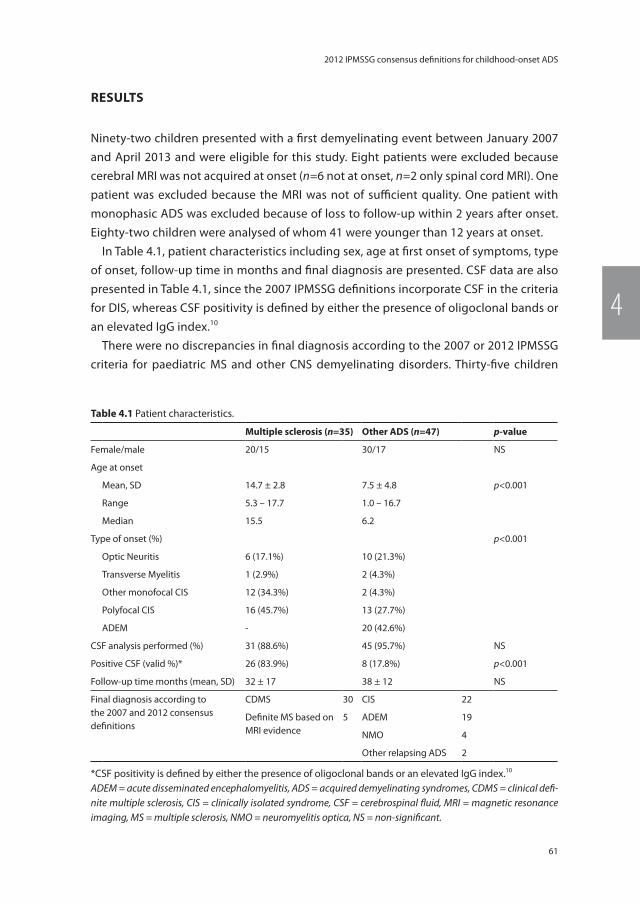
61
2012 IPMSSG consensus definitions for childhood-onset ADS
4
results
Ninety-two children presented with a first demyelinating event between January 2007 and April 2013 and were eligible for this study. Eight patients were excluded because cerebral MRI was not acquired at onset (n=6 not at onset, n=2 only spinal cord MRI). One patient was excluded because the MRI was not of sufficient quality. One patient with monophasic ADS was excluded because of loss to follow-up within 2 years after onset. Eighty-two children were analysed of whom 41 were younger than 12 years at onset.
In Table 4.1, patient characteristics including sex, age at first onset of symptoms, type of onset, follow-up time in months and final diagnosis are presented. CSF data are also presented in Table 4.1, since the 2007 IPMSSG definitions incorporate CSF in the criteria for DIS, whereas CSF positivity is defined by either the presence of oligoclonal bands or an elevated IgG index.10
There were no discrepancies in final diagnosis according to the 2007 or 2012 IPMSSG criteria for paediatric MS and other CNS demyelinating disorders. Thirty-five children
table 4.1 Patient characteristics.
multiple sclerosis (n=35) other ADs (n=47) p-value
Female/male 20/15 30/17 NS
Age at onset
Mean, SD 14.7 ± 2.8 7.5 ± 4.8 p<0.001
Range 5.3 – 17.7 1.0 – 16.7
Median 15.5 6.2
Type of onset (%) p<0.001
Optic Neuritis 6 (17.1%) 10 (21.3%)
Transverse Myelitis 1 (2.9%) 2 (4.3%)
Other monofocal CIS 12 (34.3%) 2 (4.3%)
Polyfocal CIS 16 (45.7%) 13 (27.7%)
ADEM - 20 (42.6%)
CSF analysis performed (%) 31 (88.6%) 45 (95.7%) NS
Positive CSF (valid %)* 26 (83.9%) 8 (17.8%) p<0.001
Follow-up time months (mean, SD) 32 ± 17 38 ± 12 NS
Final diagnosis according to the 2007 and 2012 consensus definitions
CDMS 30 CIS 22
Definite MS based on MRI evidence
5 ADEM 19
NMO 4
Other relapsing ADS 2
*CSF positivity is defined by either the presence of oligoclonal bands or an elevated IgG index.10
ADEM = acute disseminated encephalomyelitis, ADS = acquired demyelinating syndromes, CDMS = clinical defi-nite multiple sclerosis, CIS = clinically isolated syndrome, CSF = cerebrospinal fluid, MRI = magnetic resonance imaging, MS = multiple sclerosis, NMO = neuromyelitis optica, NS = non-significant.
Chapter 4
62
are diagnosed with MS according to both 2007 and 2012 IPMSSG definitions. Five of them did not have a second clinical event confirming CDMS during current follow-up. Two of these patients have started DMT, which may have suppressed a second clini-cal event. Children with MS were older (F=18.35, p<0.001) and have a different type of onset (X²=27.6, p<0.001) as compared to children with monophasic or other relapsing demyelinating disorders. None of the children currently diagnosed with MS presented with ADEM, as defined by a polyfocal event with encephalopathy.1
Four patients were diagnosed with NMO according to both 2007 and 2012 consensus definitions.1, 10 Three of them suffered from a relapsing disease course. Anti-aquaporin-4 antibodies were tested in 39 patients (48%) as part of their diagnostic follow-up using a cell based assay and fluorescence activated cell sorter (FACS).12 Two patients had anti-aquaporin-4 IgG seropositivity, one patient with recurrent NMO and one patient who presented with a brainstem syndrome and was diagnosed with NMO related spectrum disorder.13 Two patients had a final diagnosis of other relapsing demyelinating disorders: one patient suffered from a relapsing optic neuritis (ON) in absence of MRI lesions fulfill-ing DIT for MS diagnosis and aquaporin-4 antibodies for NMO spectrum diagnosis. The other patient suffered from a first event of ADEM followed by one event of ON without presence of new MRI lesions during follow-up.14 None of the children with ADEM-onset had a subsequent event of ADEM (multiphasic ADEM).
In Table 4.2 sensitivity and specificity of the 2007 and 2012 IPMSSG consensus defini-tions are presented with CDMS as endpoint using only the baseline characteristics and using also follow-up MRI. The 2012 IPMSSG consensus definitions had better sensitivity (79 vs 68%), NPV (88 vs 71%) and accuracy (92 vs 82%) compared to the 2007 definitions. The test characteristics differed not significantly. For the calculations of DIS and DIT 2010 McDonald MRI criteria at onset, only children who had a first MRI with gadolinium were included. Two patients who presented with ADEM fulfilled the 2010 McDonald MRI cri-teria at the first MRI and one of them also fulfilled DIS and DIT 2001 and 2010 McDonald MRI criteria at second MRI (>3 months after onset). Both these patients had negative CSF at onset and did not have a second event. Follow-up time of these patients was 53 and 58 months respectively. However, according to the 2012 IPMSSG definitions the 2010 McDonald MRI criteria do not apply for children with an ADEM-onset and both children were younger than 12 years old. Therefore according to both the 2007 as 2012 IPMSSG consensus definitions these children cannot be diagnosed as MS. In contrast, 2 other patients presenting with ADS and younger than 12 years fulfilled the 2010 McDonald MRI criteria for DIS and DIT at baseline, but because of their age they were not diag-nosed with MS at first MRI according to 2012 IPMSSG consensus definitions. Both these patients eventually showed clinical progression and thus were diagnosed with CDMS and turned out to have a false-negative test result at baseline. Applying the 2010 DIS and DIT McDonald MRI criteria at disease onset, thus irrespective of the age limit of 12

63
2012 IPMSSG consensus definitions for childhood-onset ADS
4
tabl
e 4.
2 Te
st c
hara
cter
istic
s of
the
MRI
crit
eria
incl
ude
the
2007
and
201
2 IP
MSS
G c
onse
nsus
defi
nitio
ns w
hen
appl
ied
to d
istin
guis
h M
S fr
om o
ther
acq
uire
d de
my-
elin
atin
g sy
ndro
mes
(AD
S) o
f the
CN
S.
Firs
t mri
Firs
t mri
Firs
t mri
Firs
t mri
Firs
t mri
Firs
t and
sec
ond
mri
Firs
t and
sec
ond
mri
2007
DIS
McD
onal
d 20
01 c
riter
ia
(at l
east
3 o
ut o
f 4);
- ≥9
esio
ns o
n T2
-w
eigh
ted
imag
es o
r 1
gado
liniu
m-e
nhan
c-in
g le
sion
- ≥3
Periv
entr
icul
ar
lesi
ons
- ≥1
Juxt
acor
tical
lesi
on- ≥
1 In
fran
tent
oria
l le
sion
2007
DIS
- abn
orm
al C
SF- 2
lesi
ons
on th
e M
RI o
f whi
ch 1
m
ust b
e in
the
brai
n*
2012
DIS
McD
onal
d 20
10
crite
ria (a
t lea
st 2
ou
t of 4
);- ≥
1 Sp
inal
cor
d- ≥
1 Pe
riven
tric
ular
- ≥1
Juxt
acor
tical
le
sion
- ≥1
Infr
ante
ntor
ial
lesi
on
2012
DIT
McD
onal
d 20
10
crite
ria;
- Sim
ulta
neou
s pr
esen
ce o
f as
ympt
om-
atic
gad
olin
ium
-en
hanc
ing
and
non-
enha
ncin
g le
sion
s.- E
xclu
ding
chi
ldre
n ≤1
2 ye
ars
old†
2012
DIS
+DIT
McD
onal
d 20
10
for D
IS a
nd D
IT a
t ba
selin
e.- E
xclu
ding
chi
ldre
n≤1
2 ye
ars
old†
2007
DIS
+DIT
McD
onal
d 20
01 D
IT;
- New
T2-
or g
ado-
liniu
m e
nhan
cing
le
sion
(s) m
ore
than
3 m
onth
s af
ter o
nset
on
follo
w-u
p M
RI.
- Exc
ludi
ng c
hild
ren
≤10
year
s ol
d‡
2012
DIS
+DIT
McD
onal
d 20
10 D
IT;
- New
T2
and/
or g
adol
in-
ium
-enh
anci
ng le
sion
(s)
on fo
llow
-up
MRI
, with
re
fere
nce
to a
bas
elin
e sc
an ir
resp
ectiv
e of
the
timin
g of
the
base
line
scan
- Sim
ulta
neou
s pr
es-
ence
of a
sym
ptom
atic
ga
dolin
ium
-enh
anci
ng
and
non-
enha
ncin
g le
sion
s at
any
tim
e§
Sens
itivi
ty (%
)70
7593
6358
6879
Spec
ifici
ty (%
)96
8977
100
100
100
100
PPV
(%)
9181
7110
010
010
010
0
NPV
(%)
8385
9582
8071
88
Accu
racy
(%)
8684
8386
8582
92
In th
is ta
ble
test
cha
ract
eris
tics
are
pres
ente
d w
ith C
DM
S as
end
poin
t usi
ng fi
rst a
nd s
econ
d M
RI.
* Ca
lcul
ated
for p
atie
nts
whe
n CS
F an
alys
is w
as p
erfo
rmed
(n=7
3).
† Ca
lcul
ated
for c
hild
ren
who
had
a fi
rst M
RI w
ith g
adol
iniu
m (n
=65)
.‡
Calc
ulat
ed fo
r chi
ldre
n w
ho h
ad a
sec
ond
MRI
follo
win
g 3
mon
ths
afte
r ons
et (n
=39)
.§
Calc
ulat
ed fo
r chi
ldre
n w
ho h
ad th
eir fi
rst M
RI w
ith g
adol
iniu
m a
nd/o
r had
sec
ond
MRI
, exc
ludi
ng c
hild
ren
≤12
year
s ol
d fo
r DIT
201
0 at
bas
elin
e (n
=73)
.AD
S =
acqu
ired
dem
yelin
atin
g sy
ndro
mes
, CN
S =
cent
ral n
ervo
us sy
stem
, CSF
= ce
rebr
ospi
nal fl
uid,
DIS
= d
isse
min
atio
n in
spac
e, D
IT =
dis
sem
inat
ion
in ti
me,
IPM
SSG
= In
tern
a-tio
nal P
aedi
atric
MS
Stud
y G
roup
, MRI
= m
agne
tic re
sona
nce
imag
ing,
MS
= m
ultip
le sc
lero
sis,
NPV
= n
egat
ive
pred
ictiv
e va
lue,
PPV
= p
ositi
ve p
redi
ctiv
e va
lue.
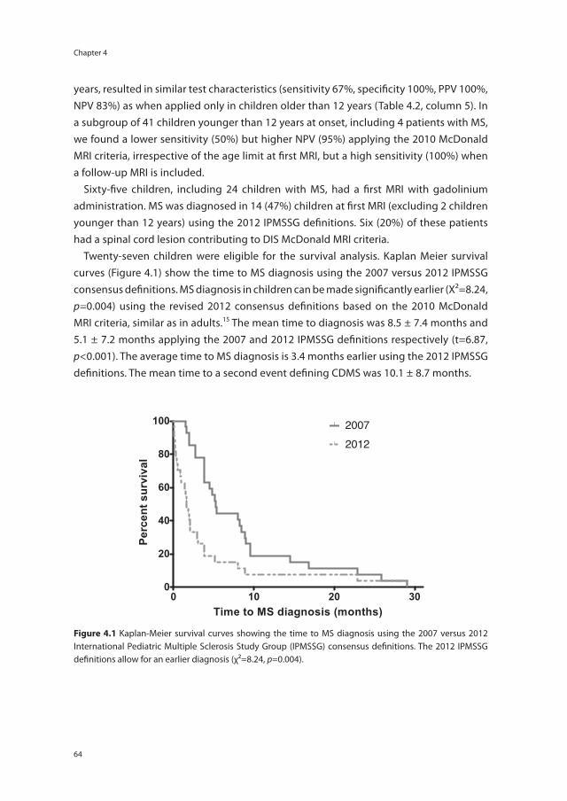
Chapter 4
64
years, resulted in similar test characteristics (sensitivity 67%, specificity 100%, PPV 100%, NPV 83%) as when applied only in children older than 12 years (Table 4.2, column 5). In a subgroup of 41 children younger than 12 years at onset, including 4 patients with MS, we found a lower sensitivity (50%) but higher NPV (95%) applying the 2010 McDonald MRI criteria, irrespective of the age limit at first MRI, but a high sensitivity (100%) when a follow-up MRI is included.
Sixty-five children, including 24 children with MS, had a first MRI with gadolinium administration. MS was diagnosed in 14 (47%) children at first MRI (excluding 2 children younger than 12 years) using the 2012 IPMSSG definitions. Six (20%) of these patients had a spinal cord lesion contributing to DIS McDonald MRI criteria.
Twenty-seven children were eligible for the survival analysis. Kaplan Meier survival curves (Figure 4.1) show the time to MS diagnosis using the 2007 versus 2012 IPMSSG consensus definitions. MS diagnosis in children can be made significantly earlier (Х²=8.24, p=0.004) using the revised 2012 consensus definitions based on the 2010 McDonald MRI criteria, similar as in adults.15 The mean time to diagnosis was 8.5 ± 7.4 months and 5.1 ± 7.2 months applying the 2007 and 2012 IPMSSG definitions respectively (t=6.87, p<0.001). The average time to MS diagnosis is 3.4 months earlier using the 2012 IPMSSG definitions. The mean time to a second event defining CDMS was 10.1 ± 8.7 months.
0 10 20 300
20
40
60
80
100 20072012
Time to MS diagnosis (months)
Perc
ent s
urvi
val
Survivalcurven=27.pzf:Data 1 - Tue Aug 09 12:30:13 2016
Figure 4.1 Kaplan-Meier survival curves showing the time to MS diagnosis using the 2007 versus 2012 International Pediatric Multiple Sclerosis Study Group (IPMSSG) consensus definitions. The 2012 IPMSSG definitions allow for an earlier diagnosis (χ²=8.24, p=0.004).
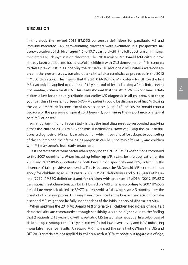
65
2012 IPMSSG consensus definitions for childhood-onset ADS
4
Discussion
In this study the revised 2012 IPMSSG consensus definitions for paediatric MS and immune-mediated CNS demyelinating disorders were evaluated in a prospective na-tionwide cohort of children aged 1.0 to 17.7 years old with the full spectrum of immune-mediated CNS demyelination disorders. The 2010 revised McDonald MRI criteria have already been studied and found useful in children with CNS demyelination.4-8 In contrast to these previous studies, not only the revised 2010 McDonald MRI criteria were consid-ered in the present study, but also other clinical characteristics as proposed in the 2012 IPMSSG definitions. This means that the 2010 McDonald MRI criteria for DIT on the first MRI can only be applied to children of 12 years and older and having a first clinical event not meeting criteria for ADEM. This study showed that the 2012 IPMSSG consensus defi-nitions allow for an equally reliable, but earlier MS diagnosis in all children, also those younger than 12 years. Fourteen (47%) MS patients could be diagnosed at first MRI using the 2012 IPMSSG definitions. Six of these patients (20%) fulfilled DIS McDonald criteria because of the presence of spinal cord lesion(s), confirming the importance of a spinal cord MRI at onset.7
An important finding in our study is that the final diagnoses corresponded applying either the 2007 or 2012 IPMSSG consensus definitions. However, using the 2012 defini-tions, a diagnosis of MS can be made earlier, which is beneficial for adequate counseling of the children and their families, as prognosis can be uncertain after ADS, and children with MS may benefit from early treatment.
Test characteristics were better when applying the 2012 IPMSSG definitions compared to the 2007 definitions. When including follow-up MRI scans for the application of the 2007 and 2012 IPMSSG definitions, both have a high specificity and PPV, indicating the absence of false positive test results. This is because the McDonald MRI criteria do not apply for children aged ≥ 10 years (2007 IPMSSG definitions) and ≥ 12 years at base-line (2012 IPMSSG definitions) and for children with an onset of ADEM (2012 IPMSSG definitions). Test characteristics for DIT based on MRI criteria according to 2007 IPMSSG definitions were calculated for 39/77 patients with a follow up scan ≥ 3 months after the onset of clinical symptoms. This may have introduced some bias as the decision to make a second MRI might not be fully independent of the initial observed disease activity.
When applying the 2010 McDonald MRI criteria to all children (regardless of age) test characteristics are comparable although sensitivity would be higher, due to the finding that 2 patients ≤ 12 years old with paediatric MS tested false negative. In a subgroup of children aged younger than 12 years old we found lower sensitivity and NPV, indicating more false negative results. A second MRI increased the sensitivity. When the DIS and DIT 2010 criteria are not applied in children with ADEM at onset but regardless of age,
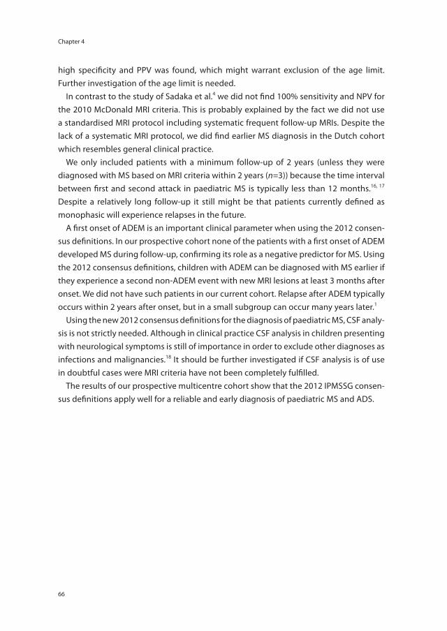
Chapter 4
66
high specificity and PPV was found, which might warrant exclusion of the age limit. Further investigation of the age limit is needed.
In contrast to the study of Sadaka et al.4 we did not find 100% sensitivity and NPV for the 2010 McDonald MRI criteria. This is probably explained by the fact we did not use a standardised MRI protocol including systematic frequent follow-up MRIs. Despite the lack of a systematic MRI protocol, we did find earlier MS diagnosis in the Dutch cohort which resembles general clinical practice.
We only included patients with a minimum follow-up of 2 years (unless they were diagnosed with MS based on MRI criteria within 2 years (n=3)) because the time interval between first and second attack in paediatric MS is typically less than 12 months.16, 17 Despite a relatively long follow-up it still might be that patients currently defined as monophasic will experience relapses in the future.
A first onset of ADEM is an important clinical parameter when using the 2012 consen-sus definitions. In our prospective cohort none of the patients with a first onset of ADEM developed MS during follow-up, confirming its role as a negative predictor for MS. Using the 2012 consensus definitions, children with ADEM can be diagnosed with MS earlier if they experience a second non-ADEM event with new MRI lesions at least 3 months after onset. We did not have such patients in our current cohort. Relapse after ADEM typically occurs within 2 years after onset, but in a small subgroup can occur many years later.1
Using the new 2012 consensus definitions for the diagnosis of paediatric MS, CSF analy-sis is not strictly needed. Although in clinical practice CSF analysis in children presenting with neurological symptoms is still of importance in order to exclude other diagnoses as infections and malignancies.18 It should be further investigated if CSF analysis is of use in doubtful cases were MRI criteria have not been completely fulfilled.
The results of our prospective multicentre cohort show that the 2012 IPMSSG consen-sus definitions apply well for a reliable and early diagnosis of paediatric MS and ADS.
67
2012 IPMSSG consensus definitions for childhood-onset ADS
4
reFerences
1. Krupp LB, Tardieu M, Amato MP, Banwell B, Chitnis T, Dale RC, et al. International Pediatric Multiple Sclerosis Study Group criteria for pediatric multiple sclerosis and immune-me-diated central nervous system demyelinating disorders: revisions to the 2007 definitions. Mult Scler 2013; 19(10): 1261-67.
2. Polman CH, Reingold SC, Banwell B, Clanet M, Cohen JA, Filippi M, et al. Diagnostic criteria for multiple sclerosis: 2010 revisions to the McDonald criteria. Ann Neurol 2011; 69(2): 292-302.
3. Chitnis T, Tenembaum S, Banwell B, Krupp L, Pohl D, Rostasy K, et al. Consensus statement: evaluation of new and existing therapeutics for pediatric multiple sclerosis. Mult Scler 2012; 18(1): 116-27.
4. Sadaka Y, Verhey LH, Shroff MM, Branson HM, Arnold DL, Narayanan S, et al. 2010 McDonald criteria for diagnosing pediatric multiple sclerosis. Ann Neurol 2012; 72(2): 211-23.
5. Sedani S, Lim MJ, Hemingway C, Wassmer E, Absoud M. Paediatric multiple sclerosis: ex-amining utility of the McDonald 2010 criteria. Mult Scler 2012; 18(5): 679-82.
6. Tantsis EM, Prelog K, Brilot F, Dale RC. Risk of multiple sclerosis after a first demyelinating syndrome in an Australian Paediatric cohort: clinical, radiological features and application of the McDonald 2010 MRI criteria. Mult Scler 2013; 19(13): 1749-59.
7. Hummel HM, Bruck W, Dreha-Kulaczewski S, Gartner J, Wuerfel J. Pediatric onset multiple sclerosis: McDonald criteria 2010 and the contribution of spinal cord MRI. Mult Scler 2013; 19(10): 1330-35.
8. Kornek B, Schmitl B, Vass K, Zehetmayer S, Pritsch M, Penzien J, et al. Evaluation of the 2010 McDonald multiple sclerosis criteria in children with a clinically isolated syndrome. Mult Scler 2012; 18(12): 1768-74.
9. Ketelslegers IA, Catsman-Berrevoets CE, Neuteboom RF, Boon M, van Dijk KG, Eikelen-boom MJ, et al. Incidence of acquired de-myelinating syndromes of the CNS in Dutch children: a nationwide study. J Neurol 2012; 259(9): 1929-35.
10. Krupp LB, Banwell B, Tenembaum S, Interna-tional Pediatric MSSG. Consensus definitions proposed for pediatric multiple sclerosis and related disorders. Neurology 2007; 68(16 Suppl 2): S7-12.
11. McDonald WI, Compston A, Edan G, Goodkin D, Hartung HP, Lublin FD, et al. Recommend-ed diagnostic criteria for multiple sclerosis: guidelines from the International Panel on the diagnosis of multiple sclerosis. Ann Neurol 2001; 50(1): 121-27.
12. Ketelslegers IA, Modderman PW, Vennegoor A, Killestein J, Hamann D, Hintzen RQ. Anti-bodies against aquaporin-4 in neuromyelitis optica: distinction between recurrent and monophasic patients. Mult Scler 2011; 17(12): 1527-30.
13. Wingerchuk DM, Lennon VA, Lucchinetti CF, Pittock SJ, Weinshenker BG. The spectrum of neuromyelitis optica. Lancet Neurol 2007; 6(9): 805-15.
14. Huppke P, Rostasy K, Karenfort M, Huppke B, Seidl R, Leiz S, et al. Acute disseminated encephalomyelitis followed by recurrent or monophasic optic neuritis in pediatric patients. Mult Scler 2013; 19(7): 941-46.
15. Runia TF, Jafari N, Hintzen RQ. Application of the 2010 revised criteria for the diagnosis of multiple sclerosis to patients with clinically isolated syndromes. Eur J Neurol 2013; 20(12): 1510-16.
16. Banwell B, Bar-Or A, Arnold DL, Sadovnick D, Narayanan S, McGowan M, et al. Clinical, environmental, and genetic determinants of multiple sclerosis in children with acute demyelination: a prospective national cohort study. Lancet Neurol 2011; 10(5): 436-45.
17. Mikaeloff Y, Suissa S, Vallee L, Lubetzki C, Ponsot G, Confavreux C, et al. First episode of acute CNS inflammatory demyelination in childhood: prognostic factors for multiple sclerosis and disability. J Pediatr 2004; 144(2): 246-52.
18. Banwell B, Ghezzi A, Bar-Or A, Mikaeloff Y, Tar-dieu M. Multiple sclerosis in children: clinical diagnosis, therapeutic strategies, and future directions. Lancet Neurol 2007; 6(10): 887-902.

2016
12
OCT
Chapter 5
Validation of MRI predictors of multiple sclerosis diagnosis in children with acute CNS demyelination
L.H. Verhey*, E.D. van Pelt*, I.A. Ketelslegers, R.F. Neuteboom, C.E. Catsman-Berrevoets, B.M. Feldman, D.L. Streiner, J.G. Sled, R.Q. Hintzen, B. Banwell.
* shared first authors
Multiple Sclerosis and Related Disorders, 2013
Chapter 5
70
AbstrAct
background In a recent Canadian prospective study of children with acute demyelin-ating syndromes (ADS), we demonstrated that the presence of T2 periventricular and T1-hypointense lesions predicted MS diagnosis. We aimed to validate these predictors in a Dutch cohort of children with ADS.
methods Participants with ADS were identified from a prospective cohort or archived dataset. MS was diagnosed based on clinical or MRI evidence of relapsing disease. Base-line MRI scans were evaluated for the presence of the two predictive parameters. Sen-sitivity, specificity, positive (LR+) and negative likelihood ratios (LR-), and positive (PPV) and negative predictive value (NPV) were calculated to evaluate the performance of the MRI parameters at classifying children as having MS or monophasic demyelination.
Findings Of 115 children identified with ADS between December 1993 and December 2009, MRI scans from 87 children (45 prospective; 47 archived) were evaluated; scans of 28 children were excluded due to incomplete or poor quality imaging. Mean duration of observation was longer in the archived group (7.1 years, SD 3.5) than the prospective cohort (3.3 years, SD 1.4). Thirty children were diagnosed with MS. Performance of the parameters was not statistically different between the prospective cohort (sensitivity 93.3% [68.1-99.8]; specificity 86.7% [69.3-96.2]; LR+ 7.0 [2.8-17.6]; LR- 0.08 [0.01-0.5]; PPV 77.8% [52.4-93.6]; NPV 96.3% [81.0-99.9]) and archived group (sensitivity 66.7% [38.4-88.2]; specificity 85.2% [66.3-95.8]; LR+ 4.5 [1.7-11.9]; LR- 0.4 [0.2-0.8]; PPV 71.4% [41.9-91.6]; NPV 82.1% [63.1-93.9]).
interpretation In an independent Dutch cohort, we confirm that the presence of ≥1 T2 periventricular and ≥1 T1-hypointense lesions reliably identifies children with MS.
71
Verhey MRI predictors for MS diagnosis in children
5
introDuction
The ability of magnetic resonance imaging (MRI) to aid in identifying children at risk for multiple sclerosis (MS) is important for early diagnosis and prompt initiation of MS-targeted therapies. With treatment trials for pediatric-onset MS currently being planned, tools to stratify children with acute demyelinating syndromes (ADS) of the central ner-vous system (CNS) into groups at highest risk for MS and those at low risk for recurrent demyelination has become increasingly valuable. We recently proposed MRI parameters associated with MS diagnosis in children with incident ADS. The presence of at least one T2 periventricular lesion and one or more T1-hypointense lesions at onset were predic-tive of MS diagnosis (sensitivity 84%, specificity 93%).1 In collaboration with the Dutch Study Group for Pediatric MS, our aim was to validate the predictive MRI parameters in an independent cohort of children with ADS.
methoDs
Participants and definitions
We included patients younger than 17 years of age with ADS. Participants were identi-fied from either an archived group or a prospective cohort under the Dutch Study Group for Pediatric MS, a nationwide network of 13 pediatric healthcare centers in the Netherlands.
Archived groupChildren less than 17 years of age who presented with an ADS, based on clinical features, between 1990 and 2007 inclusive were identified from hospital records by pediatric neurologists at 11 participating centers. These centers are located in nine large cities with complete geographic coverage of the Netherlands. Demographic, clinical and laboratory data collected at onset of ADS and at any follow-up assessments were extracted from health records by a trained individual using standardized case report forms, and subsequently entered into a centralized database. Duration of follow-up was determined based on the last clinic visit or telephone contact with the site neurologist or pediatrician. MRI scans acquired at onset, and any serial scans were retrieved from each hospital for centralized analysis.
Prospective GroupBeginning in 2008, children with incident ADS are being consecutively enrolled into a nationwide prospective study at each of the 13 participating centers (8 academic pediatric hospitals and 5 regional hospitals). To confirm nationwide inclusion of chil-
Chapter 5
72
dren with ADS, email surveys were sent out to pediatricians through the Netherlands Pediatric Surveillance Unit (response rate: 85%) in which they reported suspected cases of ADS. Uniform definitions for ADS2 and clinical assessments are conducted at onset and annually by trained neurologists at each site using standardized case report forms. In the instance that patients are unable to attend a clinic visit, neurologists at the lead site perform a telephone interview with the patient and site neurologist. MRI scans are not acquired according to a standardized research protocol at all sites; however, recom-mended sequences and scanner settings were provided to each site to increase the consistency of image acquisition. MRI scans are acquired at the time of incident ADS, at three months following ADS, and annually. Acute demyelinating syndromes were defined according to International Pediatric Multiple Sclerosis Study Group consensus definitions,2 and included optic neuritis (ON), transverse myelitis (TM), monofocal neurological deficits other than ON or TM, polyfocal deficits, and acute disseminated encephalomyelitis (ADEM) – defined by the presence of polyfocal neurological deficits and encephalopathy. Eligible children had a brain MRI scan acquired within 90 days of incident demyelinating attack. Each participant had at least two years of clinical observation following acute demyelination. Children with neuromyelitis optica were excluded.3 Participants or their guardians in the prospective cohort provided written informed consent. This study was conducted under research ethics approvals obtained previously at each participating center for the archived group and prospective cohort.
The outcome was defined as MS diagnosis, which was adjudicated by neurologists with expertise in demyelinating disorders (RFN, CEC-B, RQH) independently of the indi-vidual (LHV) who performed the analyses of baseline MRI scans. MS diagnosis was based on either a confirmed second demyelinating attack occurring at least 28 days after the incident episode,4 or on the accrual of new lesions according to the 2005 McDonald criteria for dissemination in time (DIT) in patients who had not yet experienced a second attack at the time of data analysis.5 To adjudicate whether participants met MRI criteria for DIT, available serial scans were evaluated by neurologists at the lead site (RFN, CEC-B, RQH). Children initially diagnosed with ADEM were diagnosed with MS if they had two or more non-ADEM attacks, provided that the subsequent events occurred at least 90 days after their initial ADS and the two subsequent attacks were separated by at least 28 days.2 Participants not meeting clinical or MRI criteria for MS diagnosis were classified as having monophasic demyelination at the time of data analysis.
mri analysis
MRI scans were acquired at each participating site on 1.0 or 1.5 Tesla scanners in the archived group and 1.5 Tesla scanners in the prospective study. Scans were archived as either electronic images or hardcopy films at Erasmus MC or Sophia Children’s Hospital (Rotterdam, the Netherlands). For inclusion, T1-weighted and T2-weighted or fluid at-
73
Verhey MRI predictors for MS diagnosis in children
5
tenuated inversion recovery (FLAIR) images acquired at the time of ADS were required for each patient. An individual (LHV) with expertise in neuroimaging excluded MRI scans with artifact due to patient motion or dental hardware, inadequate coverage of the whole brain, or poor grey matter–white matter contrast, according to pre-defined qual-ity criteria.1 The MRI scan acquired at the time of incident ADS for each participant was evaluated by a trained rater (LHV) blinded to clinical data. Lesions were evaluated, mea-sured, and localized according to a priori rules previously described.1 Two parameters were scored on each baseline scan as described in our original work:1 i) T1-hypointense lesions were defined as lesions hypointense relative to cortical grey matter and nonen-hancing on post-contrast T1-weighted imaging, and ii) T2 periventricular lesions were defined as lesions hyperintense on T2-weighted images abutting any portion of the lateral ventricles (including lesions in the corpus callosal white matter, but excluding deep grey matter lesions). Each scan was classified as meeting the MRI parameters when at least one T2 periventricular and one or more T1-hypointense lesions were present.
statistical analysis
Categorical data were summarized as frequency (%) and continuous data as mean (standard deviation, SD) or median (interquartile range, IQR). Univariate statistics were computed using Fisher’s exact tests, Χ2 tests, t tests and Mann-Whitney U tests as ap-propriate. We evaluated the performance of the MRI parameters against confirmed MS diagnosis by calculating sensitivity, specificity, positive predictive value (PPV), and negative predictive value (NPV).6 In addition, we also calculated likelihood ratios for a positive test (LR+) and negative test (LR-) in order to evaluate our parameters using a metric that is independent of the prevalence of the outcome of interest,6 although the relative proportion of children diagnosed with MS following an ADS event was actually similar in the Canadian and Dutch cohorts (approximately 30% during the periods of observation).1, 7-9 For the estimates of diagnostic performance, 95% confidence intervals were computed from the binomial distribution. We performed a sensitivity analysis to assess whether the performance of the MRI parameters was influenced by method of MS diagnosis (based on occurrence of a second clinical attack4 or MRI evidence of DIT alone5). Statistical analyses were conducted using Stata version 11 (College Station, Texas, USA).
results
Of 115 children and adolescents presenting with an incident ADS between December 1993 and December 2009 who were eligible, 87 were analyzed (Figure 5.1). Twenty-eight children were excluded because one or more of the required brain MRI sequences was
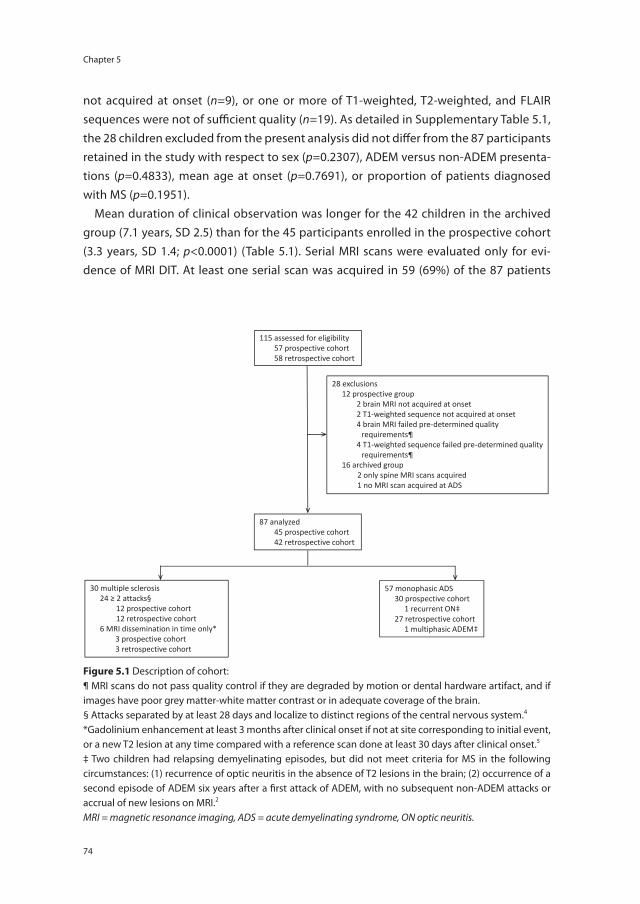
Chapter 5
74
not acquired at onset (n=9), or one or more of T1-weighted, T2-weighted, and FLAIR sequences were not of sufficient quality (n=19). As detailed in Supplementary Table 5.1, the 28 children excluded from the present analysis did not differ from the 87 participants retained in the study with respect to sex (p=0.2307), ADEM versus non-ADEM presenta-tions (p=0.4833), mean age at onset (p=0.7691), or proportion of patients diagnosed with MS (p=0.1951).
Mean duration of clinical observation was longer for the 42 children in the archived group (7.1 years, SD 2.5) than for the 45 participants enrolled in the prospective cohort (3.3 years, SD 1.4; p<0.0001) (Table 5.1). Serial MRI scans were evaluated only for evi-dence of MRI DIT. At least one serial scan was acquired in 59 (69%) of the 87 patients
115 assessed for eligibility 57 prospective cohort 58 retrospective cohort
30 multiple sclerosis 24 ≥ 2 attacks§
12 prospective cohort 12 retrospective cohort
6 MRI dissemination in time only* 3 prospective cohort 3 retrospective cohort
57 monophasic ADS 30 prospective cohort 1 recurrent ON‡ 27 retrospective cohort 1 multiphasic ADEM‡
87 analyzed 45 prospective cohort 42 retrospective cohort
28 exclusions 12 prospective group 2 brain MRI not acquired at onset 2 T1-weighted sequence not acquired at onset
4 brain MRI failed pre-determined quality requirements¶
4 T1-weighted sequence failed pre-determined quality requirements¶
16 archived group 2 only spine MRI scans acquired 1 no MRI scan acquired at ADS
Figure
Figure 5.1 Description of cohort:¶ MRI scans do not pass quality control if they are degraded by motion or dental hardware artifact, and if images have poor grey matter-white matter contrast or in adequate coverage of the brain.§ Attacks separated by at least 28 days and localize to distinct regions of the central nervous system.4
*Gadolinium enhancement at least 3 months after clinical onset if not at site corresponding to initial event, or a new T2 lesion at any time compared with a reference scan done at least 30 days after clinical onset.5
‡ Two children had relapsing demyelinating episodes, but did not meet criteria for MS in the following circumstances: (1) recurrence of optic neuritis in the absence of T2 lesions in the brain; (2) occurrence of a second episode of ADEM six years after a first attack of ADEM, with no subsequent non-ADEM attacks or accrual of new lesions on MRI.2
MRI = magnetic resonance imaging, ADS = acute demyelinating syndrome, ON optic neuritis.
75
Verhey MRI predictors for MS diagnosis in children
5
(24 had one serial scan, 23 had two, and 12 had at least three); the remaining 28 (32%) children did not have any serial imaging acquired.
Of 87 children, 30 (34%) children were diagnosed with MS, and 57 (66%) have ex-perienced monophasic ADS to date (Table 5.1). Of the 30 children diagnosed with MS, 24 (80%) experienced two or more clinical attacks,4 whereas the remaining 6 (20%) patients were diagnosed with MS based on MRI evidence of DIT5 alone. The median time from incident demyelination to MS diagnosis was shorter in the prospective group (3.6 months, IQR 2.4-8.4) than the archived group (14.4 months, IQR 6.0-20.4; p=0.019). One child in the archived group, who presented with transverse myelitis, was diagnosed with
table 5.1 Clinical and demographic characteristics of participants.
Archived group Prospective group
overall(n=42)
ms(n=15)
monophasic ADs (n=27)
overall(n=45)
ms(n=15)
monophasic ADs (n=30)
p-value*
Length of clinical observation (years)
7.1 (3.5) 8.0 (3.5) 6.6 (3.4) 3.3 (1.4) 3.3 (1.3) 3.3 (1.5) <0.0001
Age at onset (years) 9.5 (4.9) 12.1 (3.5) 8.0 (5.0) 9.1 (5.5) 15.1 (1.9) 6.1 (4.1) 0.734
Age at onset
<10 years 19 (45%) 4 (27%) 15 (56%) 26 (58%) 1 (7%) 25 (83%) 0.286
≥10 years 23 (55%) 11 (73%) 12 (44%) 19 (42%) 14 (93%) 5 (17%)
Female 22 (52%) 10 (67%) 12 (44%) 27 (60%) 7 (47%) 20 (67%) 0.521
Female : male ratio 1.1 2.0 0.8 1.5 0.9 2.0 --
Age at MS diagnosis (years)
-- 13.8 (3.9) -- -- 15.6 (1.8) -- 0.121§
Time to MS diagnosis (years)
-- 1.2 (0.5-1.7) -- -- 0.3 (0.2-0.7) -- 0.019§
2nd clinical attack -- 12 (80%) -- -- 12 (80%) -- 1.0§
ADS phenotype
ADEM 13 (31%) 1 (7%) 12 (44%) 15 (33%) 0 (0%) 15 (50%) 0.605
Transverse myelitis 5 (12%) 1 (7%) 4 (15%) 2 (4%) 1 (7%) 1 (3%)
Optic neuritis 5 (12%) 2 (13%) 3 (11%) 4 (9%) 1 (7%) 3 (10%)
Monofocal – other 8 (19%) 7 (47%) 1 (4%) 7 (16%) 5 (33%) 2 (7%)
Polyfocal 11 (26%) 4 (27%) 7 (26%) 17 (38%) 8 (53%) 9 (30%)
Abnormal MRI scan 34 (81%) 14 (93%) 20 (74%) 38 (84%) 15 (100%) 23 (77%) 0.779
*Comparison between the overall archived and prospective groups. §Comparison between children with MS in the archived group and those with MS in the prospective group. MS diagnosis based on either a confirmed second clinical attack at least 28 days after incident demyelination4, or 2005 McDonald criteria for MRI dissemination in time: gadolinium enhancement occurred at least 3 months after clinical onset if not at the site corresponding to the initial event, or a new T2 lesion was seen at any time compared with a reference scan acquired at least 30 days after clinical onset.5
Data are mean (standard deviation, SD), median (interquartile range, IQR) or number (%).MS = multiple sclerosis, ADS = acute demyelinating syndrome, ADEM = acute disseminated encephalomyelitis.
Chapter 5
76
MS after 8 years and has subsequently experienced three relapses. Five (17%) children with MS experienced their first attack prior to ten years of age. Of the 28 children who met criteria for ADEM at onset, only one was diagnosed with MS after experiencing two non-ADEM attacks.2 Of the 27 children with clinically monophasic ADEM, 17 had serial MRI scans and none had new lesions.
T2 lesions were present in 73 (84%) children; 14 (16%) had normal brain imaging at onset (1 with ADEM, 5 with monofocal ON, 5 with monofocal TM, and 3 with polyfocal neurological deficits). Of the 30 children with MS, 29 had T2-weighted lesions present on brain MRI at ADS. The one child without brain lesions presented with transverse myelitis, but subsequently developed clinical and MRI evidence of brain involvement.
Table 5.2 demonstrates the performance of the MRI parameters. Comparing the pro-spective cohort to the archived group with regards to performance of the MRI param-eters at correctly identifying children diagnosed with MS according to the occurrence of a second clinical attack or MRI evidence of DIT, sensitivity (93.3% vs. 66.7%; p=0.0686), specificity (86.7% vs. 85.2%; p=0.8706), PPV (77.8% vs. 71.4%; p=0.6783), and NPV (96.3% vs. 82.1%; p=0.0915) were not statistically different. Similarly, the 95% confidence inter-vals overlapped considerably when comparing positive and negative likelihood ratios of the prospective cohort (LR+ 7.0, 95% CI 2.8-17.6; LR- 0.08, 95% CI 0.01-0.5) to the archived group (LR+ 4.5, 95% CI 1.7-11.9; LR- 0.39, 95% CI 0.2-0.8).
In total, MRI scans of six patients with MS (5 in the archived group; 1 in the prospective cohort) did not demonstrate the MRI parameters on baseline images (20% false negative rate). Of these, one child who presented with transverse myelitis did not have brain le-sions at onset. Of the remaining five children (three of whom were under ten years of age at ADS), three children had T2 brain lesions, but no T2 periventricular or T1-hypointense lesions, and two had T2 periventricular lesions but no T1-hypointense lesions.
Eight children (4 in the archived group; 4 in the prospective cohort) were predicted to have MS based on presence of both MRI parameters, but to date have not experienced MRI or clinical evidence of relapsing disease (14% false positive rate). Of the eight par-ticipants, 6 were female and 6 were less than 10 years of age at onset. Three children presented with ADEM, 2 with monofocal deficits, and 3 with polyfocal neurological deficits.
We performed a sensitivity analysis to evaluate the effect of method of MS diagnosis on the performance of the MRI parameters. As illustrated in Table 5.2, when MS diagnosis was defined on the basis of clinical relapses only4, estimates of diagnostic performance were not significantly different from the estimates obtained when MS was diagnosed on the basis of a second attack or MRI evidence of DIT (statistical analyses not shown).
In a post hoc analysis, we evaluated the performance of the MRI parameters at cor-rectly classifying children with ADEM as having monophasic demyelination. Of the 28 children with monophasic ADEM, 25 were correctly classified as not having MS and three
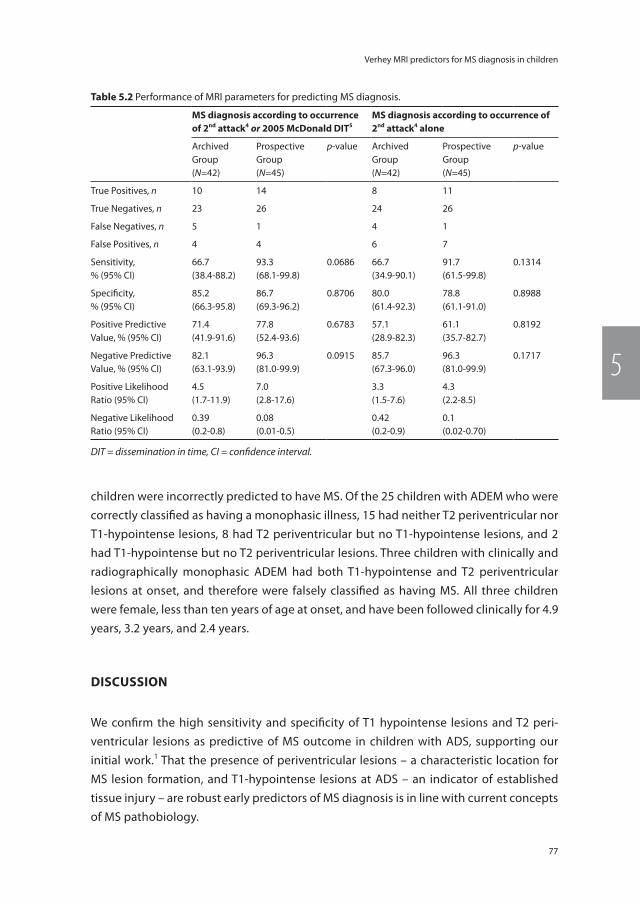
77
Verhey MRI predictors for MS diagnosis in children
5
children were incorrectly predicted to have MS. Of the 25 children with ADEM who were correctly classified as having a monophasic illness, 15 had neither T2 periventricular nor T1-hypointense lesions, 8 had T2 periventricular but no T1-hypointense lesions, and 2 had T1-hypointense but no T2 periventricular lesions. Three children with clinically and radiographically monophasic ADEM had both T1-hypointense and T2 periventricular lesions at onset, and therefore were falsely classified as having MS. All three children were female, less than ten years of age at onset, and have been followed clinically for 4.9 years, 3.2 years, and 2.4 years.
Discussion
We confirm the high sensitivity and specificity of T1 hypointense lesions and T2 peri-ventricular lesions as predictive of MS outcome in children with ADS, supporting our initial work.1 That the presence of periventricular lesions – a characteristic location for MS lesion formation, and T1-hypointense lesions at ADS – an indicator of established tissue injury – are robust early predictors of MS diagnosis is in line with current concepts of MS pathobiology.
table 5.2 Performance of MRI parameters for predicting MS diagnosis.
ms diagnosis according to occurrence of 2nd attack4 or 2005 mcDonald Dit5
ms diagnosis according to occurrence of 2nd attack4 alone
Archived Group(N=42)
Prospective Group(N=45)
p-value Archived Group(N=42)
Prospective Group(N=45)
p-value
True Positives, n 10 14 8 11
True Negatives, n 23 26 24 26
False Negatives, n 5 1 4 1
False Positives, n 4 4 6 7
Sensitivity,% (95% CI)
66.7(38.4-88.2)
93.3(68.1-99.8)
0.0686 66.7(34.9-90.1)
91.7(61.5-99.8)
0.1314
Specificity,% (95% CI)
85.2(66.3-95.8)
86.7(69.3-96.2)
0.8706 80.0(61.4-92.3)
78.8(61.1-91.0)
0.8988
Positive Predictive Value, % (95% CI)
71.4(41.9-91.6)
77.8(52.4-93.6)
0.6783 57.1(28.9-82.3)
61.1(35.7-82.7)
0.8192
Negative Predictive Value, % (95% CI)
82.1(63.1-93.9)
96.3(81.0-99.9)
0.0915 85.7(67.3-96.0)
96.3(81.0-99.9)
0.1717
Positive Likelihood Ratio (95% CI)
4.5(1.7-11.9)
7.0(2.8-17.6)
3.3(1.5-7.6)
4.3(2.2-8.5)
Negative Likelihood Ratio (95% CI)
0.39(0.2-0.8)
0.08(0.01-0.5)
0.42(0.2-0.9)
0.1(0.02-0.70)
DIT = dissemination in time, CI = confidence interval.
Chapter 5
78
The first aspect of our validation study was performed in the Dutch prospective cohort, an independent but methodologically similar study population to the Canadian Demy-elinating Disease program7 from which the MRI parameters were originally developed.1 Specifically, both studies utilized the International Pediatric MS Study Group consensus definitions2 to characterize ADS phenotype and MS diagnosis, both studies utilized the same standardized clinical reporting form, and the MRI protocols utilized were suf-ficiently similar to permit consistency in evaluation of commonly acquired sequences. The Dutch and Canadian cohorts are also similar clinically: (i) the proportion of children with ADS subsequently diagnosed with MS was 36% in the Dutch prospective cohort and 20% in Canadian prospective study; and (ii) the diagnosis of MS in children with an initial ADEM presentation was extremely rare in both cohorts (none in the Dutch cohort and one in the Canadian). Thus, in cohorts of children with typical ADS, we demonstrate robust sensitivity (Dutch cohort 93%, Canadian study 84%) and specificity (Dutch cohort 87%, Canadian study 93%) of the MRI parameters.
We then applied the MRI parameters to a dataset of archived scans of children with ADS in the Netherlands, in order to evaluate the MRI parameters in patients not enrolled in prospective research protocols. MRI scans in this archived group were obtained at either 1.0 or 1.5 Tesla, and were not obtained according to a consistent protocol. The estimate of sensitivity (67%), although not statistically significantly different, is lower than that from the prospective cohort (93%). Although all MRI scans analyzed were con-sidered to be of sufficient quality for scoring, the increased number of false negatives in the archived cohort may be due to image quality and variable sequence acquisition in the archived dataset compared with the prospective cohort. Specifically in the archived set, FLAIR imaging was either less frequently acquired or acquired in the coronal rather than axial plane, whole brain coverage was not achieved, and the contrast between grey matter and white matter on T1-weighted imaging was poorer, rendering detection of T2 lesions in the periventricular region and T1-hypointense lesions more challenging. Since common practice as well as a published MRI protocol for MS10 includes axial FLAIR and T2 sequences with contiguous three millimeter slices, we anticipate that these sequences will enhance sensitivity of our MRI parameters.
The ability to predict whether children with incident CNS demyelination will be diag-nosed with MS is important for counseling of families and for planning of patient care. A key challenge in pediatric demyelination is the ability to predict MS diagnosis across the full range of ADS presentations, particularly in the context of ADEM. Given that ADEM is particularly more common in children than adults11-13, MRI parameters that effectively stratify unselected groups of children with ADS, including those with ADEM, into high and low likelihood for MS diagnosis have significant clinical value.
Eight children (14% false positive rate) would have been predicted by our MRI param-eters to be diagnosed with MS, yet have experienced a clinically monophasic disease
79
Verhey MRI predictors for MS diagnosis in children
5
course to date (three with ADEM, three with polyfocal and two with monofocal ADS). Whether these eight children are particularly likely to reach a diagnosis of MS in the future remains to be determined as standardized serial imaging is not performed in the Dutch study, and thus MRI evidence of DIT was potentially underappreciated. Thus, while the ability to predict MS diagnosis based on initial MRI has obvious value to the immediate care of the child, it remains important to emphasize that long-term monitor-ing of children with ADS is essential to capture either new lesions on serial MRI scans or a second clinical attack.
Evaluation of the predictive validity of the MRI parameters at baseline is influenced by the method of MS diagnosis. As expected, given that MRI evidence of new lesions is often independent of clinical relapses14, 15, when restricting our analysis to only children with confirmed clinical relapses, the positive predictive value of the MRI parameters was slightly but not significantly reduced. A further confound is that six children in the current study initiated MS-targeted therapy after MRI confirmation of DIT, four of whom have not had a second clinical attack to date.
Our study has several limitations. In the archived collection of MRI scans, acquisition orientation, slice thickness, interslice gap and image contrast were not consistent, and images were acquired on 1.0 T and 1.5 T scanners. The children enrolled in the prospec-tive Dutch program were imaged with more consistent protocols, and were all imaged at 1.5 T. Our primary goal was to devise a predictive tool suited to application in the clinical context, and across variable imaging protocols. While the results of our predictive model performed better when applied to more rigorously acquired MRI scans, our parameters remained valid even within the limits of variable sequence parameters or quality. We also acknowledge the limitation of including both a retrospective and prospective patient sample in our present analysis. For the retrospective pediatric MS patients, MS diagnosis was made largely on the basis of confirmed relapsing clinical disease, given that consis-tent serial MRI studies were not available for earlier diagnostic confirmation of MS. In contrast, for the prospectively enrolled children, MRI studies were more frequently and consistently obtained, and thus new lesions on MRI were more likely to be detected, as reflected by the shorter time from first attack to MS diagnosis in this group. For our purposes, however, the outcome was not time to MS diagnosis; rather, we focused only on whether our MRI parameters correctly distinguished children with MS from those with monophasic disease. Finally, as is true for all predictive models, it is imperative to acknowledge that some children currently considered to have monophasic disease, may ultimately experience new disease activity leading to a diagnosis of MS.
Overall, we confirm that T2 lesions in the periventricular white mater (a region well-recognized as targeted in MS) and the presence of T1-hypointense lesions (suggestive of established pathology) are robust predictors of a future diagnosis of MS in children with ADS.
Chapter 5
80
reFerences
1. Verhey LH, Branson HM, Shroff MM, Callen DJ, Sled JG, Narayanan S, et al. MRI parameters for prediction of multiple sclerosis diagnosis in children with acute CNS demyelination: a prospective national cohort study. Lancet Neurol 2011; 10(12): 1065-73.
2. Krupp LB, Banwell B, Tenembaum S, Interna-tional Pediatric MSSG. Consensus definitions proposed for pediatric multiple sclerosis and related disorders. Neurology 2007; 68(16 Suppl 2): S7-12.
3. Wingerchuk DM, Lennon VA, Pittock SJ, Lucchinetti CF, Weinshenker BG. Revised diagnostic criteria for neuromyelitis optica. Neurology 2006; 66(10): 1485-89.
4. Poser CM, Paty DW, Scheinberg L, McDonald WI, Davis FA, Ebers GC, et al. New diagnostic criteria for multiple sclerosis: guidelines for research protocols. Ann Neurol 1983; 13(3): 227-31.
5. Polman CH, Reingold SC, Edan G, Filippi M, Hartung HP, Kappos L, et al. Diagnostic criteria for multiple sclerosis: 2005 revisions to the “McDonald Criteria”. Ann Neurol 2005; 58(6): 840-46.
6. Fletcher RH, Fletcher SW. Diagnosis. In: Clini-cal Epidemiology: the essentials. Lippincott Williams & Wilkins, Philadelphia 2005: page 35-58.
7. Banwell B, Bar-Or A, Arnold DL, Sadovnick D, Narayanan S, McGowan M, et al. Clinical, environmental, and genetic determinants of multiple sclerosis in children with acute demyelination: a prospective national cohort study. Lancet Neurol 2011; 10(5): 436-45.
8. Mikaeloff Y, Adamsbaum C, Husson B, Vallee L, Ponsot G, Confavreux C, et al. MRI prognostic factors for relapse after acute CNS inflamma-tory demyelination in childhood. Brain 2004; 127(Pt 9): 1942-47.
9. Neuteboom RF, Boon M, Catsman-Berrevoets CE, Vles JS, Gooskens RH, Stroink H, et al. Prognostic factors after a first attack of inflammatory CNS demyelination in children. Neurology 2008; 71(13): 967-73.
10. Simon JH, Li D, Traboulsee A, Coyle PK, Arnold DL, Barkhof F, et al. Standardized MR imaging protocol for multiple sclerosis: Consortium of MS Centers consensus guidelines. AJNR Am J Neuroradiol 2006; 27(2): 455-61.
11. Anlar B, Basaran C, Kose G, Guven A, Ha-spolat S, Yakut A, et al. Acute disseminated encephalomyelitis in children: outcome and prognosis. Neuropediatrics 2003; 34(4): 194-99.
12. Tenembaum S, Chamoles N, Fejerman N. Acute disseminated encephalomyelitis: a long-term follow-up study of 84 pediatric patients. Neurology 2002; 59(8): 1224-31.
13. Tenembaum S, Chitnis T, Ness J, Hahn JS, International Pediatric MSSG. Acute dissemi-nated encephalomyelitis. Neurology 2007; 68(16 Suppl 2): S23-36.
14. Barkhof F, Scheltens P, Frequin ST, Nauta JJ, Tas MW, Valk J, et al. Relapsing-remitting multiple sclerosis: sequential enhanced MR imaging vs clinical findings in determining disease activity. AJR Am J Roentgenol 1992; 159(5): 1041-47.
15. Sormani MP, Stubinski B, Cornelisse P, Rocak S, Li D, De SN. Magnetic resonance active lesions as individual-level surrogate for relapses in multiple sclerosis. Mult Scler 2011; 17(5): 541-49.
81
Verhey MRI predictors for MS diagnosis in children
5
suPPlementAl mAteriAl
supplementary table 5.1 Demographic and clinical characteristics of excluded patients (n=28).
Patient number
group reason for exclusion
sex ADs Age at onset, years
Diagnosis
1 Prospective Failed QC F ADEM 3.9 Monophasic ADS
2 Prospective No MRI scan F TM 3.3 Monophasic ADS
3 Prospective Failed QC F Polyfocal 12.4 Monophasic ADS
4 Prospective Failed QC M ON 10.3 Monophasic ADS
5 Prospective No T1 scan F ADEM 8.9 Monophasic ADS
6 Prospective Failed QC F ON 14.6 MS
7 Prospective Failed QC F Polyfocal 16.8 MS
8 Prospective Failed QC M ADEM 2.8 Monophasic ADS
9 Prospective Failed QC M ADEM 3.5 Monophasic ADS
10 Prospective Failed QC F ON 16.3 MS
11 Prospective No T1 scan F Monofocal 13.7 Monophasic ADS
12 Prospective No MRI scan M TM 12.5 MS
13 Archived Failed QC M Polyfocal 7.2 Relapsing ADS*
14 Archived No MRI scan F Monofocal 14.8 Monophasic ADS
15 Archived Failed QC M ADEM 4.3 Monophasic ADS
16 Archived No MRI scan M TM 8.1 Monophasic ADS
17 Archived Failed QC M TM 3.6 Monophasic ADS
18 Archived Failed QC M Polyfocal 5.9 Monophasic ADS
19 Archived Failed QC M ADEM 7.6 Monophasic ADS
20 Archived Failed QC M TM 6.8 MS
21 Archived Failed QC F ADEM 4.2 Monophasic ADS
22 Archived Failed QC F Polyfocal 12.4 MS
23 Archived No MRI scan F TM 15.4 Monophasic ADS
24 Archived Failed QC M Monofocal 8.1 Monophasic ADS
25 Archived Failed QC M ADEM 4.3 Monophasic ADS
26 Archived Failed QC M Monofocal 13.0 Monophasic ADS
27 Archived Failed QC M Polyfocal 5.7 Monophasic ADS
28 Archived Failed QC M Polyfocal 10.9 Monophasic ADS
summary statistics F: 12(43%) non-ADem:21 (75%)
mean (sD):9.0 (4.5)
ms: 6 (21%)
*This child presented with an “ADEM-like” incident demyelinating episode, and subsequently has had mul-tiple episodes of optic neuritis; brain MRI does not meet criteria for multiple sclerosis. The child has been treated with intravenous immunoglobulins, and has not experienced further clinical or MRI evidence of relapse. Until present, the child does not meet criteria for neuromyelitis optica.QC = quality control, MRI = magnetic resonance imaging, F = female, M = male, ADS = acute demyelinating syndrome, ADEM = acute disseminated encephalomyelitis, TM = transverse myelitis, ON = optic neuritis, MS = multiple sclerosis

2016
12
OCT
Chapter 6
Disease course after CIS in children versus adults: a prospective cohort study
R.M. van der Vuurst de Vries*, E.D. van Pelt*, J.Y. Mescheriakova, Y.Y.M. Wong, I.A. Ketelslegers, T.A.M. Siepman, C.E. Catsman-Berrevoets, R.F. Neuteboom, R.Q. Hintzen.
* shared first authors
Submitted
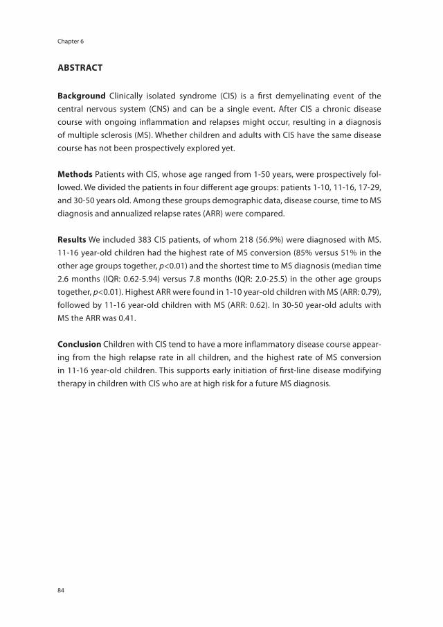
Chapter 6
84
AbstrAct
background Clinically isolated syndrome (CIS) is a first demyelinating event of the central nervous system (CNS) and can be a single event. After CIS a chronic disease course with ongoing inflammation and relapses might occur, resulting in a diagnosis of multiple sclerosis (MS). Whether children and adults with CIS have the same disease course has not been prospectively explored yet.
methods Patients with CIS, whose age ranged from 1-50 years, were prospectively fol-lowed. We divided the patients in four different age groups: patients 1-10, 11-16, 17-29, and 30-50 years old. Among these groups demographic data, disease course, time to MS diagnosis and annualized relapse rates (ARR) were compared.
results We included 383 CIS patients, of whom 218 (56.9%) were diagnosed with MS. 11-16 year-old children had the highest rate of MS conversion (85% versus 51% in the other age groups together, p<0.01) and the shortest time to MS diagnosis (median time 2.6 months (IQR: 0.62-5.94) versus 7.8 months (IQR: 2.0-25.5) in the other age groups together, p<0.01). Highest ARR were found in 1-10 year-old children with MS (ARR: 0.79), followed by 11-16 year-old children with MS (ARR: 0.62). In 30-50 year-old adults with MS the ARR was 0.41.
conclusion Children with CIS tend to have a more inflammatory disease course appear-ing from the high relapse rate in all children, and the highest rate of MS conversion in 11-16 year-old children. This supports early initiation of first-line disease modifying therapy in children with CIS who are at high risk for a future MS diagnosis.
85
Disease course after CIS in children versus adults
6
introDuction
Multiple sclerosis (MS) is a chronic inflammatory demyelinating disease of the central nervous system (CNS) which can cause a broad spectrum of neurological deficits.1 Worldwide over 2.5 million people suffer from MS.1 MS mostly affects young women in their early twenties or thirties, and although rare, MS can also occur in children.2 MS can be diagnosed when a first event of CNS demyelination, a so-called clinically isolated syndrome (CIS), is followed by a new clinical event.3-5 In addition, MS can be diagnosed after CIS when new lesions, which fulfill MS diagnostic criteria for dissemination in time and space, are detected on Magnetic Resonance Imaging (MRI).4, 5 However CIS can be a single event. Whether childhood-onset versus adulthood-onset CIS and MS reflect the same disease is unknown.6-8 Previous studies focused on MS and did not compare the disease course between children and adults after the onset of CIS.9-15 Despite existing parallels in childhood-onset and adulthood-onset MS, there are fundamental differ-ences in presentation and disease course. Several studies have reported a higher relapse rate and MRI lesion load in children than in adults with MS.10, 12, 13, 16, 17 On the other hand, the progression of disability is slower in children with MS.14, 15 It should be noted, however, that some of the previous studies included retrospective cohorts11, 14, 15, 17, and some were conducted before the immunomodulatory treatment era in children.9-11, 14, 15 Current recommendation and use of disease modifying therapies (DMT) in children with MS likely influenced their disease course and relapse rate.18 Several studies have shown initiation of first-line DMT (interferon or glatiramer acetate) in adults with CIS delays MS diagnosis and disability accumulation.19-21 In children with CIS it is not common practice yet to offer first-line DMT prior to MS diagnosis. Here, we performed a clinical prospec-tive follow-up study of 383 patients with CIS, whose age ranged from 1 - 50 years, in order to report a comparison of childhood-onset versus adulthood-onset CIS and MS.
methoDs
Patients and definitions
Patients with CIS were included between April 2006 and August 2015, either in our pro-spective cohort of adults with CIS (PRedicting the Outcome of a Demyelinating event, PROUD study)22, or in our prospective cohort study of children with a first episode of acquired demyelination of the CNS (PROUDkids).23 Both studies are ongoing multicenter observational studies conducted at Erasmus MC in Rotterdam, the Netherlands, and study protocols have been described previously.22, 23 Patients younger than 51 years old were included within 6 months after the first onset of their clinical symptoms. At baseline, a MRI scan was performed and routine laboratory tests were done. Patients
Chapter 6
86
with alternative diagnoses other than CIS were excluded. After baseline patients were reassessed at least annually.
We used a prediction rule for defining children with CIS at high risk of MS when they fulfilled two out of three criteria at onset: more than nine T2 white matter lesions, and/or at least one gadolinium-enhancing lesion on MRI, and/or positive oligoclonal bands (OCB) in cerebrospinal fluid.24, 25 MS was diagnosed in adults according to the 2010 McDonald criteria.5 Children were diagnosed with CIS and MS according to the diag-nostic criteria from the International Pediatric Multiple Sclerosis Study Group (IPMSSG).4 Clinically definite multiple sclerosis (CDMS) was defined as two attacks with (para)clinical evidence of two separate lesions as described by Poser26 and should include two non-encephalopathic events.4 The expanded disability status scale (EDSS scale) was used to define disability.27 EDSS scores were obtained at least 2 months after a relapse occurred. Secondary progressive MS is defined as a history of gradual worsening after an initial relapsing-remitting disease course, with or without exacerbations during the progres-sive course.28 This study was approved by the medical ethical committees of Erasmus MC in Rotterdam and of the other participating centers. Written informed consent was provided for all patients.
Data analysis
Statistical analysis was performed using SPSS v20 (SPSS Inc., Chicago, Illinois, USA) and GraphPad Prism 5 (GraphPad., San Diego, USA). We divided the patients in four different age groups of clinical interest, based on evidence that puberty enhances CNS autoim-munity in females29: i.e. pre-puberty, puberty, young adults and adults (patients 1-10, 11-16, 17-29, and 30-50 years old). Among these groups we compared demographic data, disease course, time to MS diagnosis and annualized relapse rate (ARR). We used the Kolmogorov-Smirnov test to assess normal distribution of the data. For the compari-son of continuous data between two groups we applied a two-tailed t-test, or when the data were not normally distributed a Mann-Whitney U test. For the analysis of multiple groups, we used the one-way-ANOVA for normally distributed data, and Kruskal-Wallis test for not normally distributed data. Categorical data were analyzed using the Chi-square or Fisher’s exact test. Kaplan-Meier survival analyses were used to analyse the time to MS diagnosis. Univariate and multivariable Cox proportional hazard regression models were used to obtain hazard ratios for MS diagnosis and EDSS 4.0 respectively. Patients who were not diagnosed with MS, or did not reach EDSS 4.0 during follow-up respectively, were considered as censored observations in those analyses. Negative binomial regression, with the natural logarithm of follow-up years after MS diagnosis as offset, was used to analyse ARR after CIS. This offset was used to correct for the different follow-up times between patients. We used a generalized estimating equations (GEE) model to compare ARR with and without disease-modifying treatment. This GEE model
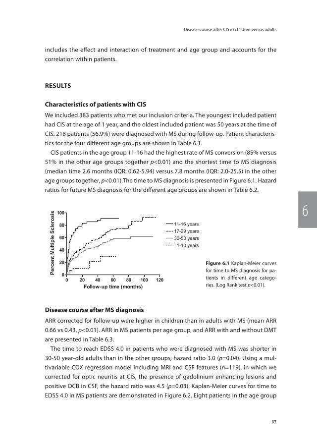
87
Disease course after CIS in children versus adults
6
includes the effect and interaction of treatment and age group and accounts for the correlation within patients.
results
characteristics of patients with cis
We included 383 patients who met our inclusion criteria. The youngest included patient had CIS at the age of 1 year, and the oldest included patient was 50 years at the time of CIS. 218 patients (56.9%) were diagnosed with MS during follow-up. Patient characteris-tics for the four different age groups are shown in Table 6.1.
CIS patients in the age group 11-16 had the highest rate of MS conversion (85% versus 51% in the other age groups together p<0.01) and the shortest time to MS diagnosis (median time 2.6 months (IQR: 0.62-5.94) versus 7.8 months (IQR: 2.0-25.5) in the other age groups together, p<0.01).The time to MS diagnosis is presented in Figure 6.1. Hazard ratios for future MS diagnosis for the different age groups are shown in Table 6.2.
Disease course after ms diagnosis
ARR corrected for follow-up were higher in children than in adults with MS (mean ARR 0.66 vs 0.43, p<0.01). ARR in MS patients per age group, and ARR with and without DMT are presented in Table 6.3.
The time to reach EDSS 4.0 in patients who were diagnosed with MS was shorter in 30-50 year-old adults than in the other groups, hazard ratio 3.0 (p=0.04). Using a mul-tivariable COX regression model including MRI and CSF features (n=119), in which we corrected for optic neuritis at CIS, the presence of gadolinium enhancing lesions and positive OCB in CSF, the hazard ratio was 4.5 (p=0.03). Kaplan-Meier curves for time to EDSS 4.0 in MS patients are demonstrated in Figure 6.2. Eight patients in the age group
1-10 years
years years years
M
S
Figure 6.1 Kaplan-Meier curves for time to MS diagnosis for pa-tients in different age catego-ries. (Log Rank test p<0.01).
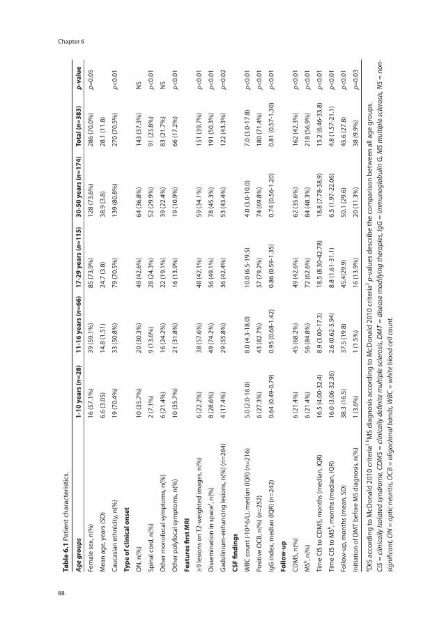
Chapter 6
88
tabl
e 6.
1 Pa
tient
cha
ract
eris
tics.
Age
grou
ps1-
10 y
ears
(n=2
8)11
-16
year
s (n
=66)
17-2
9 ye
ars
(n=1
15)
30-5
0 ye
ars
(n=1
74)
tota
l (n=
383)
p-va
lue
Fem
ale
sex,
n(%
)16
(57.
1%)
39 (5
9.1%
)85
(73,
9%)
128
(73.
6%)
286
(70.
0%)
p=0.
05
Mea
n ag
e, y
ears
(SD
)6.
6 (3
.05)
14.8
(1.5
1)24
.7 (3
.8)
38.9
(3.8
)28
.1 (1
1.8)
Cauc
asia
n et
hnic
ity, n
(%)
19 (7
0.4%
)33
(50.
8%)
79 (7
0.5%
)13
9 (8
0.8%
)27
0 (7
0.5%
)p<
0.01
type
of c
linic
al o
nset
ON
, n(%
)10
(35.
7%)
20 (3
0.3%
)49
(42.
6%)
64 (3
6.8%
)14
3 (3
7.3%
)N
S
Spin
al c
ord,
n(%
)2
(7.1
%)
9 (1
3.6%
)28
(24.
3%)
52 (2
9.9%
)91
(23.
8%)
p<0.
01
Oth
er m
onof
ocal
sym
ptom
s, n(
%)
6 (2
1.4%
)16
(24.
2%)
22 (1
9.1%
)39
(22.
4%)
83 (2
1.7%
)N
S
Oth
er p
olyf
ocal
sym
ptom
s, n(
%)
10 (3
5.7%
)21
(31.
8%)
16 (1
3.9%
)19
(10.
9%)
66 (1
7.2%
)p<
0.01
Feat
ures
firs
t mri
≥9 le
sion
s on
T2-
wei
ghte
d im
ages
, n(%
)6
(22.
2%)
38 (5
7.6%
)48
(42.
1%)
59 (3
4.1%
)15
1 (3
9.7%
)p<
0.01
Dis
sem
inat
ion
in s
pace
a , n(%
)8
(28.
6%)
49 (7
4.2%
)56
(49.
1%)
78 (4
5.3%
)19
1 (5
0.3%
)p<
0.01
Gad
olin
ium
-enh
anci
ng le
sion
s, n(
%) (
n=28
4)4
(17.
4%)
29 (5
5.8%
)36
(42.
4%)
53 (4
3.4%
)12
2 (4
3.3%
)p=
0.02
csF
findi
ngs
WBC
cou
nt (∙
10^6
/L),
med
ian
(IQR)
(n=2
16)
5.0
(2.0
-16.
0)8.
0 (4
.3-1
8.0)
10.0
(6.5
-19.
5)4.
0 (3
.0-1
0.0)
7.0
(3.0
-17.
8)p<
0.01
Posi
tive
OCB
, n(%
) (n=
252)
6 (2
7.3%
)43
(82.
7%)
57 (7
9.2%
)74
(69.
8%)
180
(71.
4%)
p<0.
01
IgG
inde
x, m
edia
n (IQ
R) (n
=242
)0.
64 (0
.49-
0.79
)0.
95 (0
.68-
1.42
)0.
86 (0
.59-
1.35
)0.
74 (0
.56-
1.20
)0.
81 (0
.57-
1.30
)p<
0.01
Follo
w-u
p
CDM
S, n
(%)
6 (2
1.4%
)45
(68.
2%)
49 (4
2.6%
)62
(35.
6%)
162
(42.
3%)
p<0.
01
MSb , n
(%)
6 (2
1.4%
)56
(84.
8%)
72 (6
2.6%
)84
(48.
3%)
218
(56.
9%)
p<0.
01
Tim
e CI
S to
CD
MS,
mon
ths
(med
ian,
IQR)
16.5
(4.0
0-32
.4)
8.9
(3.0
0-17
.3)
18.5
(8.3
0-42
.78)
18.8
(7.7
8-38
.9)
15.2
(6.4
6-33
.8)
p<0.
01
Tim
e CI
S to
MSb , m
onth
s (m
edia
n, IQ
R)16
.0 (3
.06-
32.3
6)2.
6 (0
.62-
5.94
)8.
8 (1
.61-
31.1
)6.
5 (1
.97-
22.0
6)4.
8 (1
.57-
21.1
)p<
0.01
Follo
w-u
p, m
onth
s (m
ean,
SD
)38
.3 (1
6.5)
37.5
(19.
8)45
.4(2
9.9)
50.1
(29.
6)45
.6 (2
7.8)
p<0.
01
Initi
atio
n of
DM
T be
fore
MS
diag
nosi
s, n(
%)
1 (3
.6%
)1
(1.5
%)
16 (1
3.9%
)20
(11.
3%)
38 (9
.9%
)p=
0.03
a DIS
acc
ordi
ng to
McD
onal
d 20
10 c
riter
ia5 b M
S di
agno
sis
acco
rdin
g to
McD
onal
d 20
10 c
riter
ia5 p
-val
ues
desc
ribe
the
com
paris
on b
etw
een
all a
ge g
roup
s.CI
S =
clin
ical
ly is
olat
ed sy
ndro
me,
CD
MS
= cl
inic
ally
defi
nite
mul
tiple
scle
rosi
s, D
MT
= di
seas
e m
odify
ing
ther
apie
s, Ig
G =
imm
unog
lobu
lin G
, MS
mul
tiple
scle
rosi
s, N
S =
non-
sign
ifica
nt, O
N =
opt
ic n
eurit
is, O
CB =
olig
oclo
nal b
ands
, WBC
= w
hite
blo
od ce
ll co
unt.
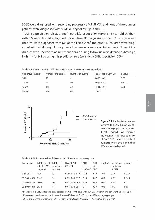
89
Disease course after CIS in children versus adults
6
30-50 were diagnosed with secondary progressive MS (SPMS), and none of the younger patients were diagnosed with SPMS during follow-up (p<0.01).
Using a prediction rule at onset (methods), 42 out of 94 (45%) 1-16 year-old children with CIS were defined at high risk for a future MS diagnosis. Of them 25 ≥12 year-old children were diagnosed with MS at the first event.4 The other 17 children were diag-nosed with MS during follow-up based on new relapses or on MRI-criteria. None of the children with CIS who remained monophasic during follow-up were defined as having a high risk for MS by using this prediction rule (sensitivity 68%, specificity 100%).
table 6.2 Hazard ratios for MS diagnosis, univariate cox regression analysis.
Age groups (years) Number of patients Number of events Hazard ratio (95% CI) p-value
1-10 28 6 0.4 (0.2-0.9) 0.03
11-16 66 56 3.6 (2.6-5.1) <0.01
17-29 115 72 1.5 (1.1-2.1) 0.01
30-50 174 84 1(ref )
Figure 6.2 Kaplan-Meier curves for time to EDSS 4.0 for MS pa-tients in age groups 1-29 and 30-50. Legend: We merged the younger age groups (1-10, 11-16, 17-29) since the patient numbers were small and their KM-curves overlapped.
table 6.3 ARR corrected for follow-up in MS patients per age group.
Age group Total years at risk after MS diagnosis
Total number of relapses
Overall ARR(95% CI)
ARR with-outDMT
ARR with DMT
p-valuea Interaction coefficient of DMT
p-valueb
0-10 (n=6) 15.4 12 0.79 (0.42-1.48) 5.22 0.43 <0.01 5.64 0.033
11-16 (n=56) 154.3 94 0.62 (0.49-0.77) 2.13 0.47 <0.01 2.08 0.040
17-30 (n=72) 200.6 104 0.52 (0.42-0.65) 1.16 0.45 <0.01 1.19 ns
30-50 (n=84) 283.6 114 0.41 (0.34-0.51) 0.81 0.37 <0.01 Ref. Ref.
a Presented p-values for the comparison of ARR with and without DMT within the different age groups.b Presented p-values for the interaction coefficient of DMT for the different age groups.ARR = annualized relapse rate, DMT = disease modifying therapies, CI = confidence interval
Chapter 6
90
Discussion
The purpose of our study was to report a comparison of childhood-onset versus adulthood-onset CIS and MS in a large prospective cohort during the current immuno-modulatory treatment era. The results of our study suggest a more inflammatory disease course, rather than a neurodegenerative disease course, in 11-16 year-old children with CIS, as this group had the highest rate of MS conversion and the shortest time to MS diagnosis, higher ARR, higher MRI lesion load and a more inflammatory CSF profile. A secondary progressive MS disease course, which reflects a more neurodegenerative in-stead of an inflammatory phase of MS, was found in eight 30-50 year-old adult patients. However, secondary progression in MS is age dependant and probably therefore has not been observed yet in our younger patients with the current follow-up.30 In addition, we found higher EDSS progression rates in adults than in children. Previous studies on childhood-onset MS versus adulthood-onset MS, have already reported a higher relapse rate in children than in adults.10, 12, 13 In our unique prospective study we followed children and adults after the first attack. Since we collected a large cohort, we could compare subgroups and draw conclusions for different age groups of clinical interest. Overall, we found a female predominance both in patients with CIS and MS, except in young children who were diagnosed with MS before puberty, of whom four out of six MS patients were boys. The difference in sex distribution in children diagnosed with MS before and after puberty and the higher rates of MS diagnoses after puberty suggests that sex hormones contribute to the onset of MS.2, 23, 29 A remarkable high rate of non-Caucasian ethnicities was observed in 11-16 year-old children with CIS. Higher rates of non-Caucasian ethnicities have been reported in childhood-onset MS and might reflect a higher vulnerability of developing MS at a younger age.23, 31, 32 A possible explanation for this might be that non-Caucasians miss certain protective genes since their ances-tors were born in countries with a low prevalence of MS.23 In the lowest age group of 1-10 years old, 28.6% of the children with CIS were non-Caucasians, while 83.3% of the children with MS were non-Caucasians.
The shorter time to MS diagnosis in 11-16 year-old children is partly explained by the current diagnostic MRI criteria which allow for an early MS diagnosis in a subgroup of the patients at first MRI, however not in children younger than 12 years of age.4, 5 Still, we observed a high rate of CDMS and short time to CDMS diagnosis in 11-16 year-old children.
Early initiation of DMT in adults with CIS might have influenced their disease course. However, a relatively small proportion of adults (12.5%) started with DMT before MS diagnosis and in a subanalysis (data not shown) we found similar results when we ex-cluded these patients. Relapse rates decrease after the initiation of DMT in both children and in adults with MS. It is unlikely our observed differences in ARR are only caused by
91
Disease course after CIS in children versus adults
6
early initiation of DMT, since a small proportion of CIS patients started with DMT prior to the MS diagnosis and pre-treatment ARR differed significantly. First-line DMT is already offered to adults with CIS who are at high risk for a future MS diagnosis, but not to chil-dren with CIS. However, DMT especially reduce MS relapse rates and have been shown beneficial in children with MS.18 In addition, DMT might prevent disability progression. Therefore early initiation of first-line DMT in children with CIS who are at high risk for MS seems logical. By using a prediction rule at onset, we found that 42 out of 94 (45%) 1-16 year-old children with CIS were defined as at high risk for MS, and could have started with first-line DMT at onset, since all 42 children were diagnosed with MS during follow-up. The utility of this prediction rule should be validated in other prospective cohorts for its usefulness and safety. A limitation of our study is that the follow-up is limited and relatively shorter in children with CIS since our prospective study in adults started prior to our prospective study in children. With the current follow-up, however, we could already demonstrate important differences in rates of MS diagnosis and relapses. It would be interesting to follow our cohort in order to compare the clinical outcome and disease progression over several decades. Another limitation of our study is a selec-tion bias since we only included patients with CIS, while in children it is known that MS can present with a spectrum of acquired demyelinating events, including ADEM.4 However, it is not expected that this influences our results much, since it is uncommon that children are diagnosed with MS after ADEM, using the current diagnostics criteria.4 In addition, there could have been a selection in patients who were reported for our prospective studies by physicians from other hospitals. Furthermore, we do not have a standard MRI protocol, gadolinium was not administered to all patients at the first event, nor was a lumbar puncture performed in all patients. Nevertheless, our data resemble clinical practice. In summary, we found a more inflammatory disease course of CIS and MS in children. This could argue for early initiation of first-line disease modifying therapy in children with CIS who are at high risk for a future MS diagnosis.
Chapter 6
92
reFerences
1. Compston A, Coles A. Multiple sclerosis. Lancet 2002; 359(9313): 1221-31.
2. Banwell B, Ghezzi A, Bar-Or A, Mikaeloff Y, Tar-dieu M. Multiple sclerosis in children: clinical diagnosis, therapeutic strategies, and future directions. Lancet Neurol 2007; 6(10): 887-902.
3. Miller DH, Chard DT, Ciccarelli O. Clinically isolated syndromes. Lancet Neurol 2012; 11(2): 157-69.
4. Krupp LB, Tardieu M, Amato MP, Banwell B, Chitnis T, Dale RC, et al. International Pediatric Multiple Sclerosis Study Group criteria for pediatric multiple sclerosis and immune-me-diated central nervous system demyelinating disorders: revisions to the 2007 definitions. Mult Scler 2013; 19(10): 1261-67.
5. Polman CH, Reingold SC, Banwell B, Clanet M, Cohen JA, Filippi M, et al. Diagnostic criteria for multiple sclerosis: 2010 revisions to the McDonald criteria. Ann Neurol 2011; 69(2): 292-302.
6. Dale RC. Paediatric MS is the same disease as adult MS: commentary. Mult Scler 2013; 19(10): 1259-1260.
7. Chitnis T. Paediatric MS is the same disease as adult MS: no. Mult Scler 2013; 19(10): 1255-1256.
8. Hintzen RQ, van Pelt DE. Paediatric MS is the same disease as adult MS: yes. Mult Scler 2013; 19(10): 1257-1258.
9. Ghezzi A, Pozzilli C, Liguori M, Marrosu MG, Milani N, Milanese C, et al. Prospective study of multiple sclerosis with early onset. Mult Scler 2002; 8(2): 115-18.
10. Boiko A, Vorobeychik G, Paty D, Devonshire V, Sadovnick D. Early onset multiple sclerosis: a longitudinal study. Neurology 2002; 59(7): 1006-10.
11. Deryck O, Ketelaer P, Dubois B. Clinical char-acteristics and long term prognosis in early onset multiple sclerosis. J Neurol 2006; 253(6): 720-23.
12. Gorman MP, Healy BC, Polgar-Turcsanyi M, Chitnis T. Increased relapse rate in pediatric-
onset compared with adult-onset multiple sclerosis. Arch Neurol 2009; 66(1): 54-59.
13. Benson LA, Healy BC, Gorman MP, Baruch NF, Gholipour T, Musallam A, et al. Elevated relapse rates in pediatric compared to adult MS persist for at least 6 years. Mult Scler Relat Disord 2014; 3(2): 186-93.
14. Simone IL, Carrara D, Tortorella C, Liguori M, Lepore V, Pellegrini F, et al. Course and prog-nosis in early-onset MS: comparison with adult-onset forms. Neurology 2002; 59(12): 1922-28.
15. Renoux C, Vukusic S, Mikaeloff Y, Edan G, Clanet M, Dubois B, et al. Natural history of multiple sclerosis with childhood onset. N Engl J Med 2007; 356: 2603-13.
16. Yeh EA, Weinstock-Guttman B, Ramanathan M, Ramasamy DP, Willis L, Cox JL, et al. Magnetic resonance imaging characteristics of children and adults with paediatric-onset multiple sclerosis. Brain 2009; 132(Pt 12): 3392-400.
17. Waubant E, Chabas D, Okuda DT, Glenn O, Mowry E, Henry RG, et al. Difference in disease burden and activity in pediatric patients on brain magnetic resonance imaging at time of multiple sclerosis onset vs adults. Arch Neurol 2009; 66(8): 967-71.
18. Chitnis T, Tenembaum S, Banwell B, Krupp L, Pohl D, Rostasy K, et al. Consensus statement: evaluation of new and existing therapeutics for pediatric multiple sclerosis. Mult Scler 2012; 18(1): 116-27.
19. Tintore M, Rovira A, Rio J, Otero-Romero S, Arrambide G, Tur C, et al. Defining high, medium and low impact prognostic factors for developing multiple sclerosis. Brain 2015; 138(Pt 7): 1863-74.
20. Comi G, Martinelli V, Rodegher M, Moiola L, Bajenaru O, Carra A, et al. Effect of glatiramer acetate on conversion to clinically definite multiple sclerosis in patients with clinically isolated syndrome (PreCISe study): a ran-domised, double-blind, placebo-controlled trial. Lancet 2009; 374(9700): 1503-11.
93
Disease course after CIS in children versus adults
6
21. Kappos L, Freedman MS, Polman CH, Edan G, Hartung HP, Miller DH, et al. Effect of early versus delayed interferon beta-1b treatment on disability after a first clinical event sugges-tive of multiple sclerosis: a 3-year follow-up analysis of the BENEFIT study. Lancet 2007; 370(9585): 389-97.
22. Runia TF, Jafari N, Siepman DA, Hintzen RQ. Fatigue at time of CIS is an independent pre-dictor of a subsequent diagnosis of multiple sclerosis. J Neurol Neurosurg Psychiatry 2015; 86(5): 543-46.
23. Ketelslegers IA, Catsman-Berrevoets CE, Neuteboom RF, Boon M, van Dijk KG, Eikelen-boom MJ, et al. Incidence of acquired de-myelinating syndromes of the CNS in Dutch children: a nationwide study. J Neurol 2012; 259(9): 1929-35.
24. Barkhof F, Rocca M, Francis G, Van Waesber-ghe JH, Uitdehaag BM, Hommes OR, et al. Validation of diagnostic magnetic resonance imaging criteria for multiple sclerosis and response to interferon beta1a. Ann Neurol 2003; 53(6): 718-724.
25. Soderstrom M, Ya-Ping J, Hillert J, Link H. Op-tic neuritis: prognosis for multiple sclerosis from MRI, CSF, and HLA findings. Neurology 1998; 50(3): 708-14.
26. Poser CM, Paty DW, Scheinberg L, McDonald WI, Davis FA, Ebers GC, et al. New diagnostic criteria for multiple sclerosis: guidelines for research protocols. Ann Neurol 1983; 13(3): 227-31.
27. Kurtzke JF. Rating neurologic impairment in multiple sclerosis: an expanded disability status scale (EDSS). Neurology 1983; 33(11): 144-52.
28. Lublin FD, Reingold SC, Cohen JA, Cutter GR, Sørensen PS, Thompson AJ, et al. Defining the clinical course of multiple sclerosis: the 2013 revisions. Neurology 2014; 83(3): 278-86.
29. Ahn JJ, O’Mahony J, Moshkova M, Hanwell HE, Singh H, Zhang MA, et al. Puberty in females enhances the risk of an outcome of multiple sclerosis in children and the development of central nervous system autoimmunity in mice. Mult Scler 2015; 21(6): 735-48.
30. Confavreux C, Vukusic S. Natural history of multiple sclerosis: a unifying concept. Brain 2006; 129(Pt 3): 606-16.
31. Chitnis T, Glanz B, Jaffin S, Healy B. Demo-graphics of pediatric-onset multiple sclerosis in an MS center population from the North-eastern United States. Mult Scler 2009; 15(5): 627-31.
32. Langer-Gould A, Zhang JL, Chung J, Yeung Y, Waubant E, Yao J. Incidence of acquired CNS demyelinating syndromes in a multiethnic cohort of children. Neurology 2011; 77(12): 1143-48.

2016
12
OCT
Chapter 7
Risk genes associated with pediatric-onset MS but not with monophasic acquired CNS demyelination
E.D. van Pelt*, J.Y. Mescheriakova*, N. Makhani, I.A. Ketelslegers, R.F. Neuteboom, S. Kundu, L. Broer, A.C.J.W. Janssens, C.E. Catsman-Berrevoets, C.M. van Duijn, B Banwell, A. Bar-Or, R.Q. Hintzen.
* shared first authors
Neurology, 2013
Chapter 7
96
AbstrAct
objective To investigate whether 57 genetic risk loci recently identified in a large-scale genome-wide association study (GWAS) in adult MS patients are also associated with a risk for pediatric-onset MS and whether they can predict MS diagnosis in children presenting with acquired demyelinating syndromes (ADS).
methods We included 188 children with ADS, of which 53 were diagnosed with MS, 466 adult-onset MS patients and 2046 adult controls in our cohort study. Weighted genetic risk scores (wGRS) were calculated to evaluate genetic effects.
results Mean wGRS was significantly higher for pediatric-onset MS patients (7.32 ± 0.53) as compared with monophasic ADS patients (7.10 ± 0.47, p=0.01) and controls (7.11 ± 0.53, p<0.01). We found no difference in mean wGRS of participants with monophasic ADS (7.10 ± 0.47) and controls (7.11 ± 0.53). The ability of the wGRS for the 57 SNPs to discriminate between children with MS and those with monophasic ADS was moderate (AUC =0.64), but improved with the addition of sex and HLA-DRB1*15 (AUC =0.70). The combined effect of 57 SNPs exceeded the effect of HLA-DRB1*15 alone in our risk models for pediatric- and adult-onset MS.
conclusion The previously reported 57 SNPs for adult-onset MS also confer increased susceptibility to pediatric-onset MS, but not to monophasic ADS.
97
Risk genes associated with MS diagnosis in children
7
introDuction
Multiple sclerosis (MS) is being increasingly diagnosed in childhood.1 In children, an ini-tial attack of central nervous system (CNS) demyelination (acquired demyelinating syn-drome or ADS) frequently remains monophasic. Approximately 21-32% of the children with ADS will display further MRI or clinical evidence of inflammatory CNS demyelination meeting diagnostic criteria for MS.2, 3 This is in contrast with adults, where the majority of patients are diagnosed with MS after an initial event of acute CNS demyelination.4 Pediatric-onset MS has been proposed as a unique time window for the study of early MS disease mechanisms. However, it is not known whether pediatric- and adult-onset MS share the same genetic risk factors. A recent Canadian study of a large ADS cohort reported that children harboring one or more HLA-DRB1*15 alleles were more likely to be confirmed to have MS, compared to ADS children lacking HLA-DRB1*15 alleles.5 While HLA alleles thus contribute to the risk of both pediatric- and adult-onset MS6, large-scale genome-wide association studies (GWAS) recently identified 57 non-HLA genetic risk loci in adult MS patients compared to controls.7 Whether these 57 single nucleotide polymorphisms (SNPs) also contribute to the risk of either MS in children, or childhood-onset ADS more generally is not known. The objective of this study was to investigate whether the 57 SNPs identified in adult-onset MS are associated with increased risk of pediatric-onset MS, and whether such SNPs distinguish children with MS from chil-dren with monophasic ADS. We utilized a previously published approach to generate compound-weighted genetic risk scores8, and compared the predictive value of the 57 SNPs with the predictive value of HLA-DRB1*15 alone, in distinguishing children with MS from children with monophasic ADS and controls from the general population.
methoDs
Patients and definitions
Children younger than 16 years 0 days (Canadian cohort) and 17 years 0 days (Dutch cohort) of age who presented with ADS between 2001 and 2009 were enrolled in either the prospective Canadian Pediatric Demyelinating Disease Study or the Dutch Study group for Pediatric MS study. The Canadian National Pediatric Demyelinating Disease Study Group consists of 23 participating pediatric health care centers across Canada. The Dutch Study Group for Pediatric MS consists of 13 participating pediatric healthcare centers in the Netherlands. Initial phenotypes were characterized by clinical history and physical examination as clinically monofocal optic neuritis (ON), clinically monofocal transverse myelitis (TM), other clinically monofocal disease, clinically polyfocal disease, or acute disseminated encephalomyelitis (ADEM9). Children were diagnosed with MS
Chapter 7
98
based either on evidence of a second clinical attack after at least 30 days or MRI evi-dence of dissemination in time.9, 10 The recent 2010 iteration of the McDonald criteria for MS diagnosis was not available at the time of clinical classification, and was not used for this work. All participants had a minimum follow-up of 2 years from initial ADS. A total of 466 adult-onset (> 18 years) MS patients with either whole blood or saliva available to extract DNA were identified through the Rotterdam MS center. DNA from a control group of 2046 unrelated European adults from the general population was obtained from individuals enrolled in the longitudinal Rotterdam Study.11 In order to control the effect of genetic variation due to ancestry, only participants with self-reported European ancestry were included in the analyses presented in the paper. Potential stratification was corrected for by genomic control and principal component analysis.
standard protocol approvals, registration and Patient consents
Institutional ethical approval by an ethical standard committee on human experimenta-tion was obtained at all 23 sites participating in the Canadian National Pediatric Demy-elinating Disease Study and all 13 sites participating in the Dutch national study and for the Rotterdam study. Written informed consent for genetic analysis was obtained from all participants and/ or their families.
snP selection and genotyping
DNA isolation and purification from saliva (Oragene DNA Purification kit, DNA Genotek ®) or whole blood samples12 was performed. Genotyping was performed using the Il-lumina Human610-Quad Bead array and the 57 risk SNPs of interest were extracted.7 An overview of these 57 risk SNPs and their odds ratios (ORs) obtained from a recent GWAS7 are presented in Supplementary Table 7.1. A tagging SNP (rs9271366) for the HLA-DRB1*15 locus was used. This SNP is in linkage disequilibrium (LD) (r2 =0.957)13 with the most often described HLA-DRB1*15 tagging SNP (rs3135388)14 in MS and is strongly correlated with the presence of HLA-DRB1*15 itself.15 We confirmed this linkage with HLA-DRB1*15 in our pediatric patients with an overlap of 97.4% for HLA-DRB1*15 allele typed using PCR amplification5 and the tagging SNP rs9271366 (r² =0.95, p<0.01). All genotyping was carried out blinded to clinical data.
genetic risk score computation
Unweighted genetic risk scores (uwGRS) were calculated by adding the total number of risk alleles for the 57 non-HLA SNPs carried by each individual. Weighted genetic risk scores (wGRS) were calculated by multiplying the number of risk alleles for each SNP with the effect size (log odd ratios) obtained from the literature7, 15 (Supplementary Table 7. 1) and then taking the sum across all 57 risk SNPs.8, 16 To assess the additional effect of HLA-DRB1*15 status (as determined by presence of the rs9271366 tagging risk SNP), we
99
Risk genes associated with MS diagnosis in children
7
also calculated the wGRS for the 57 risk SNPs with and without including HLA-DRB1*15 status. To determine how well our genetic risk scores discriminated between children with MS and monophasic ADS we constructed receiver operating characteristic (ROC) curves by plotting the sensitivity of the continuous wGRS scores against ‘1 -specificity’ and calculated the area under the ROC curve (AUC). AUC is a measure of how well the model is able to distinguish between patients and non-patients and varies between 0.5 (no discrimination) and 1 (perfect discrimination). Sex, a known risk factor in MS1, 17, 18, was also included in the final adjusted models. For comparative purposes, we tested the performance of the wGRS using a risk model for our adult-onset MS patients and controls. This study was reported based on the guideline for the Reporting of Genetic Risk Prediction Studies (GRIPS).19
statistical analysis
Statistical analyses were performed using R software.20 The Welch two-sample t-test was used to compare the means of the GRS. PredictABEL package21 was used to compute univariate ORs. For the construction of the ROC curves and computation and compari-son of the AUC values, we used PredictABEL21 and ROCR22 packages for the R software. SPSS Statistical Software (IBM Company, version 20) was used to analyze categorical and continuous variables (ex: sex and mean age) using Chi-Square and one-way-ANOVA tests and to calculate Spearman’s Rank Correlation Coefficient for HLA-DRB1*15 allele and tagging SNP rs9271366.
results
We identified 209 children with European background and with ADS. We excluded 8 patients with relapsing diseases that were not MS (multiphasic / recurrent ADEM n=2, optic neuritis after ADEM n=1, and recurrent ON n=5). Two patients with neuromyelitis optica (NMO) and 5 patients with other alternative diagnoses (vasculitis n=1, cerebel-litis n=1, CNS infection n=2 and one patient with progressive unexplained visual loss) were excluded. Three patients withdrew from the Canadian prospective study within 6 months of ADS and were excluded. Two patients were excluded because poor DNA quality did not permit accurate genotyping and one patient was excluded because data for one risk SNP was missing. A total of 188 children with ADS were therefore included in this study. Of these 188 pediatric ADS patients, 53 children were diagnosed with MS during a mean overall follow-up period of 8.1 months (range 1.1 – 37.0 months, median 5.4 months, 90th percentile 17.6 months). The mean age at ADS (13.1 years ± 3.08) of children subsequently diagnosed with MS was higher than the mean age (9.0 years ± 4.57) of those who remained monophasic (F =21.82, p<0.01). There was a greater
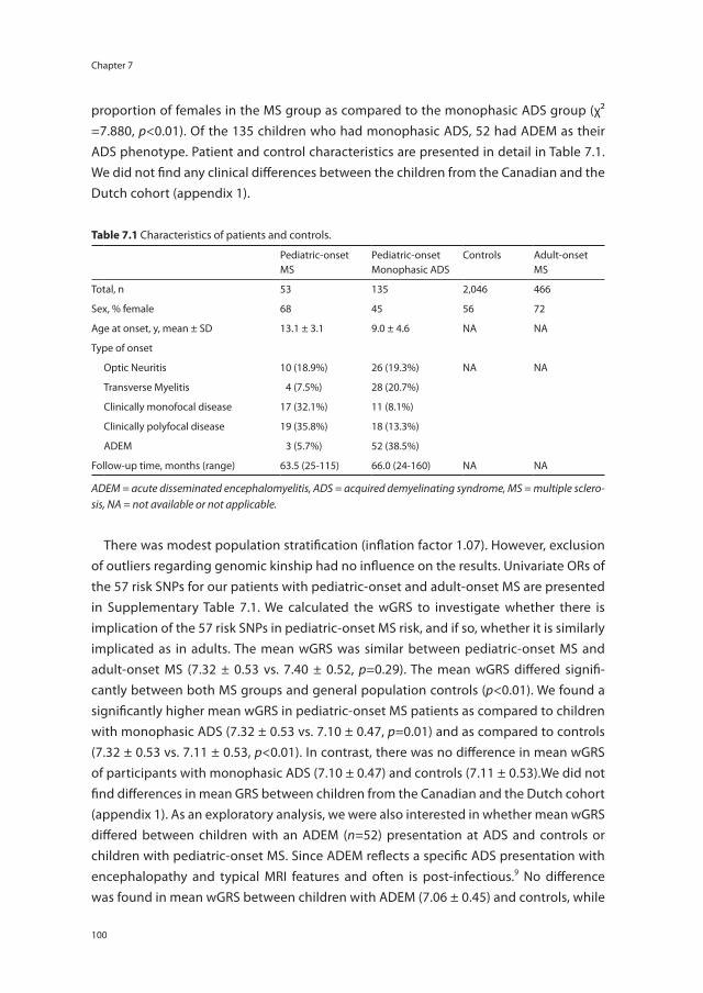
Chapter 7
100
proportion of females in the MS group as compared to the monophasic ADS group (χ² =7.880, p<0.01). Of the 135 children who had monophasic ADS, 52 had ADEM as their ADS phenotype. Patient and control characteristics are presented in detail in Table 7.1. We did not find any clinical differences between the children from the Canadian and the Dutch cohort (appendix 1).
There was modest population stratification (inflation factor 1.07). However, exclusion of outliers regarding genomic kinship had no influence on the results. Univariate ORs of the 57 risk SNPs for our patients with pediatric-onset and adult-onset MS are presented in Supplementary Table 7.1. We calculated the wGRS to investigate whether there is implication of the 57 risk SNPs in pediatric-onset MS risk, and if so, whether it is similarly implicated as in adults. The mean wGRS was similar between pediatric-onset MS and adult-onset MS (7.32 ± 0.53 vs. 7.40 ± 0.52, p=0.29). The mean wGRS differed signifi-cantly between both MS groups and general population controls (p<0.01). We found a significantly higher mean wGRS in pediatric-onset MS patients as compared to children with monophasic ADS (7.32 ± 0.53 vs. 7.10 ± 0.47, p=0.01) and as compared to controls (7.32 ± 0.53 vs. 7.11 ± 0.53, p<0.01). In contrast, there was no difference in mean wGRS of participants with monophasic ADS (7.10 ± 0.47) and controls (7.11 ± 0.53).We did not find differences in mean GRS between children from the Canadian and the Dutch cohort (appendix 1). As an exploratory analysis, we were also interested in whether mean wGRS differed between children with an ADEM (n=52) presentation at ADS and controls or children with pediatric-onset MS. Since ADEM reflects a specific ADS presentation with encephalopathy and typical MRI features and often is post-infectious.9 No difference was found in mean wGRS between children with ADEM (7.06 ± 0.45) and controls, while
table 7.1 Characteristics of patients and controls.
Pediatric-onset MS
Pediatric-onset Monophasic ADS
Controls Adult-onset MS
Total, n 53 135 2,046 466
Sex, % female 68 45 56 72
Age at onset, y, mean ± SD 13.1 ± 3.1 9.0 ± 4.6 NA NA
Type of onset
Optic Neuritis 10 (18.9%) 26 (19.3%) NA NA
Transverse Myelitis 4 (7.5%) 28 (20.7%)
Clinically monofocal disease 17 (32.1%) 11 (8.1%)
Clinically polyfocal disease 19 (35.8%) 18 (13.3%)
ADEM 3 (5.7%) 52 (38.5%)
Follow-up time, months (range) 63.5 (25-115) 66.0 (24-160) NA NA
ADEM = acute disseminated encephalomyelitis, ADS = acquired demyelinating syndrome, MS = multiple sclero-sis, NA = not available or not applicable.
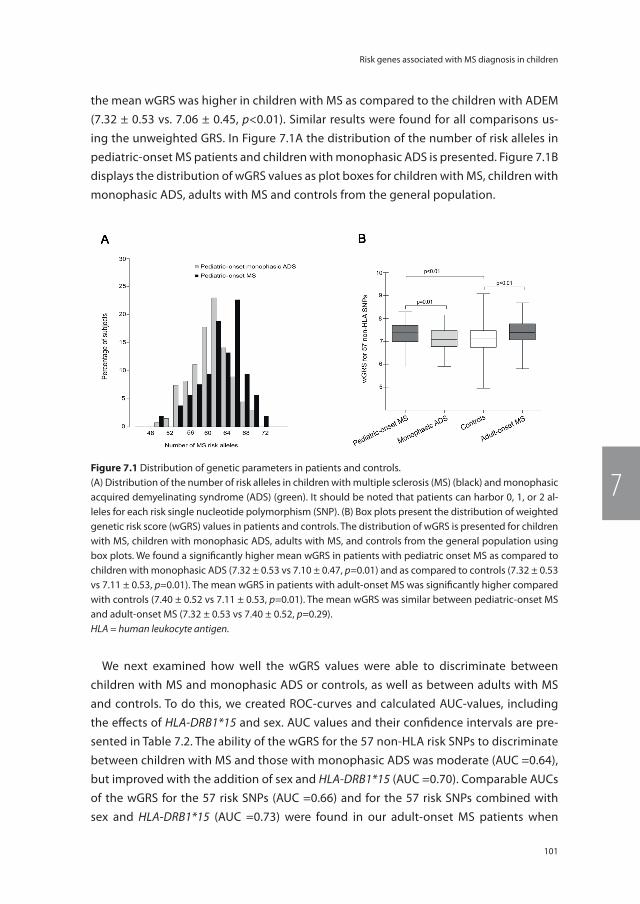
101
Risk genes associated with MS diagnosis in children
7
the mean wGRS was higher in children with MS as compared to the children with ADEM (7.32 ± 0.53 vs. 7.06 ± 0.45, p<0.01). Similar results were found for all comparisons us-ing the unweighted GRS. In Figure 7.1A the distribution of the number of risk alleles in pediatric-onset MS patients and children with monophasic ADS is presented. Figure 7.1B displays the distribution of wGRS values as plot boxes for children with MS, children with monophasic ADS, adults with MS and controls from the general population.
We next examined how well the wGRS values were able to discriminate between children with MS and monophasic ADS or controls, as well as between adults with MS and controls. To do this, we created ROC-curves and calculated AUC-values, including the effects of HLA-DRB1*15 and sex. AUC values and their confidence intervals are pre-sented in Table 7.2. The ability of the wGRS for the 57 non-HLA risk SNPs to discriminate between children with MS and those with monophasic ADS was moderate (AUC =0.64), but improved with the addition of sex and HLA-DRB1*15 (AUC =0.70). Comparable AUCs of the wGRS for the 57 risk SNPs (AUC =0.66) and for the 57 risk SNPs combined with sex and HLA-DRB1*15 (AUC =0.73) were found in our adult-onset MS patients when
A B
Figure 7.1 Distribution of genetic parameters in patients and controls.(A) Distribution of the number of risk alleles in children with multiple sclerosis (MS) (black) and monophasic acquired demyelinating syndrome (ADS) (green). It should be noted that patients can harbor 0, 1, or 2 al-leles for each risk single nucleotide polymorphism (SNP). (B) Box plots present the distribution of weighted genetic risk score (wGRS) values in patients and controls. The distribution of wGRS is presented for children with MS, children with monophasic ADS, adults with MS, and controls from the general population using box plots. We found a significantly higher mean wGRS in patients with pediatric onset MS as compared to children with monophasic ADS (7.32 ± 0.53 vs 7.10 ± 0.47, p=0.01) and as compared to controls (7.32 ± 0.53 vs 7.11 ± 0.53, p=0.01). The mean wGRS in patients with adult-onset MS was significantly higher compared with controls (7.40 ± 0.52 vs 7.11 ± 0.53, p=0.01). The mean wGRS was similar between pediatric-onset MS and adult-onset MS (7.32 ± 0.53 vs 7.40 ± 0.52, p=0.29).HLA = human leukocyte antigen.
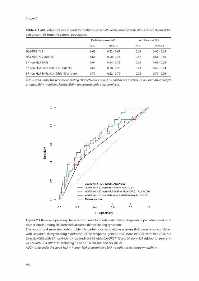
Chapter 7
102
Figure 7.2 Receiver operating characteristic curve for models identifying diagnosis of pediatric-onset mul-tiple sclerosis among children with acquired demyelinating syndrome.The results for 4 separate models to identify pediatric-onset multiple sclerosis (MS) cases among children with acquired demyelinating syndrome (ADS): weighted genetic risk score (wGRS) with HLA-DRB1*15 (black); wGRS with 57 non-HLA risk loci (red); wGRS with HLA-DRB1*15 and 57 non-HLA risk loci (green); and wGRS with HLA-DRB1*15, including 57 non-HLA risk loci and sex (blue).AUC = area under the curve, HLA = human leukocyte antigen, SNP = single nucleotide polymorphism.
table 7.2 AUC values for risk models for pediatric-onset MS versus monophasic ADS and adult-onset MS versus controls from the general population.
Pediatric-onset MS Adult-onset MS
AUC 95% CI AUC 95% CI
HLA-DRB1*15 0.60 0.52 - 0.67 0.63 0.60 - 0.65
HLA-DRB1*15 and sex 0.66 0.58 - 0.74 0.67 0.64 - 0.69
57 non-HLA SNPs 0.64 0.54 - 0.73 0.66 0.63 - 0.68
57 non-HLA SNPs and HLA-DRB1*15 0.66 0.58 - 0.75 0.71 0.69 - 0.74
57 non-HLA SNPs, HLA-DRB1*15 and sex 0.70 0.62 - 0.79 0.73 0.71 - 0.76
AUC = area under the receiver operating characteristic curve, CI = confidence interval, HLA = human leukocyte antigen, MS = multiple sclerosis, SNP = single nucleotide polymorphism.
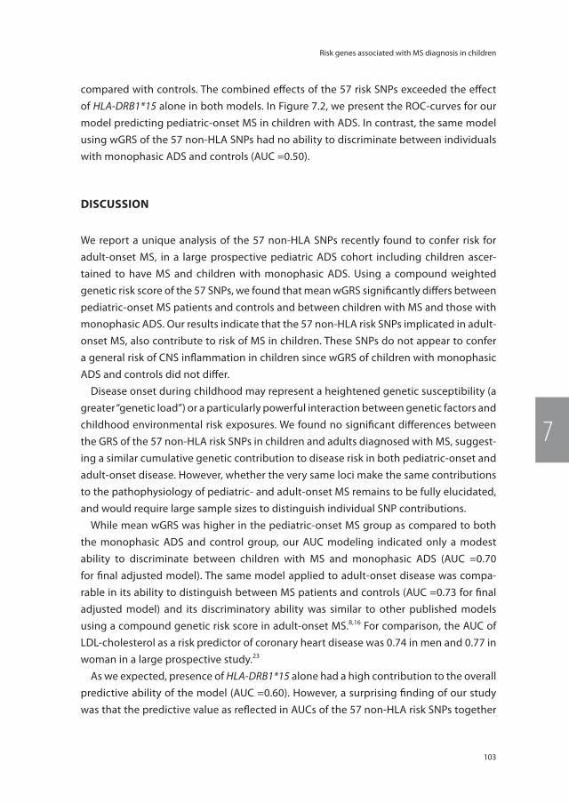
103
Risk genes associated with MS diagnosis in children
7
compared with controls. The combined effects of the 57 risk SNPs exceeded the effect of HLA-DRB1*15 alone in both models. In Figure 7.2, we present the ROC-curves for our model predicting pediatric-onset MS in children with ADS. In contrast, the same model using wGRS of the 57 non-HLA SNPs had no ability to discriminate between individuals with monophasic ADS and controls (AUC =0.50).
Discussion
We report a unique analysis of the 57 non-HLA SNPs recently found to confer risk for adult-onset MS, in a large prospective pediatric ADS cohort including children ascer-tained to have MS and children with monophasic ADS. Using a compound weighted genetic risk score of the 57 SNPs, we found that mean wGRS significantly differs between pediatric-onset MS patients and controls and between children with MS and those with monophasic ADS. Our results indicate that the 57 non-HLA risk SNPs implicated in adult-onset MS, also contribute to risk of MS in children. These SNPs do not appear to confer a general risk of CNS inflammation in children since wGRS of children with monophasic ADS and controls did not differ.
Disease onset during childhood may represent a heightened genetic susceptibility (a greater “genetic load”) or a particularly powerful interaction between genetic factors and childhood environmental risk exposures. We found no significant differences between the GRS of the 57 non-HLA risk SNPs in children and adults diagnosed with MS, suggest-ing a similar cumulative genetic contribution to disease risk in both pediatric-onset and adult-onset disease. However, whether the very same loci make the same contributions to the pathophysiology of pediatric- and adult-onset MS remains to be fully elucidated, and would require large sample sizes to distinguish individual SNP contributions.
While mean wGRS was higher in the pediatric-onset MS group as compared to both the monophasic ADS and control group, our AUC modeling indicated only a modest ability to discriminate between children with MS and monophasic ADS (AUC =0.70 for final adjusted model). The same model applied to adult-onset disease was compa-rable in its ability to distinguish between MS patients and controls (AUC =0.73 for final adjusted model) and its discriminatory ability was similar to other published models using a compound genetic risk score in adult-onset MS.8,16 For comparison, the AUC of LDL-cholesterol as a risk predictor of coronary heart disease was 0.74 in men and 0.77 in woman in a large prospective study.23
As we expected, presence of HLA-DRB1*15 alone had a high contribution to the overall predictive ability of the model (AUC =0.60). However, a surprising finding of our study was that the predictive value as reflected in AUCs of the 57 non-HLA risk SNPs together
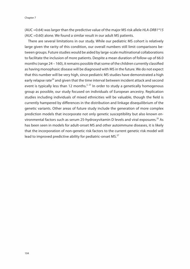
Chapter 7
104
(AUC =0.64) was larger than the predictive value of the major MS risk allele HLA-DRB1*15 (AUC =0.60) alone. We found a similar result in our adult MS patients.
There are several limitations in our study. While our pediatric MS cohort is relatively large given the rarity of this condition, our overall numbers still limit comparisons be-tween groups. Future studies would be aided by large-scale multinational collaborations to facilitate the inclusion of more patients. Despite a mean duration of follow-up of 66.0 months (range 24 – 160), it remains possible that some of the children currently classified as having monophasic disease will be diagnosed with MS in the future. We do not expect that this number will be very high, since pediatric MS studies have demonstrated a high early relapse rate24 and given that the time interval between incident attack and second event is typically less than 12 months.2, 25 In order to study a genetically homogenous group as possible, our study focused on individuals of European ancestry. Replication studies including individuals of mixed ethnicities will be valuable, though the field is currently hampered by differences in the distribution and linkage disequilibrium of the genetic variants. Other areas of future study include the generation of more complex prediction models that incorporate not only genetic susceptibility but also known en-vironmental factors such as serum 25-hydroxyvitamin D levels and viral exposures.26 As has been seen in models for adult-onset MS and other autoimmune diseases, it is likely that the incorporation of non-genetic risk factors to the current genetic risk model will lead to improved predictive ability for pediatric-onset MS.27
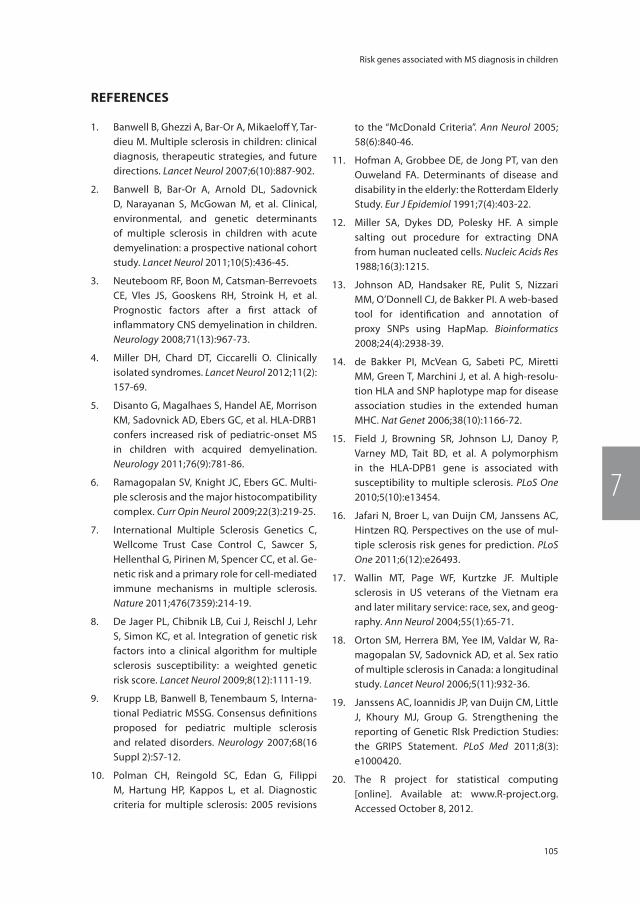
105
Risk genes associated with MS diagnosis in children
7
reFerences
1. Banwell B, Ghezzi A, Bar-Or A, Mikaeloff Y, Tar-dieu M. Multiple sclerosis in children: clinical diagnosis, therapeutic strategies, and future directions. Lancet Neurol 2007; 6(10): 887-902.
2. Banwell B, Bar-Or A, Arnold DL, Sadovnick D, Narayanan S, McGowan M, et al. Clinical, environmental, and genetic determinants of multiple sclerosis in children with acute demyelination: a prospective national cohort study. Lancet Neurol 2011; 10(5): 436-45.
3. Neuteboom RF, Boon M, Catsman-Berrevoets CE, Vles JS, Gooskens RH, Stroink H, et al. Prognostic factors after a first attack of inflammatory CNS demyelination in children. Neurology 2008; 71(13): 967-73.
4. Miller DH, Chard DT, Ciccarelli O. Clinically isolated syndromes. Lancet Neurol 2012; 11(2): 157-69.
5. Disanto G, Magalhaes S, Handel AE, Morrison KM, Sadovnick AD, Ebers GC, et al. HLA-DRB1 confers increased risk of pediatric-onset MS in children with acquired demyelination. Neurology 2011; 76(9): 781-86.
6. Ramagopalan SV, Knight JC, Ebers GC. Multi-ple sclerosis and the major histocompatibility complex. Curr Opin Neurol 2009; 22(3): 219-25.
7. International Multiple Sclerosis Genetics C, Wellcome Trust Case Control C, Sawcer S, Hellenthal G, Pirinen M, Spencer CC, et al. Ge-netic risk and a primary role for cell-mediated immune mechanisms in multiple sclerosis. Nature 2011; 476(7359): 214-19.
8. De Jager PL, Chibnik LB, Cui J, Reischl J, Lehr S, Simon KC, et al. Integration of genetic risk factors into a clinical algorithm for multiple sclerosis susceptibility: a weighted genetic risk score. Lancet Neurol 2009; 8(12): 1111-19.
9. Krupp LB, Banwell B, Tenembaum S, Interna-tional Pediatric MSSG. Consensus definitions proposed for pediatric multiple sclerosis and related disorders. Neurology 2007; 68(16 Suppl 2): S7-12.
10. Polman CH, Reingold SC, Edan G, Filippi M, Hartung HP, Kappos L, et al. Diagnostic criteria for multiple sclerosis: 2005 revisions
to the “McDonald Criteria”. Ann Neurol 2005; 58(6): 840-46.
11. Hofman A, Grobbee DE, de Jong PT, van den Ouweland FA. Determinants of disease and disability in the elderly: the Rotterdam Elderly Study. Eur J Epidemiol 1991; 7(4): 403-22.
12. Miller SA, Dykes DD, Polesky HF. A simple salting out procedure for extracting DNA from human nucleated cells. Nucleic Acids Res 1988; 16(3): 1215.
13. Johnson AD, Handsaker RE, Pulit S, Nizzari MM, O’Donnell CJ, de Bakker PI. A web-based tool for identification and annotation of proxy SNPs using HapMap. Bioinformatics 2008; 24(4): 2938-39.
14. de Bakker PI, McVean G, Sabeti PC, Miretti MM, Green T, Marchini J, et al. A high-resolu-tion HLA and SNP haplotype map for disease association studies in the extended human MHC. Nat Genet 2006; 38(10): 1166-72.
15. Field J, Browning SR, Johnson LJ, Danoy P, Varney MD, Tait BD, et al. A polymorphism in the HLA-DPB1 gene is associated with susceptibility to multiple sclerosis. PLoS One 2010; 5(10): e13454.
16. Jafari N, Broer L, van Duijn CM, Janssens AC, Hintzen RQ. Perspectives on the use of mul-tiple sclerosis risk genes for prediction. PLoS One 2011; 6(12): e26493.
17. Wallin MT, Page WF, Kurtzke JF. Multiple sclerosis in US veterans of the Vietnam era and later military service: race, sex, and geog-raphy. Ann Neurol 2004; 55(1): 65-71.
18. Orton SM, Herrera BM, Yee IM, Valdar W, Ra-magopalan SV, Sadovnick AD, et al. Sex ratio of multiple sclerosis in Canada: a longitudinal study. Lancet Neurol 2006; 5(11): 932-36.
19. Janssens AC, Ioannidis JP, van Duijn CM, Little J, Khoury MJ, Group G. Strengthening the reporting of Genetic RIsk Prediction Studies: the GRIPS Statement. PLoS Med 2011; 8(3): e1000420.
20. The R project for statistical computing [online]. Available at: www.R-project.org. Accessed October 8, 2012.
Chapter 7
106
21. Kundu S, Aulchenko YS, van Duijn CM, Jans-sens AC. PredictABEL: an R package for the assessment of risk prediction models. Eur J Epidemiol 2011; 26(4): 261-64.
22. Sing T, Sander O, Beerenwinkel N, Lengauer T. ROCR: visualizing classifier performance in R. Bioinformatics 2005; 21(20): 3940-41.
23. Wilson PW, D’Agostino RB, Levy D, Belanger AM, Silbershatz H, Kannel WB. Prediction of coronary heart disease using risk factor categories. Circulation 1998; 97(18): 1837-47.
24. Gorman MP, Healy BC, Polgar-Turcsanyi M, Chitnis T. Increased relapse rate in pediatric-onset compared with adult-onset multiple sclerosis. Arch Neurol 2009; 66(1): 54-59.
25. Mikaeloff Y, Suissa S, Vallee L, Lubetzki C, Ponsot G, Confavreux C, et al. First episode of acute CNS inflammatory demyelination in childhood: prognostic factors for multiple sclerosis and disability. J Pediatr 2004; 144(2): 246-52.
26. Handel AE, Giovannoni G, Ebers GC, Rama-gopalan SV. Environmental factors and their timing in adult-onset multiple sclerosis. Nat Rev Neurol 2010; 6(3): 156-66.
27. Simon KC, van der Mei IA, Munger KL, Ponsonby A, Dickinson J, Dwyer T, et al. Combined effects of smoking, anti-EBNA antibodies, and HLA-DRB1*1501 on multiple sclerosis risk. Neurology 2010; 74(17): 1365-71.
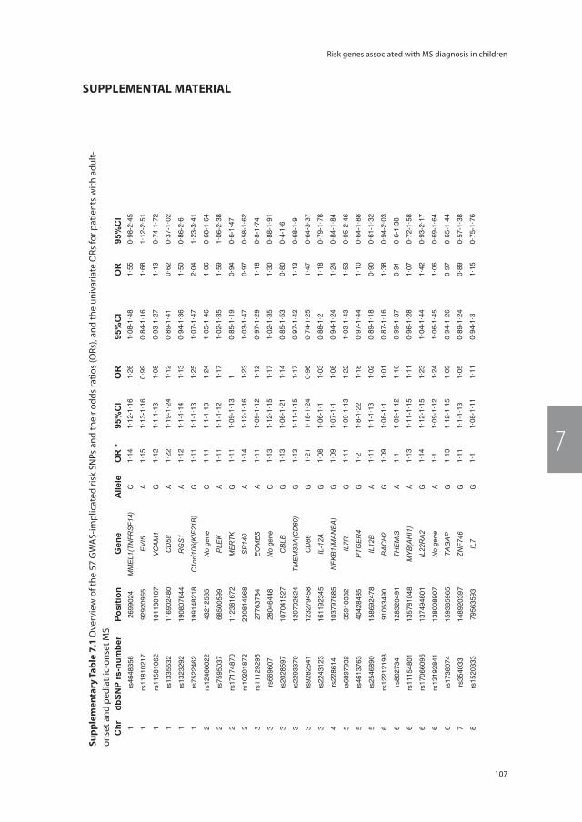
107
Risk genes associated with MS diagnosis in children
7
suPPlementAl mAteriAlsu
pple
men
tary
tab
le 7
.1 O
verv
iew
of t
he 5
7 G
WA
S-im
plic
ated
risk
SN
Ps a
nd th
eir o
dds
ratio
s (O
Rs),
and
the
univ
aria
te O
Rs fo
r pat
ient
s w
ith a
dult-
onse
t and
ped
iatr
ic-o
nset
MS.
Tabl
e1:
Ove
rvie
w o
f the
57
GW
AS-
impl
icat
ed ri
sk S
NPs
and
thei
r odd
s ra
tios
(OR
s), a
nd th
e un
ivar
iate
OR
s fo
r pat
ient
s w
ith a
dult-
onse
t and
pe
diat
ric-o
nset
MS.
Ris
kG
WAS
(n=9
772)
Adul
t-ons
et M
S (n
=466
)Pe
diat
ric-o
nset
MS
(n=5
3)C
hrdb
SNP
rs-n
umbe
r Po
sitio
nG
ene
Alle
leO
R *
95%
CI
OR
95%
CI
OR
95%
CI
1rs
4648
356
2699
024
MM
EL1
(TN
FRS
F14)
C1·
141·
12-1
·16
1·26
1·08
-1·4
81·
550·
98-2
·45
1rs
1181
0217
9292
0965
EV
I5A
1·15
1·13
-1·1
60·
990·
84-1
·16
1·68
1·12
-2·5
1
1rs
1158
1062
1011
8010
7V
CA
M1
G1·
121·
1-1·
131·
080·
93-1
·27
1·13
0·74
-1·7
2
1rs
1335
532
1169
0248
0C
D58
A1·
221·
19-1
·24
1·12
0·89
-1·4
10·
620·
37-1
·02
1rs
1323
292
1908
0764
4R
GS
1A
1·12
1·1-
1·14
1·13
0·94
-1·3
61·
500·
86-2
·6
1rs
7522
462
1991
4821
8C
1orf1
06(K
IF21
B)
G1·
111·
1-1·
131·
251·
07-1
·47
2·04
1·23
-3·4
1
2rs
1246
6022
4321
2565
No
gene
C1·
111·
1-1·
131·
241·
05-1
·46
1·06
0·68
-1·6
4
2rs
7595
037
6850
0599
PLE
KA
1·11
1·1-
1·12
1·17
1·02
-1·3
51·
591·
06-2
·38
2rs
1717
4870
1123
8167
2M
ER
TKG
1·11
1·09
-1·1
31
0·85
-1·1
90·
940·
6-1·
47
2rs
1020
1872
2308
1496
8S
P14
0A
1·14
1·12
-1·1
61·
231·
03-1
·47
0·97
0·58
-1·6
2
3rs
1112
9295
2776
3784
EO
ME
SA
1·11
1·09
-1·1
21·
120·
97-1
·29
1·18
0·8-
1·74
3rs
6696
0728
0464
48N
o ge
neC
1·13
1·12
-1·1
51·
171·
02-1
·35
1·30
0·88
-1·9
1
3rs
2028
597
1070
4152
7C
BLB
G1·
131·
06-1
·21
1·14
0·85
-1·5
30·
800·
4-1·
6
3rs
2293
370
1207
0262
4TM
EM
39A
(CD
80)
G1·
131·
11-1
·15
1·17
0·97
-1·4
21·
130·
68-1
·9
3rs
9282
641
1232
7945
8C
D86
G1·
211·
18-1
·24
0·96
0·74
-1·2
51·
470·
64-3
·37
3rs
2243
123
1611
9234
5IL
-12A
G1·
081·
06-1
·11·
030·
88-1
·21·
180·
79-1
·78
4rs
2286
1410
3797
685
NFK
B1(
MA
NB
A)
G1·
091·
07-1
·11·
080·
94-1
·24
1·24
0·84
-1·8
4
5rs
6897
932
3591
0332
IL7R
G1·
111·
09-1
·13
1·22
1·03
-1·4
31·
530·
95-2
·46
5rs
4613
763
4042
8485
PTG
ER
4G
1·2
1·8-
1·22
1·18
0·97
-1·4
41·
100·
64-1
·88
5rs
2546
890
1586
9247
8IL
12B
A1·
111·
1-1·
131·
020·
89-1
·18
0·90
0·61
-1·3
2
6rs
1221
2193
9105
3490
BA
CH
2G
1·09
1·08
-1·1
1·01
0·87
-1·1
61·
380·
94-2
·03
6rs
8027
3412
8320
491
THE
MIS
A1·
11·
09-1
·12
1·16
0·99
-1·3
70·
910·
6-1·
38
6rs
1115
4801
1357
8104
8M
YB
(AH
I1)
A1·
131·
11-1
·15
1·11
0·96
-1·2
81·
070·
72-1
·58
6rs
1706
6096
1374
9460
1IL
22R
A2
G1·
141·
12-1
·15
1·23
1·04
-1·4
41·
420·
93-2
·17
6rs
1319
2841
1380
0890
7N
o ge
neA
1·1
1·09
-1·1
21·
241·
06-1
·45
1·06
0·69
-1·6
4
6rs
1738
074
1593
8596
5TA
GA
PG
1·13
1·12
-1·1
51·
090·
94-1
·26
0·97
0·65
-1·4
4
7rs
3540
3314
8920
397
ZNF7
46G
1·11
1·1-
1·13
1·05
0·89
-1·2
40·
890·
57-1
·38
8rs
1520
333
7956
3593
IL7
G1·
11·
08-1
·11
1·11
0·94
-1·3
1·15
0·75
-1·7
6
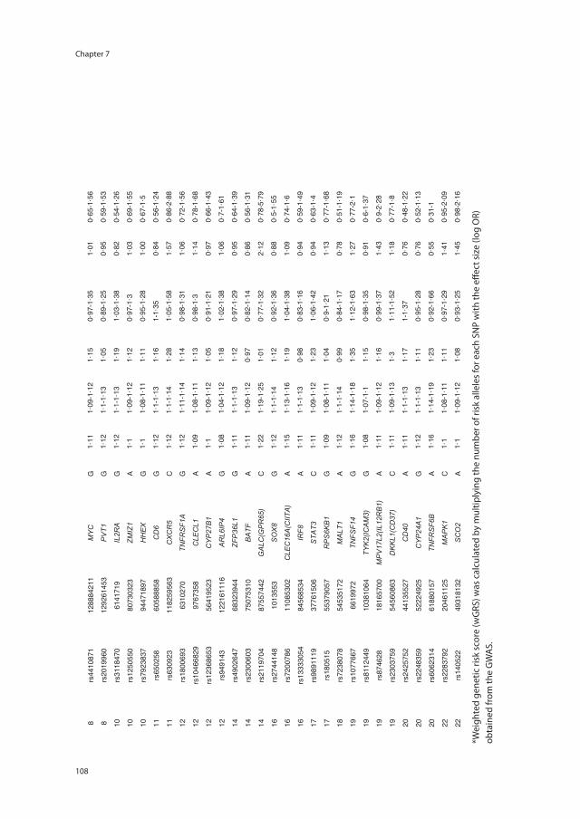
Chapter 7
108
8rs
4410
871
1288
8421
1M
YC
G1·
111·
09-1
·12
1·15
0·97
-1·3
51·
010·
65-1
·56
8rs
2019
960
1292
6145
3P
VT1
G1·
121·
1-1·
131·
050·
89-1
·25
0·95
0·59
-1·5
3
10rs
3118
470
6141
719
IL2R
AG
1·12
1·1-
1·13
1·19
1·03
-1·3
80·
820·
54-1
·26
10rs
1250
550
8073
0323
ZMIZ
1A
1·1
1·09
-1·1
21·
120·
97-1
·31·
030·
69-1
·55
10rs
7923
837
9447
1897
HH
EX
G1·
11·
08-1
·11
1·11
0·95
-1·2
81·
000·
67-1
·5
11rs
6502
5860
5888
58C
D6
G1·
121·
1-1·
131·
161-
1·35
0·84
0·56
-1·2
4
11rs
6309
2311
8259
563
CX
CR
5C
1·12
1·1-
1·14
1·28
1·05
-1·5
81·
570·
86-2
·88
12rs
1800
693
6310
270
TNFR
SF1
AG
1·12
1·11
-1·1
41·
140·
98-1
·31
1·06
0·72
-1·5
6
12rs
1046
6829
9767
358
CLE
CL1
A1·
091·
08-1
·11
1·13
0·98
-1·3
1·14
0·78
-1·6
8
12rs
1236
8653
5641
9523
CY
P27
B1
A1·
11·
09-1
·12
1·05
0·91
-1·2
10·
970·
66-1
·43
12rs
9491
4312
2161
116
AR
L6IP
4G
1·08
1·04
-1·1
21·
181·
02-1
·38
1·06
0·7-
1·61
14rs
4902
647
6832
3944
ZFP
36L1
G1·
111·
1-1·
131·
120·
97-1
·29
0·95
0·64
-1·3
9
14rs
2300
603
7507
5310
BA
TFA
1·11
1·09
-1·1
20·
970·
82-1
·14
0·86
0·56
-1·3
1
14rs
2119
704
8755
7442
GA
LC(G
PR
65)
C1·
221·
19-1
·25
1·01
0·77
-1·3
22·
120·
78-5
·79
16rs
2744
148
1013
553
SO
X8
G1·
121·
1-1·
141·
120·
92-1
·36
0·88
0·5-
1·55
16rs
7200
786
1108
5302
CLE
C16
A(C
IITA
)A
1·15
1·13
-1·1
61·
191·
04-1
·38
1·09
0·74
-1·6
16rs
1333
3054
8456
8534
IRF8
A1·
111·
1-1·
130·
980·
83-1
·16
0·94
0·59
-1·4
9
17rs
9891
119
3776
1506
STA
T3C
1·11
1·09
-1·1
21·
231·
06-1
·42
0·94
0·63
-1·4
17rs
1805
1555
3790
57R
PS
6KB
1G
1·09
1·08
-1·1
11·
040·
9-1·
211·
130·
77-1
·68
18rs
7238
078
5453
5172
MA
LT1
A1·
121·
1-1·
140·
990·
84-1
·17
0·78
0·51
-1·1
9
19rs
1077
667
6619
972
TNFS
F14
G1·
161·
14-1
·18
1·35
1·12
-1·6
31·
270·
77-2
·1
19rs
8112
449
1038
1064
TYK
2(IC
AM
3)G
1·08
1·07
-1·1
1·15
0·98
-1·3
50·
910·
6-1·
37
19rs
8746
2818
1657
00M
PV
17L2
(IL12
RB
1)A
1·11
1·09
-1·1
21·
160·
99-1
·37
1·43
0·9-
2·28
19rs
2303
759
5456
0863
DK
KL1
(CD
37)
C1·
111·
09-1
·13
1·3
1·11
-1·5
21·
180·
77-1
·8
20rs
2425
752
4413
5527
CD
40A
1·11
1·1-
1·13
1·17
1-1·
370·
760·
48-1
·22
20rs
2248
359
5222
4925
CY
P24
A1
G1·
121·
1-1·
131·
110·
95-1
·28
0·76
0·52
-1·1
3
20rs
6062
314
6188
0157
TNFR
SF6
BA
1·16
1·14
-1·1
91·
230·
92-1
·66
0·55
0·31
-1
22rs
2283
792
2046
1125
MA
PK
1C
1·1
1·08
-1·1
11·
110·
97-1
·29
1·41
0·95
-2·0
9
22rs
1405
2249
3181
32S
CO
2A
1·1
1·09
-1·1
21·
080·
93-1
·25
1·45
0·98
-2·1
6
*W
eigh
ted
gene
tic ri
sk sc
ore
(wG
RS)
was
cal
cula
ted
by m
ultip
lyin
g th
e nu
mbe
r of r
isk
alle
les f
or e
ach
SNP
with
the
effe
ct si
ze (l
og O
R)
obta
ined
from
the
GW
AS.
*Wei
ghte
d ge
netic
risk
sco
re (w
GRS
) was
cal
cula
ted
by m
ultip
lyin
g th
e nu
mbe
r of r
isk
alle
les
for e
ach
SNP
with
the
effec
t siz
e (lo
g O
R)ob
tain
ed fr
om th
e G
WA
S.

109
Risk genes associated with MS diagnosis in children
7
Appendix 1. supplementary statistical analysis
Appendix 1: supplementary statistical analysis
Canadian and Dutch cohorts
There were no overt clinical differences between the children from the Canadian and the Dutch cohort (mean age at onset, sex, type of onset, mean time of follow-up).
Mean age at onset: one-way ANOVA F = 5.17, df = 35, p = 0.68
Sex: Chi Square χ² = 2.7, df = 1, p = 0.10
Type of onset: Chi Square χ² = 9.1, df = 4, p = 0.06
Mean time of follow-up: one-way ANOVA F= 27.3, df = 30, p = 0.83
We also did not find differences in the Genetic Risk Scores between children from the Canadian and the Dutch cohort.
uwGRS 57 SNPs: Welch two-sample t-test p = 0.49
wGRS 57 SNPs: Welch two-sample t-test p = 0.55
wGRS 57 SNPs + HLA: Welch two-sample t-test p = 0.14
Controlling for possible population stratification
In order to control the effect of genetic variation due to ancestry, only participants with self-reported European ancestry were included in the analyses presented in the paper. Potential stratification was corrected by genomic control and principal component (PC) analysis. There was modest population stratification (inflation factor 1.07). However, exclusion of outliers regarding genomic kinship had no influence on the results.


2016
12
OCT
Chapter 8
Anti-MOG antibodies plead against MS diagnosis in an ADS cohort
I.A. Ketelslegers*, E.D. van Pelt*, S. Bryde, R.F. Neuteboom, C.E. Catsman-Berrevoets, D. Hamann, R.Q. Hintzen.
* shared first authors
Multiple Sclerosis, 2015
Chapter 8
112
AbstrAct
background Acquired demyelinating syndromes (ADS) in children are a group of dis-tinct first immune-mediated demyelinating events of the central nervous system (CNS). Predictive biomarkers for future diagnosis are lacking. A putative target antigen is myelin oligodendrocyte glycoprotein (MOG). We analysed the presence of MOG-antibodies in a cohort of ADS patients.
methods 117 children with ADS form a nationwide cohort were analysed with a cell-based assay, divided in 5 groups: optic neuritis (ON; n=20), transverse myelitis (TM; n=7), other monofocal ADS (n=22), polyfocal ADS without encephalopathy (n=44) and polyfo-cal ADS with encephalopathy (n=24). Additionally, 13 children with other neurological diseases (OND), 31 healthy children and 29 adult ADEM patients were tested.
results Twenty-one of the 117 children with ADS tested anti-MOG seropositive (18%). The group of patients with polyfocal ADS plus encephalopathy (ADEM) had the highest prevalence of anti-MOG seropositivity (42% versus 18% in non-encephalopathic polyfo-cal ADS patients). Forty-seven ADS children had a final diagnosis of multiple sclerosis (MS). In only one of them MOG-antibodies were detected (2%), with only borderline positivity. Only 1 out of 29 adult ADEM patients tested anti-MOG seropositive.
conclusions MOG-antibodies are strongly skewed towards ADS children that present with an ADEM-like disease-onset. The presence of such antibodies pleads against a future diagnosis of MS.
113
MOG-antibodies in childhood-onset ADS
8
introDuction
Acquired demyelinating syndromes (ADS) in children are first immune-mediated demyelinating events of the central nervous system (CNS).1, 2 The clinical spectrum is very heterogenic, including optic neuritis (ON), transverse myelitis (TM), other clinically isolated syndromes, acute disseminated encephalomyelitis (ADEM) and neuromyelitis optica (NMO). These distinct disease entities may be challenging to diagnose accurately at the first event. Disease course and prognosis are also variable and all these different subtypes of ADS can represent a first episode of multiple sclerosis (MS). It is essential to distinguish monophasic disease forms from a chronic relapsing disease like MS early in the disease course, because prompt initiation of disease-modifying treatment has been recommended for children with MS.3
Clinical evidence suggests that ADS includes several distinct disorders with different underlying pathophysiology. Preferably, certain subsets of ADS patients characterized by humoral autoimmunity might be identified through the use of disease-specific au-toantibodies. For example, NMO is now considered to be an antibody-mediated disease that is distinct from MS, on account of the discovery of the disease-specific autoantibody against aquaporin-4 (AQP4).4, 5
In the search for disease-specific autoantibodies in ADS, myelin oligodendrocyte glycoprotein (MOG) is a putative target antigen. This protein is expressed on the surface of myelin sheaths and oligodendrocytes, and thus specific to the CNS. Previous studies already showed that MOG-antibodies can cause demyelination in vitro and can induce experimental autoimmune encephalomyelitis (EAE).6, 7 Based on the current knowledge, antibodies to MOG have been detected in a subgroup of patients presenting with CNS demyelinating diseases8, 9 and are especially present in patients with ADEM10, 11, in chil-dren with very early-onset MS12, and with higher titers in the youngest children and children with ADEM.11, 13-15 But the sensitivity of MOG-antibody assays to discriminate patients with CNS demyelinating disease varies and is reported to be only as high as 46%.16 This is in part due to the patient population that is included, since the antibod-ies are more prevalent is the ADEM subgroup of ADS10-15 and might not be a sensitive marker for ADS in general. Previous studies focused mainly on specific subgroups of ADS patients based on diagnosis, like paediatric onset MS10-12, 14, 16, ADEM10, 11, 13, 14, 17 and CIS.11, 13, 14 Recently MOG-antibodies were also detected in patients with anti-AQP4 negative NMO or NMO spectrum disorders such as bilateral or recurrent ON and longi-tudinally transverse myelitis.18-26
To date, it is unsure which subgroup the children with MOG-antibodies represent within the spectrum of ADS.16 In the current study we investigated the presence of MOG-antibodies in a cohort comprised of all ADS subtypes and compared their presence between the different ADS subtypes based on clinical presentation. We hypothesized
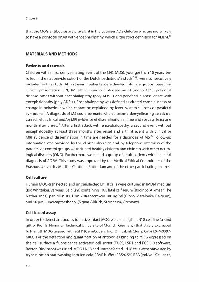
Chapter 8
114
that the MOG-antibodies are prevalent in the younger ADS children who are more likely to have a polyfocal onset with encephalopathy, which is the strict definition for ADEM.27
mAteriAls AnD methoDs
Patients and controls
Children with a first demyelinating event of the CNS (ADS), younger than 18 years, en-rolled in the nationwide cohort of the Dutch pediatric MS study2, 28, were consecutively included in this study. At first event, patients were divided into five groups, based on clinical presentation: ON, TM, other monofocal disease-onset (mono ADS), polyfocal disease-onset without encephalopathy (poly ADS –) and polyfocal disease-onset with encephalopathy (poly ADS +). Encephalopathy was defined as altered consciousness or change in behaviour, which cannot be explained by fever, systemic illness or postictal symptoms.2 A diagnosis of MS could be made when a second demyelinating attack oc-curred, with clinical and/or MRI evidence of dissemination in time and space at least one month after onset.29 After a first attack with encephalopathy, a second event without encephalopathy at least three months after onset and a third event with clinical or MRI evidence of dissemination in time are needed for a diagnosis of MS.27 Follow-up information was provided by the clinical physician and by telephone interview of the parents. As control groups we included healthy children and children with other neuro-logical diseases (OND). Furthermore we tested a group of adult patients with a clinical diagnosis of ADEM. This study was approved by the Medical Ethical Committees of the Erasmus University Medical Centre in Rotterdam and of the other participating centres.
cell culture
Human MOG-transfected and untransfected LN18 cells were cultured in IMDM medium (Bio Whittaker, Verviers, Belgium) containing 10% fetal calf serum (Bodinco, Alkmaar, The Netherlands), penicillin 100 U/ml / streptomycin 100 ug/ml (Gibco, Merelbeke, Belgium), and 50 μM 2-mercaptoethanol (Sigma-Aldrich, Steinheim, Germany).
cell-based assay
In order to detect antibodies to native intact MOG we used a glial LN18 cell line (a kind gift of Prof. B. Hemmer, Technical University of Munich, Germany) that stably expressed full-length MOG tagged with eGFP (GeneCopeia, Inc., OmicsLink Clone, Cat.# EX-M0097-M03). For the detection and quantification of antibodies binding to MOG expressed on the cell surface a fluorescence activated cell sorter (FACS, LSRII and FCS 3.0 software, Becton Dickinson) was used. MOG-LN18 and untransfected LN18 cells were harvested by trypsinization and washing into ice-cold PBAE buffer (PBS/0.5% BSA (vol/vol, Celliance,
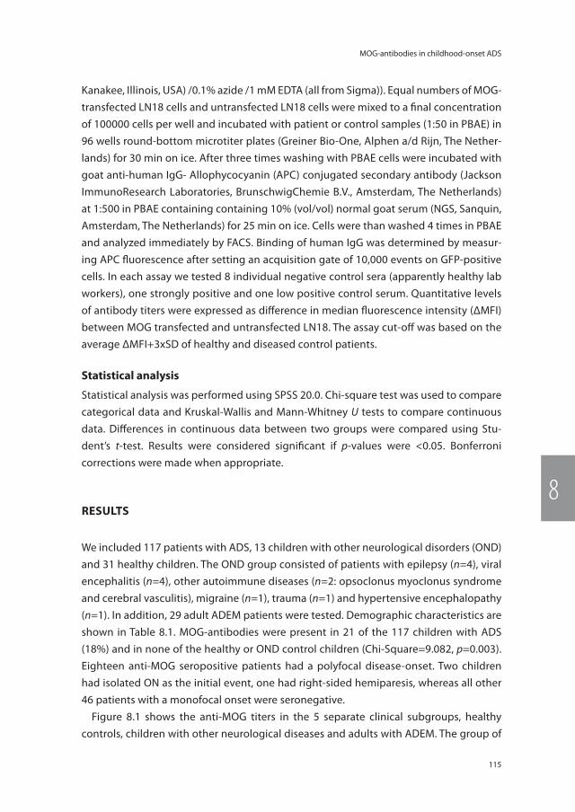
115
MOG-antibodies in childhood-onset ADS
8
Kanakee, Illinois, USA) /0.1% azide /1 mM EDTA (all from Sigma)). Equal numbers of MOG-transfected LN18 cells and untransfected LN18 cells were mixed to a final concentration of 100000 cells per well and incubated with patient or control samples (1:50 in PBAE) in 96 wells round-bottom microtiter plates (Greiner Bio-One, Alphen a/d Rijn, The Nether-lands) for 30 min on ice. After three times washing with PBAE cells were incubated with goat anti-human IgG- Allophycocyanin (APC) conjugated secondary antibody (Jackson ImmunoResearch Laboratories, BrunschwigChemie B.V., Amsterdam, The Netherlands) at 1:500 in PBAE containing containing 10% (vol/vol) normal goat serum (NGS, Sanquin, Amsterdam, The Netherlands) for 25 min on ice. Cells were than washed 4 times in PBAE and analyzed immediately by FACS. Binding of human IgG was determined by measur-ing APC fluorescence after setting an acquisition gate of 10,000 events on GFP-positive cells. In each assay we tested 8 individual negative control sera (apparently healthy lab workers), one strongly positive and one low positive control serum. Quantitative levels of antibody titers were expressed as difference in median fluorescence intensity (ΔMFI) between MOG transfected and untransfected LN18. The assay cut-off was based on the average ΔMFI+3xSD of healthy and diseased control patients.
statistical analysis
Statistical analysis was performed using SPSS 20.0. Chi-square test was used to compare categorical data and Kruskal-Wallis and Mann-Whitney U tests to compare continuous data. Differences in continuous data between two groups were compared using Stu-dent’s t-test. Results were considered significant if p-values were <0.05. Bonferroni corrections were made when appropriate.
results
We included 117 patients with ADS, 13 children with other neurological disorders (OND) and 31 healthy children. The OND group consisted of patients with epilepsy (n=4), viral encephalitis (n=4), other autoimmune diseases (n=2: opsoclonus myoclonus syndrome and cerebral vasculitis), migraine (n=1), trauma (n=1) and hypertensive encephalopathy (n=1). In addition, 29 adult ADEM patients were tested. Demographic characteristics are shown in Table 8.1. MOG-antibodies were present in 21 of the 117 children with ADS (18%) and in none of the healthy or OND control children (Chi-Square=9.082, p=0.003). Eighteen anti-MOG seropositive patients had a polyfocal disease-onset. Two children had isolated ON as the initial event, one had right-sided hemiparesis, whereas all other 46 patients with a monofocal onset were seronegative.
Figure 8.1 shows the anti-MOG titers in the 5 separate clinical subgroups, healthy controls, children with other neurological diseases and adults with ADEM. The group of
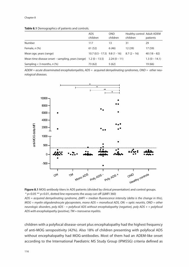
Chapter 8
116
children with a polyfocal disease-onset plus encephalopathy had the highest frequency of anti-MOG seropositivity (42%). Also 18% of children presenting with polyfocal ADS without encephalopathy had MOG-antibodies. Most of them had an ADEM-like onset according to the International Paediatric MS Study Group (IPMSSG) criteria defined as
table 8.1 Demographics of patients and controls.
ADSchildren
OND children
Healthy control children
Adult ADEM patients
Number 117 13 31 29
Female, n (%) 61 (52) 6 (46) 12 (39) 17 (59)
Mean age, years (range) 10.7 (0.5 - 17.5) 9.8 (1 - 16) 8.7 (2 – 16) 40 (18 – 82)
Mean time disease onset – sampling, years (range) 1.2 (0 – 13.5) 2.24 (0 – 11) 1.3 (0 – 14.1)
Sampling < 3 months, n (%) 73 (62) 5 (62) 19 (66)
ADEM = acute disseminated encephalomyelitis, ADS = acquired demyelinating syndromes, OND = other neu-rological diseases.
Figure 8.1 MOG-antibody titers in ADS patients (divided by clinical presentation) and control groups. * p<0.05 ** p<0.01, dotted line represents the assay cut-off (ΔMFI 360)ADS = acquired demyelinating syndrome, ΔMFI = median fluorescence intensity (delta is the change in this), MOG = myelin oligodendrocyte glycoprotein, mono ADS = monofocal ADS, ON: = optic neuritis, OND = other neurologic disorders, poly ADS - = polyfocal ADS without encephalopathy (negative), poly ADS + = polyfocal ADS with encephalopathy (positive), TM = transverse myelitis.
117
MOG-antibodies in childhood-onset ADS
8
a first polyfocal clinical CNS event with encephalopathy and a presumed inflammatory cause and usually diffuse poorly demarcated lesions on cerebral MRI, although they presented without encephalopathy, and did not qualify as suffering from MS.27 The clinical presentation and disease course of individual MOG-antibody positive children are outlined in Table 8.2. The characteristics of the MOG-antibody positive and negative paediatric patient groups are shown in Table 8.3. The MOG-antibody positive patients were 4.4 years younger on average than the MOG-antibody negative patients (p<0.001).
In the ADS cohort, 47 children had a final diagnosis of MS (mean follow-up time of 4.7 years) and except for one they were all seronegative. In contrast, of the 70 patients without MS diagnosis (mean follow-up time of 5 years) 29% was seropositive (p<0.001). Eleven of these 70 children developed a relapsing disease without fulfilling diagnostic criteria for MS. In eight of these 11 children MOG-antibodies could be detected (Table 8.2). Figure 8.2 shows the disease course of the 8 seropositive children with clinical re-lapsing disease without MS diagnosis. There were no significant differences between the monophasic and relapsing MOG-antibody seropositive patients in length of follow-up (4.02 vs 6.75 years) and in treatment (75% vs 67% were treated with Methylprednisolone after disease presentation).
Clinical recovery of the MOG-antibody positive patients at last follow-up was complete in 12 patients. Eight patients had only mild residual symptoms, like fatigue, attention or behavioural deficits or mild persisting visual loss. One patient who initially presented
Figure 8.2 Disease courses of the 8 MOG-antibody positive children with relapsing disease.ADEM = acute disseminated encephalomyelitis, BG = basal ganglia, CC = corpus callosum, MOG = myelin oli-godendrocyte glycoprotein, MRI = magnetic resonance imaging, ON = optic neuritis, TM = transverse myelitis.
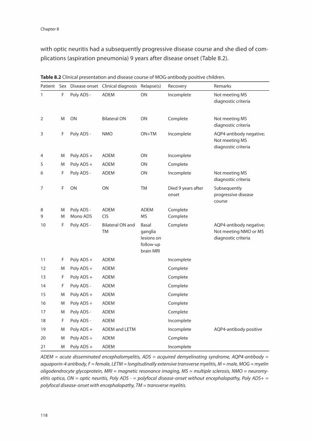
Chapter 8
118
with optic neuritis had a subsequently progressive disease course and she died of com-plications (aspiration pneumonia) 9 years after disease onset (Table 8.2).
table 8.2 Clinical presentation and disease course of MOG-antibody positive children.
Patient Sex Disease onset Clinical diagnosis Relapse(s) Recovery Remarks
1 F Poly ADS - ADEM ON Incomplete Not meeting MS diagnostic criteria
2 M ON Bilateral ON ON Complete Not meeting MS diagnostic criteria
3 F Poly ADS - NMO ON+TM Incomplete AQP4-antibody negative; Not meeting MS diagnostic criteria
4 M Poly ADS + ADEM ON Incomplete
5 M Poly ADS + ADEM ON Complete
6 F Poly ADS - ADEM ON Incomplete Not meeting MS diagnostic criteria
7 F ON ON TM Died 9 years after onset
Subsequently progressive disease course
89
MM
Poly ADS -Mono ADS
ADEMCIS
ADEMMS
CompleteComplete
10 F Poly ADS - Bilateral ON and TM
Basal ganglia lesions on follow-up brain MRI
Complete AQP4-antibody negative; Not meeting NMO or MS diagnostic criteria
11 F Poly ADS + ADEM Incomplete
12 M Poly ADS + ADEM Complete
13 F Poly ADS + ADEM Complete
14 F Poly ADS - ADEM Complete
15 M Poly ADS + ADEM Complete
16 M Poly ADS + ADEM Complete
17 M Poly ADS - ADEM Complete
18 F Poly ADS - ADEM Incomplete
19 M Poly ADS + ADEM and LETM Incomplete AQP4-antibody positive
20 M Poly ADS + ADEM Complete
21 M Poly ADS + ADEM Incomplete
ADEM = acute disseminated encephalomyelitis, ADS = acquired demyelinating syndrome, AQP4-antibody = aquaporin-4 antibody, F = female, LETM = longitudinally extensive transverse myelitis, M = male, MOG = myelin oligodendrocyte glycoprotein, MRI = magnetic resonance imaging, MS = multiple sclerosis, NMO = neuromy-elitis optica, ON = optic neuritis, Poly ADS - = polyfocal disease-onset without encephalopathy, Poly ADS+ = polyfocal disease-onset with encephalopathy, TM = transverse myelitis.
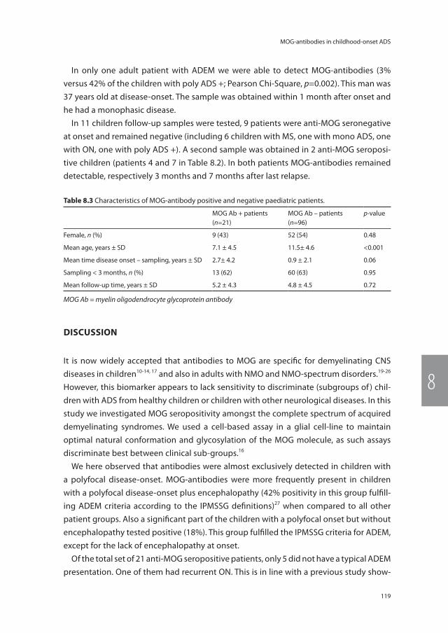
119
MOG-antibodies in childhood-onset ADS
8
In only one adult patient with ADEM we were able to detect MOG-antibodies (3% versus 42% of the children with poly ADS +; Pearson Chi-Square, p=0.002). This man was 37 years old at disease-onset. The sample was obtained within 1 month after onset and he had a monophasic disease.
In 11 children follow-up samples were tested, 9 patients were anti-MOG seronegative at onset and remained negative (including 6 children with MS, one with mono ADS, one with ON, one with poly ADS +). A second sample was obtained in 2 anti-MOG seroposi-tive children (patients 4 and 7 in Table 8.2). In both patients MOG-antibodies remained detectable, respectively 3 months and 7 months after last relapse.
Discussion
It is now widely accepted that antibodies to MOG are specific for demyelinating CNS diseases in children10-14, 17 and also in adults with NMO and NMO-spectrum disorders.19-26 However, this biomarker appears to lack sensitivity to discriminate (subgroups of ) chil-dren with ADS from healthy children or children with other neurological diseases. In this study we investigated MOG seropositivity amongst the complete spectrum of acquired demyelinating syndromes. We used a cell-based assay in a glial cell-line to maintain optimal natural conformation and glycosylation of the MOG molecule, as such assays discriminate best between clinical sub-groups.16
We here observed that antibodies were almost exclusively detected in children with a polyfocal disease-onset. MOG-antibodies were more frequently present in children with a polyfocal disease-onset plus encephalopathy (42% positivity in this group fulfill-ing ADEM criteria according to the IPMSSG definitions)27 when compared to all other patient groups. Also a significant part of the children with a polyfocal onset but without encephalopathy tested positive (18%). This group fulfilled the IPMSSG criteria for ADEM, except for the lack of encephalopathy at onset.
Of the total set of 21 anti-MOG seropositive patients, only 5 did not have a typical ADEM presentation. One of them had recurrent ON. This is in line with a previous study show-
table 8.3 Characteristics of MOG-antibody positive and negative paediatric patients.
MOG Ab + patients (n=21)
MOG Ab – patients (n=96)
p-value
Female, n (%) 9 (43) 52 (54) 0.48
Mean age, years ± SD 7.1 ± 4.5 11.5± 4.6 <0.001
Mean time disease onset – sampling, years ± SD 2.7± 4.2 0.9 ± 2.1 0.06
Sampling < 3 months, n (%) 13 (62) 60 (63) 0.95
Mean follow-up time, years ± SD 5.2 ± 4.3 4.8 ± 4.5 0.72
MOG Ab = myelin oligodendrocyte glycoprotein antibody
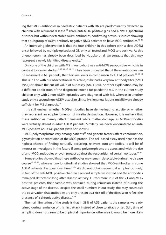
Chapter 8
120
ing that MOG-antibodies in paediatric patients with ON are predominantly detected in children with recurrent disease.18 Three anti-MOG positive girls had a NMO (spectrum) disorder, but without detectable AQP4-antibodies, confirming previous studies showing that a subgroup of AQP4-antibody negative NMO patients do have MOG-antibodies.19-26
An interesting observation is that the four children in this cohort with a clear ADEM onset followed by multiple episodes of ON only, all tested anti-MOG seropositive. As this phenomenon has already been described by Huppke et al, we suggest that this may represent a newly identified disease entity.30
Only one of the children with MS in our cohort was anti-MOG seropositive, which is in contrast to former studies.9-12, 14, 17, 19, 21 It has been discussed that if these antibodies can be measured in MS patients, the titers are lower in comparison to ADEM patients.11, 13, 14 This is in line with our observation in this child, as he had a very low antibody titer (ΔMFI 395) just above the cut-off value of our assay (ΔMFI 360). Another explanation may be a different application of the diagnostic criteria for paediatric MS. In the current study children only with 2 non-ADEM episodes were diagnosed with MS, whereas in another study only a second non-ADEM attack or clinically silent new lesions on MRI were already sufficient for MS diagnosis.14
It is still unclear whether MOG-antibodies have demyelinating activity or whether they represent an epiphenomenon of myelin destruction. However, it is unlikely that these antibodies merely reflect fulminant white matter damage, as MOG-antibodies were virtually absent in adult ADEM patients. Similarly, we never encountered an anti-MOG positive adult MS patient (data not shown).
MOG polymorphisms vary among patients31 and genetic factors affect conformation, glycosylation or expression of the MOG protein. The cell-based assay used here has the highest chance of finding naturally occurring, relevant auto-antibodies. It will be of interest to investigate in the future if some polymorphisms are associated with the rise of anti-MOG antibodies or even protect against the recognition of certain epitopes.16, 32
Some studies showed that these antibodies may remain detectable during the disease course10, 12, 18, whereas two longitudinal studies showed that MOG-antibodies in some ADEM patients disappear over time.11, 14 We did not obtain sequential samples routinely. In two of the anti-MOG positive children a second sample was tested and the antibodies remained detectable long after disease activity. Furthermore in 6 of the 21 anti-MOG positive patients, their sample was obtained during remission instead of during the active stage of the disease. Despite the small numbers in our study, this may contradict the observation that antibodies are only present as a kick-off of the disease or reflect the presence of a chronic active disease.8, 14
The main limitation of the study is that in 38% of ADS patients the samples were ob-tained during remission of this first attack instead of close to attack onset. Still, time of sampling does not seem to be of pivotal importance, otherwise it would be more likely
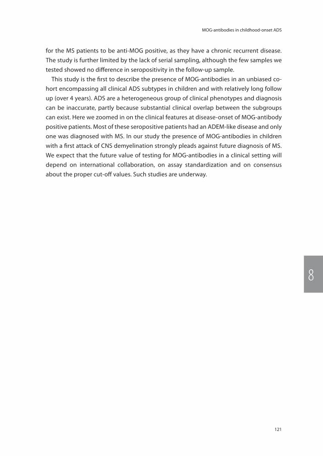
121
MOG-antibodies in childhood-onset ADS
8
for the MS patients to be anti-MOG positive, as they have a chronic recurrent disease. The study is further limited by the lack of serial sampling, although the few samples we tested showed no difference in seropositivity in the follow-up sample.
This study is the first to describe the presence of MOG-antibodies in an unbiased co-hort encompassing all clinical ADS subtypes in children and with relatively long follow up (over 4 years). ADS are a heterogeneous group of clinical phenotypes and diagnosis can be inaccurate, partly because substantial clinical overlap between the subgroups can exist. Here we zoomed in on the clinical features at disease-onset of MOG-antibody positive patients. Most of these seropositive patients had an ADEM-like disease and only one was diagnosed with MS. In our study the presence of MOG-antibodies in children with a first attack of CNS demyelination strongly pleads against future diagnosis of MS. We expect that the future value of testing for MOG-antibodies in a clinical setting will depend on international collaboration, on assay standardization and on consensus about the proper cut-off values. Such studies are underway.
Chapter 8
122
reFerences
1. Banwell B, Kennedy J, Sadovnick D, Arnold DL, Magalhaes S, Wambera K, et al. Incidence of acquired demyelination of the CNS in Canadian children. Neurology 2009; 72(3): 232-39.
2. Ketelslegers IA, Catsman-Berrevoets CE, Neuteboom RF, Boon M, van Dijk KG, Eikelen-boom MJ, et al. Incidence of acquired de-myelinating syndromes of the CNS in Dutch children: a nationwide study. J Neurol 2012; 259(9): 1929-35.
3. Ghezzi A, Banwell B, Boyko A, Amato MP, An-lar B, Blinkenberg M, et al. The management of multiple sclerosis in children: a European view. Mult Scler 2010; 16(10): 1258-67.
4. Lennon VA, Kryzer TJ, Pittock SJ, Verkman AS, Hinson SR. IgG marker of optic-spinal multiple sclerosis binds to the aquaporin-4 water channel. J Exp Med 2005; 202(4): 473-77.
5. Ketelslegers IA, Modderman PW, Vennegoor A, Killestein J, Hamann D, Hintzen RQ. Anti-bodies against aquaporin-4 in neuromyelitis optica: distinction between recurrent and monophasic patients. Mult Scler 2011; 17(12): 1527-30.
6. Schluesener HJ, Sobel RA, Linington C, Weiner HL. A monoclonal antibody against a myelin oligodendrocyte glycoprotein induces re-lapses and demyelination in central nervous system autoimmune disease. J Immunol 1987; 139(12): 4016-21.
7. Linington C, Bradl M, Lassmann H, Brunner C, Vass K. Augmentation of demyelination in rat acute allergic encephalomyelitis by circulating mouse monoclonal antibodies directed against a myelin/oligodendrocyte glycoprotein. Am J Pathol 1988; 130(3): 443-54.
8. Lalive PH, Menge T, Delarasse C, Della Gaspera B, Pham-Dihn D, Villoslada P, et al. Antibodies to native myelin oligodendrocyte glycoprotein are serologic markers of early inflammation in multiple sclerosis. Proc Natl Acad Sci USA 2006; 103(7): 2280-85.
9. Zhou D, Srivastava R, Nessler S, Grummel V, Sommer N, Bruck W, et al. Identification of a pathogenic antibody response to native
myelin oligodendrocyte glycoprotein in mul-tiple sclerosis. Proc Natl Acad Sci USA 2006; 103(50): 19057-62.
10. O’Connor KC, McLaughlin KA, De Jager PL, Chitnis T, Bettelli E, Xu C, et al. Self-antigen tetramers discriminate between myelin au-toantibodies to native or denatured protein. Nat Med 2007; 13(2): 211-17.
11. Di Pauli F, Mader S, Rostasy K, Schanda K, Bajer-Kornek B, Ehling R, et al. Temporal dynamics of anti-MOG antibodies in CNS demyelinating diseases. Clin Immunol 2011; 138(3): 247-54.
12. McLaughlin KA, Chitnis T, Newcombe J, Franz B, Kenndy J, McArdel S, et al. Age-dependent B cell autoimmunity to a myelin surface anti-gen in pediatric multiple sclerosis. J Immunol 2009; 183(6): 4067-76.
13. Brilot F, Dale RC, Selter RC, Grummel V, Kalluri SR, Aslam M, et al. Antibodies to native my-elin oligodendrocyte glycoprotein in children with inflammatory demyelinating central nervous system disease. Ann Neurol 2009; 66(6): 833-42.
14. Probstel AK, Dornmair K, Bittner R, Sperl P, Jenne D, Magalhaes S, et al. Antibodies to MOG are transient in childhood acute disseminated encephalomyelitis. Neurology 2011; 77(6): 580-88.
15. Selter RC, Brilot F, Grummel V, Kraus V, Cepok S, Dale RC, et al. Antibody responses to EBV and native MOG in pediatric inflammatory demyelinating CNS diseases. Neurology 2010; 74(21): 1711-15.
16. Reindl M, Di Pauli F, Rostasy K, Berger T. The spectrum of MOG autoantibody-associated demyelinating diseases. Nat Rev Neurol 2013; 9(8): 455-61.
17. Lalive PH, Hausler MG, Maurey H, Mikaeloff Y, Tardieu M, Wiendl, et al. Highly reactive anti-myelin oligodendrocyte glycoprotein antibodies differentiate demyelinating dis-eases from viral encephalitis in children. Mult Scler 2011; 17(3): 297-302.
18. Rostasy K, Mader S, Schanda K, Huppke P,
123
MOG-antibodies in childhood-onset ADS
8
Gärtner J, Kraus V, et al. Anti-myelin oligoden-drocyte glycoprotein antibodies in pediatric patients with optic neuritis. Arch Neurol 2012; 69(6): 752-56.
19. Mader S, Gredler V, Schanda K, Rostasy K, Dujmovic I, Pfaller K, et al. Complement acti-vating antibodies to myelin oligodendrocyte glycoprotein in neuromyelitis optica and related disorders. J Neuroinflammation 2011; 8: 184.
20. Kitley J, Woodhall M, Waters P, Leite MI, De-venney E, Craig J, et al. Myelin-oligodendro-cyte glycoprotein antibodies in adults with a neuromyelitis optica phenotype. Neurology 2012; 79(12): 1273-77.
21. Rostasy K, Mader S, Hennes E, Schanda K, Gredler V, Guenther A, et al. Persisting myelin oligodendrocyte glycoprotein antibodies in aquaporin-4 antibody negative pediatric neuromyelitis optica. Mult Scler 2013; 19(8): 1052-59.
22. Kitley J, Waters P, Woodhall M, Leite MI, Murchison A, George J, et al. Neuromyelitis optica spectrum disorders with aquaporin-4 and myelin-oligodendrocyte glycoprotein antibodies: a comparative study. JAMA Neurol 2014; 71(3): 276-83.
23. Sato DK, Callegaro D, Lana-Peixoto MA, Waters PJ, Jorge FMD, Takahashi T, et al. Distinction between MOG antibody-positive and AQP4 antibody-positive NMO spectrum disorders. Neurology 2014; 82(6): 474-81.
24. Tanaka M, Tanaka K. Anti-MOG antibodies in adult patients with demyelinating disorders of the central nervous system. J Neuroim-munol 2014; 270(1-2): 98-99.
25. Höftberger R, Sepulveda M, Armangue T, Blanco Y, Rostasy K, Cobo Calvo A, et al. Antibodies to MOG and AQP4 in adults with neuromyelitis optica and suspected limited forms of the disease. Mult Scler 2015; 21(7): 866-74.
26. Ramanathan S, Reddel SW, Henderson A, Par-ratt JD, Barnett M, Gatt PN, et al. Antibodies to myelin oligodendrocyte glycoprotein in bilateral and recurrent optic neuritis. Neurol Neuroimmunol Neuroinflamm 2014; 1(4): e40.
27. Krupp LB, Tardieu M, Amato MP, Banwell B, Chitnis T, Dale RC, et al. International Pediatric Multiple Sclerosis Study Group criteria for pediatric multiple sclerosis and immune-me-diated central nervous system demyelinating disorders: revisions to the 2007 definitions. Mult Scler 2013; 19(10): 1261-67.
28. Neuteboom RF, Boon M, Catsman-Berrevoets CE, Vles JS, Gooskens RH, Stroink H, et al. Prognostic factors after a first attack of inflammatory CNS demyelination in children. Neurology 2008; 71(13): 967-73.
29. Polman CH, Reingold SC, Edan G, Filippi M, Hartung HP, Kappos L, et al. Diagnostic criteria for multiple sclerosis: 2005 revisions to the “McDonald Criteria”. Ann Neurol 2005; 58(6): 840-46.
30. Huppke P, Rostasy K, Karenfort M, Huppke B, Seidl R, Leiz S, et al. Acute disseminated encephalomyelitis followed by recurrent or monophasic optic neuritis in pediatric patients. Mult Scler 2013; 19(7): 941-46.
31. Ohlenbusch A, Pohl D, Hanefeld F. Myelin oligodendrocyte gene polymorphisms and childhood multiple sclerosis. Pediatr Res 2002; 52(2): 175-79.
32. Mayer MC, Breithaupt C, Reindl M, Schanda K, Rostasy K, Berger T, et al. Distinction and temporal stability of conformational epitopes on myelin oligodendrocyte glycoprotein recognized by patients with different inflam-matory central nervous system diseases. J Immunol 2013; 191(7): 3594-04.

2016
12
OCT
Chapter 9
Gray matter related proteins are associated with childhood-onset Multiple Sclerosis
V. Singh, E.D. van Pelt, M.P. Stoop, C. Stingl, I.A. Ketelslegers, R.F. Neuteboom, C.E. Catsman-Berrevoets, T.M. Luider, R.Q. Hintzen.
Neurology: Neuroimmunology and Neuroinflammation, 2015
Chapter 9
126
AbstrAct
objective To identify CSF biomarkers for multiple sclerosis (MS) in children with an initial acquired CNS demyelinating syndromes (ADS).
methods CSF was collected from a cohort of 39 children with initial ADS, of them 18 were diagnosed with MS and 21 had a monophasic disease course. Proteomic analysis of trypsinised CSF (20 µl) was performed by nano liquid chromatography Orbitrap mass spectrometry. Univariate statistical analysis was used to identify differentially abundant proteins between childhood-onset MS and monophasic ADS.
results A total of 2260 peptides corresponding with 318 proteins were identified in the total set of samples. Of these 2260 peptides, 88 were identified as most distinctive be-tween MS and ADS. 53 peptides, corresponding to 14 proteins, had higher abundance in children with MS as compared to monophasic ADS. Twelve out of these 14 proteins were linked to neuronal functions and structures such as synapses, axons and CNS proteases (example: neurofascin, carboxypeptidase E, brevican core protein, and contactin-2). The other two were functionally related to immune function. The 35 peptides identified with decreased abundance in children with MS corresponded to seven proteins. Six of them linked to innate immune function (example: haptoglobin, haptoglobin-related protein, c4b-binding protein alpha chain and monocyte differentiation antigen CD14) and one to cellular adhesion (protein diaphanous homolog 1).
conclusion At first onset of ADS, CSF of children diagnosed with MS showed increased abundance of CNS gray matter related proteins, whereas CSF of children with a mono-phasic disease course showed increased abundance of innate immunity related proteins.
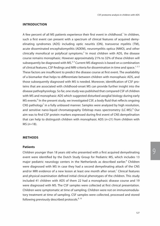
127
CSF proteome analysis in children with ADS
9
introDuction
A few percent of all MS patients experience their first event in childhood.1 In children, such a first event can present with a spectrum of clinical features of acquired demy-elinating syndromes (ADS) including optic neuritis (ON), transverse myelitis (TM), acute disseminated encephalomyelitis (ADEM), neuromyelitis optica (NMO), and other clinically monofocal or polyfocal symptoms.2 In most children with ADS, the disease course remains monophasic. However approximately 21% to 32% of these children will subsequently be diagnosed with MS.3, 4 Current MS diagnosis is based on a combination of clinical features, CSF findings and MRI-criteria for dissemination in time and space.1, 3, 5 These factors are insufficient to predict the disease course at first event. The availability of a biomarker that helps to differentiate between children with monophasic ADS, and those subsequently diagnosed with MS is needed. Moreover, identification of CSF pro-teins that are associated with childhood-onset MS can provide further insight into the disease pathophysiology. So far, one study was published that compared CSF of children with MS and monophasic ADS which suggested disturbed axoglial biology during early MS events.6 In the present study, we investigated CSF, a body fluid that reflects ongoing CNS pathology7 in a fully unbiased manner. Samples were analyzed by high resolution, and sensitive nano-liquid chromatography Orbitrap mass spectrometry (LC-MS).8 Our aim was to find CSF protein markers expressed during first event of CNS demyelination that can help to distinguish children with monophasic ADS (n=21) from children with MS (n=18).
methoDs
Patients
Children younger than 18 years old who presented with a first acquired demyelinating event were identified by the Dutch Study Group for Pediatric MS, which includes 13 major pediatric neurology centers in the Netherlands as described earlier.9 Children were diagnosed with MS in case they had a second demyelinating attack of the CNS and/or MRI evidence of a new lesion at least one month after onset.1 Clinical features and physical examination defined initial clinical phenotypes of the children. This study included 41 children with ADS of them 22 had a monophasic disease course and 19 were diagnosed with MS. The CSF samples were collected at first clinical presentation. Children were symptomatic at time of sampling. Children were not on immunomodula-tory treatment at time of sampling. CSF samples were collected, processed and stored following previously described protocols.8, 10
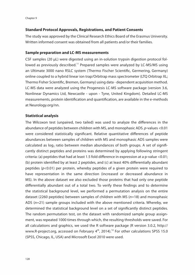
Chapter 9
128
standard Protocol Approvals, registrations, and Patient consents
The study was approved by the Clinical Research Ethics Board of the Erasmus University. Written informed consent was obtained from all patients and/or their families.
sample preparation and lc-ms measurements
CSF samples (20 µL) were digested using an in-solution trypsin digestion protocol fol-lowed as previously described.11 Prepared samples were analyzed by LC-MS/MS using an Ultimate 3000 nano RSLC system (Thermo Fischer Scientific, Germering, Germany) online coupled to a hybrid linear ion trap/Orbitrap mass spectrometer (LTQ Orbitrap XL; Thermo Fisher Scientific, Bremen, Germany) using data - dependent acquisition method. LC-MS data were analyzed using the Progenesis LC-MS software package (version 3.6, Nonlinear Dynamics Ltd, Newcastle - upon - Tyne, United Kingdom). Detailed LC-MS measurements, protein identification and quantification, are available in the e-methods at Neurology.org/nn.
statistical analysis
The Wilcoxon test (unpaired, two tailed) was used to analyze the differences in the abundance of peptides between children with MS, and monophasic ADS. p-values <0.01 were considered statistically significant. Relative quantitative differences of peptide abundances between samples of children with MS and monophasic ADS samples were calculated as log2 ratio between median abundances of both groups. A set of signifi-cantly distinct peptides and proteins was determined by applying following stringent criteria: (a) peptides that had at least 1.5 fold difference in expression at a p-value <0.01; (b) protein identified by at least 2 peptides, and (c) at least 40% differentially abundant peptides (p<0.01) per protein, whereby peptides of a given protein were required to have representation in the same direction (increased or decreased abundance in MS). In the above dataset we also excluded those proteins that had only one peptide differentially abundant out of a total two. To verify these findings and to determine the statistical background level, we performed a permutation analysis on the entire dataset (2260 peptides) between samples of children with MS (n=18) and monophasic ADS (n=21) sample groups included with the above mentioned criteria. Whereby, we determined the statistical background level on a set of significantly distinct peptides. The random permutation test, on the dataset with randomized sample group assign-ment, was repeated 1000 times through which, the resulting thresholds were saved. For all calculations and graphics, we used the R software package (R version 3.0.2, http://www.R-project.org, accessed on February 4th, 2014).12 For other calculations SPSS 15.0 (SPSS, Chicago, IL, USA) and Microsoft Excel 2010 were used.
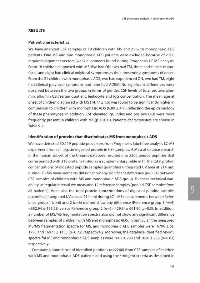
129
CSF proteome analysis in children with ADS
9
results
Patient characteristics
We have analyzed CSF samples of 18 children with MS and 21 with monophasic ADS patients. One MS and one monophasic ADS patients were excluded because of <200 required alignment vectors (weak alignment) found during Progenesis LC-MS analysis. From 18 children diagnosed with MS, five had ON, two had TM, three had clinical mono-focal, and eight had clinical polyfocal symptoms as their presenting symptoms of onset. From the 21 children with monophasic ADS, two had experienced ON, two had TM, eight had clinical polyfocal symptoms and nine had ADEM. No significant differences were observed between the two groups in terms of gender, CSF levels of total protein, albu-min, albumin CSF/serum quotient, leukocyte and IgG concentration. The mean age at onset of children diagnosed with MS (14.17 ± 1.5) was found to be significantly higher in comparison to children with monophasic ADS (6.89 ± 4.9), reflecting the epidemiology of these phenotypes. In addition, CSF elevated IgG index and positive OCB were more frequently present in children with MS (p ≤ 0.01). Patients characteristics are shown in Table 9.1.
identification of proteins that discriminates ms from monophasic ADs
We have detected 50,119 peptide precursors from Progenesis label-free analysis LC-MS experiment from all trypsin digested protein in CSF samples. A Mascot database search in the human subset of the Uniprot database resulted into 2260 unique peptides that corresponded with 318 proteins (listed as a supplementary Table e-1). The total protein concentrations of digested peptide samples quantified (integrated UV area at 214 nm) during LC–MS measurements did not show any significant difference (p=0.54) between CSF samples of children with MS and monophasic ADS group. To check technical vari-ability, at regular interval we measured 12 reference samples (pooled CSF samples from all patients). Here, also the total protein concentrations of digested peptide samples quantified (integrated UV area at 214 nm) during LC – MS measurements between Refer-ence group 1 (n=6) and 2 (n=6) did not show any difference (Reference group 1 (n=6) =382.94 ± 153.28; versus Reference group 2 (n=6), 429.34± 661.90, p=0.3). In addition, a number of MS/MS fragmentation spectra also did not show any significant difference between samples of children with MS and monophasic ADS. In particular, the measured MS/MS fragmentation spectra for MS, and monophasic ADS samples were 16796 ± SD 1795 and 16971 ± 1153 (p=0.72) respectively. Moreover, the database identified MS/MS spectra for MS and monophasic ADS samples were 1607 ± 280 and 1626 ± 256 (p=0.83) respectively.
Comparing abundance of identified peptides (n=2260) from CSF samples of children with MS and monophasic ADS patients and using the stringent criteria as described in
Chapter 9
130
tabl
e 9.
1 Cl
inic
al c
hara
cter
istic
s of
chi
ldre
n w
ith m
ultip
le s
cler
osis
and
mon
opha
sic
acqu
ired
dem
yelin
atin
g sy
ndro
me.
mon
opha
sic
AD
s (n
=21)
ms
(n=1
8)p
Valu
ea
mea
n ±
sDm
edia
nra
nge
mea
n ±
sDm
edia
nra
nge
mon
opha
sic
AD
s vs
ms
Age
at o
nset
, yea
rs6.
89 ±
4.9
15.
811.
14 -
17.1
114
.17
± 1.
5014
.29
11.1
4 - 1
6.21
p<0.
01
Sex,
% fe
mal
es71
.4N
/AN
/A61
.1N
/AN
/AN
S
Prot
ein,
g/L
0.36
± 0
.21
(n=2
1)0.
30.
18-1
.15
0.33
± 0
.14
(n=1
7)0.
320.
19-0
.75
NS
Alb
umin
, g/L
0.23
± 0
.20
(n=1
1)0.
160.
11-0
.79
0.18
± 0
.07
(n=1
4)0.
170.
08-0
.36
NS
Leuk
ocyt
es x
106
37.1
2 ±
34.9
1 (n
=20)
28.5
1-11
820
.98
± 25
.68
(n=1
7)13
1.0-
87N
S
IgG
, g/L
0.05
± 0
.07
(n=1
1)0.
030.
01-0
.27
0.05
± 0
.03
(n=1
3)0.
050.
03-0
.12
NS
IgG
inde
x0.
61 ±
0.1
0 (n
=12)
0.58
0.5-
0.79
1.36
± 0
.72
(n=1
7)1
0.67
-3p<
0.01
Elev
ated
IgG
inde
xb , n (%
)2
(16.
7)N
/AN
/A16
(94.
1)N
/AN
/Ap<
0.01
Posi
tive
OCB
, n (%
)2
(12.
5) (n
=16)
N/A
N/A
14 (8
7.5)
(n=1
6)N
/AN
/Ap<
0.01
Rela
psin
g di
seas
e, n
(%)
0 (0
)N
/AN
/A18
(100
)N
/AN
/Ap<
0.01
This
tabl
e sh
ows
clin
ical
cha
ract
eris
tics
and
rout
ine
CSF
findi
ngs
of c
hild
ren
with
MS
(at d
isea
se o
nset
) and
chi
ldre
n w
ith m
onop
hasi
c A
DS.
Dat
a ar
epr
esen
ted
as th
e m
ean
± SD
or m
edia
n an
d ra
nge.
In c
ase
of m
issi
ng d
ata,
the
num
ber o
f pat
ient
s w
ith a
vaila
ble
data
is in
dica
ted
in p
aren
thes
es.
a Cal
cula
ted
by M
ann-
Whi
tney
test
and
p <
0.0
1 w
as c
onsi
dere
d si
gnifi
cant
.b Ig
G in
dex
cons
ider
ed e
leva
ted
0.68
or h
ighe
r.4
ADS
= ac
quire
d de
mye
linat
ing
synd
rom
e; M
S =
mul
tiple
scle
rosi
s; N
A =
not a
pplic
able
; NS
= no
t sig
nific
ant;
OCB
= o
ligoc
lona
l ban
ds.
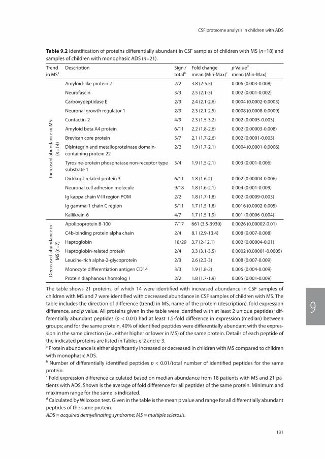
131
CSF proteome analysis in children with ADS
9
table 9.2 Identification of proteins differentially abundant in CSF samples of children with MS (n=18) and samples of children with monophasic ADS (n=21).
Trend in MSa
Description Sign./totalb
Fold changemean (Min-Max)c
p Valued
mean (Min-Max)
Incr
ease
d ab
unda
nce
in M
S(n
=14)
Amyloid-like protein 2 2/2 3.8 (2-5.5) 0.006 (0.003-0.008)
Neurofascin 3/3 2.5 (2.1-3) 0.002 (0.001-0.002)
Carboxypeptidase E 2/3 2.4 (2.1-2.6) 0.0004 (0.0002-0.0005)
Neuronal growth regulator 1 2/3 2.3 (2.1-2.5) 0.0008 (0.0008-0.0009)
Contactin-2 4/9 2.3 (1.5-3.2) 0.002 (0.0005-0.003)
Amyloid beta A4 protein 6/11 2.2 (1.8-2.6) 0.002 (0.00003-0.008)
Brevican core protein 5/7 2.1 (1.7-2.6) 0.002 (0.0001-0.005)
Disintegrin and metalloproteinase domain-containing protein 22
2/2 1.9 (1.7-2.1) 0.0004 (0.0001-0.0006)
Tyrosine-protein phosphatase non-receptor type substrate 1
3/4 1.9 (1.5-2.1) 0.003 (0.001-0.006)
Dickkopf-related protein 3 6/11 1.8 (1.6-2) 0.002 (0.00004-0.006)
Neuronal cell adhesion molecule 9/18 1.8 (1.6-2.1) 0.004 (0.001-0.009)
Ig kappa chain V-III region POM 2/2 1.8 (1.7-1.8) 0.002 (0.0009-0.003)
Ig gamma-1 chain C region 5/11 1.7 (1.5-1.8) 0.0016 (0.0002-0.005)
Kallikrein-6 4/7 1.7 (1.5-1.9) 0.001 (0.0006-0.004)
Dec
reas
ed a
bund
ance
in
MS
(n=7
)
Apolipoprotein B-100 7/17 661 (3.5-3930) 0.0026 (0.00002-0.01)
C4b-binding protein alpha chain 2/4 8.1 (2.9-13.4) 0.008 (0.007-0.008)
Haptoglobin 18/29 3.7 (2-12.1) 0.002 (0.00004-0.01)
Haptoglobin-related protein 2/4 3.3 (3.1-3.5) 0.0002 (0.00001-0.0005)
Leucine-rich alpha-2-glycoprotein 2/3 2.6 (2.3-3) 0.008 (0.007-0.009)
Monocyte differentiation antigen CD14 3/3 1.9 (1.8-2) 0.006 (0.004-0.009)
Protein diaphanous homolog 1 2/2 1.8 (1.7-1.9) 0.005 (0.001-0.009)
The table shows 21 proteins, of which 14 were identified with increased abundance in CSF samples of children with MS and 7 were identified with decreased abundance in CSF samples of children with MS. The table includes the direction of difference (trend) in MS, name of the protein (description), fold expression difference, and p value. All proteins given in the table were identified with at least 2 unique peptides; dif-ferentially abundant peptides (p < 0.01) had at least 1.5-fold difference in expression (median) between groups; and for the same protein, 40% of identified peptides were differentially abundant with the expres-sion in the same direction (i.e., either higher or lower in MS) of the same protein. Details of each peptide of the indicated proteins are listed in Tables e-2 and e-3.a Protein abundance is either significantly increased or decreased in children with MS compared to children with monophasic ADS.b Number of differentially identified peptides p < 0.01/total number of identified peptides for the same protein.c Fold expression difference calculated based on median abundance from 18 patients with MS and 21 pa-tients with ADS. Shown is the average of fold difference for all peptides of the same protein. Minimum and maximum range for the same is indicated.d Calculated by Wilcoxon test. Given in the table is the mean p value and range for all differentially abundant peptides of the same protein.ADS = acquired demyelinating syndrome; MS = multiple sclerosis.
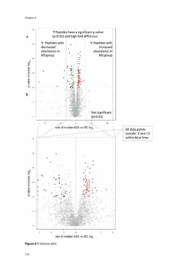
Chapter 9
132
Figure 9.1 Volcano plot.
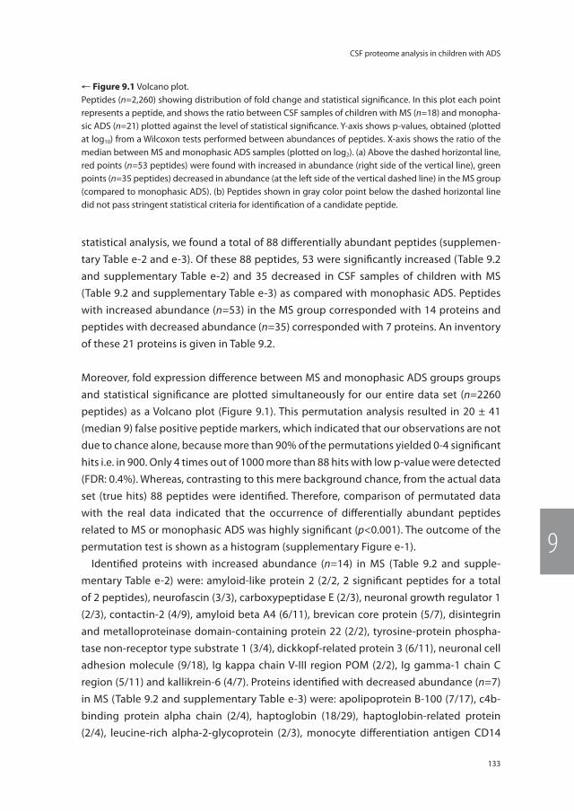
133
CSF proteome analysis in children with ADS
9
statistical analysis, we found a total of 88 differentially abundant peptides (supplemen-tary Table e-2 and e-3). Of these 88 peptides, 53 were significantly increased (Table 9.2 and supplementary Table e-2) and 35 decreased in CSF samples of children with MS (Table 9.2 and supplementary Table e-3) as compared with monophasic ADS. Peptides with increased abundance (n=53) in the MS group corresponded with 14 proteins and peptides with decreased abundance (n=35) corresponded with 7 proteins. An inventory of these 21 proteins is given in Table 9.2.
Moreover, fold expression difference between MS and monophasic ADS groups groups and statistical significance are plotted simultaneously for our entire data set (n=2260 peptides) as a Volcano plot (Figure 9.1). This permutation analysis resulted in 20 ± 41 (median 9) false positive peptide markers, which indicated that our observations are not due to chance alone, because more than 90% of the permutations yielded 0-4 significant hits i.e. in 900. Only 4 times out of 1000 more than 88 hits with low p-value were detected (FDR: 0.4%). Whereas, contrasting to this mere background chance, from the actual data set (true hits) 88 peptides were identified. Therefore, comparison of permutated data with the real data indicated that the occurrence of differentially abundant peptides related to MS or monophasic ADS was highly significant (p<0.001). The outcome of the permutation test is shown as a histogram (supplementary Figure e-1).
Identified proteins with increased abundance (n=14) in MS (Table 9.2 and supple-mentary Table e-2) were: amyloid-like protein 2 (2/2, 2 significant peptides for a total of 2 peptides), neurofascin (3/3), carboxypeptidase E (2/3), neuronal growth regulator 1 (2/3), contactin-2 (4/9), amyloid beta A4 (6/11), brevican core protein (5/7), disintegrin and metalloproteinase domain-containing protein 22 (2/2), tyrosine-protein phospha-tase non-receptor type substrate 1 (3/4), dickkopf-related protein 3 (6/11), neuronal cell adhesion molecule (9/18), Ig kappa chain V-III region POM (2/2), Ig gamma-1 chain C region (5/11) and kallikrein-6 (4/7). Proteins identified with decreased abundance (n=7) in MS (Table 9.2 and supplementary Table e-3) were: apolipoprotein B-100 (7/17), c4b-binding protein alpha chain (2/4), haptoglobin (18/29), haptoglobin-related protein (2/4), leucine-rich alpha-2-glycoprotein (2/3), monocyte differentiation antigen CD14
← Figure 9.1 Volcano plot.Peptides (n=2,260) showing distribution of fold change and statistical significance. In this plot each point represents a peptide, and shows the ratio between CSF samples of children with MS (n=18) and monopha-sic ADS (n=21) plotted against the level of statistical significance. Y-axis shows p-values, obtained (plotted at log10) from a Wilcoxon tests performed between abundances of peptides. X-axis shows the ratio of the median between MS and monophasic ADS samples (plotted on log2). (a) Above the dashed horizontal line, red points (n=53 peptides) were found with increased in abundance (right side of the vertical line), green points (n=35 peptides) decreased in abundance (at the left side of the vertical dashed line) in the MS group (compared to monophasic ADS). (b) Peptides shown in gray color point below the dashed horizontal line did not pass stringent statistical criteria for identification of a candidate peptide.
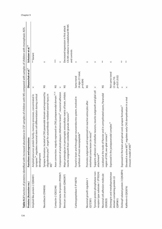
Chapter 9
134
tabl
e 9.
3 Fu
nctio
n of
pro
tein
s id
entifi
ed w
ith in
crea
sed
abun
danc
e in
CSF
sam
ples
of c
hild
ren
with
MS
com
pare
d w
ith s
ampl
es o
f chi
ldre
n w
ith m
onop
hasi
c A
DS.
Prot
eins
(Acc
essi
on n
o.)
Func
tion
s an
d ex
pres
sion
sD
haun
chak
et a
l.6sc
hutz
er e
t al.13
Am
yloi
d-lik
e pr
otei
n 2
(Q06
481)
Am
yloi
d pr
ecur
sor p
rote
in fa
mily
, mem
ory
proc
esse
s, co
ncen
trat
ed in
sy
naps
es18
, reg
ulat
es n
euro
nal s
tem
cel
l diff
eren
tiatio
n du
ring
cort
ical
de
velo
pmen
t
×**
Var
iant
Neu
rofa
scin
(O94
856)
Syna
pse
form
atio
n, lo
cate
d at
CN
S pa
rano
dal d
omai
n an
d ex
pres
sed
by
olig
oden
droc
ytes
20, t
arge
t for
aut
oant
ibod
y-m
edia
ted
axon
al in
jury
in
MS21
NS
×
Cont
actin
-2 (Q
0224
6)A
xon
conn
ectio
n, m
ajor
ly e
xpre
ssed
on
CNS
juxt
apar
anod
al d
omai
n19, 2
0N
S**
*
Am
yloi
d be
ta A
4 pr
otei
n (P
0506
7)Co
mpo
nent
of a
myl
oid
plaq
ues
(Alz
heim
er b
rain
s)35
, neu
rona
l adh
esio
n×
×
Brev
ican
cor
e pr
otei
n (Q
96G
W7)
Maj
or p
rote
ogly
can
in p
eris
ynap
tic e
xtra
cellu
lar m
atrix
of b
rain
, inh
ibits
ne
urite
out
grow
th fr
om c
ereb
ella
r gra
nule
neu
rons
23N
SD
ecre
ased
exp
ress
ion
in fi
rst a
ttac
k CI
S-M
S re
lativ
e to
est
ablis
hed
RR-M
S an
d co
ntro
ls
Carb
oxyp
eptid
ase
E (P
1687
0)Fo
und
in b
rain
and
thro
ugho
ut n
euro
endo
crin
e sy
stem
, inv
olve
d in
sy
nthe
sis
of m
ost n
euro
pept
ide36
Sam
e tr
end
(4 s
ign.
/21
tota
l, p=
0.00
4)
×
Neu
rona
l gro
wth
regu
lato
r 1
(Q7Z
3B1)
Prom
otes
out
grow
th a
nd e
xpre
ssed
on
reac
tive
astr
ocyt
es a
fter
en
torh
inal
cor
tex
lesi
on (i
n m
ice)
37N
S×
Tyro
sine
-pro
tein
pho
spha
tase
non
-re
cept
or ty
pe s
ubst
rate
1 (P
7832
4)Su
ppor
ts a
dhes
ion
of c
ereb
ella
r neu
rons
, neu
rite
outg
row
th a
nd g
lial c
ell
atta
chm
ent28
××
Neu
rona
l cel
l adh
esio
n m
olec
ule
(Q92
823)
Loca
lized
at t
he n
ode
of R
anvi
er a
nd in
unm
yelin
ated
axo
ns, P
aran
odal
re
gion
of C
NS,
axo
-glia
l con
tact
19N
S**
Dis
inte
grin
and
met
allo
prot
eina
se
dom
ain-
cont
aini
ng p
rote
in 2
2 (Q
9P0K
1)
Expr
esse
d in
the
juxt
apar
anod
al c
ompl
ex19
Not
sam
e tr
end
(2 s
ign.
/10;
p=
0.01
,0.0
2)
×
Dic
kkop
f-rel
ated
pro
tein
3 (Q
9UBP
4)Ex
pres
sed
in th
e br
ain
and
spin
al c
ord,
syn
apse
form
atio
n13×
**
Kalli
krei
n-6
(Q92
876)
Elev
ated
in a
ctiv
e M
S26, r
egul
ates
ear
ly C
NS
dem
yelin
atio
n in
a v
iral
(mou
se) m
odel
of M
S26×
**
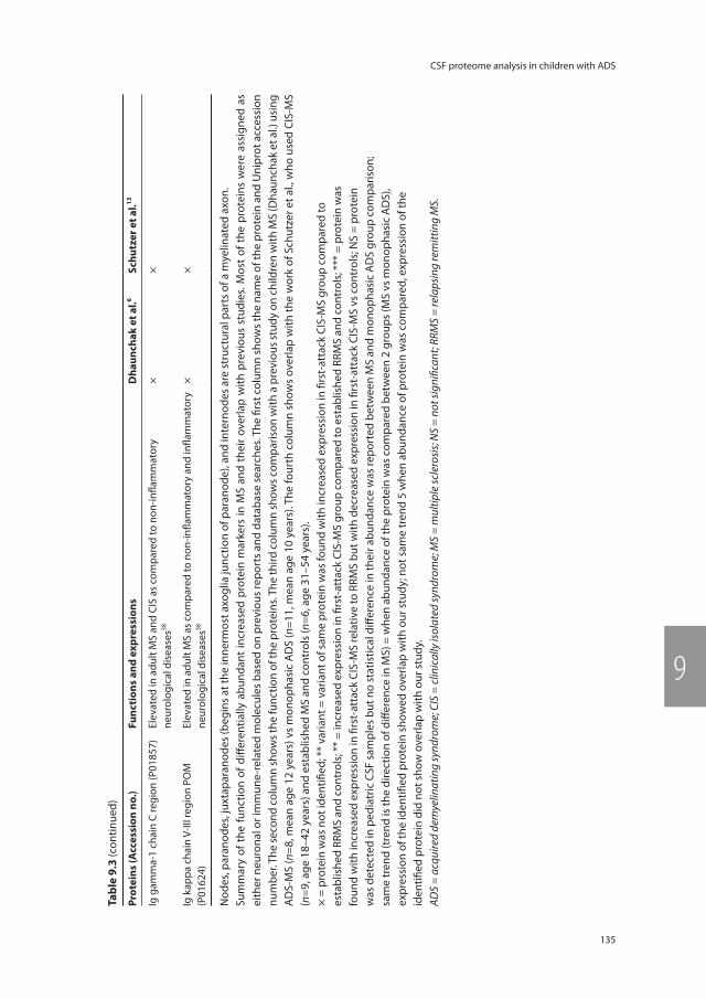
135
CSF proteome analysis in children with ADS
9
tabl
e 9.
3 (c
ontin
ued)
Prot
eins
(Acc
essi
on n
o.)
Func
tion
s an
d ex
pres
sion
sD
haun
chak
et a
l.6sc
hutz
er e
t al.13
Ig g
amm
a-1
chai
n C
regi
on (P
0185
7)El
evat
ed in
adu
lt M
S an
d CI
S as
com
pare
d to
non
-infla
mm
ator
y ne
urol
ogic
al d
isea
ses38
××
Ig k
appa
cha
in V
-III r
egio
n PO
M
(P01
624)
Elev
ated
in a
dult
MS
as c
ompa
red
to n
on-in
flam
mat
ory
and
infla
mm
ator
y ne
urol
ogic
al d
isea
ses38
××
Nod
es, p
aran
odes
, jux
tapa
rano
des
(beg
ins
at th
e in
nerm
ost a
xogl
ia ju
nctio
n of
par
anod
e), a
nd in
tern
odes
are
str
uctu
ral p
arts
of a
mye
linat
ed a
xon.
Sum
mar
y of
the
func
tion
of d
iffer
entia
lly a
bund
ant
incr
ease
d pr
otei
n m
arke
rs in
MS
and
thei
r ov
erla
p w
ith p
revi
ous
stud
ies.
Mos
t of
the
pro
tein
s w
ere
assi
gned
as
eith
er n
euro
nal o
r im
mun
e-re
late
d m
olec
ules
bas
ed o
n pr
evio
us re
port
s an
d da
taba
se s
earc
hes.
The
first
col
umn
show
s th
e na
me
of th
e pr
otei
n an
d U
nipr
ot a
cces
sion
nu
mbe
r. Th
e se
cond
col
umn
show
s the
func
tion
of th
e pr
otei
ns. T
he th
ird c
olum
n sh
ows c
ompa
rison
with
a p
revi
ous s
tudy
on
child
ren
with
MS
(Dha
unch
ak e
t al.)
usi
ng
AD
S-M
S (n
=8, m
ean
age
12 y
ears
) vs
mon
opha
sic
AD
S (n
=11,
mea
n ag
e 10
yea
rs).
The
four
th c
olum
n sh
ows
over
lap
with
the
wor
k of
Sch
utze
r et a
l., w
ho u
sed
CIS-
MS
(n=9
, age
18–
42 y
ears
) and
est
ablis
hed
MS
and
cont
rols
(n=6
, age
31–
54 y
ears
).×
= pr
otei
n w
as n
ot id
entifi
ed; *
* va
riant
= v
aria
nt o
f sam
e pr
otei
n w
as fo
und
with
incr
ease
d ex
pres
sion
in fi
rst-
atta
ck C
IS-M
S gr
oup
com
pare
d to
esta
blis
hed
RRM
S an
d co
ntro
ls; *
* =
incr
ease
d ex
pres
sion
in fi
rst-
atta
ck C
IS-M
S gr
oup
com
pare
d to
est
ablis
hed
RRM
S an
d co
ntro
ls; *
** =
pro
tein
was
foun
d w
ith in
crea
sed
expr
essi
on in
firs
t-at
tack
CIS
-MS
rela
tive
to R
RMS
but w
ith d
ecre
ased
exp
ress
ion
in fi
rst-
atta
ck C
IS-M
S vs
con
trol
s; N
S =
prot
ein
was
det
ecte
d in
ped
iatr
ic C
SF s
ampl
es b
ut n
o st
atis
tical
diff
eren
ce in
thei
r abu
ndan
ce w
as re
port
ed b
etw
een
MS
and
mon
opha
sic
AD
S gr
oup
com
paris
on;
sam
e tr
end
(tre
nd is
the
dire
ctio
n of
diff
eren
ce in
MS)
= w
hen
abun
danc
e of
the
prot
ein
was
com
pare
d be
twee
n 2
grou
ps (M
S vs
mon
opha
sic
AD
S),
expr
essi
on o
f the
iden
tified
pro
tein
sho
wed
ove
rlap
with
our
stu
dy; n
ot s
ame
tren
d 5
whe
n ab
unda
nce
of p
rote
in w
as c
ompa
red,
exp
ress
ion
of th
eid
entifi
ed p
rote
in d
id n
ot s
how
ove
rlap
with
our
stu
dy.
ADS
= ac
quire
d de
mye
linat
ing
synd
rom
e; C
IS =
clin
ical
ly is
olat
ed sy
ndro
me;
MS
= m
ultip
le sc
lero
sis;
NS
= no
t sig
nific
ant;
RRM
S =
rela
psin
g re
mitt
ing
MS.
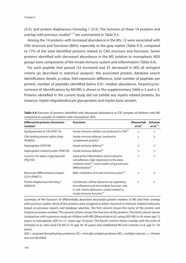
Chapter 9
136
(3/3), and protein diaphanous homolog 1 (2/2). The function of these 14 proteins and overlap with previous studies6, 13 are summarized in Table 9.3.
Among the 14 proteins with increased abundance in the MS, 12 were associated with CNS structure and functions (86%), especially to the gray matter (Table 9.3), compared to 17% of the total identified proteins related to CNS structure and functions. Seven proteins identified with decreased abundance in the MS (relative to monophasic ADS group) were components of the innate immune system and inflammation (Table 9.4).
For each peptide that passed (53 increased and 35 decreased in MS) all stringent criteria (as described in statistical analysis): the associated protein, database search identification details, p-value, fold expression difference, total number of peptides per protein, number of peptides identified below 0.01, median abundance, frequency/oc-currence of identifications by MS/MS is shown in the supplementary Table e-2 and e-3. Proteins identified in the current study did not exhibit any myelin related proteins, for instance; myelin oligodendrocyte glycoprotein and myelin basic protein.
table 9.4 Function of proteins identified with decreased abundance in CSF samples of children with MS compared to samples of children with monophasic ADS.
Differential proteins (Accession number)
Functions Dhaunchaket al.6
schutzer et al.13
Apolipoprotein B-100 (P04114) Innate immune related, not produced in CNS39 × ×
C4b-binding protein alpha chain (P04003)
Innate immune defense, involved in complement activity31
× ×
Haptoglobin (P00738) Innate immune defense29 × ×
Haptoglobin-related protein (P00739) Innate immune defense40 × ×
Leucine-rich alpha-2-glycoprotein (P02750)
Induced by inflammation and involved in cell adhesion, high expression in the deep cerebral cortex25, novel marker of granulocytic differentiation29
× ×
Monocyte differentiation antigen CD14 (P08571)
Main modulator of innate immune system32 × ×
Protein diaphanous homolog 1 (O60610)
Coordinates cellular dynamics by regulating microfilament and microtubule function, role in cell–matrix adhesions, variant related to innate immune function34
× ×
Summary of the function of differentially abundant decreased protein markers in MS and their overlap with previous studies. Most of the proteins were assigned as either neuronal or immune-related molecules based on previous reports and database searches. The first column shows the name of the protein and Uniprot accession number. The second column shows the function of the proteins. The third column shows comparison with a previous study on children with MS (Dhaunchak et al.) using ADS-MS (n=8, mean age 12 years) vs monophasic ADS (n=11, mean age 10 years). The fourth column shows overlap with the work of Schutzer et al., who used CIS-MS (n=9, age 18–42 years) and established MS and controls (n=6, age 31–54 years).ADS = acquired demyelinating syndrome; CIS = clinically isolated syndrome; MS = multiple sclerosis. × = Protein was not identified.
137
CSF proteome analysis in children with ADS
9
We examined for the influence of age of onset on the 88 candidate peptides abun-dances for children with MS and monophasic ADS by correlation analysis. We found a mean coefficient of determination (± SD) for MS 0.04 ± 0.06, and for monophasic ADS 0.05 ± 0.05. Thus, by correlation analysis no significant correlation was found between age and peptide abundance for children with MS and monophasic ADS for all 88 pep-tides.
Discussion
In the current study, we have used LC-MS proteomic approach to search for differences in CSF proteome between children with MS and monophasic ADS in children. Benefit of this Orbitrap technique is the possibility to identify relatively vast amounts of different peptides, and assess their abundances, in a small sample volume. We observed a striking difference between the two groups (children with monophasic ADS versus MS), using stringent statistical criteria. We searched for the known functions of the 88 peptides corresponding to 21 distinctive proteins (14 increased and 7 decreased in abundance in MS), using biological databases (www.geneontology.org and www.nextprot.org) and literature. Recently, two research groups6, 13 demonstrated the identification of axoglial and gray matter proteins using mass spectrometry in CSF of MS patients . Similar to our study, Dhaunchak and co-workers6 compared CSF of 8 children with MS with 11 children with monophasic ADS. The overlap of some proteins in the MS group is noticeable (e.g. carboxypeptidase E), despite clear differences in sample handling such as depletion of abundant proteins with possible carrier function for other proteins, and exclusion of proteins with less than 5 kDa weight (Table 9.3).
Our results show overlap with molecules identified in the CSF of adult acute onset MS cases by Schutzer et al.13, who performed the mass spectrometry analysis in CSF on CIS cases versus established relapsing remitting MS (RR-MS) and controls (Table 9.3). They showed proteins that distinguished these CIS patients from both established RR-MS and controls. For example, they showed a significant increase of kallikrein-6, dickkopf-related protein 3 at first clinical onset in comparison to established RR-MS and controls (Table 9.3). They also showed a significant increase in contactin-2 (neuronal membrane protein) in first-attack MS patients relative to established RR-MS (Table 9.3).13
Our findings illustrate that the neurodegenerative arm of MS neuropathology is already active at the earliest stage of clinical disease, also in children. Consistent with the findings of others and our own previous studies6, 14-16, we again observed a striking lack of myelin proteins in these clear-cut cases of acute demyelination. This may not directly imply absence of such free proteins in this type of pathology, but rather it may reflect the specific physicochemical properties of the hydrophobic myelin components,
Chapter 9
138
and perhaps different pathways of elimination from the CSF (e.g. via draining macro-phages).17 In any case, we must be cautious in using the dominant presence of CNS gray matter over white matter proteins as proof that neurodegeneration is a primary event in MS and would precede demyelination. The presence of CNS gray matter may simply represent damage by inflammation, and the molecules identified may lead to a better understanding of this presumed inflammation induced neurodegeneration. It should be stressed that not all differentially abundant proteins were overrepresented in MS; some were underrepresented, pointing at more complex mechanisms, such as a pertur-bation in the physiology of the axoglial apparatus (Table 2 and Tables e-2 and e-3).6 A confounding factor in our study could be the fact that, due to the skewed occurrence of monophasic disease at younger age, both groups were not matched according to age. We doubt, however whether this has influenced the results, as we did not see an age effect in our groups on the abundance of the 21 identified proteins.
Of 14 proteins with increased abundance in MS (Table 9.2 and 9.3), two were associ-ated with the amyloid beta A4 protein family. Amyloid-like protein 2 is mainly concen-trated at neuronal synapses18 and has a role in memory processes. While, amyloid beta A4 protein is associated with neurite growth, neuronal adhesion and axonogenesis (Table 9.3). Six (of 14) proteins: contactin-219, neurofascin20, neuronal growth regulator 1, brevican core protein6, neuronal cell adhesion molecule19 and disintegrin and metal-loproteinase domain-containing protein 2219 are shown to be located at the paranodal and, juxtaparanodal region of the CNS of myelinated axons (Table 9.3). Contactin-2 is axonal glycoprotein which is shown at juxtaparanodal domain of myelinated axons.13 Neurofascin plays a role in the assembly of nodes of Ranvier in the CNS.21 Two isoforms of neurofascin are shown to interact with contactin-associated protein and contactin-1 to form paranodal junction, that attaches the myelin loop to the axon; and helps to sepa-rate voltage gated sodium channels at node and potassium channels at juxtparanodal region.20 Disruption of neurofascin localization shows early changes preceding demy-elination and remyelination in MS.22 Interestingly, contactin-2 and neurofascin are previ-ously reported as autoimmune targets in MS.20 Neuronal growth regulator 1 is shown to be located at paranodal region of CNS and play a role in axo-glia contact at the node of Ranvier. Identification of contactin-2 and neurofascin in CSF of children with MS is consistent with a previous study6 however, they did not found significant difference in CSF of children with MS and monophasic ADS (Table 9.2 and 9.3). Brevican core protein is known as CNS specific proteoglycan19 at the surface of neuroglial sheaths, where it is enriched in perisynaptic extracellular matrix.23
Among the proteins with increased abundance in MS (of 14), the proteases/peptidases protein named as carboxypeptidase E, plays a role in synthesis of most neuropeptides.24 Next, disintegrin and metalloproteinase domain-containing protein 22 are highly ex-pressed in brain and localized at juxtaparanode.19 This molecule presumed to be work
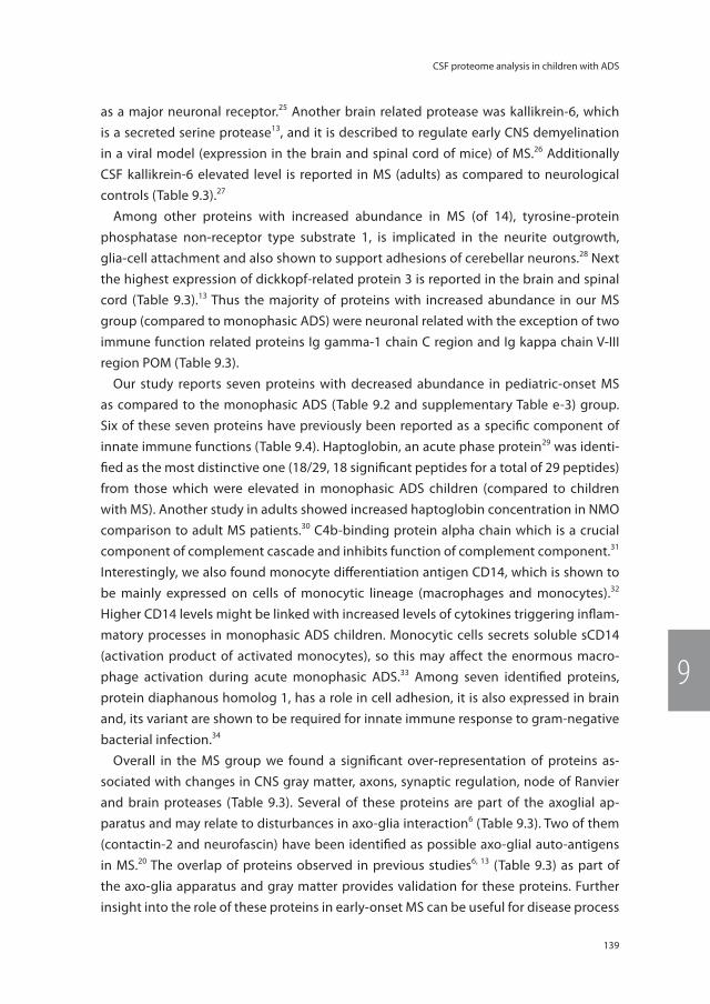
139
CSF proteome analysis in children with ADS
9
as a major neuronal receptor.25 Another brain related protease was kallikrein-6, which is a secreted serine protease13, and it is described to regulate early CNS demyelination in a viral model (expression in the brain and spinal cord of mice) of MS.26 Additionally CSF kallikrein-6 elevated level is reported in MS (adults) as compared to neurological controls (Table 9.3).27
Among other proteins with increased abundance in MS (of 14), tyrosine-protein phosphatase non-receptor type substrate 1, is implicated in the neurite outgrowth, glia-cell attachment and also shown to support adhesions of cerebellar neurons.28 Next the highest expression of dickkopf-related protein 3 is reported in the brain and spinal cord (Table 9.3).13 Thus the majority of proteins with increased abundance in our MS group (compared to monophasic ADS) were neuronal related with the exception of two immune function related proteins Ig gamma-1 chain C region and Ig kappa chain V-III region POM (Table 9.3).
Our study reports seven proteins with decreased abundance in pediatric-onset MS as compared to the monophasic ADS (Table 9.2 and supplementary Table e-3) group. Six of these seven proteins have previously been reported as a specific component of innate immune functions (Table 9.4). Haptoglobin, an acute phase protein29 was identi-fied as the most distinctive one (18/29, 18 significant peptides for a total of 29 peptides) from those which were elevated in monophasic ADS children (compared to children with MS). Another study in adults showed increased haptoglobin concentration in NMO comparison to adult MS patients.30 C4b-binding protein alpha chain which is a crucial component of complement cascade and inhibits function of complement component.31 Interestingly, we also found monocyte differentiation antigen CD14, which is shown to be mainly expressed on cells of monocytic lineage (macrophages and monocytes).32 Higher CD14 levels might be linked with increased levels of cytokines triggering inflam-matory processes in monophasic ADS children. Monocytic cells secrets soluble sCD14 (activation product of activated monocytes), so this may affect the enormous macro-phage activation during acute monophasic ADS.33 Among seven identified proteins, protein diaphanous homolog 1, has a role in cell adhesion, it is also expressed in brain and, its variant are shown to be required for innate immune response to gram-negative bacterial infection.34
Overall in the MS group we found a significant over-representation of proteins as-sociated with changes in CNS gray matter, axons, synaptic regulation, node of Ranvier and brain proteases (Table 9.3). Several of these proteins are part of the axoglial ap-paratus and may relate to disturbances in axo-glia interaction6 (Table 9.3). Two of them (contactin-2 and neurofascin) have been identified as possible axo-glial auto-antigens in MS.20 The overlap of proteins observed in previous studies6, 13 (Table 9.3) as part of the axo-glia apparatus and gray matter provides validation for these proteins. Further insight into the role of these proteins in early-onset MS can be useful for disease process
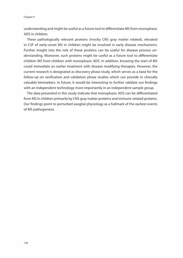
Chapter 9
140
understanding and might be useful as a future tool to differentiate MS from monophasic ADS in children.
These pathologically relevant proteins (mostly CNS gray matter related), elevated in CSF of early-onset MS in children might be involved in early disease mechanisms. Further insight into the role of these proteins can be useful for disease process un-derstanding. Moreover, such proteins might be useful as a future tool to differentiate children MS from children with monophasic ADS. In addition, knowing the start of MS could immediate an earlier treatment with disease modifying therapies. However, the current research is designated as discovery phase study, which serves as a base for the follow-up on verification and validation phase studies which can provide in clinically valuable biomarkers. In future, it would be interesting to further validate our findings with an independent technology more importantly in an independent sample group.
The data presented in this study indicate that monophasic ADS can be differentiated from MS in children primarily by CNS gray matter proteins and immune-related proteins. Our findings point to perturbed axoglial physiology as a hallmark of the earliest events of MS pathogenesis.
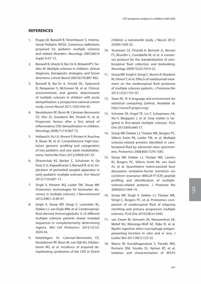
141
CSF proteome analysis in children with ADS
9
reFerences
1. Krupp LB, Banwell B, Tenembaum S, Interna-tional Pediatric MSSG. Consensus definitions proposed for pediatric multiple sclerosis and related disorders. Neurology 2007; 68(16 Suppl 2): S7-12.
2. Banwell B, Ghezzi A, Bar-Or A, Mikaeloff Y, Tar-dieu M. Multiple sclerosis in children: clinical diagnosis, therapeutic strategies, and future directions. Lancet Neurol 2007; 6(10): 887-902.
3. Banwell B, Bar-Or A, Arnold DL, Sadovnick D, Narayanan S, McGowan M, et al. Clinical, environmental, and genetic determinants of multiple sclerosis in children with acute demyelination: a prospective national cohort study. Lancet Neurol 2011; 10(5): 436-45.
4. Neuteboom RF, Boon M, Catsman-Berrevoets CE, Vles JS, Gooskens RH, Stroink H, et al. Prognostic factors after a first attack of inflammatory CNS demyelination in children. Neurology 2008; 71(13): 967-73.
5. Holland H, Xu LX, Ahnert P, Kirsten H, Koschny R, Bauer M, et al. Comprehensive high-reso-lution genomic profiling and cytogenetics of two pediatric and one adult medulloblas-toma. Pathol Res Pract 2013; 209(9): 541-47.
6. Dhaunchak AS, Becker C, Schulman H, De Faria O Jr, Rajasekharan S, Banwell B, et al. Im-plication of perturbed axoglial apparatus in early pediatric multiple sclerosis. Ann Neurol 2012; 71(5): 601-13.
7. Singh V, Hintzen RQ, Luider TM, Stoop MP. Proteomics technologies for biomarker dis-covery in multiple sclerosis. J Neuroimmunol 2012; 248(1-2): 40-47.
8. Singh V, Stoop MP, Stingl C, Luitwieler RL, Dekker LJ, van Duijn MM, et al. Cerebrospinal-fluid-derived immunoglobulin G of different multiple sclerosis patients shares mutated sequences in complementarity determining regions. Mol Cell Proteomics 2013; 12(12): 3924-34.
9. Ketelslegers IA, Catsman-Berrevoets CE, Neuteboom RF, Boon M, van Dijk KG, Eikelen-boom MJ, et al. Incidence of acquired de-myelinating syndromes of the CNS in Dutch
children: a nationwide study. J Neurol 2012; 259(9): 1929-35.
10. Teunissen CE, Petzold A, Bennett JL, Berven FS, Brundin L, Comabella M, et al. A consen-sus protocol for the standardization of cere-brospinal fluid collection and biobanking. Neurology 2009; 73(22): 1914-22.
11. Stoop MP, Singh V, Stingl C, Martin R, Khademi M, Olsson T, et al. Effects of natalizumab treat-ment on the cerebrospinal fluid proteome of multiple sclerosis patients. J Proteome Res 2013; 12(3): 1101-07.
12. Team RC. R: A language and environment for statistical computing [online]. Available at: http://www.R-project.org/.
13. Schutzer SE, Angel TE, Liu T, Schepmoes AA, Xie F, Bergquist J, et al. Gray matter is tar-geted in first-attack multiple sclerosis. PLoS One 2013; 8(9): e66117.
14. Stoop MP, Dekker LJ, Titulaer MK, Burgers PC, Sillevis Smitt PA, Luider TM, et al. Multiple sclerosis-related proteins identified in cere-brospinal fluid by advanced mass spectrom-etry. Proteomics 2008; 8(8): 1576-1585.
15. Stoop MP, Dekker LJ, Titulaer MK, Lamers RJ, Burgers PC, Sillevis Smitt PA, van Gool AJ, et al. Quantitative matrix-assisted laser desorption ionization-fourier transform ion cyclotron resonance (MALDI-FT-ICR) peptide profiling and identification of multiple-sclerosis-related proteins. J Proteome Res 2009; 8(3): 1404-14.
16. Stoop MP, Singh V, Dekker LJ, Titulaer MK, Stingl C, Burgers PC, et al. Proteomics com-parison of cerebrospinal fluid of relapsing remitting and primary progressive multiple sclerosis. PLoS One 2010; 5(8): e12442.
17. van Zwam M, Samsom JN, Nieuwenhuis EE, Melief MJ, Wierenga-Wolf AF, Dijke IE, et al. Myelin ingestion alters macrophage antigen-presenting function in vitro and in vivo. J Leukoc Biol 2011; 90(1): 123-32.
18. Wasco W, Gurubhagavatula S, Paradis MD, Romano DM, Sisodia SS, Hyman BT, et al. Isolation and characterization of APLP2
Chapter 9
142
encoding a homologue of the Alzheimer’s associated amyloid beta protein precursor. Nat Genet 1993; 5(1): 95-100.
19. Faivre-Sarrailh C, Devaux JJ. Neuro-glial inter-actions at the nodes of Ranvier: implication in health and diseases. Front Cell Neurosci 2013; 7: 196.
20. Derfuss T, Linington C, Hohlfeld R, Meinl E. Axo-glial antigens as targets in multiple scle-rosis: implications for axonal and grey matter injury. J Mol Med (Berl) 2010; 88(8): 753-61.
21. Mathey EK, Derfuss T, Storch MK, Williams KR, Hales K, Woolley DR, et al. Neurofascin as a novel target for autoantibody-mediated axo-nal injury. J Exp Med 2007; 204(10): 2363-72.
22. Howell OW, Palser A, Polito A, Melrose S, Zonta B, Scheiermann C, et al. Disruption of neurofascin localization reveals early changes preceding demyelination and remyelination in multiple sclerosis. Brain 2006; 129(Pt 12): 3173-85.
23. Yamada H, Fredette B, Shitara K, Hagihara K, Miura R, Ranschot B, Stallcup WB, et al. The brain chondroitin sulfate proteoglycan brevi-can associates with astrocytes ensheathing cerebellar glomeruli and inhibits neurite outgrowth from granule neurons. J Neurosci 1997; 17(20): 7784-95.
24. Vilijn MH, Das B, Kessler JA, Fricker LD. Cul-tured astrocytes and neurons synthesize and secrete carboxypeptidase E, a neuropeptide-processing enzyme. J Neurochem 1989; 53(5): 1487-93.
25. Ozkaynak E, Abello G, Jaegle M, van Berge L, Hamer D, Kegel L, et al. Adam22 is a major neuronal receptor for Lgi4-mediated Schwann cell signaling. J Neurosci 2010; 30(10): 3857-64.
26. Scarisbrick IA, Yoon H, Panos M, Larson N, Blaber SI, Blaber M, et al. Kallikrein 6 regulates early CNS demyelination in a viral model of multiple sclerosis. Brain Pathol 2012; 22(5): 709-22.
27. Hebb AL, Bhan V, Wishart AD, Moore CS, Rob-ertson GS. Human kallikrein 6 cerebrospinal levels are elevated in multiple sclerosis. Curr Drug Discov Technol 2010; 7(2): 137-140.
28. Latour S, Tanaka H, Demeure C, Mateo V, Rubio M, Brown EJ, et al. Bidirectional nega-tive regulation of human T and dendritic cells by CD47 and its cognate receptor signal-regulator protein-alpha: down-regulation of IL-12 responsiveness and inhibition of dendritic cell activation. J Immunol 2001; 167(5): 2547-54.
29. Donnell LC, Druhan LJ, BR. A. Molecular characterization and expression analysis of leucine-rich alpha2-glycoprotein, a novel marker of granulocytic differentiation. J Leu-koc Biol 2002; 72: 478-485.
30. Chang KH, Tseng MY, Ro LS, Lyu RK, Tai YH, Chang HS, et al. Analyses of haptoglobin level in the cerebrospinal fluid and serum of patients with neuromyelitis optica and multiple sclerosis. Clin Chim Acta 2013; 417: 26-30.
31. Chung LP, Reid KB. Structural and functional studies on C4b-binding protein, a regula-tory component of the human complement system. Biosci Rep 1985; 5(10-11): 855-865.
32. Kurne A, Sayat G, Aydin OF, Turgutoglu N, Terzi M, Sackesen C, et al. Lack of association of the CD14/C -- 159T polymorphism with susceptibility and progression parameters in Turkish multiple sclerosis patients. J Neuroim-munol 2012; 250(1-2): 83-86.
33. Kitchens RL, Thompson PA. Modulatory effects of sCD14 and LBP on LPS-host cell in-teractions. J Endotoxin Res 2005; 11(4): 225.
34. Huh JR, Foe I, Muro I, Hong Chen C, Hong Seol J, Yoo JIS, et al. The Drosophila inhibitor of apoptosis (IAP) DIAP2 is dispensable for cell survival, required for the innate im-mune response to gram-negative bacterial infection, and can be negatively regulated by the reaper/hid/grim family of IAP-binding apoptosis inducers. J Biol Chem 2007; 282(3): 2056-68.
35. Jacobsen KT, Iverfeldt K. Amyloid precursor protein and its homologues: a family of pro-teolysis dependent receptors. Cell Mol Life Sci 2009; 66(14): 2299–18.
36. Koshimizu H, Senatorov V, Loh YP, Gozes I. Neuroprotective protein and carboxypepti-dase E. J Mol Neurosci 2009; 39(1-2): 1–8.
143
CSF proteome analysis in children with ADS
9
37. Schäfer M, Brauer AU, Savaskan NE, Rathjen FG, Brummendorf T. Neurotractin/kilon pro-motes neurite outgrowth and is expressed on reactive astrocytes after entorhinal cortex lesion. Mol Cell Neurosci 2005; 29(4): 580–590.
38. Stoop MP, Coulier L, Rosenling T, Shi S, Smolinska AM, Buydens L, et al. Quantitative proteomics and metabolomics analysis of normal human cerebrospinal fluid samples. Mol Cell Proteomics 2010; 9(9): 2063–75.
39. Pepe G, Chimienti G, Liuzzi GM, Lamanuzzi BL, Nardulli M, Lolli F, et al. Lipoprotein(a) in the cerebrospinal fluid of neurological pa-tients with bloodcerebrospinal fluid barrier dysfunction. Clin Chem 2006; 52(11): 2043–48.
40. Nielsen MJ, Petersen SV, Jacobsen C, Oxvig C, Rees D, Møller HJ, et al. Haptoglobinrelated protein is a high-affinity hemoglobin-binding plasma protein. Blood 2006; 108(8): 2846–49.
Chapter 9
144
suPPlementAl mAteriAl
Supplemental data at Neurology.org/nn http://nn.neurology.org/content/2/5/e155Supplemental MethodsSupplementary Figure e-1. Permutation test.Supplementary Table e-1: All identified peptides 2260 (corresponded to 318 proteins).Supplementary Table e-2: Peptides (n=53) identified with increased abundance in MS as compared to monophasic ADS group and passed all the criteria for candidate peptide.Supplementary Table e-3: Peptides (n=35) identified with decreased abundance in MS as compared to monophasic ADS group and passed all the criteria for candidate peptide.

2016
12
OCT
Chapter 10
General discussion

147
General discussion
10
Acquired demyelinating syndromes (ADS) cover a broad spectrum of inflammatory de-myelinating syndromes of the CNS, of which multiple sclerosis (MS) is the most common subtype. This thesis focuses on two relatively rare clinical subtypes of ADS: neuromyelitis optica spectrum disorders (NMOSD) and childhood-onset ADS including MS. Awareness and recognition of uncommon ADS subtypes are of importance for clinicians, since those require a distinct diagnostic and therapeutic approach. In this thesis we aimed to reveal the spectrum of ADS by describing the clinical features of NMOSD and childhood-onset ADS, in order to improve the diagnostic process. Here, the most important findings from our studies are outlined and discussed in relation to other studies. Future directions and steps for further research are discussed.
PArt one: neuromyelitis oPticA sPectrum DisorDers
Demographic features of nmosD
NMOSD is a rare variant of CNS inflammation, previously often misdiagnosed as MS.1, 2 However, treatment regimens and prognosis differ significantly between NMOSD and MS and therefore it is important to distinguish these diseases. Prior to our study, in-cidence numbers of NMOSD in the Netherlands were unknown. Based on results of a national centralized NMO expert center, we could asses the mean nationwide incidence of AQP4-IgG seropositive NMOSD in the Netherlands is 0.09 per 100,000 people per year (chapter 2). The Dutch incidence is within the range of previous reported incidence rates which range from 0.05 – 0.4 per 100,000 people.3 It should be noted that these are minimum incidence figures, since mild cases and forme fruste types of the disease could have been missed. The incidence of NMOSD in the Netherlands is estimated more than twice as high in non-Caucasians. Africans seem to be more predisposed to NMOSD than whites.3-5 This is supported by a recently reported high incidence of AQP4-IgG sero-positive NMOSD patients of 0.65 per 100,000 people in a population with 90% people of Afro-Caribbean origin.4 Moreover, Afro-Caribbean NMOSD patients are younger at dis-ease onset, have more multifocal attacks and have a higher likelihood of visual disability than Caucasian patients.6 Differences in clinical features and outcomes between ethnic groups indicate the need for a future tailored treatment approach for individual groups.
The recently revised diagnostic criteria for NMOSD broaden the clinical spectrum of NMO.7 Most importantly, the new diagnostic criteria allow for an earlier NMOSD diagno-sis8, 9 even under the assumption of an unknown AQP4-IgG status.8 Previously, patients had to have optic neuritis (ON) and transverse myelitis (TM) in order to fulfill definite NMO criteria.10 Now, AQP4-IgG seropositive patients can be diagnosed with NMOSD af-ter one event of isolated ON, TM or area postrema syndrome.7 In patients without AQP4-IgG, dissemination in space with two different core clinical characteristics (Table 1.1)
Chapter 10
148
and exclusion of alternative diagnoses are required for NMOSD diagnosis. Recognition of NMOSD patients can be a diagnostic challenge since not all NMOSD patients present with classical bilateral ON and longitudinally extensive TM (LETM). Spinal cord lesions can be short in a minority of the NMOSD cases11, and sometimes even asymptomatic.12 Clues to a NMOSD diagnosis are a non-Caucasian ethnicity, ON with severe vision loss and minimum response to steroids, preceding ON or brainstem syndrome, personal or serological evidence of autoimmunity and a brain MRI without MS lesions.12 More awareness and better recognition of the broadening clinical spectrum of NMOSD might increase the incidence of NMOSD in the future.
the search for new prognostic markers in nmosD
Despite the high specificity of AQP4-IgG for NMOSD, the antibody is present in ap-proximately 77% of the NMO cases and lacks sensitivity to diagnose all patients with NMOSD.7, 13-15 Therefore, researchers focused on identifying new diagnostic and prog-nostic biomarkers for the AQP4-IgG seronegative NMOSD patients.16, 17 In the past years, MOG autoantibodies were evaluated as a potential novel marker for NMOSD.18, 19 In the Dutch cohort we have confirmed the presence of MOG-IgG in 33% of the AQP4-IgG seronegative patients with a clinical NMOSD phenotype7, including limited forms as recurrent ON, bilateral ON and LETM1 (chapter 3). In addition to previous studies, we evaluated MRIs for NMO-specific cerebral lesions, which typically occur at sites with high aquaporin-4 expression.20, 21 NMO-specific cerebral lesions were absent in the MOG-IgG seropositive patients which is likely explained by the different underlying disease mechanisms.16, 22 In general, MOG-IgG seropositive patients with a clinical NMOSD phenotype have a relative benign disease course, which is often monophasic, and if relapses occur those are less severe.23-25 However, MOG-IgG seropositive cases with a worse disease course including ongoing relapses and disability progression have been observed.24-27 At the NMO expert center at Erasmus MC, we follow a few MOG-IgG sero-positive patients who suffered from frequent disabling relapses and required chronic immunosuppressant therapies. This demonstrates the importance of an individualized treatment approach and careful consideration of chronic treatment initiation in a sub-group of MOG-IgG seropositive patients.
Reported MOG-IgG seropositivity rates as percentage of the AQP4-IgG seronegative NMOSD cases, including limited forms of NMO like recurrent ON or LETM, range between 7-39%.23-35 Double seropositive cases (both AQP4-IgG and MOG-IgG) are extremely rare, limited to only a few cases.25, 28, 31, 35 Since study protocols and inclusion criteria differ between various cohorts, it is not easy to compare MOG-IgG seropositivity rates. At the NMO expert center at Erasmus MC we were also interested in limited forms of NMOSD and included patients with bilateral ON, recurrent ON and LETM. This and referral bias to expert clinics might contribute to differences in reported MOG-IgG seropositivity rates.
149
General discussion
10
PArt one: neuromyelitis oPticA sPectrum DisorDers
main findings
• The incidence of AQP4-IgG seropositive NMOSD in the Netherlands is 0.09 per 100,000 people per year.
• In approximately one-third of the Dutch AQP4-IgG seronegative patients with a clinical NMOSD phenotype MOG-IgG is detected.
• MOG-IgG seropositive patients with a clinical NMOSD phenotype generally have a more favorable monophasic disease course than AQP4-IgG seropositive NMOSD patients.
clinical implications
• MOG-IgG should be tested in AQP4-IgG seronegative patients with a clinical NMOSD phenotype as part of their standard diagnostic work up.
Future studies should collect serial serum samples to study if MOG-IgG persists in pa-tients with a clinical NMOSD phenotype. Furthermore, MOG-IgG seropositive patients should be followed in order to investigate if they truly have a more favorable disease course than AQP4-IgG seropositive NMOSD patients. Collaborative international stud-ies in search of new additional biomarkers for NMOSD diagnosis and for establishing the prognosis and therapeutic management of individual patients are promising.17, 36, 37 Recently, it has been questioned whether MOG-IgG seropositivity justifies a diagnosis of NMOSD.22 Clinical phenotypes of MOG-IgG seropositive and AQP4-IgG seropositive patients overlap. However, there are important differences in their underlying disease mechanisms.16, 22 Neuromyelitis optica refers to the classical syndrome as first described by Eugene Devic in 1894.38 The old nomenclature became out-dated, since the spec-trum of NMOSD is growing and includes limited forms of NMOSD as isolated ON, or isolated TM, or brainstem syndromes.1, 2, 7 Elevated creatine kinase (CK) levels39-41, and hypothalamic endocrinopathies, including symptoms as galactorrhoea42, are examples of rare features of NMOSD. Some have proposed the novel umbrella term autoimmune aquaporin-4 channelopathy as this term better suits the broad clinical spectrum, which includes more than inflammation of the optic nerve and spinal cord.2 In addition, this term clearly refers to the underlying disease mechanism. Major disadvantage of this term is that it excludes the AQP4-IgG seronegative NMOSD patients, who share clinical similarities with the AQP4-IgG seropositive NMOSD patients.43
PArt two: AcQuireD DemyelinAting synDromes in chilDren
Diagnosing childhood-onset ADs
In children with ADS, the most relevant risk parameters for MS can be found amongst MRI parameters, which can help to identify the children in whom an incident event of ADS represent the first attack of MS. In the past years, several MRI criteria have been developed for the diagnosis of ADS, including MS, in children and for the distinction from other diseases.44-46
Chapter 10
150
The current IPMSSG diagnostic definitions for childhood-onset MS, incorporate the 2010 revised McDonald MRI criteria for MS.47, 48 Aims of the 2010 MRI criteria were to simplify the existing criteria49, 50, and to allow for an earlier definite MS diagnosis. Unique for the 2010 McDonald criteria is that MS can be diagnosed in patients who fulfill criteria for dissemination in time and space, already after a single attack based on a single baseline MRI.48 With the 2010 McDonald MRI criteria for relapsing-remitting MS, detection of CSF OCB is not obligatory to confirm MS diagnosis. However, in clinical practice CSF analysis is often performed in children presenting with CNS demyelination in order to exclude alternative diagnoses, most importantly infectious diseases, prior to the initiation of acute treatment with intravenous methylprednisolone. Previous studies reported the 2010 McDonald MRI criteria allow for an early MS diagnosis in children.51-55 But the diagnostic accuracy is less when applied in young children and children with ADEM.51, 53 Therefore the 2012 IPMSSG stated the McDonald 2010 MRI criteria cannot be applied in children less than 12 years old and in children with ADEM at the first event.47 In chapter 4 we evaluated the utility of the most recent MRI criteria, as per the 2012 IPMSSG consensus definitions in a Dutch prospective cohort including children with the full spectrum of ADS. Here, we demonstrated the 2012 IPMSSG consensus definitions allow for an equally reliable but earlier MS diagnosis in all children (including children ≤12) than with the 2007 IPMSSG definitions. This is beneficial for the adequate counsel-ling of patients and parents, who face their lives with insecurity. In addition, an earlier MS diagnosis allows for earlier initiation of disease modifying therapy (DMT), which might be of long-term benefit.56-58
The IPMSSG 2012 definitions also include a revised definition of MS diagnosis after ADEM. However, using the IPMSSG definitions, a definite MS diagnosis is observed less often than previously reported59 in children initially presenting with ADEM (<10%).47, 60-62 We endorse the IPMSSG panel’s advice not to apply the 2010 McDonald MRI criteria in children with a first event of ADEM. Children with ADEM usually have large and enhanc-ing lesions47, 59, 60, 63, which could lead to false-positive MS diagnoses. Other MRI criteria are more useful to discriminate children with ADEM from those with MS.46 In the Dutch cohort it was confirmed that the Callen MS-ADEM criteria (i.e. fulfilling at least 2 out of 3: a) absence of a diffuse bilateral lesion pattern, b) presence of black holes and c) ≥2 periven-tricular lesions) were the most useful criteria to differentiate MS from ADEM in children.64
The Canadian study group proposed new MRI parameters for the prediction of MS diagnosis in children with ADS.60 In their large-scale prospective cohort study including 284 children with ADS, 14 MRI parameters were assessed for MS risk prediction using a multivariable prediction model. Here, 57 children were diagnosed with MS during follow-up. Two MRI parameters were associated with an increased likelihood of MS diagnosis: T1-hypointense lesions and periventricular lesions (Figure 10.1). MS risk was highest when both parameters were present. In collaboration with our Canadian col-
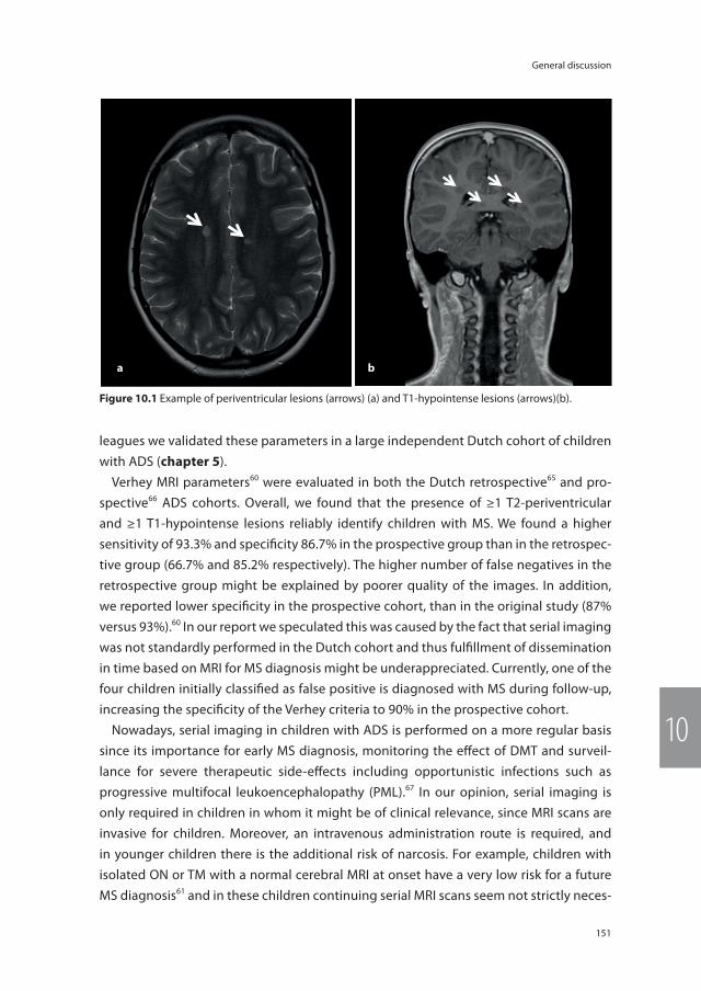
151
General discussion
10
leagues we validated these parameters in a large independent Dutch cohort of children with ADS (chapter 5).
Verhey MRI parameters60 were evaluated in both the Dutch retrospective65 and pro-spective66 ADS cohorts. Overall, we found that the presence of ≥1 T2-periventricular and ≥1 T1-hypointense lesions reliably identify children with MS. We found a higher sensitivity of 93.3% and specificity 86.7% in the prospective group than in the retrospec-tive group (66.7% and 85.2% respectively). The higher number of false negatives in the retrospective group might be explained by poorer quality of the images. In addition, we reported lower specificity in the prospective cohort, than in the original study (87% versus 93%).60 In our report we speculated this was caused by the fact that serial imaging was not standardly performed in the Dutch cohort and thus fulfillment of dissemination in time based on MRI for MS diagnosis might be underappreciated. Currently, one of the four children initially classified as false positive is diagnosed with MS during follow-up, increasing the specificity of the Verhey criteria to 90% in the prospective cohort.
Nowadays, serial imaging in children with ADS is performed on a more regular basis since its importance for early MS diagnosis, monitoring the effect of DMT and surveil-lance for severe therapeutic side-effects including opportunistic infections such as progressive multifocal leukoencephalopathy (PML).67 In our opinion, serial imaging is only required in children in whom it might be of clinical relevance, since MRI scans are invasive for children. Moreover, an intravenous administration route is required, and in younger children there is the additional risk of narcosis. For example, children with isolated ON or TM with a normal cerebral MRI at onset have a very low risk for a future MS diagnosis61 and in these children continuing serial MRI scans seem not strictly neces-
a b
Figure 10.1 Example of periventricular lesions (arrows) (a) and T1-hypointense lesions (arrows)(b).
Chapter 10
152
sary. Clinical follow-up and surveillance for new symptoms are sufficient in this specific category of children who are at low risk for a future MS diagnosis.
This year novel MRI criteria for the diagnosis of MS were reported.68 The 2016 MS di-agnostic MRI criteria include two new CNS sites which can contribute to dissemination in space: optic nerve lesions and cortical lesions. These criteria need to be validated in pediatric and adult cohorts of CNS demyelination. Furthermore, non-conventional imaging techniques as diffusion tensor imaging (DTI) should be further studied, as these images could be used as a prognostic marker in children presenting with ADS.69 Func-tional MRI (fMRI) can provide insight into the adaptive and compensatory mechanisms in childhood-onset MS.70, 71 Combined with neuropsychological assessments, fMRI might provide relevant implications for counselling and rehabilitation strategies.
children versus adults: outcome and management
There are important differences in childhood-onset versus adulthood-onset MS. Chil-dren with MS can present with a broad spectrum of demyelinating syndromes, includ-ing ADEM.72 In addition, the differential diagnosis in children is far more complex.73-75 Children with MS have more frequent and more severe attacks.76-78 Still, children with MS take longer to reach stages of progressive disability, but do so at a younger age.79-81 However, these observations are based on studies conducted prior to the immuno-modulatory treatment era in children.76, 79, 80 Current recommendation and use of DMT in children with MS, likely influences their disease course.82 In chapter 6 we compared the disease course in 383 children and adults presenting with CIS. Over 50 percent of the included patients were diagnosed with MS during follow-up. 11-16 year-old children had the highest rate of MS conversion and the shortest time to MS diagnosis. Overall we found a female predominance, but as in line with previous studies not in young children who were diagnosed with MS before puberty.83, 84 This suggests sex hormones might contribute to the onset of MS.85 Moreover, we found that children with MS more frequently had a non-Caucasian ethnicity than adults with MS. Previous studies reported a higher vulnerability for ADS and a future MS diagnosis in non-Caucasian children.66, 86-89 The exact underlying mechanism is unclear, but it might be that those children lack certain protective genes since their ancestors were born in countries with a low preva-lence of MS.66, 90 In addition, a higher load of certain environmental risk factors, as for example lower vitamin D levels in Afro-American children, might contribute to a higher MS vulnerability in non-Caucasian children. Moreover, non-Caucasian children with MS have a higher relapse rate and more severe relapses.91, 92 This indicates the need for fu-ture tailored treatment protocols for individual groups based on their disease course. In chapter 6 we also validated a prediction rule for a future definite MS diagnosis and early first-line treatment initiation in 11-16 year-old children. In adult CIS patients with a high risk for a future MS diagnosis, it is already common practice to start with DMT at onset.93
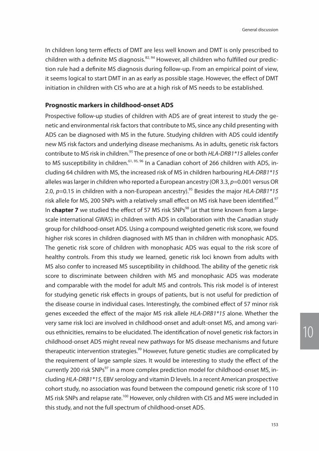
153
General discussion
10
In children long term effects of DMT are less well known and DMT is only prescribed to children with a definite MS diagnosis.82, 94 However, all children who fulfilled our predic-tion rule had a definite MS diagnosis during follow-up. From an empirical point of view, it seems logical to start DMT in an as early as possible stage. However, the effect of DMT initiation in children with CIS who are at a high risk of MS needs to be established.
Prognostic markers in childhood-onset ADs
Prospective follow-up studies of children with ADS are of great interest to study the ge-netic and environmental risk factors that contribute to MS, since any child presenting with ADS can be diagnosed with MS in the future. Studying children with ADS could identify new MS risk factors and underlying disease mechanisms. As in adults, genetic risk factors contribute to MS risk in children.95 The presence of one or both HLA-DRB1*15 alleles confer to MS susceptibility in children.61, 95, 96 In a Canadian cohort of 266 children with ADS, in-cluding 64 children with MS, the increased risk of MS in children harbouring HLA-DRB1*15 alleles was larger in children who reported a European ancestry (OR 3.3, p=0.001 versus OR 2.0, p=0.15 in children with a non-European ancestry).95 Besides the major HLA-DRB1*15 risk allele for MS, 200 SNPs with a relatively small effect on MS risk have been identified.97 In chapter 7 we studied the effect of 57 MS risk SNPs98 (at that time known from a large-scale international GWAS) in children with ADS in collaboration with the Canadian study group for childhood-onset ADS. Using a compound weighted genetic risk score, we found higher risk scores in children diagnosed with MS than in children with monophasic ADS. The genetic risk score of children with monophasic ADS was equal to the risk score of healthy controls. From this study we learned, genetic risk loci known from adults with MS also confer to increased MS susceptibility in childhood. The ability of the genetic risk score to discriminate between children with MS and monophasic ADS was moderate and comparable with the model for adult MS and controls. This risk model is of interest for studying genetic risk effects in groups of patients, but is not useful for prediction of the disease course in individual cases. Interestingly, the combined effect of 57 minor risk genes exceeded the effect of the major MS risk allele HLA-DRB1*15 alone. Whether the very same risk loci are involved in childhood-onset and adult-onset MS, and among vari-ous ethnicities, remains to be elucidated. The identification of novel genetic risk factors in childhood-onset ADS might reveal new pathways for MS disease mechanisms and future therapeutic intervention strategies.99 However, future genetic studies are complicated by the requirement of large sample sizes. It would be interesting to study the effect of the currently 200 risk SNPs97 in a more complex prediction model for childhood-onset MS, in-cluding HLA-DRB1*15, EBV serology and vitamin D levels. In a recent American prospective cohort study, no association was found between the compound genetic risk score of 110 MS risk SNPs and relapse rate.100 However, only children with CIS and MS were included in this study, and not the full spectrum of childhood-onset ADS.
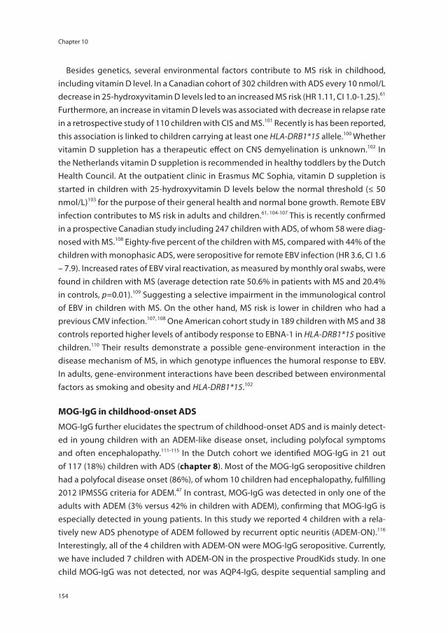
Chapter 10
154
Besides genetics, several environmental factors contribute to MS risk in childhood, including vitamin D level. In a Canadian cohort of 302 children with ADS every 10 nmol/L decrease in 25-hydroxyvitamin D levels led to an increased MS risk (HR 1.11, CI 1.0-1.25).61 Furthermore, an increase in vitamin D levels was associated with decrease in relapse rate in a retrospective study of 110 children with CIS and MS.101 Recently is has been reported, this association is linked to children carrying at least one HLA-DRB1*15 allele.100 Whether vitamin D suppletion has a therapeutic effect on CNS demyelination is unknown.102 In the Netherlands vitamin D suppletion is recommended in healthy toddlers by the Dutch Health Council. At the outpatient clinic in Erasmus MC Sophia, vitamin D suppletion is started in children with 25-hydroxyvitamin D levels below the normal threshold (≤ 50 nmol/L)103 for the purpose of their general health and normal bone growth. Remote EBV infection contributes to MS risk in adults and children.61, 104-107 This is recently confirmed in a prospective Canadian study including 247 children with ADS, of whom 58 were diag-nosed with MS.108 Eighty-five percent of the children with MS, compared with 44% of the children with monophasic ADS, were seropositive for remote EBV infection (HR 3.6, CI 1.6 – 7.9). Increased rates of EBV viral reactivation, as measured by monthly oral swabs, were found in children with MS (average detection rate 50.6% in patients with MS and 20.4% in controls, p=0.01).109 Suggesting a selective impairment in the immunological control of EBV in children with MS. On the other hand, MS risk is lower in children who had a previous CMV infection.107, 108 One American cohort study in 189 children with MS and 38 controls reported higher levels of antibody response to EBNA-1 in HLA-DRB1*15 positive children.110 Their results demonstrate a possible gene-environment interaction in the disease mechanism of MS, in which genotype influences the humoral response to EBV. In adults, gene-environment interactions have been described between environmental factors as smoking and obesity and HLA-DRB1*15.102
mog-igg in childhood-onset ADs
MOG-IgG further elucidates the spectrum of childhood-onset ADS and is mainly detect-ed in young children with an ADEM-like disease onset, including polyfocal symptoms and often encephalopathy.111-115 In the Dutch cohort we identified MOG-IgG in 21 out of 117 (18%) children with ADS (chapter 8). Most of the MOG-IgG seropositive children had a polyfocal disease onset (86%), of whom 10 children had encephalopathy, fulfilling 2012 IPMSSG criteria for ADEM.47 In contrast, MOG-IgG was detected in only one of the adults with ADEM (3% versus 42% in children with ADEM), confirming that MOG-IgG is especially detected in young patients. In this study we reported 4 children with a rela-tively new ADS phenotype of ADEM followed by recurrent optic neuritis (ADEM-ON).116 Interestingly, all of the 4 children with ADEM-ON were MOG-IgG seropositive. Currently, we have included 7 children with ADEM-ON in the prospective ProudKids study. In one child MOG-IgG was not detected, nor was AQP4-IgG, despite sequential sampling and
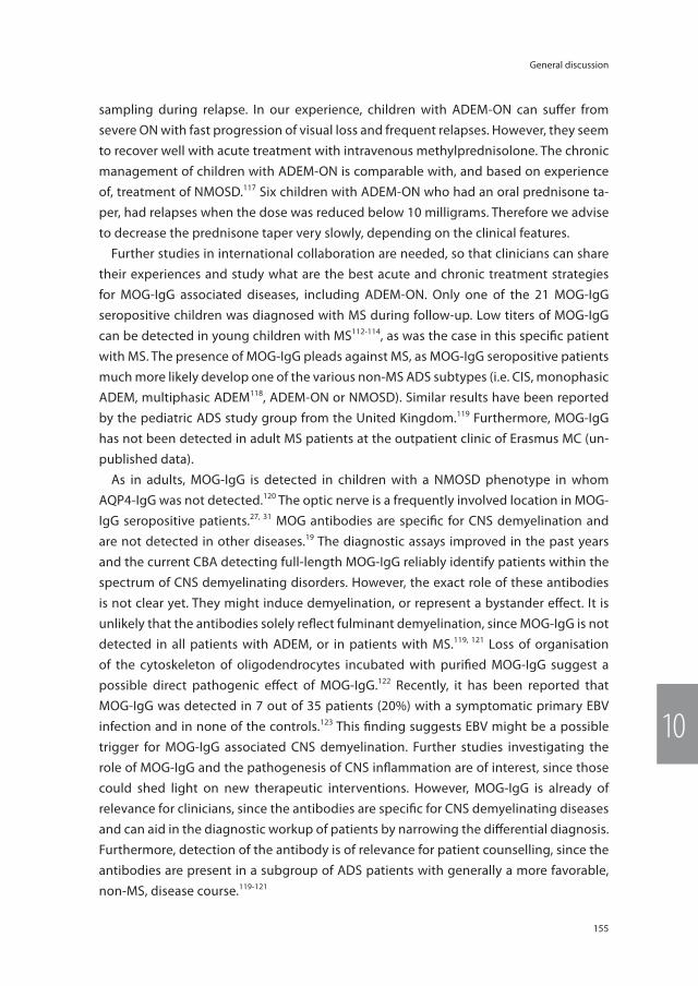
155
General discussion
10
sampling during relapse. In our experience, children with ADEM-ON can suffer from severe ON with fast progression of visual loss and frequent relapses. However, they seem to recover well with acute treatment with intravenous methylprednisolone. The chronic management of children with ADEM-ON is comparable with, and based on experience of, treatment of NMOSD.117 Six children with ADEM-ON who had an oral prednisone ta-per, had relapses when the dose was reduced below 10 milligrams. Therefore we advise to decrease the prednisone taper very slowly, depending on the clinical features.
Further studies in international collaboration are needed, so that clinicians can share their experiences and study what are the best acute and chronic treatment strategies for MOG-IgG associated diseases, including ADEM-ON. Only one of the 21 MOG-IgG seropositive children was diagnosed with MS during follow-up. Low titers of MOG-IgG can be detected in young children with MS112-114, as was the case in this specific patient with MS. The presence of MOG-IgG pleads against MS, as MOG-IgG seropositive patients much more likely develop one of the various non-MS ADS subtypes (i.e. CIS, monophasic ADEM, multiphasic ADEM118, ADEM-ON or NMOSD). Similar results have been reported by the pediatric ADS study group from the United Kingdom.119 Furthermore, MOG-IgG has not been detected in adult MS patients at the outpatient clinic of Erasmus MC (un-published data).
As in adults, MOG-IgG is detected in children with a NMOSD phenotype in whom AQP4-IgG was not detected.120 The optic nerve is a frequently involved location in MOG-IgG seropositive patients.27, 31 MOG antibodies are specific for CNS demyelination and are not detected in other diseases.19 The diagnostic assays improved in the past years and the current CBA detecting full-length MOG-IgG reliably identify patients within the spectrum of CNS demyelinating disorders. However, the exact role of these antibodies is not clear yet. They might induce demyelination, or represent a bystander effect. It is unlikely that the antibodies solely reflect fulminant demyelination, since MOG-IgG is not detected in all patients with ADEM, or in patients with MS.119, 121 Loss of organisation of the cytoskeleton of oligodendrocytes incubated with purified MOG-IgG suggest a possible direct pathogenic effect of MOG-IgG.122 Recently, it has been reported that MOG-IgG was detected in 7 out of 35 patients (20%) with a symptomatic primary EBV infection and in none of the controls.123 This finding suggests EBV might be a possible trigger for MOG-IgG associated CNS demyelination. Further studies investigating the role of MOG-IgG and the pathogenesis of CNS inflammation are of interest, since those could shed light on new therapeutic interventions. However, MOG-IgG is already of relevance for clinicians, since the antibodies are specific for CNS demyelinating diseases and can aid in the diagnostic workup of patients by narrowing the differential diagnosis. Furthermore, detection of the antibody is of relevance for patient counselling, since the antibodies are present in a subgroup of ADS patients with generally a more favorable, non-MS, disease course.119-121
Chapter 10
156
search for novel biomarkers in childhood-onset ADs
Possible new biomarkers for CNS demyelination can be identified in the exciting field of proteomics. The large-scale study of proteins involved in ADS can provide further insight into the pathophysiology of MS. We studied the CSF proteome of 39 children with ADS, of whom 18 children were subsequently diagnosed with MS (chapter 9). In CSF collected at the first clinical event of CNS demyelination, we found peptides relating to 21 proteins to be differentially abundant in children with monophasic ADS and MS. Fourteen proteins had higher abundance in children with MS than in children with monophasic ADS. Of these proteins, 12 were linked to neuronal function and gray matter, illustrating neuro-degeneration already occurs early in MS. This finding is supported by previous studies which reported an abundance of gray matter proteins in early MS.124 Furthermore, some of the identified proteins overlap with a previous study, including CSF of 8 children with MS and 11 children with monophasic ADS, in which axoglial proteins were detected in the children with MS.125 The presence of black holes on MRI at the first event46 and lim-ited age-expected brain growth and brain atrophy in children with MS126 endorse neu-rodegeneration occurs at an early stage. Seven proteins were abundant in children with monophasic ADS, of which 6 proteins have a role in innate immunity. This demonstrates a more innate inflammatory disease mechanism in children with monophasic ADS, which might be explained by the children with an ADEM-like onset in this group, who often have a monophasic event preceded by infections. Our discovery study of the CSF proteome in children with ADS indicates different underlying disease mechanisms in monophasic ADS and MS. Future larger-sample size studies, preferably in international collaboration, are needed in order to investigate whether certain proteins relate to clinical symptoms and if these proteins can predict the outcome of MS diagnosis and relapse rate.
PArt two: AcQuireD DemyelinAting synDromes in chilDren
main findings
• In children with ADS, MOG-IgG identifies a subset of young children with an ADEM-like phenotype without a future MS diagnosis.
• 57 risk SNPs for adult-onset MS are associated with MS risk in children with ADS. The compound effect of 57 minor risk alleles exceeded the effect of the major MS risk allele HLA-DRB1*15.
• MRI is an important diagnostic tool in child-hood onset ADS. We found that the Verhey MRI criteria and IPMSSG 2012 consensus definitions reliably identify children with MS.
• Children with CIS have a more inflammatory disease course than adults with CIS, apparent from a higher rate of future MS diagnoses and more frequent relapses.
clinical implications
• MOG-IgG should be tested in children presenting with CNS inflammatory syndromes as part of their standard diagnostic work up.
• The IPMSSG 2012 definitions for childhood-onset ADS, including MS, are validated.
• We offer a clinical prediction rule, which has to be validated, for early DMT initiation in 11-16 year-old children with CIS who are at high risk for a future MS diagnosis.
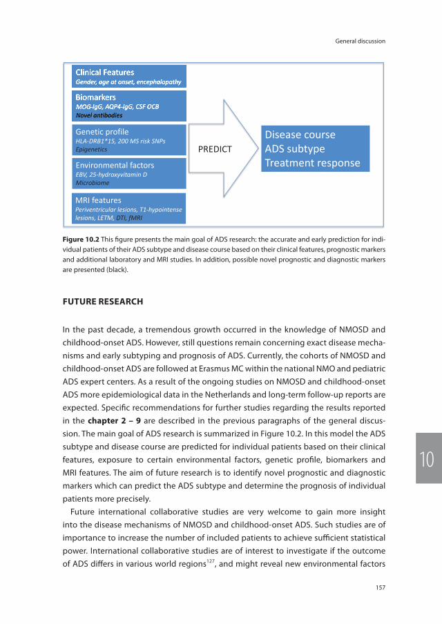
157
General discussion
10
Future reseArch
In the past decade, a tremendous growth occurred in the knowledge of NMOSD and childhood-onset ADS. However, still questions remain concerning exact disease mecha-nisms and early subtyping and prognosis of ADS. Currently, the cohorts of NMOSD and childhood-onset ADS are followed at Erasmus MC within the national NMO and pediatric ADS expert centers. As a result of the ongoing studies on NMOSD and childhood-onset ADS more epidemiological data in the Netherlands and long-term follow-up reports are expected. Specifi c recommendations for further studies regarding the results reported in the chapter 2 – 9 are described in the previous paragraphs of the general discus-sion. The main goal of ADS research is summarized in Figure 10.2. In this model the ADS subtype and disease course are predicted for individual patients based on their clinical features, exposure to certain environmental factors, genetic profi le, biomarkers and MRI features. The aim of future research is to identify novel prognostic and diagnostic markers which can predict the ADS subtype and determine the prognosis of individual patients more precisely.
Future international collaborative studies are very welcome to gain more insight into the disease mechanisms of NMOSD and childhood-onset ADS. Such studies are of importance to increase the number of included patients to achieve suffi cient statistical power. International collaborative studies are of interest to investigate if the outcome of ADS diff ers in various world regions127, and might reveal new environmental factors
Clinical Features Gender, age at onset, encephalopathy
Biomarkers MOG-IgG, AQP4-IgG, CSF OCB Novel antibodies
Clinical FeaturesGender, age at onset, encephalopathy
BiomarkersMOG-IgG, AQP4-IgG, CSF OCBNovel antibodies
MRI features Periventricular lesions, T1-hypointense lesions, LETM, DTI, fMRI
Disease course ADS subtype Treatment response
Genetic profile HLA-DRB1*15, 200 MS risk SNPs Epigenetics
Environmental factors EBV, 25-hydroxyvitamin D Microbiome
PREDICT
Figure 10.2 This fi gure presents the main goal of ADS research: the accurate and early prediction for indi-vidual patients of their ADS subtype and disease course based on their clinical features, prognostic markers and additional laboratory and MRI studies. In addition, possible novel prognostic and diagnostic markers are presented (black).
Chapter 10
158
involved in the pathogenesis of ADS. Ongoing prospective studies in NMOSD and childhood-onset ADS include the search for novel prognostic biomarkers and require large-scale standardized storage of blood and CSF samples (biobanking).128 Novel bio-markers are needed to further specify the subtypes of ADS and determine the prognosis of individual patients. AQP4-IgG and MOG-IgG are useful markers in patients presenting with a first event of ADS and identify patients who are likely to have diseases distinct from MS.7, 19 However, AQP4-IgG and MOG-IgG lack sensitivity to accurately subtype all patients with ADS. Future serum and CSF immunohistochemistal studies might reveal novel antibodies associated with ADS.
An exciting new focus in MS research is the gut microbiome. The gut microbiome has an important role in normal immune function.129 The composition of the gut microbiota might represent a propagating factor for inflammatory signalling in MS.130 A recent Ger-man study with a mouse model for human MS, presented at the 31st Congress of the Eu-ropean Committee for Treatment and Research in Multiple Sclerosis (ECTRIMS), reported that mice who received a poop transplant from a monozygotic twin with MS got worse disease than mice who received a poop transplant from the healthy twin.131 These results suggest that the gut microbiome represent a propagating factor for MS rather than just a consequence of MS. American studies including relatively small sample sizes, already reported some microbiota stems are associated with pediatric MS and a higher relapse rate.132, 133 Further studies in this novel field, including children with a European diet, are exciting as those studies might identify novel (supportive) therapeutic strategies. Likewise, such studies would be interesting in other autoimmune diseases, including NMOSD.
Furthermore, international collaboration would allow for larger sample sizes and genome-wide association studies in patients with various ethnicities. Such large-scale genetic studies might reveal new specific risk genes for childhood-onset ADS and for NMOSD. In addition, international collaboration would allow discovering the field of epigenetics, which is the study of heritable changes in genome function without un-derlying modifications in their nucleotide sequence.134 Epigenetic modifications are the crossroad between environmental and genetic modifications and might reveal future diagnostic and therapeutic targets in ADS patients.
Future prospective studies including a broader spectrum of CNS inflammation with ADS and other autoimmune diseases (for example sarcoidosis, vasculitis or SLE) would be of great interest to study early prognostic and diagnostic markers in patients pre-senting with an incident event of CNS inflammation. Such an approach would resemble clinical practice, but is hampered by the rarity of these disorders.
Future research will focus on the improvement of treatment strategies. Novel thera-peutic trials are underway both in the field of NMOSD and MS.2, 135-137 As the number of chronic treatment possibilities is growing, a future individual treatment approach is
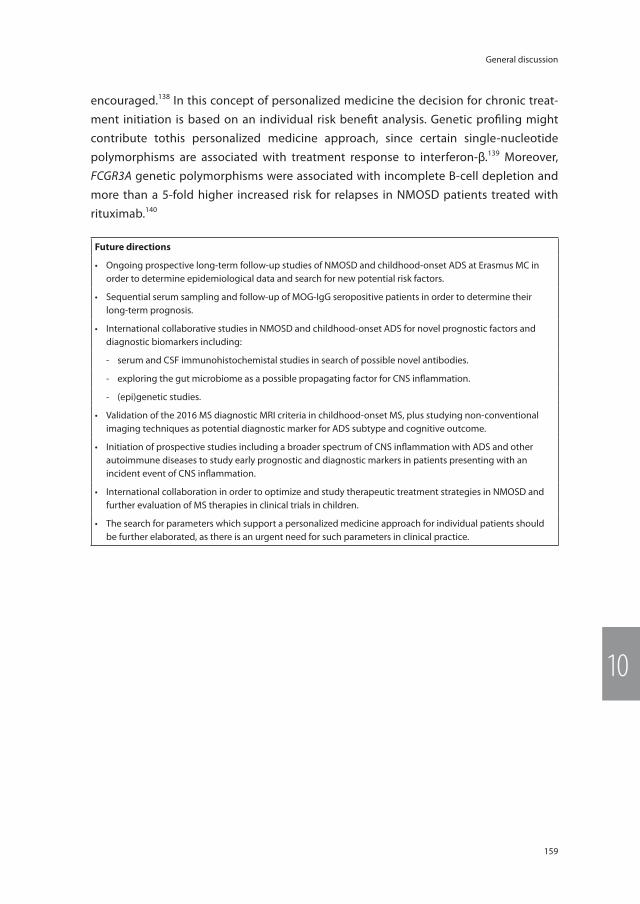
159
General discussion
10
encouraged.138 In this concept of personalized medicine the decision for chronic treat-ment initiation is based on an individual risk benefit analysis. Genetic profiling might contribute tothis personalized medicine approach, since certain single-nucleotide polymorphisms are associated with treatment response to interferon-β.139 Moreover, FCGR3A genetic polymorphisms were associated with incomplete B-cell depletion and more than a 5-fold higher increased risk for relapses in NMOSD patients treated with rituximab.140
Future directions
• Ongoing prospective long-term follow-up studies of NMOSD and childhood-onset ADS at Erasmus MC in order to determine epidemiological data and search for new potential risk factors.
• Sequential serum sampling and follow-up of MOG-IgG seropositive patients in order to determine their long-term prognosis.
• International collaborative studies in NMOSD and childhood-onset ADS for novel prognostic factors and diagnostic biomarkers including:
- serum and CSF immunohistochemistal studies in search of possible novel antibodies.
- exploring the gut microbiome as a possible propagating factor for CNS inflammation.
- (epi)genetic studies.
• Validation of the 2016 MS diagnostic MRI criteria in childhood-onset MS, plus studying non-conventional imaging techniques as potential diagnostic marker for ADS subtype and cognitive outcome.
• Initiation of prospective studies including a broader spectrum of CNS inflammation with ADS and other autoimmune diseases to study early prognostic and diagnostic markers in patients presenting with an incident event of CNS inflammation.
• International collaboration in order to optimize and study therapeutic treatment strategies in NMOSD and further evaluation of MS therapies in clinical trials in children.
• The search for parameters which support a personalized medicine approach for individual patients should be further elaborated, as there is an urgent need for such parameters in clinical practice.
Chapter 10
160
reFerences
1. Wingerchuk DM, Lennon VA, Lucchinetti CF, Pittock SJ, Weinshenker BG. The spectrum of neuromyelitis optica. Lancet Neurol 2007; 6(9): 805-15.
2. Pittock SJ, Lucchinetti CF. Neuromyelitis optica and the evolving spectrum of auto-immune aquaporin-4 channelopathies: a decade later. Ann N Y Acad Sci 2016; 1366(1): 20-39.
3. Pandit L, Asgari N, Apiwattanakul M, Palace J, Paul F, Leite MI, et al. Demographic and clini-cal features of neuromyelitis optica: A review. Mult Scler 2015; 21(7): 845-53.
4. Flanagan EP, Cabre P, Weinshenker BG, St Sauver J, Jacobson DJ, Majed M, et al. Epide-miology of aquaporin-4 autoimmunity and neuromyelitis optica spectrum. Ann Neurol Published Online First: 17 Feb 2016. doi: 10.1002/ana.24617.
5. Mealy MA, Wingerchuk DM, Greenberg BM, Levy M. Epidemiology of neuromyelitis optica in the United States: a multicenter analysis. Arch Neurol 2012; 69(9): 1176-80.
6. Kitley J, Leite MI, Nakashima I, Waters P, McNeillis B, Brown R, et al. Prognostic factors and disease course in aquaporin-4 antibody-positive patients with neuromyelitis optica spectrum disorder from the United Kingdom and Japan. Brain 2012; 135(Pt 6): 1834-49.
7. Wingerchuk DM, Banwell B, Bennett JL, Cabre P, Carroll W, Chitnis T, et al. International con-sensus diagnostic criteria for neuromyelitis optica spectrum disorders. Neurology 2015; 85(2): 177-89.
8. Hyun JW, Jeong IH, Joung A, Kim SH, Kim HJ. Evaluation of the 2015 diagnostic criteria for neuromyelitis optica spectrum disorder. Neurology 2016; 86(19): 1772-79.
9. Chitnis T, Ness J, Krupp L, Waubant E, Hunt T, Olsen CS, et al. Clinical features of neuro-myelitis optica in children: US Network of Pediatric MS Centers report. Neurology 2016; 86(3): 245-52.
10. Wingerchuk DM, Lennon VA, Pittock SJ, Lucchinetti CF, Weinshenker BG. Revised
diagnostic criteria for neuromyelitis optica. Neurology 2006; 66(10): 1485-89.
11. Flanagan EP, Weinshenker BG, Krecke KN, Len-non VA, Lucchinetti CF, McKeon A, et al. Short myelitis lesions in aquaporin-4-IgG-positive neuromyelitis optica spectrum disorders. JAMA Neurol 2015; 72(1): 81-87.
12. Flanagan EP, Weinshenker BG, Krecke KN, Pittock SJ. Asymptomatic myelitis in neuro-myelitis optica and autoimmune aquaporin-4 channelopathy. Neurol Clin Pract 2015; 5(2): 175-77.
13. Jarius S, Wildemann B. Aquaporin-4 antibod-ies (NMO-IgG) as a serological marker of neuromyelitis optica: a critical review of the literature. Brain Pathol 2013; 23(6): 661-83.
14. Waters PJ, McKeon A, Leite MI, Rajasekharan S, Lennon VA, Villalobos A, et al. Serologic diagnosis of NMO: a multicenter comparison of aquaporin-4-IgG assays. Neurology 2012; 78(9): 665-71.
15. Sato DK, Nakashima I, Takahashi T, Misu T, Waters P, Kuroda H, et al. Aquaporin-4 antibody-positive cases beyond current di-agnostic criteria for NMO spectrum disorders. Neurology 2013; 80(24): 2210-16.
16. Bernard-Valnet R, Liblau RS, Vukusic S, Marignier R. Neuromyelitis optica: a positive appraisal of seronegative cases. Eur J Neurol 2015; 22(12): 1511-18.
17. Melamed E, Levy M, Waters PJ, Sato DK, Ben-nett JL, John GR, et al. Update on biomarkers in neuromyelitis optica. Neurol Neuroimmunol Neuroinflamm 2015; 2(4): e134.
18. Reindl M, Di Pauli F, Rostasy K, Berger T. The spectrum of MOG autoantibody-associated demyelinating diseases. Nat Rev Neurol 2013; 9(8): 455-61.
19. Ramanathan S, Dale RC, Brilot F. Anti-MOG antibody: The history, clinical phenotype, and pathogenicity of a serum biomarker for demyelination. Autoimmun Rev 2016; 15(4): 307-24.
20. Kim HJ, Paul F, Lana-Peixoto MA, Tenembaum S, Asgari N, Palace J, et al. MRI characteristics
161
General discussion
10
of neuromyelitis optica spectrum disorder: an international update. Neurology 2015; 84(11): 1165-73.
21. Pittock SJ, Weinshenker BG, Lucchinetti CF, Wingerchuk DM, Corboy JR, Lennon VA. Neuromyelitis optica brain lesions localized at sites of high aquaporin 4 expression. Arch Neurol 2006; 63(7): 964-68.
22. Zamvil SS, Slavin AJ. Does MOG Ig-positive AQP4-seronegative opticospinal inflam-matory disease justify a diagnosis of NMO spectrum disorder? Neurol Neuroimmunol Neuroinflamm 2015; 2(1): e62.
23. Kitley J, Waters P, Woodhall M, Leite MI, Murchison A, George J, et al. Neuromyelitis optica spectrum disorders with aquaporin-4 and myelin-oligodendrocyte glycoprotein antibodies: a comparative study. JAMA Neurol 2014; 71(3): 276-83.
24. Sato DK, Callegaro D, Lana-Peixoto MA, Waters PJ, Jorge FMD, Takahashi T, et al. Distinction between MOG antibody-positive and AQP4 antibody-positive NMO spectrum disorders. Neurology 2014; 82(6): 474-81.
25. Hoftberger R, Sepulveda M, Armangue T, Blanco Y, Rostasy K, Cobo Calvo A, et al. Antibodies to MOG and AQP4 in adults with neuromyelitis optica and suspected limited forms of the disease. Mult Scler 2015; 21(7): 866-74.
26. Piccolo L, Woodhall M, Tackley G, Jurynczyk M, Kong Y, Domingos J, et al. Isolated new on-set ‘atypical’ optic neuritis in the NMO clinic: serum antibodies, prognoses and diagnoses at follow-up. J Neurol 2016; 263(2): 370-79.
27. Ramanathan S, Reddel SW, Henderson A, Par-ratt JD, Barnett M, Gatt PN, et al. Antibodies to myelin oligodendrocyte glycoprotein in bilateral and recurrent optic neuritis. Neurol Neuroimmunol Neuroinflamm 2014; 1(4): e40.
28. Mader S, Gredler V, Schanda K, Rostasy K, Dujmovic I, Pfaller K, et al. Complement acti-vating antibodies to myelin oligodendrocyte glycoprotein in neuromyelitis optica and related disorders. J Neuroinflammation 2011; 8: 184.
29. Kitley J, Woodhall M, Waters P, Leite MI, De-venney E, Craig J, et al. Myelin-oligodendro-cyte glycoprotein antibodies in adults with a neuromyelitis optica phenotype. Neurology 2012; 79(12): 1273-77.
30. Tanaka M, Tanaka K. Anti-MOG antibodies in adult patients with demyelinating disorders of the central nervous system. J Neuroim-munol 2014; 270(1-2): 98-99.
31. Martinez-Hernandez E, Sepulveda M, Rostasy K, Hoftberger R, Graus F, Harvey RJ, et al. An-tibodies to aquaporin 4, myelin-oligodendro-cyte glycoprotein, and the glycine receptor alpha1 subunit in patients with isolated optic neuritis. JAMA Neurol 2015; 72(2): 187-93.
32. Probstel AK, Rudolf G, Dornmair K, Collongues N, Chanson JB, Sanderson NS, et al. Anti-MOG antibodies are present in a subgroup of pa-tients with a neuromyelitis optica phenotype. J Neuroinflammation 2015; 12: 46.
33. Nakajima H, Motomura M, Tanaka K, Fujikawa A, Nakata R, Maeda Y, et al. Antibodies to myelin oligodendrocyte glycoprotein in idiopathic optic neuritis. BMJ Open 2015; 5(4): e007766.
34. Cobo-Calvo A, Sepulveda M, Bernard-Valnet R, Ruiz A, Brassat D, Martinez-Yelamos S, et al. Antibodies to myelin oligodendrocyte glyco-protein in aquaporin 4 antibody seronegative longitudinally extensive transverse myelitis: Clinical and prognostic implications. Mult Scler 2016; 22(3): 312-19.
35. Sepulveda M, Armangue T, Sola-Valls N, Ar-rambide G, Meca-Lallana JE, Oreja-Guevara C, et al. Neuromyelitis optica spectrum disorders: Comparison according to the phe-notype and serostatus. Neurol Neuroimmunol Neuroinflamm 2016; 3(3): e225.
36. Kremer S, Renard F, Achard S, Lana-Peixoto MA, Palace J, Asgari N, et al. Use of Advanced Magnetic Resonance Imaging Techniques in Neuromyelitis Optica Spectrum Disorder. JAMA Neurol 2015; 72(7): 815-22.
37. Weinshenker BG, Barron G, Behne JM, Ben-nett JL, Chin PS, Cree BA, et al. Challenges and opportunities in designing clinical trials for neuromyelitis optica. Neurology 2015; 84(17): 1805-15.
Chapter 10
162
38. Devic E. Myélite subaiguë compliquée de névrite optique. Bull Méd 1894; 8: 1033-34.
39. Guo Y, Lennon VA, Popescu BF, Grouse CK, Topel J, Milone M, et al. Autoimmune aqua-porin-4 myopathy in neuromyelitis optica spectrum. JAMA Neurol 2014; 71(8): 1025-29.
40. Suzuki N, Takahashi T, Aoki M, Misu T, Ko-nohana S, Okumura T, et al. Neuromyelitis optica preceded by hyperCKemia episode. Neurology 2010; 74(19): 1543-45.
41. Di Filippo M, Franciotta D, Massa R, Di Grego-rio M, Zardini E, Gastaldi M, et al. Recurrent hyperCKemia with normal muscle biopsy in a pediatric patient with neuromyelitis optica. Neurology 2012; 79(11): 1182-84.
42. Watanabe M, Furusho K, Takahashi T, Tamaoka A. Galactorrhea in a Patient With Aquaporin-4 Antibody-positive Neuromyelitis Optica Spectrum Disorder: A Case Report and Re-view of the Literature. Neurologist 2015; 20(6): 101-03.
43. Jiao Y, Fryer JP, Lennon VA, Jenkins SM, Quek AM, Smith CY, et al. Updated estimate of AQP4-IgG serostatus and disability outcome in neuromyelitis optica. Neurology 2013; 81(14): 1197-204.
44. Mikaeloff Y, Adamsbaum C, Husson B, Vallee L, Ponsot G, Confavreux C, et al. MRI prognostic factors for relapse after acute CNS inflamma-tory demyelination in childhood. Brain 2004; 127(Pt 9): 1942-47.
45. Callen DJ, Shroff MM, Branson HM, Lotze T, Li DK, Stephens D, et al. MRI in the diagnosis of pediatric multiple sclerosis. Neurology 2009; 72(11): 961-67.
46. Callen DJ, Shroff MM, Branson HM, Li DK, Lotze T, Stephens D, et al. Role of MRI in the differentiation of ADEM from MS in children. Neurology 2009; 72(11): 968-73.
47. Krupp LB, Tardieu M, Amato MP, Banwell B, Chitnis T, Dale RC, et al. International Pediatric Multiple Sclerosis Study Group criteria for pediatric multiple sclerosis and immune-me-diated central nervous system demyelinating disorders: revisions to the 2007 definitions. Mult Scler 2013; 19(10): 1261-67.
48. Polman CH, Reingold SC, Banwell B, Clanet M, Cohen JA, Filippi M, et al. Diagnostic criteria for multiple sclerosis: 2010 revisions to the McDonald criteria. Ann Neurol 2011; 69(2): 292-302.
49. McDonald WI, Compston A, Edan G, Goodkin D, Hartung HP, Lublin FD, et al. Recommend-ed diagnostic criteria for multiple sclerosis: guidelines from the International Panel on the diagnosis of multiple sclerosis. Ann Neurol 2001; 50(1): 121-27.
50. Polman CH, Reingold SC, Edan G, Filippi M, Hartung HP, Kappos L, et al. Diagnostic criteria for multiple sclerosis: 2005 revisions to the “McDonald Criteria”. Ann Neurol 2005; 58(6): 840-46.
51. Sadaka Y, Verhey LH, Shroff MM, Branson HM, Arnold DL, Narayanan S, et al. 2010 McDonald criteria for diagnosing pediatric multiple sclerosis. Ann Neurol 2012; 72(2): 211-23.
52. Sedani S, Lim MJ, Hemingway C, Wassmer E, Absoud M. Paediatric multiple sclerosis: ex-amining utility of the McDonald 2010 criteria. Mult Scler 2012; 18(5): 679-82.
53. Tantsis EM, Prelog K, Brilot F, Dale RC. Risk of multiple sclerosis after a first demyelinating syndrome in an Australian Paediatric cohort: clinical, radiological features and application of the McDonald 2010 MRI criteria. Mult Scler 2013; 19(13): 1749-59.
54. Kornek B, Schmitl B, Vass K, Zehetmayer S, Pritsch M, Penzien J, et al. Evaluation of the 2010 McDonald multiple sclerosis criteria in children with a clinically isolated syndrome. Mult Scler 2012; 18(12): 1768-74.
55. Hummel HM, Bruck W, Dreha-Kulaczewski S, Gartner J, Wuerfel J. Pediatric onset multiple sclerosis: McDonald criteria 2010 and the contribution of spinal cord MRI. Mult Scler 2013; 19(10): 1330-35.
56. Edan G, Kappos L, Montalban X, Polman CH, Freedman MS, Hartung HP, et al. Long-term impact of interferon beta-1b in patients with CIS: 8-year follow-up of BENEFIT. J Neurol Neurosurg Psychiatry 2014; 85(11): 1183-89.
57. Kappos L, O’Connor P, Radue EW, Polman C, Hohlfeld R, Selmaj K, et al. Long-term
163
General discussion
10
effects of fingolimod in multiple sclerosis: the randomized FREEDOMS extension trial. Neurology 2015; 84(15): 1582-91.
58. Comi G, Martinelli V, Rodegher M, Moiola L, Bajenaru O, Carra A, et al. Effect of glatiramer acetate on conversion to clinically definite multiple sclerosis in patients with clinically isolated syndrome (PreCISe study): a ran-domised, double-blind, placebo-controlled trial. Lancet 2009; 374(9700): 1503-11.
59. Mikaeloff Y, Caridade G, Husson B, Suissa S, Tardieu M, Neuropediatric KSGotFNS. Acute disseminated encephalomyelitis cohort study: prognostic factors for relapse. Eur J Paediatr Neurol 2007; 11(2): 90-95.
60. Verhey LH, Branson HM, Shroff MM, Callen DJ, Sled JG, Narayanan S, et al. MRI parameters for prediction of multiple sclerosis diagnosis in children with acute CNS demyelination: a prospective national cohort study. Lancet Neurol 2011; 10(12): 1065-73.
61. Banwell B, Bar-Or A, Arnold DL, Sadovnick D, Narayanan S, McGowan M, et al. Clinical, environmental, and genetic determinants of multiple sclerosis in children with acute demyelination: a prospective national cohort study. Lancet Neurol 2011; 10(5): 436-45.
62. Ketelslegers IA, Visser IE, Neuteboom RF, Boon M, Catsman-Berrevoets CE, Hintzen RQ. Disease course and outcome of acute dis-seminated encephalomyelitis is more severe in adults than in children. Mult Scler 2011; 17(4): 441-48.
63. Tenembaum S, Chitnis T, Ness J, Hahn JS, International Pediatric MSSG. Acute dissemi-nated encephalomyelitis. Neurology 2007; 68(16 Suppl 2): S23-36.
64. Ketelslegers IA, Neuteboom RF, Boon M, Catsman-Berrevoets CE, Hintzen RQ, Dutch Pediatric MSSG. A comparison of MRI criteria for diagnosing pediatric ADEM and MS. Neu-rology 2010; 74(18): 1412-15.
65. Neuteboom RF, Boon M, Catsman-Berrevoets CE, Vles JS, Gooskens RH, Stroink H, et al. Prognostic factors after a first attack of inflammatory CNS demyelination in children. Neurology 2008; 71(13): 967-73.
66. Ketelslegers IA, Catsman-Berrevoets CE, Neuteboom RF, Boon M, van Dijk KG, Eikelen-boom MJ, et al. Incidence of acquired de-myelinating syndromes of the CNS in Dutch children: a nationwide study. J Neurol 2012; 259(9): 1929-35.
67. Kappos L, Bates D, Edan G, Eraksoy M, Garcia-Merino A, Grigoriadis N, et al. Natalizumab treatment for multiple sclerosis: updated recommendations for patient selection and monitoring. Lancet Neurol 2011; 10(8): 745-58.
68. Filippi M, Rocca MA, Ciccarelli O, De Stefano N, Evangelou N, Kappos L, et al. MRI criteria for the diagnosis of multiple sclerosis: MAG-NIMS consensus guidelines. Lancet Neurol 2016; 15(3): 292-303.
69. Tillema JM, Leach J, Pirko I. Non-lesional white matter changes in pediatric multiple sclerosis and monophasic demyelinating disorders. Mult Scler 2012; 18(12): 1754-59.
70. Akbar N, Giorgio A, Till C, Sled JG, Doesburg SM, De Stefano N, et al. Alterations in Functional and Structural Connectivity in Pediatric-Onset Multiple Sclerosis. PLoS One 2016; 11(1): e0145906.
71. Pasto L, Portaccio E, Goretti B, Ghezzi A, Lori S, Hakiki B, et al. The cognitive reserve theory in the setting of pediatric-onset multiple sclerosis. Mult Scler Published Online First: 11 Feb 2016. doi: 10.1177/1352458516629559.
72. Banwell B, Kennedy J, Sadovnick D, Arnold DL, Magalhaes S, Wambera K, et al. Incidence of acquired demyelination of the CNS in Canadian children. Neurology 2009; 72(3): 232-39.
73. Venkateswaran S, Banwell B. Pediatric mul-tiple sclerosis. Neurologist 2010; 16(2): 92-105.
74. Hahn JS, Pohl D, Rensel M, Rao S, Interna-tional Pediatric MSSG. Differential diagnosis and evaluation in pediatric multiple sclerosis. Neurology 2007; 68(16 Suppl 2): S13-22.
75. Bigi S, Banwell B. Pediatric multiple sclerosis. J Child Neurol 2012; 27(11): 1378-83.
76. Boiko A, Vorobeychik G, Paty D, Devonshire V, Sadovnick D, University of British Columbia MSCN. Early onset multiple sclerosis: a longi-tudinal study. Neurology 2002; 59(7): 1006-10.
Chapter 10
164
77. Gorman MP, Healy BC, Polgar-Turcsanyi M, Chitnis T. Increased relapse rate in pediatric-onset compared with adult-onset multiple sclerosis. Arch Neurol 2009; 66(1): 54-59.
78. Benson LA, Healy BC, Gorman MP, Baruch NF, Gholipour T, Musallam A, et al. Elevated relapse rates in pediatric compared to adult MS persist for at least 6 years. Mult Scler Relat Disord 2014; 3(2): 186-93.
79. Simone IL, Carrara D, Tortorella C, Liguori M, Lepore V, Pellegrini F, et al. Course and prog-nosis in early-onset MS: comparison with adult-onset forms. Neurology 2002; 59(12): 1922-28.
80. Renoux C, Vukusic S, Mikaeloff Y, Edan G, Clanet M, Dubois B, et al. Natural history of multiple sclerosis with childhood onset. N Engl J Med 2007; 356(25): 2603-13.
81. Harding KE, Liang K, Cossburn MD, Ingram G, Hirst CL, Pickersgill TP, et al. Long-term out-come of paediatric-onset multiple sclerosis: a population-based study. J Neurol Neurosurg Psychiatry 2013; 84(2): 141-47.
82. Chitnis T, Tenembaum S, Banwell B, Krupp L, Pohl D, Rostasy K, et al. Consensus statement: evaluation of new and existing therapeutics for pediatric multiple sclerosis. Mult Scler 2012; 18(1): 116-27.
83. Banwell B, Krupp L, Kennedy J, Tellier R, Tenembaum S, Ness J, et al. Clinical features and viral serologies in children with multiple sclerosis: a multinational observational study. Lancet Neurol 2007; 6(9): 773-81.
84. Huppke B, Ellenberger D, Rosewich H, Friede T, Gartner J, Huppke P. Clinical presentation of pediatric multiple sclerosis before puberty. Eur J Neurol 2014; 21(3): 441-46.
85. Ahn JJ, O’Mahony J, Moshkova M, Hanwell HE, Singh H, Zhang MA, et al. Puberty in females enhances the risk of an outcome of multiple sclerosis in children and the development of central nervous system autoimmunity in mice. Mult Scler 2015; 21(6): 735-48.
86. Chitnis T, Glanz B, Jaffin S, Healy B. Demo-graphics of pediatric-onset multiple sclerosis in an MS center population from the North-
eastern United States. Mult Scler 2009; 15(5): 627-31.
87. Kennedy J, O’Connor P, Sadovnick AD, Perara M, Yee I, Banwell B. Age at onset of multiple sclerosis may be influenced by place of resi-dence during childhood rather than ancestry. Neuroepidemiology 2006; 26(3): 162-67.
88. Langer-Gould A, Zhang JL, Chung J, Yeung Y, Waubant E, Yao J. Incidence of acquired CNS demyelinating syndromes in a multiethnic cohort of children. Neurology 2011; 77(12): 1143-48.
89. Absoud M, Lim MJ, Chong WK, De Goede CG, Foster K, Gunny R, et al. Paediatric acquired demyelinating syndromes: incidence, clinical and magnetic resonance imaging features. Mult Scler 2013; 19(1): 76-86.
90. Ascherio A, Munger KL, Lunemann JD. The initiation and prevention of multiple sclero-sis. Nat Rev Neurol 2012; 8(11): 602-12.
91. Boster AL, Endress CF, Hreha SA, Caon C, Perumal JS, Khan OA. Pediatric-onset multiple sclerosis in African-American black and European-origin white patients. Pediatr Neurol 2009; 40(1): 31-33.
92. Fay AJ, Mowry EM, Strober J, Waubant E. Relapse severity and recovery in early pedi-atric multiple sclerosis. Mult Scler 2012; 18(7): 1008-12.
93. Tintore M, Rovira A, Rio J, Otero-Romero S, Arrambide G, Tur C, et al. Defining high, medium and low impact prognostic factors for developing multiple sclerosis. Brain 2015; 138(Pt 7): 1863-74.
94. Ghezzi A, Banwell B, Boyko A, Amato MP, An-lar B, Blinkenberg M, et al. The management of multiple sclerosis in children: a European view. Mult Scler 2010; 16(10): 1258-67.
95. Disanto G, Magalhaes S, Handel AE, Morrison KM, Sadovnick AD, Ebers GC, et al. HLA-DRB1 confers increased risk of pediatric-onset MS in children with acquired demyelination. Neurology 2011; 76(9): 781-86.
96. Boiko AN, Gusev EI, Sudomoina MA, Alek-seenkov AD, Kulakova OG, Bikova OV, et al. Association and linkage of juvenile MS with
165
General discussion
10
HLA-DR2(15) in Russians. Neurology 2002; 58(4): 658-60.
97. Hintzen R, International Multiple Sclerosis Genetics Consortium (IMSGC). Personal com-munication. 2016.
98. International Multiple Sclerosis Genetics C, Wellcome Trust Case Control C, Sawcer S, Hellenthal G, Pirinen M, Spencer CC, et al. Ge-netic risk and a primary role for cell-mediated immune mechanisms in multiple sclerosis. Nature 2011; 476(7359): 214-19.
99. Bashinskaya VV, Kulakova OG, Boyko AN, Favorov AV, Favorova OO. A review of ge-nome-wide association studies for multiple sclerosis: classical and hypothesis-driven approaches. Hum Genet 2015; 134(11-12): 1143-62.
100. Graves JS, Barcellos LF, Shao X, Noble J, Mowry EM, Quach H, et al. Genetic predictors of relapse rate in pediatric MS. Mult Scler Published Online First: 14 Jan 2016. doi: 10.1177/1352458515624269.
101. Mowry EM, Krupp LB, Milazzo M, Chabas D, Strober JB, Belman AL, et al. Vitamin D status is associated with relapse rate in pediatric-onset multiple sclerosis. Ann Neurol 2010; 67(5): 618-24.
102. Hedstrom AK, Alfredsson L, Olsson T. Envi-ronmental factors and their interactions with risk genotypes in MS susceptibility. Curr Opin Neurol 2016; 29(3): 293-98.
103. Wielders JP, Muskiet FA, van de Wiel A. [Shed-ding new light on vitamin D-reassessment of an essential prohormone] Nieuw licht op vitamine D. Ned Tijdschr Geneeskd 2010; 154: A1810.
104. Ascherio A, Munger KL. Environmental risk factors for multiple sclerosis. Part I: the role of infection. Ann Neurol 2007; 61(4): 288-99.
105. Alotaibi S, Kennedy J, Tellier R, Stephens D, Banwell B. Epstein-Barr virus in pediatric mul-tiple sclerosis. JAMA 2004; 291(15): 1875-79.
106. Pohl D, Krone B, Rostasy K, Kahler E, Brunner E, Lehnert M, et al. High seroprevalence of Epstein-Barr virus in children with multiple sclerosis. Neurology 2006; 67(11): 2063-65.
107. Waubant E, Mowry EM, Krupp L, Chitnis T, Yeh EA, Kuntz N, et al. Common viruses associated with lower pediatric multiple sclerosis risk. Neurology 2011; 76(23): 1989-95.
108. Makhani N, Banwell B, Tellier R, Yea C, Mc-Govern S, O’Mahony J, et al. Viral exposures and MS outcome in a prospective cohort of children with acquired demyelination. Mult Scler 2016; 22(3): 385-88.
109. Yea C, Tellier R, Chong P, Westmacott G, Marrie RA, Bar-Or A, et al. Epstein-Barr virus in oral shedding of children with multiple sclerosis. Neurology 2013; 81(16): 1392-99.
110. Waubant E, Mowry EM, Krupp L, Chitnis T, Yeh EA, Kuntz N, et al. Antibody response to common viruses and human leukocyte antigen-DRB1 in pediatric multiple sclerosis. Mult Scler 2013; 19(7): 891-95.
111. O’Connor KC, McLaughlin KA, De Jager PL, Chitnis T, Bettelli E, Xu C, et al. Self-antigen tetramers discriminate between myelin au-toantibodies to native or denatured protein. Nat Med 2007; 13(2): 211-17.
112. Brilot F, Dale RC, Selter RC, Grummel V, Kalluri SR, Aslam M, et al. Antibodies to native my-elin oligodendrocyte glycoprotein in children with inflammatory demyelinating central nervous system disease. Ann Neurol 2009; 66(6): 833-42.
113. Di Pauli F, Mader S, Rostasy K, Schanda K, Bajer-Kornek B, Ehling R, et al. Temporal dynamics of anti-MOG antibodies in CNS demyelinating diseases. Clin Immunol 2011; 138(3): 247-54.
114. Probstel AK, Dornmair K, Bittner R, Sperl P, Jenne D, Magalhaes S, et al. Antibodies to MOG are transient in childhood acute disseminated encephalomyelitis. Neurology 2011; 77(6): 580-88.
115. Selter RC, Brilot F, Grummel V, Kraus V, Cepok S, Dale RC, et al. Antibody responses to EBV and native MOG in pediatric inflammatory demyelinating CNS diseases. Neurology 2010; 74(21): 1711-15.
116. Huppke P, Rostasy K, Karenfort M, Huppke B, Seidl R, Leiz S, et al. Acute disseminated encephalomyelitis followed by recurrent
Chapter 10
166
or monophasic optic neuritis in pediatric patients. Mult Scler 2013; 19(7): 941-46.
117. Longoni G, Banwell B, Filippi M, Yeh EA. Rituximab as a first-line preventive treatment in pediatric NMOSDs: Preliminary results in 5 children. Neurol Neuroimmunol Neuroinflamm 2014; 1(4): e46.
118. Baumann M, Hennes EM, Schanda K, Karen-fort M, Kornek B, Seidl R, et al. Children with multiphasic disseminated encephalomyelitis and antibodies to the myelin oligodendro-cyte glycoprotein (MOG): Extending the spectrum of MOG antibody positive diseases. Mult Scler Published Online First: 11 Feb 2016. doi: 10.1177/1352458516631038.
119. Hacohen Y, Absoud M, Deiva K, Hemingway C, Nytrova P, Woodhall M, et al. Myelin oligodendrocyte glycoprotein antibodies are associated with a non-MS course in children. Neurol Neuroimmunol Neuroinflamm 2015; 2(2): e81.
120. Lechner C, Baumann M, Hennes EM, Schanda K, Marquard K, Karenfort M, et al. Antibodies to MOG and AQP4 in children with neuromy-elitis optica and limited forms of the disease. J Neurol Neurosurg Psychiatry 2016; 87(8): 897-905.
121. Baumann M, Sahin K, Lechner C, Hennes EM, Schanda K, Mader S, et al. Clinical and neuroradiological differences of paediatric acute disseminating encephalomyelitis with and without antibodies to the myelin oligo-dendrocyte glycoprotein. J Neurol Neurosurg Psychiatry 2015; 86(3): 265-72.
122. Dale RC, Tantsis EM, Merheb V, Kumaran RY, Sinmaz N, Pathmanandavel K, et al. Antibod-ies to MOG have a demyelination phenotype and affect oligodendrocyte cytoskeleton. Neurol Neuroimmunol Neuroinflamm 2014; 1(1): e12.
123. Kakalacheva K, Regenass S, Wiesmayr S, Azzi T, Berger C, Dale RC, et al. Infectious Mononucleosis Triggers Generation of IgG Auto-Antibodies against Native Myelin Oligo-dendrocyte Glycoprotein. Viruses Published online: 12 Feb 2016. doi: 10.3390/v8020051.
124. Schutzer SE, Angel TE, Liu T, Schepmoes AA, Xie F, Bergquist J, et al. Gray matter is tar-geted in first-attack multiple sclerosis. PLoS One 2013; 8(9): e66117.
125. Dhaunchak AS, Becker C, Schulman H, De Faria O, Jr., Rajasekharan S, Banwell B, et al. Implication of perturbed axoglial apparatus in early pediatric multiple sclerosis. Ann Neurol 2012; 71(5): 601-13.
126. Aubert-Broche B, Fonov V, Narayanan S, Arnold DL, Araujo D, Fetco D, et al. Onset of multiple sclerosis before adulthood leads to failure of age-expected brain growth. Neurol-ogy 2014; 83(23): 2140-46.
127. Waldman A, Ghezzi A, Bar-Or A, Mikaeloff Y, Tardieu M, Banwell B. Multiple sclerosis in children: an update on clinical diagnosis, therapeutic strategies, and research. Lancet Neurol 2014; 13(9): 936-48.
128. Teunissen CE, Petzold A, Bennett JL, Berven FS, Brundin L, Comabella M, et al. A consen-sus protocol for the standardization of cere-brospinal fluid collection and biobanking. Neurology 2009; 73(22): 1914-22.
129. Berer K, Mues M, Koutrolos M, Rasbi ZA, Boziki M, Johner C, et al. Commensal microbiota and myelin autoantigen cooperate to trigger autoimmune demyelination. Nature 2011; 479(7374): 538-41.
130. Mielcarz DW, Kasper LH. The gut microbiome in multiple sclerosis. Curr Treat Options Neurol 2015; 17(4): 344.
131. Hohlfeld R. Lecture: Modulating microbiota: friend or foe? 31ste congress of the European Committee for Treatment and Research in Multiple Sclerosis (ECTRIMS) 7-10 Oct 2015.
132. Tremlett H, Fadrosh DW, Faruqi AA, Hart J, Roalstad S, Graves J, et al. Gut microbiota composition and relapse risk in pediatric MS: A pilot study. J Neurol Sci 2016; 363: 153-57.
133. Tremlett H, Fadrosh DW, Faruqi AA, Zhu F, Hart J, Roalstad S, et al. Gut microbiota in early pediatric multiple sclerosis: a case-control study. Eur J Neurol 2016; 23(8): 1308-21.
134. Zhang Z, Zhang R. Epigenetics in autoim-mune diseases: Pathogenesis and prospects
167
General discussion
10
for therapy. Autoimmun Rev 2015; 14(10): 854-63.
135. Papadopoulos MC, Bennett JL, Verkman AS. Treatment of neuromyelitis optica: state-of-the-art and emerging therapies. Nat Rev Neurol 2014; 10(9): 493-506.
136. Kessler RA, Mealy MA, Levy M. Treatment of Neuromyelitis Optica Spectrum Disorder: Acute, Preventive, and Symptomatic. Curr Treat Options Neurol 2016; 18(1): 2.
137. Radick L, Mehr SR. The Latest Innovations in the Drug Pipeline for Multiple Sclerosis. Am Health Drug Benefits 2015; 8(8): 448-53.
138. Comabella M, Sastre-Garriga J, Montalban X. Precision medicine in multiple sclerosis: biomarkers for diagnosis, prognosis, and treatment response. Curr Opin Neurol 2016; 29(3): 254-62.
139. Mahurkar S, Moldovan M, Suppiah V, Sorosina M, Clarelli F, Liberatore G, et al. Response to interferon-beta treatment in multiple scle-rosis patients: a genome-wide association study. Pharmacogenomics J Published Online First: 22 Mar 2016. doi: 10.1038/tpj.2016.20.
140. Kim SH, Jeong IH, Hyun JW, Joung A, Jo HJ, Hwang SH, et al. Treatment Outcomes With Rituximab in 100 Patients With Neuromyelitis Optica: Influence of FCGR3A Polymorphisms on the Therapeutic Response to Rituximab. JAMA Neurol 2015; 72(9): 989-95.


2016
12
OCT
Chapter 11
Summary | Samenvatting

171
Summary
11
summAry
Acquired demyelinating syndromes (ADS) are a group of immune mediated CNS inflam-matory diseases, of which multiple sclerosis (MS) is the most common subtype. ADS cover a broad spectrum of clinical phenotypes with either a monophasic or relapsing disease course. These syndromes overlap and therefore can be difficult to recognize and distinguish. However, awareness and recognition of the various ADS subtypes are of importance for clinicians, since those require a distinct diagnostic and therapeutic ap-proach. This thesis focuses on two relatively rare subtypes of ADS: neuromyelitis optica spectrum disorders (NMOSD) and childhood-onset MS.
chapter 1 describes the heterogeneous spectrum of ADS, and the current knowledge of NMOSD and childhood-onset ADS respectively. NMOSD is a rare variant of MS, char-acterized by optic neuritis and transverse myelitis. In the majority of NMOSD patients serum aquaporin-4 antibodies (AQP4-IgG) are present. Next to this, the clinical spectrum of ADS in children, as well as the risk factors for childhood-onset MS and its disease course are described.
The first part of this thesis goes into NMOSD in the Netherlands. One of the goals of our research project was to gain epidemiological figures of NMOSD in the Netherlands. Here, AQP4-IgG is tested in one centralized Dutch NMO expert centre, which provides a unique change to assess a nationwide incidence figure. In chapter 2 we report ap-proximately 15 AQP4-IgG seropositive NMOSD patients are detected per year in the Netherlands. The mean incidence of AQP4-IgG seropositive NMOSD was calculated at 0.09 per 100,000 people, which is nearly one in a million. However, it is estimated that 77% of the NMOSD patients are AQP4-IgG seropositive, meaning nearly one quarter of the NMOSD patients are not identified by AQP4-IgG testing. Another goal of our study was to identify new possible biomarkers for AQP4-IgG seronegative NMOSD patients.
In chapter 3 we studied if antibodies directed to myelin oligodendrocyte glycoprotein (MOG-IgG) are a possible diagnostic and prognostic marker for AQP4-IgG seronegative NMOSD patients. We found that 20 patients out of 61 AQP4-IgG seronegative patients were MOG-IgG seropositive. MOG-IgG seropositive NMOSD patients more frequently were males and had a Caucasian ethnicity compared with AQP4-IgG seropositive NMOSD patients. In addition, MOG-IgG seropositive NMOSD patients more frequently had coincident optic neuritis and transverse myelitis, and in general had a more favor-able monophasic disease course than AQP4-IgG seropositive NMOSD patients.
The second part of this thesis describes the spectrum of ADS in children. The main goal of our studies on childhood-onset ADS is to find potential markers which can identify the children in whom an incident event of ADS represents the first attack of MS. In chapters 4 and 5 the role of magnetic resonance imaging (MRI) was studied as a diagnostic and differentiating tool for children with ADS. We validated the 2012 revised diagnostic defi-
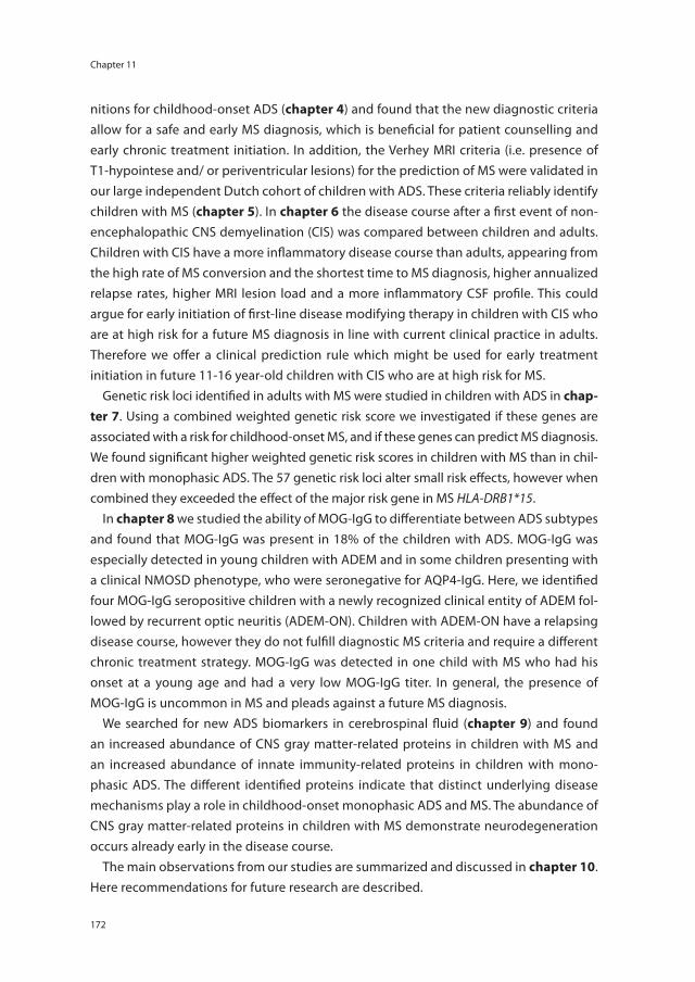
Chapter 11
172
nitions for childhood-onset ADS (chapter 4) and found that the new diagnostic criteria allow for a safe and early MS diagnosis, which is beneficial for patient counselling and early chronic treatment initiation. In addition, the Verhey MRI criteria (i.e. presence of T1-hypointese and/ or periventricular lesions) for the prediction of MS were validated in our large independent Dutch cohort of children with ADS. These criteria reliably identify children with MS (chapter 5). In chapter 6 the disease course after a first event of non-encephalopathic CNS demyelination (CIS) was compared between children and adults. Children with CIS have a more inflammatory disease course than adults, appearing from the high rate of MS conversion and the shortest time to MS diagnosis, higher annualized relapse rates, higher MRI lesion load and a more inflammatory CSF profile. This could argue for early initiation of first-line disease modifying therapy in children with CIS who are at high risk for a future MS diagnosis in line with current clinical practice in adults. Therefore we offer a clinical prediction rule which might be used for early treatment initiation in future 11-16 year-old children with CIS who are at high risk for MS.
Genetic risk loci identified in adults with MS were studied in children with ADS in chap-ter 7. Using a combined weighted genetic risk score we investigated if these genes are associated with a risk for childhood-onset MS, and if these genes can predict MS diagnosis. We found significant higher weighted genetic risk scores in children with MS than in chil-dren with monophasic ADS. The 57 genetic risk loci alter small risk effects, however when combined they exceeded the effect of the major risk gene in MS HLA-DRB1*15.
In chapter 8 we studied the ability of MOG-IgG to differentiate between ADS subtypes and found that MOG-IgG was present in 18% of the children with ADS. MOG-IgG was especially detected in young children with ADEM and in some children presenting with a clinical NMOSD phenotype, who were seronegative for AQP4-IgG. Here, we identified four MOG-IgG seropositive children with a newly recognized clinical entity of ADEM fol-lowed by recurrent optic neuritis (ADEM-ON). Children with ADEM-ON have a relapsing disease course, however they do not fulfill diagnostic MS criteria and require a different chronic treatment strategy. MOG-IgG was detected in one child with MS who had his onset at a young age and had a very low MOG-IgG titer. In general, the presence of MOG-IgG is uncommon in MS and pleads against a future MS diagnosis.
We searched for new ADS biomarkers in cerebrospinal fluid (chapter 9) and found an increased abundance of CNS gray matter-related proteins in children with MS and an increased abundance of innate immunity-related proteins in children with mono-phasic ADS. The different identified proteins indicate that distinct underlying disease mechanisms play a role in childhood-onset monophasic ADS and MS. The abundance of CNS gray matter-related proteins in children with MS demonstrate neurodegeneration occurs already early in the disease course.
The main observations from our studies are summarized and discussed in chapter 10. Here recommendations for future research are described.
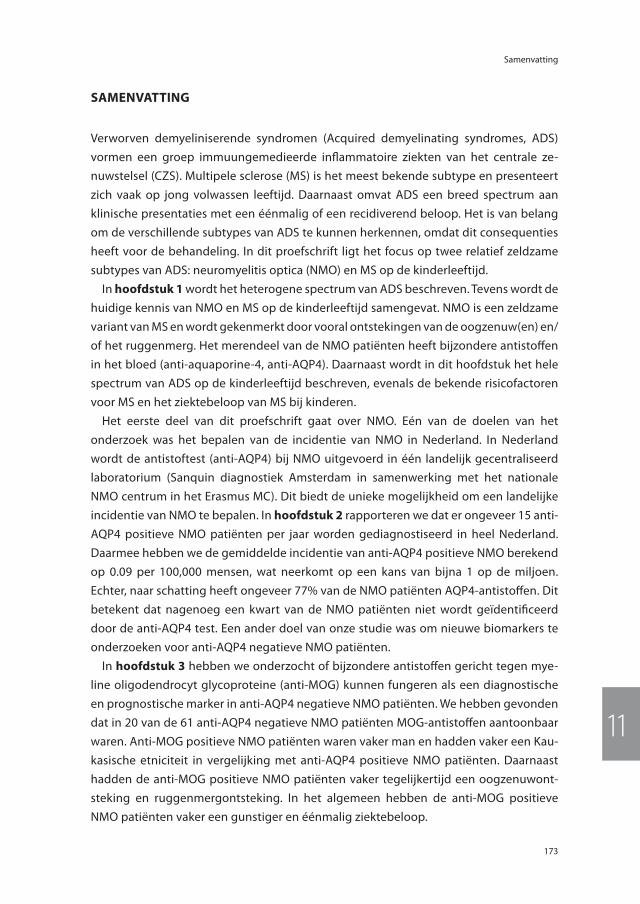
173
Samenvatting
11
sAmenVAtting
Verworven demyeliniserende syndromen (Acquired demyelinating syndromes, ADS) vormen een groep immuungemedieerde inflammatoire ziekten van het centrale ze-nuwstelsel (CZS). Multipele sclerose (MS) is het meest bekende subtype en presenteert zich vaak op jong volwassen leeftijd. Daarnaast omvat ADS een breed spectrum aan klinische presentaties met een éénmalig of een recidiverend beloop. Het is van belang om de verschillende subtypes van ADS te kunnen herkennen, omdat dit consequenties heeft voor de behandeling. In dit proefschrift ligt het focus op twee relatief zeldzame subtypes van ADS: neuromyelitis optica (NMO) en MS op de kinderleeftijd.
In hoofdstuk 1 wordt het heterogene spectrum van ADS beschreven. Tevens wordt de huidige kennis van NMO en MS op de kinderleeftijd samengevat. NMO is een zeldzame variant van MS en wordt gekenmerkt door vooral ontstekingen van de oogzenuw(en) en/of het ruggenmerg. Het merendeel van de NMO patiënten heeft bijzondere antistoffen in het bloed (anti-aquaporine-4, anti-AQP4). Daarnaast wordt in dit hoofdstuk het hele spectrum van ADS op de kinderleeftijd beschreven, evenals de bekende risicofactoren voor MS en het ziektebeloop van MS bij kinderen.
Het eerste deel van dit proefschrift gaat over NMO. Eén van de doelen van het onderzoek was het bepalen van de incidentie van NMO in Nederland. In Nederland wordt de antistoftest (anti-AQP4) bij NMO uitgevoerd in één landelijk gecentraliseerd laboratorium (Sanquin diagnostiek Amsterdam in samenwerking met het nationale NMO centrum in het Erasmus MC). Dit biedt de unieke mogelijkheid om een landelijke incidentie van NMO te bepalen. In hoofdstuk 2 rapporteren we dat er ongeveer 15 anti-AQP4 positieve NMO patiënten per jaar worden gediagnostiseerd in heel Nederland. Daarmee hebben we de gemiddelde incidentie van anti-AQP4 positieve NMO berekend op 0.09 per 100,000 mensen, wat neerkomt op een kans van bijna 1 op de miljoen. Echter, naar schatting heeft ongeveer 77% van de NMO patiënten AQP4-antistoffen. Dit betekent dat nagenoeg een kwart van de NMO patiënten niet wordt geïdentificeerd door de anti-AQP4 test. Een ander doel van onze studie was om nieuwe biomarkers te onderzoeken voor anti-AQP4 negatieve NMO patiënten.
In hoofdstuk 3 hebben we onderzocht of bijzondere antistoffen gericht tegen mye-line oligodendrocyt glycoproteine (anti-MOG) kunnen fungeren als een diagnostische en prognostische marker in anti-AQP4 negatieve NMO patiënten. We hebben gevonden dat in 20 van de 61 anti-AQP4 negatieve NMO patiënten MOG-antistoffen aantoonbaar waren. Anti-MOG positieve NMO patiënten waren vaker man en hadden vaker een Kau-kasische etniciteit in vergelijking met anti-AQP4 positieve NMO patiënten. Daarnaast hadden de anti-MOG positieve NMO patiënten vaker tegelijkertijd een oogzenuwont-steking en ruggenmergontsteking. In het algemeen hebben de anti-MOG positieve NMO patiënten vaker een gunstiger en éénmalig ziektebeloop.
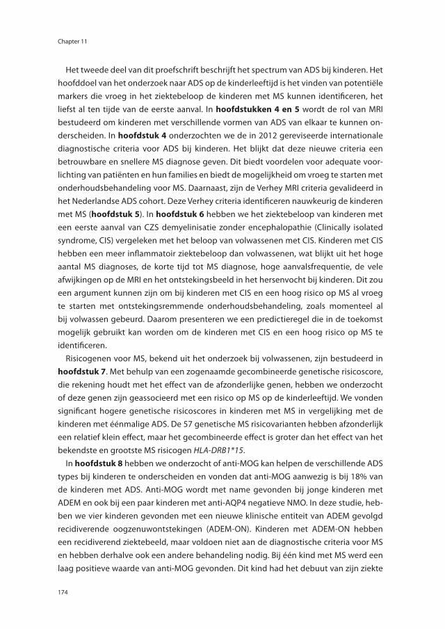
Chapter 11
174
Het tweede deel van dit proefschrift beschrijft het spectrum van ADS bij kinderen. Het hoofddoel van het onderzoek naar ADS op de kinderleeftijd is het vinden van potentiële markers die vroeg in het ziektebeloop de kinderen met MS kunnen identificeren, het liefst al ten tijde van de eerste aanval. In hoofdstukken 4 en 5 wordt de rol van MRI bestudeerd om kinderen met verschillende vormen van ADS van elkaar te kunnen on-derscheiden. In hoofdstuk 4 onderzochten we de in 2012 gereviseerde internationale diagnostische criteria voor ADS bij kinderen. Het blijkt dat deze nieuwe criteria een betrouwbare en snellere MS diagnose geven. Dit biedt voordelen voor adequate voor-lichting van patiënten en hun families en biedt de mogelijkheid om vroeg te starten met onderhoudsbehandeling voor MS. Daarnaast, zijn de Verhey MRI criteria gevalideerd in het Nederlandse ADS cohort. Deze Verhey criteria identificeren nauwkeurig de kinderen met MS (hoofdstuk 5). In hoofdstuk 6 hebben we het ziektebeloop van kinderen met een eerste aanval van CZS demyelinisatie zonder encephalopathie (Clinically isolated syndrome, CIS) vergeleken met het beloop van volwassenen met CIS. Kinderen met CIS hebben een meer inflammatoir ziektebeloop dan volwassenen, wat blijkt uit het hoge aantal MS diagnoses, de korte tijd tot MS diagnose, hoge aanvalsfrequentie, de vele afwijkingen op de MRI en het ontstekingsbeeld in het hersenvocht bij kinderen. Dit zou een argument kunnen zijn om bij kinderen met CIS en een hoog risico op MS al vroeg te starten met ontstekingsremmende onderhoudsbehandeling, zoals momenteel al bij volwassen gebeurd. Daarom presenteren we een predictieregel die in de toekomst mogelijk gebruikt kan worden om de kinderen met CIS en een hoog risico op MS te identificeren.
Risicogenen voor MS, bekend uit het onderzoek bij volwassenen, zijn bestudeerd in hoofdstuk 7. Met behulp van een zogenaamde gecombineerde genetische risicoscore, die rekening houdt met het effect van de afzonderlijke genen, hebben we onderzocht of deze genen zijn geassocieerd met een risico op MS op de kinderleeftijd. We vonden significant hogere genetische risicoscores in kinderen met MS in vergelijking met de kinderen met éénmalige ADS. De 57 genetische MS risicovarianten hebben afzonderlijk een relatief klein effect, maar het gecombineerde effect is groter dan het effect van het bekendste en grootste MS risicogen HLA-DRB1*15.
In hoofdstuk 8 hebben we onderzocht of anti-MOG kan helpen de verschillende ADS types bij kinderen te onderscheiden en vonden dat anti-MOG aanwezig is bij 18% van de kinderen met ADS. Anti-MOG wordt met name gevonden bij jonge kinderen met ADEM en ook bij een paar kinderen met anti-AQP4 negatieve NMO. In deze studie, heb-ben we vier kinderen gevonden met een nieuwe klinische entiteit van ADEM gevolgd recidiverende oogzenuwontstekingen (ADEM-ON). Kinderen met ADEM-ON hebben een recidiverend ziektebeeld, maar voldoen niet aan de diagnostische criteria voor MS en hebben derhalve ook een andere behandeling nodig. Bij één kind met MS werd een laag positieve waarde van anti-MOG gevonden. Dit kind had het debuut van zijn ziekte
175
Samenvatting
11
op zeer jonge leeftijd. In het algemeen, komt anti-MOG niet voor bij MS en pleit de aanwezigheid van anti-MOG tegen een toekomstige diagnose MS.
We hebben gezocht naar nieuwe biomarkers in het hersenvocht van kinderen met MS (hoofdstuk 9) en vonden een overschot van eiwitten gerelateerd aan de grijze stof van het CZS in het hersenvocht van kinderen met MS. In het hersenvocht van kinderen met een éénmalige vorm van ADS hebben we een overschot gevonden van eiwitten die gerelateerd zijn aan het aangeboren afweersysteem. Deze verschillende geïdenti-ficeerde eiwitten suggereren dat verschillende ziektemechanismen een rol spelen bij éénmalige ADS en MS op de kinderleeftijd. Het overschot van eiwitten gerelateerd aan de CZS grijze stof in het hersenvocht laat zien dat neurodegeneratie al vroeg in het ziektebeloop optreedt.
De belangrijkste bevindingen uit onze studies worden samengevat en bediscussieerd in hoofdstuk 10. Hier worden ook aanbevelingen gedaan voor toekomstig onderzoek.


2016
12
OCT
Chapter 12
Epilogue
Dankwoord
Authors and Affiliations
About the author
Publications
PhD portfolio
Abbreviations

179
Dankwoord
12
DAnkwoorD
Eindelijk is het zover en ligt het resultaat van 4 jaar promotieonderzoek voor u. Dit proefschrift had nooit tot stand kunnen komen zonder de samenwerking met, en de hulp van velen! Daarom wil ik hier graag iedereen die een bijdrage heeft geleverd aan het proefschrift hartelijk bedanken.
Op de eerste plaats wil ik graag alle kinderen en jongeren die meedoen aan de kinder ADS studie bedanken. Vele van jullie heb ik op de polikliniek in het Sophia jaren gevolgd en zien opgroeien van tieners tot jongvolwassenen, waarbij jullie tegenwoordig je verkering meenemen naar het spreekuur in plaats van pa of ma. Het heeft me enorm gemotiveerd om te zien wat een doorzetters jullie zijn! Dank ook aan alle ouders en familie die regelmatig mee naar Rotterdam zijn afgereisd. Daarnaast wil ik ook alle NMO patiënten bedanken voor hun bereidheid om deel te nemen aan onderzoek. Velen van u ken ik persoonlijk en heb ik regelmatig gezien op de polikliniek. Ik hoop van harte dat de internationale samenwerking op gebied van ADS op de kinderleeftijd en NMO gaat leiden tot verdere doorbraken in de kennis en de behandeling van deze zeldzame aandoeningen.
Daarnaast wil ik graag alle verwijzers, kinderartsen en (kinder)neurologen bedanken. Zonder u was het niet mogelijk geweest om twee relatief grote cohorten te verzamelen van kinderen met ADS en patiënten met NMO.
Mijn promotor, Prof.dr. R.Q. Hintzen, beste Rogier, bedankt voor de kans die je me hebt gegeven om dit patiëntgebonden onderzoek te doen. Ik bewonder je scherpe blik en de manier waarop je kansen creëert voor samenwerking met andere onderzoeksgroepen. Ik heb erg veel van je geleerd op het gebied van wetenschappelijk onderzoek, maar ook op gebied van patiëntenzorg in het expertisecentrum voor NMO. Dank voor alle kansen en het vertrouwen dat je me hebt gegeven om dit project tot een goed einde te brengen.
Mijn copromotor, Dr. R.F. Neuteboom, Rinze, dank voor je begeleiding de afgelopen jaren met name in het wetenschappelijk onderzoek, maar ook zeker op de polikliniek in het Sophia heb ik veel van je geleerd. Als een coach heb je me door het promotietraject geleid en zo hoop ik in de toekomst ook nog veel van je te mogen leren.
De kleine promotiecommissie. Dr. C.E. Catsman-Berrevoets, beste Coriene, het enthou-siasme voor wetenschappelijk onderzoek is eigenlijk bij jou begonnen met het keuzeon-derzoek naar Niet Aangeboren Hersenletsel bij kinderen en jongeren. Toen kwam ik er-achter dat het doen van onderzoek en het presenteren van eigen onderzoeksresultaten, destijds bij de NVKN in Madurodam, heel erg leuk is. Ik ben je heel dankbaar dat je me hebt aangedragen toen er een opvolgster nodig was voor het kinder ADS onderzoek. Dr. Annemarie van Rossum, hartelijk bedankt voor uw tijd en voor het beoordelen van het manuscript, ik kijk uit naar de gedachtewisseling tijdens de verdediging. Prof.dr. Michèl
Chapter 12
180
Willemsen, dank u wel voor het beoordelen van het manuscript, en voor uw betrokken-heid bij de nationale kinder ADS studie en het kritisch meelezen van de validatiestudie van de 2012 IPMSSG criteria in het bijzonder.
De overige leden van de commissie, Prof.dr. Pieter van Doorn, Prof.dr. Aniki Rothova en Dr. Dörte Hamann wil ik graag bedanken voor hun bereidheid plaats te willen nemen in de promotiecommissie. Beste Dörte, wat ben ik blij dat je in de commissie zit. Bedankt voor het altijd warme ontvangst, en de leerzame en vooral gezellige dagen bij Sanquin Diagnostiek in Amsterdam.
Alle collega’s van het multidisciplinaire kinder MS centrum: Sarita, Femke, Yannick, Jetty, Karlijn, Leontien, Jeroen, Roelie, Petra, Adri, Marjolein, Margreet (2x) en Anke. Wat is het fijn om in zo’n super team samen te werken. Dit is echt patiënt prominent! Sarita, we zijn ongeveer tegelijkertijd in het MS-centrum begonnen en hebben samen de transitiepoli opgezet. Dank voor de fijne samenwerking, je bent een topper die altijd voor de patiënten klaar staat! Heel veel succes met de volgende stap in jouw carrière de Master Advanced Nursing Practice.
Alle collega’s, zowel oud als nieuw, van het MS centrum ErasMS: Dorine, Josje, Immy, Nagmeh, Tessel, Julia, Roos, Yu Yi, Hanne, Yvonne, Leonieke, Beatrijs en Els. Heel veel dank voor de fijne samenwerking en alle gezelligheid! Immy, mijn voorgangster, je bent echt mijn steun en toeverlaat in het onderzoek. Je bent altijd bereid om kritisch mee te denken en daar werden de stukken altijd alleen maar beter van. Wat was het fijn om uit jouw ervaringen te putten en te horen ‘het hoort er allemaal bij’, daarna kon ik altijd weer meer een positieve blik verder. Bedankt voor al je hulp! Ik ben heel blij dat je als mijn paranimf achter me staat. Dorine, als een soort van manager van het MS centrum heb je het superdruk, toch kun je altijd bij jou terecht. Je bent een voorbeeld hoe ik graag neuroloog wil worden. Julia, dank je wel voor de gezellige onderzoekstijd. Ik ben trots op het genetica artikel waar we samen aan hebben gewerkt. Yu Yi, je begon als geneeskunde student met een onderzoeksproject naar de ADEM MRIs. Wat fijn dat je bent teruggekomen en dat we (in sneltreinvaart) hebben samengewerkt aan het ADS onderzoek. Heel veel succes met alle spannende nieuwe projecten waar je nu mee bezig bent! Roos, hardloopmaatje tijdens congressen, hopelijk mogen we binnenkort eens een fles bubbels opentrekken!
Alle coauteurs van de artikelen ontzettend bedankt, Dr. Vaibhav Singh en Dr. Maartje Boon in het bijzonder. Speciale dank gaat uit naar de coauteurs uit Canada. Leonard Verhey, dank je wel voor de fijne samenwerking! Ik heb van je geleerd om goed naar de MRI te kijken en heb leuke herinneringen aan de tijd dat je in Nederland was. Dr. Naila Makhani, Prof. Brenda Banwell and Prof. Amit Bar-Or, thank you very much for your col-laboration with the genetic study and the great result we have accomplished together. Naila, it was a pleasure to welcome you in Rotterdam.
181
Dankwoord
12
Alle onderzoekcollega’s van de 22ste en natuurlijk kamergenoten Bianca, Esther, Ca-rina, Christine, Laurike, Daniëlle, Marienke, Joyce, Agnes, het is niet altijd bevorderlijk voor het schrijven om met zoveel dames op één kamer te werken, maar het is in ieder geval wel altijd heel gezellig.
Dank ook aan alle collega’s, specialisten en arts-assistenten van de afdeling neurolo-gie. Ik kijk uit naar de verdere opleidingstijd. Stephan, handig dat je me voorging, dank voor je praktische promotietips. Jacqueline, dank je wel voor je praktische hulp en het prikken van de datum, Yes!
Ook wil ik graag alle collega’s van de laboratoria immunologie, virologie en genetische epidemiologie bedanken. Marié-Jose, Annet, Marvin, Gijs, Malou, Jamie, Lisa, heel veel tijd heb ik niet op het lab doorgebracht. Daarbuiten zagen we elkaar regelmatig, bij de MS onderzoeksdagen, kinder MS dagen, journal clubs of sportieve events, dank jullie wel voor de gezellige tijd.
En tot slot de belangrijkste mensen in mijn leven. Lieve (schoon)familie en vrienden, zo hèhè, het is nu eindelijk af! Bedankt dat jullie zo ontzettend met me mee hebben geleefd en voor alle afleiding die jullie hebben geboden. Het was heel fijn dat jullie altijd interesse hadden in mijn onderzoek. Speciale dank gaat uit naar mijn paranimf en vriendin sinds de collegebanken in Rotterdam, Chantal. Inmiddels zijn we een paar verhuizingen verder, maar gelukkig lukt het ons nog steeds om elkaar regelmatig te zien. Wat gaaf dat je vandaag naast me staat!
Lieve pap en mam, bedankt dat jullie altijd voor ons klaar staan en alle mijlpalen zo enthousiast meevieren. Van jullie leerde ik om door te zetten en ervoor te gaan, maar vooral ook om te genieten van het leven. Wat een goed gevoel dat jullie onvoorwaarde-lijke trots zijn, wat ik ook doe.
Lieve Thom, zonder jou kan ik het me allemaal niet voorstellen. Dank je wel voor al je geduld, relativering, peptalks, alle afleiding en bovenal dat we zo heerlijk kunnen lachen samen. Ik ben hartstikke gek op je <3
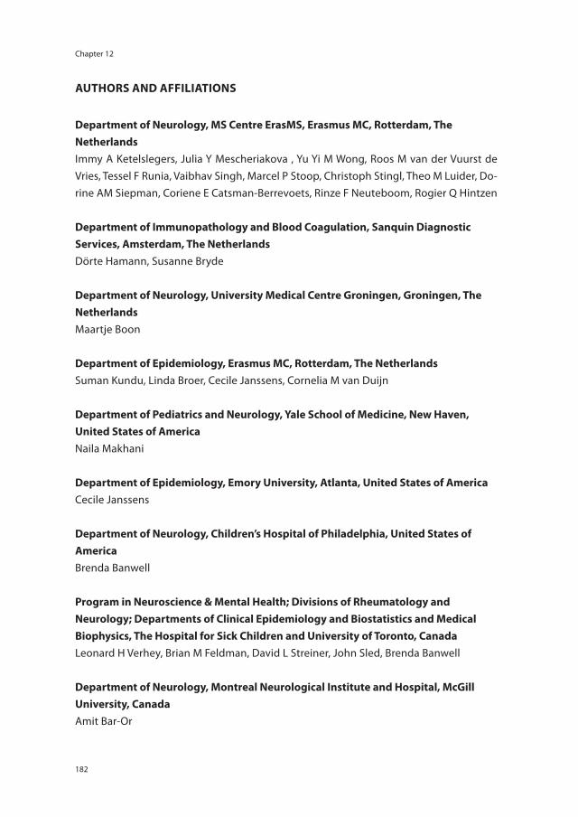
Chapter 12
182
Authors AnD AFFiliAtions
Department of neurology, ms centre erasms, erasmus mc, rotterdam, the netherlandsImmy A Ketelslegers, Julia Y Mescheriakova , Yu Yi M Wong, Roos M van der Vuurst de Vries, Tessel F Runia, Vaibhav Singh, Marcel P Stoop, Christoph Stingl, Theo M Luider, Do-rine AM Siepman, Coriene E Catsman-Berrevoets, Rinze F Neuteboom, Rogier Q Hintzen
Department of immunopathology and blood coagulation, sanquin Diagnostic services, Amsterdam, the netherlandsDörte Hamann, Susanne Bryde
Department of neurology, university medical centre groningen, groningen, the netherlandsMaartje Boon
Department of epidemiology, erasmus mc, rotterdam, the netherlandsSuman Kundu, Linda Broer, Cecile Janssens, Cornelia M van Duijn
Department of Pediatrics and neurology, yale school of medicine, new haven, united states of AmericaNaila Makhani
Department of epidemiology, emory university, Atlanta, united states of AmericaCecile Janssens
Department of neurology, children’s hospital of Philadelphia, united states of AmericaBrenda Banwell
Program in neuroscience & mental health; Divisions of rheumatology and neurology; Departments of clinical epidemiology and biostatistics and medical biophysics, the hospital for sick children and university of toronto, canadaLeonard H Verhey, Brian M Feldman, David L Streiner, John Sled, Brenda Banwell
Department of neurology, montreal neurological institute and hospital, mcgill university, canadaAmit Bar-Or
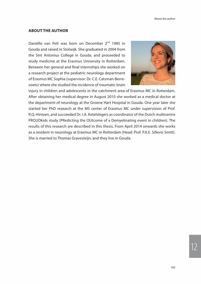
183
About the author
12
About the Author
Daniëlle van Pelt was born on December 2nd 1985 in Gouda and raised in Stolwijk. She graduated in 2004 from the Sint Antonius College in Gouda, and proceeded to study medicine at the Erasmus University in Rotterdam. Between her general and final internships she worked on a research project at the pediatric neurology department of Erasmus MC Sophia (supervisor: Dr. C.E. Catsman-Berre-voets) where she studied the incidence of traumatic brain injury in children and adolescents in the catchment area of Erasmus MC in Rotterdam. After obtaining her medical degree in August 2010 she worked as a medical doctor at the department of neurology at the Groene Hart Hospital in Gouda. One year later she started her PhD research at the MS center of Erasmus MC under supervision of Prof. R.Q. Hintzen, and succeeded Dr. I.A. Ketelslegers as coordinator of the Dutch multicentre PROUDkids study (PRedicting the OUtcome of a Demyelinating event in children). The results of this research are described in this thesis. From April 2014 onwards she works as a resident in neurology at Erasmus MC in Rotterdam (Head: Prof. P.A.E. Sillevis Smitt). She is married to Thomas Gravesteijn, and they live in Gouda.
Chapter 12
184
PublicAtions
TF Runia*, eD van Pelt*, RQ Hintzen. Recent gains in clinical multiple sclerosis research. CNS and Neurological Disorders - Drug Targets 2012;11(5):497-505.
LH Verhey*, eD van Pelt*, IA Ketelslegers, RF Neuteboom, CE Catsman-Berrevoets, BM Feldman, DL Streiner, JG Sled, RQ Hintzen, B Banwell. Validation of MRI predictors of multiple sclerosis diagnosis in children with acute CNS demyelination. Multiple Sclerosis and Related Disorders 2013;2(3):193-199.
RQ Hintzen, eD van Pelt. Paediatric MS is the same disease as adult MS: yes. Multiple Sclerosis Journal 2013;19(10):1257-1258.
eD van Pelt*, JY Mescheriakova*, N Makhani, IA Ketelslegers, RF Neuteboom, S Kundu, L Broer, C Janssens, CE Catsman-Berrevoets, CM van Duijn, B Banwell, A Bar-Or, RQ Hintzen. Risk genes associated with pediatric-onset MS but not with monophasic acquired CNS demyelination. Neurology 2013;81(23):1996-2001.
eD van Pelt, RF Neuteboom, IA Ketelslegers, M Boon, CE Catsman-Berrevoets, RQ Hint-zen; Dutch Study Group for Paediatric MS. Application of the 2012 revised diagnostic definitions for paediatric multiple sclerosis and immune-mediated central nervous system demyelination disorders. Journal of Neurology, Neurosurgery and Psychiatry 2014;85(7):790-794.
IA Ketelslegers*, eD van Pelt*, S Bryde, RF Neuteboom, CE Catsman-Berrevoets, D Hamann, RQ Hintzen. Anti-MOG antibodies plead against MS diagnosis in an Acquired Demyelinating Syndromes cohort. Multiple Sclerosis Journal 2015;21(12):1513-1520.
E Melamed, M Levy, PJ Waters, DK Sato, JL Bennett, GR John, DC Hooper, A Saiz, A Bar-Or, HJ Kim, L Pandit, MI Leite, N Asgari, N Kissani, RQ Hintzen, R Marignier, S Jarius, J Marcelletti, TJ Smith, MR Yeaman, MH Han, O Aktas, M Apiwattanakul, B Banwell, D Bichuetti, S Broadley, P Cabre, T Chitnis, J De Seze, K Fujihara, B Greenberg, K Hellwig, S Iorio , E Klawiter, I Kleiter, M Lana-Peixoto, I Nakashima, K O’Connor K, J Palace, F Paul, N Prayoonwiwat, K Ruprecht, O Stuve, T Tedder, S Tenembaum, JP Garrahan, B Aires, K van Herle, eD van Pelt, P Villoslada, E Waubant, B Weinshenker, D Wingerchuk, J Würfel, S Zamvil. Update on biomarkers in neuromyelitis optica. Neurology: Neuroimmunology and Neuroinflammation 2015;2(4):e134.
185
Publications
12
S Kremer, F Renard, S Achard, MA Lana-Peixoto, J Palace, N Asgari, EC Klawiter, SN Ten-embaum, B Banwell, BM Greenberg, JL Bennett, M Levy, P Villoslada, A Saiz, K Fujihara, KH Chan, S Schippling, F Paul, HJ Kim, J de Seze, JT Wuerfel; Guthy-Jackson Charitable Foundation (GJCF) Neuromyelitis Optica (NMO) International Clinical Consortium and Biorepository, P Cabre, R Marignier, T Tedder, eD van Pelt, S Broadley, T Chitnis, D Wing-erchuk, L Pandit, MI Leite, M Apiwattanakul, I Kleiter, N Prayoonwiwat, M Han, K Hellwig, K van Herle, G John, DC Hooper, I Nakashima I, D Sato, MR Yeaman, E Waubant, S Zamvil, O Stüve, O Aktas, TJ Smith, A Jacob, K O’Connor. Use of Advanced Magnetic Resonance Imaging Techniques in Neuromyelitis Optica Spectrum Disorder. JAMA Neurology 2015;72(7):815-822.
V Singh, eD van Pelt, MP Stoop, C Stingl, IA Ketelslegers, RF Neuteboom, CE Catsman-Berrevoets, TM Luider, RQ Hintzen. Gray matter-related proteins are associated with childhood-onset multiple sclerosis. Neurology: Neuroimmunology and Neuroinflamma-tion 2015;2(5):e155.
eD van Pelt*, YYM Wong*, IA Ketelslegers, D Hamann, RQ Hintzen. Neuromyelitis optica spectrum disorders: comparison of clinical and MRI characteristics of AQP4-IgG versus MOG-IgG seropositive cases in the Netherlands. European Journal of Neurology 2016;23(3):580-587.
JR Scheepe*, YYM Wong*, eD van Pelt, IA Ketelslegers, CE Catsman-Berrevoets, J van den Hoek, RQ Hintzen, RF Neuteboom. Neurogenic lower urinary tract dysfunction in the early disease phase of paediatric multiple sclerosis. Multiple Sclerosis Journal Pub-lished online 20 November 2015. doi:10.1177/1352458515618541
eD van Pelt, YYM Wong, IA Ketelslegers, TAM Siepman, D Hamann, RQ Hintzen. Incidence of AQP4-IgG seropositive neuromyelitis optica spectrum disorders in the Netherlands: about one in a million. Multiple Sclerosis Journal: Experimental, Translational and Clinical Published online 26 January 2016. doi:10.1177/2055217315625652.
P Waters, M Reindl, A Saiz, K Schanda, F Tuller, V Kral, P Nytrova, O Sobek, HH Nielsen, T Barington, ST Lillevang, Z Illes, K Rentzsch, A Berthele, T Berki, L Granieri, A Bertolotto, B Giometto, L Zuliani, D Hamann, eD van Pelt, RQ Hintzen, R Höftberger, C Costa, M Comabella, X Montalban, M Tintoré, A Siva, A Altintas, G Deniz, M Woodhall, J Palace, F Paul, HP Hartung, O Aktas, S Jarius, B Wildemann, C Vedeler, A Ruiz, MI Leite, P Trillen-berg, M Probst, S Saschenbrecker, A Vincent, R Marignier. Multicentre comparison of a diagnostic assay: aquaporin-4 antibodies in neuromyelitis optica. Journal of Neurology, Neurosurgery and Psychiatry 2016;87(9):1005-1015.
Chapter 12
186
YYM Wong, eD van Pelt, IA Ketelslegers, CE Catsman-Berrevoets, RQ Hintzen, RF Neute-boom. Evolution of MRI abnormalities in paediatric acute disseminated encephalomyeli-tis. Accepted for publication in European Journal of Paediatric Neurology, 2016.
R van der Vuurst de Vries*, eD van Pelt*, JY Mescheriakova, YYM Wong, IA Ketelslegers, TAM Siepman, CE Catsman-Berrevoets, RF Neuteboom, RQ Hintzen. Disease course after CIS in children versus adults: a prospective cohort study. Submitted.
other PublicAtions
eD van Pelt, A de Kloet, SR Hilberink, SAM Lambregts, E Peeters, ME Roebroeck, CE Catsman-Berrevoets. The incidence of traumatic brain injury in young people in the catchment area of the University Hospital Rotterdam, The Netherlands. European Journal of Paediatric Neurology 2011;15(6)519-526.
eD van Pelt, JC den Hollander, HJ de Vries, Y van Beek, DC Melles, WI van der Meijden, PJJ van Genderen. A tropical disease characterised by rapidly progressive skin lesions and haemolytic anaemia. The Netherlands Journal of Medicine 2011;69(9):400.
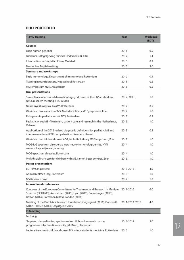
187
PhD Portfolio
12
PhD PortFolio
1. PhD training year workload (ects)
courses
Basic human genetics 2011 0.5
Basiscursus Regelgeving Klinisch Onderzoek (BROK) 2012 1.4
Introduction in GraphPad Prism, MolMed 2015 0.3
Biomedical English writing 2015 3.0
seminars and workshops
Basic immunology, Department of Immunology, Rotterdam 2012 0.5
Training in transition care, Hogeschool Rotterdam 2013 0.5
MS symposium NVN, Amsterdam 2016 0.5
oral presentations
Surveillance of acquired demyelinating syndromes of the CNS in children: NSCK research meeting, TNO Leiden
2012, 2013 1.0
Neuromyelitis optica, ErasMS Rotterdam 2012 0.5
Workshop rare variants of MS, Multidisciplinary MS Symposium, Ede 2012 1.0
Risk genes in pediatric onset ADS, Rotterdam 2013 0.5
Pediatric onset MS - Treatment, patient care and research in the Netherlands, Odense
2013 1.0
Application of the 2012 revised diagnostic definitions for pediatric MS and immune-mediated CNS demyelination disorders, Hasselt
2013 0.5
Workshop on childhood-onset ADS, Multidisciplinary MS Symposium, Ede 2013 1.0
MOG-IgG spectrum disorders: a new neuro-immunologic entity, NVN wetenschappelijke vergadering
2014 1.0
MOG-spectrum diseases, Rotterdam 2014 1.0
Multidisciplinary care for children with MS, samen beter congres, Zeist 2015 1.0
Poster presentations
ECTRIMS (4 posters) 2013-2016 4.0
Annual MolMed Day, Rotterdam 2013 1.0
MS Research days 2012 1.0
international conferences
Congress of the European Committees for Treatment and Research in Multiple Sclerosis (ECTRIMS); Amsterdam (2011), Lyon (2012), Copenhagen (2013), Boston (2014), Barcelona (2015), London (2016)
2011-2016 6.0
Meeting of the Dutch MS Research foundation; Oegstgeest (2011), Doorweth (2012), Hasselt (2013), Oegstgeest 2015
2011-2013, 2015 4.0
2. teaching
Lecturing
‘Acquired demyelinating syndromes in childhood’, research master programme infection & immunity (MolMed), Rotterdam
2012-2014 3.0
Lecture ‘treatment childhood-onset MS’, minor students medicine, Rotterdam 2013 1.0

Chapter 12
188
Supervising/guiding master thesis
Aditi Broos February – July 2013 master student infection & immunity, Rotterdam
2013 2.5
Yu Yi M Wong June – August 2013 medical student Radboud MC, Nijmegen 2013 2.5
3. other
Guiding pre-university students in their final school year with their research paper on NMO (Laura Scholten, Eline Feitsma, Tessa Brouwer, Zaheda Sadoghy) and MS (Charlotte Hoffman).
2011, 2012 1.0
Video colleges pediatric MS, MS en wetenschap 2013 1.0
Co-investigator in clinical MS trials (Fingolimod, Natalizumab, Ocrelizumab, ParadigMS)
2011-2016 2.0
189
Abbreviations
12
AbbreViAtions
ADEM Acute disseminated encephalomyelitisADEM-ON Acute disseminated encephalomyelitis followed by recurrent optic neuritisADS Acquired demyelinating syndromesAID Autoimmune disease AUC Area under the curveAQP4 Aquaporin-4 ARR Annualized relapse rate CBA Cell-based assay CD Cluster of differentiationCDMS Clinically definite multiple sclerosisCI Confidence intervalCIS Clinically isolated syndromeCK Creatine kinase CNS Central nervous systemCRION Chronic relapsing inflammatory optic neuropathyCSF Cerebrospinal fluidDIS Dissemination in spaceDIT Dissemination in timeDMT Disease modifying therapy ΔMFI Delta mean fluorescence intensity DNA Deoxyribonucleic acidEAE Experimental autoimmune encephalomyelitisEDSS Expanded disability severity scale EBV Epstein-Barr virusFACS Fluorescence activated cell sortingFLAIR Fluid-attenuated inversion recoveryGEE Generalized estimating equationsGRIPS Guideline for the reporting of genetic risk prediction studiesGRS Genetic risk scoreGWAS Genomic-wide association studyHEK293 Human embryonic kidney cell lineHLA Human leukocyte antigen HPV Human papilloma virusICU Intensive care unitIgG Immunoglobulin GIgM Immunoglobulin MIL InterleukinIPMSSG International Pediatric Multiple Sclerosis Study Group
Chapter 12
190
IQR Inter quartile rangeLC-MS Nano-liquid chromatography mass spectrometry LD Linkage disequilibrium LETM Longitudinally extensive transverse myelitisLN18 Human malignant glioma cell line LR Likelihood ratioMC Medical centerMOG Myelin oligodendrocyte glycoproteinMRI Magnetic resonance imagingMS Multiple sclerosisN Number of patientsNA Not applicableNS Not significantNMO Neuromyelitis optica NMOSD Neuromyelitis optica spectrum disordersNPV Negative predictive valueNSCK Nederlands Signalerings Centrum Kindergeneeskunde (Netherlands Pediatric Surveillance Unit). OCB Oligoclonal bandsON Optic neuritis OND Other neurological diseasesOR Odds ratio PML Progressive multifocal leukoencephalopathy Poly ADS + Polyfocal ADS with encephalopathyPoly ADS - Polyfocal ADS without encephalopathyPP-MS Primary progressive multiple sclerosisPPV Positive predictive valuePROUDkids PRedicting the OUtcome of a Demyelinating event in children studyQC Quality controlROC Receiver operating characteristicRR Relative riskRR-MS Relapsing-remitting multiple sclerosis SD Standard deviation SLE Systemic lupus erythematosus SNP Single nucleotide polymorphismSP-MS Secondary progressive multiple sclerosisTM Transverse myelitis WBC White blood cell countwGRS Weighted genetic risk score
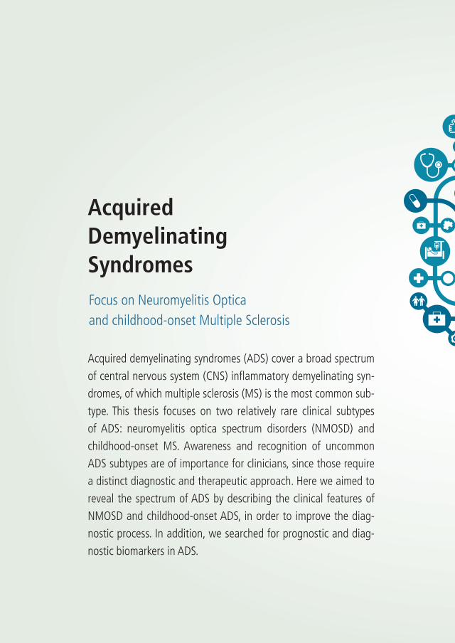
2016
12
OCT
Acquired Demyelinating Syndromes Focus on Neuromyelitis Optica and childhood-onset Multiple Sclerosis
Acquired demyelinating syndromes (ADS) cover a broad spectrum of central nervous system (CNS) inflammatory demyelinating syn-dromes, of which multiple sclerosis (MS) is the most common sub-type. This thesis focuses on two relatively rare clinical subtypes of ADS: neuromyelitis optica spectrum disorders (NMOSD) and childhood-onset MS. Awareness and recognition of uncommon ADS subtypes are of importance for clinicians, since those require a distinct diagnostic and therapeutic approach. Here we aimed to reveal the spectrum of ADS by describing the clinical features of NMOSD and childhood-onset ADS, in order to improve the diag-nostic process. In addition, we searched for prognostic and diag-nostic biomarkers in ADS.