Acls Study Guide September 2011
18
1 IU Health ACLS Study Guide Preparing for your upcoming ACLS Class REVISED SEPTEMBER 2011 ON APRIL 1, 2011—WE BEGAN TEACHING THE 2010 AHA GUIDELINES . WE HIGHLY RECOMMEND REVIEWING THE NEW ALGORYHMS FOUND IN THE 2010 ACLS Textbook available through AHA and other retail outlets. The AHA also requests the following in preparation for your ACLS course: 1. CPR/AED competency (this will be practiced and evaluated in the ACLS course). 2. Understand the 10 cardiac cases found in the ACLS Provider Manual 3. Understand the ACLS algorithms for the cases in the ACLS Provider Manual 4. Complete the online ACLS Pre-course Self-Assessment on ACLS ECGs and pharmacology (ACLS Student Website www.heart.org/eccstudent ) To successfully pass the ACLS course, AHA requires you successfully manage a mega- code. A mega-code is hands-on, dynamic, in real time practice of treating a life-threatening cardiac emergency. The cardiac emergency will include that may include ventricular fibrillation, ventricular tachycardia with or without pulses, asystole, pulseless electrical activity, bradycardia and more. In addition, you’re required to pass a written exam with a score of ≥ 84%. In managing a code or cardiac arrest, you will be required to recognize and correctly identify basic life-threatening rhythms or arrhythmias. You’ll be required to assess the patient’s general condition and effectively treat the patient according to ACLS algorithms and recommendations using the defibrillator and basic cardiac drugs. During this course, you will have to perform roles that may be outside your standard scope of practice during the mega-code practice and testing portion of the class. You’ll be required to be a “team leader” in managing the code, requesting defibrillation, synchronized cardioversion, CPR, and the correct cardiac drug and dosage when indicated according to ACLS guidelines. In managing the mega-code, the team leader will assign their team members to specific roles and responsibilities including: CPR, respiratory management, use of the defibrillator/monitor, selecting drugs out of the code cart, recorder, etc. Though you’ll have sufficient time to practice running mega-codes and you’ll watch audio-visual programs presented by the AHA, course preparation is highly recommended to make your experience valuable toward your education and your successful completion of the
-
Upload
zarah-jane-rull-nate -
Category
Documents
-
view
67 -
download
0
description
Acls STUDY GUIDE
Transcript of Acls Study Guide September 2011

1
IU Health ACLS Study Guide Preparing for your upcoming ACLS Class
REVISED SEPTEMBER 2011
ON APRIL 1, 2011—WE BEGAN TEACHING THE 2010 AHA GUIDELINES. WE HIGHLY RECOMMEND REVIEWING THE NEW ALGORYHMS FOUND IN THE 2010 ACLS Textbook available through AHA and other retail outlets.
The AHA also requests the following in preparation for your ACLS course: 1. CPR/AED competency (this will be practiced and evaluated in the ACLS course). 2. Understand the 10 cardiac cases found in the ACLS Provider Manual 3. Understand the ACLS algorithms for the cases in the ACLS Provider Manual 4. Complete the online ACLS Pre-course Self-Assessment on ACLS ECGs and
pharmacology (ACLS Student Website www.heart.org/eccstudent )
To successfully pass the ACLS course, AHA requires you successfully manage a mega-
code. A mega-code is hands-on, dynamic, in real time practice of treating a life-threatening
cardiac emergency. The cardiac emergency will include that may include ventricular fibrillation,
ventricular tachycardia with or without pulses, asystole, pulseless electrical activity, bradycardia
and more. In addition, you’re required to pass a written exam with a score of ≥ 84%.
In managing a code or cardiac arrest, you will be required to recognize and correctly
identify basic life-threatening rhythms or arrhythmias. You’ll be required to assess the patient’s
general condition and effectively treat the patient according to ACLS algorithms and
recommendations using the defibrillator and basic cardiac drugs.
During this course, you will have to perform roles that may be outside your standard
scope of practice during the mega-code practice and testing portion of the class. You’ll be
required to be a “team leader” in managing the code, requesting defibrillation, synchronized
cardioversion, CPR, and the correct cardiac drug and dosage when indicated according to ACLS
guidelines. In managing the mega-code, the team leader will assign their team members to
specific roles and responsibilities including: CPR, respiratory management, use of the
defibrillator/monitor, selecting drugs out of the code cart, recorder, etc.
Though you’ll have sufficient time to practice running mega-codes and you’ll watch
audio-visual programs presented by the AHA, course preparation is highly recommended to
make your experience valuable toward your education and your successful completion of the

2
class. You’re encouraged to purchase or borrow an ACLS Provider Manual that offers direction
to a web site and a pre-course assessment. The online pre-course assessment has shown to be
of high value by those who have attended our classes. In addition, the provider manual will
provide you with ACLS algorhythm pocket guides that can be used during your mega-code
testing as a reference. The textbook itself is also a great resource in preparation for your course
and the written exam.
To offer further guidelines in your preparation for the ACLS course, we at I.U. Health
Emergency Response Training Institute /Emergency Cardiac Care have provided you with the very basic rhythms and arrhythmias covered in the ACLS course. This is not to be considered a replacement for the ACLS Student Manual, the online pre-course assessment and other resources offered by the AHA. Again—we highly encourage you to get a provider manual and access the website found in the manual for study.
BLS CPR BLS CPR changed in 2010. The primary change is from the “ABC” format to “CAB.” After establishing unresponsiveness and calling for a code, check for a pulse in 5 to 10 seconds then begin compressions immediately, within 10 seconds of arriving at the patient’s side. After thirty compressions, give your first two breaths. The 30:2 ratio then will continue. “Look, listen and feel” for breathing has been removed from the new guidelines. One other significant change in 2010, the use of an AED is now indicated for infants. Here are the basic steps in BLS:
1. Check for responsiveness 2. Call for help and an AED 3. Check for pulse and simultaneously scan the chest for breathing. 4. Begin the thirty compressions (within 10 seconds of arriving at the
patient) 5. Give two breaths—continue 30:2 ratio. 6. Apply the AED as soon as it arrives
NOTES: ____________________________________________________________________________________________________________________________________________________________________________________________________________________________________________________________________________________________________________________________________________________________________________________________________________________________________________________________________________________

3
Sinus Bradycardia (sinus rhythm with a rate less than 50)
Is your patient stable or unstable?
Stable- monitor closely
Unstable/ Symptomatic – this patient is showing signs of poor perfusion (their heart rate is not fast enough to deliver an adequate volume of blood to the body and requires treatment/ intervention) for example: hypotensive feels faint, decreased or altered mental status, cool or clammy/diaphoretic.
Administer 0.5mg Atropine. If Atropine is ineffective you now have three options:
administer Dopamine 2-10 mcg/kg/minute OR
Administer Epinephrine infusion: 2 – 10 mcg per minute
Prepare and provide external transcutaneous pacing.
Note: if the patient is severely hypotensive, transcutaneous pacing may or may not capture and not be able to produce an adequate profusing pressure. The epinephrine drip or dopamine drip with transvenous pacing with expert consultation should now be considered.
NOTES: ____________________________________________________________________________________________________________________________________________________________________________________________________________________________________________________________________________________________________________________________________________________________________________________________________________________________________________________________________________________

4
Heart Blocks
Is your patient stable or unstable?
Stable- monitor closely, seek expert consultation
Unstable/ Symptomatic – this patient is showing signs of poor perfusion
(their heart rate is not fast enough to deliver an adequate volume of blood to
the body and requires treatment/ intervention) for example: low B/P, feels
faint, decreased or altered mental status, cool or clammy/diaphoretic
Administer 0.5mg Atropine. But don’t rely on atropine in Mobitz
type II, second-degree block or third-degree block with a new wide
QRS complex.
For an unstable Mobitz type II second-degree or third-degree heart
block patient be prepared for transcutaneous pacing
NOTES: ____________________________________________________________________________________________________________________________________________________________________________________________________________________________________________________________________________________________________________________________________________________________________________________________________________________________________________________________________________________

5
Supraventricular Tachycardia- SVT (SUSTAINED rapid
narrow complex tachycardia with a rate greater than 150)
Is your patient stable or unstable?
Stable- Attempt vagal maneuvers like bearing down or a good hard cough. If you’re a physician, try carotid massage (I.U. Health policy states only physicians can perform carotid massage).
If vagal maneuvers aren’t successful in slowing their heart rate, administer 6 mg. of Adenosine. What is unique about administering Adenosine is that it is a fast-push and fast-acting drug. It may cause a second or two of asystole. Therefore, it is only given with a physician order and a physician at the bedside per I.U. Health policy. Patient also must be monitored.
If the first dose of 6 mg isn’t successful, ACLS allow you to repeat the Adenosine providing 12 mg the second and third time if needed.
Unstable/ Symptomatic – this patient is showing signs of poor perfusion (low B/P, feels faint, decreased or altered mental status, cool or clammy/diaphoretic) it may be due to their heart rate is too fast to deliver an adequate volume of blood to the body and requires rapid treatment/ intervention. Provide synchronized cardioversion of 50 – 100 joules.
NOTES: ____________________________________________________________________________________________________________________________________________________________________________________________________________________________________________________________________________________________________________________________________________________________________________________________________________________________________________________________________________________

6
Monomorphic Ventricular Tachycardia- VT (SUSTAINED)
>30 seconds rapid wide complex tachycardia)
First and important question: Does your patient have a pulse with this rhythm?
If yes, he/she does have a pulse, then is you patient stable? o With a PULSE and UNSTABLE (low b/p, weak, clammy, cold, ashen,
unresponsive or lethargic)
Deliver immediate synchronized cardioversion at 100J.
Evaluate the rhythm post cardioversion and consider a second attempt at a higher energy level if needed.
With a PULSE and STABLE, treat with adenosine only if regular and monomorphic.
Consider antiarrythmic infusion i.e. Procainamide or Amiodarone IV drip.
NOTES: ____________________________________________________________________________________________________________________________________________________________________________________________________________________________________________________________________________________________________________________________________________________________________________________________________________________________________________________________________________________
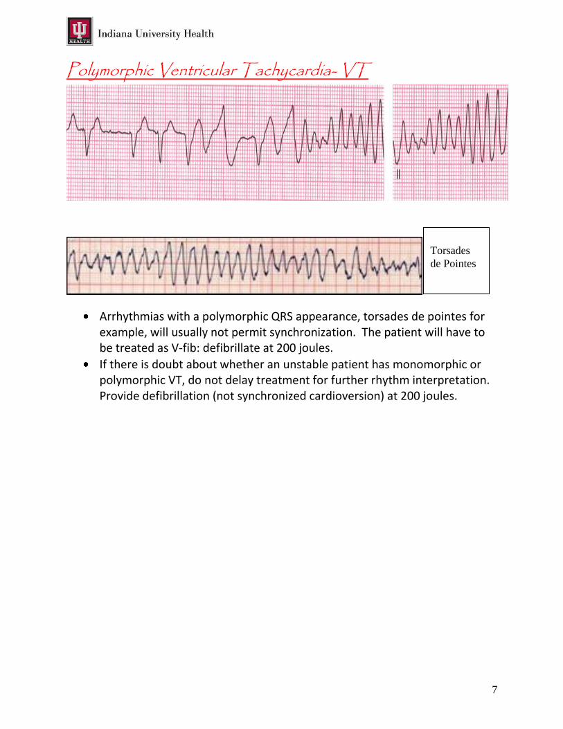
7
Polymorphic Ventricular Tachycardia- VT
Arrhythmias with a polymorphic QRS appearance, torsades de pointes for example, will usually not permit synchronization. The patient will have to be treated as V-fib: defibrillate at 200 joules.
If there is doubt about whether an unstable patient has monomorphic or polymorphic VT, do not delay treatment for further rhythm interpretation. Provide defibrillation (not synchronized cardioversion) at 200 joules.
Torsades
de Pointes

8
VENTRICULAR FIBRILLATION-
VFib is a chaotic and disorganized rhythm that generates absolutely no perfusion! The heart is quivering as it is dying and requires IMMEDIATE defibrillation…do not delay! The sooner the heart in VF can be defibrillated, the higher the chances of successfully converting to an organized rhythm.
Quickly….
Rapidly assemble your team
Begin chest compressions
Apply defibrillator (hands-free) pads to patient, clear your co-workers from touching the patient or the bed and deliver 200J shock as quickly as you can.
Immediately after the shock is delivered, resume compressions and bag mask ventilations. (CPR should not stop for more than 10 seconds.)
You will continue CPR for 2 minutes (make sure your timer/recorder is tracking this for you) and prepare your first drug – your first medication will be Epinephrine 1mg, but do not administer it yet. This is also a good time to get IV or IO access if not already established.
At 2 minutes clear to reevaluate your rhythm- if VF persists charge and defibrillate a second time at 200J, clear the patient and deliver the shock. Immediately resume compressions (make sure to rotate compressor and person bagging every 2 minutes for optimal compressions- you will get tired quickly)
During this 2 minute cycle administer the Epinephrine an prepare the second medication- Amiodarone 300mg.

9
Again at the 2 minute mark clear to reevaluate your rhythm- if VF persists charge and defibrillate at 200J, again resume compressions immediately after the shock is delivered.
During this 2 minute cycle administer the Amiodarone 300mg and prepare your next dose of Epinephrine, 1 mg.
These 2 minute cycles of rhythm check- shock if indicated- CPR- administer med will continue as long as VF or pulseless VT persists.
NOTES: ____________________________________________________________________________________________________________________________________________________________________________________________________________________________________________________________________________________________________________________________________________________________________________________________________________________________________________________________________________________
Asystole- Ventricular Standstill – Absolutely no electrical or
mechanical activity!
Asystole requires immediate intervention
Begin compressions and airway management, good CPR.
Administer Epinephrine 1mg IVP as soon as it’s available.
1 mg of Epinephrine (1:10,000 used in cardiac arrest) is giving every 3-5 minutes and there is no maximum dose.
A critical step to restoring a perfusing rhythm is to quickly identify one of the underlying/reversible causes that most frequently lead to asystole. The most common are known as the H’s & T’s! As a team leader you should run through the list for consideration.
H’s & T’s are as follows: Hypoxia, Hypothermia, Hypo/Hyperkalemia, Hydrogen Ion (acidosis), Hypovolemia, Toxins, Tension Pneumothorax, Tamponade, Thrombus (coronary or pulmonary)

10
Pulseless Electrical Activity (PEA) – Electrical Activity without
mechanical contractility, you have an organized rhythm, but the heart isn’t pumping .
What do you do if you are in a code and you find an organized rhythm on the monitor?
CHECK FOR A PULSE! If you have a rhythm and no pulse you are in PEA
Begin compressions and airway management / good CPR at a ratio of 30:2.
Administer Epinephrine 1:10,000 1mg IVP
A critical step to restoring a perfusing rhythm is to quickly identify one of the underlying/reversible causes that most frequently lead to PEA. The most common are known as the H’s & T’s! As a team leader you should run through the list for consideration.
H’s & T’s are as follows: Hypoxia, Hypothermia, Hypo/Hyperkalemia, Hydrogen Ion (acidosis), Hypovolemia, Toxins, Tension Pneumothorax, Tamponade, Thrombus (coronary or pulmonary)
Remember- PEA is not always a Sinus Rhythm and it is not always a slow PEA. It can look like any organized rhythm.
NOTES: ____________________________________________________________________________________________________________________________________________________________________________________________________________________________________________________________________________________________________________________________________________________________________________________________________________________________________________________________________________________
NO PULSE

11
During class you will have opportunity to get hands-on practice with each of these rhythms and their appropriate algorithms. If you study the content the application in the simulation labs will pull it all together for you and your instructor will help tie it all together and answer any additional questions you may have. This content is not intended to replace your ACLS provider manual- you are still highly encouraged to have the manual or borrow one provided by your department in preparation for and while attending class.

12
ACLS Pharmacology
Drugs for Pulseless Arrest VF/VT
Epinephrine 1:10,000: 1mg IV/IO followed by 20 ml flush; repeat throughout code every
3-5 minutes. There is no maximum dose. When is Epi administered? Asystole, PEA, VF, Pulseless VT….in other words….when there isn’t a pulse, this is the first drug given. .
Vasopressin: 40 units can replace the first or second dose of Epinephrine.
Amiodarone: 300mg IV/IO push. Second dose (if needed) 150 mg IV/IO push.
-Antiarrhythmic medication that has been shown to increase chance of return of pulse after defibrillation of a shockable rhythm, VF or pulseless VT. NOTES: ____________________________________________________________________________________________________________________________________________________________________________________________________________________________________________________________________________________________________________________________________________________________________________________________________________________________________________________________________________________
Drugs for Pulseless Arrest PEA or Asystole
Epinephrine: 1mg IV/IO followed by 20ml flush; repeat throughout code every 3-5 minutes.
There is no maximum dose. NOTES: ____________________________________________________________________________________________________________________________________________________________________________________________________________________________________________________________________________________________________________________________________________________________________________________________________________________________________________________________________________________
13
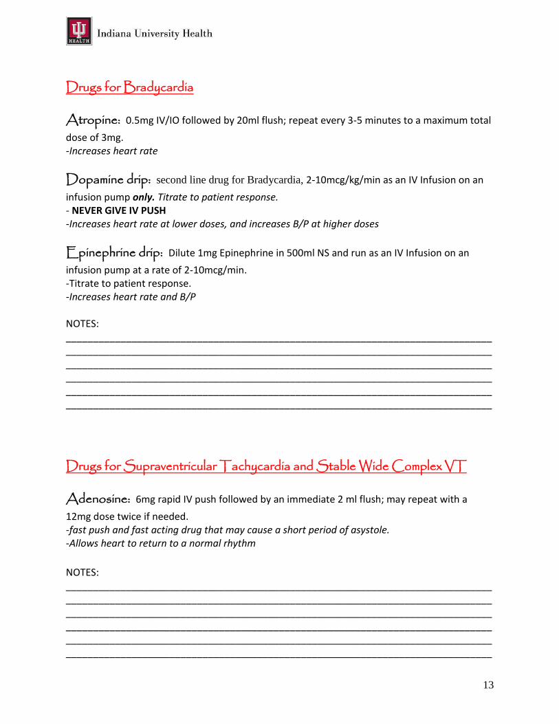
Drugs for Bradycardia
Atropine: 0.5mg IV/IO followed by 20ml flush; repeat every 3-5 minutes to a maximum total
dose of 3mg. -Increases heart rate
Dopamine drip: second line drug for Bradycardia, 2-10mcg/kg/min as an IV Infusion on an
infusion pump only. Titrate to patient response. - NEVER GIVE IV PUSH -Increases heart rate at lower doses, and increases B/P at higher doses
Epinephrine drip: Dilute 1mg Epinephrine in 500ml NS and run as an IV Infusion on an
infusion pump at a rate of 2-10mcg/min. -Titrate to patient response. -Increases heart rate and B/P NOTES: ____________________________________________________________________________________________________________________________________________________________________________________________________________________________________________________________________________________________________________________________________________________________________________________________________________________________________________________________________________________
Drugs for Supraventricular Tachycardia and Stable Wide Complex VT
Adenosine: 6mg rapid IV push followed by an immediate 2 ml flush; may repeat with a
12mg dose twice if needed. -fast push and fast acting drug that may cause a short period of asystole. -Allows heart to return to a normal rhythm
NOTES: ____________________________________________________________________________________________________________________________________________________________________________________________________________________________________________________________________________________________________________________________________________________________________________________________________________________________________________________________________________________

14
Drugs for ACS (Acute Coronary Syndromes) MONA
M = Morphine
O = Oxygen (for oxygen saturation less than 94%)
N = Nitrates
A = Aspirin
It is preferable to do a 12 Lead ECG, if available, prior to the administration of nitroglycerin to verify your patient’s cardiac rhythm status. Morphine: Initial dose is 2 to 4 mg IV over 1 to 5 minutes,, aadminister slowly and only titrate
to effect -May administer to patients with suspected ischemic pain unresponsive to oxygen and nitrates -Before administering morphine, be sure that the patient’s systolic blood pressure is >90 mm Hg and they are not hypovolemic -Remember to reassess and repeat vital signs between doses -At IU Health, Fentanyl may be an alternative pain medication to morphine for the unstable patient (ie, systolic blood pressure <90 mm Hg, hypovolemia, bradycardia, etc.)
Nitroglycerin: 1 spray sublingually every 3-5 minutes up to a total of 3 sprays
-Indications — Standard therapy for all patients with new pain/discomfort suggestive of ACS -Give within minutes of arrival -Nitroglycerin decreases pain in ischemia and is a vasodilator. -Before administering nitroglycerin, be sure that the patient’s systolic blood pressure is >90 mm Hg and they should not have bradycardia or tachycardia. -Do not give nitroglycerin if the patient has had an erectile dysfunction drug within the past week.
Aspirin: 160 to 325 mg given as soon as possible, non-coated baby or adult aspirin may be
used -Indications — Standard therapy for all patients with new pain/discomfort suggestive of ACS -Give within minutes of arrival -Aspirin irreversibly binds to platelets and partially inhibits platelet function. -Before administering aspirin, make sure patient does not have an aspirin allergy, or recent or active GI bleeding. NOTES: ____________________________________________________________________________________________________________________________________________________________________________________________________________________________________________________________________________________________________________________________________________________________________________________________________________________________________________________________________________________

15
How to Calculate the EKG ♥ Rate
In preparation for your ACLS it is not only important for you to be able to identify the rhythms but determine the rate as well. Here is the nitty gritty to help you accomplish this task.
EKG paper is comprised of many boxes: each of the following can be
used in different methods to calculate a strips correct rate.
1 tiny box=0.04 second
1 large box=(5 tiny boxes)0.20 second
6 second strip=30 large boxes
1 minute strip=300 large boxes
1 minute strip=1500 tiny boxes

16
The Countdown Method: This does not require a 3-second or 6-second
strip and can be used easily at the bedside. It is accomplished by memorizing this sequence: 300, 150, 100, 75, 60, 50, 40, and 30. You find a QRS complex that falls close to a heavy line and count large boxes between the QRS complexes using these numbers. See example below. This method only works for regular rhythms.
The 1500 Method: Count the tiny squares between identical points on 2
consecutive QRS complexes. Then divide 1500 by the number of tiny boxes to obtain the heart rate.
The 300 Method: Count the number of large squares between 2 consecutive
QRS complexes…then divide 300 by the number of large boxes to obtain the heart rate.

17
The 3 Second Strip Method: Using a 3 second strip (15 large squares=3
seconds), count the number of QRS complexes and multiply by 20 to obtain the heart rate.
The 6 Second Strip Method: Using a 6 second strip (30 large squares=6
seconds), count the number of QRS complexes and multiply by 10 to obtain the heart rate.

18
References
Advanced Circulatory Systems, Inc., (2008). ResQpod clinical information. Retrieved August 13,
2009, from Advanced Circulatory Systems, Inc. Web site:
http://www.advancedcirculatory.com/newsroom/Clinical_Information.htm
Butler, J., Busti, A., & Gonzales, L. (2007). ECG and pharmacology student workbook. Dallas,
Texas: American Heart Association.
Field, John M. (Ed.). (2006). Advanced cardiovascular life support provider manual. Dallas,
Texas: American Heart Association.
Field, John M. (Ed.). (2008). Advanced cardiac life support resource text. Dallas, Texas:
American Heart Association.
Ralston, M., Hazinski, M, F., Zaritsky, A, L., & Schexnayder, S, M. (Eds.). (2006). Pediatric
advanced life support course guide. Dallas, Texas: American Heart Association.
Ralston, M., Hazinski, M, F., Zaritsky, A, L., & Schexnayder, S, M. (Eds.). (2006). Pediatric
advanced life support provider manual. Dallas, Texas: American Heart Association.
Vidacare, (2008). Vidacare-EZ-IO. Retrieved August 13, 2009, from Vidacare-EZ-IO Home Web
site: http://www.vidacare.com/ez-io/index.html