Acidosis and alkalosis
103
Biochemical basis of Acid Base Balance
-
Upload
nakul-poudel -
Category
Education
-
view
1.006 -
download
1
Transcript of Acidosis and alkalosis
Biochemical basis of Acid Base Balance

Q.How acid base balance is maintained in normal human being?
• Normal body pH=7.4 equivalent to 40 nmole H
con. Per liter. This level is very important for normal biological activity. this level of pH should be maintained for proper functioning of the body. on an average, the pH range may fluctuate from 7.35-7.45.
• During normal metabolic activity, body produces both acid and bases but the acid production is greater than the base production.

• So body is a net acid producer.in a normal adult two types of metabolic acid are produced.
• 1.Volatile acid- 15 mole/day in the form of co2
• 2. Non volatile acid-70 mEq/day in the form of H2so4, HCl, H3Po4.
• These metabolic acid has a major consequences in alter the normal body pH.

• Volatile acids are excreted through lung via pulmonary route.non volatile acids are excreted through kidney via urine.
• Before their excreation respiratory system takes some time and kidney system also takes certain time.

Excreation of volatile acid:
• Co2 produced in cellular level due to metabolic activity diffused into blood.in blood co2 is transported in three form.
• A.in dissolved state=7%• B. in the form of HCO3
-&H+ (H2Co3)=70%• In combination of Hb=23%• From blood CO2 is diffused into alveoli of
lungs and their into the atmosphere.

Transport of carbon dioxide in the blood

Approach
• History - subjective information concerning events, environment, trauma, medications, poisons, toxins
• Physical examination - objective information assessing organ system status and function
• Differentials - potential reasons for presentation• Clinical and laboratory studies - degree of changes
from normal• Compensation - assessment of response to initial
problem

8
pH Review• pH = - log [H+]• H+ is really a proton• Range is from 0 - 14• If [H+] is high, the solution is acidic; pH < 7• If [H+] is low, the solution is basic or alkaline ; pH > 7

9

10

11
• Acids are H+ donors.• Bases are H+ acceptors, or give up OH- in
solution.• Acids and bases can be:
–Strong – dissociate completely in solution • HCl, NaOH
–Weak – dissociate only partially in solution• Lactic acid, carbonic acid

12
The Body and pH• Homeostasis of pH is tightly controlled• Extracellular fluid = 7.4• Blood = 7.35 – 7.45• < 6.8 or > 8.0 death occurs• Acidosis (acidemia) below 7.35• Alkalosis (alkalemia) above 7.45

13
14
Small changes in pH can produce major disturbances
• Most enzymes function only with narrow pH ranges• Acid-base balance can also affect electrolytes (Na+, K+, Cl-)• Can also affect hormones

15
The body produces more acids than bases
• Acids take in with foods• Acids produced by metabolism of lipids and proteins• Cellular metabolism produces CO2.• CO2 + H20 ↔ H2CO3 ↔ H+ + HCO3
-

16
Control of Acids1. Buffer systems
Take up H+ or release H+ as conditions changeBuffer pairs – weak acid and a baseExchange a strong acid or base for a weak oneResults in a much smaller pH change

17
Bicarbonate buffer• Sodium Bicarbonate (NaHCO3) and carbonic acid (H2CO3)• Maintain a 20:1 ratio : HCO3
- : H2CO3
HCl + NaHCO3 ↔ H2CO3 + NaCl
NaOH + H2CO3 ↔ NaHCO3 + H2O

18
Phosphate buffer• Major intracellular buffer• H+ + HPO4
2- ↔ H2PO4-
• OH- + H2PO4- ↔ H2O + H2PO4
2-

19
Protein Buffers
• Includes hemoglobin.• Carboxyl group gives up H+ • Amino Group accepts H+

20
2. Respiratory mechanisms• Exhalation of carbon dioxide• Powerful, but only works with volatile acids• Doesn’t affect fixed acids like lactic acid• CO2 + H20 ↔ H2CO3 ↔ H+ + HCO3
-
• Body pH can be adjusted by changing rate and depth of breathing

21
3. Kidney excretion• Can eliminate large amounts of acid• Can also excrete base• Can conserve and produce bicarb ions• Most effective regulator of pH• If kidneys fail, pH balance fails

22
Rates of correction• Buffers function almost instantaneously• Respiratory mechanisms take several minutes to hours• Renal mechanisms may take several hours to days

23

24

25
Acid-Base Imbalances• pH< 7.35 acidosis• pH > 7.45 alkalosis• The body response to acid-base imbalance is called compensation• May be complete if brought back within normal limits• Partial compensation if range is still outside norms.

26
Compensation• If underlying problem is metabolic, hyperventilation or hypoventilation can help : respiratory compensation.• If problem is respiratory, renal mechanisms can bring about metabolic compensation.

27
Acidosis• Principal effect of acidosis is depression of the
CNS through ↓ in synaptic transmission.• Generalized weakness• Deranged CNS function the greatest threat• Severe acidosis causes
–Disorientation–coma –death

28
Alkalosis• Alkalosis causes over excitability of the central
and peripheral nervous systems.• Numbness• It can cause :
– Nervousness– muscle spasms or tetany – Convulsions – Loss of consciousness– Death

29

30
Respiratory Acidosis• Carbonic acid excess caused by blood levels of CO2 above 45 mm Hg. • Hypercapnia – high levels of pCO2 in blood• Chronic conditions:
– Depression of respiratory center in brain that controls breathing rate – drugs or head trauma– Paralysis of respiratory or chest muscles– Asthma,Pneumonia,Emphysema

31
Respiratory Acidosis• Acute conditons:
– Adult Respiratory Distress Syndrome– Pulmonary edema– Pneumothorax

32
Compensation for Respiratory Acidosis
• Kidneys eliminate hydrogen ion and retain bicarbonate ion.• Mechanism:↓pH→↑H+→H ion+ HCO3
- → H2CO3 → CO2 + H20 →pH backs towards normal.

33
Signs and Symptoms of Respiratory Acidosis
• Breathlessness• Restlessness• Lethargy and disorientation• Tremors, convulsions, coma• Respiratory rate rapid, then gradually
depressed• Skin warm and flushed due to vasodilation
caused by excess CO2

34
Treatment of Respiratory Acidosis• Restore ventilation• IV lactate solution• Treat underlying dysfunction or disease

35

36
Respiratory Alkalosis• Carbonic acid deficit• pCO2 less than 35 mm Hg (hypocapnea)• Most common acid-base imbalance• Primary cause is hyperventilation• Hysteria• Hyperapnoea at high altitude.• Meningitis,enchephalitis.• Hepatic failure

37
Respiratory Alkalosis• Conditions that stimulate respiratory
center:– Oxygen deficiency at high altitudes– Pulmonary disease and Congestive heart
failure – caused by hypoxia – Acute anxiety– Fever, anemia– Early salicylate intoxication– Cirrhosis– Gram-negative sepsis

38
Compensation of Respiratory Alkalosis
• By renal system• Kidneys conserve hydrogen ion• Excrete bicarbonate ion.
39
Treatment of Respiratory Alkalosis• Treat underlying cause• Breathe into a paper bag• IV Chloride containing solution – Cl- ions replace lost bicarbonate ions

40

41
Metabolic Acidosis
• Decreased pH due to HCO3- deficit is called
metabolic acidosis.• Bicarbonate deficit - blood concentrations of
bicarb drop below 22mEq/L• Causes:
– Loss of bicarbonate through diarrhea or renal dysfunction
– DM, Accumulation of acids (lactic acid or ketones)
– Failure of kidneys to excrete H+

• Ingestion of acid• Formation of excessive quantities of
metabolic acid in the body.• Loss of excessive alkali from the body.• Intravenous administration of metabolic
acid.• Poisoning by acidic eg. Acetyl salicylates
(aspirin) and methyl alcohol.

43
Symptoms of Metabolic Acidosis• Headache, lethargy• Nausea, vomiting, diarrhea• Coma• Death

44
Compensation for Metabolic Acidosis
• By respiratory system• Increased ventilation• Renal excretion of hydrogen ions if possible• K+ exchanges with excess H+ in ECF• ( H+ into cells, K+ out of cells)• Mechanism: ↓pH→↑respiration→ ↓ pCO2 to match the lowered HCO3
- →pH backs towards normal.

45
Treatment of Metabolic Acidosis• IV lactate solution

46

47
Metabolic Alkalosis• Bicarbonate excess - concentration in blood is greater than 26 mEq/L• Causes:
– Excess vomiting = loss of stomach acid– Excessive use of alkaline drugs– Certain diuretics– Endocrine disorders– Heavy ingestion of antacids– Severe dehydration

48
Compensation for Metabolic Alkalosis
• Alkalosis most commonly occurs with renal dysfunction, so can’t count on kidneys• Respiratory compensation difficult – hypoventilation limited by hypoxia

49
Symptoms of Metabolic Alkalosis• Respiration slow and shallow• Hyperactive reflexes ; tetany• Often related to depletion of electrolytes• Atrial tachycardia• Dysrhythmias

50
Treatment of Metabolic Alkalosis• Electrolytes to replace those lost• IV chloride containing solution• Treat underlying disorder

51

52
Diagnosis of Acid-Base Imbalances1. Note whether the pH is low (acidosis) or high (alkalosis)2. Decide which value, pCO2 or HCO3
- , is outside the normal range and could be the cause of the problem. If the cause is a change in pCO2, the problem is respiratory. If the cause is HCO3- the problem is metabolic.

Acid-Base BiochemistryPhysiology
2 different processesBicarbonate regeneration (incorrectly reabsorption)Hydrogen ion excretion

Acid-Base Biochemistry
Importance of Renal Bicarbonate Regeneration • Bicarbonate is freely filtered through the glomerulus so
plasma and glomerular filtrate have the same bicarbonate concentration
• At normal GFR approx 4300 mmol of bicarbonate would be filtered in 24 hr
• Without re-generation of bicarbonate the buffering capacity of the body would be depleted causing acidotic state
• In health virtually all the filtered bicarbonate is recovered


Acid-Base Biochemistry
• Renal Bicarbonate Regeneration involves the enzyme carbonate dehydratase (carbonic anhydrase)
• Luminal side of the renal tubular cells impermeable to bicarbonate ions
• Carbonate dehydratase catalyses the formation of CO2 and H2O from carbonic acid (H2CO3) in the renal tubular lumen
• CO2 diffuses across the luminal membrane into the tubular cells

Acid-Base Biochemistry
within the renal tubular cells carbonate dehydratase catalyses the formation of carbonic acid (H2CO3) from CO2 and H2OCarbonic acid then dissociates into H+ and HCO3-
The bicarbonate ions pass into the extracellular fluid and the hydrogen ions are secreted back into the lumen in exchange for sodium ions which pass into the extracellular fluidExchange of sodium and hydrogen ions an active process involving Na+/K+/H+ ATP pumpK+ important in electrolyte disturbances of acid-base

Acid-Base Biochemistry
Regeneration of bicarbonate does not involve net excretion of hydrogen ionsHydrogen ion excretion requires the same reactions occurring in the renal tubular cells but also requires a suitable buffer in urinePrincipal buffer system in urine is phosphate80% of phosphate in glomerular filtrate is in the form of the divalent anion HPO4
2- This combines with hydrogen ionsHPO4
2- + H+ ↔ H2PO4-
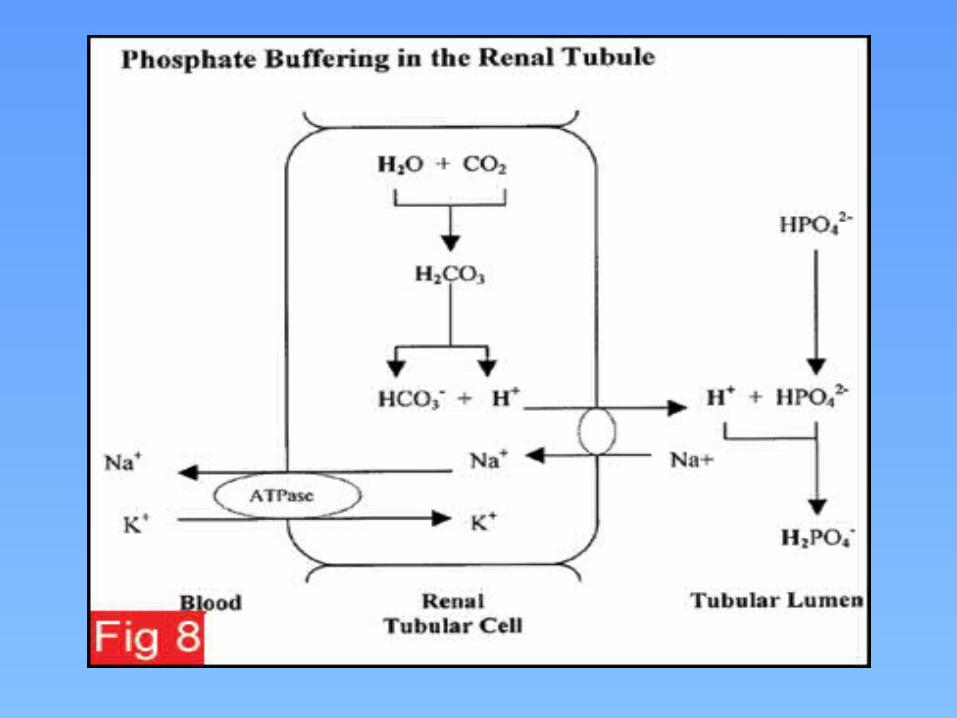

Acid-Base Biochemistry
Hydrogen ion excretion capacityThe minimum urine pH that Can be generated is 4.6 ( 25µmol/L)Normal urine output is 1.5LWithout the phosphate buffer system the free excretion of Hydrogen ions is less than 1/1000 of the acid produced by normal metabolism

Acid-Base Biochemistry
The phosphate buffer system increases hydrogen ion excretion capacity to 30-40 mmol/24 hoursIn times of chronic overproduction of acid another urine buffer systemAmmonia

Acid-Base Biochemistry
Ammonia produced by deamination of glutamine in renal tubular cells Catalysed by glutaminase which is induced by chronic acidosisAllows increased ammonia production and hence increased hydrogen ion excretion via ammonium ionsNH3 + H+ ↔ NH4
+

Acid-Base Biochemistry
At normal intracellular pH most ammonia is present as ammonium ions which can’t diffuse out of the cellDiffusion of ammonia out of the cell disturbs the equilibrium between ammonia and ammonium ions causing more ammonia to be formedHydrogen ions formed at the same time!These are used up by the deamination of glutamine to glutamate during gluconeogenesis


Acid-Base Biochemistry
Carbon dioxide transportCarbon dioxide produced by aerobic respiration diffuses out of cells and into the ECFA small amount combines with water to form carbonic acid decreasing the pH of ECFIn red blood cells metabolism is anaerobic and very little CO2 is produced hence it diffuses into red cells down a concentration gradient to form carbonic acid (carbonate dehydratase) buffered by haemoglobin .

Acid-Base Biochemistry
Haemoglobin has greatest buffering capacity when it is dexoygenated hence the buffering capacity increases as oxygen is lost to the tissues Net effect is that carbon dioxide is converted to bicarbonate in red cellsBicarbonate diffuses out of red cells down concentration gradient and chloride ions diffuse in to maintain electrochemical neutrality (chloride shift)


• Acid-Base Biochemistry
In the lungs this process is reversedHaemoglobin is oxygenated reducing its buffering capacity and generating hydrogen ionsThese combine with bicarbonate to form CO2 which diffuses into the alveoliBicarbonate diffuses into the cells from the plasma


72

Reference ranges and points
Parameter Reference range Reference point
pH 7.35-7.45 7.40
PCO2 33-44 mm Hg 40 mm Hg
PO2 75-105 mm Hg
HCO3- 22-28 mEq/L 24mEq/L
Anion gap 8-16 mEq/L 12 mEq/L
Osmolar gap <10 mOsm/L

Treatment of Metabolic Alkalosis• Electrolytes to replace those lost• IV chloride containing solution• Treat underlying disorder
74

75
Diagnosis of Acid-Base Imbalances1. Note whether the pH is low (acidosis) or high (alkalosis)2. Decide which value, pCO2 or HCO3
- , is outside the normal range and could be the cause of the problem. If the cause is a change in pCO2, the problem is respiratory. If the cause is HCO3- the problem is metabolic.

Delta ratio
Delta ratio Assessment
<0.4 Hyperchloraemic normal anion gap acidosis
0.4 – 0.8 Combined high AG and normal AG acidosisNote that the ratio is often <1 in acidosis associated with renal failure
1 - 2
Uncomplicated high-AG acidosisLactic acidosis: average value 1.6 DKA more likely to have a ratio closer to 1 due to urine ketone loss (if patient not dehydrated)
>2 Pre-existing increased [HCO3
-]:concurrent metabolic alkalosispre-existing compensated respiratory acidosis
𝛥 ratio = Anion gap/ [HCO𝛥 𝛥 3-] = (AG – 12)/(24 -
[HCO3-])

CompensationPrimary
DisturbancepH HCO3
- PCO2 Compensation
Respiratory acidosis <7.35 Compensatory increase
Primary increase
Acute: 1-2 mEq/L increase in HCO3
- for every 10 mm Hg increase in PCO2Chronic: 3-4 mEq/L increase in HCO3
- for every 10 mm Hg increase in PCO2
Respiratory alkalosis >7.45 Compensatory decrease
Primary decrease
Acute: 1-2 mEq/L decrease in HCO3
- for every 10 mm Hg decrease in PCO2
Chronic: 4-5 mEq/L decrease in HCO3
- for every 10 mm Hg decrease in PCO2
Metabolic acidosis <7.35 Primary decrease
Compensatory decrease
1.2 mm Hg decrease in PCO2 for every 1 mEq/L decrease in HCO3
-
Metabolic alkalosis >7.45 Primary increase
Compensatory increase
0.6-0.75 mm Hg increase in PCO2 for every 1 mEq/L increase in HCO3
-
, PCO2 should not rise above 55 mm Hg in compensation

Respiratory acidosis
PCO2 greater than expectedAcute or chronicCauses
excess CO2 in inspired air(rebreathing of CO2-containing expired air, addition of CO2 to inspired air, insufflation of CO2 into body cavity)
decreased alveolar ventilation(central respiratory depression & other CNS problems, nerve or muscle disorders, lung or chest wall defects, airway disorders, external factors)
increased production of CO2
(hypercatabolic disorders)

Racid acuteA 65-year-old man with a history of emphysema comes to the physician with a 3-hour history of shortness of breath.
pH 7.18
PO2 61 mm Hg
PCO2 58 mm Hg
HCO3- 26 mEq/L
History suggests hypoventilation, supported by increased PCO2 and lower than anticipated PO2.
Respiratory acidosis (acute) due to no renal compensation.

DescriptionpH 7.18
PO2 61 mm Hg
PCO2 58 mm Hg
HCO3- 26 mEq/L
1-2 mEq/L increase in HCO3- for every 10 mm Hg increase
in PCO2.
PCO2 increase = 58-40 = 18 mm Hg.
HCO3- increase predicted = (1-2) x (18/10) = 2-4 mEq/L
add to 24 mEq/L (reference point) = 26-28 mEq/L

Racid chronicA 56-year-old woman with COPD is brought to the physician with a 3-hour history of severe epigastric pain.
pH 7.39
PO2 62 mm Hg
PCO2 52 mm Hg
HCO3- 29 mEq/L
History suggests hypoventilation, supported by increased PCO2.
Respiratory acidosis (chronic) with renal compensation.

DescriptionpH 7.39
PO2 62 mm Hg
PCO2 52 mm Hg
HCO3- 29 mEq/L
3-4 mEq/L increase in HCO3- for every 10 mm Hg increase
in PCO2.
PCO2 increase = 52-40 = 12 mm Hg.
HCO3- increase predicted = (3-4) x (12/10) = 4-5 mEq/L
add to 24 mEq/L (reference point) = 28-29 mEq/L

Respiratory alkalosis
PCO2 less than expected
Acute or chronic
Causes increased alveolar ventilation
(central causes, direct action via respiratory center; hypoxaemia, act via peripheral chemoreceptors; pulmonary causes, act via intrapulmonary receptors; iatrogenic, act directly on ventilation)

Ralk acuteA 17-year-old woman is brought to the physician with a 3-hour history of epigastric pain and nausea. She admits taking a large dose of aspirin. Her respirations are full and rapid.
pH 7.57PO2 104 mm HgPCO2 25 mm HgHCO3
- 23 mEq/LHistory suggests hyperventilation, supported by decreased PCO2.Respiratory alkalosis (acute) due to no renal compensation.

DescriptionpH 7.57PO2 104 mm HgPCO2 25 mm HgHCO3
- 23 mEq/L
1-2 mEq/L decrease in HCO3- for every 10 mm Hg decrease
in PCO2.
PCO2 decrease = 40-25 = 15 mm Hg.
HCO3- decrease predicted = (1-2) x (15/10) = 2-3 mEq/L
subtract from 24 mEq/L (reference point) = 21-22 mEq/L

Ralk chronicA 81-year-old woman with a history of anxiety is brought to the physician with a 2-hour history of shortness of breath. She has been living at 9,000 ft elevation for the past 1 month. Her respirations are full at 20/min.
pH 7.44PO2 69 mm HgPCO2 24 mm HgHCO3
- 16 mEq/LHistory suggests hyperventilation, supported by decreased PCO2.Respiratory alkalosis (chronic) with renal compensation.

DescriptionpH 7.44PO2 69 mm HgPCO2 24 mm HgHCO3
- 16 mEq/L
4-5 mEq/L decrease in HCO3- for every 10 mm Hg decrease
in PCO2.
PCO2 decrease = 40-24 = 16 mm Hg.
HCO3- decrease predicted = (4-5) x (16/10) = 6-8 mEq/L
subtract from 24 mEq/L (reference point) = 16-18 mEq/L

Metabolic acidosis
Plasma HCO3- less than expected
Gain of strong acid or loss of base
Alternatively, high anion gap or normal anion gap metabolic acidosis
Causes high anion-gap acidosis (normochloremic)
(ketoacidosis, lactic acidosis, renal failure, toxins) normal anion-gap acidosis (hyperchloremic)
(renal, gastrointestinal tract, other)

Macid high AGA 20-year-old man with a history of diabetes is brought to the emergency department with a 3-day history of feeling ill. He is non-adherent with his insulin. Urine ketones are 2+ and glucose is 4+.
pH 7.26 Na+ 136 mEq/LPO2 110 mm Hg K+ 4.8 mEq/LPCO2 19 mm Hg Cl- 101 mEq/LHCO3
- 8 mEq/L CO2, total 10 mEq/LGlucose 343 mg/dL Urea 49 mg/dL
Creatinine 1 mg/dLHistory suggests d NC iabetic ketoacidosis.Metabolic acidosis with appropriate respiratory compensation.

DescriptionpH 7.26 Na+ 136 mEq/LPO2 110 mm Hg K+ 4.8 mEq/LPCO2 19 mm Hg Cl- 101 mEq/LHCO3
- 8 mEq/L Glucose 343 mg/dLUrea 49 mg/dL
AG = 136-101-8=27 mEq/L Creatinine 1 mg/dL1.2 mm Hg decrease in PCO2 for every 1 mEq/L decrease in HCO3
-.HCO3
- decrease = 24-8 = 16 mEq/L PCO2 decrease predicted = 1.2 x 16 = 19 mm Hg.subtract from 40 mm Hg (reference point) = 21 mm Hg

Macid normal AGA 43-year-old man comes to the physician with a 3-day history of diarrhea. He has decreased skin turgor.
pH 7.31 Na+ 134 mEq/LPO2 -- mm Hg K+ 2.9 mEq/LPCO2 31 mm Hg Cl- 113 mEq/LHCO3
- 16 mEq/L Urea 74 mgl/dLCreatinine 3.4 mmol/L
History is limited.Metabolic acidosis with respiratory compensation.

DescriptionpH 7.31 Na+ 134 mEq/LPO2 -- mm Hg K+ 2.9 mEq/LPCO2 31 mm Hg Cl- 113 mEq/LHCO3
- 16 mEq/L Urea 74 mg/dLCreatinine 3.4 mg/dL
AG = 134-113-16=5 mEq/L1.2 mm Hg decrease in PCO2 for every 1 mEq/L decrease in HCO3
-.HCO3
- decrease = 24-16 = 8 mEq/L PCO2 decrease predicted = 1.2 x 8 = 10 mm Hg.subtract from 40 mm Hg (reference point) = 30 mm Hg

Metabolic alkalosis
Plasma HCO3- greater than expected
Loss of strong acid or gain of base
Causes (2 ways to organize) loss of H+ from ECF via kidneys (diuretics) or gut (vomiting) gain of alkali in ECF from exogenous source (IV NaHCO3
infusion) or endogenous source (metabolism of ketoanions)or addition of base to ECF (milk-alkali syndrome) Cl- depletion (loss of acid gastric juice) K+ depletion (primary/secondary hyperaldosteronism) Other disorders (laxative abuse, severe hypoalbuminaemia)

Urinary Chloride
Spot urine Cl- less than 10 mEq/L often associated with volume depletion respond to saline infusion common causes - previous thiazide diuretic therapy,
vomiting (90% of cases)
Spot urine Cl- greater than 20 mEq/L often associated with volume expansion and hypokalemia resistant to therapy with saline infusion causes: excess aldosterone, severe K+ deficiency, current
diuretic therapy, Bartter syndrome

Calculate the anion gap AG = Na – Cl – HCO3 (normal 12 ± 2) AG corrected = AG + 2.5[4 – albumin] If there is an anion Gap then calculate the Delta/delta
gap (step 6). Only need to calculate delta gap (excess anion gap) when there is an anion gap to determine additional hidden metabolic disorders (nongap metabolic acidosis or metabolic alkalosis)
If there is no anion gap then start analyzing for non-anion acidosis

Malk high Urine Cl-
An 83-year-old woman is brought to the physician with a 1-week history of weakness and poor appetite.
pH 7.58 Na+ 145 mEq/LPO2 60 mm Hg K+ 1.9 mEq/LPCO2 56 mm Hg Cl- 86 mEq/LHCO3
- 52 mEq/L Urine Cl- 74 mEq/LHistory is limited.Metabolic alkalosis with respiratory compensation.The cause is unknown, most likely excess adrenocortical activity, current diuretic therapy, or idiopathic.

EXAMPLE • Calculate Anion gap
ABG 7.23/17/235 on 50% VM BMP Na 123/ Cl 97/ HCO3 7/BUN 119/ Cr 5/ Albumin
4.
AG = Na – Cl – HCO3 (normal 12 ± 2) 123 – 97 – 7 = 19
• No need to correct for albumin as it is 4

EXAMPLE : Delta Gap ABG 7.23/17/235 on 50% VM BMP Na 123/ Cl 97/ HCO3 7/BUN 119/ Cr 5/ Albumin
4.
• Delta gap = (actual AG – 12) + HCO3 • (19-12) +7 = 14 • Delta gap < 18 -> additional non-gap
metabolic acidosis• So Metabolic acidosis anion and non anion
gap

DescriptionpH 7.58 Na+ 145 mEq/LPO2 60 mm Hg K+ 1.9 mEq/LPCO2 56 mm Hg Cl- 86 mEq/LHCO3
- 52 mEq/L Urine Cl- 74 mEq/L0.6-0.75 mm Hg increase in PCO2 for every 1 mEq/L increase in HCO3
-.HCO3
- increase = 52-24 = 28 mEq/L PCO2 increase predicted = 0.6-0.75 x 28 = 17-21 mm Hg.add to 40 mm Hg (reference point) = 57-61 mm Hg

Malk low Urine Cl-
An 24-year-old woman is brought to the physician with a 3-month history of weakness and fatigue. Blood pressure is 90/60 mm Hg.
pH 7.52 Na+ 137 mEq/LPO2 78 mm Hg K+ 2.6 mEq/LPCO2 49 mm Hg Cl- 90 mEq/LHCO3
- 39 mEq/L Urine Cl- 5 mEq/LHistory and physical examination suggests bulimia.Metabolic alkalosis with respiratory compensation.The cause is most likely bulimia.

DescriptionpH 7.52 Na+ 137 mEq/LPO2 78 mm Hg K+ 2.6 mEq/LPCO2 49 mm Hg Cl- 90 mEq/LHCO3
- 39 mEq/L Urine Cl- 5 mEq/L0.6-0.75 mm Hg increase in PCO2 for every 1 mEq/L increase in HCO3
-.HCO3
- increase = 39-24 = 15 mEq/L PCO2 increase predicted = 0.6-0.75 x 15 = 9-12 mm Hg.add to 40 mm Hg (reference point) = 49-52 mm Hg

What is the primary disorder?
What disorder is present? pH pCO2 or HCO3
Respiratory Acidosis pH low pCO2 high
Metabolic Acidosis pH low HCO3 low
Respiratory Alkalosis pH high pCO2 low
Metabolic Alkalosis pH high HCO3 high

Special Cases
• Pregnancy – hyperventilation (respiratory alkalosis), hyperemesis (metabolic alkalosis or acidosis), maternal ketosis (metabolic acidosis)
• Children – low bicarbonate reserve (N=12-16 mEq/L), low acid excretion reserve, inborn errors in metabolism, diabetes, and poisoning (all metabolic acidosis)