Mineralization process during acellular cementogenesis in ...
Analyst
PAPER
Cite this: Analyst, 2020, 145, 4867
Received 21st February 2020,Accepted 13th May 2020
DOI: 10.1039/d0an00380h
rsc.li/analyst
Acellular oxidative potential assay for screening ofamorphous silica nanoparticles†
Dalibor Breznan, ‡a Nazila Nazemof,‡b Filip Kunc,c Myriam Hill,d
Djordje Vladisavljevic,d James Gomes,b Linda J. Johnston, c Renaud Vincenta,e
and Prem Kumarathasan*a,b
Silica nanoparticles (SiNPs) are used in a wide range of consumer products, engineering and medical
applications, with likelihood of human exposure and potential health concerns. It is essential to generate
toxicity information on SiNP forms and associated physicochemical determinants to conduct risk assess-
ment on these new materials. To address this knowledge gap, we screened a panel of custom synthesized,
well-characterized amorphous SiNPs pristine and surface-modified (–C3-COOH, –C11-COOH, –NH2, –PEG)
of 5 different sizes: (15, 30, 50, 75, 100 nm) for their oxidative potential using an acellular assay. The assay
is based on oxidation of dithiothreitol (DTT) by reactive oxygen species and can serve as a surrogate test
for oxidative stress. These materials were characterized for size distribution, aggregation, crystallinity,
surface area, surface modification, surface charge and metal content. Tests for association between oxi-
dative potential of SiNPs and their physicochemical properties were carried out using analysis of variance
and correlation analyses. These test results suggest that the size of amorphous SiNPs influenced their oxi-
dative potential irrespective of the surface modification, with 15 nm exhibiting relatively higher oxidative
potential compared to the other sizes. Furthermore, SiNP surface area, surface modification and agglom-
eration in solution also appeared to affect oxidative potential of these SiNPs. These findings indicate that
physicochemical properties are critical in influencing the oxidative behaviour of amorphous SiNPs, with
potential to trigger cellular oxidative stress and thus toxicity, when exposed. This information advances
our understanding of potential toxicities of these amorphous SiNPs and supports risk assessment efforts
and the design of safer forms of silica nanomaterials.
Introduction
Engineered nanoparticles (NPs) provide challenges for hazardidentification and risk evaluation due to a lack of reliablephysicochemical and toxicity data, creating difficulties for gov-ernment agencies to establish effective safety evaluation guide-lines. Among the various engineered nanomaterials, amor-
phous silica nanoparticles (SiNPs) have received relativelymore attention and have been frequently used as additives tocosmetics, drugs, printer toners, varnishes and food.1–4 Due totheir biocompatibility, easy surface functionalization and resis-tance to biodegradation in the cellular environment, silicananoparticles are currently being synthesized on a largescale for biomedical and biotechnology applications such ascancer therapy, in gene carriers, drug delivery and enzymeimmobilization.5–10
The growing use of nanomaterials including SiNPs raisesconcerns in terms of their potential harmful effects to humanhealth and the environment and places them at a high priorityfor toxicity screening and risk assessment by regulatoryauthorities.11,12 However, toxicological studies on SiNPs are farbehind the pace of their production and applications. Thereare emerging reports on the analyses of the toxicological pro-perties of SiNPs.12–16 The physicochemical properties of SiNPshighly depend on the synthetic method for their productionand can influence the toxicity of these materials.1,17 Uniquephysicochemical properties of engineered nanomaterials suchas particle size (surface area and size distribution), aggrega-
†Electronic supplementary information (ESI) available. See DOI: 10.1039/d0an00380h‡These authors contributed equally to this work.
aEnvironmental Health Science and Research Bureau, Health Canada, Ottawa, ON,
K1A 0K9, Canada. E-mail: [email protected];
Fax: +613-946-2600; Tel: +613-218-4530bInterdisciplinary School of Health Sciences, University of Ottawa, Ottawa, ON,
K1N 7K4, CanadacMetrology Research Centre, National Research Council Canada, Ottawa, Ontario,
K1A 0R6, CanadadNew Substances Assessment and Control Bureau, Health Canada, Ottawa, ON, K1A
0K9, CanadaeDepartment of Biochemistry, Microbiology and Immunology, University of Ottawa,
Ottawa, ON, K1H 8M5, Canada
This journal is © The Royal Society of Chemistry 2020 Analyst, 2020, 145, 4867–4879 | 4867
Ope
n A
cces
s A
rtic
le. P
ublis
hed
on 2
8 M
ay 2
020.
Dow
nloa
ded
on 1
/1/2
022
7:07
:21
AM
. T
his
artic
le is
lice
nsed
und
er a
Cre
ativ
e C
omm
ons
Attr
ibut
ion
3.0
Unp
orte
d L
icen
ce.
View Article OnlineView Journal | View Issue
tion/agglomeration, chemical composition (purity, crystalli-nity, electronic properties) and surface structure (surface reac-tivity, surface groups, inorganic or organic coating) can deter-mine the fate of NP–cell interactions.
Formation of reactive oxygen species (ROS) has beensuggested as one of the mechanisms by which SiNPs couldexert toxicity.18–20 The excessive production of ROS-mediatedoxidative stress triggered by exposures to environmental pollu-tants such as complex mixtures of air pollution particles con-taining ultrafine and nano-scale components can be associ-ated with disruption in the normal mechanism of cellularsignaling leading to various disease processes includingcardiovascular disease, COPD, diabetes, cancer, Alzheimer’sdisease.21–23 The assessment of oxidative potential as a surro-gate for ROS generation has been proposed as a useful metricto measure the capacity of particulate matter (PM) to oxidisetarget molecules in biological systems.24 It is an attractivemeasure because it integrates various biologically relevant pro-perties, including size, surface and chemical composition.25
Both cellular26–28 and acellular29–32 methods have been devel-oped to measure the oxidative potential of NPs.
Methodological approaches for redox activity are basedeither on direct measurement of ROS in cellular environmentusing high performance liquid chromatography (HPLC), elec-tron spin resonance spectroscopy (ESR) and fluorescence-based methods33 or indirect measurement using particle-induced depletion of the reductants (e.g. DTT) or the antioxi-dants (e.g. vitamin C, glutathione and uric acid)34,35 in acellu-lar tests. Acellular methods require a less controlled environ-ment, are less costly and can be relatively rapid for high-throughput identification of NP hazard. Furthermore, in vitroacellular screening for oxidative potential of nanomaterialscan be of value in terms of predicting relative reactivities andthus potencies of these particles when exposed to cells.
Among all available acellular methods, the dithiothreitol(DTT) assay is one of the commonly used assays to study oxi-dative potential associated with micron-sized environmentalparticles, such as ambient air particulate matter with complexmatrices.30 This assay can serve as an initial screening step toidentify the oxidative potential of particles prior to conductingmore extensive cell or animal exposure studies. The DTT assayis based on the presence of redox-active chemical species inparticles to oxidize DTT to its disulfide form. In general, theredox active species, such as transition metals, conjugatedunsaturated organic species in complex air pollutant matricescan donate electron to dissolved molecular oxygen, formingsuperoxide, a ROS, which in turn can form other ROS, such ashydrogen peroxide which in the presence of metals, such asiron can lead to the formation of hydroxyl radical (•OH). Oneof the limitations noted for this assay was its inability to dis-criminate between ROS, for instance, its inability to measurespecifically •OH generation.33 The DTT assay is a fast and in-expensive assay and therefore is a candidate assay for high-throughput screening for oxidative potential.36,37 Nevertheless,the engineered nanomaterials, such as pure amorphous silicananoparticles do not contain redox active metals or organicspecies to participate in redox-cycling reactions. Therefore itcan be challenging to apply DTT assay to measure oxidativepotential of such particles and thus the sensitivity of the assaycan be questionable. Yet, the simplicity, cost and speed of theDTT assay are attractive as a first pass screening tool for oxi-dative potential measurement of nanomaterials compared toin vitro or in vivo exposure studies and thus warrants feasibilitytesting with these materials.
The objective of this study was to test the applicability ofthe cell-free assay based on DTT oxidation to screen varioussizes (15, 30, 50, 75, 100 nm) of pristine and surface-modifiedSiNPs (–C3-COOH, –C11-COOH, –NH2, –PEG) for their oxi-
Fig. 1 Schematic drawing of the general analytical approach.
Paper Analyst
4868 | Analyst, 2020, 145, 4867–4879 This journal is © The Royal Society of Chemistry 2020
Ope
n A
cces
s A
rtic
le. P
ublis
hed
on 2
8 M
ay 2
020.
Dow
nloa
ded
on 1
/1/2
022
7:07
:21
AM
. T
his
artic
le is
lice
nsed
und
er a
Cre
ativ
e C
omm
ons
Attr
ibut
ion
3.0
Unp
orte
d L
icen
ce.
View Article Online
dative potential, and to identify the physicochemical pro-perties of SiNPs that can influence their oxidative potential bytesting for associations between them. The analytical approachis presented in Fig. 1. To the best of our knowledge, this studyis the first report for a comprehensive screening of nanoformsof amorphous SiNPs using the acellular DTT assay.
ExperimentalMaterials
Dithiothreitol (DTT), 5,5′-dithiobis-(2-nitrobenzoic acid)(DTNB), 1 M phosphate buffer, di-tert-butyl peroxide (DTBP),tert-butyl hydroperoxide (TBHP), Fe(II) sulphate hydrate andultrapure water were obtained from Sigma Aldrich (Oakville,ON, Canada). Dulbecco’s Modified Eagle’s Medium (phenolred-free) and with phenol red, fetal bovine serum (FBS)and phosphate buffered-saline (10×) were purchased fromFisher Scientific (Nepean, ON, Canada). Custom synthesizedwell-characterized pristine amorphous silica nanoparticles(SiNPs) of different sizes (15, 30, 50, 75, 100 nm) and thecorresponding surface modified –C3-COOH, –C11-COOH,–(OH)2Si(OCH2CH2)nOCH3 (n = 9–12) (–PEG) and–(OH)2Si(CH2)2CH2NH2 (–NH2) variants were purchased fromAdvanced Quantum Materials Inc. (AQM, Edmonton, AB,Canada).
For comparative purposes, reference particles were includedin the experiments. Micron-sized standard reference materials(SRM-1879 silicon dioxide, SiO2 and SRM-154b titaniumdioxide, TiO2) were obtained from the National Institute ofStandards and Technology (Gaithersburg, MD, USA). Ottawaambient air EHC-6802 particles were included to represent anurban particulate matter standard. The preparation andcharacterization of EHC-6802 particles has been previouslydescribed.38 A reference nanoparticle, 12 nm-sized amorphoussilica was also included in the experiment (cat. # 718483;Sigma Aldrich, Oakville, ON, Canada).
Characterization of physicochemical properties
The pristine SiNPs were synthesized using modified-Stöbersol–gel based process that included a calcination step. Thesame batch of a specific size of the pristine SiNPs was usedto prepare the corresponding surface modified counterparts.The physicochemical properties of the particles were ana-lyzed by transmission electron microscopy (TEM) for size dis-tribution, X-ray diffraction (XRD) to assess the crystallinity,Fourier transform infrared spectroscopy (FT-IR) for func-tional groups and thermogravimetric analysis (TGA) for func-tional group loss, by AQM. In addition, Brunauer–Emmett–Teller (BET) analyses for surface area, quantitative nuclearmagnetic resonance (qNMR) analyses for surface modifi-cations, dynamic light scattering (DLS) and zeta potential foragglomeration size and surface charge in aqueous mediaand inductively coupled plasma mass spectrometry (ICP-MS/AES) for elemental analysis were conducted as part of thisstudy.
Electron microscopy
Bright field transmission electron microscopy (TEM) imageswere taken with a JEOL 2010 TEM (with LaB6 electron gun)using accelerating voltage of 200 kV. TEM samples were pre-pared by depositing a drop of ethanol or toluene solution (ca.0.1 mg mL−1) of the pristine or functionalized SiO2 nano-particle suspensions onto carbon coated copper grid (obtainedfrom Electron Microscopy Inc.). The nanoparticle sizes weredetermined upon averaging the dimensions of at least 50–100particles chosen manually using ImageJ software (version1.45).
X-ray powder diffraction (XRD)
XRD data were obtained using an Inel MPD Multi PurposeDiffractometer equipped with a CPS 120 curved position sensi-tive X-ray detector and copper Kα (8.047 KeV energy) radiationsource. The samples were made by depositing the SiNPs on aSi (111) wafer.
Fourier transform infrared (FT-IR) spectroscopy
FT-IR spectroscopic analyses were performed using a ThermoNicolet Magna 750 IR Spectrometer. Samples for FT-IR analysiswere prepared by drop coating a methanol solution of the pris-tine and surface-functionalized SiNPs onto an electronic-gradeSi-wafer (N-type, 100 surface, 100 mm thickness and 10 ohmcm resistivity).
Thermogravimetric analysis (TGA)
TGA was performed using a Mettler Toledo Star TGA/DSCsystem. Pristine and surface-functionalized SiNP samples wereplaced in a Pt pan and heated under Ar atmosphere from 35 to700 °C at a rate of 10 °C min−1 or 25 °C min−1 as indicated.
Quantitative nuclear magnetic resonance (qNMR)
Functional group contents of SiNPs were quantified by dis-solution of the NPs in basic solution, followed by solutionNMR using a Bruker Avance 400 Mz spectrometer, accordingto a published protocol.39 SiNPs (4–10 mg) were treated withsodium deuteroxide solution in D2O (0.65 mL, 0.4 M) and dis-persed by sonication in an ultrasonic bath for 10 min. Sampleswere then placed in a heated mixer and shaken at 45 °C for16 hours, after which they were brought to room temperature;maleic acid calibrant solution in D2O (20 μL, 98.62 mM) wasadded and the sample was vortexed. These samples were trans-ferred into a 5 mm NMR tube and the NMR measurement wasconducted within 24 hours. Triplicate analyses were per-formed. Due to the high-throughput requirements, NMR ana-lyses were carried out using a zg30 pulse program instead ofzg90 as typically used for quantitative NMR experiments. Thisstep introduces a difference of ≤5% in the quantification, butshortens the experiment duration by a factor of 4. Spectralwidth was 20 ppm with 6.175 ppm transmitter frequencyoffset. 32 scans with 2 dummy scans were recorded at 6 s relax-ation delay. Maximal receiver gain was set prior to eachmeasurement. All acquired spectra were phase corrected
Analyst Paper
This journal is © The Royal Society of Chemistry 2020 Analyst, 2020, 145, 4867–4879 | 4869
Ope
n A
cces
s A
rtic
le. P
ublis
hed
on 2
8 M
ay 2
020.
Dow
nloa
ded
on 1
/1/2
022
7:07
:21
AM
. T
his
artic
le is
lice
nsed
und
er a
Cre
ativ
e C
omm
ons
Attr
ibut
ion
3.0
Unp
orte
d L
icen
ce.
View Article Online
manually and baseline correction was done by 5th order poly-nomial fit. Baseline-resolved diagnostic signals of the func-tional moiety were identified, integrated, and the average inte-gral was compared with the integral of the calibrant, maleicacid.
ICP-MS/AES
Metal content of pristine SiNP samples was analyzed usinginductively coupled plasma-mass spectrometry/atomic emis-sion spectroscopy (ICP-MS/AES, Varian Vista-Pro, Mulgrave,Australia) after acid-digestion following the previously reportedprocedure.40 In brief, the various SiNPs were digested in 50%HNO3 for 8 h at 80 °C and after filtration via a 0.22 mm filterand the filtrates were analyzed by ICP-MS/AES.41 A reagentblank was analyzed by ICP-MS/AES and the blank values weresubtracted from the sample analysis results to obtain theactual metal concentrations in these samples. All analyseswere done in duplicate.
Brunauer–Emmett–Teller analysis (BET)
The BET method with nitrogen absorption was utilized toobtain the specific surface areas for the SiNPs. ASAP 2020instrument (Micrometrics, Norcross, GA, USA) was used forthe gas-adsorption based SiNP surface area analyses. Prior todegassing, the SiNP samples contained in the sample tubewere weighed. The samples were degassed under vacuum to apressure of 10 mm per Hg at 80 °C, followed by a seconddegassing step at a pressure of 300 µm per Hg for 90 minutes.The temperature was increased at 100 °C min−1 to 120 °C atwhich time the degassing was continued for 12 hours. TheSiNP samples were cooled to 20 °C and complete degassingwas verified. The specific surface area was determined by themultipoint BET method.
Particle preparations
All the amorphous SiNP variants and reference particles(EHC-6802 urban dust particles and micron-sized SiO2 andTiO2) were re-suspended in ultrapure water (Sigma-Aldrich,Oakville, ON, Canada) to a concentration of 500 μg mL−1. Thesuspensions were vortexed for 30 s and sonicated (6 cycles of30 s on/off with 50–60% amplitude) on ice by using a probesonicator (120 W, 20 KHz; Model CL-18, Fisher Scientific,Ottawa, ON, Canada). Sonicated samples were vortexed for 30 sand agitated by a rocking shaker. The suspended particleswere employed to conduct the oxidative potential analyses.
Dynamic light scattering (DLS) and zeta potential
The particle stocks were vortexed for 30 s and sonicated in awater bath sonicator (100 W, 20 kHz; Branson Ultrasonics,Danbury, CT, USA) for 20 min prior to sample preparation asfollows; A 10 μl aliquot of aqueous suspensions of pristine orsurface-modified SiNP variants (500 μg mL−1) was combinedwith 100 μL of 0.5 M potassium phosphate buffer (pH 7.4) anda final volume of 500 μL was made up with ultrapure water toobtain 10 μg mL−1 SiNP concentration for hydrodynamic dia-meter measurement by DLS. The samples were mixed using
Eppendorf Thermomixer R dry-block shaker (Eppendorf,Mississauga, ON) for 10 min at 37 °C. Subsequently, thesamples were vortexed for 10 s and transferred into cuvettesfor measurements taken immediately and after 20 min incu-bation at room temperature (RT). Identical sample preparationwas used for the zeta potential analyses. The DLS (z-averageand polydispersity index) and zeta potential values wereobtained using the Zetasizer Nano ZS (Malvern Instruments,UK) as reported earlier.42 All measurements were conducted intriplicate.
Oxidative potential analyses
The procedure reported by Janssen et al. was followed toperform the dithiothreitol (DTT) assay after optimization withminor modifications.25 Briefly, 10 μl of pristine or surface-modified SiNP suspension in ultrapure water at 500 μg mL−1
was incubated in the dark room with gentle agitation in a ther-momixer at 550 rpm (Eppendorf Thermomixer R) with 100 μlof 0.5 M potassium phosphate buffer (pH 7.4) and 340 μl ofultrapure water for 10 min at 37 °C. Then, 50 μl of 1 mM DTTwas added to the incubation vial and vortexed for 10 s. A100 μl aliquot of the DTT reaction mixture was withdrawn andtransferred to a vial containing 10 μl of 1.5 mM 5,5′-dithiobis(2-nitrobenzoic acid) (DTNB) to quench the oxidation reactionat different time periods (3, 10, 20, 60 min). The reactionbetween the remainder of the reduced DTT and DTNB is fastand forms 2-nitro-5-thiobenzoic acid (TNB), which is stable inthe final solution for at least 2 h at RT. This mixture was centri-fuged at 10 000g for 40 s and the supernatant was transferredto 96-well plates. The absorbance was measured at 412 nmwith a POLARstar Omega plate reader spectrophotometer(BMG Lab tech, Ortenberg, Germany).
Since both DTT and TNB are sensitive to light, care wastaken to exclude light as much as possible by working in adark room and sealing the 96-well plate with aluminium foilduring the experiment. Initially, the reaction conditions wereoptimized using the reference particles, as well as pristine andCOOH-modified SiNPs. The time period of 20 min was chosenas the optimal period for the DTT oxidation by these SiNPsand was used to screen the oxidative potential of all SiNP var-iants. A subset of SiNP variants (15 and 75 nm pristine andsurface-modified SiNPs) were then subjected to free radicalinitiators DTBP or TBHP prior to conducting the DTT assay toassess the extent of oxidative potential amplification, if poss-ible. Here, 100 µM TBHP or DTBP was initially reacted at RTwith 20 µM Fe(II) sulfate hydrate and phosphate buffer (0.5 M;pH 7.4) and incubated for 15 min at 37 °C with continuousagitation using a thermomixer (550 rpm). Ten μL of the SiNPsuspension (500 µg mL−1) in water was added to the incubatedmixture and continuously shaken. After 10 min, DTT assay wascarried out as described earlier. All analyses were conducted induplicate.
Statistical analyses
The presented data for the DTT assay were normalized to thecorresponding blanks and to the sum of the plate responses
Paper Analyst
4870 | Analyst, 2020, 145, 4867–4879 This journal is © The Royal Society of Chemistry 2020
Ope
n A
cces
s A
rtic
le. P
ublis
hed
on 2
8 M
ay 2
020.
Dow
nloa
ded
on 1
/1/2
022
7:07
:21
AM
. T
his
artic
le is
lice
nsed
und
er a
Cre
ativ
e C
omm
ons
Attr
ibut
ion
3.0
Unp
orte
d L
icen
ce.
View Article Online
per each experiment. The experimental data were assessed forstatistical significance by Kruskal–Wallis method or a two-wayanalysis of variance (ANOVA), as appropriate, using SigmaPlotv12.5 (Systat Software, San Jose, CA, USA). Where the assump-tions of normality and equal variance were not met, the datawere rank-transformed prior to conducting the ANOVA. For theKruskal–Wallis method, Dunn’s test was applied for the pair-wise multiple comparisons (α = 0.05). For the two-way ANOVA,the multiple comparisons analysis was conducted using theHolm–Sidak post hoc test (α = 0.05). Nanoparticle size (size)and surface modification (mod) were applied as factors in thetwo-way ANOVA. Pearson product moment correlation wasused to test for associations between oxidative potential valuesfor the different SiNP forms and their physicochemicalproperties.
Results and discussionPhysicochemical characterization
In this study, a set of custom synthesized amorphous SiNPsincluding pristine and surface-modified particles of varioussizes were assessed for their in vitro acellular oxidative pro-perties. Since the assessment of physicochemical properties ofSiNPs is critical to understand nanomaterial reactivity andtheir relative potencies in biological systems, the SiNP variantswere characterized for key physicochemical parameters. Thesemeasurements included TEM of pristine and surface-modifiedSiNP variants for size analyses of dry, primary particles andthe BET method to determine the surface area of the SiNPs.Agglomeration behaviour of the SiNPs in solution-state wasassessed using DLS method, while the electrophoretic mobilityof the SiNPs in aqueous suspension was determined by zetapotential analysis.
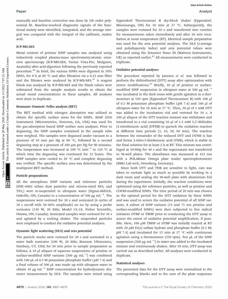
The TEM analysis results for the pristine and surface-modi-fied SiNPs (Fig. 2 and S1†) showed that among the pristineSiNPs, in dry form, the 15 nm-sized particles showed a highertendency to agglomerate than the SiNPs of other sizes. Also, asthe size of pristine SiNPs increased, the particles were spheri-cal with uniform particle size distribution. Similar behaviourwas observed with surface-coated SiNP particles with 15 nmsize being agglomerated across all surface modifications com-pared to the other sizes. In contrast, the 100 nm surface-
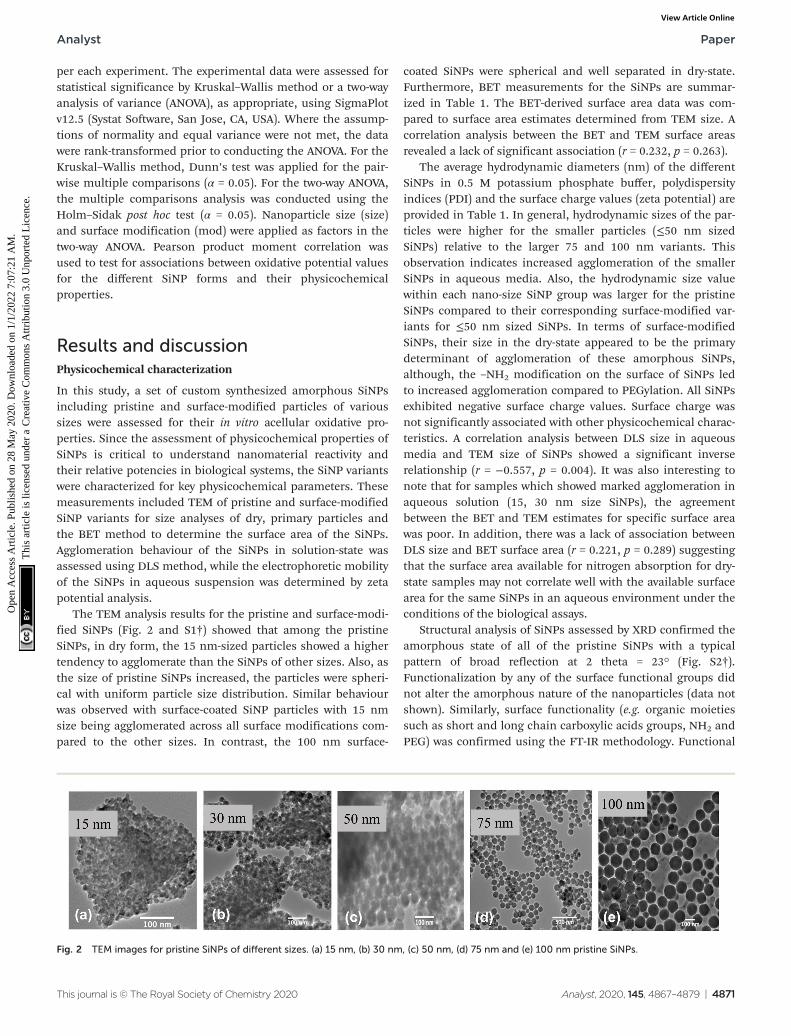
coated SiNPs were spherical and well separated in dry-state.Furthermore, BET measurements for the SiNPs are summar-ized in Table 1. The BET-derived surface area data was com-pared to surface area estimates determined from TEM size. Acorrelation analysis between the BET and TEM surface areasrevealed a lack of significant association (r = 0.232, p = 0.263).
The average hydrodynamic diameters (nm) of the differentSiNPs in 0.5 M potassium phosphate buffer, polydispersityindices (PDI) and the surface charge values (zeta potential) areprovided in Table 1. In general, hydrodynamic sizes of the par-ticles were higher for the smaller particles (≤50 nm sizedSiNPs) relative to the larger 75 and 100 nm variants. Thisobservation indicates increased agglomeration of the smallerSiNPs in aqueous media. Also, the hydrodynamic size valuewithin each nano-size SiNP group was larger for the pristineSiNPs compared to their corresponding surface-modified var-iants for ≤50 nm sized SiNPs. In terms of surface-modifiedSiNPs, their size in the dry-state appeared to be the primarydeterminant of agglomeration of these amorphous SiNPs,although, the –NH2 modification on the surface of SiNPs ledto increased agglomeration compared to PEGylation. All SiNPsexhibited negative surface charge values. Surface charge wasnot significantly associated with other physicochemical charac-teristics. A correlation analysis between DLS size in aqueousmedia and TEM size of SiNPs showed a significant inverserelationship (r = −0.557, p = 0.004). It was also interesting tonote that for samples which showed marked agglomeration inaqueous solution (15, 30 nm size SiNPs), the agreementbetween the BET and TEM estimates for specific surface areawas poor. In addition, there was a lack of association betweenDLS size and BET surface area (r = 0.221, p = 0.289) suggestingthat the surface area available for nitrogen absorption for dry-state samples may not correlate well with the available surfacearea for the same SiNPs in an aqueous environment under theconditions of the biological assays.
Structural analysis of SiNPs assessed by XRD confirmed theamorphous state of all of the pristine SiNPs with a typicalpattern of broad reflection at 2 theta = 23° (Fig. S2†).Functionalization by any of the surface functional groups didnot alter the amorphous nature of the nanoparticles (data notshown). Similarly, surface functionality (e.g. organic moietiessuch as short and long chain carboxylic acids groups, NH2 andPEG) was confirmed using the FT-IR methodology. Functional
Fig. 2 TEM images for pristine SiNPs of different sizes. (a) 15 nm, (b) 30 nm, (c) 50 nm, (d) 75 nm and (e) 100 nm pristine SiNPs.
Analyst Paper
This journal is © The Royal Society of Chemistry 2020 Analyst, 2020, 145, 4867–4879 | 4871
Ope
n A
cces
s A
rtic
le. P
ublis
hed
on 2
8 M
ay 2
020.
Dow
nloa
ded
on 1
/1/2
022
7:07
:21
AM
. T
his
artic
le is
lice
nsed
und
er a
Cre
ativ
e C
omm
ons
Attr
ibut
ion
3.0
Unp
orte
d L
icen
ce.
View Article Online
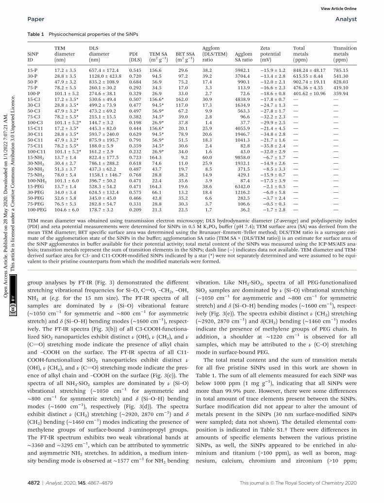
group analyses by FT-IR (Fig. 3) demonstrated the differentstretching vibrational frequencies for Si–O, CvO, –CH2, –OH,NH2 at (e.g. for the 15 nm size). The FT-IR spectra of allsamples are dominated by ν (Si–O) vibrational feature(∼1050 cm−1 for symmetric and ∼800 cm−1 for asymmetricstretch) and δ (Si–O–H) bending modes (∼1600 cm−1), respect-ively. The FT-IR spectra (Fig. 3(b)) of all C3-COOH-functiona-lized SiO2 nanoparticles exhibit distinct ν (OH), ν (CHx), and ν
(CvO) stretching mode indicate the presence of alkyl chainand –COOH on the surface. The FT-IR spectra of all C11-COOH-functionalized SiO2 nanoparticles exhibit distinct ν
(OH), ν (CHx), and ν (CvO) stretching mode indicate the pres-ence of alkyl chain and –COOH on the surface (Fig. 3(c)). Thespectra of all NH2-SiO2 samples are dominated by ν (Si–O)vibrational stretching (∼1050 cm−1 for asymmetric and∼800 cm−1 for symmetric stretch) and δ (Si–O–H) bendingmodes (∼1600 cm−1), respectively (Fig. 3(d)). The spectraexhibit distinct ν (CH2) stretching (∼2920, 2870 cm−1) and δ
(CH2) bending (∼1460 cm−1) modes indicating the presence ofmethylene groups of surface-bound 3-aminopropyl groups.The FT-IR spectrum exhibits two weak vibrational bands at∼3360 and ∼3295 cm−1, which can be attributed to symmetricand asymmetric NH2 stretches. In addition, a medium inten-sity bending mode is observed at ∼1577 cm−1 for NH2 bending
vibration. Like NH2-SiO2, spectra of all PEG-functionalizedSiO2 samples are dominated by ν (Si–O) vibrational stretching(∼1050 cm−1 for asymmetric and ∼800 cm−1 for symmetricstretch) and δ (Si–O–H) bending modes (∼1600 cm−1), respect-ively (Fig. 3(e)). The spectra exhibit distinct ν (CH2) stretching(∼2920, 2870 cm−1) and δ(CH2) bending (∼1460 cm−1) modesindicate the presence of methylene groups of PEG chain. Inaddition, a shoulder at ∼1220 cm−1 is observed for allsamples, which may be attributed to the ν (C–O) stretchingmode in surface-bound PEG.
The total metal content and the sum of transition metalsfor all five pristine SiNPs used in this work are shown inTable 1. The sum of all elements measured for each SiNP wasbelow 1000 ppm (1 mg g−1), indicating that all SiNPs weremore than 99.9% pure. However, there were some differencesin total amount of trace elements present between the SiNPs.Surface modification did not appear to alter the amount ofmetals present in the SiNPs (30 nm surface-modified SiNPswere sampled; data not shown). The detailed elemental com-position is indicated in Table S1.† There were differences inamounts of specific elements between the various pristineSiNPs, as well, the SiNPs appeared to be enriched in alu-minium and titanium (>100 ppm), as well as boron, mag-nesium, calcium, chromium and zirconium (>10 ppm;
Table 1 Physicochemical properties of the SiNPs
SiNPID
TEMdiameter(nm)
DLSdiameter(nm)
PDI(DLS)
TEM SA(m2 g−1)
BET SSA(m2 g−1)
Agglom(DLS/TEM)ratio
AgglomSA ratio
Zetapotential(mV)
Totalmetals(ppm)
Transitionmetals(ppm)
15-P 17.2 ± 3.5 657.4 ± 172.4 0.545 156.6 29.6 38.2 5982.1 −15.9 ± 1.2 848.24 ± 48.17 785.1530-P 28.8 ± 3.5 1128.0 ± 423.8 0.720 94.5 97.2 39.2 3704.4 −13.4 ± 2.8 615.55 ± 8.44 541.3050-P 47.9 ± 3.2 835.2 ± 108.9 0.684 56.9 75.2 17.4 990.1 −12.0 ± 2.1 902.74 ± 19.11 828.0375-P 78.2 ± 5.5 260.1 ± 30.2 0.292 34.5 17.0 3.3 113.9 −16.6 ± 2.3 476.36 ± 4.55 419.10100-P 101.1 ± 5.2 274.6 ± 38.1 0.329 26.9 33.0 2.7 72.6 −18.6 ± 0.8 401.62 ± 10.96 359.9415-C3 17.2 ± 3.5* 530.6 ± 49.4 0.507 156.6* 162.0 30.9 4838.9 −17.8 ± 0.7 — —30-C3 28.8 ± 3.5* 499.2 ± 73.9 0.477 94.5* 117.0 17.3 1634.9 −24.7 ± 1.3 — —50-C3 47.9 ± 3.2* 473.2 ± 69.2 0.497 56.9* 67.2 9.9 563.3 −27.8 ± 1.7 — —75-C3 78.2 ± 5.5* 215.1 ± 15.5 0.382 34.5* 39.0 2.8 96.6 −32.2 ± 2.3 — —100-C3 101.1 ± 5.2* 144.7 ± 3.2 0.198 26.9* 37.8 1.4 37.7 −29.9 ± 2.5 — —15-C11 17.2 ± 3.5* 445.3 ± 82.0 0.444 156.6* 20.1 25.9 4055.9 −21.4 ± 4.5 — —30-C11 28.8 ± 3.5* 593.7 ± 240.0 0.629 94.5* 78.9 20.6 1946.7 −34.8 ± 2.8 — —50-C11 47.9 ± 3.2* 875.9 ± 195.7 0.791 56.9* 51.5 18.3 1041.3 −21.7 ± 1.6 — —75-C11 78.2 ± 5.5* 188.0 ± 5.9 0.359 34.5* 30.6 2.4 82.8 −35.8 ± 2.4 — —100-C11 101.1 ± 5.2* 161.2 ± 2.9 0.232 26.9* 34.0 1.6 43.0 −32.0 ± 2.9 — —15-NH2 13.7 ± 1.4 822.4 ± 177.5 0.723 164.3 9.2 60.0 9858.0 −6.7 ± 1.7 — —30-NH2 30.4 ± 2.7 786.1 ± 288.2 0.618 74.6 11.0 25.9 1932.1 −14.9 ± 2.6 — —50-NH2 51.3 ± 3.7 437.3 ± 62.2 0.487 43.7 19.7 8.5 371.5 −8.5 ± 3.3 — —75-NH2 78.0 ± 5.4 1158.1 ± 146.7 0.768 28.8 38.2 14.9 429.1 −15.9 ± 0.7 — —100-NH2 101.1 ± 6.0 396.7 ± 50.2 0.471 22.4 35.6 3.9 87.4 −17.5 ± 3.9 — —15-PEG 13.7 ± 1.4 528.3 ± 54.2 0.471 164.3 19.6 38.6 6342.0 −2.1 ± 0.5 — —30-PEG 34.0 ± 3.4 624.5 ± 132.4 0.575 66.1 13.2 18.4 1216.2 −6.0 ± 5.8 — —50-PEG 52.6 ± 5.8 345.0 ± 45.0 0.466 42.8 35.2 6.6 282.5 −3.7 ± 2.4 — —75-PEG 76.5 ± 5.3 282.8 ± 54.7 0.331 28.8 30.3 3.7 106.6 −0.5 ± 0.3 — —100-PEG 104.6 ± 6.0 178.7 ± 3.2 0.209 21.3 22.5 1.7 36.2 −1.7 ± 2.8 — —
TEM mean diameter was obtained using transmission electron microscopy; DLS hydrodynamic diameter (Z-average) and polydispersity index(PDI) and zeta potential measurements were determined for SiNPs in 0.5 M K3PO4 buffer (pH 7.4); TEM surface area (SA) was derived from themean TEM diameter; BET specific surface area was determined using the Brunauer–Emmett–Teller method; DLS/TEM ratio is a surrogate esti-mate of the agglomeration state of the SiNPs in the buffer; agglomeration SA ratio (TEM SA × (DLS/TEM ratio)) is an estimate for surface area ofthe SiNP agglomerates in buffer available for their potential activity; total metal content of the SiNPs was measured using the ICP-MS/AES ana-lysis; transition metals represent the sum of transition elements in the SiNPs; dash line (−) indicates data not available. TEM diameter and TEM-derived surface area for C3- and C11-COOH-modified SiNPs indicated by a star (*) were not separately determined and were assumed to be equi-valent to their pristine counterparts from which the modified materials were formed.
Paper Analyst
4872 | Analyst, 2020, 145, 4867–4879 This journal is © The Royal Society of Chemistry 2020
Ope
n A
cces
s A
rtic
le. P
ublis
hed
on 2
8 M
ay 2
020.
Dow
nloa
ded
on 1
/1/2
022
7:07
:21
AM
. T
his
artic
le is
lice
nsed
und
er a
Cre
ativ
e C
omm
ons
Attr
ibut
ion
3.0
Unp
orte
d L
icen
ce.
View Article Online

Table S1†). Chemical analyses data on pristine SiNPs revealeddifferences in the levels of contaminant metals, implyingperhaps differences in the batches of source materials used inthe synthesis of these nanomaterials or the additives used insynthesis (Table S1†). For instance, there were increased levelsof alkaline earth metals, transition metals, and metalloidsnamely, barium, strontium, titanium, copper, zirconium,cerium, aluminum, lead and boron in the ≤50 nm sized SiNPscompared to the 75 and 100 nm variants. These metals areknown to contribute to oxidative stress reactions and similarlyto toxicity effects in biological systems.43–47
Surface density of the organic functional groups on surface-modified SiNP variants was determined using TGA and qNMR.Thermogravimetric weight losses under inert argon atmos-pheres for the pristine SiNPs are illustrated in Fig. 4. The massloss profiles were relatively simple with initial loss of watermolecules adsorbed on the surface, for the pristine SiNPs at
temperatures ∼250 °C (Fig. 4). There was a variable mass lossat higher temperature, which based on previous literature48
may be due to condensation of surface hydroxyl groups. Aswell, potential contaminants introduced during the synthesisprocess and possibly also loss of matrix components couldcontribute to the observed mass loss. TGA profiles for thesurface modified SiNPs showed loss of water at low tempera-ture, followed by mass loss between ∼250 and 700 °C that istentatively assigned to surface functional groups (Fig. 4).Moreover, the mass loss between 250 and 700 °C (i.e., eliminat-ing the water contribution), an assumed silica density of 2.2 gcm−3, and a surface area calculated from the mean TEM dia-meter were used to derive an estimate of surface coverage. Thesurface coverage estimates are presented in Table 2 and showthat NH2-modified SiNPs have markedly higher surface densityvalues relative to PEG-modified SiNPs. The C3-COOH and C11-COOH-modified SiNPs featured intermediate levels of surface
Fig. 3 Functional group (FT-IR spectra) analysis for 15 nm-sized SiNPs; (a) pristine, (b) C3-COOH, (c) C11-COOH, (d) NH2, (e) PEG-modified SiNPs.
Analyst Paper
This journal is © The Royal Society of Chemistry 2020 Analyst, 2020, 145, 4867–4879 | 4873
Ope
n A
cces
s A
rtic
le. P
ublis
hed
on 2
8 M
ay 2
020.
Dow
nloa
ded
on 1
/1/2
022
7:07
:21
AM
. T
his
artic
le is
lice
nsed
und
er a
Cre
ativ
e C
omm
ons
Attr
ibut
ion
3.0
Unp
orte
d L
icen
ce.
View Article Online

coverage (Table 2). Note that these estimates ignore any massloss due to matrix components or condensation of surface sila-nols in the same temperature range as the loss of functionalgroup.
Quantitative analysis of the functional group content wascarried out using a previously reported method that relies ondissolution of silica NPs in base, followed by measurement ofthe 1H NMR spectrum and quantification of the released func-tional group by comparison to an internal standard.39
Representative spectra are shown in Fig. 5 for the four functio-nalized 15 nm SiNPs. For C3-COOH and NH2-modified SiNPsthe three methylene signals (a, b, c) were well-separated fromthe impurity signals due to residual ethanol and all three wereused for integration. For C11-COOH-modified SiNPs, peak “d”
corresponds to the interior methylene groups of the undecylchain and was not included in the integration, due to theinterference from the trace ethanol signal. Peak “d” was alsoexcluded from the integration for the polyethylene glycol(PEG)-modified silica NPs since the PEG chain length is notknown. The functional group content measured by qNMR isprovided in Table 2. Comparison of NMR and TGA data indi-cates that the two methods give similar results for PEG-functio-nalized SiNPs, with the exception of the 30 nm samples. Thisfinding agrees well with the previous study in which it wasobserved that SiNPs functionalized with a large molecularweight group such as PEG revealed a similarity by NMR andTGA, due to the increased mass loss for a large functionalgroup in the TGA.48 The agreement was poor for the othersamples, with TGA giving estimates that are both lower andhigher than the NMR values. Note that the structural identifi-cation of the functional groups by NMR significantly increasesthe confidence in this method for quantification of the func-tional group content. Across all particles assessed, the corre-lation between qNMR and TGA methods in quantification offunctional groups on the surface of SiNPs revealed a signifi-cant association (r = 0.607, p = 0.005) between the two tech-niques. However, detailed analyses within each surface modifi-cation revealed a strong association between the two methodsfor PEG-modified SiNPs (r = 0.960, p = 0.01) only. While thehigher temperature losses observed in the thermograms forthe surface-modified SiNPs are attributable to functionalgroup loss, absolute determination of functional group loss
Fig. 4 TGA analysis of 15 nm-sized SiNPs; (a) pristine, (b) C3-COOH, (c)C11-COOH, (d) NH2, (e) PEG-modified SiNPs.
Table 2 Functional group content of the modified SiNPs
SiNP ID
Mass lossfrom TGA(wt%)
TGA(µmol g−1)
Surfacedensity TGA(μmol m−2)
qNMR(µmol g−1)
Fraction ofmonolayercoverage(qNMR)
15-C3 5.15 351 3.6 522 ± 55 1.530-C3 4.04 267 7.0 401 ± 8 1.950-C3 11.12 679 15.1 370 ± 6 2.975-C3 3.52 239 12.8 124 ± 3 1.6100-C3 1.72 113 9.0 139 ± 7 2.315-C11 8.32 325 2.1 196 ± 13 0.630-C11 9.76 378 4.0 375 ± 1 1.850-C11 13.72 532 9.4 221 ± 11 1.875-C11 6.55 256 7.3 129 ± 5 1.7100-C11 3.57 140 5.2 129 ± 1 2.215-NH2 18.9 1232 7.5 757 ± 137 1.730-NH2 9.98 619 8.3 1360 ± 110 6.850-NH2 9.07 620 14.2 1310 ± 10 12.175-NH2 9.97 667 21.8 269 ± 6 3.6100-NH2 5.12 320 14.3 458 ± 83 7.715-PEG 13.92 180 1.1 196 ± 5 0.430-PEG 12.66 171 2.6 235 ± 14 1.350-PEG 7.42 107 2.5 121 ± 4 1.175-PEG 7.55 103 3.6 106 ± 3 1.4100-PEG 4.78 64 3.0 70 ± 2 1.2
The TGA mass loss between ∼250 and 700 °C was assumed to be due toonly the organic functional group and was used to calculate the functionalgroup content (μmol g−1) and surface density. qNMR was also used to esti-mate the functional group content (μmol g−1) and to calculate the corres-ponding fraction of monolayer coverage, assuming that a full monolayercorresponds to 1.3 molecules per nm2 of the SiNP surface.
Paper Analyst
4874 | Analyst, 2020, 145, 4867–4879 This journal is © The Royal Society of Chemistry 2020
Ope
n A
cces
s A
rtic
le. P
ublis
hed
on 2
8 M
ay 2
020.
Dow
nloa
ded
on 1
/1/2
022
7:07
:21
AM
. T
his
artic
le is
lice
nsed
und
er a
Cre
ativ
e C
omm
ons
Attr
ibut
ion
3.0
Unp
orte
d L
icen
ce.
View Article Online

can still be confounded by mass loss due to other componentsin this temperature regimen, as observed for their pristinecounterparts. This complication explains the lack of corre-lation between qNMR and TGA data for the majority of theindividual surface-modified SiNPs (–C3-COOH, –C11-COOH,–NH2). Thus it is challenging to use TGA results as a measureof the organic functional group contents associated withsurface modifications, unless a reliable correction method canbe applied for the mass loss that corresponds to surfacegroups other than the intended surface modifications.48
Oxidative potential
There are reports on the application of the DTT oxidationassay to quantify the oxidative potential of ambient air pol-lution particles consisting of complex matrices containingtransition metals and various organics that can participate inredox-cycling mechanisms.49–51 On the contrary, engineerednanomaterials are prepared for intended use and are rela-tively pure compared to air particles and the applicability ofthe DTT assay for oxidative potential screening requiresexploration. Here, we applied the DTT assay to assess oxi-dative potential of synthesized SiNPs that do not contain
redox-cycling species in its chemical composition.Nevertheless, our findings revealed that all reference particles(EHC-6802, TiO2, SiO2, and SiNP 12 nm) and SiNPs used inthis study were able to oxidize DTT in aqueous media underthe experimental conditions applied, at various time points(3–60 min; Fig. S3†). In addition, some variability in the0–3 min time point was observed across the particles, indicat-ing that the DTT oxidation reaction is rapid. Therefore, thereactivity at 3 minutes was selected to represent the initialreaction of DTT and SiNPs, prior to the quenching withDTNB. Here, DTNB reacts with the free “thiol” groups presentin unreacted DTT. Based on the time course, data for 20 minis reported subsequently, since the extent of the DTT oxi-dation at this time point for all SiNPs, appeared to be themost stable across all particles and suitable for further ana-lysis. Time-related changes in the oxidation of DTT by thevarious SiNPs exhibited an apparent decrease in the DTT oxi-dation at 60 min compared to that at 20 min for most par-ticles (Fig. S3†). The apparent decrease in the DTT oxidationat 60 min (longer time period) may be attributed to amasking effect caused by alternate reactions of DTT thatresult in reaction products with a free “thiol” group which in
Fig. 5 Determination of functional group content of SiNPs by dissolution qNMR. The H2O signal at 4.7 ppm has been removed and the spectra areexpanded for display purposes. The letters indicate the carbon atom in the functional group structure corresponding to the peak in the NMR spec-trum. The star indicates the presence of an EtOH peak. (a) 15 nm C11-COOH; (b) 15 nm C3-COOH; (c) 15 nm NH2; (d) 15 nm PEG.
Analyst Paper
This journal is © The Royal Society of Chemistry 2020 Analyst, 2020, 145, 4867–4879 | 4875
Ope
n A
cces
s A
rtic
le. P
ublis
hed
on 2
8 M
ay 2
020.
Dow
nloa
ded
on 1
/1/2
022
7:07
:21
AM
. T
his
artic
le is
lice
nsed
und
er a
Cre
ativ
e C
omm
ons
Attr
ibut
ion
3.0
Unp
orte
d L
icen
ce.
View Article Online

turn can react with DTNB (instead of disulfide-containingproduct) and absorb at the wavelength measured forDTT-DTNB product.
The relative oxidative potential values of the amorphousSiNPs revealed significant-size related changes (2-way ANOVA:p < 0.05; Fig. 6(a)) at 20 min (but not at 60 min of reactiontime). Note that the lower the DTNB absorbance, the higherwill be DTT oxidation to the disulfide product. All tested SiNPsindicated generally low redox reactivity in the DTT-based assay.The DTT oxidation results indicated that 15 and 30 nm sizepristine SiNPs were relatively more reactive (p < 0.05) com-pared to the larger size pristine SiNP variants and that surfacemodifications did not show any deviations from this effect.
The 15 nm SiNPs, regardless of modification type, were signifi-cantly more reactive in comparison to the 30 nm SiNPs (two-way ANOVA, size main effect, p = 0.002; 15 nm versus 30 nm, p< 0.001). It was also interesting to note that both amorphous12 nm SiNP, which is a reference material, and the test pristine15 nm SiNP exhibited similar levels of oxidation of DTT.Among the reference particles, EHC-6802 had the highestactivity in DTT depletion after a 20 min reaction, while TiO2
had the least activity (Kruskal–Wallis test, p = 0.006; EHC-6802versus TiO2, p < 0.05; Fig. 6(a)). Low redox activity of TiO2 sus-pension with the acellular assay DCFH-DA has also previouslybeen reported.52 The difference between EHC-6802 and theother reference particles used in this work may be related to
Fig. 6 Intrinsic oxidative capacity of SiNPs and reference particles based on DTT depletion (a) at selected 20 min time point, and (b) the oxidationof DTT ± 100 µM tert-butyl peroxide and 20 μM Fe2+ by 15 and 75 nm SiNPs after 20 min co-incubation. The increased DTT substrate depletion isindicated by lower values of the DTNB absorbance (below 1.0).
Paper Analyst
4876 | Analyst, 2020, 145, 4867–4879 This journal is © The Royal Society of Chemistry 2020
Ope
n A
cces
s A
rtic
le. P
ublis
hed
on 2
8 M
ay 2
020.
Dow
nloa
ded
on 1
/1/2
022
7:07
:21
AM
. T
his
artic
le is
lice
nsed
und
er a
Cre
ativ
e C
omm
ons
Attr
ibut
ion
3.0
Unp
orte
d L
icen
ce.
View Article Online
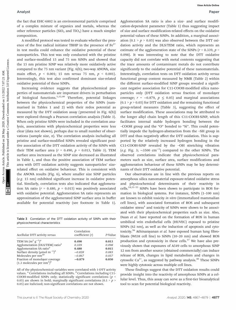
the fact that EHC-6802 is an environmental particle comprisedof a complex mixture of organics and metals, whereas theother reference particles (SiO2 and TiO2) have a much simplercomposition.
A modified protocol was tested to evaluate whether the pres-ence of the free radical initiator TBHP in the presence of Fe2+
in test media could enhance the oxidative potential of thesenanoparticles. This test was only conducted with the pristineand surface-modified 15 and 75 nm SiNPs and showed thatthe 15 nm pristine SiNP was relatively more oxidatively activecompared to the 75 nm variant (Fig. 6(b); two-way ANOVA, sizemain effect, p < 0.001; 15 nm versus 75 nm, p < 0.001).Interestingly, this test also confirmed dominant size-relatedoxidative potential of these SiNPs.
Increasing evidence suggests that physicochemical pro-perties of nanomaterials are important drivers in perturbationof biological functions.1,12,40 Therefore, potential linkagesbetween the physicochemical properties of the SiNPs (sum-marised in Tables 1 and 2) with their redox potential asmeasured by the DTT oxidation assay (presented in Fig. 6(b))were explored through a Pearson correlation analysis (Table 3).When only pristine SiNPs were included in the correlation ana-lysis, associations with physicochemical properties were lessclear (data not shown), perhaps due to small number of obser-vations (sample size, n). The correlation analysis including allpristine and surface-modified SiNPs revealed significant posi-tive association of the DTT oxidation activity of the SiNPs withtheir TEM surface area (r = 0.490, p = 0.013, Table 3). TEMsurface area increased as the SiNP size decreased as illustratedin Table 1, and thus the positive association of TEM surfacearea with DTT oxidation activity suggests nanoparticles’ size-related effect on oxidative behaviour. This is consistent withthe ANOVA results (Fig. 6), where smaller size SiNP particles(e.g. 15 nm) exhibited significant increase in oxidative poten-tial. Similarly, correlation tests also indicated that agglomera-tion SA ratio (r = 0.480, p = 0.015) was positively associatedwith oxidative potential. Agglomeration SA ratio represents anapproximation of the agglomerated SiNP surface area in bufferavailable for potential reactivity (see footnote in Table 1).
Agglomeration SA ratio is also a size- and surface modifi-cation-dependent parameter (Table 1) thus suggesting impactof size and surface modification-related effects on the oxidativepotential values of these SiNPs. In addition, a marginal associ-ation (0.1 > p > 0.05) was also observed between the DTT oxi-dation activity and the DLS/TEM ratio, which represents anestimate of the agglomeration state of the SiNPs (r = 0.339, p =0.098). It was interesting to note that the DTT oxidativecapacity did not correlate with metal contents suggesting thatthe trace amounts of contaminant metals do not contributesignificantly to the oxidative potential of these nanomaterials.Interestingly, correlation tests on DTT oxidation activity versusfunctional group content measured by NMR (Table 2) withinthe different surface-modified SiNP groups revealed a signifi-cant negative association for C11-COOH-modified silica nano-particles only (DTT oxidation versus fraction of monolayercoverage; r = −0.879, p ≤ 0.05) and marginal associations(0.1 > p > 0.05) for DTT oxidation and the remaining functionalgroup-related measures (Table 3), suggesting the effect ofsurface modification. These associations may be attributed tothe longer alkyl chain length of this C11-COOH-SiNP, whichfacilitates internal stable hydrogen bonding between the–COOH group and the “O” bonded to “Si”. This could poten-tially impede the hydrogen-abstraction from the –SH group inDTT and thus negatively affect the DTT oxidation. This is sup-ported by the relatively increased hydrogen bonding in theC11-COOH-SiNP revealed by the –OH stretching vibration(e.g. Fig. 3; ∼3500 cm−1) compared to the other SiNPs. Theobserved correlations indicate that physicochemical para-meters such as size, surface area, surface modifications andagglomeration behaviour of these SiNPs may be key determi-nants of their DTT oxidative potential.
Our observations are in line with the previous reports onamorphous silica nanomaterial exposure-related oxidative stressand physicochemical determinants of their reactivity incells.18,53–55 SiNPs have been shown to participate in ROS for-mation in biological systems. For instance, SiNPs (1–100 nm)are known to exhibit toxicity in vitro (immortalized mammaliancell lines), with associated formation of ROS and subsequentoxidative stress1 and toxicity of SiNPs were shown to be associ-ated with their physicochemical properties such as size. Also,Duan et al. have reported on the formation of ROS in humanumbilical vein endothelial cells (HUVEC) exposed to pristineSiNPs (62 nm), as well as the induction of apoptosis and cyto-toxicity.56 Athinarayanan et al. have exposed human lung fibro-blasts (WI38 cell line) to SiNPs (10–20 nm) and showed ROSproduction and cytotoxicity in these cells.57 We have also pre-viously shown that exposures of A549 cells to amorphous SiNP12 nm from another source (obtained commercially) can inducerelease of ROS, changes in lipid metabolism and changes incytosolic Ca2+, as suggested by pathway analysis.40 These SiNPswere highly cytotoxic across multiple cell lines.
These findings suggest that the DTT oxidation results couldprovide insight into the reactivity of amorphous SiNPs at a cel-lular level. Thus, this assay can serve as a first-tier bioanalyticaltool to scan for potential biological reactivity.
Table 3 Correlation of the DTT oxidation activity of SiNPs with theirphysicochemical characteristics
Acellular DTT activity versusCorrelationcoefficient (r) P-Value
TEM SA (m2 g−1)a 0.490 0.013Agglomeration (DLS/TEM) ratioa 0.339 0.098Agglomeration SA ratioa 0.480 0.015Surface density (μmol m−2)b −0.828 0.083Molecules per nm2b −0.867 0.057Fraction of monolayer coverage(1.3 molecules per nm2)b
−0.879 0.050
All of the physicochemical variables were correlated with 1-DTT activityvalues. aCorrelations including all SiNPs. b Correlations including C11-COOH-modified SiNPs only; statistically significant correlations (p <0.05) are shown in bold; marginally significant correlations (0.1 > p >0.05) are italicized; non-significant correlations are not shown.
Analyst Paper
This journal is © The Royal Society of Chemistry 2020 Analyst, 2020, 145, 4867–4879 | 4877
Ope
n A
cces
s A
rtic
le. P
ublis
hed
on 2
8 M
ay 2
020.
Dow
nloa
ded
on 1
/1/2
022
7:07
:21
AM
. T
his
artic
le is
lice
nsed
und
er a
Cre
ativ
e C
omm
ons
Attr
ibut
ion
3.0
Unp
orte
d L
icen
ce.
View Article Online

Conclusion
Smaller-sized amorphous pristine SiNPs (15 nm) exhibitgreater potential to oxidize DTT, as well as to agglomerate com-pared to relatively larger-sized SiNPs. Also, physicochemicalproperties including TEM-derived surface area, impacted bysize of SiNPs and agglomeration SA ratio, impacted by SiNPsize and surface modification were positively correlated withthe oxidative potential of these amorphous SiNP variants,while surface coverage of organics was negatively correlatedwith the DTT oxidation only for the C11-COOH-modifiedSiNPs, also exhibiting the effect of surface modification. Thisstudy reveals the capacity of the DTT assay to serve as an initialacellular screening tool to predict SiNP reactivities, and futurework on these SiNPs in biological systems can unravel therelationship between their oxidative potential and related cel-lular toxicity characteristics.
Conflicts of interest
There are no conflicts of interest to declare.
Acknowledgements
Chemicals Management Plan (CMP) and NanotechnologySection nano research fund from NSACB, Health Canada. Thepanel of the amorphous SiNPs was synthesized by AppliedQuantum Materials Inc. in Edmonton, AB, Canada. We aregrateful to Dr Nimal DeSilva at the Department of Earth andEnvironmental Sciences, University of Ottawa for conductingthe ICP-MS/AES analysis. We are grateful to Drs Dharani Dasand Yong-Lai Feng at Health Canada, for critical reviews of themanuscript. We thank Bing Wang, Health Canada and DanielHong (CO-OP student, University of Waterloo) for DLS techni-cal assistance, Monica Sourial (Capstone course field place-ment student, Carleton University) for her assistance with theDTT assay and Floyd Toll, NRC for assistance with BETmeasurements.
References
1 D. Napierska, L. C. Thomassen, D. Lison, J. A. Martens andP. H. Hoet, Part. Fibre Toxicol., 2010, 7, 39.
2 L. R. Khot, S. Sankaran, J. M. Maja, R. Ehsani andE. W. Schuster, Crop Prot., 2012, 35, 64–70.
3 M. R. Kasaai, J. Nanotechnol., 2015, 2015, 852394.4 A. Brinch, S. F. Hansen, N. B. Hartmann and A. Baun,
Nanomaterials, 2016, 6, 33.5 V. Vijayanathan, T. Thomas and T. J. Thomas, Biochemistry,
2002, 41, 14085–14094.6 L. R. Hirsch, R. J. Stafford, J. A. Bankson, S. R. Sershen,
B. Rivera, R. E. Price, J. D. Hazle, N. J. Halas and J. L. West,Proc. Natl. Acad. Sci. U. S. A., 2003, 100, 13549–13554.
7 M. Benezra, O. Penate-Medina, P. B. Zanzonico, D. Schaer,H. Ow, A. Burns, E. DeStanchina, V. Longo, E. Herz, S. Iyer,J. Wolchok, S. M. Larson, U. Wiesner and M. S. Bradbury,J. Clin. Invest., 2011, 121, 2768–2780.
8 A. Bitar, N. M. Ahmad, H. Fessi and A. Elaissari, DrugDiscovery Today, 2012, 17, 1147–1154.
9 L. Tang and J. Cheng, Nano Today, 2013, 8, 290–312.10 J. Chen, Z. Guo, H. Tian and X. Chen, Mol. Ther. – Methods
Clin. Dev., 2016, 3, 1602.11 WHO, Nanotechnology and human health: Scientific evi-
dence and risk governance, Report of the WHO expertmeeting 10–11 December 2012, Bonn, Germany,Copenhagen, WHO Regional Office for Europe, 2013.
12 S. Murugadoss, D. Lison, L. Godderis, S. Van Den Brule,J. Mast, F. Brassinne, N. Sebaihi and P. H. Hoet, Arch.Toxicol., 2017, 91, 2967–3010.
13 J. Y. Kim, J. H. Park, M. Kim, H. Jeong, J. Hong,R. S. Chuck and C. Y. Park, Sci. Rep., 2017, 7, 14566.
14 L. Chen, J. Liu, Y. Zhang, G. Zhang, Y. Kang, A. Chen,X. Feng and L. Shao, Nanomedicine, 2018, 13, 1939–1962.
15 Z. Du, S. Chen, G. Cui, Y. Yang, E. Zhang, Q. Wang,M. F. Lavin, A. J. Yeo, C. Bo, Y. Zhang, C. Li, X. Liu,X. Yang, C. Peng and H. Shao, Int. J. Mol. Med., 2019, 43,1229–1240.
16 K. Lee, J. Lee, M. Kwak, Y. L. Cho, B. Hwang, M. J. Cho,N. G. Lee, J. Park, S. H. Lee, J. G. Park, Y. G. Kim, J. S. Kim,T. S. Han, H. S. Cho, Y. J. Park, S. J. Lee, H. G. Lee,W. K. Kim, I. C. Jeung, N. W. Song, K. H. Bae and J. K. Min,J. Nanobiotechnol., 2019, 17, 24.
17 H. Zhang, D. R. Dunphy, X. Jiang, H. Meng, B. Sun,D. Tarn, M. Xue, X. Wang, S. Lin, Z. Ji, R. Li, F. L. Garcia,J. Yang, M. L. Kirk, T. Xia, J. I. Zink, A. Nel andC. J. Brinker, J. Am. Chem. Soc., 2012, 134, 15790–15804.
18 E. J. Park and K. Park, Toxicol. Lett., 2009, 184, 18–25.19 F. Wang, F. Gao, M. Lan, H. Yuan, Y. Huang and J. Liu,
Toxicol. In Vitro, 2009, 23, 808–815.20 Y. Ye, J. Liu, M. Chen, L. Sun and M. Lan, Environ. Toxicol.
Pharmacol., 2010, 29, 131–137.21 C. Terzano, F. Di Stefano, V. Conti, E. Graziani and
A. Petroianni, Eur. Rev. Med. Pharmacol. Sci., 2010, 14, 809–821.
22 M. Lodovici and E. Bigagli, J. Toxicol., 2011, 2011,487074.
23 A. Valavanidis, T. Vlachogianni, K. Fiotakis and S. Loridas,Int. J. Environ. Res. Public Health, 2013, 10, 3886–3907.
24 P. J. Borm, F. Kelly, N. Künzli, R. P. Schins andK. Donaldson, Occup. Environ. Med., 2007, 64, 73–74.
25 N. A. H. Janssen, A. Yang, M. Strak, M. Steenhof,B. Hellack, M. E. Gerlofs-Nijland, T. Kuhlbusch, F. Kelly,R. Harrison, B. Brunekreef, G. Hoek and F. Cassee, Sci.Total Environ., 2014, 472, 572–581.
26 V. Bonvallot, A. Baeza-Squiban, A. Baulig, S. Brulant,S. Boland, F. Muzeau, R. Barouki and F. Marano,Am. J. Respir. Cell Mol. Biol., 2001, 25, 515–521.
27 A. Kubátová, L. C. Dronen, M. J. Picklo andS. B. Hawthorne, Chem. Res. Toxicol., 2006, 19, 255–261.
Paper Analyst
4878 | Analyst, 2020, 145, 4867–4879 This journal is © The Royal Society of Chemistry 2020
Ope
n A
cces
s A
rtic
le. P
ublis
hed
on 2
8 M
ay 2
020.
Dow
nloa
ded
on 1
/1/2
022
7:07
:21
AM
. T
his
artic
le is
lice
nsed
und
er a
Cre
ativ
e C
omm
ons
Attr
ibut
ion
3.0
Unp
orte
d L
icen
ce.
View Article Online
28 T. Xia, M. Kovochich, J. Brant, M. Hotze, J. Sempf,T. Oberley, C. Sioutas, J. I. Yeh, M. R. Wiesner andA. E. Nel, Nano Lett., 2006, 6, 1794–1807.
29 T. Shi, R. P. F. Schins, A. M. Knaapen, T. Kuhlbusch,M. Pitz, J. Heinrich and P. J. A. Borm, J. Environ. Monit.,2003, 5, 550–556.
30 A. K. Cho, C. Sioutas, A. H. Miguel, Y. Kumagai,D. A. Schmitz, M. Singh, A. Eiguren-Fernandez andJ. R. Froines, Environ. Res., 2005, 99, 40–47.
31 I. S. Mudway, G. Fuller, D. Green, C. Dunster and F. J. Kelly,Report: Quantifying the London specific component ofPM10 oxidative activity, University of London, Defra, UK,2011.
32 B. Zomer, L. Collé, A. Jedyńska, G. Pasterkamp, I. Kooterand H. Bloemen, Anal. Bioanal. Chem., 2011, 401, 2945–2954.
33 Q. Xiong, H. Yu, R. Wang, J. Wei and V. Verma, Environ. Sci.Technol., 2017, 51, 6507–6514.
34 I. S. Mudway, S. T. Duggan, C. Venkataraman, G. Habib,F. J. Kelly and J. Grigg, Part. Fibre Toxicol., 2005, 2, 6.
35 J. G. Ayres, P. Borm, F. R. Cassee, V. Castranova,K. Donaldson, A. Ghio, R. M. Harrison, R. Hider, F. Kelly,I. M. Kooter, F. Marano, R. L. Maynard, I. Mudway, A. Nel,C. Sioutas, S. Smith, A. Baeza-Squiban, A. Cho, S. Dugganand J. Froines, Inhalation Toxicol., 2008, 20, 75–99.
36 P. J. O’Brien, Chem.-Biol. Interact., 1991, 80, 1–41.37 G. L. Squadrito, R. Cueto, B. Dellinger and W. A. Pryor, Free
Radicals Biol. Med., 2001, 31, 1132–1138.38 E. M. Thomson, A. Williams, C. L. Yauk and R. Vincent,
Part. Fibre Toxicol., 2009, 6, 6.39 F. Kunc, V. Balhara, A. Brinkmann, Y. Sun, D. M. Leek and
L. J. Johnston, Anal. Chem., 2018, 90, 13322–13330.40 D. Breznan, D. D. Das, J. S. O’Brien, C. MacKinnon-Roy,
S. Nimesh, N. Q. Vuong, S. Bernatchez, N. DeSilva, M. Hill,P. Kumarathasan and R. Vincent, Nanotoxicology, 2017, 11,223–235.
41 M. K. Kim and W. K. Jo, Int. Arch. Occup. Environ. Health,2006, 80, 40–50.
42 D. D. Das, Y. Yang, J. S. O’Brien, D. Breznan, S. Nimesh,S. Bernatchez, M. Hill, A. Sayari, R. Vincent andP. Kumarathasan, J. Nanomater., 2014, 12, e176015.
43 K. Bhattacharya, M. Davoren, J. Boertz, R. P. Schins,E. Hoffmann and E. Dopp, Part. Fibre Toxicol., 2009, 6, 17.
44 K. Jomova and M. Valko, Toxicology, 2011, 283, 65–87.45 E. Asadpour, H. R. Sadeghnia, A. Ghorbani and
M. T. Boroushaki, Iran. J. Pharm. Res., 2014, 13, 1141–1148.46 E. Cheraghi, A. Golkar, K. Roshanaei and B. Alani,
Int. J. Fertil. Steril., 2017, 11, 166–175.47 D. Schwotzer, M. Niehof, D. Schaudien, H. Kock,
T. Hansen, C. Dasenbrock and O. Creutzenberg,J. Nanobiotechnol., 2018, 16, 16.
48 F. Kunc, V. Balhara, Y. Sun, M. Daroszewska, Z. J. Jakubek,M. Hill, A. Brinkmann and L. J. Johnston, Analyst, 2019,144, 5589–5599.
49 Y. Kumagai, S. Koide, K. Taguchi, A. Endo, Y. Nakai,T. Yoshikawa and N. Shimojo, Chem. Res. Toxicol., 2002, 15,483–489.
50 M. Y. Chung, R. A. Lazaro, D. Lim, J. Jackson, J. Lyon,D. Rendulic and A. S. Hasson, Environ. Sci. Technol., 2006,40, 4880–4886.
51 Y. Wang, C. Arellanes, D. B. Curtis and S. E. Paulson,Environ. Sci. Technol., 2010, 44, 4070–4075.
52 L. K. Limbach, P. Wick, P. Manser, R. N. Grass, A. Bruininkand W. J. Stark, Environ. Sci. Technol., 2007, 41, 4158–4163.
53 H. Nabeshi, T. Yoshikawa, K. Matsuyama, Y. Nakazato,S. Tochigi, S. Kondoh, T. Hirai, T. Akase, K. Nagano, Y. Abe,Y. Yoshioka, H. Kamada, N. Itoh, S. Tsunoda andY. Tsutsumi, Part. Fibre Toxicol., 2011, 8, 1.
54 M. Ahamed, Hum. Exp. Toxicol., 2013, 32, 186–195.55 A. Nemmar, S. Beegam, P. Yuvaraju, J. Yasin, A. Shahin and
B. H. Ali, Cell. Physiol. Biochem., 2014, 34, 255–265.56 J. Duan, Y. Yu, Y. Li, Y. Yu and Z. Sun, Biomaterials, 2013,
34, 5853–5862.57 J. Athinarayanan, V. S. Periasamy, M. A. Alsaif, A. A. Al-
Warthan and A. A. Alshatwi, Cell Biol. Toxicol., 2014, 30,89–100.
Analyst Paper
This journal is © The Royal Society of Chemistry 2020 Analyst, 2020, 145, 4867–4879 | 4879
Ope
n A
cces
s A
rtic
le. P
ublis
hed
on 2
8 M
ay 2
020.
Dow
nloa
ded
on 1
/1/2
022
7:07
:21
AM
. T
his
artic
le is
lice
nsed
und
er a
Cre
ativ
e C
omm
ons
Attr
ibut
ion
3.0
Unp
orte
d L
icen
ce.
View Article Online