Accumulated Hard Tissue Debris Produced during Reciprocating ...
6
Accumulated Hard Tissue Debris Produced during Reciprocating and Rotary Nickel-Titanium Canal Preparation Gustavo De-Deus, DDS, MSc, PhD,* Juliana Marins, DDS, MSc, PhD, † Emmanuel Jo~ ao Nogueira Leal Silva, DDS, MSc, PhD,* Erick Souza, DDS, MSc, PhD, ‡ Felipe Gonc ¸alves Belladonna, DDS, MSc, § Claudia Reis, DDS, MSc, PhD, ¶ Alessandra Silveira Machado, MSc, k Ricardo Tadeu Lopes, MSc, DSc, k Marco Aur elio Versiani, DDS, MSc, PhD,** Sidnei Paciornik, MSc, PhD, †† and Aline Almeida Neves, DDS, MSc, PhD ‡‡ Abstract Introduction: This study compared the amount of hard tissue debris produced after different apical enlarge- ment with single-file reciprocating systems (WaveOne [Dentsply Maillefer, Baillaigues, Switzerland] and Reciproc [VDW, Munich, Germany]) and a conventional multifile rotary system (BioRaCe [FKG Dentaire, La- Chaux-de-Fonds, Switzerland]) using micro–computed tomographic imaging. Methods: Thirty moderately curved mesial roots of mandibular molars presenting 2 independent root canals were selected and scanned at an isotropic resolution of 14.16 mm. The sample was as- signed to 3 groups (n = 10) with respect to the root length and degree of curvature of the mesial root ac- cording to the system used for the root canal prepara- tion: Reciproc, WaveOne, and BioRaCe. Second and third scans were taken after the root canals were pre- pared up to ISO sizes 25 and 40, respectively. The matched images of the mesial canals, before and after preparation, were examined from the furcation level to the apex to evaluate the amount of hard tissue debris (%). Data were statistically compared using a general linear model for repeated-measures with a significance level set at 5%. Results: Instrumentation systems per se did not influence the amount of hard tissue accumu- lation (P > .05), whereas a significant reduction in the percentage of hard tissue debris was observed after sequential enlargement in all groups (P < .05). Conclu- sions: None of the systems yielded root canals completely free from packed hard tissue debris. The increased final apical size resulted in significantly less debris accumulation for both reciprocating and rotary systems. (J Endod 2015;41:676–681) Key Words Hard tissue debris, micro–computed tomographic imaging, nickel-titanium, recipro- cating motion, root canal preparation, rotary system I n 2011, Paqu e et al (1) reopened the discussion about the substantial amount of debris packed in the fins, isthmuses, irregularities, and ramifications of the root canal system after preparation using an innovative approach based on micro–computed tomographic (micro-CT) imaging. Micro-CT imaging allows monitoring the accumula- tion and removal of radiopaque structures in the main space of the root canal and its recesses and isthmuses during and after instrumentation (2–4) while preserving the sample integrity (5). Hard tissue debris accumulation has been considered an undesirable side effect of the shaping procedures and may be considered more clinically relevant than the smear layer per se because it could easily harbor bacterial contents away from the disinfection procedures (1). It has been shown that the currently used irrigation systems and solutions are unable to render root canals completely free from packed hard tissue debris (1–4, 6–8), which raises the need for preparation protocols that reduce debris buildup (3). The introduction of new preparation systems based on the use of only 1 instrument through a reciprocating motion has raised new perspectives for the mechanical prep- aration of the root canal space (5). The reciprocating working motion consists on a forward counterclockwise rotation to cut dentin and a shorter clockwise rotation to relieve the instrument (9). Overall, research findings on reciprocating systems report a decrease in preparation time (9), increased cyclic fatigue life (10), and similar shaping ability compared with multiple-file systems (5, 11, 12). In the literature, it remains controversial whether the use of reciprocating movement per se is able to influence the final amount of packed hard tissues debris in the root canal (3, 9). Likewise, the impact of further apical enlargement on the accumulation of hard tissue debris is still unknown. Within this background, the current study was designed to compare the volume of hard tissue debris accumulated in mesial root canals of mandibular molars prepared by single-file reciprocating systems (WaveOne [Dentsply Maillefer, Baillaigues, From the *Department of Endodontics, Grande Rio University, Duque de Caxias, Rio de Janeiro; † Department of Endodontics, Rio de Janeiro State University, Rio de Janeiro, Rio de Janeiro; ‡ Department of Dentistry II, Federal University of Maranh~ ao, S~ ao Luis, Maranh~ ao; § Department of Endodontics, Fluminense Federal University, Niter oi, Rio de Janeiro ¶ Department of Endodontics, University of Esp ırito Santo, Vit oria, Esp ırito Santo; k Nuclear Engineering Program, Federal University of Rio de Janeiro, Rio de Janeiro, Rio de Janeiro; **Department of Restorative Dentistry, Dental School of Ribeir~ ao Preto, University of S~ ao Paulo, S~ ao Paulo, S~ ao Paulo; †† Department of Material Engineering, PUC-Rio, Rio de Janeiro, Rio de Janeiro; and ‡‡ Department of Pediatric Dentistry and Orthodontics, Federal University of Rio de Janeiro, Rio de Janeiro, Rio de Janeiro, Brazil. Address requests for reprints to Prof Gustavo De-Deus, Av Henrique Dodsworth, 85, Apto. 808-Lagoa, Rio de Janeiro, RJ, Brazil 22061-030. E-mail address: [email protected] 0099-2399/$ - see front matter Copyright ª 2015 American Association of Endodontists. http://dx.doi.org/10.1016/j.joen.2014.11.028 Basic Research—Technology 676 De-Deus et al. JOE — Volume 41, Number 5, May 2015
Transcript of Accumulated Hard Tissue Debris Produced during Reciprocating ...
Basic Research—Technology
Accumulated Hard Tissue Debris Produced duringReciprocating and Rotary Nickel-Titanium Canal PreparationGustavo De-Deus, DDS, MSc, PhD,* Juliana Marins, DDS, MSc, PhD,†
Emmanuel Jo~ao Nogueira Leal Silva, DDS, MSc, PhD,* Erick Souza, DDS, MSc, PhD,‡
Felipe Goncalves Belladonna, DDS, MSc,§ Claudia Reis, DDS, MSc, PhD,¶
Alessandra Silveira Machado, MSc,kRicardo Tadeu Lopes, MSc, DSc,
k
Marco Aur�elio Versiani, DDS, MSc, PhD,** Sidnei Paciornik, MSc, PhD,††
and Aline Almeida Neves, DDS, MSc, PhD‡‡
Abstract
Introduction: This study compared the amount of hardtissue debris produced after different apical enlarge-ment with single-file reciprocating systems (WaveOne[Dentsply Maillefer, Baillaigues, Switzerland] andReciproc [VDW, Munich, Germany]) and a conventionalmultifile rotary system (BioRaCe [FKG Dentaire, La-Chaux-de-Fonds, Switzerland]) using micro–computedtomographic imaging. Methods: Thirty moderatelycurved mesial roots of mandibular molars presenting 2independent root canals were selected and scanned atan isotropic resolution of 14.16 mm. The sample was as-signed to 3 groups (n = 10) with respect to the rootlength and degree of curvature of the mesial root ac-cording to the system used for the root canal prepara-tion: Reciproc, WaveOne, and BioRaCe. Second andthird scans were taken after the root canals were pre-pared up to ISO sizes 25 and 40, respectively. Thematched images of the mesial canals, before and afterpreparation, were examined from the furcation levelto the apex to evaluate the amount of hard tissue debris(%). Data were statistically compared using a generallinear model for repeated-measures with a significancelevel set at 5%. Results: Instrumentation systems perse did not influence the amount of hard tissue accumu-lation (P > .05), whereas a significant reduction in thepercentage of hard tissue debris was observed aftersequential enlargement in all groups (P < .05). Conclu-sions: None of the systems yielded root canalscompletely free from packed hard tissue debris. Theincreased final apical size resulted in significantly lessdebris accumulation for both reciprocating and rotarysystems. (J Endod 2015;41:676–681)From the *Department of Endodontics, Grande Rio University, DuJaneiro, Rio de Janeiro; ‡Department of Dentistry II, Federal UniversiNiter�oi, Rio de Janeiro ¶Department of Endodontics, University of EJaneiro, Rio de Janeiro, Rio de Janeiro; **Department of Restora††Department of Material Engineering, PUC-Rio, Rio de Janeiro, Riode Janeiro, Rio de Janeiro, Rio de Janeiro, Brazil.
Address requests for reprints to Prof Gustavo De-Deus, Av [email protected]/$ - see front matter
Copyright ª 2015 American Association of Endodontists.http://dx.doi.org/10.1016/j.joen.2014.11.028
676 De-Deus et al.
Key WordsHard tissue debris, micro–computed tomographic imaging, nickel-titanium, recipro-cating motion, root canal preparation, rotary system
In 2011, Paqu�e et al (1) reopened the discussion about the substantial amount ofdebris packed in the fins, isthmuses, irregularities, and ramifications of the root canal
system after preparation using an innovative approach based on micro–computedtomographic (micro-CT) imaging. Micro-CT imaging allows monitoring the accumula-tion and removal of radiopaque structures in the main space of the root canal and itsrecesses and isthmuses during and after instrumentation (2–4) while preserving thesample integrity (5).
Hard tissue debris accumulation has been considered an undesirable side effect ofthe shaping procedures and may be considered more clinically relevant than the smearlayer per se because it could easily harbor bacterial contents away from the disinfectionprocedures (1). It has been shown that the currently used irrigation systems and solutionsare unable to render root canals completely free from packed hard tissue debris (1–4,6–8), which raises the need for preparation protocols that reduce debris buildup (3).
The introduction of new preparation systems based on the use of only 1 instrumentthrough a reciprocating motion has raised new perspectives for the mechanical prep-aration of the root canal space (5). The reciprocating working motion consists on aforward counterclockwise rotation to cut dentin and a shorter clockwise rotation torelieve the instrument (9). Overall, research findings on reciprocating systems reporta decrease in preparation time (9), increased cyclic fatigue life (10), and similarshaping ability compared with multiple-file systems (5, 11, 12). In the literature, itremains controversial whether the use of reciprocating movement per se is able toinfluence the final amount of packed hard tissues debris in the root canal (3, 9).Likewise, the impact of further apical enlargement on the accumulation of hardtissue debris is still unknown.
Within this background, the current study was designed to compare the volume ofhard tissue debris accumulated in mesial root canals of mandibular molars prepared bysingle-file reciprocating systems (WaveOne [Dentsply Maillefer, Baillaigues,
que de Caxias, Rio de Janeiro; †Department of Endodontics, Rio de Janeiro State University, Rio dety of Maranh~ao, S~ao Luis, Maranh~ao; §Department of Endodontics, Fluminense Federal University,sp�ırito Santo, Vit�oria, Esp�ırito Santo; kNuclear Engineering Program, Federal University of Rio detive Dentistry, Dental School of Ribeir~ao Preto, University of S~ao Paulo, S~ao Paulo, S~ao Paulo;de Janeiro; and ‡‡Department of Pediatric Dentistry and Orthodontics, Federal University of Rio
nrique Dodsworth, 85, Apto. 808-Lagoa, Rio de Janeiro, RJ, Brazil 22061-030. E-mail address:
JOE — Volume 41, Number 5, May 2015
Basic Research—Technology
Switzerland] and Reciproc [VDW, Munich, Germany]) and a conven-tional multifile rotary system (BioRaCe [FKG Dentaire, La-Chaux-de-Fonds, Switzerland]) at 2 different apical sizes using micro-CTtechnology. The following hypotheses were tested:1. Single-file reciprocating systems produce less hard tissue debrisaccumulation than a conventional multifile system.
2. Single-file reciprocating systems produce similar hard tissue accu-mulation.
3. Larger apical preparation is an effective antidebris strategy.
Materials and MethodsSample Size Estimation
A repeated measures analysis of variance, within-between inter-action was selected from the F tests family in G*Power 3.1.7 softwarefor Windows (Heinrich Heine, Universit€at D€usseldorf). Because of thelack of previous studies evaluating hard tissue debris accumulationafter canal preparation with reciprocating instruments at differentapical enlargements, the effect size for this study was establishedfrom the medium convention (0.2526, derived from n2 = 0.06).An alpha-type error of 0.05, power beta of 0.95, correlation amongrepeated measures of 0.7, nonsphericity correction of 1, numberof groups (within subjects) of 2, and number of measurements (be-tween subjects) of 3 were also specified. Twenty-six teeth were indi-cated as the total sample size required to observe significantdifferences.
Specimen SelectionFrom a pool of 300 mandibular first molar teeth, moderately
curved mesial roots (10�–20�) were selected applying Schneider’smethod (13) in digitized buccolingual radiographs using AxioVision4.5software (Carl Zeiss Vision GmbH, Hallbergmoos, Germany). In addi-tion, the inclusion criteria comprised only mandibular molars present-ing 2 independent root canals in the mesial root (Vertucci type IIconfiguration system) in which the final apical gauging allowed foran ISO size 10 hand K-file (Dentsply Maillefer) to be placed to the work-ing length (WL). Based on these criteria, 44 mesial molar roots wereselected. After resection of the distal root at the furcation level, 14 teethwere discarded, and 30 mesial roots were disinfected in 0.5% chlora-mine T, stored in distilled water at 4�C, and used within 6 months afterextraction.
The specimens were randomly assigned (http://www.random.org) into 3 experimental groups (n = 10) according to the systemused for the chemomechanical preparation: Reciproc, WaveOne,and BioRaCe. After checking for normality assumption (P > .05,Shapiro-Wilk test), the degree of homogeneity of the groups, withrespect to the root length and degree of curvature of the mesialroot, was statistically confirmed (P> .05, 2-way analysis of variance).For the experimental procedures, the apex of the mesial roots wassealed with hot glue, and the roots were embedded into a thin filmof polyvinyl siloxane.
Root Canal PreparationRoot canals were accessed and patency confirmed by inserting a
size 10 hand K-file through the apical foramen before and after comple-tion of root canal preparation. For all groups, a glide path was createdby scouting a stainless steel size 15 K-file (Dentsply Maillefer) up to theWL, which was established by deducting 1 mm from the canal length. Ineach group, instruments were driven with the VDW Silver motor (VDWGmbH) according to each manufacturer’s instructions, and a singleexperienced operator performed all preparations.
JOE — Volume 41, Number 5, May 2015
Group 1: Reciproc SystemReciproc R25 (25/0.08) (VDW GmbH) was introduced into the
canal until resistance was felt and then activated in reciprocating mo-tion. The instrument was moved in an apical direction using an in-and-out pecking motion of about 3 mm in amplitude with a light apicalpressure. After 3 pecking motions, the instrument was removed fromthe canal, and its flutes were cleaned off. This procedure was performeduntil the instrument reached the WL. Afterward, the Reciproc R40 in-strument (40/0.06) (VDW GmbH) was used with the same protocol.
Group 2: WaveOne SystemWaveOne Primary (25/0.08) and Large (40/0.08) instruments
(Dentsply Maillefer) were used to the WL following the same protocoldescribed for group 1.
Group 3: BioRaCe SystemBioRaCe instruments (FKG Dentaire) were used in a crown-down
manner according to the manufacturer’s instructions using thefollowing sequence: BR0 (25/0.08), BR1 (15/0.05), BR2 (25/0.04),BR3 (25/0.06), BR4 (35/0.04), and BR5 (40/0.04) instruments. Themotor was adjusted to 500–600 rpm and 1 N/cm2. After 4 gentle in-and-out motion strokes, the instrument was removed from the canaland cleaned until the WL was reached.
After the glide path and each nickel-titanium file, root canals wereirrigated with 2 mL 5.25% NaOCl for 1 minute delivered by a VATEAperistaltic pump (ReDent-Nova, Ra’anana, Israel) at a 2-mL/min rateconnected to a 30-G Endo-Eze Tip (Ultradent Products Inc, South Jor-dan, UT) inserted up to 2 mm from the apical foramen. Aspiration wasperformed with a SurgiTip (Ultradent Products Inc) attached to a high-speed suction pump. After canal preparation to size 40, an additionalrinse with either 18 mL/9 min (for the rotary group) or 24 mL/12min (for the reciprocating groups) NaOCl was performed to equalizethe amount and time of irrigant used within the groups. A final rinse with5 mL 17% EDTA (pH = 7.7) delivered at a 1-mL/min rate for 5 minutesfollowed by a 5-minute 5-mL rinse with bidistilled water was performedfor both groups. Hence, a total volume of 40 mL irrigant was used percanal in a total time of 25 minutes. Then, canals were dried with absor-bent paper points (Dentsply Maillefer).
Micro-CT ScanningThree high-resolution scans were accomplished per tooth:
1. Before treatment2. After root canal preparation up to ISO size 253. After root canal preparation up to ISO size 40
Teeth were placed inside a custom-made epoxy resin holder (Ø =18 mm) and adapted into a sample holder of a micro-CT device (Sky-Scan 1173; Bruker-microCT, Kontich, Belgium). Scanning procedureswere performed at an isotropic resolution of 14.16mm, 70 kV, 114mA,360� rotation around the vertical axis, rotation step of 0.5�, and cameraexposure time of 250 milliseconds. Acquired projection images werereconstructed into cross-sectional slices (NRecon v.1.6.9; Bruker-microCT) using standardized parameters for beam hardening (40%)and ring artifact correction of 10 as well as similar contrast limits.The volume of interest was selected extending from the furcation levelto the apex of the root, resulting in the acquisition of 700 to 900 trans-verse cross sections per tooth.
Quantitative Tridimensional Image AnalysisEvaluation procedures have been described elsewhere in details
(4). Image stacks, before and after canal preparation, were registered
Hard Tissue Debris 677
Basic Research—Technology
using an automatic superimposition, and volumes of matched root ca-nals before and after preparation were calculated. Material with densitysimilar to dentin in instrumented canal regions, which were previouslyoccupied by air, was considered to be debris (2, 4). Accumulated hardtissue debris was calculated as the percentage volume of the originalcanal anatomy after intersecting the stacks of original andinstrumented root canal space. All image analysis operations wereundertaken using the ImageJ software v.1.49n implementation (Fiji,Madison, WI) interface (14). Subsequently, the images obtained afterdebris quantification were 3-dimensionally rendered using the plugin3D Viewer (Internationale Medieninformatik, HTW Berlin, Berlin, Ger-many) and qualitatively evaluated with CTVol v.2.2.1 software (Bruker-microCT).Statistical AnalysisRaw data normal distribution was confirmed using the Shapiro-
Wilk test (P> .05). Because of the dependent nature of the study design,a general linear model for repeatedmeasures (SPSS for Windows v17.0;SPSS Inc, Chicago, IL) was selected for analysis. File sizes were tested asthe within-subject effects, whereas instrumentation systems were set asbetween-subject effects. Significance was established at a = 5%.
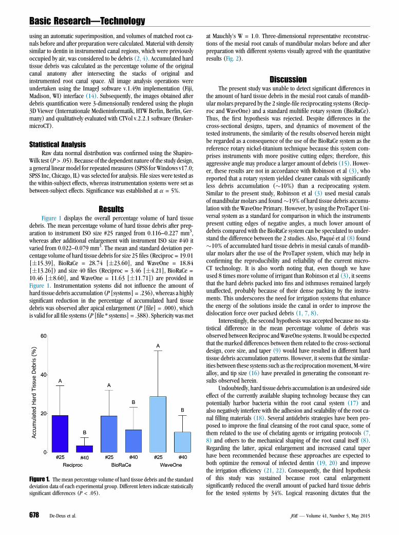
ResultsFigure 1 displays the overall percentage volume of hard tissue
debris. The mean percentage volume of hard tissue debris after prep-aration to instrument ISO size #25 ranged from 0.116–0.227 mm3,whereas after additional enlargement with instrument ISO size #40 itvaried from 0.022–0.079 mm3. The mean and standard deviation per-centage volume of hard tissue debris for size 25 files (Reciproc = 19.01[�15.39], BioRaCe = 28.74 [�23.60], and WaveOne = 18.84[�13.26]) and size 40 files (Reciproc = 3.46 [�4.21], BioRaCe =10.46 [�8.60], and WaveOne = 11.63 [�11.71]) are provided inFigure 1. Instrumentation systems did not influence the amount ofhard tissue debris accumulation (P [systems] = .236), whereas a highlysignificant reduction in the percentage of accumulated hard tissuedebris was observed after apical enlargement (P [file] = .000), whichis valid for all file systems (P [file * systems] = .388). Sphericity wasmet
Figure 1. The mean percentage volume of hard tissue debris and the standarddeviation data of each experimental group. Different letters indicate statisticallysignificant differences (P < .05).
678 De-Deus et al.
at Mauchly’s W = 1.0. Three-dimensional representative reconstruc-tions of the mesial root canals of mandibular molars before and afterpreparation with different systems visually agreed with the quantitativeresults (Fig. 2).
DiscussionThe present study was unable to detect significant differences in
the amount of hard tissue debris in the mesial root canals of mandib-ular molars prepared by the 2 single-file reciprocating systems (Recip-roc and WaveOne) and a standard multifile rotary system (BioRaCe).Thus, the first hypothesis was rejected. Despite differences in thecross-sectional designs, tapers, and dynamics of movement of thetested instruments, the similarity of the results observed herein mightbe regarded as a consequence of the use of the BioRaCe system as thereference rotary nickel-titanium technique because this system com-prises instruments with more positive cutting edges; therefore, thisaggressive angle may produce a larger amount of debris (15). Howev-er, these results are not in accordance with Robinson et al (3), whoreported that a rotary system yielded cleaner canals with significantlyless debris accumulation (�10%) than a reciprocating system.Similar to the present study, Robinson et al (3) used mesial canalsof mandibular molars and found�19% of hard tissue debris accumu-lation with the WaveOne Primary. However, by using the ProTaper Uni-versal system as a standard for comparison in which the instrumentspresent cutting edges of negative angles, a much lower amount ofdebris compared with the BioRaCe system can be speculated to under-stand the difference between the 2 studies. Also, Paqu�e et al (8) found�10% of accumulated hard tissue debris in mesial canals of mandib-ular molars after the use of the ProTaper system, which may help inconfirming the reproducibility and reliability of the current micro-CT technology. It is also worth noting that, even though we haveused 8 times more volume of irrigant than Robinson et al (3), it seemsthat the hard debris packed into fins and isthmuses remained largelyunaffected, probably because of their dense packing by the instru-ments. This underscores the need for irrigation systems that enhancethe energy of the solutions inside the canal in order to improve thedislocation force over packed debris (1, 7, 8).
Interestingly, the second hypothesis was accepted because no sta-tistical difference in the mean percentage volume of debris wasobserved between Reciproc andWaveOne systems. It would be expectedthat the marked differences between them related to the cross-sectionaldesign, core size, and taper (9) would have resulted in different hardtissue debris accumulation patterns. However, it seems that the similar-ities between these systems such as the reciprocationmovement, M-wirealloy, and tip size (16) have prevailed in generating the consonant re-sults observed herein.
Undoubtedly, hard tissue debris accumulation is an undesired sideeffect of the currently available shaping technology because they canpotentially harbor bacteria within the root canal system (17) andalso negatively interfere with the adhesion and sealability of the root ca-nal filling materials (18). Several antidebris strategies have been pro-posed to improve the final cleansing of the root canal space, some ofthem related to the use of chelating agents or irrigating protocols (7,8) and others to the mechanical shaping of the root canal itself (8).Regarding the latter, apical enlargement and increased canal taperhave been recommended because these approaches are expected toboth optimize the removal of infected dentin (19, 20) and improvethe irrigation efficiency (21, 22). Consequently, the third hypothesisof this study was sustained because root canal enlargementsignificantly reduced the overall amount of packed hard tissue debrisfor the tested systems by 34%. Logical reasoning dictates that the
JOE — Volume 41, Number 5, May 2015
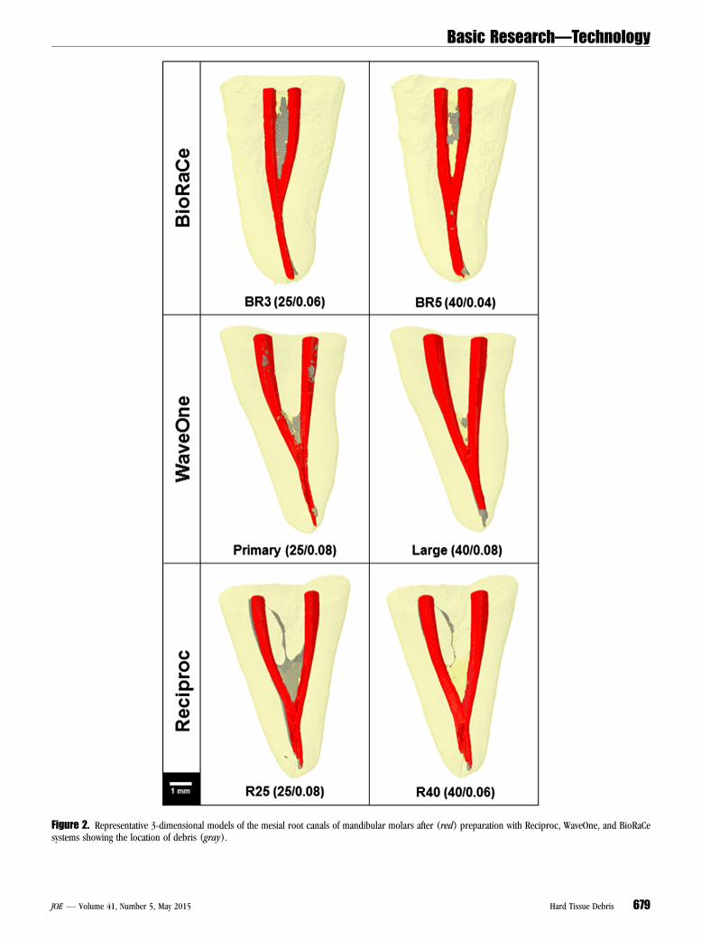
Figure 2. Representative 3-dimensional models of the mesial root canals of mandibular molars after (red) preparation with Reciproc, WaveOne, and BioRaCesystems showing the location of debris (gray).
Basic Research—Technology
JOE — Volume 41, Number 5, May 2015 Hard Tissue Debris 679
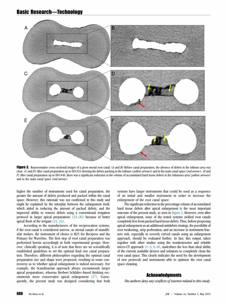
Figure 3. Representative cross-sectional images of a given mesial root canal. (A and B) Before canal preparation, the absence of debris in the isthmus area wasclear. (C and D) After canal preparation up to ISO #25 showing the debris packing in the isthmus (yellow arrows) and in the main canal space (red arrow). (E andF) After canal preparation up to ISO #40, there was a significant reduction in the volume of accumulated hard tissue debris in the isthmuses area (yellow arrows)and in the main canal space (red arrow).
Basic Research—Technology
higher the number of instruments used for canal preparation, thegreater the amount of debris produced and packed within the canalspace. However, this rationale was not confirmed in this study andmight be explained by the interplay between the enlargement itself,which aided in reducing the amount of packed debris, and theimproved ability to remove debris using a conventional irrigationprotocol in larger apical preparations (23–26) because of betterapical flush of the irrigant (21, 22).
According to the manufacturers of the reciprocation systems,if the root canal is considered narrow, as mesial canals of mandib-ular molars, the instrument of choice is R25 for Reciproc and thePrimary for WaveOne. The first step of root canal preparation wasperformed herein accordingly in both experimental groups. How-ever, clinically speaking, it is of note that there are no scientificallyestablished guidelines to set the optimal final size canal prepara-tion. Therefore, different philosophies regarding the optimal canalpreparation size and shape were proposed, resulting in some con-troversy as to whether apical enlargement is indeed necessary. Forexample, the Scandinavian approach always recommends largerapical preparations, whereas Herbert Schilder–based thinking rec-ommends more conservative apical enlargement (27). Conse-quently, the present study was designed considering that both
680 De-Deus et al.
systems have larger instruments that could be used as a sequenceof an initial and smaller instrument in order to increase theenlargement of the root canal space.
The significant reduction in the percentage volume of accumulatedhard tissue debris after apical enlargement is the most importantoutcome of the present study, as seen in Figure 3. However, even afterapical enlargement, none of the tested systems yielded root canalscompletely free from packed hard tissue debris. Thus, before proposingapical enlargement as an additional antidebris strategy, the possibility ofroot weakening, strip perforation, and an increase in instrument frac-ture risk, especially in severely curved canals using an enlargementapproach, should be evaluated further. In fact, this output, takentogether with other studies using the nondestructive and reliablemicro-CT approach (1–3, 6–8), underlines the less than ideal abilityof the current available devices and solutions to completely clean theroot canal space. This clearly indicates the need for the developmentof new protocols and instruments able to optimize the root canalspace cleaning.
AcknowledgmentsThe authors deny any conflicts of interest related to this study.
JOE — Volume 41, Number 5, May 2015
Basic Research—Technology
References1. Paqu�e F, Laib A, Gautschi H, Zehnder M. Hard-tissue debris accumulation analysis
by high-resolution computed tomography scans. J Endod 2009;35:1044–7.2. Robinson JP, Lumley PJ, Claridge E, et al. An analytical Micro CT methodology for
quantifying inorganic dentine debris following internal tooth preparation. J Dent2012;40:999–1005.
3. Robinson JP, Lumley PJ, Cooper PR, et al. Reciprocating root canal techniqueinduces greater debris accumulation than a continuous rotary technique as as-sessed by 3-dimensional micro-computed tomography. J Endod 2013;39:1067–70.
4. De-Deus G, Marins J, Neves AA, et al. Assessing accumulated hard-tissue debris us-ing micro-computed tomography and free software for image processing and anal-ysis. J Endod 2014;40:271–6.
5. Versiani MA, Steier L, De-Deus G, et al. Micro–computed tomography study of oval-shaped canals prepared with the Self-adjusting File, Reciproc, WaveOne, and Pro-Taper Universal systems. J Endod 2013;39:1060–6.
6. Endal U, Shen Y, Knut A, et al. A high-resolution computed tomographic study ofchanges in root canal isthmus area by instrumentation and root filling. J Endod2011;37:223–7.
7. Paqu�e F, Boessler C, Zehnder M. Accumulated hard tissue debris levels in mesialroots of mandibular molars after sequential irrigation steps. Int Endod J 2011;44:148–53.
8. Paqu�e F, Al-Jadaa A, Kfir A. Hard-tissue debris accumulation created by conven-tional rotary versus self-adjusting file instrumentation in mesial root canal systemsof mandibular molars. Int Endod J 2012;45:413–8.
9. B€urklein S, Hinschitza K, Dammaschke T, et al. Shaping ability and cleaning effec-tiveness of two single-file systems in severely curved root canals of extracted teeth:Reciproc and WaveOne versus Mtwo and ProTaper. Int Endod J 2012;45:449–61.
10. Kiefner P, Ban M, De-Deus G. Is the reciprocating movement per se able to improvethe cyclic fatigue resistance of instruments? Int Endod J 2014;47:430–6.
11. Stern S, Patel S, Foschi F, et al. Changes in centring and shaping ability using threenickel-titanium instrumentation techniques analysed by micro-computed tomogra-phy (mCT). Int Endod J 2012;45:514–23.
12. Gergi R, Osta N, Bourbouze G, et al. Effects of three nickel titanium instrument sys-tems on root canal geometry assessed by micro-computed tomography. Int Endod J2015;48:162–70.
JOE — Volume 41, Number 5, May 2015
13. Schneider SW. A comparison of canal preparations in straight and curved root ca-nals. Oral Surg Oral Med Oral Pathol 1971;32:271–5.
14. Schneider CA, Rasband WS, Eliceiri KW. NIH Image to ImageJ: 25 years of imageanalysis. Nat Methods 2012;9:671–5.
15. Junior EC, da Fonseca TS, da Frota MF, et al. Cleaning capacity of hybrid instrumen-tation technique using reamer with alternating cutting edges system files: histologicalanalysis. Contemp Clin Dent 2014;5:203–8.
16. You SY, Kim HC, Bae KS, et al. Shaping ability of reciprocating motion in curved rootcanals: a comparative study with micro-computed tomography. J Endod 2011;37:1296–300.
17. Nair PN, Henry S, Cano V, et al. Microbial status of apical root canal system of humanmandibular first molars with primary apical periodontitis after ‘‘one-visit’’ endodon-tic treatment. Oral Surg Oral Med Oral Pathol Oral Radiol Endod 2005;99:231–52.
18. Hu X, Ling J, Gao Y. Effects of irrigation solutions on dentin wettability and rough-ness. J Endod 2010;36:1064–7.
19. Fornari VJ, Silva-Sousa YT, Vanni JR, et al. Histological evaluation of the effectivenessof increased apical enlargement for cleaning the apical third of curved canals. IntEndod J 2010;43:988–94.
20. de Melo Ribeiro MV, Silva-Sousa YT, Versiani MA, et al. Comparison of the cleaningefficacy of Self-adjusting File and rotary systems in the apical third of oval-shapedcanals. J Endod 2013;39:398–401.
21. Boutsioukis C, Gogos C, Verhaagen B, et al. The effect of apical preparation size onirrigant flow in root canals evaluated using an unsteady Computational Fluid Dy-namics model. Int Endod J 2010;43:874–81.
22. Boutsioukis C, Gogos C, Verhaagen B, et al. The effect of root canal taper on theirrigant flow: evaluation using an unsteady Computational Fluid Dynamics model.Int Endod J 2010;43:909–16.
23. Lumley PJ. Cleaning efficacy of two apical preparation regimens following shapingwith hand files of greater taper. Int Endod J 2000;33:262–5.
24. Albrecht LJ, Baumgartner JC, Marshall JG. Evaluation of apical debris removal usingvarious sizes and tapers of ProFile GT files. J Endod 2004;30:425–8.
25. Falk KW, Sedgley CM. The influence of preparation size on the mechanical efficacy ofroot canal irrigation in vitro. J Endod 2005;31:742–5.
26. Usman N, Baumgartner JC, Marshall JG. Influence of instrument size on root canaldebridement. J Endod 2004;30:110–2.
27. Peters O. Current challenges and concepts in the preparation of root canal systems:a review. J Endod 2004;30:559–67.
Hard Tissue Debris 681