
Accreditation in Level 1 Echocardiography Information Pack … · · 2018-04-19Page 1...
28
Page 1 Accreditation in Level 1 Echocardiography Information Pack This pack is for the use of all candidates undergoing the accreditation process and becomes effective as of 02/04/2018. This document supersedes all previous versions.
Transcript of Accreditation in Level 1 Echocardiography Information Pack … · · 2018-04-19Page 1...

Page 1
Accreditation in Level 1 Echocardiography
Information Pack
This pack is for the use of all candidates undergoing the accreditation process and
becomes effective as of 02/04/2018.
This document supersedes all previous versions.

Page 2
Contents
Welcome message from Accreditation Chair .................................................................................. 3 Introduction and Aims ................................................................................................................... 3 Summary of process requirements ................................................................................................ 4 Details of the practical assessment ................................................................................................ 5 Component 1 - Logbook ................................................................................................................ 5 Component 2 - Live scanning ......................................................................................................... 6 Component 3 - Identification of Level 1 pathologies ...................................................................... 6 Outcomes and process for re-attempts .......................................................................................... 7 Appendix 1 -Suggested Reading List ............................................................................................... 8 Appendix 2 -Training syllabus for BSE Level 1accreditation ....................................................... 9-14 Appendix 3 - Level 1 Curriculum based assessment tool ............................................................... 15 Appendix 4 -Suggested format for a report .................................................................................. 17 Appendix 5 -Level 1 logbook summary sheet ............................................................................... 19 Appendix 6 - Examples of component 1 practical assessment mark sheets ................................... 20 Appendix 7 -Examples of component 2 practical assessment sheets............................................. 21 Appendix 8 - Identification of Level 1 pathologies - get it right ..................................................... 24 Appendix 9 - Supervisor statement to accompany the practical assessment ................................. 26 Appendix 10 - BSE policy on the non-anonymisation of patient data ............................................ 27 Appendix 11 - Final checklist for candidates ................................................................................. 29

Page 3
Welcome message from Accreditation Chair
Dear Candidate, Welcome to the British Society of Echocardiography. The process underlying accreditation is set up to assist the echocardiographer in training and it is important that you read all the information carefully before commencing your logbook.
Level 1 Echocardiography is designed to be accessible to echocardiography practitioners from a wide variety of backgrounds, and has, at its ultimate aim, the achievement and maintenance of high standards of clinical echocardiography to rule out life threatening and immediately reversible pathology in a time frame appropriate to the acute emergency patient. Since Level 1 studies are not intended to fulfil the minimum dataset for complete assessment of the adult heart, it is vital that the practitioner understands that Level 1 exists only within the umbrella of a supporting Level 2 service, and that the knowledge of when to ask for Level 2 review is both key to safety and a criteria for passing the accreditation process. A list of accredited members is maintained on the BSE website. The accreditation process enables the standard of proficiency required for each specific accreditation to be set at a high enough level to command the respect of our professional colleagues. We want to make it possible for as many members as possible to obtain accreditation, and not to put any unnecessary barriers in their way. The Level 1 Echocardiography practical assessment is held multiple times each year in several venues around the UK and Republic of Ireland. Full details and registration forms are on the website www.bsecho.org. Please let us know if we can assist you in this process in any specific way, or if you have constructive feedback to offer the accreditation committee then please just get in touch. Good luck with your accreditation process. Best wishes Claire Colebourn Chair, BSE Accreditation Committee
Introduction and Aims

Page 4
• Accreditation is run as a service for members of the British Society of Echocardiography and is not a compulsory or regulatory certificate of competence or excellence.
• Accredited members are expected to be able to perform and report echocardiographic studies unsupervised. Within this framework accredited members are expected to know when a Level 2 echocardiographic study is indicated.
• Accreditation is a minimum requirement and cannot be regarded as a guarantee of competence.
• Please refer to the Level 1 minimum data set (on the BSE website) for the details of the echocardiographic views to be collected for a full Level 1 study.
• The Accreditation process comprises a practical assessment including demonstration of selected echo views on a normal volunteer or a simulator in an exam setting, review of the required log-book and correct identification of key pathologies in a viva setting.
• Echo skills can only be maintained by continued education and practical involvement in echocardiography. The importance of this is underlined by limiting Accreditation to 5 years after which re accreditation must be sought.
Summary of process requirements
• You must be a member of the British Society of Echocardiography
• Your training must be overseen by a Supervisor with BSE accreditation (Transthoracic/Transoesophageal/Community/Critical Care/Stress Echo). Other people may be considered as Supervisors but this would have to be approved prospectively by the BSE.
• You should address all queries regarding accreditation to: BSE Accreditation Administrator, address details are available on the website www.bsecho.org, Tel: 020 7345 5185, Fax: 020 7345 5186, Email: [email protected]
• You should register for the Practical assessment using the form found on the BSE website. This will advise the date and location of the next examination.
• You must acquire your logbook before attending the Practical assessment.
• The logbook cases should be collected over a period of no more than 12 months with the Practical assessment being taken no later than 6 months after the end of the collection period. You must submit:
➢ A logbook containing 75 reports of a specific case mix. There is no reduction in numbers for holding other forms of certification (for example FICE, FEEL, FATE etc).
➢ The Level 1 Curriculum based competency assessment tool (appendix 3).
• Extensions to the 12 month logbook deadline may be granted only following periods of parental or extended sick leave or in exceptional circumstances. Extension requests must be submitted in writing to the Chair (c/o the accreditation administration office) before the case COLLECTION deadline. Extension request forms can be obtained by visiting the BSE website. (www.bsecho.org). Requests received after the case deadline may not be reviewed.

Page 5
• EXTENSIONS ARE NOT GUARANTEED. A non-refundable charge of £100 will be made for each extension request regardless of the outcome.
• A fee of £115 is charged for the complete accreditation process and this includes your first year BSE membership fees. This fee is payable, in advance upon registration for the practical assessment. Candidates who are unsuccessful in the practical assessment will be charged a reduced fee of £40 to re-sit. Candidates are entitled to one re-attempt at accreditation, after which the entire process must be undertaken again.
• The full training syllabus is available in appendix 2.
• Appeals - Please see the appeals section on the website for details. Details of the Practical assessment
All candidates will be required to attend a practical assessment within 18 months of beginning to collect their cases. The practical assessment sessions will be held regularly throughout the year. Dates and locations will be published on the BSE website and you can apply for a place in advance. Candidates will be given an appointment time and should arrive at the venue up to 30 minutes prior to this. Latecomers will only be admitted in exceptional circumstances. All candidates will be required to bring the following items:
• Final checklist (appendix 11)
• Logbook summary sheet (appendix 5)
• Supervisor statement (appendix 9)
• Curriculum based competency assessment tool (appendix 3) The assessment will consist of three components. The first components will review the logbook. Component A – Logbook
• The Logbook should comprise details of 75 transthoracic cases personally performed and reported by you during the specified period of 12 months. It is not acceptable to include cases reported by you that have been performed by someone else.
• The quality and context of your logbook reports should be checked by your supervisor and submitted online, prior to attendance at the practical assessment.
• You must bring your curriculum based competency assessment tool (see appendix 3) for review during component A of the practical assessment.
• All cases should be reported in accordance with the structure in Appendix 4. Every report should contain the clinical indication for the study, and a conclusion from the images that references the indication.
• Logbooks and cases must be fully anonymised – please read the BSE Policy on the non-anonymisation of Patient data in appendix 10. A major breach of this policy will result in a fail regardless of other aspects of performance.
• All patient identifying data must be removed including: full date of birth, name or address. See appendix 10.

Page 6
• All cases have been collected in accordance with local requirements for data protection i.e. your trust policy.
• The signature and full name of the candidate is included.
• Logbook submission: The logbook should be submitted via the online logbook portal only.
• The logbook should reflect the normal case-load of acutely unwell adult patients:
➢ At least 5 cases demonstrate left ventricular functional impairment ➢ At least 5 cases demonstrate right ventricular functional impairment ➢ At least 3 cases should demonstrate aortic valve disease ➢ At least 3 cases should demonstrate mitral valve disease ➢ At least 3 cases should demonstrate hypovolaemia ➢ At least 2 cases should demonstrate pericardial effusions ➢ At least 1 case should demonstrate a dilated aortic root
Component B - Live scanning
• The candidate will be asked to demonstrate acquisition of the Level 1 views on a normal volunteer or a simulator (see appendix 7). The assessor will be present in the room and may help adjust the echo machine buttons as directed by the candidate if the machine is unfamiliar. This will be done in a specified timescale.
• The candidate will be required to demonstrate comprehensive 2D image optimisation: o Depth o Width o Gain o Focus
• The candidate will be required to demonstrate comprehensive colour flow Doppler optimisation:
o Box position o Box size o Baseline o Scale
Component C Identification of Level 1 pathologies
• This component will assess the correct interpretation of pre-recorded key pathologies. The candidate will be required to accurately read and report two echocardiograms to pass this station. The following pathologies may be presented:
➢ Normal ➢ Hypovolaemia ➢ Severe aortic stenosis

Page 7
➢ Aortic regurgitation ➢ Disrupted or dysfunctional mitral valve ➢ Dilated aortic root ➢ Left ventricular dysfunction ➢ Right heart strain and dysfunction ➢ Pericardial fluid, TTE signs of tamponade
Outcomes and process for re-attempts Once you have completed the practical assessment you will be deemed to have passed the
accreditation process if you were successful in all three parts and will receive your certificate prior to leaving the assessment. If you are unsuccessful at any component, you will be deemed to have been unsuccessful at this sitting of the practical assessment. You will be provided with constructive feedback to facilitate a re-attempt of those components that you failed. To re-attempt accreditation, you will need to pass all three components at a single sitting. A re-attempt is subject to a fee of £40 and must take place within 6 months of the initial attempt. Candidates are entitled to one re-attempt, after which the entire process must be undertaken again as per the rules above, with commencement of a new logbook.

Page 8
Appendix 1: Suggested Reading List The syllabus is set by the accreditation committee of the British Society of Echocardiography and is presented as a guide to candidates. The reading list is provided by the accreditation committee of the British Society of Echocardiography. There are many excellent books on echocardiography and some examples are listed below. In addition to those listed there are many small basic texts which are a useful introduction to the subject.
• Leeson P. (2012). Echocardiography (Oxford Specialist Handbooks in Cardiology). Oxford University Press.
• Colebourn C, Newton J. (2017). Acute and Critical Care Echocardiography. Oxford University Press.
• Kaddoura S. (2016). Echo made easy. Elsevier.

Page 9
Appendix 2: Training syllabus for BSE Level 1 accreditation Topics that candidates should have a good knowledge of: General Concepts 1. The role of TTE in the emergency setting
• Awareness of the potential for TTE to guide first-line management in the emergency setting
• Awareness of important pathology that can be missed by Level 1 TTE
• Appropriate action and inaction in relation to clinical findings • Awareness of indications for immediate expert assistance
• Knowledge of the indications for Level 1 echocardiography in acutely ill patients 1.1 Service Provision
• Awareness of the role of Level 1 echocardiography within the acute hospital service. • Awareness of the role of Level 1 echocardiography within the parent service, including
local mechanisms for immediate support and review. • Awareness of equipment maintenance including infection control.
1.2 Professional relationships
• Awareness of providing patient explanation relevant to the clinical setting
• Awareness of maintaining professional interdepartmental relationships with colleagues
1.3 Reporting and Documentation
• Knowledge of standard report structure for Level 1 echocardiography
• Awareness of the distinction and importance of both a technical and clinical report • Awareness of the Data Protection Act with respect to echocardiography reporting
• Awareness of the need for appropriate storage systems for Level 1 echocardiograms to facilitate immediate remote review, storage and audit.
2. Imaging Physics & Instrumentation
2.1 Ultrasound Transducers
• Knowledge of the piezo-electric effect
2.2 Imaging physics
• Knowledge of appropriate imaging frequencies in adults
• Knowledge of the effect of harmonics on imaging quality • Knowledge of 2D mode and M Mode imaging methods
• Awareness of ‘parallel processing’ and influence on frame rate and image quality
• Knowledge of reverberation artefacts
• Knowledge of factors limiting detection of small targets

Page 10
2.3 Echo Instrumentation
• Knowledge of machine controls including: o Depth, width, focus o Overall gain & compression o Time gain compensation o Lateral gain compensation o Colour flow Doppler (box position, sizing, baseline and range)
2.4 Optimising Images
• Awareness of the importance of optimal patient positioning
• Appreciation of the importance of the use of echo gel and the relevant infection risk
• Knowledge of all standard Level 1 views (see appendix 7)
• Awareness of other standard TTE views (not Level 1) • Knowledge of optimisation of resolution: axial, lateral and temporal • Knowledge of appropriate focus position (even if you have trained on a machine with automatic focus)
3. Doppler
3.1 Principles of Doppler
• Knowledge of the generation of the Doppler effect by red blood cells and ultrasound waves
• Knowledge of the effect of beam angle errors on Doppler velocities • Knowledge of the effect of aliasing when using colour Doppler
• Appreciation of the effect of packet size/colour mode/sector size on frame rate
• Knowledge of the colour display: ‘BART’ convention
• Knowledge of the use of colour maps to show velocity scales
4. Cardiac Anatomy and Physiology
4.1 Anatomy of the thorax
• Knowledge of thoracic anatomy including vascular structures
4.2 Gross anatomy of the heart
• Knowledge of the nomenclature of the cardiac chambers and valves • Knowledge of the relationships between the cardiac chambers, valves and blood vessels
• Knowledge of the pericardial reflections
4.3 Cardiac anatomy and physiology as demonstrated by echocardiography
• Knowledge of echocardiographic anatomy: o Chamber o Valves

Page 11
o Great vessels o Pericardium
4.4. Coronary anatomy and relationship to LV function
• Knowledge of the anatomy of the major coronary arteries
• Knowledge of the derived regional blood supply to the cardiac walls
• Knowledge of the standard left ventricular wall nomenclature according to the 17-segment model
• Knowledge of the appearance of normal and abnormal left ventricular systolic myocardial function, including large territory regional wall motion abnormalities
4.5 The Cardiac Cycle
• Knowledge of the temporal relationships of the ECG/chamber pressures/valve movements
• Knowledge of the relationship of valve movements to heart sounds
• Knowledge of the effect of spontaneous unsupported ventilation on the cardiac cycle
5. Cardiac functional measurements
5.1 2D and M-mode measurements
• Awareness of the effect of off-axis images on 2D and M-mode measurements
• Knowledge of normal range for: o Left ventricular internal diameter in diastole (LVIDd) o Tricuspid annular plane systolic excursion (TAPSE)
5.2 Methods for determining systolic function and cardiac work
• Knowledge of the visual qualitative differentiation between normal and impaired LV systolic function including the appearance of large regional wall motion abnormalities • Awareness of the influence of volume status/vasoactive medication on the above
5.3 IVC
• Knowledge of the normal patterns of IVC movement with the respiratory cycle • Knowledge of the effects of acute pathologies on the IVC
o Hypovolaemia o Obstructive shock of any cause
6. Mitral valve
6.1 Normal Mitral Valve
• Knowledge of the 2D and colour Doppler characteristics of the normal mitral valve
6.2 Mitral stenosis

Page 12
• Recognition of valvular calcification • Recognition of restricted mitral valve leaflet opening
6.3 Mitral regurgitation
• Recognition of: o Mitral valve prolapse o Flail leaflet o Failure of leaflet coaptation
• Assessment of severity o Colour jet size in relation to LA
7. Aortic Valve
7.1 Normal Aortic Valve
• Knowledge of the 2D, M-mode and colour Doppler characteristics of the normal aortic valve
7.2 Aortic stenosis
• Recognition of valvular calcification • Recognition of restricted aortic valve leaflet opening
7.3 Aortic regurgitation
• Assessment of severity o Distance travelled by colour jet size in relation to LV cavity and MV
Visual assessment of the width of the colour jet within the LV
7.4 Aortic root
• Visual assessment of aortic root size: either normal or larger than normal
8. Myocardial ischaemia
8.1 Early post-infarction complications
• Recognition of post-infarction complications o LV dysfunction o Papillary muscle rupture and flail mitral valve leaflet o Free wall perforation and tamponade
9. Pericardial fluid
9.1 Echocardiographic features of pericardial fluid
• Recognition of a pericardial effusion as distinct to a pleural effusion
• Appreciation of the importance of speed of fluid accumulation rather than volume size

Page 13
9.2 Features of tamponade
• Recognition of the progressive signs of cardiac tamponade o Collapse of the RA o Diastolic and then systolic collapse of the RV free-wall o Splinting of the IVC
10. Assessment of right heart function
• Knowledge of pathological causes of acute right heart dysfunction
• Knowledge of RV size and functional assessment by o visual assessment o TAPSE
• Appreciation of the effect on septal motion of volume and pressure overload including: o ‘D’ deformity o paradoxical septal motion
11. The post cardiac arrest patient
• Awareness of the technical considerations inherent in peri-arrest echocardiography
• Knowledge of the relationship between peri-arrest echo and the ALS algorithm • Knowledge of the process and role of focused peri-arrest echocardiography in excluding:
o Cardiac tamponade o Gross left ventricular overload and failure o Gross hypovolaemia o Massive pulmonary embolus o Gross RV impairment
• Limitations of the technique
12. Findings/clinical settings in the critically ill which should trigger expert help
• Echo windows insufficient to answer the clinical question
• Significant regional or global LV dysfunction
• Evidence of post myocardial infarction complications
• Mitral valvular dysfunction o 2D evidence of poor opening or other leaflet dysfunction o Significant colour flow Doppler jet
• Aortic valvular dysfunction o 2D evidence of poor opening or other leaflet dysfunction o Significant colour flow Doppler jet
• Presence of pericardial fluid
• Any unexpected 2D finding, for example intra-cardiac mass or a visually enlarged aortic root.

Page 14
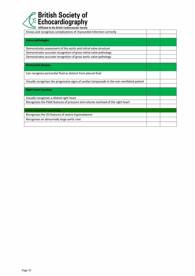
Appendix 3: Level 1 Curriculum based competency assessment tool
• The following competency statements relate to the Level 1 syllabus given in Appendix 2.
• At each hospital where you train in echocardiography you must have a supervisor who is BSE accredited in any modality, who has personally observed you scanning and corrected your technique on multiple occasions. He or she should sign the competencies shown below when he or she is satisfied that you are competent and can perform and report unsupervised to level 1 standard.
• This competency checklist should be brought to your practical assessment.
• The competency assessment should ideally demonstrate progress over the entire period of your echocardiographic training and is therefore intended to be formative not summative: please do not leave this to the end but use it as a guide to your training and development over time.
Principles of using Level 1 TTE
Date Signed
Demonstrates theoretical knowledge of the role of TTE in the emergency patient
Relays clinical findings to the critical care team in an appropriate and timely manner
Demonstrates consistent and appropriate referral of echo findings requiring expert help as per Level 1 Echocardiography syllabus part 13
Imaging physics and instrumentation
Demonstrates theoretical knowledge of ultrasound physics to allow full and accurate use of imaging equipment
Knows how to and routinely optimise all images in accordance with this
Doppler instrumentation
Demonstrates accurate use of colour Doppler with attention to: Box size and position, gain setting, scale and baseline
Anatomy and physiology
Demonstrates knowledge of cardiac anatomy
Measurements and calculation
Measures 2D distances from point to point accurately
Measures M-mode distances from leading edge to leading edge accurately
Demonstrates accurate qualitative assessment of ventricular performance
Demonstrates correct interpretation of chamber sizes and IVC behaviour for volume assessment
Myocardial infarction
Recognises and assesses large territory regional wall motion abnormalities
Page 15
Knows and recognises complications of myocardial infarction correctly
Valve pathologies
Demonstrates assessment of the aortic and mitral valve structure
Demonstrates accurate recognition of gross mitral valve pathology
Demonstrates accurate recognition of gross aortic valve pathology
Pericardial disease
Can recognise pericardial fluid as distinct from pleural fluid
Visually recognises the progressive signs of cardiac tamponade in the non-ventilated patient
Right heart function
Visually recognises a dilated right heart
Recognises the PSAX features of pressure and volume overload of the right heart
Other important pathology
Recognises the 2D features of severe hypovolaemia
Recognises an abnormally large aortic root

Page 16
Appendix 4: Suggested format for a report Each report must contain a documented clinical question, a comprehensive report of Level 1 Echocardiographic findings and a conclusion integrating the above. PLEASE NOTE – ALL REPORTS SUBMITTED IN THE LOGBOOK AND ACCOMPANYING THE CASES MUST BE ANONYMISED AS PER APPENDIX 10.

Page 17
Level 1 Echocardiogram report
Patient name
MRN/NHS no
DOB
Gender
Left ventricular size
Normal size LVIDD (cm)……………………
Small cavity LVIDD (cm)……………………
Large cavity LVIDD (cm)………………..
Unable to assess
Left ventricular function
Normal movement Globally impaired (more than mild)
Major regional wall motion abnormality
Unable to assess
Right ventricular size
Normal Small cavity Enlarged Unable to assess
Right ventricular function
Normal TAPSE (mm)
Impaired TAPSE (mm)
Unable to assess
AV structure
Normal 2D structure Heavily calcified Valve eversion seen Unable to assess
AV function
Normal function Restricted opening Significant AR on CFD Unable to assess
MV structure
Normal 2D structure Heavily calcified Valve eversion seen
Unable to assess
MV function
Normal function Restricted opening Significant MR on CFD Unable to assess
Aortic root Visually normal size May be dilated
Unable to assess
IVC Small and/or collapsing Normal movement with respiration
Large and/or non-collapsing
Unable to assess
Pericardial fluid No pericardial fluid seen Trivial Significant, +/- signs of tamponade
Unable to assess
Pleural effusion present
Yes No
Conclusion (referenced to the clinical question)
Is this a training report?
Scan/report checked and approved by:
Does the patient need a Level 2 study?
(findings reported in red require immediate expert help)
Referring physician informed?
(training reports are not to be used for patient care unless checked and approved)
Name and signature
Indications for study
Date of study
Haemodynamics
Referring Clinician
Page 18
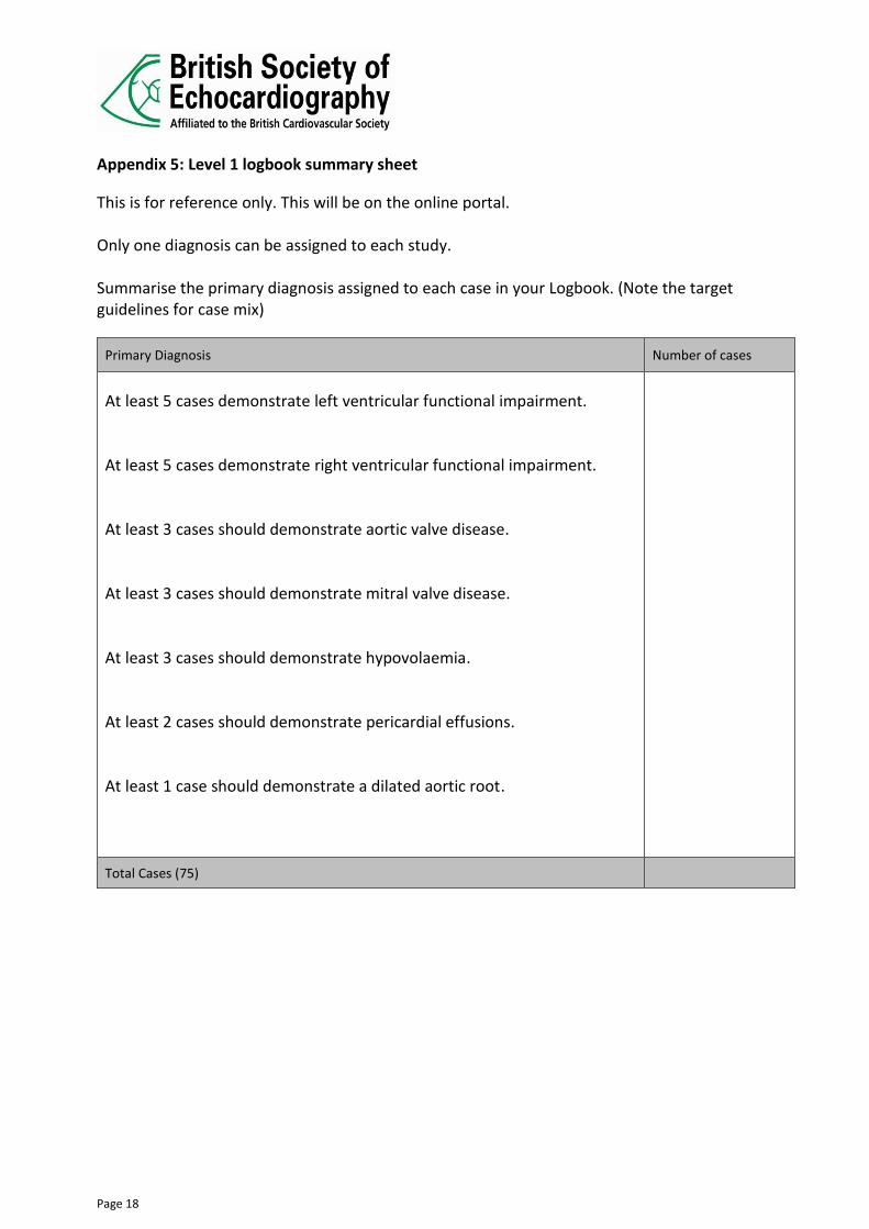
Appendix 5: Level 1 logbook summary sheet
This is for reference only. This will be on the online portal. Only one diagnosis can be assigned to each study. Summarise the primary diagnosis assigned to each case in your Logbook. (Note the target guidelines for case mix)
Primary Diagnosis Number of cases
At least 5 cases demonstrate left ventricular functional impairment. At least 5 cases demonstrate right ventricular functional impairment. At least 3 cases should demonstrate aortic valve disease. At least 3 cases should demonstrate mitral valve disease. At least 3 cases should demonstrate hypovolaemia. At least 2 cases should demonstrate pericardial effusions. At least 1 case should demonstrate a dilated aortic root.
Total Cases (75)

Page 19
Appendix 6: Examples of Component A Practical Assessment mark sheets
Logbook YES NO Comments
Logbook submitted
All cases collected within 12-month period
75 TTE reports performed and reported by the candidate
All cases fully anonymised
Correct case mix
All reports with full name and signature
Summary sheet present
Supervisor/Mentor statement present
Final check list present
Reports 1 2 3 4 5 6 7 8 9 10 11 12 13 14 15 Comments
Fully Anonymised
Indication for echo present
Appropriate accurate measurements present
Do measurements match descriptions
All parts of heart described
Descriptions complete
Appropriate to request
Conclusion present
Pass or fail

Page 20
Appendix 7: Examples of Component B Practical Assessment Sheets
Component B, Timeframe chart
Each candidate assessment will last for 25 minutes.
16 Minutes: You will have 16 minutes to obtain and acquire 8 images.
4 Minutes: You will be given an additional 4 minutes to repeat any of the views they wish to.
In the event of them doing this the 1st image will be ignored and the 2nd acquisition will be marked.
5 Minutes: The remaining 5 minutes will be used to enter details into the U/S system at the
start of the scan and save images
Performance Competency
Criteria F BF BP P Weighting Guidance Max Score
Checks patient identity
Checks patient identity using 3 unique identifiers
0 1 2 3 3 Checks the correct patient identity. Award P if 3 unique identifiers are checked, BP if 2 unique identifiers are checked, BF if 1 unique identifier is checked and F if no checks are made.
2D PLAX
Pays attention to detail and is able to recognise/acquire a good image
0 1 2 3 5 Acquisition of good quality 2D image in required timeframe. Award P if high quality optimised image. BP if clinically satisfactory image with limited optimization. BF if unable to accurately acquire image although is able to identify remedial measures. F if unable to reproduce image which reflects the PLAX in the specific model.
2D PLAX with CFM over the
aortic and mitral valves
Pays attention to detail and is able to recognise/acquire a good image
0 1 2 3 5 Acquisition of good quality 2D image demonstrating Colour Doppler assessment in required timeframe. Award P if high quality optimised image. BP if clinically satisfactory image with limited optimization. BF if unable to accurately acquire image although is able to identify remedial measures. F if unable to reproduce image which reflects the PLAX with CFM in the specific model.
Page 21
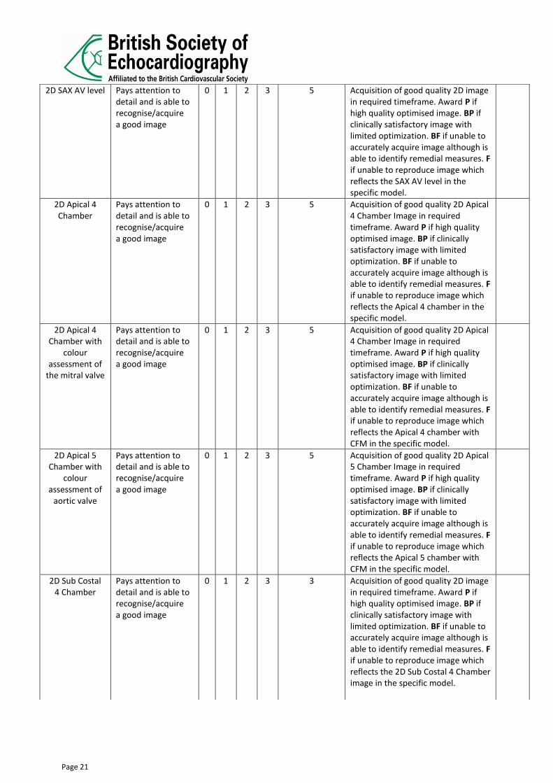
2D SAX AV level
Pays attention to detail and is able to recognise/acquire a good image
0 1 2 3 5 Acquisition of good quality 2D image in required timeframe. Award P if high quality optimised image. BP if clinically satisfactory image with limited optimization. BF if unable to accurately acquire image although is able to identify remedial measures. F if unable to reproduce image which reflects the SAX AV level in the specific model.
2D Apical 4 Chamber
Pays attention to detail and is able to recognise/acquire a good image
0 1 2 3 5 Acquisition of good quality 2D Apical 4 Chamber Image in required timeframe. Award P if high quality optimised image. BP if clinically satisfactory image with limited optimization. BF if unable to accurately acquire image although is able to identify remedial measures. F if unable to reproduce image which reflects the Apical 4 chamber in the specific model.
2D Apical 4 Chamber with
colour assessment of
the mitral valve
Pays attention to detail and is able to recognise/acquire a good image
0 1 2 3 5 Acquisition of good quality 2D Apical 4 Chamber Image in required timeframe. Award P if high quality optimised image. BP if clinically satisfactory image with limited optimization. BF if unable to accurately acquire image although is able to identify remedial measures. F if unable to reproduce image which reflects the Apical 4 chamber with CFM in the specific model.
2D Apical 5 Chamber with
colour assessment of
aortic valve
Pays attention to detail and is able to recognise/acquire a good image
0 1 2 3 5 Acquisition of good quality 2D Apical 5 Chamber Image in required timeframe. Award P if high quality optimised image. BP if clinically satisfactory image with limited optimization. BF if unable to accurately acquire image although is able to identify remedial measures. F if unable to reproduce image which reflects the Apical 5 chamber with CFM in the specific model.
2D Sub Costal 4 Chamber
Pays attention to detail and is able to recognise/acquire a good image
0 1 2 3 3 Acquisition of good quality 2D image in required timeframe. Award P if high quality optimised image. BP if clinically satisfactory image with limited optimization. BF if unable to accurately acquire image although is able to identify remedial measures. F if unable to reproduce image which reflects the 2D Sub Costal 4 Chamber image in the specific model.

Page 22
2D IVC view
Pays attention to detail and is able to recognise/acquire a good image
0 1 2 3 3 Acquisition of good quality 2D image in required timeframe. Award P if high quality optimised image. BP if clinically satisfactory image with limited optimization. BF if unable to accurately acquire image although is able to identify remedial measures. F if unable to reproduce image which reflects the 2D IVC view in the specific model.
Modification of patient position
to optimise image quality
Pays attention to detail and is able to consider manipulation of the patient either positional or with the aid of respiratory manoeuvre’s
0 1 2 3 5 Demonstration of manoeuvers to assist in securing high quality echocardiography images in required timeframe. Award P if high quality optimised image. F if unable to demonstrate skills of manipulation in the specific model.

Page 23
Appendix 8: Component C Identification of Level 1 pathologies
This section will assess recognition of pre-recorded echocardiograms of common emergency pathologies that can be assessed with Level 1 Echocardiography. The candidate will fill out a standard Level 1 Echocardiography report for each pathology in the presence of the examiner who will discuss the findings with the candidate. Two cases will be shown. Both must be reported correctly to pass this section of the exam. In the event of a borderline fail on one case a third case may be used. If either case is a clear fail or both cases are a borderline fail the candidate will fail this component.
➢ Normal ➢ Hypovolaemia ➢ Severe aortic stenosis ➢ Aortic regurgitation ➢ Disrupted or dysfunctional mitral valve ➢ Dilated aortic root ➢ Left ventricular dysfunction ➢ Right heart strain and dysfunction ➢ Pericardial fluid, TTE signs of tamponade
BSE Level 1 – Component C marking sheet
Candidate name Date
Candidate no
Case no
Left ventricular size
Normal size LVIDD (cm)……………………
Small cavity LVIDD (cm)……………………
Large cavity LVIDD (cm)………………..
Unable to assess
Correct1 Minor2 error
Major error3
Left ventricular function
Normal movement Globally impaired (more than mild)
Major regional wall motion abnormality
Unable to assess
Right ventricular size Normal Small cavity Enlarged Unable to assess
Right ventricular function
Normal TAPSE (mm)
Impaired TAPSE (mm)
Unable to assess
AV structure
Normal 2D structure
Heavily calcified Valve eversion seen
Unable to assess
AV function
Normal function Restricted opening Significant AR on CFD
Unable to assess
MV structure
Normal 2D structure
Heavily calcified Valve eversion seen
Unable to assess

Page 24
MV function
Normal function Restricted opening Significant MR on CFD
Unable to assess
Aortic root Visually normal size
May be dilated
Unable to assess
IVC Small and/or collapsing
Normal movement with respiration
Large and/or non-collapsing
Unable to assess
Pericardial fluid No pericardial fluid seen
Trivial Significant, +/- signs of tamponade
Unable to assess
Pleural effusion present
Yes No
Conclusion (referenced to the clinical question)
Referral for Level 2 study?
1Correct = Correct interpretation 2Minor error = minor reporting error that does not affect key findings 3Major error = reporting error that either misses key pathology or incorrectly describes normal findings as
abnormal
Pass/Fail Pass = all components correctly assessed.
Borderline fail = up to two minor reporting errors i.e does not affect the key findings.
Fail = reporting error which either misses the key pathology or incorrectly describes normal findings as abnormal.
Comments
Examiner name
Examiner signature

Page 25
Appendix 9: Supervisor statement to accompany the Practical Assessment
Candidate’s name ____________________________________________
Initial
I certify that the candidate has undergone a programme of training in echocardiography
I certify I have observed the candidate scanning and I am satisfied that he/she is competent in Level 1 Echocardiography.
I certify that the candidate has reached a standard of training to be able to independently perform and report a Level 1 transthoracic echocardiographic study. He/she has reached all of the mandated competencies. I have signed off the candidate’s competency sheet.
I certify that the candidate above has performed and reported the cases included in the accompanying Log Book within a 12-month period.
I certify that all cases are fully anonymised (no patient’s personal details such as names, full date of births or addresses) as per Appendix 11
I certify that all cases are signed with name printed of the candidate
I certify that these cases are being handed in as per Trust Policy Guidelines
Supervisor’s name: ____________________________________________ Signature: ______________________________ Date: _______________________ I am satisfied that the candidate above has performed and reported the cases included in the accompanying Log Book within a 12-month period. Medical/Technical Head of Echocardiography’s name: __________________________ Signature: _______________________________ Date: _________________________ Notes: The Head of Echocardiography is usually the lead clinician or consultant cardiologist with overall responsibility for echocardiography. This may be a representative from a local Cardiology department who has personally observed the candidate scanning and is satisfied that they have the ability to perform and report echos independently.

Page 26
Appendix 10: BSE Policy on the Non-Anonymisation of Patient Data
Introduction The duty of confidentiality arises out of the common law of confidentiality, professional obligations and also staff employment contracts. Breach of confidence may lead to disciplinary measures, bring into question professional reputation and possibly result in legal proceedings. Guidance is provided to NHS staff in the ‘NHS Code of Practice on Confidentiality’ (November 2003). http://www.dh.gov.uk/prod_consum_dh/groups/dh_digitalassets/@dh/@en/documents/digitalasset/dh_4069254.pdf Patient information that can identify individual patients is confidential and must not be used or disclosed. In contrast, anonymised information is not confidential and may be used. Key identifiable information includes: Patient’s name, address, full post code, date of birth; NHS number and local identifiable codes; anything
else that may be used to identify a patient directly or indirectly. For example, rare diseases, drug treatment or statistical analyses which have very small numbers within a small population may allow individuals to be identified. Anonymisation requires the removal of such information from all reports and images. For accreditation purposes, BSE Administrators and BSE Assessors must not be able to identify the patient from the detail or combination of details given.
Speakers presenting on behalf of the BSE at meetings and speakers on courses/meetings awarded BSE re- accreditation points must ensure that all presentation material is anonymised. Guidance to candidates submitting Logbooks and Cases for Accreditation The NHS Code of Practice on confidentiality means that evidence submitted for the practical part of the Accreditation process must have all patient identification removed.
In order for evidence to be considered to have been anonymised, BSE Administrators and BSE Assessors must not be able to see any of the identifiers listed above. As age is relevant to the assessment either the age or year of birth must be provided however a full date of birth must not be shown. Reports Please note that correction fluid may still allow data to be visible if you look at the back of the page, as does placing a sticker over the patient data. Marker pen often fades so that data may be correctly disguised at the point of anonymisation but not when brought to a Practical Assessment session We therefore advise:

Page 27
Use the online portal and electronically delete all patient information except age and gender
Or: cutting out the patient data Or:Deleting data electronically prior to printing Or:Using corrective fluid or marker pen, then photocopying the sheet Major breach: One or more examples of detailed patient demographics (e.g. name and address) OR One or more examples of patient data sufficient to allow a patient to be traced in any way
Minor breach: Examples of patient identifiable information found within the logbook. These might include, for example, first name or date of birth but insufficient information to identify the patient. In the event of a major breach: The candidate will automatically fail and candidate will be informed of the fail and notified of the reason for it. The Chair of the Accreditation Committee will be notified of all major breaches and will make the decision as to whether the Head of Information Governance at the candidate’s place of employment should be informed.
In the event of a minor breach: The candidate will be informed of the breach and notified of the reason for it. This will be taken into account in the marking scheme. The final decision remains at the discretion of the Chair of the Accreditation Committee.
Page 28
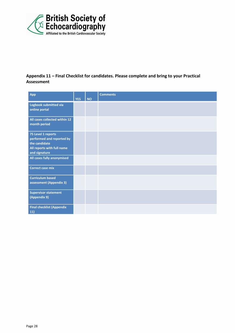
Appendix 11 – Final Checklist for candidates. Please complete and bring to your Practical Assessment
App
YES
NO Comments
Logbook submitted via
online portal
All cases collected within 12
month period
75 Level 1 reports
performed and reported by
the candidate All reports with full name
and signature
All cases fully anonymised
Correct case mix
Curriculum based
assessment (Appendix 3)
Supervisor statement
(Appendix 9)
Final checklist (Appendix
11)