Academy of Veterinary Dental Technicians of 2020 AVDT...orthodontics, prosthodontics, and...
54
Academy of Veterinary Dental Technicians Credentials Packet 2018-2020 Class of 2020
Transcript of Academy of Veterinary Dental Technicians of 2020 AVDT...orthodontics, prosthodontics, and...

Academy of Veterinary Dental Technicians
Credentials Packet 2018-2020
Class of 2020

Academy of Veterinary Dental Technicians – Class of 2020
Credentials Packet 2018-2020
AVDT Credentials Packet© Revised 10/2017
- 2 -
TABLE OF CONTENTS
AVDT Extraction Position Statement, Hours and Specialty Training Requirements Page 3-4 Case Logs – Guidelines and Requirements Page 5-9 Case Reports – Guidelines, Requirements, and Examples Page 9-23 Dental Radiography Requirement Page 24 Equipment List Requirement Page 25 Reading List Page 25 AVDC/AVDT Approved Case Log Abbreviations/Nomenclature Page 26-37 Form 1: Waiver, Release, and Indemnity Agreement Page 38 Form 2: Skills Form Page 39 Form 3a: Specialty Training Wet Lab Form Page 40 Form 3b: Specialty Training Lecture Form Page 41 Form 4: Equipment List Page 42-44 Form 6: Case Log Cadaver Verification Form Page 45 Form 7: Dental Radiography Cadaver Use Form Page 46 Form 8: Mentor/Mentee Contacts and Case Log Verification Form Page 47 Form 9: Mentee Shadowing Hours Verification Form Page 48 Canine Dental Chart For Case Logs & Case Reports Page 49 Feline Dental Chart For Case Logs & Case Reports Page 50 Credentials Packet Format Page 51-53 Credentials Packet Submission Process and Checklist Page 54

Academy of Veterinary Dental Technicians – Class of 2020
Credentials Packet 2018-2020
AVDT Credentials Packet© Revised 10/2017
- 3 -
AVDT Extraction Position Statement
The AVDT does not condone, endorse nor recommend that veterinary technicians, credentialed or not, perform
dental extractions. Extraction of teeth is oral surgery and should be performed by a licensed veterinarian, per the
AVMA. Several states allow for veterinary technicians to perform extractions, but the wording is often vague,
some stating simple extractions only, others simply list extractions as a task that can be performed by a
credentialed veterinary technician.
The duties of a veterinary technician during a dental procedure include charting, performing dental cleaning
(sub-gingival and supra-gingival) and polishing, intraoral radiographs, performing nerve blocks, assisting with
the dental procedures and oral surgery. Diagnosing dental disease, prescribing treatment options and
medications, and performing oral surgery are duties for the veterinarian. The veterinary technician may and
should assist the veterinarian with these duties, thereby ensuring the patient receives the most thorough and
efficient dental care.
A Veterinary Technician Specialist (VTS) in Dentistry is a credentialed technician with a special interest in
dentistry and oral surgery. They have extensive knowledge and training in these areas (endodontics, exodontics,
orthodontics, prosthodontics, and periodontics), however they are not taught nor licensed to perform oral
surgery. They may instruct veterinarians on proper extraction techniques under the direct supervision of a
licensed veterinarian.
Always refer to your state’s veterinary practice act for the duties that you may perform legally.
Hours The mentee must spend at least 4000 hours during the Specialist program practicing veterinary technology. At
least 2780 of these hours must be spent within the dental setting. Dentistry hours can be accumulated via any of
the following routes:
✓ Providing client education such as: how to brush teeth, explaining a disease process, your clinics tx
recommendations, etc. (anything related to client dentistry education)
✓ Scheduling/assisting with dental consultations
✓ Performing/assisting dental procedures (this includes holding the mouth while the veterinarian is
suturing, running anesthesia on dental patients, etc.)
✓ Performing INTRA ORAL dental radiographs (skull films do not count)
✓ Discharging dental patients and going over homecare instructions
✓ Creating client handouts for dental related topics for your clinic
✓ Shadowing hours required to complete your case logs
✓ Dental related CE hours that you have accumulated during your two year mentorship program
o If you plan to include your hours obtained while attending a dental related CE, confirmation of
these hours can be incorporated in your letter from a supervising veterinarian and office manger
(see below).
When you submit your credential packet in January 2020, you will be required to submit THREE - FOUR
documents along with your packet confirming you have worked a minimum of 2780 hours in veterinary
dentistry:

Academy of Veterinary Dental Technicians – Class of 2020
Credentials Packet 2018-2020
AVDT Credentials Packet© Revised 10/2017
- 4 -
1. A letter from a supervising veterinarian who can attest that 75% of your time was spent in dentistry &
confirming your total hours of 2780.
2. A letter from your practice manager who can attest that 75% of your time was spent in dentistry &
confirming your total hours of 2780.
3. A summary of time worked from a timesheet printed out from your employer proving your required
hours have been met
4. The fourth documentation is only applicable IF you are inlcuding hours while shaowing. You must
complete and sign Form 9. If this form is not completed and signed by the mentee and supervising
veterinary (DAVDC or FAVD), these hours will be null and void.
Any applicant who cannot confirm their hours have been met, or were unable to meet the required hours,
will not be allowed to submit their credentials packet in January 2020.
Specialty Training In addition to meeting the general requirements, the mentee must successfully complete wet lab training and
attend lectures in advanced dentistry procedures. Teaching a wet lab or lecture or writing a veterinary dentistry
continuing education article does not qualify as CE attendance.
Training and CE credit is accepted from credentialed members of the following organizations:
• Academy of Veterinary Dental Technicians (www.avdt.us); Academy of Veterinary Dentistry
(www.avdonline.org); American Veterinary Dental College (AVDC.org); Foundation for Veterinary
Dentistry (www.f4vd.com)
• Dental local and regional anesthesia CE obtained from a Diplomate of The American College of
Veterinary Anesthesia and Analgesia (ACVAA) or Veterinary Technician Specialist(Anesthesia and
Analgesia) will be accepted. Any other CE from any other veterinarian, veterinary technician or
veterinary technician specialty will NOT be accepted. *If you are an international mentee and have limited resources and/or are unsure if CE in your country will be
accepted, please contact the credential chair for further assistance.
A list of CE meetings can be found at each of the above websites, at the Veterinary Dental Forum website
(www.veterinarydentalforum.com), the AVDT website (www.avdt.us) or in the Journal of Veterinary Dentistry.
The mentee must complete the "Specialty Training Form" (Forms 3a and b) and give proof of attendance for
each event you attended to show you have completed both the 25 hours of wet lab training and attended 15
hours of advanced dentistry lectures. A photocopy of a document provided by the organization or speaker is
proof of attendance. Cancelled checks or other documents will not be accepted as proof of attendance. You
must provide detailed course descriptions provided by the organization presenting the CE as proof that the
continuing education was related to veterinary dental care, and the lab or lectures must fit into one of the
categories listed below. Participation and attendance at wet labs and lectures must be completed during the
two-year Specialist training program between January 2018 and December 31, 2019.
Wet Labs The mentee must complete a total of 25 hours of wet labs within each of the following disciplines:
• Dental Prophylaxis – 5 hours

Academy of Veterinary Dental Technicians – Class of 2020
Credentials Packet 2018-2020
AVDT Credentials Packet© Revised 10/2017
- 5 -
• Periodontics – 5 hours
• Prosthodontics – 2.5 hours
• Radiology – 6 hours
• Endodontics – 2.5 hours
• Dental Local and Regional Anesthesia – 4 hours
Advanced Dental Procedures Lectures The mentee must attend a total of 15 hours of lectures in advanced dentistry procedures:
• Endodontics –2.5 hours
• Prosthodontics – 2.5 hours
• Orthodontics – 2.5 hours
• Oral Surgery – 2.5 hours
• Oral Pathology – 2.5 hours
• Advanced Periodontal Therapy– 2.5 hours AVDT approved wet labsand lectures may beavailable at the following conferences or training centers:
1. National Conference of Veterinary Technician Specialty Academies
2. AnimalDentalTrainingCenter (www.animaldentalcenter.com)
3. Veterinary Dental Forum (www.veterinarydentalforum.com)
4. North American Veterinary Conference (www.navc.com)
5. Western States Veterinary Conference (www.wvc.org)
6. Central States Veterinary Conference (www.thecvc.com)
7. American Veterinary Medical Association (www.avma.org)
8. AmericanAnimalHospital Association (www.aahanet.org)
9. AnimalDentalCareTrainingCenter (www.vetdentalclasses.com) To receive credit for other courses, a written request to and written approval from the AVDT Credentials
Chair is required. All CE obtained at the Veterinary Dental Forum will be accepted if it fits into the
categories above.A maximum of 2 lecture hours may be obtained through online sources.Skype and Face
Time are not acceptable methods to obtain CE hours. A maximum of 5 wetlab hours and 3 lecture hours
may be obtained in a non-traditional setting. Please direct any questions to the credentials chair.

Academy of Veterinary Dental Technicians – Class of 2020
Credentials Packet 2018-2020
AVDT Credentials Packet© Revised 10/2017
- 6 -
Case Logs Completed Chronological and Categorical Case Logs with a minimumof 75 dentistry cases. If only 75 cases
are submitted, a single unacceptable case could result in your credentials packet being rejected.However,
please do not submit more than an additional 3 cases per a category. Categorical case log formsand
chronological case log forms are provided to you.Make sure each entry in your log is complete. If the animal's
weight (lbs. or kg.), age, or sex is unknown, enter "unknown" in the case log. Only include cases seen between
January 1, 2018 andDecember 31, 2019.Remember, all experience must be within the two-year Specialist
program. Please highlight the five cases used for your case reports. Standard diagnostic and procedural
abbreviations, as defined by the AVDC (revised 2017), are included on page 27. If your clinic commonly uses
abbreviations that do not appear on the list, please define these at the beginning of your log.
MINIMUM REQUIRED CASELOAD The AVDT prefers the mentee assist in procedures marked (*). However, if that is not possible, proof of CE in
this area is required. This must be highlighted on the case log and indicated on Forms 4a and/or 4b, as
appropriate. A cadaver may be used in a maximum of two cases from any category marked (**). These cases
are left to the discretion of the mentee, but must be certified and supervised by a Diplomate of the
AmericanVeterinaryDentalCollege or a Fellow of the Academy of Veterinary Dentistry (Form 6).
Oral Medicine
OM Cases requiring involved diagnostic tests (e.g. anesthesia and biopsy and/or radiographs, sialography, masticatory
muscle EMG, CBCT, or where laboratory tests beyond complete blood count and biochemical profile are used) but which
do not include a specific treatment procedure that is included under a treatment code
below.……………………………………………………..............................5
Periodontics
PE1 Complete prophylaxis not requiring involved periodontal treatment....................................12
PE2 Involved periodontal scaling and root planing to include placement of a perioceutic
medication when no PE3 or PE4 procedure is performed......................................................5
PE3 Simple periodontal surgery (gingivectomy/gingivoplasty, open curettage
or flap procedure, except those combined with bone grafting)................................................3
PE4* Involved periodontal treatment (osseous surgery, increasing attachment height, bone
augmentation, gingival grafting, and guided tissue regeneration, periodontal splinting)…....1
Note: extraction followed by placement of a bone promoting or substitute material is not a PE4 case.
Endodontics** (all including routine restoration of access openings)
EN1 Mature canal endodontic obturation - nonsurgical..................................................................7
EN2 Vital pulp therapy (partial vital pulpotomy)............................................................................2
EN3* Surgical endodontic treatment, apexification, replacement of avulsed or luxated teeth,
splinting of tooth with horizontally fractured root with follow up endodontic evaluation......1
Restorative Dentistry**
RE Restorative procedures (requiring gingival flap exposure, occlusal table cavity
preparation, other involved restoration including routine restoration of endodontic access
openings, dentin bonding only is NOT considered a RE case)..............................................5
Oral Surgery
OS1 Simple (closed) dental extractions, crown amputations (tooth resorption)..............................15
OS2 Involved dental extractions (open or closed, requiring tooth sectioning, bone removal

Academy of Veterinary Dental Technicians – Class of 2020
Credentials Packet 2018-2020
AVDT Credentials Packet© Revised 10/2017
- 7 -
or other procedures in addition to elevator and forceps work). A full mouth extraction
patient may be logged as one OS2 case...................................................................................12
OS3** Mandibular or maxillary fracture fixation (using dental acrylic splint, body of mandible
fracture fixation with wire, pins, screws or plate, symphyseal separation fixation)................1
OS4** Involved oral surgical procedures (TMJ condylectomy, repair of existing palatal
defects and oronasal fistulas, maxillectomy, mandibulectomy)...............................................1
OS5** Miscellaneous soft tissue oral surgery (resection of traumatic cheek or sublingual granuloma
hyperplasia; salivary gland surgery, removal of oral masses not requiring maxillectomy or
mandibulectomy, laser surgery for stomatitis, operculectomy, does not include therapy laser
treatments that do not directly treat the oral cavity)................................................1
Prosthodontics
PR* Crown and/or bridge preparation, impression and cementation (including canine,
incisor and carnassial teeth).......................................................................................................1
Orthodontics
OR* Malocclusion treatment plans (including detailed consultation and evaluation of the bite or
bite registration), extraction of deciduous teeth or permanent teeth causing malocclusion,
management of clinical malocclusion (crown amputation, application of incline plane), and
management of clinical malocclusions (use of and active force orthodontic
device)…………………………………………………...........................................................1
Other Species
EX Dental procedures on animals other than dog and cat, (does not include beak trims, does include occlusal leveling
in equine patients) …………………….............................................................2
CASE LOG GUIDELINES This document is a guideline for the mentor of an AVDT applicant and for the applicant. It is a basis for review
of logs, but does not mean that all case logs the mentor reviews will be approved by the Credentials Committee
of the AVDT.
To make construction and management of the case logs as user friendly as possible for applicants, the AVDT
provides Excel based spreadsheet files with templates and instructions for logging cases and related activities.
Use of this spreadsheet is required by the Credentials Committee.
This document contains the following:
1. Case Log Categories
2. Guidelines for Counting Cases
3. Format for Submission of Case logs
Required Case Logs A detailed list of a minimum of75 veterinary dental cases seen during the AVDT approved training period is
required. (There is no limit to the number of cases that can be submitted.) Cases that were treated prior to the
applicant’s AVDT training program acceptance date cannot be included in the case log. The case log is to
conform to the guidelines, nomenclature and abbreviations described below. Along with your case logs, you
will be required to pick and submit ONE completed dental chart from each category that will

Academy of Veterinary Dental Technicians – Class of 2020
Credentials Packet 2018-2020
AVDT Credentials Packet© Revised 10/2017
- 8 -
demonstrate your ability to accurately and properly chart various cases. You WILL NOT submit a
dental chart for all 50+ cases.Please use the generic dental charts provided with your credential packets
for each case log.However, a completed dental chart must be available for all logged cases but need not be
submitted unless requested by the AVDT. The cases are to be listed under the categories stated in the AVDT
case log categories listing.
For submission as part of a credentials application package, the full Categorical and Chronological log is
required including cases logged during the entire AVDT training period.
Chronological Log: The cases are to be numbered sequentially and listed chronologically. The required format
is that the chronological log should contain all of the appropriate AVDC codes that pertain to the case.
Categorical Log: The cases are to be listed under the categories and subcategories stated in the AVDT case log
categories listing. The Categorical Case Log is to begin with a table listing the number of cases seen in each
category. For each case listed, the case number is to be the same number assigned to the listing of that case in
the chronological list. The categorical log should contain ONLY the codes pertaining to the category that the
case is included in.
The completed chronological logs demonstrate your understanding of how to log the complete case whereas the
categorical log demonstrates more focused understanding of the specific logged procedure.
If an applicant remains in a training program for more than two years, cases in the log that are more than two
years old cannot be counted towards meeting the AVDT total case logs. However, the applicant is not required
to revise the individual case log numbers following deletion of cases that are no longer allowable. Any report to
the AVDT that includes a case log in which the most recent two year chronological log list does not start with
case number one is to note the reason for not starting with one as the initial case in the chronological log.
Miscellaneous Cases When a case does not appear to fit into any of the AVDT categories, the applicant is to request clarification
from the AVDTCredentials Committee Chair via email. When necessary, the Credentials Chair will forward
the query to the Board for consideration.
Guidelines for Counting Cases An AVDT case is defined as either the performance or assistance in a dental discipline. A maximum of one
case may be logged from any single treatment episode of a particular animal. If this patient is anesthetized later
for another dental procedure, it may be used as an additional entry.
Review of Case Logs and Reports by Mentor
Categorical and Chronological Logs of cases performed during the two year Specialist program are to be
submitted as part of the applicant’s report to their mentor. One half of logs and case reports should be
completed for midterm review by your mentor.The completed logs and reports must be submitted to your
mentor for approval at least one month prior to packet submission.

Academy of Veterinary Dental Technicians – Class of 2020
Credentials Packet 2018-2020
AVDT Credentials Packet© Revised 10/2017
- 9 -
Requirements for Case Logs 1. Category
List the category you are assigning this case to, using the Case Log Categories to determine the category
(see pages 4&5).
2. Number
Consecutive throughout an applicants program, listed in date order. If an applicant remains in the
training program for more than two years, cases in the log that are now more than two years old can no
longer be counted.
3. Date
Month, day, and year procedures were performed.
4. Patient Name
List the patient’s name or clinic case number.
5. Signalment
Species, age,breed,weight, and gender of the patient.
6. Diagnosis
List the diagnosis(es) made using the approved AVDC abbreviations. If you use any other
abbreviations, a key to these abbreviations must be included in your submitted packet.
7. Dental Procedure
List the dental procedures performed using the approved AVDC abbreviations. If you use any other
abbreviations, a key to these abbreviations must be included in your submitted packet. Individual teeth
treated are to be identified. The AVDT requires that the modified Triadan system of tooth identification
be used.
8. Radiographs
This column is checked if radiographs were taken.
9. Anesthetic
List general anesthetic protocol (generic drug names preferred).DO NOT listpreanesthetic or local
anesthetic drugs given.
11. Use of the AVDT Excel based case log template is required for applicants.
Logging Equine/Pocket Pet Procedures for “Float”:Additional Equine abbreviations are on page 36
Diagnosis: T/O/EP or abnormal elongation of enamel points
Treatment: OD or odontoplasty
Example: T/O/EP 106-111, 206-211, 306-311, 406-411
OD 106-111, 206-211, 306-311, 406-44

Academy of Veterinary Dental Technicians – Class of 2020
Credentials Packet 2018-2020
AVDT Credentials Packet© Revised 10/2017
- 10 -
Case Reports Five Case Reports. Included in this packet are two examples of case reports submitted by a successful applicant.
Case report guidelines are outlined below. Each case report will be a total of 12 pages in length. The body of the
case report(s) must be typed, double spaced, and no more than 10 pages in length. The eleventh page is
designated for the references, required pictures and radiographs. The twelfth, and final page, will be the
dental chart associated with the case report(s).Please use the generic dental charts provided with your
credential packets for all case reports.Pictures AND radiographs are required. Cases for your reports
must appear in your case log. Case reports that are not clearly highlighted in your case logs will not
receive points.
Select five various cases from your log that will demonstrate your expertise in dentistry nursing skills. The case
reports should describe in detail, how the patient was diagnosed and treated. The case report must also be used
to demonstrate how you used your knowledge and experience to assist the veterinarian in diagnosing and
treating the patient. These case reports do not need to be cases that are done at exotic facilities such as a zoo or
wildlife sanctuary. Your mentor will be a great resource to help you pick cases that will be successful as case
reports.
Be sure that information such as patient's name, owner's name, identification number, and the date the case was
seen is included in the report. This information is used to determine if the case is entered in the case log. The
case reports should describe, in detail, how the patient was diagnosed and treated. The case reports must also be
used to demonstrate how you used your knowledge and experience to assist the veterinarian in diagnosing and
treating the patient. It is important that the information in your case reports be clearly understood. Present each
case in a logical manner, check spelling and grammar, and define any abbreviations. It is important that you
show that you participated in the case and were not just an observer. Pertinent anesthesia information should be
included; i.e. route and dose of sedation drugs, induction drugs, maintenance drugs, and any local or regional
drugs administered. Consider some of the following ways of demonstrating your knowledge and expertise:
1. Show how your observations, examination and history taking assisted the veterinarian with the
development of an effective dental plan.
2. Explain why an observation was important or why you asked a certain question during the
procedure.
3. Describe how an observation you made helped to avoid a possible complication.
4. Describe the procedures you performed and assisted with. Explain why the procedure was
performed.
5. Describe the anesthetic protocol that was chosen for this patient and how you maintained the
patient's condition during the procedure.
6. Show your understanding of the problem being treated.
CASE REPORT GUIDELINES A case report is an opportunity to show good dental concepts and the ability to deliver a well written and well-
documented scientific paper about a case performed well by current standards. The use of advanced technology
or skill in the reported cases is not required. Your mentor and the Credentials Committee will evaluate each of
the items below.Case reports where a technician is doing any surgical treatments, including surgical
extractions, will automatically receive zero points regardless of the mentees state regulations. Please refer
to our AVDT position statement on page 3 and/or contact the current Credential Chair with any additional
questions regarding this guideline.

Academy of Veterinary Dental Technicians – Class of 2020
Credentials Packet 2018-2020
AVDT Credentials Packet© Revised 10/2017
- 11 -
Introduction
Present the topic of the case report.
Case Report
1. History
A. Include a signalment and presenting problem or chief complaint.
B. Describe lesions/problem. You can refer to teeth in this manner: the right maxillary fourth premolar
(108) on the first mention subsequently you may refer to the tooth as 108
C. Describe past dental history.
D. Describe past medical history if relevant.
E. Describe any other relevant problems.
2. Diagnosis
A. Thoroughly describe the oral exam findings during the consultation. Describe what you saw-
gingivitis index, calculus index, etc. Include the physical exam findings, radiographs, MRI, etc.
B. Demonstrate attention to the patient as a whole. Perform appropriate preoperative diagnostics and
laboratory tests.
3. Problem List
A. Provide an accurate assessment.
B. Mention all oral lesions observed.
C. Mention differential diagnosis and their rule-outs.
4 Treatment Plan
A. Discuss different modalities for treatment and their prognosis.
B. If other lesions are apparent, you should mention them and note if treated or nottreated.
C. Address any potential genetic impact of the condition, if applicable.
5. Treatment
A. Describe the procedure including technique, instruments, and materials in detail using proper
terminology. Make sure to list the bur numbers that were used in the procedure. Highlight your
involvement in the procedure. Make sure that all pathology listed in the case log is addressed.
(Cases with less pathology may be easier to fit into the 5 page limit.)
B. Include your anesthetic management: using appropriate anesthetic protocol, drugs (generic name
preferred), dosages, route of administration, and monitoring.
C. Demonstrate appropriate peri-operative care: vital functions monitoring and support, intravenous
fluid administration, control of body temperature, etc. Adequate pain management is very
important.
D. Include dosages in mg and administration routes of any medication used or prescribed. Use generic
names.
E. Include postoperative radiographsand their assessment if indicated.
F. Describe the handling of any complications.
G. Provide adequate photos to support your report. Provide adequate and accurate captions, labeled
pre- and postoperative radiographs for procedures and intra operative radiographs for root canal
therapy. Again, make sure to label your radiographs and photographs.
6. Postoperative Care
A. Describe instructions given to client, including medication dispensed for home use and dental home
care procedure.
Academy of Veterinary Dental Technicians – Class of 2020
Credentials Packet 2018-2020
AVDT Credentials Packet© Revised 10/2017
- 12 -
Discussion
A. Discuss any point relative to your case.
B. Briefly review the literature on the disease condition and/or procedure in question, if appropriate.
C. Discuss pertinent aspects of the diagnostic work up.
D. Include all pertinent client education related to your case. How did you educate the owner about this
case? Are there any preventative measures the owner could take in the future? Recommendations?
E. Provide references to support your statements. Number references consecutively in the order
which they are first mentioned in the text. Identify them by Arabic numerals in superscript. Please
provide at least 2 references per case report using multiple sources. Format the references as per
https://owl.english.purdue.edu/owl/resource/560/06/
F. Discuss any particular ways the case was different from expected, and how the problems were
managed.
Conclusion
A. What conclusion, if any, could be drawn from the case?
Technical Details
A. Each case report should be no longer than ten pages in Times New Roman 12 point font. Margins
should be 1 inch on each side. References and pictures should be limited to one additional page.
Pictures AND radiographs are required. Teeth may be listed as right upper fourth premolar (108)
and thereafter may be referred to as 108. BPM may be used for breaths per minute and beats per
minute. Bur types and numbers should be listed. Do not list equipment manufacturers. Make sure
to designate that the tube is cuffed or uncuffed. There is not need to list fine or course prophy paste
but if flour pumice is used it should be stated and why.
B. It is required that your mentor review your case reports.
C. Read your manuscript while playing the role of a critic. Keep it technical. Please remember that
this is scientific writing, and spelling and grammar are very important.Plagiarismwillresult in
severe penalties.
D. Above all, prepare your case reports early enough to seek pre-approval by your mentor, leaving
enough time to edit and return it again well before your Credentials Packet deadline.
Case report example #1:
“Katie” Burdick—a periodontal disease case

Academy of Veterinary Dental Technicians – Class of 2020
Credentials Packet 2018-2020
AVDT Credentials Packet© Revised 10/2017
- 13 -
Case Log #122—8/16/2012
“Katie”, an 11-year-old, 29.9kg, spayed female Chesapeake Bay Retriever dog, presented for an oral
examination due to heavy dental calculus and halitosis. Earlier that week, she had pre-anesthetic laboratory
testing consisting of a chemistry panel and a complete blood count; both were within normal limits. “Katie” was
not on any medications and her owner did not provide dental home care. The most recent professional dental
cleaning with full mouth radiographs was performed over four years ago. Findings from that procedure included
moderate dental calculus (calculus index 2), mild gingivitis (gingivitis index 1), generalized abrasion (AB) on
most cusps, and a 6mm periodontal pocket between the right maxillary fourth premolar and first molar (108 and
109) that had been treated with closed ultrasonic periodontal debridement and perioceutic application.
Radiographically, “Katie’s” teeth looked relatively healthy with early signs of periodontitis and less than 25%
attachment loss (Periodontal Disease Stage 2). At that time, “Katie’s” owner was instructed to return in 6
months for a follow up anesthestized dental cleaning and oral exam to see if her periodontal disease has
progressed or not between the 108 and 109. However, she had not returned for her recommended re-evaluation
later that year.
Conscious physical examination revealed no abnormalities. The patient had an ideal body condition
score of 3 out of 5 and was normally hydrated. Her heart rate was 82 bpm, respiratory rate was 18 rpm, and
body temperature was 100.3°F. Her conscious oral examination revealed a normal occlusion, severe dental
calculus (calculus index 3), moderate gingivitis (gingivitis index 3), and generalized abrasion on most cusp.The
level of dental calculus on her teeth made it difficult to evaluate the crowns for discoloration or fractures. She
had very obvious halitosis. There were no other abnormalities noted on her conscious oral examination. The
owner was presented with a treatment plan; which included, general anesthesia for an oral examination,
professional scaling and polishing of the teeth, and full mouth dental radiographs. The owner consented to this
treatment plan and asked to be called during the procedure if any additional treatments were indicated.

Academy of Veterinary Dental Technicians – Class of 2020
Credentials Packet 2018-2020
AVDT Credentials Packet© Revised 10/2017
- 14 -
Katie” was administered an intramuscular pre-anesthetic sedative consisting of dexmedetomidine
(0.1mg at 4µg/kg) and butorphanol (6.5mg at 0.2mg/kg). The technician also administered a subcutaneous
injection of carprofen (65mg at 2.2mg/kg) as an analgesic. Once the patient was sedate enough to accept 100%
oxygen administered through a mask, the technician placed her in sternal recumbancy on a circulating warm
water blanket and covered her with an additional warm water blanket to maintain body temperature. The
technician then aseptically placed a 22 gauge intravenous catheter in the right cephalic vein, and administered a
balanced electrolyte solution (299mL/hr, at 10mL/kg/hr) throughout anesthesia to support normal blood
pressure. Once the patient was connected to intravenous fluids, the technician drew up propofol at 3mg/kg and
administered it slowly via the catheter until the patient was relaxed enough to allow intubation (10mg total).
The technician then placed a size 14 cuffed endotracheal tube, and connected the patient to a rebreathing
anesthetic circuit. The patient’s eyes were lubricated with a petrolatum ophthalmic ointment and the technician
maintained anesthesia with a mixture of isoflurane (1.75% to 2.0%) and oxygen—isoflurane concentration was
adjusted as needed based on the patient’s vital signs and response to stimuli. Anesthetic monitoring included
visual assessment, reflex activity, body temperature, oxygen saturation (pulse oximetry), heart rate, respiratory
rate, blood pressure, and end-tidal carbon dioxide. The technician monitored these values continuously, and
recorded the latter five parameters in the patient’s anesthetic log every five minutes.
Once the patient was at a stable plane of anesthesia, the technician rinsed the patient’s mouth with a
0.12% chlorhexidine oral rinse to help decrease the amount of aresolized bacteria during the dental procedure.
The technician then took full mouth dental radiographs using a size 2 direct digital sensor plate. All teeth
appeared normal radiographically, including the right maxillary quadrant where the patient had been treated
with ultrasonic periodontal debridement and perioceutic application in 2008 (Fig. 1). Due to the patient’s heavy
dental calculus (Fig.2), the veterinarian and technician decided to proceed with the dental cleaning before
performing a comprehensive oral examination. The technician performed a complete supragingival and

Academy of Veterinary Dental Technicians – Class of 2020
Credentials Packet 2018-2020
AVDT Credentials Packet© Revised 10/2017
- 15 -
subgingival scaling, using a broad-tipped insert for the ultrasonic scaler supragingivally and a periodontal-
specific insert on low power subgingivally.While scaling subgingivally, the technician noticed a potentially
deep periodontal pocket that wrapped slightly from mesiobuccal to the mesiopalatal aspect of the 108, and
checked its depth with a periodontal probe (Fig. 3). The pocket registered at 9mm, which the technician
recorded on the patient’s dental chart and brought to the attention of the veterinarian. Because teeth with
periodontal pockets of this depth require either extensive open periodontal surgery or extraction, the technician
did not attempt further closed root planing or subgingival scaling of 1081. After the technician gently dried all
of the teeth using the three-way syringe to check for any chalky-looking calculus deposits left behind, the teeth
were polished using a fine-grit flour pumice paste and an oscillating disposable prophy head. Any leftover paste
was rinsed away with distilled water from the three-way syringe.
The veterinarian and technician performed a comprehensive oral examination. The oral cavity was first
examined which included the following: extraoral, mandibular lymph nodes, buccal mucosa, tongue, hard and
soft palate, tonsils, and pharynx. No abnormalities were discovered on this examination. with gingival probing
and dental charting. Because the patient’s heavy dental calculus had been removed during her scaling and
polishing, all surfaces of her teeth were able to be evaluated for abnormalities. As noted on the conscious oral
examination, the patient had generalized abrasion and moderate gingivitis (GI2). Because her gingiva bled when
probed, her gingivitis was considered to be moderate instead of mild or marginal2. Other than the 9mm pocket
associated with 108, periodontal probing did not reveal any sulcal depths greater than 3mm. There was a 2mm
of gingival recession associated with the mesiobuccal aspect of 109 and none of the patient’s worn teeth had
pulp exposure or radiographic changes, and all appeared to have intact tertiary dentin. No treatment was
indicated for these teeth2. The patient’s halitosis had improved after the scaling and polishing, but had not
entirely disappeared. Halitosis associated with periodontal disease is mainly caused by volatile sulfur
compounds that are produced during anaerobic bacterial respiration and tissue destruction2. Based on the oral

Academy of Veterinary Dental Technicians – Class of 2020
Credentials Packet 2018-2020
AVDT Credentials Packet© Revised 10/2017
- 16 -
examination and gingival probing, the veterinarian was able to make a diagnosis of stage 4 periodontal disease.
In stage 4 periodontal disease, the attachment loss between tooth root and alveolar bone is greater than 50%,
which in dogs can mean periodontal pockets that exceed 7mm3. Periodontal disease is staged by both
attachment loss as well as radiographic changes, although in “Katie’s” case there did not appear to be any
radiographic abnormalities associated with her 9mm pocket of 108. Because a radiograph is only a two-
dimensional image, it may be difficult to detect vertical bone loss radiographically, especially on an area with
significant bony superimposition such as the palatal aspect of a multi-rooted tooth4.
After making the diagnosis of stage 4 periodontal disease, the veterinarian called the owner to discuss
treatment options. Surgical extraction of the affected tooth or teeth is frequently warranted for stage 4
periodontal disease. This level of disease can be addressed with aggressive periodontal surgery to debride the
tooth roots and promote tissue reattachment, but it still carries a poor prognosis3. To try and preserve a tooth
with stage 4 periodontal disease, annual professional dental cleanings will need to supplemented with daily
home care. Depending on the patient’s individual response, reevaluation and follow-up professional care might
be needed as often as every 3-4 months3, 5. After discussion of all of these factors, the owner chose to have 108
surgically extracted rather than try and preserve it with periodontal surgery daily home care.
At this point, the technician administered an intramuscular injection of morphine (9mg at 0.3mg/kg) to
the patient and prepared a mayo stand with instruments and supplies for the surgical extraction of 108. A local
nerve block of 0.8mL of a 1:4 mixture of 2% lidocaine to 0.5% bupivacaine was injected into the right
infraorbital foramen. The technician aspirated prior to injecting the local nerve block to ensure it was not being
injected into the patient’s bloodstream. The veterinarian used a #15 scalpel blade to gently sever the gingival
attachment around 108 to create a full-thickness triangular mucoperionsteal flap. After this, a single vertical
releasing incision was made just mesial to the juga of the mesiobuccal root to create a buccal mucoperiosteal
flap. Because the maxillary fourth premolars are large, multi-rooted teeth, and two out of the three roots of 108

Academy of Veterinary Dental Technicians – Class of 2020
Credentials Packet 2018-2020
AVDT Credentials Packet© Revised 10/2017
- 17 -
were still fully surrounded by alveolar bone, nonsurgical extraction was not an option6. The gingival tissues
were elevated using a periosteal elevator, and 1/3 of the alveolar buccal bone was removed using a #4 round
cutting bur. A #557 crosscut fissure bur was then used to section the tooth into three pieces, each with its own
root, and the individual roots were elevated using a luxator and surgical elevators in sizes 1-4. Once the roots
were mobile, they were removed with extraction forceps. Alveoloplasty was performed using a diamond round
bur to smooth the alveolar buccal bone in preparation for flap closure. After increasing flap elasticity by
incising the periosteum with a #15 scalpel blade, the veterinarian sutured the flap closed with 4-0 chromic gut,
in a simple interrupted pattern (Fig. 4).The technician took a post-extraction radiograph to confirm complete
removal of all tooth and root structures (Fig. 5). As a final step, the technician applied a waxy polymer plaque
preventative paste that will remain on the enamel surface of her teeth for 7 days and will reduce the amount of
plaque build-up during the initial extraction site healing process.
In preparation for recovering the patient, the technician rinsed the patient’s mouth using distilled water
from the three-way syringe, and checked the oral cavity for any remaining gauze or debris. The technician
turned the isoflurane vaporizer off, and the patient was left in lateral recumbancy on 100% oxygen for five
minutes. The patient’s post-operative vitals were normal, except for her temperature which was 97.7°F. She
remained connected to all aforementioned anesthetic monitoring equipment until she was extubated. After five
minutes on 100% oxygen, the patient was disconnected entirely from the anesthesia machine and recovered
until extubation on room air. Once the patient’s swallowing reflex returned, the patient was extubated and
moved into a recovery cage with a heating unit for continued monitoring. Her temperature was monitored post-
operatively until it rose to a more normal body temperature of 100.5°F. She recovered from anesthesia
uneventfully, and was bright, alert and responsive and walking on a leash without difficulty at time of
discharge. During the discharge appointment, the technician instructed the owner to feed “Katie” soft food and
to avoid hard chew toys and oral play for 10 to 14 days following the extraction to prevent disruption of the

Academy of Veterinary Dental Technicians – Class of 2020
Credentials Packet 2018-2020
AVDT Credentials Packet© Revised 10/2017
- 18 -
sutures.The patient was sent home with carprofen tablets (100mg at 3.3mg/kg PO BID x 5 days) to be started
that evening. The patient was scheduled to return for an extraction site recheck 14 days following her
procedure.
During the recheck exam in 14 days, once the extraction site is healed, the technician will demonstrate
how to provide dental homecare, including proper toothbrush technique and application of the waxy polymer
plaque preventative, which should be applied once a week. There are several ways to incorporate a stress-free
toothbrush routine. It is a matter of finding the right way for each individual animal and their needs. It is
recommended that owner’s brush their pet’s teeth daily. When first introducing a toothbrush and toothpaste to a
pet, it is important to take it one step at a time. The goal is to not struggle and fight with the pet. It should be fun
for the pet with positive reinforcement. If the owner is unable to perform daily toothbrushing, it may be
recommended that the owner commit to more frequent professional, anesthetized dental procedures to maintain
a healthy/comfortable oral status. It was recommended to “Katie’s” owner to find a Veterinary Oral Health
Council (VOHC)-approved toothpaste with a flavor that “Katie” enjoys. Next, find a schedule that works best
for the owner, whether it be every morning, or every night. For the first week, the technician instructed the
owner to allow “Katie” to lick the toothpaste off of her finger, like it’s a tasty treat. For the second week, the
owner would indruce her finger into “Katie’s” mouth and rub her gums with toothpaste on her finger. Once
“Katie” has adapted to her owner’s finger rubbing her gums, the owner may introduce a soft bristeled
toothbrush with toothpaste. The technician stressed the importance of finding an appropriately sized toothbrush
that works best for “Katie’s,” size and to ensure the bristles are not too hard. If the owner brushes the teeth with
hard bristles and/or too aggressively, the owner could create pseudopockets, gingival recession and sensitive,
bleeding gums. These negative effects of toothbrushing could cause ‘Katie” to resist homecare because of the
discomfort it’s causing. Once “Katie” and her owner have found a good schedule and “Katie” is desensitized to
the toothbrush, the owner was instructed to focus on brushing the buccal and labial aspects of “Katie’s” teeth,

Academy of Veterinary Dental Technicians – Class of 2020
Credentials Packet 2018-2020
AVDT Credentials Packet© Revised 10/2017
- 19 -
since the palatal and lingual aspects are hard to reach on an awake pet. Starting in the back of her mouth, and
moving rostral, the owner should brush 3-5 strokes per quadrant, with the toothbrush directed 45 degrees
towards the gingiva to disrupt plaque near the gingival sulcus. Lastly, the technician informed the owner that
some areas are harder to reach than others, so not to get discouraged if she’s unable to brush the teeth as
thoroughly as she’d like. The owner scheduled a 3 month followup exam with the technician to asses the
homecare routine. However, if the owner was having difficulties with homecare prior to the 3 month recheck,
the owner was encouraged to come in sooner.
The technician also suggested trying an approved VOHC water additive to help control plaque
formation. The owner was told that the prognosis for periodontal disease is extremely variable, and depends on
the patient’s own immune response as well as both the professional oral care and home care that the patient
receives2. Because of this, the owner was encouraged to schedule “Katie” for an anesthetized oral examination,
dental cleaning, and radiographs in 6 months to monitor potential progression of periodontal disease.
Periodontal disease is the inflammation of the supporting structures of the teeth which is defined as the
periodontium. The periodontium consists of the attached gingiva, the periodontal ligament, the cementum, and
the alveolar bone. Several studies have shown periodontal disease to be the most commonly diagnosed disease
of dogs and cats3. Although periodontal disease has complex, multi-faceted pathophysiology, it has its
beginnings in the bacteria that inhabit the pellicle—the thin layer of glycoproteins deposited on teeth by saliva.
As the bacteria count rises, their by-products cause the pellicle to thicken into plaque; this process takes about
24 hours. These bacteria are mostly aerobic and gram-positive3. As the plaque thickens, it develops
subgingivally, where it causes inflammation of the tissues which results in gingivitis. Plaque is soft and sticky
and can be removed by toothbrushing. Gingivitis is reversible if plaque is removed. If plaque is not removed,
over the next 2-3 days, calcium compounds from the saliva mineralize and harden the plaque—at which point it
is termed dental calculus. Calculus cannot be removed by toothbrushing5. As the calculus thickens, it irritates

Academy of Veterinary Dental Technicians – Class of 2020
Credentials Packet 2018-2020
AVDT Credentials Packet© Revised 10/2017
- 20 -
the gingiva, provides a rough surface for more plaque to adhere, and provides an oxygen-poor environment for
new species of bacteria to populate. These bacteria are typically anaerobic and gram-negative, and their
metabolic by-products create more severe inflammation and tissue destruction of a tooth’s supporting structures.
At this point, reversible gingivitis has become irreversible periodontitis—the active destruction of periodontal
tissues. Only about 25% of bacteria cultured from healthy canine subgingival tissues are anaerobic, but when
periodontal disease is present the anaerobes make up as much as 95% of the bacterial population. There is a host
component to this process as well. As the patient’s immune system tries to destroy the invading bacteria it may
end up damaging the periodontal tissues as well. Because different animals will have different immune
responses, two dogs with similar plaque burdens may develop widely differing degrees of periodontal disease 3.
As periodontal disease progresses, the gingiva detach and recede from the alveolar bone which form periodontal
pockets; this is where more plaque and anaerobic bacteria develop. Eventually, the periodontal ligament and
alveolar bone degenerate and the teeth become mobile which ultimately results in tooth loss3.
Animals with periodontal disease may present with a wide range of signs and symptoms, depending on
the severity of their condition. In earlier stages, gingivitis, halitosis and calculus deposition may be the only
signs. As periodontal disease worsens, patients may develop additional symptoms such as gingival recession,
root exposure, purulent discharge around teeth, mobile teeth, oral pain, and ulcerated gingiva3, 5. Because the
disease is often undetected by owners in its early stages, the halitosis associated with advanced periodontal
disease is commonly the initiating factor for the visit to the veterinarian, as it was in “Katie’s” case5. The goal in
treating periodontal disease is to remove all contributing factors of inflammation from the patient’s oral cavity
and reestablish healthy periodontal tissues. This is accomplished by removing all supra- and subgingival plaque
and calculus from teeth providing periodontal therapy or oral surgery if indicated by the presence of periodontal
pockets or gingival recession, and extracting teeth that cannot be preserved with treatment or surgery3.
Following professional dental care, there are many products available for owners to use at home to help combat

Academy of Veterinary Dental Technicians – Class of 2020
Credentials Packet 2018-2020
AVDT Credentials Packet© Revised 10/2017
- 21 -
plaque formation. These include specially formulated diets and treats, water additives, plaque preventative gels
and waxes, oral rinses, and toothbrushes/pastes2. Dedicated home care by the owner will help maintain a
healthy periodontium between professional dental cleanings, and is an essential component in the long-term
preservation of oral health.
Figure 1: radiographs of 108
Figure 2: the right maxilla, pre-dental cleaning Figure 3: a 9mm periodontal pocket
Figure 4: post-extraction and scaling/polishing Figure 5: post-extraction radiograph
References

Academy of Veterinary Dental Technicians – Class of 2020
Credentials Packet 2018-2020
AVDT Credentials Packet© Revised 10/2017
- 22 -
1. Holmstrom SE, Frost P, Eisner ER. Dental Prophylaxis and Periodontal Disease
Stages.Veterinary Dental Techniques for the Small Animal Practitioner. 3rd ed., Philadelphia:
Saunders; 2004: 175-232.
2. Lobprise, Heidi B. Blackwell’s Five-Minute Veterinary Consult Clinical Companion—Small
Animal Dentistry.Ames: Blackwell Publishing Professional, 2007; 3-13, 163-165, 172-180.
3. Wiggs RB, Lobprise HB, Periodontology. Veterinary Dentistry—Principles and Practice.
Philadelphia: Lippincott-Raven; 1997: 186-231.
4. DuPont G, DeBowes L. Atlas of Dental Radiography in Dogs and Cats. St. Louis: Saunders;
2009: 134-141.
5. Perrone JR. Small Animal Dental Procedures for Veterinary Technicians and Nurses. Ames:
Wiley-Blackwell, 2013; 106-115.
6. Holmstrom SE, Frost P, Eisner ER. Exodontics. Veterinary Dental Techniques for the Small
Animal Practitioner. 3rd ed., Philadelphia: Saunders; 2004: 291-338.

Academy of Veterinary Dental Technicians – Class of 2020
Credentials Packet 2018-2020
AVDT Credentials Packet© Revised 10/2017
- 23 -

Academy of Veterinary Dental Technicians – Class of 2020
Credentials Packet 2018-2020
AVDT Credentials Packet© Revised 10/2017
- 24 -
Dental Radiography Requirement The mentee must provide one complete set of intra-oral dental radiographs of a dog and and one complete set
of intra-oral dental radiographs of a cat to show proficiency in dental radiography. Digital radiographs are
acceptable and encouraged. Digital films must NOT be altered or enhanced in any way. The radiographic requirement is fulfilled as follows: full-mouth series of a live dog and cat or dog and cat
cadaver with permanent and complete dentition. Radiographs must include all roots. If necessary 2 views
may be used to show both the crown and root of larger patients. Whole skull radiographs are unacceptable.
Cadaver radiographs are accepted and they do not need to be intubated (see Form 7). Radiographs should be
mounted and labeled appropriately: identifying client, patient, date, animal age and breed. Labeling
requirements are noted in Veterinary Dental Techniques, 3rd Ed; (Holmstrom, S.E., Frost P., Eisner E.R., WB
Saunders, 2004) and Small Animal Dental Procedures for Veterinary Technicians and Nurses; (Perrone, J.R.,
Wiley-Blackwell, 2013)
AVDT CHECKLIST FOR SUBMITTING RADIOGRAPHS
• All adult teeth to be evaluated are clearly visible.Radiographs must include complete permanent
dentition.
• The maxillary teeth should have the crowns facing downward and the roots upwards.
• The mandibular teeth have the crowns facing upward and the root downwards.
• When viewing the right side of the mouth, the anterior teeth are on the right side.
• When viewing the left side of the mouth, the anterior teeth are on the left side of the radiograph.
• Proper angulation is used.
• There is no foreshortening or elongation.
• Adequate visualization crowns and apices–at least 2mm space around each, 2 views acceptable
• Exposure/developing technique is adequate.
• No artifacts are seen on the image.
• Correct contrast and density of the radiograph.
• File sheets should be labeled according to AVDC guidelines—radiographs should be mounted and
labeled appropriately, identifying client, patient, date, animal age, breed and Triadan number
Submitting a Radiograph Set High quality .jpg images or images imbedded in a Word.doc are required. This can be done by submitting
digital dental radiographs, by scanning radiographs using a high resolution scanner, or using a digital camera to
photograph radiographs directly off a view box. To improve the quality of photographed images:
• Use a camera with a “macro” focus capability so that the radiograph fills the entire frame;
• Block off unwanted areas on the view box with black or other dark colored paper;
• Check that the long axis of the lens is perpendicular to the radiograph surface;
• Turn off the camera flash;
• Turn off the lights in the room;
• Use a tripod – this will result in a sharper image when a longer exposure time is needed. Keep the
radiograph at the edge of the view box so that the image can remain perpendicular to the axis of the
camera lens.

Academy of Veterinary Dental Technicians – Class of 2020
Credentials Packet 2018-2020
AVDT Credentials Packet© Revised 10/2017
- 25 -
Equipment List Verification Form A copy of the equipment list verification form (Form 4) must be completed and signed by the mentee and the
mentee’s supervising veterinarian. If this form is turned in but not signed, the mentee will not receive any points
for section.
READING LIST Class of 2020 Required reading list:
Bellows, Jan. Feline Dentistry: Oral Assessment, Treatment, and Preventative Care. Wiley-Blackwell, 2010.
Dupont, Gregg A. and DeBowes, Linda J. Atlas of Dental Radiography in Dogs and Cats. Saunders, 2009.
Holmstrom, Steven E. Veterinary Dentistry: A Team Approach, Second Edition. WB Saunders, 2013.
Kesel, M. Lynne. Veterinary Dentistry for the Small Animal Technician, First Edition. IowaState Press, 2000.
Niemiec, Brook A. Small Animal Dental, Oral & Maxillofacial Disease: A Color Handbook. Second Edition
Manson Publishing, 2011.
Perrone, Jeanne R. Small Animal Dental Procedures for Veterinary Technicians and Nurses. Wiley-Blackwell,
2012.
Suggested reading list: Journal of Veterinary Dentistry (previous 2 years prior to exam): F4VD membership required. (800-
332-AVDS)
Bartolomucci, Linda R. Dental Instruments: A Pocket Guide, 4th Edition. Saunders, 2011.
Wiggs, Robert B. and Lobprise, Heidi B. Veterinary Dentistry Principles & Practice. Lippincott-Raven, 1997.
Step by Step Compendium. May be ordered through the Foundation of Veterinary Dentistry:
(http://www.f4vd.com/compendia.html)
Mentees should also look at other dental handbooks and periodicals available, including technician magazines,
which offer special features on dentistry.

Academy of Veterinary Dental Technicians – Class of 2020
Credentials Packet 2018-2020
AVDT Credentials Packet© Revised 10/2017
- 26 -
AVDC Abbreviation List: The AVDC diagnostic/treatment abbreviation list should be used for all case logs. If your clinic uses a different abbreviation, or there is not an abbreviation listed for a case, the mentee must create an abbreviation key at the top of BOTH case log spreadsheets and their dental chart examples.This list was sent along with the mentees credentials packet. Additional Abbreviations
Furcation Involvement/Exposure
Stage 1 (F1, furcation involvement) exists when a periodontal probe extends less than half way under
the crown in any direction of a multirooted tooth with attachment loss.
Stage 2 (F2, furcation involvement) exists when a periodontal probe extends greater than half way
under the crown of a multirooted tooth with attachment loss but not through and through.
Stage 3 (F3, furcation exposure) exists when a periodontal probe extends under the crown of a
multirooted tooth, through and through from one side of the furcation out the other.
Tooth Mobility
Stage 0 (M0) Physiologic mobility up to 0.2 mm.
Stage 1 (M1) The mobility is increased in any direction other than axial over a distance of more than
0.2 mm and up to 0.5 mm.
Stage 2 (M2) The mobility is increased in any direction other than axial over a distance of more than
0.5 mm and up to 1.0 mm.
Stage 3 (M3) The mobility is increased in any direction than axial over a distance exceeding 1.0 mm
or any axial movement.

Academy of Veterinary Dental Technicians – Class of 2020
Credentials Packet 2018-2020
AVDT Credentials Packet© Revised 10/2017
- 27 -
Types of Resorption, Based on Radiographic Appearance
On a radiograph of a tooth with type 1 (T1) appearance, a focal or multifocal radiolucency is present
in the tooth with otherwise normal radiopacity and normal periodontal ligament space.
On a radiograph of a tooth with type 2 (T2) appearance, there is narrowing or disappearance of the
periodontal ligament space in at least some areas and decreased radiopacity of part of the tooth.
On a radiograph of a tooth with type 3 (T3) appearance, features of both type 1 and type 2 are
present in the same tooth. A tooth with this appearance has areas of normal and narrow or lost
periodontal ligament space, and there is focal or multifocal radiolucency in the tooth and decreased
radiopacity in other areas of the tooth.
Abbreviations: A tooth with a Stage 4b lesion that has a type 2 radiographic appearance would be
abbreviated TR-S4b-T2

Academy of Veterinary Dental Technicians – Class of 2020
Credentials Packet 2018-2020
AVDT Credentials Packet© Revised 10/2017
- 28 -

Academy of Veterinary Dental Technicians – Class of 2020
Credentials Packet 2018-2020
AVDT Credentials Packet© Revised 10/2017
- 29 -
Radiographic Examples of Types of Tooth Resorption:

Academy of Veterinary Dental Technicians – Class of 2020
Credentials Packet 2018-2020
AVDT Credentials Packet© Revised 10/2017
- 30 -
Classification of Dental Occlusion in Dogs
An ideal occlusion can be described as perfect interdigitation of the upper and lower teeth. In the dog, the ideal tooth positions in the arches are defined by the occlusal, inter-arch and interdental relationships of the teeth of the archetypal dog (i.e. wolf). This ideal relationship with the mouth closed can be defined by the following:
The maxillary incisor teeth are all positioned rostral to the corresponding mandibular incisor teeth. The crown cusps of the mandibular incisor teeth contact the cingulum of the maxillary incisor teeth. The mandibular canine tooth is inclined labially and bisects the interproximal (interdental) space between the opposing maxillary third incisor tooth and canine tooth. The maxillary premolar teeth do not contact the mandibular premolar teeth. The crown cusps of the mandibular premolar teeth are positioned lingual to the arch of the maxillary premolar teeth. The crown cusps of the mandibular premolar teeth bisect the interproximal (interdental) spaces rostral to the corresponding maxillary premolar teeth. The mesial crown cusp of the maxillary fourth premolar tooth is positioned lateral to the space between the mandibular fourth premolar tooth and the mandibular first molar tooth.
Normal occlusion in a dog:

Academy of Veterinary Dental Technicians – Class of 2020
Credentials Packet 2018-2020
AVDT Credentials Packet© Revised 10/2017
- 31 -
A malocclusion is any deviation from normal occlusion described above. Malocclusion may be due to abnormal
positioning of a tooth or teeth (dental malocclusion) or due to asymmetry or other deviation of bones which support the
dentition (skeletal malocclusion).
Terms of malocclusion:
Neutroclusion (Class 1 malocclusion; MAL/1): A normal rostral-caudal relationship of the maxillary and mandibular
dental arches with malposition of one or more individual teeth.
Mandibular distoclusion (Class 2 malocclusion; MAL/2): An abnormal rostral-caudal relationship between the dental
arches in which the mandibular arch occludes caudal to its normal position relative to the maxillary arch. Example:

Academy of Veterinary Dental Technicians – Class of 2020
Credentials Packet 2018-2020
AVDT Credentials Packet© Revised 10/2017
- 32 -
Mandibular mesioclusion (Class 3 malocclusion; MAL/3): An abnormal rostral-caudal relationship between the dental
arches in which the mandibular arch occludes rostral to its normal position relative to the maxillary arch. Example:

Academy of Veterinary Dental Technicians – Class of 2020
Credentials Packet 2018-2020
AVDT Credentials Packet© Revised 10/2017
- 33 -
Dental malocclusions
Neutroclusion - Class 1 Malocclusion (MAL1): A normal rostrocaudal relationship of the maxillary and mandibular dental arches with malposition of one or more individual teeth. Distoversion(MAL1/DV) describes a tooth that is in its anatomically correct position in the dental arch but which is abnormally angled in a distal direction. Mesioversion (MAL1/MV) describes a tooth that is in its anatomically correct position in the dental arch but which is abnormally angled in a mesial direction. Linguoversion (MAL1/LV) describes a tooth that is in its anatomically correct position in the dental arch but which is abnormally angled in a lingual direction.
Palatoversion (MAL1/PV) describes a tooth that is in its anatomically correct position in the dental arch but which is
abnormally angled in a palatal direction.
Labioversion (MAL1/LABV) describes an incisor or canine tooth that is in its anatomically correct position in the dental
arch but which is abnormally angled in a labial direction.
Buccoversion (MAL1/BV) describes a premolar or molar tooth that is in its anatomically correct position in the dental

Academy of Veterinary Dental Technicians – Class of 2020
Credentials Packet 2018-2020
AVDT Credentials Packet© Revised 10/2017
- 34 -
arch but which is abnormally angled in a buccal direction.
Crossbite (CB) describes a malocclusion in which a mandibular tooth or teeth have a more buccal or labial position than
the antagonist maxillary tooth. It can be classified as rostral or caudal:
In rostral crossbite (CB/R): One or more of the mandibular incisor teeth is labial to the opposing maxillary incisor teeth
when the mouth is closed. Similar to posterior crossbite in human terminology.
In caudal crossbite (CB/C): One or more of the mandibular cheek teeth is buccal to the opposing maxillary cheek teeth
when the mouth is closed. Similar to posterior crossbite in human terminology.
Skeletal malocclusions: Symmetrical skeletal malocclusion is defined in Terms of Malocclusion (Classes 1-3) at the top of this section. Asymmetrical Skeletal Malocclusion:
Aymmetrical Skeletal Malocclusions:
Maxillomandibular Asymmetry - Class 4 Malocclusion: (MAL4) Asymmetry in a rostrocaudal, side-to-side, or dorsoventral direction:
Maxillomandibular asymmetry in a rostrocaudal direction (MAL4/RC) occurs when mandibular mesioclusion or
distoclusion is present on one side of the face while the contralateral side retains normal dental alignment.
Maxillomandibular asymmetry in a side-to-side direction (MAL4/STS) occurs when there is loss of the midline
alignment of the maxilla and mandible.
Maxillomandibular asymmetry in a dorsoventral direction (MAL4/DV) results in an open bite, which is defined as an
abnormal vertical space between opposing dental arches when the mouth is closed.
The expression "wry bite" is a layman term that has been used to describe a wide variety of unilateral occlusal
abnormalities. Because "wry bite" is non-specific, its use is not recommended. AVDC Abbreviations for malocclusions
in dogs:
The diagnosis for a patient with malocclusion can be abbreviated as: MAL ( malocclusion)/1 or 2 or 3 (= malocclusion
class designation)/specific malocclusion abbreviation and tooth or teeth number(s).
Example: MAL/1/RXB202 for a dog with class 1 occlusion and a rostral crossbite of the second incisor.

Academy of Veterinary Dental Technicians – Class of 2020
Credentials Packet 2018-2020
AVDT Credentials Packet© Revised 10/2017
- 35 -
If multiple teeth have the same malocclusion, include the tooth numbers with a comma in between e.g.
MAL/1/RXB202,302.
Equine Abbreviations and Structures:
Equine Dental Anatomical Structures Infundibulum (INF): Enamel cup-like infolding from the occlusal surface in incisors (one) and maxillary cheek teeth (two) Diastema (D): Space between teeth in a jaw. Pulp horn (PH): An elongation of the pulp chamber extending towards the cusps of brachyodont teeth; an elongation of the common pulp chamber extending towards the occlusal surface of equine cheek teeth (numbers refer to the Du Toit numbering system, e.g., PH1, PH2, etc.). Regular secondary dentin: Dentin produced on the periphery of the pulp after the tooth has come into occlusion which gradually reduces the size of the pulp horns Irregular secondary dentin: Physiological dentin that is laid down last, sub-occlusally in the center of the pulp horn which prevents pulp exposure with normal tooth wear Sinus (SIN): Paranasal cavity within a bone Conchofrontal sinus (SIN/CF): Compound term for the frontal sinus and the dorsal conchal sinus, which are continuous in equines Caudal maxillary sinus (SIN/CMX): Cavity in equines separated by the maxillary septum from the rostral maxillary sinus; communicating with the frontal and sphenopalatine sinuses Rostral maxillary sinus (SIN/RMX): Cavity in equines separated by the maxillary septum from the caudal maxillary sinus; opening freely into the ventral conchal sinus Sphenopalatine sinus (SIN/SP): Continued cavity formed by the sphenoid and palatine sinuses in equines; opening into the caudal maxillary sinus Ventral conchal sinus (SIN/VC): Cavity enclosed by the caudal part of the ventral concha.
Return to Top
Equine Dental Abnormalities and Procedures Tooth elongation (T/EL): Abnormal intraoral and/or periapical extension of the coronal and/or apical portion of the tooth Open diastema (D/O): Pathological widening of the interproximal space that has similar widths at the gingival margin and occlusal surface Valve diastema (D/V): Pathological widening of the interproximal space that is considerably wider at the gingival margin than at the occlusal surface Temporal teratoma (TT): Vestigial dental structure in the vicinity of the temporal bone as a result of failure of closure of the first branchial cleft usually associated with swelling or a sinus tract at the base of the pinna of the ear; also known as heterotopic polyodontia or “ear tooth” and erroneously called dentigerous cyst in the horse
Academy of Veterinary Dental Technicians – Class of 2020
Credentials Packet 2018-2020
AVDT Credentials Packet© Revised 10/2017
- 36 -
Pulp horn defect (PH/D): Pulp horn exposure or defective secondary dentin overlying a pulp horn noted on the occlusal surface of cheek teeth which may or may not be vital Infundibular caries (CA/INF): Caries of the maxillary cheek teeth infundibulae; grade 1 involving cementum only; grade 2 also involving enamel; grade 3 also involving dentin; grade 4 affecting the structural integrity of the tooth; use CA/INF/D for distal infundibular caries and CA/INF/M for mesial infundibular caries Peripheral caries (CA/PER): - Caries affecting the periphery of the cheek teeth; grade 1 involving cementum only; grade 2 also involving enamel; grade 3 also involving dentin; grade 4 affecting the structural integrity of the tooth Shear mouth (SHE): Abnormally increased occlusal angulation of the cheek teeth; for example >40° for mandibular cheek teeth and >25° for maxillary cheek teeth Sinusitis (SIN/IN): Inflammation of the sinus (e.g. SIN/IN/RMX = rostral maxillary sinusitis) Primary sinusitis: Inflammation of the sinus associated with bacterial infection of the sinuses without any detectable predisposing cause. Secondary sinusitis: Inflammation of the sinus associated with bacterial infection where a predisposing cause such as periapical disease or intra-sinus growth is present Dental sinusitis: Sinusitis caused by periapical disease of the caudal cheek teeth Diastema odontoplasty (or widening) (D/ODY): Removal of interproximal dental tissue to avoid entrapment of food between teeth Sinoscopy (SIN/EN): Endoscopic examination of the sinus using a trephine portal Sinus flap (SIN/F): Surgical access to the sinus via a skin and bone flap; use SIN/CF/F for a chonchofrontal sinus flap and SIN/MX/F for a maxillary sinus flap Sinus lavage (SIN/LAV): Flushing of the sinus
Equine Tooth Extraction and Related Procedures
Trephination (TRP): Surgical access to a structure of interest via skin incision and removal of a circular piece of bone using a trephine Sinus trephination (SIN/TRP): Access to the sinus via a trephined hole Closed extraction with odontoplasty (XS/ODY): Removal of interproximal crown tissue to facilitate transoral extraction of a tooth Extraction of a tooth after apicoectomy and repulsion (XSS/APX/RPL): Extraction of a tooth after apicoectomy and repulsion which is performed via TRP, SIN/TRP or SIN/F Transbuccal extraction (XSS/BUC): Extraction of a tooth after buccotomy Transbuccal extraction with alveolectomy (XSS/BUC/ALV): Extraction of a tooth after buccotomy and alveolectomy Transcommissural extraction (XSS/COM): Extraction of a tooth after commissurotomy

Academy of Veterinary Dental Technicians – Class of 2020
Credentials Packet 2018-2020
AVDT Credentials Packet© Revised 10/2017
- 37 -
Transcommissural extraction with alveolectomy (XSS/COM/ALV): Extraction of a tooth after commissurotomy and alveolectomy Minimally invasive buccotomy extraction (XSS/MIB): Extraction of a tooth via minimally invasive buccotomy (small incision made for introduction of straight instrumentation to elevate, section or drill into a cheek tooth for the purpose of facilitating its transoral extraction) Extraction via repulsion (XSS/RPL): Extraction of a tooth after repulsion which is performed via TRP, SIN/TRP or SIN/F Please take note of this link: https://www.avdc.org/Nomenclature/Nomen-Intro.html There is A LOT of information on this website that can be beneficial for your case logs and case reports.

Academy of Veterinary Dental Technicians – Class of 2020
Credentials Packet 2018-2020
AVDT Credentials Packet© Revised 10/2017
- 38 -
Form 1 WAIVER, RELEASE, AND INDEMNITY AGREEMENT
I hereby submit my credentials to the Academy of Veterinary Dental Technicians for consideration for
examination in accordance with its rules, and enclose the required fee. I agree that prior to, or subsequent to my
examination, the Board may investigate my standing as a technician, including my reputation for complying
with the standards of ethics of the profession. I understand and agree that the credential fee is nonrefundable.
I agree to abide by the decisions of the Board of Directors of the Academy of Veterinary Dental Technicians
and thereby voluntarily release, discharge, and relinquish any and all actions or causes of actions against the
Academy of Veterinary Dental Technicians and each and all of its member, directors, officers, examiners and
assigns from and against any liability whatsoever in respect of any decisions or acts that they may make in
connection with this credentials packet, the grades on such examinations and/or granting or issuance, or failure
thereof, of any certificate, except as specifically provided by the Constitution and Bylaws of this organization. I
agree to exempt and relieve, defend and indemnify, and hold harmless the Academy of Veterinary Technicians,
and each and all of its members, directors, officers and assigns against any and all claims, demands and/or
proceedings, including court costs and attorney's fees, brought by or prosecuted for my benefit, extended to all
claims of every kind and nature whatsoever whether known or unknown at this time. I further agree that any
certificate that may be granted and issued to me shall be and remain the property of the Academy of Veterinary
Dental Technicians. Active membership in the AVDT, once accepted, will remain in effect as long as my paid
dues are current and I fulfill all recertification requirements.
I certify that all information provided by me in this credentials packet is true and correct. I acknowledge that I
have read, understand, and agree to abide by the above two paragraphs.
____________________________________ _________________________________
(Signature) (Date)
_________________________________________________
(Please print your name)

Academy of Veterinary Dental Technicians – Class of 2020
Credentials Packet 2018-2020
AVDT Credentials Packet© Revised 10/2017
- 39 -
Form 2 SKILLS FORM
Name____________________________________________________________
You are required to state whether or not you have mastered the skills on this form. Mastery is defined as
being able to perform the task safely, with a high degree of success, without being coached or prompted.
Mastery requires having performed the task in a wide variety of patients and situations.TheAVDT is
aware that some states and provinces may not allow a task to be performed by a credentialed veterinary
technician. The AVDT requires that a Diplomate of the American Veterinary Dental College or a
VTS(Dentistry) attest to your ability to perform the tasks listed below.
Skill (Applies to both dogs and cats)
Mastered
Diplomate of AVDC or VTS(D) who can attest to mentee's mastery of skill
Identify normal dentition and eruption
schedules
Identify abnormal pathology
Charting techniques
Use of hand instruments
Use of power scaling units
Subgingival scaling, root planing and
curettage
Taking whole mouth alginate impressions.
Experience needed – mastery not required.
Making stone laboratory models.
Experience desired – mastery not required.
Instrument identification and use sequence
in:
a. Pulpotomy
b. Pulpectomy
c. Extractions (non-surgical)
d. Extractions (surgical)
e. Periodontal surgery
f. Oral surgery
Intraoral Radiology positioning, film
processing and mounting
Maintenance of hand instruments,
equipment and dental delivery systems

Academy of Veterinary Dental Technicians – Class of 2020
Credentials Packet 2018-2020
AVDT Credentials Packet© Revised 10/2017
- 40 -
Form 3a SPECIALTY TRAINING FORM WET LABS
Name/Applicant Number: ____________________________________________________
SUBJECT REQUIRED
HOURS HOURS DOCUMENTED
TITLE/SPEAKER/LOCATION/COURSE DESCRIPTION * (highlight if used in case log)
Dental
Prophylaxis 5
Periodontics 5
Prosthodontics 2.5
Radiology 6
Endodontics 2.5
Regional/Local
Anesthesia 4.0
*More pages may be attached if needed. See page 3, Specialty Training, for required documentation.

Academy of Veterinary Dental Technicians – Class of 2020
Credentials Packet 2018-2020
AVDT Credentials Packet© Revised 10/2017
- 41 -
Form 3b SPECIALTY TRAINING FORM ADVANCED DENTAL PROCEDURES LECTURES
Name/Applicant Number: _____________________________________________
SUBJECT REQUIRED
HOURS HOURS
DOCUMENTED TITLE/SPEAKER/LOCATION/COURSE DESCRIPTION * (highlight if used in case log)
Endodontics 2.5
Prosthodontics 2.5
Orthodontics
2.5
Oral Surgery 2.5
Oral Pathology 2.5
Advanced Perio.
Therapy 2.5
*More pages may be attached if needed. See page 3, Specialty Training, for required documentation.

Academy of Veterinary Dental Technicians – Class of 2020
Credentials Packet 2018-2020
AVDT Credentials Packet© Revised 10/2017
- 42 -
Form 4 EQUIPMENT LIST Equipment Requirements for AVDT Name/Applicant Number: _____________________________________________
Below is the “Required Instruments In Your Practice” and the “Knowledge of Equipment List.” The required
section must be initialed by a Supervising Veterinarian, Diplomate of the American Veterinary Dental College
or your menor who can attest that you have those instruments readily available to you in your practice. Please
be aware, all instruments listed below, even the knowledge of equipment, are considered testing
materials. Study ALL instruments below!
Required Instruments In Your Practice Required Check if
present
Supervising
Veterinarian,
DAVDC or
mentor initials
Safety Glasses/Face Shield X
Surgical Mask X
Exam Gloves X
Ultrasonic or Sonic Scaler with Tips X
Hand Scaler(s): Check those that are present at your
clinic:
Sickle Scaler Other:________________
Jacquette Scaler
Morse Scaler
Nebraska Scaler
X
Hand Curette(s): Check those that are present at your
clinic:
Barnhart
Columbia Other:________________
McCall
Gracey
X
Periodontal Probe/explorer X
Dental mirror
Compressed Air System with high speed, low speed and
3 way syringe
X
Handpiece cleaning, conditioning spray or lubricating
oil
X
Prophy Angle X
Prophy Cup/paste X
Perioceutic Medication X
Bone Graft Material X
Periosteal elevator X
Winged, straight, and/or luxating oral surgical elevators X

Academy of Veterinary Dental Technicians – Class of 2020
Credentials Packet 2018-2020
AVDT Credentials Packet© Revised 10/2017
- 43 -
Extraction forceps X
Bone Curette X
Oral surgery suturing instruments X
Root tip forceps and /or pick (these are different!) X
Chlorhexidine Oral Rinse X
High Speed Cutting burs X
High Speed Finishing burs X
Dental X-ray Unit X
Digital CR and/or DR system and software X
Arkansas Sharpening Stone, Arkansas Conical Stone
and Honing Oil
X
Knowledge Of Equipment Required Knowle
dge of
Check if
present
Electrosurgical Unit X
Chairside Developer X
Automatic Developer X
Film Clips X
Size 0 Film X
Size 2 Film X
Size 4 Film X
View Box X
Endodontic Equipment Required Knowle
dge of
Check if
present
Endodontic File Organizer X
Endodontic File Stops X
Endodontic Ruler X
College-tipped pliers X
Paper Points X
Endodontic Broaches X
Endodontic Files & Reamers:
-H-files
-K-files
-Reamers
X
File Sterilizer X
Gates Glidden X
Peeso Reamers X
Finger Plugger X
Finger Spreader X
Electronically Heated Spreader X
Irrigation Needles X
Irrigation Solution X
Chelating Agent X
GuttaPercha Heater X

Academy of Veterinary Dental Technicians – Class of 2020
Credentials Packet 2018-2020
AVDT Credentials Packet© Revised 10/2017
- 44 -
GuttaPercha Points X
Calcium Hydroxide Powder/Cement X
ZOE or Other Sealant/Cement X
Spiral Paste Fillers X
Flour Pumice X
Bonding composite resin(s) X
Calcium Hydroxide Powder/Cement X
Restorative Equipment Required Knowle
dge of
Check if
present
Beavertail/plastic filling instrument X
Shofu discs X
Mandrel X
Light Cure Gun X
Dental Chisel X
Dental Hatchet X
Excavator X
Amalgamator X
Amalgam Condenser (Plugger) X
Amalgam Carver X
Mixing Spatula X
Mixing Pads X
Dentinal Bonding Agent(s) X
Glass Ionomer Products X
VI. Orthodontics Required Knowle
dge of
Check if
present
Impression Trays X
Rubber Mixing Bowl X
Mixing Spatula X
Vibrator X
Model Trimmer X
Alginate and/or putty X
Polyvinyl siloxane impression material X
Articulator X
Orthodontic Wire X
Orthodontic Buttons X
Orthodontic Chain X
Articulating paper X
Bite wax X
Dental Acrylic X
Surgical Wire X
Dental Pliers X
Dental Wire cutters X
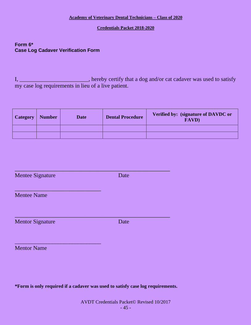
Academy of Veterinary Dental Technicians – Class of 2020
Credentials Packet 2018-2020
AVDT Credentials Packet© Revised 10/2017
- 45 -
Form 6* Case Log Cadaver Verification Form
I, ________________________, hereby certify that a dog and/or cat cadaver was used to satisfy
my case log requirements in lieu of a live patient.
Category Number Date Dental Procedure Verified by: (signature of DAVDC or
FAVD)
______________________________________________________
Mentee Signature Date
______________________________
Mentee Name
______________________________________________________
Mentor Signature Date
______________________________
Mentor Name
*Form is only required if a cadaver was used to satisfy case log requirements.

Academy of Veterinary Dental Technicians – Class of 2020
Credentials Packet 2018-2020
AVDT Credentials Packet© Revised 10/2017
- 46 -
Form 7* Dental Radiography – Cadaver Use
I, ______________________________, hereby certify that a cadaver dog and/or cat cadaver
was used in my dental radiography requirement in lieu of a live patient.
______________________________________________________
Mentee Signature Date
______________________________
Mentee Name
______________________________________________________
Mentor Signature Date
______________________________
Mentor Name
*Form is only required if a cadaver is used to satisfy radiography requirement.

Academy of Veterinary Dental Technicians – Class of 2020
Credentials Packet 2018-2020
AVDT Credentials Packet© Revised 10/2017
- 47 -
Form 8 MENTOR/MENTEE CONTACTS and CASE LOG/CE VERIFICATION
The mentor and mentee met on the following dates via the following form of communication:
Date
Mentee
Initials
Mentor
Initials
Method of
Communication
(i.e.: in person, email,
phone, etc.)
Nature of Meeting
– Topic(s) Covered
_______________________________ _______________________________
Mentee Signature Mentor Signature±
_______________________________ ________________________________
Mentee Name Mentor Name
± By signing this form, the Mentor is verifying regular contact with the Mentee, as well as
verifying having reviewed the Mentee’s Credentials Packet (including all logs) and CE
documentation.

Academy of Veterinary Dental Technicians – Class of 2020
Credentials Packet 2018-2020
AVDT Credentials Packet© Revised 10/2017
- 48 -
Form 9 Mentee Shadowing Hours Verification Form
The mentee must complete this form if they’d like their shadowing hours to count towards their total hours at
the end of their two year mentorship. The mentee AND supervising veterinarian (DAVDC or FAVD)at the
clinic to which these hours were accumulated at must sign this form. If a signature is not present when the
mentee turns in this form these hours will be null and void.
Date
Clinic name
Shadowing hours
obtained
_______________________________ _______________________________
Mentee Signature Verified by: (signature of DAVDC or FAVD)±
_______________________________ ________________________________
Mentee Name DAVDC or FAVD Name
±By signing this form, the supervising veterinarian (DAVDC or FAVD) is verifying the mentee
obtained shadowing hours at their clinic.
*** IF you’ve obtained shadowing hours at multiple clinics, please make copies of this form- do not
have multiple clinics on one form. ***
Canine Dental Chart

Academy of Veterinary Dental Technicians – Class of 2020
Credentials Packet 2018-2020
AVDT Credentials Packet© Revised 10/2017
- 49 -
104 204
04 104
404 304
ABBREVIATIONS:
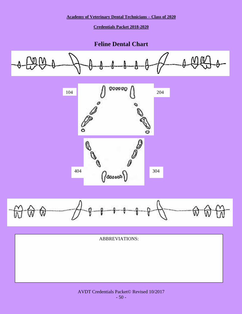
Academy of Veterinary Dental Technicians – Class of 2020
Credentials Packet 2018-2020
AVDT Credentials Packet© Revised 10/2017
- 50 -
104 204
404 304
Feline Dental Chart
ABBREVIATIONS:

Academy of Veterinary Dental Technicians – Class of 2020
Credentials Packet 2018-2020
AVDT Credentials Packet© Revised 10/2017
- 51 -
CREDENTIALS PACKET FORMAT You must submit two copies of your credentials packet to our AVDT google drive: the AVDT Master
Folder and one additional “anonymous” folder with your randomly assigned number.
Final instructions on submission of your credentials packet will be sent to you by December 31, 2019. At
that time, you will be assigned a random number to be used as identification on one copy of your packet.
This “anonymous” packet will be sent to two reviewers for grading. Your Master packet (the AVDT Master
Folder) should include your full identification. It is very important that your anonymous packet not
contain your name or initials on ANY document.Note: It is your responsibility to notify, in writing, the
secretary of the AVDT within 30 days of any changes to this contact information. Please notify the
Credentials Chair immediately with any changes to your email address.
1. AVDT Master Folder ONLY: Signed copy of "Waiver, Release and Indemnity Agreement" (Form 1). Scan or photograph this form and save as a PDF file, and name it: First Last (names) - Form1.pdf
2. AVDT Master Folder ONLY: Letter of recommendation from supervising veterinarian. Thisletter should
detail such things as training program, ethical behavior, quality of skills, and your relationship to the
person writing the letter. Scan or photograph your letter and save as a PDF file and name it: First Last –
LetterofRec.pdf
3. AVDT Master Folder ONLY: Signed copy of “Mentor/Mentee Contacts and Case Log/CE Verification”
(Form 8). Scan or photograph this form and save as a PDF file, and name it: First Last - Form8.pdf
4. AVDT Master Folder ONLY: Signed copies of the “AVDT Program Hours Documents” (listed on page
2-3). Scan and save as a PDF file, and name it: FirstLast – Hours Verification Form 1.pdf,
FirstLast – Hours Verification Form 2.pdf, FirstLast – Hours Verification Form 3.pdf, FirstLast –
Hours Verification Form 4.pdf
5. AVDT Master Folder ONLY:Signed copy of the “AVDT Shadowing Hours Verification Form. Scan
and save as a PDF file, and name it: FirstLast – Shadowing Hours Verification Form 9.pdf
6. AVDT Master Folder ONLY: Signed copy of “Equipment List” (Form 4). Scan this form and save as a
PDF file, and name it: First Last – Form4.pdf
7. AVDT Master Folder ONLY: A copy of blank dental record forms (feline, canine, or other).
DentaLabels are not acceptable as a complete dental record. Save as PDF files in your Master Folder.
Please name the files: First Last - Canine Chart.pdf (or Feline) in your Master folder
8. Completed “Skills Form” (Form 2). You are required to state whether or not you have mastered the skill
on the form. Mastery is defined as being able to perform the task safely, with a high degree of
success, without being coached or prompted. Mastery requires having performed the task in a wide
variety of patients and situations. AVDT is aware that some states or provinces may not allow a task to
be performed by a veterinary technician. AVDT requires that a veterinarian, who has mastered the skill,
attest to your ability to perform the task. Scan or photograph this form and save as a PDF file, and name
it: First Last - Form2.pdf in your Master Folder and Applicant### - Form2.pdf in your Anonymous folder.
Academy of Veterinary Dental Technicians – Class of 2020
Credentials Packet 2018-2020
AVDT Credentials Packet© Revised 10/2017
- 52 -
9. Completed “Specialty Training Forms” (Forms 3a and b). A copy of this form is saved in both your Master
Folder and Anonymous Folder. After adding CE certificates, please change the names of the files to: First
Last – Form3a OR 3b.pdfin your Master Folder and Applicant### - Form3a OR 3b.pdfin your Anonymous
Folder.
10. A copy of “Case Log Cadaver Verification” (Form 6). This is only if cadavers are used. Scan or
photograph this form and save it as a PDF file, and name it: First Last - Form6.pdfin your Master folder and
Applicant### - Form6in your Anonymous folder.
11. A copy of “Dental Radiography - Cavader Use” (Form 7). This is only if cadavers are used.Scan or
photograph this form and save it as a Word, JPEG or PDF file, and name it: First Last - Form5 in your Master
folder and Applicant### - Form5in your Anonymous folder.
12. Completed Categorical Case Logs and Chronological Case Logs with a minimum of 50 cases. Copies of
these logs are saved in both your Master folder and Anonymous folder. Please change the names and convert
the files to pdfs. The name should be: First Last - Cat Log.pdf in your Master folder and Applicant### -
CatLog.pdf in your Anonymous folder. Please change the names and convertthe files to pdfs: First Last -
Chrono Log.pdf in your Master folder and Applicant### - Chrono Log.pdf in your Anonymous folder.
13. Five Case Reports. Save as PDF files, and name them: First Last - Report 1 in your Master folder and
Applicant### - Report1.pdfin your Anonymous folder. Name them Report1, Report2, Report3, etc.
14. Dental Radiography Requirement. Save as a JPEG file or embedded in a Word.doc, and name them:
First Last - CanineRads (or Feline) in the Master folder and Applicant#### - CanineRads (or
Feline) in your Anonymous folder.
15. Submit you dues/packet administration fee of $75 to Mary Berg, AVDT treasurer. Additional
details will be provided.
*** Please follow these instructions carefully!! Also, make sure to white out or block your name on any files included in your anonymous packet. Failure to comply with these instructions will result in denial of your packet! ***
† Please note that documents requiring a signature must be scanned or photographed. Forms not requiring
signatures may be typed directly on inside the file. All files must be saved with the appropriate names. Please
call or email the Credentials Chair with any questions.

Academy of Veterinary Dental Technicians – Class of 2020
Credentials Packet 2018-2020
AVDT Credentials Packet© Revised 10/2017
- 53 -
CREDENTIAL PACKET SUBMISSION PROCESS
If you choose to duplicate any form using a word processing program, use the same size and style of font, and
the same number of pages. It is required that you keep a back up copy of your credentials
packet in case of technology failure and for your own reference. All information included in the
original should be in your copy. No packets will be returned to you at the end of the review process. All
packets will be destroyed after review. A notice will be emailed to provide instructions for you dues/packet fee
to be paid.
Do not modify any form.
The folders of your credentials packet should be sent via email in a compressed zip drive to:
Renae White, CVT, VTS (Dentistry)
AVDT Credentials Chair
The credentials packet must be received on or before January 31, 2020. Packets received after this date will
not be considered for the 2020 examination process. Please keep the Credentials Chair and your
mentor up to date on your email address, as this will be used as our primary source of
communication.
Mentor/Mentee Checklist Master Folder Only (all forms below must also be in the Master Folder with original signatures):
Form 1: Waiver, Release, and Indemnity Agreement Signed by Mentee Letter of Recommendation from supervising veterinarian Form 8: Mentor/Mentee Contacts and Case Log Verification Form Signed by Mentor and Mentee AVDT Program Hours Documentations: Signed by Mentee, supervising veterinian, office manager
If you are including your Shadowing Hours Verification Form (form 9), mentee and supervising DAVDC or FAVD at said clinic(s) must sign form
Form 4: Equipment List Signed by Mentee and Supervising veterinarian Blank Dental Records (Canine and Feline)
Anonymous Folder:
Form 2: Skills Form Signed by supervising DVM Form 3a: Specialty Training Wet Lab Form Form 3b: Specialty Training Lecture Form Proof of CE Attendance Form 6: Case Log Cadaver Verification Form Signed by Mentor, Mentee, and supervising DAVDC or FAVD Form 7: Dental Radiograph Cadaver Form Signed by Mentor and Mentee Categorical and Chronological Case Logs
Academy of Veterinary Dental Technicians – Class of 2020
Credentials Packet 2018-2020
AVDT Credentials Packet© Revised 10/2017
- 54 -
Five Case Reports Dental Radiography Requirement (Canine and Feline)