Academic Sciences · Original Article ISOLATION, CLONING AND EXPRESSION OF RECOMBINANT...
5
Original Article ISOLATION, CLONING AND EXPRESSION OF RECOMBINANT STAPHYLOKINASE GENE AGAINST THROMBOSIS YERASI GNANA PRASAD REDDY 1 , RAJENDRAN PRAKASH 1 AND SINGARAM ANANDAKUMAR 1* 1 VMKV Engineering College, Periyaseeragapadi, Salem- 636308, Tamilnadu, India. Email: [email protected] Received: 04 Feb 2014 Revised and Accepted: 25 Feb 2014 ABSTRACT Objective: Recombinant technology has crucial impact in therapy development. In microbial environment, pathogenic organism such as staphylococcus aureus produces staphylokinase protein. This protein has major role in thrombolysis. In clinical, non-pathogenic organism well known as Escherichia coli used for recombinant drug synthesis. Methods: In the present study, staphylokinase (SAK) gene is isolated from S. aureus. Sample of pus is collected from wound and infected patients. This S. aureus retrieves from sample. The staphylokinase gene is isolated and injected into a vector such as pET-32 a, b, c (+). This cloned vector transformed into salt induced E. coli strain (DH5α). Results: The mature recombinant Sak protein is expressed in E.coli culture. Further, recombinant staphylokinase is extracted and examined by SDS- PAGE and the present of staphylokinase was observed. Conclusion: Recombinant staphylokinase has major crucial role in thrombotic disorders and I will used to design drug against thrombosis without side effect. Keywords: Staphylokinase, E. coli, pET-32a, Recombinant, Thrombolysis. INTRODUCTION In therapeutic system, Staphylococcus aureus play crucial role in the pathogenic world. Despite, S. aureus was expressed many significant proteins, especially sak protein. Extracellular protein Staphylokinase (SAK) was excreted by S. aureus strains. SAK gene encodes 163 amino acids and a mature protein consists of 136 amino acids. Ordinarily, methicillin-resistant Staphylococcus aureus (MRSA) strains were produced staphylokinase protein. The staphylokinase structural gene was encoded N-acetylmuramoyl L-alanine amidase in methicillin resistant staphylococcus aureus MRSA. In therapeutic purpose, staphylokinase was explicit the properties of a thrombolytic agent. This staphylokinase protein interacted with plasminogen and converted into proteolytic enzyme plasmin, which staphylokinase digested fibrin clots. Staphylokinase was cleaved C3b and IgG and inhibiting phagocytosis. It was also regulated by agr gene regulator [1-4]. Pathologies were involved in the failure of hemostasis and production of thrombolytic agents. Staphylokinase was one of the thrombolytic agent and other fibrinolytic agents such as tissues type plasminogen activator (t-PA) and urokinase. These agents were utilized for clot dissolution [5]. Generally, MRSA strains produced staphylokinase protein, it caused various diseases. In clinical studies, staphylokinase gene introduced into non-pathogenic Escherichia coli and it was produced recombinant staphylokinase (r-SAK) protein used for thrombolysis. Recombinant SAK was found more meaningful effect than streptokinase for dissolution of platelet-rich arterial thrombi [6, 7]. Thrombotic disorders were one of the major impacts in human death owing to the thrombosis. Thrombi usually related to stroke, peripheral occlusive disease, pulmonary embolism, myocardial infarction and deep vein thrombosis. Therapy for these diseases was well recognized. Staphylokinase, plasminogen activator and urokinase was widely used thrombolytic agents. Among these agents, staphylokinase play major imperative role in fibrinolysis. In this research, staphylokinase gene was isolated from MRSA strains and MRSA strains were separated based on thrombolysis activity using a blood sample from patients. Then, this significant gene was introduced into Escherichia coli and produced recombinant staphylokinase (r-SAK) proteins against fibrinolysis [8-10]. Fig. 1: Schematic representation of overall experimental flow chart. A) Mechanism of fibrinolysis by staphylokinase. B) Experimental theoretical view. International Journal of Pharmacy and Pharmaceutical Sciences ISSN- 0975-1491 Vol 6, Issue 4, 2014 Innovare Academic Sciences
Transcript of Academic Sciences · Original Article ISOLATION, CLONING AND EXPRESSION OF RECOMBINANT...

Original Article
ISOLATION, CLONING AND EXPRESSION OF RECOMBINANT STAPHYLOKINASE GENE AGAINST THROMBOSIS
YERASI GNANA PRASAD REDDY1, RAJENDRAN PRAKASH1 AND SINGARAM ANANDAKUMAR1*
1VMKV Engineering College, Periyaseeragapadi, Salem- 636308, Tamilnadu, India.
Email: [email protected]
Received: 04 Feb 2014 Revised and Accepted: 25 Feb 2014
ABSTRACT
Objective: Recombinant technology has crucial impact in therapy development. In microbial environment, pathogenic organism such as staphylococcus aureus produces staphylokinase protein. This protein has major role in thrombolysis. In clinical, non-pathogenic organism well known as Escherichia coli used for recombinant drug synthesis.
Methods: In the present study, staphylokinase (SAK) gene is isolated from S. aureus. Sample of pus is collected from wound and infected patients. This S. aureus retrieves from sample. The staphylokinase gene is isolated and injected into a vector such as pET-32 a, b, c (+). This cloned vector transformed into salt induced E. coli strain (DH5α).
Results: The mature recombinant Sak protein is expressed in E.coli culture. Further, recombinant staphylokinase is extracted and examined by SDS-PAGE and the present of staphylokinase was observed.
Conclusion: Recombinant staphylokinase has major crucial role in thrombotic disorders and I will used to design drug against thrombosis without side effect.
Keywords: Staphylokinase, E. coli, pET-32a, Recombinant, Thrombolysis.
INTRODUCTION
In therapeutic system, Staphylococcus aureus play crucial role in the pathogenic world. Despite, S. aureus was expressed many significant proteins, especially sak protein. Extracellular protein Staphylokinase (SAK) was excreted by S. aureus strains. SAK gene encodes 163 amino acids and a mature protein consists of 136 amino acids. Ordinarily, methicillin-resistant Staphylococcus aureus (MRSA) strains were produced staphylokinase protein. The staphylokinase structural gene was encoded N-acetylmuramoyl L-alanine amidase in methicillin resistant staphylococcus aureus MRSA. In therapeutic purpose, staphylokinase was explicit the properties of a thrombolytic agent. This staphylokinase protein interacted with plasminogen and converted into proteolytic enzyme plasmin, which staphylokinase digested fibrin clots. Staphylokinase was cleaved C3b and IgG and inhibiting phagocytosis. It was also regulated by agr gene regulator [1-4].
Pathologies were involved in the failure of hemostasis and production of thrombolytic agents. Staphylokinase was one of the thrombolytic agent and other fibrinolytic agents such as tissues type plasminogen activator (t-PA) and urokinase. These agents were utilized for clot dissolution [5]. Generally, MRSA strains produced staphylokinase protein, it caused various diseases. In clinical studies, staphylokinase gene introduced into non-pathogenic Escherichia coli and it was produced recombinant staphylokinase (r-SAK) protein used for thrombolysis. Recombinant SAK was found more meaningful effect than streptokinase for dissolution of platelet-rich arterial thrombi [6, 7].
Thrombotic disorders were one of the major impacts in human death owing to the thrombosis. Thrombi usually related to stroke, peripheral occlusive disease, pulmonary embolism, myocardial infarction and deep vein thrombosis. Therapy for these diseases was well recognized. Staphylokinase, plasminogen activator and urokinase was widely used thrombolytic agents. Among these agents, staphylokinase play major imperative role in fibrinolysis. In this research, staphylokinase gene was isolated from MRSA strains and MRSA strains were separated based on thrombolysis activity
using a blood sample from patients. Then, this significant gene was introduced into Escherichia coli and produced recombinant staphylokinase (r-SAK) proteins against fibrinolysis [8-10].
Fig. 1: Schematic representation of overall experimental flow chart. A) Mechanism of fibrinolysis by staphylokinase. B)
Experimental theoretical view.
International Journal of Pharmacy and Pharmaceutical Sciences
ISSN- 0975-1491 Vol 6, Issue 4, 2014
Innovare Academic Sciences
Anandkumar et al. Int J Pharm Pharm Sci, Vol 6, Issue 4, 266-270
267
MATERIALS AND METHODS
Isolation, characterization and cloning of SAK gene, expression of staphylokinase protein determinations were carried out by following methods such as, sample preparations, Isolation and methicillin-resistant detections, biochemical and morphological studies of Staphylococcus aureus, Isolation of genomic DNA and agarose gel electrophoresis, Sak gene cloning into E.coli (DH5α) and SDS-PAGE for determination of expressions and separation of staphylokinase protein.
Sample Preparation
Clinical samples of pus were collected from different sources of wound infection of infected patients from Government Mohan Kumaramangalam Hospital and Gopi Hospital, Salem, Tamilnadu. Clinical samples from the infected patients were collected in sterilized plastic container, stored and transported using insulated container and brought to the laboratory for bacteriological analysis.
Isolation and methicillin-resistant detection
Bacteria were found in clinical sample by morphological observation. Especially, S. aureus was isolated and characterized by spread plate and biochemical methods.
Two culture media used for isolated and methicillin-resistant detection such as, staphylococcus specific media 110 and MeReSa media (Acme Progen Biotech India Pvt Ltd, http://www.acmeprogen.com/). Staphylococcus aureus were grown on the specific media 110 and only Staphylococcus aureus were grown MeReSa media. The method was followed by mathias et al., 2012 [22].
Biochemical and morphological studies
Biochemical and morphological test for the confirmation of Staphylococcus aureus carried out by the method suggested by Sogaard et al., 2007 and Rubin et al., 2010 [23, 24].
Isolation of genomic DNA and agarose gel electrophoresis
The genomic DNA was isolated from specialized Staphylococcus aureus colonies and run agarose gel electrophoresis was confirmed the expression of MecA and SAK gene. Genomic DNA isolation and agarose gel preparations were carried out as suggested by Sowmya et al., 2012 [25]. Further SAK gene was sequenced and amplified using PCR.
SAK gene PCR and cloning
Isolated SAK gene was amplified using PCR. The primers were designed such as, forward primer - 5’ AGAGATTGATTGTGAAAGAAGTGTT 3’ and reverse primer - 3’ CGAAGTACCTGCCTAAAAAAGGAT 5’. The amplified SAK gene was directly subcloned into a vector such as pET-32a by using restriction enzyme BamHI and EcoRI and ligated using T4 DNA ligase. Vector pET-32a was bought from Acme Progen Biotech India Pvt Ltd. Cloned vector was transformed into Escherichia coli competent cells. Transformed cells were identified by colonies formation suing antibiotic (ampicilin) containing medium with the presence of X-gal and IPTG (Isopropyl β-D-1-thiogalactopyranoside). Protocol and preparations of all these techniques were followed by Pulicherla et al., 2013 [10].
SDS-PAGE
Sample preparations and protein separations were followed by Pulicherla et al., 2013 [10]. The transformed DNA expression and protein separation were carried out by using SDS-PAGE. Cultured cells containing the Sak gene were grown up to 0.8 – 1 optical density and induced with appropriate inducer (IPTG for BL 21) for 4 h at 37°C. The induced protein expression profiles were resolved on 15% SDS-PAGE.
The Staphylokinase was visualized on the gel at the position corresponding to the reference or standard protein. Recombinant Sak gene expressions and protein isolation was done by using SDS-PAGE.
RESULTS
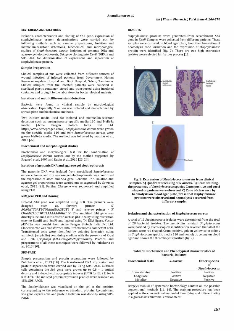
Staphylokinase proteins were generated from recombinant SAK gene in E.coli. Samples were collected from different patients. These samples were cultured on blood agar plate, from the observation of heomolysis zone formation and the expression of staphylokinase protein were identified (fig. 2). There are two high expression isolates were selected for further process [11].
Fig. 2: Expression of Staphylococcus aureus from clinical samples. A) Quadrant streaking of S. aureus. B) Gram staining,
the presences of Staphylococcus species Gram positive and cocci shaped organisms were observed. C) Zone of clearance by heomolysis on blood agar plate, present of staphylokinase
proteins were observed and heomolysis occurred from different sample.
Isolation and characterization of Staphylococcus aureus
A total of 13 Staphylococcus isolates were determined from the total of 28 bacterial isolates. The methicillin resistant Staphylococcus were notified by micro scopical identification revealed that all of the isolates were rod shaped, Gram positive, golden yellow color colony on Staphylococcus specific media 110 and hemolytic colony on blood agar and shown the thrombolysis positive (fig. 2).
Table 1: Biochemical and Phenotypical characteristics of bacterial isolates
Biochemical tests S. aureus Other species of
Staphylococcus Gram staining Positive Positive
Coagulase Positive Negative Motality Negative Positive
Bergeys manual of systematic bacteriology contain all the possible conventional methods [11, 14]. The staining procedure has been applied as the conventional method of identifying and differentiating in a givenssssss microbial environment.

Anandkumar et al. Int J Pharm Pharm Sci, Vol 6, Issue 4, 266-270
268
Table 2: Biochemical chara8cteristics of methicillin resistant Staphylococcus species.
TEST MRSA Gram stain +ve
Under light microscope Cocci in clusters Nutrient Agar Golden yellow color colony
Blood agar Beta hemolytic colony
Genomic DNA isolation and PCR
Bacterial genome DNA was extracted from whole cells by using a standard method or a commercial system. The organism was found highly gram positive, it does not undergo cell wall rupture by lysozyme action. So the whole cell has been frozen in liquid nitrogen, ground well and then the DNA was extracted and purified by commercial processes. Thus isolated genomic
Fig. 3: Isolation of MecA and Sak genes using Gel documentation. A) Isolation of MecA gene was spotted. B) Isolation and
identification of SAK gene 492 bp was observed by using agarose gel electrophoresis. C) PCR amplified SAK gene with Promoter. D) Cloned SAK gene was isolated from E.coli and
amplified SAK gene was observed from subclone.
DNA was run in agarose gel for the conformation of the isolated DNA. From gel documentation of the genomic DNA, it has been identified that total length of the DNA arrived about 492bp when compare to that of the marker gene in the lane 1 (fig. 3).
The next step process also called cycle sequencing. Both the forward and reverse sequences were used as the template. Universal primers were used M13 forward and reverse primers. The forward Primer - 5’ AGAGATTGATTGTGAAAGAAGTGTT 3’ and the reverse Primer – 5’ CGAAGTACCTGCCTAAAAAAGGAT 3’.
The broad based or universal primers complementary to the conserved regions of SAK were used so that the region can be
amplified from any bacteria. The amplified DNA has been run in agarose gel and then the bands are eluted by the gel elution kit. The gel documentation shown that the amplified DNA fragment was exactly 492 bp (fig. 3). From this result, that amplified DNA was conformed as Sak gene [13, 14]. Subcloning of the gene of interest SAK gene into destination vector such as pET-32a. Restriction enzymes were used to excise the gene of interest (SAK gene) such as BamHI and EcoRI. Subcloned gene was amplified by using PCR [10, 14].
Fig. 4: Amplified Sak gene sequence from PCR. Amplified gene was isolated from agarose gel and sequenced to determine the
present of SAK gene.
Cloning into E. coli
The amplified Sak gene was inserted into the destination vector such as pET-32a by using the restriction enzyme and T4DNA ligase. This ligated DNA vector pET-32a was transformed into E. coli competent cells (fig. 5). The transformants were screened by growing the transformed cell in the LB agar medium in the combination of IPTG and ampicillin. The selection was based on the formation of white colonies, which it was called as transformants and the blue colonies were the natural vectors or plasmids of the E. coli cells. But the result shown that, there was no such blue colonies and the whole of our target DNA has been transformed [9, 15, 16].
Expression Analysis of Staphylokinase
SDS-PAGE, sodium dodecyl sulfate polyacrylamide gel electrophoresis widely used to separate proteins according to their electrophoretic mobility such as length of the polypeptide chain and its charge. Transformed E.coli culture was produced staphylokinase proteins. All protein expressions were analyzed by running on 15% SDS-PAGE and a very clear 28.6 KDa protein band was identified against a high molecular weight protein ladder (fig. 6). The observed results in the present investigation were coincided with the similar expression patterns in E. coli as an extra cellular protein. Literal expressions of 28.6 KDa SAK protein was separated by using SDS-PAGE. This gel documentation was shown the production of recombinant protein. These recombinant staphylokinase proteins were used as thrombolytic or fibrinolytic agent. In clinical, these proteins also used therapeutic agent for many thrombotic disorders [10, 14, 16].

Anandkumar et al. Int J Pharm Pharm Sci, Vol 6, Issue 4, 266-270
269
Fig. 5: The structure of plasmid vector pET-32a vector.
DISCUSSIONS
Some Staphylococcus strains synthesized a family of enzymatic proteins that carried the potential to activated plasminogen [1].
Fig. 6: Expression of recombinant SAK protein form gel documentation and SDS-PAGE. A) Gel documentation of Plasmid DNA. B) Transformation of ligated vector into E.coli competent
cells, white colonies were observed and confirmed the presence of transformants. C) Transformed E.coli cells were cultured and
produced recombinant staphylokinase proteins (r-SAK). Recombinant SAK proteins 28.6 KDa were separated and
observed by using SDS-PAGE.
The plasminogen activator of Staphylococcus aureus has been isolated. Staphylokinase family of four plasminogen activators called STAN, STA-CM-I, STA-CN-II, STAR-C [6]. Staphylococcus aureus was produced thrombolytic protein such as staphylokinase (SAK). Staphylokinase was best microbial plasminogen activator [17]. The morphological and biochemical studies were better understood [18]. Recombinant staphylokinase proteins were produced via bacterial cloning such as Escherichia coli (fig. 6) [19]. These recombinant staphylokinase proteins were shown to fibrin clot lysis in human plasma [20]. A Fibrinolytic protein has been isolated from other species including microfungi, leeches, mosquitoes and snakes [21]. Many of these proteins, recombinant staphylokinase proteins have crucial impact in thrombolysis [8].
CONCLUSIONS
The staphylokinase proteins were isolated from MRSA strains. This recombinant SAK gene was produced staphylokinase by using E.coli as a host. Staphylococcus aureus was also pathogenic but E.coli was non-pathogenic bacterium. This non-pathogenic recombinant protein was used as fibrinolytic agent. Production of recombinant staphylokinase proteins were very simple process, low cost, high efficiency, non-pathogenic, non-toxic, high yield and no side effect. Following these kind of advantage, many pathologists used recombinant SAK gene to produced recombinant staphylokinase proteins and used as fibrinolysis agent against variety of thrombotic disorders. It was a best method to treated thrombotic disorders by using recombinant SAK proteins against fibrinolysis.
ACKNOWLEDGEMENTS
The authors are earnest thank to Dr. (Late) S. Ganeshmanikandan for valuable guidance and encouragement. The authors also thank to Dr. A. Nagappan, Principal and Dr.
C.K. Hindumathy, Dean- Biosciences, Vinayaka Mission’s Kirupananda Variyar Engineering College, Salem, Tamil Nadu for their support for accomplishing this work.
REFERENCES
1. Vesterberg K, Vesterberg O. Studies of staphylokinase. J Med Microbiol 1972; 5(4): 441-50.
2. Bokarewa MI, Jin T, Tarkowski A. Staphylococcus aureus: Staphylokinase. Int J Biochem Cell Biol 2006; 38(4): 504-9.
3. Koch TK, Reuter M, Barthel D, Bohm S, van den Elsen J, Kraiczy P, et al. Staphylococcus aureus proteins Sbi and Efb recruit human plasmin to degrade complement C3 and C3b. PLoS One 2012; 7(10): e47638.
4. Chen CJ, Unger C, Hoffmann W, Lindsay JA, Huang YC, Gotz F. Characterization and comparison of 2 distinct epidemic community-associated methicillin-resistant Staphylococcus aureus clones of ST59 lineage. PLoS One 2013; 8(9): e63210.
5. Rother J, Ford GA, Thijs VN. Thrombolytics in acute ischaemic stroke: historical perspective and future opportunities. Cerebrovasc Dis 2013; 35(4): 313-9.
6. Collen D, De Cock F, Stassen JM. Comparative immunogenicity and thrombolytic properties toward arterial and venous thrombi of streptokinase and recombinant staphylokinase in baboons. Circulation 1993; 87(3): 996-1006.
7. Li CJ, Huang J, Yang ZJ, Cao KJ. Thrombolytic efficacy of native recombinant staphylokinase on femoral artery thrombus of rabbits. Acta Pharmacol Sin 2007; 28(1): 58-65.
8. Moussa M, Ibrahim M, El Ghazaly M, Rohde J, Gnoth S, Anton A, et al. Expression of recombinant staphylokinase in the methylotrophic yeast Hansenula polymorpha. BMC Biotechnol 2012; 12: 96.
9. Kwiecinski J, Josefsson E, Mitchell J, Higgins J, Magnusson M, Foster T, et al. Activation of plasminogen by staphylokinase reduces the severity of Staphylococcus aureus systemic infection. J Infect Dis 2010; 202(7): 1041-9.
10. Pulicherla KK, Kumar A, Gadupudi GS, Kotra SR, Rao KR. In vitro characterization of a multifunctional staphylokinase variant with reduced reocclusion, produced from salt inducible E. coli GJ1158. Biomed Res Int 2013; 2013: 297305.

Anandkumar et al. Int J Pharm Pharm Sci, Vol 6, Issue 4, 266-270
270
11. Wieckowska-Szakiel M, Sadowska B, Rózalska B. Staphylokinase production by clinical Staphylococcus aureus strains. Pol J Microbiol 2007; 56(2): 97-102.
12. Paabu. Precise protocol and the problems faced during conventional PCR. Mol evolutions 1989; 26: 1289-1296.
13. Sako T, Tsuchida N. Nucleotide sequence of the staphylokinase gene from Staphylococcus aureus. Nucleic Acids Res 1983; 11(22): 7679-93.
14. Mandi N, Soorapaneni S, Rewanwar S, Kotwal P, Prasad B, Mandal G, et al. High yielding recombinant Staphylokinase in bacterial expression system—cloning, expression, purification and activity studies. Protein Expr Purif 2008; 64: 69–75.
15. Sang Jun Lee, Chul Kim, Dong Min Kim, Kwang Hee Bae, Si Myung Byun. High level secretion of recombinant staphylokinase into periplasm of Escherichia coli. Biotechnol Lett 1998; 20: 113-116.
16. Kowalski M, Brown G, Bieniasz M, Oszajca K, Chabielska E, Pietras T. Cloning and expression of a new recombinant thrombolytic and anthithrombotic agent - a staphylokinase variant. Acta Biochim Pol 2009; 56(1): 41-53.
17. Vanderschueren SMF, Lijnen HR, Collen D. Properties of Staphylokinase and its Potential as a Thrombolytic Agent. Fibrinolysis 1995; 9: 87-90.
18. Okada K, Ueshima S, Fukao H, Matsuo O. Analysis of complex formation between plasmin(ogen) and staphylokinase or streptokinase. Arch Biochem Biophys 2001; 393: 339–41.
19. Schlott B, Hartmann M, Guhrs KH, Birchhirschfeid E, Pohl HD, Vanderschueren S, et al. High yield production and purification of recombinant staphylokinase for thrombolytic therapy. Biotechnology 1994; 12: 185– 9.
20. Matsuo O, Okada K, Fukao H, Tomioka Y, Ueshima S, Watanuki M, et al. Thrombolytic properties of staphylokinase. Blood 1990; 76: 925–9.
21. Barwald G, Jahn G, Volzke KD. Microbiological isolation of a protease with fibrinolytic effect from Aspergillus ochraceus. Folia Haematol Int Mag Klin Morphol Blutforsch 1974; 101: 83– 93.
22. Mathias MT, Horsley MB, Mawn LA, Laquis SJ, Cahill KV, Foster J, et al. Atypical presentations of orbital cellulitis caused by methicillin-resistant Staphylococcus aureus. Ophthalmology 2012; 119: 1238-43.
23. Sogaard M, Norgaard M, Schonheyder HC. First notification of positive blood cultures and the high accuracy of the gram stain report. J Clin Microbiol 2007; 45: 1113-7.
24. Rubin JE, Bayly MK, Chirino-Trejo M. Comparison of dog and rabbit plasmas in the tube coagulase test for Staphylococcus aureus. J Vet Diagn Invest 2010; 22: 770-1.
25. Sowmya N, Thakur MS, Manonmani HK. Rapid and simple DNA extraction method forthe detection of enterotoxigenic Staphylococcus aureus directlyfrom food samples: comparison of PCR and LAMP methods. J Appl Microbiol 2012; 113: 106-13.
http://www.ncbi.nlm.nih.gov/pubmed?term=Chabielska%20E%5BAuthor%5D&cauthor=true&cauthor_uid=19018330
http://www.ncbi.nlm.nih.gov/pubmed?term=Chabielska%20E%5BAuthor%5D&cauthor=true&cauthor_uid=19018330
http://www.ncbi.nlm.nih.gov/pubmed?term=Chabielska%20E%5BAuthor%5D&cauthor=true&cauthor_uid=19018330