Acaaddemmiicc Sccii eenncess · from dried plant by using kitchen mixer grinder (Bajaj electronics...
7
Research Article ISOLATION AND STRUCTURAL ELUCIDATION OF FLAVONOIDS FROM AQUATIC FERN AZOLLA MICROPHYLLA AND EVALUATION OF FREE RADICAL SCAVENGING ACTIVITY K. SELVARAJ, RANJANA CHOWDHURY * , CHIRANJIB BHATTACHARJEE Department of Chemical Engineering, Jadavpur University, Jadavpur, Kolkata 700032, India. Email: [email protected] Received: 26 Jun 2013, Revised and Accepted: 06 Aug 2013 ABSTRACT Objective: The present study was designed for isolation of bioactive polyphenolic compounds from methanol extract of Azolla microphylla and their subsequent characterization. Methods: The flavonoid compounds were isolated and characterized by using thin layer chromatography (TLC), purified by preparative thin layer chromatography (PTLC) and were identified using High performance chromatography (HPLC). Their structures and chemical bonds were analyzed using Ultraviolet-Visible spectrophotomery (UV spec), Fourier Transform-Infra Red spectroscopy (FTIR) and Nuclear magnetic resonance NMR ( 13 C and 1 H) techniques. Results: Two flavonoids were identified as rutin and quercetin. The isolated compounds showed a potent antioxidant radical scavenging activity, as assessed by non-physiological assays like DPPH (2, 2-diphenyl-1-picrylhydrazyl), ABTS (2, 2’-Azinobis (3-ethylbenzothiazoline-6-sulphonic acid) diammonium salt) and FRAP (Ferric reducing antioxidant power). Conclusion: For the first time rutin and quercetin have been isolated successfully from the macrophyte aquatic fern Azolla microphylla under the present study. The isolation of the above characterized flavonoids would be useful to prepare plant-based pharmaceutical preparation to treat various complications linked with human diseases. Keywords: Azolla microphylla, DPPH, ABTS, FRAP, Rutin, Quercetin. INTRODUCTION Azolla microphylla is an aquatic fern that freely floats on the surface of the water body. It is a pteridophyte plantae belonging to the Salvinacea family [1]. It has been traditionally used as a green manure for wetland paddy fields for the fixation of atmospheric- nitrogen (N2) with the help of Azolla-anabaena - a cyanobacterium [2]. They can accumulate elements like P and K and minerals from the environment. These essential elements become available to the soil on the decomposition of Azolla microphylla [3, 4]. This fern also serves as one of the main components in the food for the omnivorous– phytoplanktonophagous tilapia (Oreochromis niloticus) [5, 6]. One or more Azolla species are found worldwide in wetlands [7]. The phytochemical investigation on the Azolla microphylla shows that tannins, phenols, sugar, anthroquinone glycosides and steroids are present [8]. Azolla microphylla is also rich in protein, vitamin and minerals and is used as food supplements for cattle, pigs, ducks and chickens resulting in increased milk production, enhancement of weight of cattle, pigs, ducks and broiler chickens and for production of eggs with layered yolk, as compared to conventional ones[9,10]. More than 4000 chemically unique flavonoids have been isolated and identified in plant extracts obtained through solvent extraction processes [11, 12]. More recently, a few flavonoids have also been isolated from microorganisms as their secondary metabolites [13]. Flavonoids are of great importance for the bioactivities, related to their antioxidant activities and many enzymatic reactions, resulting in a decrease of platelet activation and aggregation, against cardiovascular diseases, cancer chemoprevention and anti- inflammatory activity [14-20].Although from phytochemical analysis of Azolla microphylla, it is evident that this fern is rich in flavonoids, no work has so far been reported on the isolation of flavonoids from them. The present study focuses on the isolation and free radical scavenger activities of flavonoids from methanolic extract of Azolla microphylla. Two flavonoids namely, rutin and quercetin have been isolated from Azolla microphylla. Their structures have been elucidated through UV, FTIR, 13 C NMR, and 1 H NMR. MATERIALS AND METHODS Chemicals and Plant material Rutin, Quercetin, 2, 2-diphenyl-1-picrylhydrazyl (DPPH), 2, 4, 6- tripyridyl-s-triazine (TPTZ) and 2, 2’-Azinobis (3- ethylbenzothiazoline-6-sulphonic acid) diammonium salt (ABTS) were obtained from Sigma–Aldrich, MO, USA. Folin-Ciocalteu’s reagent and gallic acid were obtained from Himedia laboratories Pvt. Ltd. Mumbai, India. Other chemicals and solvents such as silica gel (GF254), IR grade potassium bromide, methanol, petroleum ether, chloroform, ethyl acetate and formic acid were purchased from Merck, India Ltd. Azolla microphylla, an aquatic fern, was purchased from Vivekananda Kendra-NARDEP (Natural Resources Development Project), Vivekanandapuram, Kanyakumari, Tamil Nadu, India. Sample Preparation The whole plant, Azolla microphylla were washed thoroughly with tap water followed by rinsing with double distilled water and shade drying for 7days. The fine powder (≈60 mesh size) was obtained from dried plant by using kitchen mixer grinder (Bajaj electronics Ltd, India). The plant powder was sterilized at 121˚C for 15 min. The plant powder was stored under desiccator for further studies. Instruments Nuclear Magnetic Resonance ( 1 H and 13 C) spectra were recorded in either CD3OD or CDCl3 on a Bruker NMR Avance with TCI cyroprobe operating at 600MHz. UV absorption was carried out by Varian, Cary 50 UV-Visible double beam spectrophotometer. High performance liquid chromatography (HPLC) was performed on Model LC-8, Shimadzu, Japan and FT-IR spectrum was generated using Perkin Elmer, Spectrum 100 instrument. Extraction of flavonoids Solvent extraction of dried powder (40g) of Azolla microphylla was done using 2L of 80% methanol (50mL/g of sample) in a soxhlet extractor for 24h. The extract was concentrated by evaporation (40- 50 ° C) in a rotary vacuum evaporator. The concentrated methonolic extract (10mL) was suspended in 50mL of distilled water and was further extracted successively with petroleum ether, chloroform, and ethyl acetate. For each solvent, extraction procedure was repeated thrice for complete extraction. The petroleum ether extract (Fraction-I) was discarded because it contained fatty substances. The cyanidine test was performed for the chloroform (Fraction-II) and ethyl acetate (Fraction-III) extracts for further selection. The ethyl acetate extract (Fraction-III) was analyzed for flavonoids using chromatographic separation. International Journal of Pharmacy and Pharmaceutical Sciences ISSN- 0975-1491 Vol 5, Suppl 3, 2013 A A c c a a d d e e m mi i c c S S c c i i e e n n c c e e s s
Transcript of Acaaddemmiicc Sccii eenncess · from dried plant by using kitchen mixer grinder (Bajaj electronics...

Research Article
ISOLATION AND STRUCTURAL ELUCIDATION OF FLAVONOIDS FROM AQUATIC FERN AZOLLA MICROPHYLLA AND EVALUATION OF FREE RADICAL SCAVENGING ACTIVITY
K. SELVARAJ, RANJANA CHOWDHURY*, CHIRANJIB BHATTACHARJEE
Department of Chemical Engineering, Jadavpur University, Jadavpur, Kolkata 700032, India. Email: [email protected]
Received: 26 Jun 2013, Revised and Accepted: 06 Aug 2013
ABSTRACT
Objective: The present study was designed for isolation of bioactive polyphenolic compounds from methanol extract of Azolla microphylla and their subsequent characterization.
Methods: The flavonoid compounds were isolated and characterized by using thin layer chromatography (TLC), purified by preparative thin layer chromatography (PTLC) and were identified using High performance chromatography (HPLC). Their structures and chemical bonds were analyzed using Ultraviolet-Visible spectrophotomery (UV spec), Fourier Transform-Infra Red spectroscopy (FTIR) and Nuclear magnetic resonance NMR (13C and 1H) techniques.
Results: Two flavonoids were identified as rutin and quercetin. The isolated compounds showed a potent antioxidant radical scavenging activity, as assessed by non-physiological assays like DPPH (2, 2-diphenyl-1-picrylhydrazyl), ABTS (2, 2’-Azinobis (3-ethylbenzothiazoline-6-sulphonic acid) diammonium salt) and FRAP (Ferric reducing antioxidant power).
Conclusion: For the first time rutin and quercetin have been isolated successfully from the macrophyte aquatic fern Azolla microphylla under the present study. The isolation of the above characterized flavonoids would be useful to prepare plant-based pharmaceutical preparation to treat various complications linked with human diseases.
Keywords: Azolla microphylla, DPPH, ABTS, FRAP, Rutin, Quercetin.
INTRODUCTION
Azolla microphylla is an aquatic fern that freely floats on the surface of the water body. It is a pteridophyte plantae belonging to the Salvinacea family [1]. It has been traditionally used as a green manure for wetland paddy fields for the fixation of atmospheric- nitrogen (N2) with the help of Azolla-anabaena - a cyanobacterium [2]. They can accumulate elements like P and K and minerals from the environment. These essential elements become available to the soil on the decomposition of Azolla microphylla [3, 4]. This fern also serves as one of the main components in the food for the omnivorous– phytoplanktonophagous tilapia (Oreochromis niloticus) [5, 6]. One or more Azolla species are found worldwide in wetlands [7]. The phytochemical investigation on the Azolla microphylla shows that tannins, phenols, sugar, anthroquinone glycosides and steroids are present [8]. Azolla microphylla is also rich in protein, vitamin and minerals and is used as food supplements for cattle, pigs, ducks and chickens resulting in increased milk production, enhancement of weight of cattle, pigs, ducks and broiler chickens and for production of eggs with layered yolk, as compared to conventional ones[9,10]. More than 4000 chemically unique flavonoids have been isolated and identified in plant extracts obtained through solvent extraction processes [11, 12]. More recently, a few flavonoids have also been isolated from microorganisms as their secondary metabolites [13]. Flavonoids are of great importance for the bioactivities, related to their antioxidant activities and many enzymatic reactions, resulting in a decrease of platelet activation and aggregation, against cardiovascular diseases, cancer chemoprevention and anti-inflammatory activity [14-20].Although from phytochemical analysis of Azolla microphylla, it is evident that this fern is rich in flavonoids, no work has so far been reported on the isolation of flavonoids from them. The present study focuses on the isolation and free radical scavenger activities of flavonoids from methanolic extract of Azolla microphylla. Two flavonoids namely, rutin and quercetin have been isolated from Azolla microphylla. Their structures have been elucidated through UV, FTIR, 13C NMR, and 1 H NMR.
MATERIALS AND METHODS
Chemicals and Plant material
Rutin, Quercetin, 2, 2-diphenyl-1-picrylhydrazyl (DPPH), 2, 4, 6-tripyridyl-s-triazine (TPTZ) and 2, 2’-Azinobis (3-
ethylbenzothiazoline-6-sulphonic acid) diammonium salt (ABTS) were obtained from Sigma–Aldrich, MO, USA. Folin-Ciocalteu’s reagent and gallic acid were obtained from Himedia laboratories Pvt. Ltd. Mumbai, India. Other chemicals and solvents such as silica gel (GF254), IR grade potassium bromide, methanol, petroleum ether, chloroform, ethyl acetate and formic acid were purchased from Merck, India Ltd. Azolla microphylla, an aquatic fern, was purchased from Vivekananda Kendra-NARDEP (Natural Resources Development Project), Vivekanandapuram, Kanyakumari, Tamil Nadu, India.
Sample Preparation
The whole plant, Azolla microphylla were washed thoroughly with tap water followed by rinsing with double distilled water and shade drying for 7days. The fine powder (≈60 mesh size) was obtained from dried plant by using kitchen mixer grinder (Bajaj electronics Ltd, India). The plant powder was sterilized at 121˚C for 15 min. The plant powder was stored under desiccator for further studies.
Instruments
Nuclear Magnetic Resonance (1H and 13C) spectra were recorded in either CD3OD or CDCl3 on a Bruker NMR Avance with TCI cyroprobe operating at 600MHz. UV absorption was carried out by Varian, Cary 50 UV-Visible double beam spectrophotometer. High performance liquid chromatography (HPLC) was performed on Model LC-8, Shimadzu, Japan and FT-IR spectrum was generated using Perkin Elmer, Spectrum 100 instrument.
Extraction of flavonoids
Solvent extraction of dried powder (40g) of Azolla microphylla was done using 2L of 80% methanol (50mL/g of sample) in a soxhlet extractor for 24h. The extract was concentrated by evaporation (40-50°C) in a rotary vacuum evaporator. The concentrated methonolic extract (10mL) was suspended in 50mL of distilled water and was further extracted successively with petroleum ether, chloroform, and ethyl acetate. For each solvent, extraction procedure was repeated thrice for complete extraction. The petroleum ether extract (Fraction-I) was discarded because it contained fatty substances. The cyanidine test was performed for the chloroform (Fraction-II) and ethyl acetate (Fraction-III) extracts for further selection. The ethyl acetate extract (Fraction-III) was analyzed for flavonoids using chromatographic separation.
International Journal of Pharmacy and Pharmaceutical Sciences
ISSN- 0975-1491 Vol 5, Suppl 3, 2013
AAccaaddeemmiicc SScciieenncceess
Chowdhury et al. Int J Pharm Pharm Sci, Vol 5, Suppl 3, 743-749
744
Isolation of flavonoids by thin layer chromatography (TLC)
The glass plates (100×200mm) coated with silica gel G (0.2-0.3mm) paste were dried naturally (atmospheric). Subsequently they were activated at 100 °C for 30 minutes and were cooled at room temperature (≈ 25oC). The extract was separated by TLC with the following mobile phases: ethyl acetate: n-butanol: water (50:30:10 v/v), ethyl acetate: acetic acid: water (60:30:10) and benzene: acetic acid: water (60:35: 5 v/v). The plates were developed using ammonia fumes and visualized under UV lamp. The Rf values of separated bands were calculated.
Purification of flavonoids by preparative thin layer chromatography (PTLC)
The extract was reduced to 5mL and loaded in preparative TLC plates coated with silica gel GF254 (0.4-0.5mm). The chromatogram was developed with benzene: acetic acid: water (60:35:5 v/v) and examined under UV lamp. The fluorescing spots were scraped out from the 100 plates and extracted with ethanol. The diluted mixture was then centrifuged at 10000 rpm for 10minutes at 4˚C. The supernatant was collected for further experimental (HPLC, FT-IR, NMR) analysis. The eluted compounds were co-chromatographed along with standard flavonoids.
High Performance Liquid Chromatography analysis
The isolated compounds (in ethanol) were filtered through membrane filter (Millipore, USA) and were injected (10µL) through the BDS Hypersil RP-C18 column (Thermo, 5µm, 120Å, 250mm × 4.6mm) at column temperature 25˚C. The mobile phase composed of methanol, water and formic acid (70:30:1 vol. %) was eluted at a flow rate of 1mL/min and the effluent was monitored at 280nm by UV detector. The peaks were detected and compared with the standards.
Spectral analysis
The isolated compounds 1 and 2 were dissolved in methanol and their maximum UV absorption ranges were recorded using the UV-Vis double-beam spectrophotometer. The compounds 1 and 2 were dissolved in CDCl3 and 1H and 13C NMR spectra using NMR spectroscopy with TCI Cyroprobe were recorded. Tetramethylsilane (TMS) was used as an internal standard. The chemical shift values were reported in ppm (δ) unit and the coupling constants (J) are in Hz. FTIR spectra of the compounds 1 and 2 were measured using IR grade potassium bromide (KBr). The compounds 1 and 2 were separately mixed with 200mg KBr to obtain round disc with the help of hydraulic press. Round disc was later subjected to FTIR in the range of 4000-400cm-1 at a resolution of 4cm-1.
In vitro radical scavenging assays
DPPH scavenging assay
The 2, 2-diphenyl-1-picrylhydrazil (DPPH˙*) free radical scavenging activity of the isolated compounds (1 and 2) was determined according to previous method [19]. To 0.1mL of aqueous solution (10-50µg/mL) of each isolated compound, 3mL of ethanolic solution of DPPH (0.1µM) was added. The mixture was shaken vigorously and allowed to stand for 30minutes in the dark, and the absorbance was measured at 517nm against a blank. The capability to scavenge the free radical DPPH (%DPPHSC) was calculated using the formula;
%DPPHSC = (A0 - A1) × 100/A0
Where A0= absorbance of the control; A1 = absorbance of the sample.
ABTS scavenging assay
ABTS˙* radical scavenging activity was measured according to the reference method [21] with some modifications. The ABTS˙* was generated by the reaction between 7mM ABTS (2, 2’-Azinobis (3-ethylbenzothiazoline-6-sulphonic acid) diammonium salt) solution and 2.45mM potassium persulphate solution incubated in the dark at room temperature for 16h. Before use, the absorbance at 734nm was adjusted to 0.700(±0.0020) by dilution with ethanol. To 3mL of the ABTS solution, 0.1mL of aqueous solution of each isolated compound (1-100µg/mL) was mixed vigorously. The reaction mixture was incubated for 6min and the absorbance was determined at 734nm by a UV-vis spectrophotometer. A standard curve was obtained by using rutin in 80% ethanol. The % ABTS
which was scavenged (%ABTSSC) was calculated using the formula;
% ABTSSC = (A0 - A1) × 100/A0
Where A0= absorbance of the control; A1 = absorbance of the sample.
Ferric reducing antioxidant potential (FRAP) assay
The ferric reducing antioxidant power activity was measured according to the reference method [22]. The FRAP reagent was prepared using 300mM acetate buffer (3.1g Sodium acetate, and 16mL Acetic acid) at pH 3.6, 10mM TPTZ (2,4,6-tripyridyl -s-triazine) solution in 40mM hydrochloric acid solution, and 20mM FeCl3.6H2O solution in distilled water. The acetate buffer (25mL) and TPTZ (2.5mL) were mixed together with FeCl3.6H2O (2.5mL). The temperature of the solution was adjusted to 37˚C before it was used. The different concentrations (10-100µg/mL) of the isolated compounds (1mL each) were allowed to react with the FRAP solution (3mL) for 30min under dark conditions. The absorbance was measured at 593nm and the reducing power was expressed as percentage ferric reducing activity of the compounds.
Statistical analysis
All experiments were repeated at least three times. Results are reported as mean ± standard deviation.
RESULTS AND DISCUSSIONS
Physical and Chemical Characteristics of isolated compounds
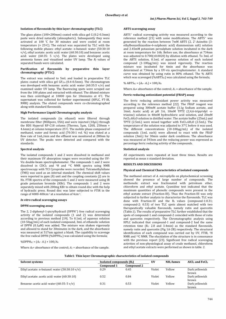
The methanol extract of A. microphylla on phytochemical screening showed the presence of large number of compounds. The methonolic extract was fractionated with petroleum ether, chloroform and ethyl acetate. Cyanidine test indicated that the maximum quantities of phenolic compounds were present in the ethyl acetate extract (Fraction-III). Thus the Fraction-III was only subjected to further analysis to characterize the flavonoids. TLC was done with Fraction-III and the Rf values (compound-1:0.31; compound-2: 0.53) of two TLC spots almost matched with two therapeutically valuable flavonoids, namely rutin and quercetin (Table.1). The results of preparative TLC further established that the spots of compound-1 and compound-2 coincided with those of rutin and quercetin respectively. The Chromatographic analysis using HPLC indicated that compound-1 and compound-2 had the same retention time (Rt: 2.8 and 3.4min) as the standard flavonoids, namely rutin and quercetin (Fig 1A-2B) respectively. The structural identification of each compound was carried out by UV, FTIR, 1H NMR and 13C NMR. The elucidation of the structure is in consonance with the previous report [23]. Significant free radical scavenging activities of non-physiological assay of crude methanol, chloroform and ethyl acetate extracts were performed as shown in table. 2.
[
Table1: Thin layer Chromatographic characteristics of isolated compounds
Solvent systems Isolated compounds (Rf) UV NH3 fumes AlCl3 and FeCl3 Compound 1 Compound 2
Ethyl acetate: n-butanol: water (50:30:10 v/v) 0.29 0.45 Violet Yellow Dark yellowish brown
Ethyl acetate: acetic acid: water (60:30:10) 0.52 0.84 Violet Yellow Dark yellowish brown
Benzene: acetic acid: water (60:35: 5 v/v) 0.31 0.53 Violet Yellow Dark yellowish brown

Chowdhury et al. Int J Pharm Pharm Sci, Vol 5, Suppl 3, 743-749
745
Fig. 1A: HPLC chromatogram of Isolated Rutin
Fig. 1B: HPLC chromatogram of Standard Rutin
Fig. 2A: HPLC chromatogram of Isolated Quercetin

Chowdhury et al. Int J Pharm Pharm Sci, Vol 5, Suppl 3, 743-749
746
Fig. 2B: HPLC chromatogram of Standard Quercetin
Table 2: Antioxidant activity of various extracts of Azolla microphylla
Sample DPPHsc (%) ABTSsc (%) % Ferric reducing power (%) Methanol extract 88.42 50.3 55 Chloroform 86.69 48.2 52 Ethyl acetate 92.61 55.4 56
Compound-1
Light yellow powder (ethanol); m. p. 190 ˚C; λmax. 300nm; mol. formula: C27 H30 O16; IR (KBr) Vmax cm-1: 3408, 3321 (O-H stretching), 2924,2843( CH2-stretching), 2714 (C-H bonding), 1462( C=O groups) and 1383(C-OH vibrations) (shown in Figure 3A); 1H-NMR (600MHz in CH3OD, δ ppm) 3.33-3.64 (m,12H of sugar moieties), 3.81(d, J=1.15Hz, 1H-Rham), 1.10(3H, d, J=6Hz, CH3-Rham), 4.52(4H, d, J=7.8Hz, H-1 Glu), 5.11 (1H, d, J=2Hz H-6), 6.19 (1H, d, J=2Hz, H-8), 6.38(1H, d, J=8Hz, H-5’), 7.66 (1H, m, H-2’, H-6’); 13C –NMR (600MHz in CH3OD, δ ppm): shown in Table 3. It was characterized as 3, 3’, 4’, 5, 7-pentahydroxy flavones-3-rutinoside (rutin) [24].
Compound-2
Pale yellow powder (ethanol); m. p. 300 ˚C; λmax. 360nm; mol. formula: C15 H10 O7; IR (KBr) Vmax cm-1:3428, 3369 (O-H stretching), 2986 (CH2-stretching), 2872 (CH-stretching), 1457(C=O), 1362 (C-OH vibrations) (shown in figure 3B); 1H-NMR (600MHz in CH3OD, δ ppm) 6.18(1H, d, J=2Hz, H-6), 6.38(1H, d, J=2Hz, H-8), 6.88(1H, d, J=8Hz, H-5’), 7.63(1H, d, J=7.5Hz, H-6’), 7.72(1H, d, J=2Hz, H-2’); 13C-NMR(600MHz in CH3OD, δ ppm): shown in Table 3. It was characterized as 2-(3, 4-Dihydroxyphenyl)-3, 5, 7-trihydroxy-4H-1-benzopyran-4-one (quercetin) [25].
Fig. 3A: FTIR spectra of rutin

Chowdhury et al. Int J Pharm Pharm Sci, Vol 5, Suppl 3, 743-749
747
Fig. 3B: FTIR spectra of Quercetin
Table 3: 13C-NMR data for isolated rutin and quercetin in comparison to 13C-NMR of rutin and quercetin obtained by the references [24, 25].
Position Compound 1 Reference rutin Compound 2 Reference quercetin 2 158.5 158.5 148.8 147.9 3 135.7 135.6 137.3 137.2 4 179.4 179.4 177.4 177.3 5 163.0 162.5 162.5 162.5 6 100.0 99.9 99.3 99.3 7 166.0 166.0 165.6 165.7 8 95.0 94.8 94.5 94.4 9 159.4 159.3 158.3 158.2 10 105.7 105.6 104.6 104.4 1’ 123.7 123.1 124.2 124.1 2’ 117.8 117.6 116.0 116.0 3’ 145.9 145.8 146.3 146.2 4’ 149.9 149.7 148.1 148.7 5’ 116.1 116.1 116.3 116.2 6’ 123.2 123.5 121.7 121.6 1’’ 104.8 104.7 2’’ 75.8 75.7 3’’ 77.2 77.2 4’’ 71.5 71.4 5’’ 78.2 78.1 6’’ 68.6 68.6 1’’’ 102.5 102.4 2’’’ 72.3 72.0 3’’’ 72.2 72.2 4’’’ 74.0 73.9 5’’’ 69.8 69.7 6’’’ 18.0 17.9
Anti-free radical activity of quercetin and rutin
The antioxidant activity of isolated rutin and quercetin was measured by different non- physiological methods. Mainly DPPH, ABTS radical scavenging and ferric (FRAP) reducing assays have been made. The antioxidant parameter is influenced by various factors such as oxidation substrate, oxidation mechanism and reaction medium [26]. The DPPH radical can be reduced by the en-1, 2-diol and dien-1, 4-diole moieties in antioxidants [27]. From the scavenging activity of the stable DPPH, ABTS cation radical and ferric reducing power assay of presently isolated rutin and quercetin it was observed that the flavonoids and their glycosides exhibited potent antioxidant activity.
Fig.4 and 5 respectively show the potential free radical scavenging activity against DPPH and ABTS free radicals
scavenging activity as a function of concentration of rutin and quercetin in the range of 92.6% at 35µg/ml and 91.5% at 30µg/ml concentration, whereas ABTS radical scavenging value of rutin and quercetin was 93.5% in 35µg/ml and 90.8% in 35µg/ml respectively. From the analysis of the figures, it is evident that free radical scavenging activities increase with increase of concentration of both rutin and quercetin up to a certain level and beyond this concentration saturation is observed. For scavenging activity against DPPH, the concentrations of rutin and quercetin corresponding to saturation value are 35µg/ml and 30µg/ml respectively. For scavenging activity against ABTS, the concentrations of rutin and quercetin corresponding to saturation value are 35µg/ml and 35µg/ml respectively.

Chowdhury et al. Int J Pharm Pharm Sci, Vol 5, Suppl 3, 743-749
748
Fig. 4: Scavenging of DPPH radical potential of rutin and quercetin in DPPH assay
Fig. 5: Scavenging of ABTS radical potential of rutin and quercetin in ABTS assay
It has been reported that ferric reducing power is associated with antioxidant activity and may serve as a significant reflection of the antioxidant activity [28]. The ferric reductive potential of the presently isolated rutin and quercetin were also dose dependent as shown in Fig. 6. The ferric reducing power of rutin and quercetin correlate similarly as the antioxidant activity with concentration of flavonoids. The concentrations of rutin and
quercetin corresponding to saturation of ferric reducing power are 40µg/ml for 85% and 40µg/ml for 80% respectively. The reducing properties are generally associated with the presence of reductones, which have been shown to exert antioxidant action by breaking the free radical chain by donating a hydrogen atom. Thus, the bioactive flavonoids isolated under the present investigation are potent antioxidants.
Fig. 6: Ferric reducing power of rutin and quercetin in ferric reducing antioxidant power assay

Chowdhury et al. Int J Pharm Pharm Sci, Vol 5, Suppl 3, 743-749
749
For bio-evaluation studies, rutin is used for the treatment of various conditions related to capillary bleeding and increased capillary fragility and permeability [29]. The combination of flavonoids such as rutin and quercetin has been frequently used in the allergic conditions. The quercetin alone exerts cytotoxic effects against the human cancer cell line [30]. Additionally, flavonoids are also widely used in food industry for the preservation of food to elongate the shelf life by preventing or delaying the oxidation process [31].The study suggests that the bioactive flavonoids, namely rutin and quercetin may be extracted from Azolla microphylla in an inexpensive route and the plant may be a potential source for the isolation of these bioactive flavonoids. Further studies are, however, required to investigate the hypoglycemic, anti-hepatotoxicity, anticancer and anti-inflammatory activities of the flavonoids, when applied to living animals.
CONCLUSION
It may be mentioned that two bioactive flavonoids have been isolated successfully from the macrophyte aquatic fern Azolla microphylla under the present study. On the basis of spectral data, the compounds were identified as rutin and quercetin. The in-vitro free radical scavenging assays of the compounds strongly indicate their potent antioxidant activity.
ACKNOWLEDGMENTS
The authors are grateful to Prof. T. K. Pal, Bioequivalence study center, Department of Pharmaceutical Technology, Jadavpur University and Prof. E. Padmanaban, Indian Institute of Chemical Biology, Kolkata for HPLC and NMR measurements respectively.
REFERENCES
1. Wagner, G.M. Azolla: a review of its biology and utilization. The Botanical review. 1997., 63: 1-26.
2. Lumpkin, T.A., Plucknett, D.L. Azolla: botany, physiology and use as green manure. Economic Botany. 1980., 34: 111–53.
3. Arora, A., Singh, P.K. Comparison of biomass productivity and nitrogen fixing potential of Azolla SPP. J. Biomass and Bioenergy. 2003., 24: 175-178.
4. Ashton, P., Walmsley, R. The aquatic fern Azolla and its Anabaena symbiont. Endeavour. 1976., 35: 39-43.
5. Fiogbe, E. D., Micha, J.C., Van, H.C. Use of a natural aquatic fern, Azolla microphylla, as a main component in food for the omnivorous–phytoplanktonophagous tilapia, Oreochromis niloticus L. J. Appl. Ichthyol. 2004., 20: 517–520.
6. Antoine, T., Carraro, S., Micha, J.C.,Van, H.C. Comparative appetency for Azolla of Cichlasoma and Oreochromis (Tilapia). Aquaculture. 1986., 53:95–99.
7. Waseem, R., Preeti, R and Suchit, A. J. Azolla: An aquatic pteridophyte with great potential. Int. J. Research in Biological Sciences. 2012., 2: 68-72.
8. Abraham, G., Vidhu, A. A preliminary examination of the phytochemical profile of Azolla microphylla with respect to Seasons. Asia Pacific Journal of Tropical Biomedicine. 2012., S: 1392-1395.
9. Van, H. C. Azolla and its multiple use with emphasis on Africa. Food and Agriculture Organization, Rome. 1989., 21:112-116.
10. Khah, A.N., Talab, M. M. The use of Azolla in lactating cows. Iranian J. Agric. Sci. 1992., 23: 47-56.
11. Christian, W. H., Christian, G.H., Karl H. O. Isolation and characterization of methoxylated flavones in the flowers of Primula veris by liquid chromatography and mass spectrometry. J. Chromatography A. 2000., 870:453-462.
12. Mahesh, C. M., Vidya, P. Isolation and Identification of flavonoid “Quercetin” from Citrullus colocynthis (Linn.) Schrad. Asian J. Exp. Science. 2008., 22:137-142.
13. Leonard, E., Yan, Y., Fowler, Z.L., Li, Z., Lim C.G., Kok-Hong Lim,K.H., Koffas, M.A.G. Strain Improvement of Recombinant Escherichia coli for Efficient Production of Plant Flavonoids. Molecular Pharmaceutics. 2008., 5: 257-265.
14. Yao, L. H., Jiang, Y. M., Shi, J., Tomás-Barberán, F. A., Datta, N., Singanusong, R., Chen, S. S. Flavonoids in food and their health benefits. Plant Foods Hum. Nutr. 2004., 59: 113-122.
15. Sadhu, S. K., Okuyama, E., Fujimoto, H., Ishibashi, M., Yesilada, E. Prostaglandin inhibitory and antioxidant components of Cistus laurifolius, a Turkish medicinal plant. J. Ethnopharmacol. 2006., 108:371-378.
16. Kong, L.D., Abliz, Z., Zhou, C.X., Li, L. J., Cheng, C. H. K., Tan, R. X. Glycosides and xanthine oxidase inhibitors from Conyza bonariensis. Phytochemistry. 2001., 58:645-651.
17. Sadik, C. D., Sies, H., Schewe, T. Inhibition of 15-lipoxyigenase by flavonoids: structure-activity relations and mode of action. Biochem. Pharmacol. 2003., 65:773-781.
18. Fayez, M.A., Cai, H., Tunstall, R., Steward, W. P., Gescher, A. J. Differential modulation of cyclooxygenase-mediated prostaglandin production by the putative cancer chemopreventive flavonoids tricin, apigenin and quercetin. Cancer Chemother. Pharmacol. 2006., 58: 816-825.
19. Sampath, M and Vasathi, M. Isolation, structural elucidation of Flavonoids from Polyalthia longifolia (SONN.) thawaits and evaluation of antibacterial, antioxidant and anticancer potential. International Journal of Pharmacy and Pharmaceutical Sciences. 2013., 5: 336-341.
20. Khatri, D.K., Juvekar, P and Juvekar, A.R. Phytochemical investigation and in vitro antioxidant activities indigofera cordifolia seed extracts. International Journal of Pharmacy and Pharmaceutical Sciences. 2013., 5: 71-75.
21. Nakbi, A., Issaoui, M., Dabbou, S., Koubaa, N., Echbili, A., Hammami, M., Attia, N. Evaluation of antioxidant activities of phenolic compounds from two extra virgin olive oils. J. Food Compos. Anal. 2010., 23:711-715.
22. Re, R., Pellegrini, N., Proteggente, A., Pannala, A., Yang, M., Evans, C.R. Antioxidant activity applying an improved ABTS radical cation decolorization assay. Free Radic. Biol. Med. 1999., 26: 1231–1237.
23. Benzie, I. F. F., Strain, J. J. The ferric reducing ability of plasma (FRAP) as a measure of “antioxidant power”: the FRAP assay. Anal. Biochem. 1996., 239:70–76.
24. Guvenalp, Z., Nurcan, K., Kazaz, C., Yusuf, K., Omur, D. L. Chemical Constituents of Galium tortumense. Turk. J. Chem. 2006., 30:515-523.
25. Guvenalp, Z., Omur, D. L. Flavonol Glycosides from Asperula arvensis L. Turk.J.Chem. 2005., 29: 163-169.
26. Duh, P. D. Antioxidant activity of burdock (Arctium lappa L.): its scavenging effect on free radical and active oxygen. Journal of the American oil Chemists’ Society. 1998., 75:455-461.
27. Blois, M. S. Antioxidant determinations by the use of a stable free radical. Nature. 1958., 181: 1199-1200.
28. Amarowicza, R., Peggb, R., Rahimi-Moghaddamc, P., Barld, B., Weilc, J. Free-radical scavenging capacity and antioxidantactivity of selected plant species from the Canadianprairies. Food Chem. 2004., 84: 551–562.
29. Ambrose, A. M., Deeds, F. Effect of rutin on permeability of cutaneous capillaries. J. Pharmacol and Exp.Therap. 1947., 90: 359-363.
30. Mario, A., Raquel, M., Elena, L., Laura, B., Luis, G. Influence of quercetin and rutin on growth and antioxidant defense system of a human hepatoma cell line (HepG2). Eur. J. Nutr. 2006., 45: 19-28.
31. Dietrych-szostak, D., Oleszek, W. Effect of processing on the flavonoid content in Buckwheat (Fagopyrum esculentum Moench) grain. Journal of Agri. and food chem. 1999., 47:4384-4387.