ABSTRACT KEYWORDS - worldwidejournals.com · Radiological images of pulmonary TB, ... that can...
3
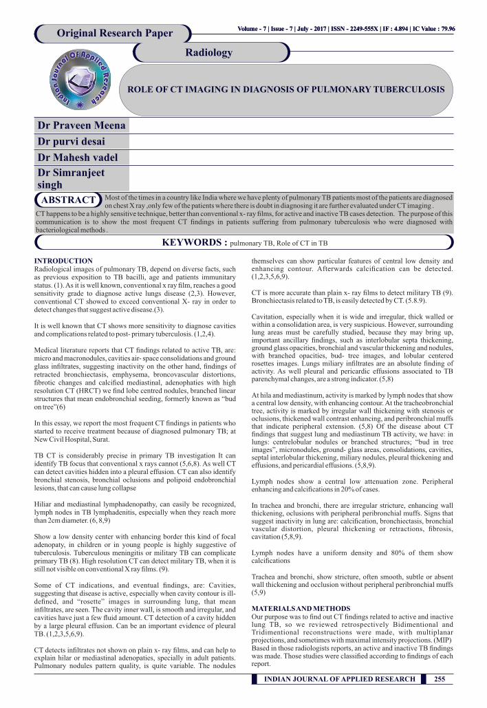
Dr Praveen Meena Dr purvi desai Dr Mahesh vadel Dr Simranjeet singh INTRODUCTION Radiological images of pulmonary TB, depend on diverse facts, such as previous exposition to TB bacilli, age and patients immunitary status. (1). As it is well known, conventional x ray film, reaches a good sensitivity grade to diagnose active lungs disease (2,3). However, conventional CT showed to exceed conventional X- ray in order to detect changes that suggest active disease.(3). It is well known that CT shows more sensitivity to diagnose cavities and complications related to post- primary tuberculosis. (1,2,4). Medical literature reports that CT findings related to active TB, are: micro and macronodules, cavities air- space consolidations and ground glass infiltrates, suggesting inactivity on the other hand, findings of retracted bronchiectasis, emphysema, broncovascular distortions, fibrotic changes and calcified mediastinal, adenophaties with high resolution CT (HRCT) we find lobe centred nodules, branched linear structures that mean endobronchial seeding, formerly known as “bud on tree”(6) In this essay, we report the most frequent CT findings in patients who started to receive treatment because of diagnosed pulmonary TB; at New Civil Hospital, Surat. TB CT is considerably precise in primary TB investigation It can identify TB focus that conventional x rays cannot (5,6,8). As well CT can detect cavities hidden into a pleural effusion. CT can also identify bronchial stenosis, bronchial oclusions and polipoid endobronchial lesions, that can cause lung collapse Hiliar and mediastinal lymphadenopathy, can easily be recognized, lymph nodes in TB lymphadenitis, especially when they reach more than 2cm diameter. (6, 8,9) Show a low density center with enhancing border this kind of focal adenopaty, in children or in young people is highly suggestive of tuberculosis. Tuberculous meningitis or military TB can complicate primary TB (8). High resolution CT can detect military TB, when it is still not visible on conventional X ray films. (9). Some of CT indications, and eventual findings, are: Cavities, suggesting that disease is active, especially when cavity contour is ill- defined, and “rosette” images in surrounding lung, that mean infiltrates, are seen. The cavity inner wall, is smooth and irregular, and cavities have just a few fluid amount. CT detection of a cavity hidden by a large pleural effusion. Can be an important evidence of pleural TB. (1,2,3,5,6,9). CT detects infiltrates not shown on plain x- ray films, and can help to explain hilar or mediastinal adenopaties, specially in adult patients. Pulmonary nodules pattern quality, is quite variable. The nodules themselves can show particular features of central low density and enhancing contour. Afterwards calcification can be detected. (1,2,3,5,6,9). CT is more accurate than plain x- ray films to detect military TB (9). Bronchiectasis related to TB, is easily detected by CT. (5.8.9). Cavitation, especially when it is wide and irregular, thick walled or within a consolidation area, is very suspicious. However, surrounding lung areas must be carefully studied, because they may bring up, important ancillary findings, such as interlobular septa thickening, ground glass opacities, bronchial and vascular thickening and nodules, with branched opacities, bud- tree images, and lobular centered rosettes images. Lungs miliary infiltrates are an absolute finding of activity. As well pleural and pericardic effusions associated to TB parenchymal changes, are a strong indicator. (5,8) At hila and mediastinum, activity is marked by lymph nodes that show a central low density, with enhancing contour. At the tracheobronchial tree, activity is marked by irregular wall thickening with stenosis or oclusions, thickened wall contrast enhancing, and peribronchial muffs that indicate peripheral extension. (5,8) Of the disease about CT findings that suggest lung and mediastinum TB activity, we have: in lungs: centrelobular nodules or branched structures; “bud in tree images”, micronodules, ground- glass areas, consolidations, cavities, septal interlobular thickening, miliary nodules, pleural thickening and effusions, and pericardial effusions. (5,8,9). Lymph nodes show a central low attenuation zone. Peripheral enhancing and calcifications in 20% of cases. In trachea and bronchi, there are irregular stricture, enhancing wall thickening, oclusions with peripheral peribronchial muffs. Signs that suggest inactivity in lung are: calcification, bronchiectasis, bronchial vascular distortion, pleural thickening or retractions, fibrosis, cavitation (5,8,9). Lymph nodes have a uniform density and 80% of them show calcifications Trachea and bronchi, show stricture, often smooth, subtle or absent wall thickening and occlusion without peripheral peribronchial muffs (5,9) MATERIALS AND METHODS Our purpose was to find out CT findings related to active and inactive lung TB, so we reviewed retrospectively Bidimentional and Tridimentional reconstructions were made, with multiplanar projections, and sometimes with maximal intensity projections. (MIP) Based in those radiologists reports, an active and inactive TB findings was made. Those studies were classified according to findings of each report. Most of the times in a country like India where we have plenty of pulmonary TB patients most of the patients are diagnosed on chest X ray ,only few of the patients where there is doubt in diagnosing it are further evaluated under CT imaging . CT happens to be a highly sensitive technique, better than conventional x- ray films, for active and inactive TB cases detection. The purpose of this communication is to show the most frequent CT findings in patients suffering from pulmonary tuberculosis who were diagnosed with bacteriological methods . ABSTRACT KEYWORDS : pulmonary TB, Role of CT in TB Volume - 7 | Issue - 7 | July - 2017 | 4.894 ISSN - 2249-555X | IF : | IC Value : 79.96 Volume - 7 | Issue - 7 | July - 2017 | 4.894 ISSN - 2249-555X | IF : | IC Value : 79.96 ROLE OF CT IMAGING IN DIAGNOSIS OF PULMONARY TUBERCULOSIS Original Research Paper Radiology INDIAN JOURNAL OF APPLIED RESEARCH 255
Transcript of ABSTRACT KEYWORDS - worldwidejournals.com · Radiological images of pulmonary TB, ... that can...
Dr Praveen Meena
Dr purvi desai
Dr Mahesh vadel
Dr Simranjeet singh
INTRODUCTION Radiological images of pulmonary TB, depend on diverse facts, such as previous exposition to TB bacilli, age and patients immunitary status. (1). As it is well known, conventional x ray film, reaches a good sensitivity grade to diagnose active lungs disease (2,3). However, conventional CT showed to exceed conventional X- ray in order to detect changes that suggest active disease.(3).
It is well known that CT shows more sensitivity to diagnose cavities and complications related to post- primary tuberculosis. (1,2,4).
Medical literature reports that CT findings related to active TB, are: micro and macronodules, cavities air- space consolidations and ground glass infiltrates, suggesting inactivity on the other hand, findings of retracted bronchiectasis, emphysema, broncovascular distortions, fibrotic changes and calcified mediastinal, adenophaties with high resolution CT (HRCT) we find lobe centred nodules, branched linear structures that mean endobronchial seeding, formerly known as “bud on tree”(6)
In this essay, we report the most frequent CT findings in patients who started to receive treatment because of diagnosed pulmonary TB; at New Civil Hospital, Surat.
TB CT is considerably precise in primary TB investigation It can identify TB focus that conventional x rays cannot (5,6,8). As well CT can detect cavities hidden into a pleural effusion. CT can also identify bronchial stenosis, bronchial oclusions and polipoid endobronchial lesions, that can cause lung collapse
Hiliar and mediastinal lymphadenopathy, can easily be recognized, lymph nodes in TB lymphadenitis, especially when they reach more than 2cm diameter. (6, 8,9)
Show a low density center with enhancing border this kind of focal adenopaty, in children or in young people is highly suggestive of tuberculosis. Tuberculous meningitis or military TB can complicate primary TB (8). High resolution CT can detect military TB, when it is still not visible on conventional X ray films. (9).
Some of CT indications, and eventual findings, are: Cavities, suggesting that disease is active, especially when cavity contour is ill- defined, and “rosette” images in surrounding lung, that mean infiltrates, are seen. The cavity inner wall, is smooth and irregular, and cavities have just a few fluid amount. CT detection of a cavity hidden by a large pleural effusion. Can be an important evidence of pleural TB. (1,2,3,5,6,9).
CT detects infiltrates not shown on plain x- ray films, and can help to explain hilar or mediastinal adenopaties, specially in adult patients. Pulmonary nodules pattern quality, is quite variable. The nodules
themselves can show particular features of central low density and enhancing contour. Afterwards calcification can be detected. (1,2,3,5,6,9).
CT is more accurate than plain x- ray films to detect military TB (9). Bronchiectasis related to TB, is easily detected by CT. (5.8.9).
Cavitation, especially when it is wide and irregular, thick walled or within a consolidation area, is very suspicious. However, surrounding lung areas must be carefully studied, because they may bring up, important ancillary findings, such as interlobular septa thickening, ground glass opacities, bronchial and vascular thickening and nodules, with branched opacities, bud- tree images, and lobular centered rosettes images. Lungs miliary infiltrates are an absolute finding of activity. As well pleural and pericardic effusions associated to TB parenchymal changes, are a strong indicator. (5,8)
At hila and mediastinum, activity is marked by lymph nodes that show a central low density, with enhancing contour. At the tracheobronchial tree, activity is marked by irregular wall thickening with stenosis or oclusions, thickened wall contrast enhancing, and peribronchial muffs that indicate peripheral extension. (5,8) Of the disease about CT findings that suggest lung and mediastinum TB activity, we have: in lungs: centrelobular nodules or branched structures; “bud in tree images”, micronodules, ground- glass areas, consolidations, cavities, septal interlobular thickening, miliary nodules, pleural thickening and effusions, and pericardial effusions. (5,8,9).
Lymph nodes show a central low attenuation zone. Peripheral enhancing and calcifications in 20% of cases.
In trachea and bronchi, there are irregular stricture, enhancing wall thickening, oclusions with peripheral peribronchial muffs. Signs that suggest inactivity in lung are: calcification, bronchiectasis, bronchial vascular distortion, pleural thickening or retractions, fibrosis, cavitation (5,8,9).
Lymph nodes have a uniform density and 80% of them show calcifications
Trachea and bronchi, show stricture, often smooth, subtle or absent wall thickening and occlusion without peripheral peribronchial muffs (5,9)
MATERIALS AND METHODS Our purpose was to find out CT findings related to active and inactive lung TB, so we reviewed retrospectively Bidimentional and Tridimentional reconstructions were made, with multiplanar projections, and sometimes with maximal intensity projections. (MIP) Based in those radiologists reports, an active and inactive TB findings was made. Those studies were classified according to findings of each report.
Most of the times in a country like India where we have plenty of pulmonary TB patients most of the patients are diagnosed on chest X ray ,only few of the patients where there is doubt in diagnosing it are further evaluated under CT imaging .
CT happens to be a highly sensitive technique, better than conventional x- ray films, for active and inactive TB cases detection. The purpose of this communication is to show the most frequent CT findings in patients suffering from pulmonary tuberculosis who were diagnosed with bacteriological methods .
ABSTRACT
KEYWORDS : pulmonary TB, Role of CT in TB
Volume - 7 | Issue - 7 | July - 2017 | 4.894ISSN - 2249-555X | IF : | IC Value : 79.96Volume - 7 | Issue - 7 | July - 2017 | 4.894ISSN - 2249-555X | IF : | IC Value : 79.96
ROLE OF CT IMAGING IN DIAGNOSIS OF PULMONARY TUBERCULOSIS
Original Research Paper
Radiology
INDIAN JOURNAL OF APPLIED RESEARCH 255
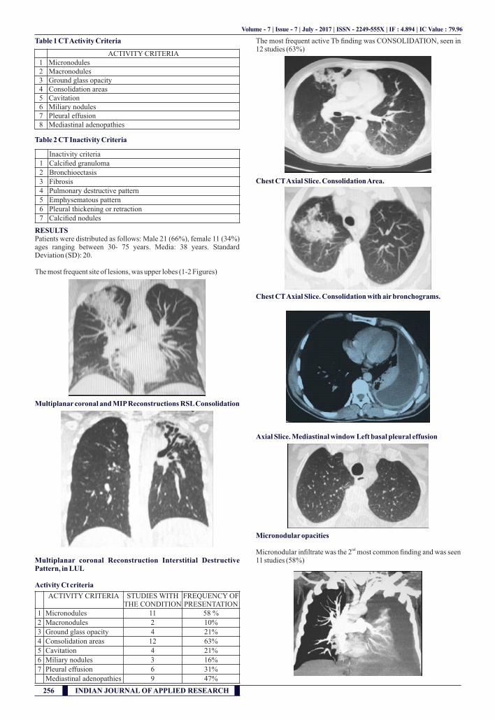
Table 1 CT Activity Criteria
Table 2 CT Inactivity Criteria
RESULTS Patients were distributed as follows: Male 21 (66%), female 11 (34%) ages ranging between 30- 75 years. Media: 38 years. Standard Deviation (SD): 20.
The most frequent site of lesions, was upper lobes (1-2 Figures)
Multiplanar coronal and MIP Reconstructions RSL Consolidation
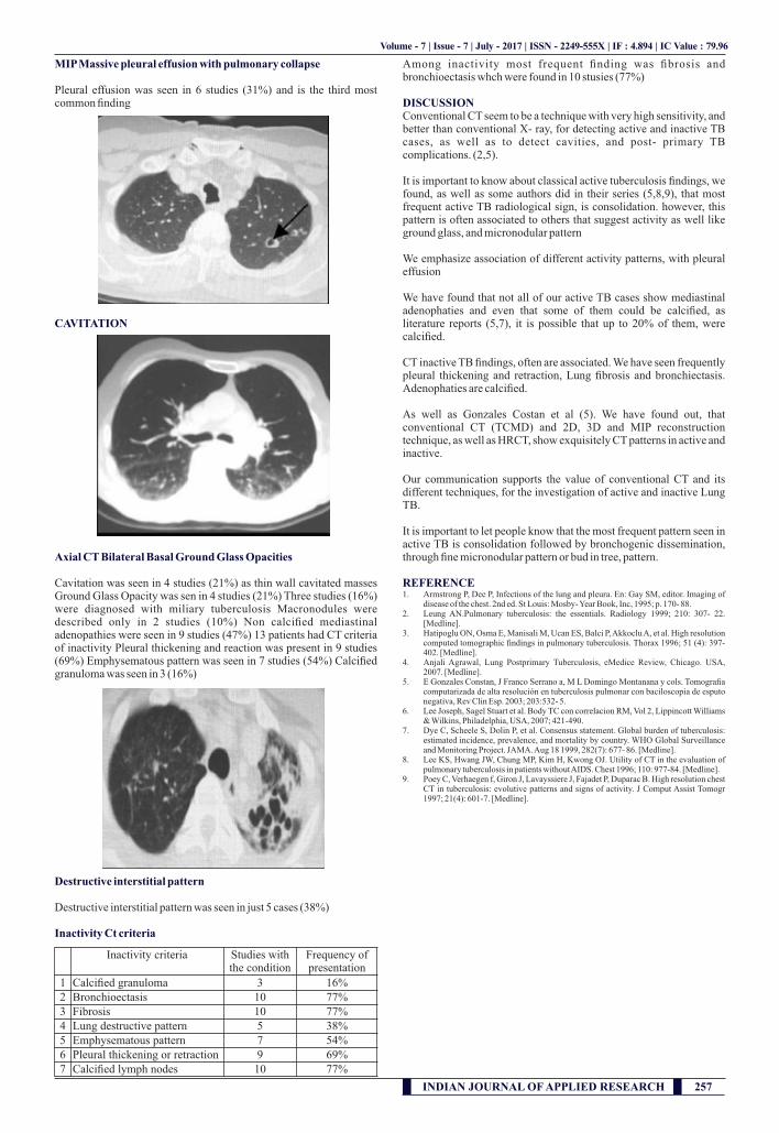
Multiplanar coronal Reconstruction Interstitial Destructive Pattern, in LUL
Activity Ct criteria
The most frequent active Tb finding was CONSOLIDATION, seen in 12 studies (63%)
Chest CT Axial Slice. Consolidation Area.
Chest CT Axial Slice. Consolidation with air bronchograms.
Axial Slice. Mediastinal window Left basal pleural effusion
Micronodular opacities
ndMicronodular infiltrate was the 2 most common finding and was seen 11 studies (58%)
Volume - 7 | Issue - 7 | July - 2017 | 4.894ISSN - 2249-555X | IF : | IC Value : 79.96
ACTIVITY CRITERIA 1 Micronodules 2 Macronodules3 Ground glass opacity4 Consolidation areas5 Cavitation 6 Miliary nodules 7 Pleural effusion 8 Mediastinal adenopathies
Inactivity criteria 1 Calcified granuloma 2 Bronchioectasis3 Fibrosis4 Pulmonary destructive pattern5 Emphysematous pattern6 Pleural thickening or retraction 7 Calcified nodules
ACTIVITY CRITERIA STUDIES WITH THE CONDITION
FREQUENCY OF PRESENTATION
1 Micronodules 11 58 %
2 Macronodules 2 10%
3 Ground glass opacity 4 21%
4 Consolidation areas 12 63%
5 Cavitation 4 21%
6 Miliary nodules 3 16%
7 Pleural effusion 6 31%
Mediastinal adenopathies 9 47%
256 INDIAN JOURNAL OF APPLIED RESEARCH
MIP Massive pleural effusion with pulmonary collapse
Pleural effusion was seen in 6 studies (31%) and is the third most common finding
CAVITATION
Axial CT Bilateral Basal Ground Glass Opacities
Cavitation was seen in 4 studies (21%) as thin wall cavitated masses Ground Glass Opacity was sen in 4 studies (21%) Three studies (16%) were diagnosed with miliary tuberculosis Macronodules were described only in 2 studies (10%) Non calcified mediastinal adenopathies were seen in 9 studies (47%) 13 patients had CT criteria of inactivity Pleural thickening and reaction was present in 9 studies (69%) Emphysematous pattern was seen in 7 studies (54%) Calcified granuloma was seen in 3 (16%)
Destructive interstitial pattern
Destructive interstitial pattern was seen in just 5 cases (38%)
Inactivity Ct criteria
Among inactivity most frequent finding was fibrosis and bronchioectasis whch were found in 10 stusies (77%)
DISCUSSION Conventional CT seem to be a technique with very high sensitivity, and better than conventional X- ray, for detecting active and inactive TB cases, as well as to detect cavities, and post- primary TB complications. (2,5).
It is important to know about classical active tuberculosis findings, we found, as well as some authors did in their series (5,8,9), that most frequent active TB radiological sign, is consolidation. however, this pattern is often associated to others that suggest activity as well like ground glass, and micronodular pattern
We emphasize association of different activity patterns, with pleural effusion
We have found that not all of our active TB cases show mediastinal adenophaties and even that some of them could be calcified, as literature reports (5,7), it is possible that up to 20% of them, were calcified.
CT inactive TB findings, often are associated. We have seen frequently pleural thickening and retraction, Lung fibrosis and bronchiectasis. Adenophaties are calcified.
As well as Gonzales Costan et al (5). We have found out, that conventional CT (TCMD) and 2D, 3D and MIP reconstruction technique, as well as HRCT, show exquisitely CT patterns in active and inactive.
Our communication supports the value of conventional CT and its different techniques, for the investigation of active and inactive Lung TB.
It is important to let people know that the most frequent pattern seen in active TB is consolidation followed by bronchogenic dissemination, through fine micronodular pattern or bud in tree, pattern.
REFERENCE 1. Armstrong P, Dee P, Infections of the lung and pleura. En: Gay SM, editor. Imaging of
disease of the chest. 2nd ed. St Louis: Mosby- Year Book, Inc, 1995; p. 170- 88. 2. Leung AN.Pulmonary tuberculosis: the essentials. Radiology 1999; 210: 307- 22.
[Medline]. 3. Hatipoglu ON, Osma E, Manisali M, Ucan ES, Balci P, Akkoclu A, et al. High resolution
computed tomographic findings in pulmonary tuberculosis. Thorax 1996; 51 (4): 397- 402. [Medline].
4. Anjali Agrawal, Lung Postprimary Tuberculosis, eMedice Review, Chicago. USA, 2007. [Medline].
5. E Gonzales Constan, J Franco Serrano a, M L Domingo Montanana y cols. Tomografia computarizada de alta resolución en tuberculosis pulmonar con baciloscopia de esputo negativa, Rev Clin Esp. 2003; 203:532- 5.
6. Lee Joseph, Sagel Stuart et al. Body TC con correlacion RM, Vol 2, Lippincott Williams & Wilkins, Philadelphia, USA, 2007; 421-490.
7. Dye C, Scheele S, Dolin P, et al. Consensus statement. Global burden of tuberculosis: estimated incidence, prevalence, and mortality by country. WHO Global Surveillance and Monitoring Project. JAMA. Aug 18 1999, 282(7): 677- 86. [Medline].
8. Lee KS, Hwang JW, Chung MP, Kim H, Kwong OJ. Utility of CT in the evaluation of pulmonary tuberculosis in patients without AIDS. Chest 1996; 110: 977-84. [Medline].
9. Poey C, Verhaegen f, Giron J, Lavayssiere J, Fajadet P, Duparac B. High resolution chest CT in tuberculosis: evolutive patterns and signs of activity. J Comput Assist Tomogr 1997; 21(4): 601-7. [Medline].
Volume - 7 | Issue - 7 | July - 2017 | 4.894ISSN - 2249-555X | IF : | IC Value : 79.96
Inactivity criteria Studies with the condition
Frequency of presentation
1 Calcified granuloma 3 16%2 Bronchioectasis 10 77%3 Fibrosis 10 77%4 Lung destructive pattern 5 38%5 Emphysematous pattern 7 54%6 Pleural thickening or retraction 9 69%7 Calcified lymph nodes 10 77%
INDIAN JOURNAL OF APPLIED RESEARCH 257