Abstract - ir.nctu.edu.tw
10
Article Characterization of canine monocyte-derived dendritic cells with phenotypic and functional differentiation Yu-Shan Wang,* Kwan-Hwa Chi,* Kuang-Wen Liao, Cheng-Chi Liu, Chiao-Lei Cheng, Yi-Chun Lin, Chiung-Hsiang Cheng, Rea-Min Chu Abstract For therapeutic purposes, large numbers of dendritic cells (DCs) are essential. In this study, we used 2% autologous canine plasma, granulocyte/macrophage colony-stimulating factor (GM-CSF), fms-like tyrosine kinase 3 ligand (Flt3L), and interleukin 4 (IL-4) in generating monocyte-derived DCs from peripheral blood mononuclear cells of dogs. The plasma enriched the population of CD14-positive monocytes by greatly enhancing the efficiency of monocyte adherence, the proportion of adherent cells increasing from 6.6% with 10% fetal bovine serum to 15.3% with 2% autologous canine plasma. Culturing the adherent monocytes for 6 d with human GM-CSF, canine IL-4, and human Flt3L significantly increased the yield of DCs, more than 90% of which were CD14-negative. Because, in the presence of lipopolysaccharide (LPS), monocytes that were CD14-positive expressed tumor necrosis factor a much more than DCs with low levels of CD14, it is important to decrease the numbers of CD14-positive cells in generating monocyte-derived DCs. With flow cytometry and real-time reverse-transcriptase-mediated polymerase chain reaction assays, we found that in canine immature DCs (iDCs) the expression of DLA class II molecules, CD1a, CD11c, CD40, and CD86 was high and the expression of CD80, CD83, and CD14 either low or negative. During maturation (stimulated by LPS), the expression of CD1a, CD40, CD83, and CD80 was upregulated. However, the expression of DLA class II molecules, CD11c, and CD86 was not increased in mature DCs. Incubating the iDCs with LPS decreased antigen uptake and increased the cells’ immunostimulatory capacity (assessed by the allogeneic mixed-lymphocyte reaction), indicating that LPS accelerates the functional maturation of DCs. This protocol may facilitate the use of DCs in cellular immunotherapy. Résumé Lorsqu’utilisées à des fins thérapeutiques, de grandes quantités de cellules dendritiques (DCs) sont essentielles. Dans la présente étude nous avons utilisé du plasma canin autologue à 2 %, du facteur stimulant de colonies des granulocytes/macrophages (GM-CSF), un analogue du ligand de la tyrosine kinase 3 (Flt3L), et de l’interleukine 4 (IL-4) afin de générer des DCs de chien dérivés des monocytes à partir des cellules mononucléaires du sang périphérique. Le plasma autologue a été utilisé pour enrichir des monocytes CD14 1 : l’efficacité d’adhérence des monocytes était grandement augmentée, la proportion de cellules adhérentes augmentant de 6,6 % avec du sérum fœtal de veau à 10 % à 15,3 % avec le plasma à 2 %. La cultivation des monocytes adhérents pendant 6 jours avec du GM-CSF et Flt3L humain et du IL-4 canin augmenta de manière significative la récolte de DCs, et plus de 90 % des DCs étaient CD14 2 . Étant donné qu’en présence de lipopolysaccharide (LPS) les monocytes qui étaient CD14 1 exprimaient le facteur de nécrose tumorale a beaucoup plus que les DCs avec un bas niveau de CD14, il est important de diminuer le nombre de cellules CD14 1 lorsque l’on génère des DCs dérivées de monocytes. Avec la cytométrie en flux et la réaction d’amplification en chaîne par la polymérase en temps réel utilisant la transcriptase réverse, nous avons trouvé que chez DCs canines immatures l’expression de DLA de classe II, CD1a, CD11c, CD40, et CD86 était élevée et que l’expression de CD80, CD83, et CD14 était soit faible ou négative. Durant la maturation (stimulée par le LPS), il y avait une régulation à la hausse de CD1a, CD40, CD83, et CD80. Toutefois, il n’y avait pas d’augmentation des DLA de classe II, CD11c, et CD86 dans les DCs matures. L’incubation de DCs immatures avec du LPS diminuait l’absorption d’antigènes et augmentait la capacité d’immunostimulation (évaluée par la réaction lymphocytaire allogénique mixte), indiquant ainsi que le LPS accélérait la maturation fonctionnelle de DCs. Ce protocole pourrait faciliter l’utilisation de DCs lors d’immunothérapie cellulaire. (Traduit par Docteur Serge Messier) Department of Veterinary Medicine, National Taiwan University (Wang, Liu, Chiao-Lei Cheng, Lin, Chiung-Hsiang Cheng, Chu), Department of Radiation Therapy and Oncology, Shin Kong Wu Ho-Su Memorial Hospital (Wang, Chi), and Department of Biological Science & Technology, National Chiao Tung University (Liao), Taipei, Taiwan, Republic of China. * These authors contributed equally to this work. Address all correspondence and reprint requests to Dr. Rea-Min Chu, #1, Roosevelt Road, Section 4, Taipei, Taiwan, 10617, Republic of China; telephone: 886 2 2368 6570; fax: 886 2 2365 5147; e-mail: [email protected] Received Dec. 13, 2005. Accepted August 1, 2006. 2007;71:165–174 The Canadian Journal of Veterinary Research 165
Transcript of Abstract - ir.nctu.edu.tw
Article
Characterization of canine monocyte-derived dendritic cells with phenotypic and functional differentiation
Yu-Shan Wang,* Kwan-Hwa Chi,* Kuang-Wen Liao, Cheng-Chi Liu, Chiao-Lei Cheng, Yi-Chun Lin, Chiung-Hsiang Cheng, Rea-Min Chu
A b s t r a c tFor therapeutic purposes, large numbers of dendritic cells (DCs) are essential. In this study, we used 2% autologous canine plasma, granulocyte/macrophage colony-stimulating factor (GM-CSF), fms-like tyrosine kinase 3 ligand (Flt3L), and interleukin 4 (IL-4) in generating monocyte-derived DCs from peripheral blood mononuclear cells of dogs. The plasma enriched the population of CD14-positive monocytes by greatly enhancing the efficiency of monocyte adherence, the proportion of adherent cells increasing from 6.6% with 10% fetal bovine serum to 15.3% with 2% autologous canine plasma. Culturing the adherent monocytes for 6 d with human GM-CSF, canine IL-4, and human Flt3L significantly increased the yield of DCs, more than 90% of which were CD14-negative. Because, in the presence of lipopolysaccharide (LPS), monocytes that were CD14-positive expressed tumor necrosis factor a much more than DCs with low levels of CD14, it is important to decrease the numbers of CD14-positive cells in generating monocyte-derived DCs. With flow cytometry and real-time reverse-transcriptase-mediated polymerase chain reaction assays, we found that in canine immature DCs (iDCs) the expression of DLA class II molecules, CD1a, CD11c, CD40, and CD86 was high and the expression of CD80, CD83, and CD14 either low or negative. During maturation (stimulated by LPS), the expression of CD1a, CD40, CD83, and CD80 was upregulated. However, the expression of DLA class II molecules, CD11c, and CD86 was not increased in mature DCs. Incubating the iDCs with LPS decreased antigen uptake and increased the cells’ immunostimulatory capacity (assessed by the allogeneic mixed-lymphocyte reaction), indicating that LPS accelerates the functional maturation of DCs. This protocol may facilitate the use of DCs in cellular immunotherapy.
R é s u m éLorsqu’utilisées à des fins thérapeutiques, de grandes quantités de cellules dendritiques (DCs) sont essentielles. Dans la présente étude nous avons utilisé du plasma canin autologue à 2 %, du facteur stimulant de colonies des granulocytes/macrophages (GM-CSF), un analogue du ligand de la tyrosine kinase 3 (Flt3L), et de l’interleukine 4 (IL-4) afin de générer des DCs de chien dérivés des monocytes à partir des cellules mononucléaires du sang périphérique. Le plasma autologue a été utilisé pour enrichir des monocytes CD141 : l’efficacité d’adhérence des monocytes était grandement augmentée, la proportion de cellules adhérentes augmentant de 6,6 % avec du sérum fœtal de veau à 10 % à 15,3 % avec le plasma à 2 %. La cultivation des monocytes adhérents pendant 6 jours avec du GM-CSF et Flt3L humain et du IL-4 canin augmenta de manière significative la récolte de DCs, et plus de 90 % des DCs étaient CD142. Étant donné qu’en présence de lipopolysaccharide (LPS) les monocytes qui étaient CD141 exprimaient le facteur de nécrose tumorale a beaucoup plus que les DCs avec un bas niveau de CD14, il est important de diminuer le nombre de cellules CD141 lorsque l’on génère des DCs dérivées de monocytes. Avec la cytométrie en flux et la réaction d’amplification en chaîne par la polymérase en temps réel utilisant la transcriptase réverse, nous avons trouvé que chez DCs canines immatures l’expression de DLA de classe II, CD1a, CD11c, CD40, et CD86 était élevée et que l’expression de CD80, CD83, et CD14 était soit faible ou négative. Durant la maturation (stimulée par le LPS), il y avait une régulation à la hausse de CD1a, CD40, CD83, et CD80. Toutefois, il n’y avait pas d’augmentation des DLA de classe II, CD11c, et CD86 dans les DCs matures. L’incubation de DCs immatures avec du LPS diminuait l’absorption d’antigènes et augmentait la capacité d’immunostimulation (évaluée par la réaction lymphocytaire allogénique mixte), indiquant ainsi que le LPS accélérait la maturation fonctionnelle de DCs. Ce protocole pourrait faciliter l’utilisation de DCs lors d’immunothérapie cellulaire.
(Traduit par Docteur Serge Messier)
Department of Veterinary Medicine, National Taiwan University (Wang, Liu, Chiao-Lei Cheng, Lin, Chiung-Hsiang Cheng, Chu), Department of Radiation Therapy and Oncology, Shin Kong Wu Ho-Su Memorial Hospital (Wang, Chi), and Department of Biological Science & Technology, National Chiao Tung University (Liao), Taipei, Taiwan, Republic of China.
* These authors contributed equally to this work.
Address all correspondence and reprint requests to Dr. Rea-Min Chu, #1, Roosevelt Road, Section 4, Taipei, Taiwan, 10617, Republic of China; telephone: 886 2 2368 6570; fax: 886 2 2365 5147; e-mail: [email protected]
Received Dec. 13, 2005. Accepted August 1, 2006.
2007;71:165–174 TheCanadianJournalofVeterinaryResearch 165

166 TheCanadianJournalofVeterinaryResearch 2000;64:0–00
I n t r o d u c t i o nDendritic cells (DCs) play fundamental roles in both innate and
adaptive immune responses (1,2) and are critical in fighting tumor growth by presenting tumor-specific antigens to initiate an effective immune response (3). The precursors of DCs circulate in the blood and have an intermediate differentiation/activation phenotype. They represent a small (less than 1%) proportion of blood mono-nuclear cells (MNCs). The precursors move from the circulation into the tissues, where they acquire a fully mature APC phenotype after interaction with pathogens or other stimuli. The key surface molecules, such as CD40, CD80, and CD86, which are involved in costimulation and serve as markers of mature DCs (mDCs), are over-expressed in humans and mice (4). Owing to the naturally low DC numbers in tissues, techniques to generate large numbers of DCs in culture from either proliferating CD34-positive progenitors (5,6) or nonproliferating CD14-positive monocytic precursors are essential for DC immunotherapy (7,8). Currently, monocyte-derived DCs are the most widely used, because they can be generated relatively simply, without the need for cytokine pretreatment of the donor. The progeny are at present the best characterized DC population, and they have been the most commonly used in clinical trials in humans (9–11). Bone-marrow-derived DCs are too invasive for routine use.
Several studies have demonstrated that canine DCs can be cul-tured from peripheral blood monocytes or bone marrow (12–17), and CD1a, CD11c, major histocompatibility complex (MHC) class II molecules, and CD86 have been investigated in surface phenotype studies (15–17). However, information is still lacking on the other important surface molecules associated with DCs in canine species, such as CD40, CD80, and CD83, which are commonly used for char-acterization of the DC maturation state in humans. In this study, we developed a procedure that could efficiently generate a relatively large amount of canine DCs from peripheral blood mononuclear cells (PBMCs). Using real-time reverse-transcription polymerase chain reaction (RT-PCR), we compared the expression of CD80, CD83, and CD86 in immature DCs (iDCs) and mDCs and determined the functional profiles of the monocyte-derived DCs. These results will facilitate the use of canine DCs for further immunotherapy research and clinical application.
M a t e r i a l s a n d m e t h o d s
Generation of DCsPeripheral blood was obtained from 7 healthy beagles. All dogs
had been dewormed regularly and vaccinated against distemper, leptospirosis, parvovirus infection, and hepatitis. We isolated PBMCs from heparinized whole blood by standard gradient centrifuga-tion with Ficoll-Hypaque (density, 1.077; Amersham Biosciences, Piscataway, New Jersey, USA) (18). The PBMCs were harvested from the interface, washed twice, and resuspended in Roswell Park Memorial Institute (RPMI)-1640 medium (Life Technologies, Gaithersburg, Maryland, USA) supplemented with 100 U/mL of penicillin, 100 mg/mL of streptomycin, and 2 mM of L-glutamine. For testing the isolation efficiency of adherence, different concentra-tions (2% and 10%) of fetal bovine serum (FBS; Life Technologies)
or autologous canine plasma were added to the medium and the PBMCs allowed to adhere to a 25-cm2 flask (1 3 107 cells/mL) for 24 h at 37°C. To obtain iDCs, we removed nonadherent cells by gentle pipetting with phosphate-buffered saline (PBS), pH 7.4. These procedures were repeated 3 times. To evaluate the adherence efficiency of monocytes, the adherent cells were removed by treat-ment with 3 mL of versene (0.02% ethylene diamine tetraacetic acid [EDTA] in PBS, pH 7.4) for 30 min and assayed by flow cytometry. To generate DCs, we cultured the adherent cells for another 6 d in DC medium that was composed of RPMI-1640 medium supplemented as above but also with 800 U/mL of human granulocyte–macrophage colony-stimulating factor (GM-CSF; Leucomax; Schering-Plough, Kenilworth, New Jersey, USA), 500 U/mL of canine interleukin 4 (IL-4; R&D Systems, Minneapolis, Minnesota, USA), and 10% FBS (12,19,20). This medium with or without 25 ng/mL of human fms-like tyrosine kinase 3 ligand (Flt3L; R&D Systems) was used to test its effect on the generation of DCs. The GM-CSF and Flt3L are bioactive for canine cells (12). Fresh medium and cytokines were added every 3 d. To prepare mature, activated DCs, iDCs were incubated for 48 h with 10 mg/mL of lipopolysaccharide (LPS) from Escherichia coli serotype O128:B12 (Sigma Chemical Company, St. Louis, Missouri, USA), and then the putative iDCs and mDCs were harvested for morphologic, phenotypic, or functional analyses. The percentages of monocytes were determined by flow cytometry from the presence of CD14 after staining with CD14 antibody.
Morphologic examinationEach DC preparation was examined with a phase-contrast micro-
scope (Nikon, Tokyo, Japan). For cytologic study, iDCs were smeared on a glass slide, allowed to air dry, and stained with May-Grünwald Giemsa stain (Diff-Quick; Gamidor, Abingdon, England). For trans-mission electron microscopy (TEM), iDC pellets obtained after cen-trifugation at 250 3 g were fixed in 2.5% glutaraldehyde in 0.1 M PBS, pH 7.4, at 4°C overnight and postfixed in 1% osmium tetroxide at room temperature for 1 h. The blocks of iDCs were dehydrated in graded ethanol solutions, then embedded with Spurr’s resin kit (EMS, Washington, Pennsylvania, USA). Ultrathin sections were cut with a diamond knife (Ultracut E; Reichert-Jung, Vienna, Austria), stained with a 5% aqueous solution of uranyl acetate for 20 min, and then stained with Reynold’s lead citrate for 4 min. Randomly selected specimens were examined by TEM with a JEOL 1200EXII (JEOL USA, Peabody, Massachusetts, USA).
Flow cytometryCommercial monoclonal antibodies were used to analyze the
canine DC surface antigens (Table I), and goat IgG antibody against mouse antigen conjugated with fluorescein isothiocyanate (FITC; Serotec, Oxford, England) was used as the secondary antibody. Using procedures described previously (18), we stained PBMCs, nonadherent cells, adherent cells, and putative and LPS-treated iDCs. Briefly, for direct immunofluorescence analysis, 50 000 cells were washed twice with fluorescence-activated cell sorting (FACS) buffer (PBS, 1% BSA, and 0.02% sodium azide, pH 7.4). Cells were incubated for 30 min, on ice and in the dark, with isotype control or specific mouse monoclonal antibodies to detect CD1a, CD40, CD80, CD83, and CD86. For indirect immunofluorescence analysis,

2000;64:0–00 TheCanadianJournalofVeterinaryResearch 167
cells were incubated with isotype control or specific monoclonal antibodies against CD3, CD11c, CD14, CD21, dog leukocyte antigen (DLA) II, and DEC 205 (CD205). Cells were washed and further stained with FITC-conjugated goat IgG antibody against mouse antigen for 30 min. Finally, all cells were washed and suspended in FACS buffer containing 5 mg/mL of propidium iodide. The surface immunofluorescence of 1 3 104 viable cells was measured with a FACSCalibur flow cytometer (Becton Dickinson, Mountain View, California, USA). Fluorescence intensities were analyzed with Cell Quest software (Becton Dickinson).
Immunoprecipitation and immunoblottingThe cross-reactivity with canine CD40 molecules of iAb, the
monoclonal antibody against human CD40 (clone LOB7/6; Serotec), was evaluated as follows. The iAb can be used in flow cytometry and immunoprecipitation but cannot be used for Western blotting. A rabbit polyclonal antibody against CD40, wAb (CSS-180; StressGen Biotechnologies, San Diego, California, USA), cross-reacts with canine species but can be used only for Western blotting. Thus, our strategy was to immunoprecipitate the canine PBMC lysate with the iAb and then use the eluate for Western blotting with the wAb. We lysed the PBMCs with 1 mL of ice-cold lysis buffer contain-ing 0.5% Nonidet P-40, 1 mM EDTA, 1 mM Na3VO4, 50 mM NaF, and 10% protease inhibitor mixture (Sigma) in PBS for immuno-precipitation or in 250 mL of Laemmli sample buffer (Bio-Rad, Hercules, California, USA) for analysis of CD40 molecules. For immunoprecipitation, the lysates were incubated with 1 mg of iAb for 24 h at 4°C. The precipitates were further reacted with protein A–Sepharose beads (Amersham Biosciences) and eluted by 5 min of boiling in Laemmli sample buffer. Electrophoretic separation of the immunoprecipitated proteins or cell lysates was done in 10% acrylamide gels, and bands were transferred onto Immobilon NC membranes (Millipore, Bedford, Massachusetts, USA). For immuno-blotting, the membranes were probed with wAb at a 1/200 dilution. The blots were incubated with species-specific secondary antibodies
conjugated with horseradish peroxidase (Jackson ImmunoResearch Laboratories, West Grove, Pennsylvania, USA). The signals were revealed by enhanced chemiluminescence (ECL system; Amersham Biosciences).
Real-time RT-PCRReal-time RT-PCR was performed as previously described (21).
Total RNA was extracted from iDCs and mDCs with TRIzol (Gibco-BRL, Grand Island, New York, USA) and then reverse-transcribed with SuperScript II RT (Gibco-BRL) and oligo(dT) primers. Real-time RT-PCR was performed on the ABI Prism 5700 (Applied Biosystems) in 96-well optical reaction plates with the use of SYBR Green PCR Master Mix in accord with the manufacturer’s instructions. Briefly, each well contained a 50-mL reaction mixture that contained 25 mL of the master mix, 1 mL each of the forward and reverse prim-ers, 21 mL of water, and 2 mL of cDNA samples. The SYBR green dye was measured at 530 nm during the extension phase. The threshold cycle (Ct) value reflects the cycle number at which the
Table I. Monoclonal antibodies used for surface phenotype assays of canine monocyte-derived dendritic cells (DCs)
Specificity Clonea ImmunoglobulinclassHumanCD1a-FITC NA1/34 MouseIgG2aCanineCD3 CA17.2A12 MouseIgG1CanineCD11c CA11.6A1 MouseIgG1CanineCD21 CA2.1D6 MouseIgG1HumanCD14 TUK4b MouseIgG2aHumanCD40-FITC LOB7/6 MouseIgG2aMouseCD80-FITC 16-10A1c HamsterIgG2aHumanCD83-FITC HB15ec MouseIgG1HumanCD83-FITC HB15ad MouseIgG2bHumanCD86-RPE BU63 MouseIgG1CanineDLAclassII CA2.1C12 MouseIgG1MouseDEC205 NLDC-145 RatIgG2aaSources: bDako (Glostrup,Denmark), cPharmingen (SanDiego,California, USA), d Immunotech (Marseilles, France), and, for theremainder,Serotec(Oxford,England).
Figure 1. Efficiency of monocyte isolation from canine peripheral blood mononuclear cells (PBMCs), as determined by flow cytometry. The PBMCs were isolated by Ficoll gradient (A) and cultured in flasks with medium alone or with supplementary plasma or fetal bovine serum (FBS). After 24 h, non-adherent cells were washed and adherent cells removed. Panels B to G rep-resent 3 experiments yielding similar results. The proportion of monocytes is in the upper right corner of each panel and the proportion of lymphocytes in the lower right. SSC-H — side scatter; FSC-H — forward scatter.

168 TheCanadianJournalofVeterinaryResearch 2000;64:0–00
fluorescence generated within a reaction crosses a given threshold. The Ct value assigned to each well thus reflects the point during the reaction at which sufficient amplicons have been accumulated. The relative mRNA amount in each sample was calculated from its Ct value in comparison with the Ct value of the housekeep-ing gene b-actin. The results were presented, in arbitrary units, as 22(Ct of target gene 2 Ct of housekeeping gene) (22Ct) as described by the manufacturer. The purity of the amplified product was deter-mined as a single peak of the dissociation curve. Real-time RT-PCR was conducted in duplicate for each sample, and the mean value was calculated. This procedure was performed in at least 2 or 3 independent experiments. The primers for canine CD80 and CD86 assays were designed with the use of Primer 3 (www-genome.wi.mit.edu/cgi-bin/primer/primer3_www.cgi) and the canine-specific sequences for CD80 (403765) and CD86 (403764) in GenBank. The primers for CD83 were designed within exons from the conserved regions of the human and mouse CD83 gene sequences; the sequence of the PCR product corresponded with the predicted canine sequence in the Ensembl Database (www.ensembl.org). Primer Express software (Applied Biosystems) was used to design b-actin and tumor necrosis factor a (TNF-a) primer pairs (22). The real-time RT-PCR products were confirmed by electrophoresis in 1% agarose gel and stained with ethidium bromide to detect primer con-tamination. The following were the sequences of the primers used: CD80, forward 59-ATGGATTACACAGCGAAGTGGAGAA-39 and reverse 59-AGGCGCAGAGCCATAATCACGAT-39; CD83, forward 59-CAGTCATATAAAAGCTATGGTGAGATGC-39 and reverse 59-AGATGAAAAGGCCCTGCTGGGG-39; CD86, forward 59-ATG- TATCTCAGATGCACTATGGAAC-39 and reverse 59-TTCTCTT- TGCCTCTGTATAGCTCGT-39; b-actin, forward GACCCTGAA- GTACCCCATTGAG and reverse TTGTAGAAGGTGTGGTGCCAGAT; TNF-a , forward GAGCCGACGTGCCAATG and reverse CAACCCATCTGACGGCACTA.
Measurement of endocytotic activityEndocytotic activity was assessed by incubating cells for 2 h
with FITC-Dextran (100 mg/mL) (Sigma) at 4°C or 37°C. Cells were washed extensively with PBS. Nonspecific binding of FITC-Dextran to the cell surface was measured by incubating the cells at 4°C (23).
Allogeneic mixed-lymphocyte reaction (MLR)Freshly prepared DCs were washed and combined with untreated
PBMCs from an unrelated beagle. Mitomycin C-treated DCs were added (in triplicate) in graded doses to 1 3 105 PBMCs per well in 96-well flat-bottom plates and cocultivated for 5 d in RPMI-1640 medium supplemented with 100 U/mL of penicillin, 100 mg/mL of streptomycin, 2 mM L-glutamine, and 10% FBS. Then, 1 mCi of tritiated thymidine (New England Nuclear, Boston, Massachusetts, USA) was added to each well. After 16 h, cells were harvested, washed, transferred to glass fiber membranes, and counted in a scintillation mix in an imaging reader (Canberra Packard, Schwadorf, Austria) (24,25).
Statistical analysisAll results were expressed as means (and standard deviation)
and were analyzed with a 2-tailed Student’s t test. Differences were considered statistically significant at P , 0.05.
R e s u l t s
Generation of canine monocyte-derived DCsTo generate DCs, we incubated canine PBMCs for 24 h in medium
containing 2% or 10% FBS or autologous canine plasma. Adherence of monocytes to the flasks was best with 2% autologous canine
Table II. Adherence of peripheral blood mononuclear cells (PBMCs) to flasks containing culture medium and various amounts of fetal bovine serum (FBS) or autologous canine plasma (ACP)
Percentage,mean(andstandarddeviation) OfCD141
OfPBMCs monocytes CD141 isolatedfromSupplement Cellcategory Allcells Lymphocytes monocytes PBMCsa
FBS 10% Nonadherent 93.4(2.5) 67.74(12.38) 17.43(3.80) Adherent 6.6(2.5) 2.54(0.92) 95.01(0.65) 6.27(2.38) 2% Nonadherent 95.4(3.3) 69.67(12.00) 16.07(3.43) Adherent 4.6(3.3) 7.20(3.68) 89.88(3.02) 4.13(2.97)ACP 10% Nonadherent 92.3(3.2) 70.55(11.04) 14.38(4.80) Adherent 7.6(3.2) 8.48(1.21) 86.34(2.17) 6.56(2.76) 2% Nonadherent 84.6(2.4) 73.48(10.34) 13.90(3.15) Adherent 15.3(2.4) 8.49(5.33) 88.35(4.19) 13.52(2.12)Neither Nonadherent 71.4(8.4) 71.05(4.65) 11.72(3.67) Adherent 28.6(8.4) 60.72(21.43) 33.39(20.12) 9.55(2.85)aAdherentmonocytesafterremovalofcontaminatinglymphocytes.Thenumberswereobtainedbymultiplyingthepercentageofadherentcellsunder“Allcells”bythepercentageofCD14-positivemonocytes.

2000;64:0–00 TheCanadianJournalofVeterinaryResearch 169
plasma, the proportion of adherent monocytes among PBMCs being at least 2.3 times greater than with FBS (Table II). Flow cytometry showed typical morphologic distributions of the PBMCs (Figure 1A). Adherent cells (Figures 1C, 1E, and 1G) had purer monocyte popu-lations than nonadherent cells (Figures 1B, 1D, and 1F). Culture of adherent cells in 2% autologous canine plasma (Figure 1E) or 2% FBS (Figure 1G) greatly increased the purity of the monocyte population, from 39.84% without the serum or plasma to 92.78% with the plasma and 92.83% with the FBS, whereas culture without the plasma or serum (Figure 1C) resulted in a high rate of contami-nation by lymphocytes: 53.95%, compared with 3.67% for culture with the plasma and 4.34% for culture with the FBS (Figures 1E and G). These procedures were repeated 3 times; the mean values are recorded in Table II.
To further evaluate the purity of the monocyte populations, we measured the expression of CD14, a monocyte marker, on the PBMCs. Again, culture in 2% autologous canine plasma yielded the highest mean proportion of CD14-positive monocytes from PBMCs (13.52% out of 15.3% [Table II]). Although culture with-out serum or plasma yielded the highest proportion of adher-ent cells from PBMCs, most of the cells were lymphocytes, less than 10% being monocytes. Adding 2% autologous plasma to the medium increased the proportion of CD14-positive mono-cytes by at least 40% and to as much as 220%, compared with the other groups, and thus was chosen for the adherence step in our experiments.
Among nonadherent PBMCs the proportion of CD14-positive cells was very low (less than 10%) compared with the proportions of CD3-positive cells (64.40%) and CD21-positive cells (33.24%). Adherent PBMCs had high proportions of CD14-positive cells
(83.14%) and were negative for both CD3 and CD21 (Figure 2). These results suggested that the adherence procedure excluded the lymphocytes (nonadherent) and thus enriched the population of CD14-positive monocytes. When adherent PBMCs were cultured for 6 d with GM-CSF, IL-4, and Flt3L, the CD14 positivity dramatically decreased, to 6.32%, indicating that the monocytes had differenti-ated into DCs.
After 6 d of culture of the purified monocytes, the cells were stained with propidium iodide to exclude dead cells during flow cytometry (Figure 2C, left). Gating for the propidium-iodide- negative cells resulted in a nonlymphoid population with a larger size distribution (Figure 2C, right), presumably iDCs. Other popu-lations were usually obscure. This procedure produced 0.5 to 1 3 106 putative iDCs per 20 mL of peripheral blood. Adding human Flt3L to the medium, we harvested at least 4 times more putative iDCs (2.0 to 4.0 3 106). In both cases, the purity of the cells was 80% to 85%, as determined by May-Grünwald Giemsa staining.
Morphologic features of the iDCsThe adherent PBMCs were examined by phase-contrast and
light microscopy, as well as by TEM. The putative iDCs exhibited typical dendritic cytoplasmic projections (Figure 3A). Although the
Figure 2A. Analysis of CD14 expression by flow cytometry after culture of PBMCs with 2% autologous canine plasma and further culture of adher-ent cells with human granulocyte–macrophage colony-stimulating factor (GM-CSF), canine interleukin 4 (IL-4), and human fms-like tyrosine kinase 3 ligand (Flt3L) for 6 d to obtain putative dendritic cells (DCs). Data for the test dogs are indicated by the dashed lines and data for the isotype control by the solid black lines.
Figure 2B. Analysis of the expression of CD3 (left panels) and CD21 (right panels) in the same manner as for CD14.
Figure 2C. After the 6 d of culture, the putative immature DCs (iDCs) were stained with propidium iodide (left); with gating for the propidium-iodide-negative dead cells (right), the nonlymphoid population of presumptive iDCs is represented.

170 TheCanadianJournalofVeterinaryResearch 2000;64:0–00
cells varied in size and shape, each cell had numerous dendrites radiating from the surface and a large lobulated nucleus (Figure 3B). Ultrastructurally, the dendrites had surface irregularities: long, narrow cytoplasmic protrusions (Figure 3C). These iDCs had a plasmacytoid appearance and contained extensive amounts of endo-plasmic reticulum (Figure 3C).
Phenotype of the iDCsMost of the tested commercial monoclonal antibodies reacted
with the putative iDCs; 1 mouse antibody (DEC205) and 2 human antibodies (CD83 and CD86) did not cross-react with the canine cells. Human CD80 antibody had been approved as cross-reactive with canine antigens by the manufacturer. We used immuno-precipitation and Western blotting to confirm that the antibody against human CD40 that we used in this experiment cross-reacted with the canine CD40 molecule. As shown in Figure 4, canine CD40 (48 kDa) was immunoprecipitated by iAb and probed by wAb; thus, the antibody that we used reacts with canine CD40 antigen.
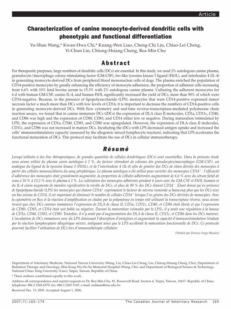
As Figure 5 shows, flow cytometry demonstrated that the putative iDCs expressed high levels of CD1a, CD40, CD11c, and DLA class II molecules; the level of CD80, like that of CD14, was very low. Two days after LPS treatment, the levels of CD1a, CD40, and CD80 were significantly increased (P , 0.05), indicating DC maturation. We also used real-time RT-PCR to detect CD80, CD83, and CD86 expression on the putative DCs to determine the state of maturation, with b-actin amplified in all samples from reverse-transcribed RNA as a control of the quality of the RNA preparations and PCR analyses. We found that the mRNA gene expression levels of CD80 and CD83 were significantly increased (P , 0.05), a mean of 132 (standard deviation 26)-fold and 901 (84)-fold, on mDCs after LPS treatment. However, the gene expression of CD86 was not significantly different between iDCs and mDCs.
Endocytotic activityImmature DCs display potent endocytotic activity, which decreases
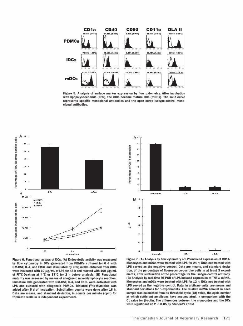
upon maturation. We evaluated endocytotic activity of the putative DCs before and after LPS treatment by measuring the phagocytosis of mannose-receptor-mediated FITC-Dextran. Uptake was sig-nificantly lower (P , 0.05) after LPS treatment than before (38.1% [0.38%] versus 55.9% [2.87%]) (Figure 6A).
Mixed lymphocyte reactionTo test whether the putative DCs stimulated by LPS had increased
immunostimulatory capacity, an indication of DC maturation, we conducted an allogeneic MLR, with PBMCs as the responder cells and fresh mitomycin-C-treated autologous PBMCs from the same donors as controls. The putative DCs treated with both mitomycin C and LPS stimulated the allogeneic PBMCs much more (P , 0.05) than the fresh mitomycin-C-treated autologous PBMCs or the mitomycin-C-treated putative iDCs (Figure 6B).
Capacity to produce TNF-aFlow cytometry revealed that the monocytes expressed high levels
of CD14 when differentiated into DCs with upregulated CD1a and CD40 (Figure 5) but almost none during DC maturation (Figure 7A).
Figure 4. Immunoprecipitation for canine CD40. Left: a lysate of PBMCs was immunoprecipitated with monoclonal antibody against CD40. Right: Western blot of the eluate with a rabbit polyclonal antibody against CD40.
Figure 3. Putative iDCs derived from adherent CD14-positive canine mono-cytes after culture for 6 d with GM-CSF, IL-4, and Flt3L as seen with phase-contrast microscopy (3 25), (B) May-Grünwald Giemsa staining (3 1000), and (C) transmission electron microscopy. Each image represents more than 6 fields of DCs from 2 PBMC preparations. Note the numerous typi-cal dendritic cytoplasmic projections (A), large lobulated nucleus (B), and long, narrow cytoplasmic protrusions from the dendrites, with extensive endoplasmic reticulum (C). Bar — 1 mm.
A
B
C
2000;64:0–00 TheCanadianJournalofVeterinaryResearch 171
Figure 5. Analysis of surface marker expression by flow cytometry. After incubation with lipopolysaccharide (LPS), the iDCs became mature DCs (mDCs). The solid curve represents specific monoclonal antibodies and the open curve isotype-control mono-clonal antibodies.
100 101 102 103 104 100 101 102 103 104 100 101 102 103 104 100 101 102 103 104 100 101 102 103 104
100 101 102 103 104 100 101 102 103 104 100 101 102 103 104 100 101 102 103 104 100 101 102 103 104
100 101 102 103 104 100 101 102 103 104 100 101 102 103 104 100 101 102 103 104 100 101 102 103 104
Figure 6. Functional assays of DCs. (A) Endocytotic activity was measured by flow cytometry in DCs generated from PBMCs cultured for 6 d with GM-CSF, IL-4, and Flt3L and stimulated by LPS; mDCs obtained from iDCs were incubated with 10 mg/mL of LPS for 48 h and reacted with 100 mg/mL of FITC-Dextran at 4°C or 37°C for 2 h before analysis. (B) Functional maturity was assessed by means of allogeneic mixed-lymphocyte reaction. Immature DCs generated with GM-CSF, IL-4, and Flt3L were activated with LPS and cultured with allogeneic PBMCs. Tritiated (3H)-thymidine was added after 5 d of incubation. Scintillation counts were done after 16 h. Data are means, and standard deviation, in counts per minute (cpm) for triplicate wells in 3 independent experiments.
3 H-t
hym
idin
e in
corp
orat
ion,
cpm
Per
cent
age
of F
ITC
-Dex
tran
-pos
itive
cel
ls
Figure 7. (A) Analysis by flow cytometry of LPS-induced expression of CD14. Monocytes and mDCs were treated with LPS for 24 h; iDCs not treated with LPS served as the negative control. Data are means, and standard devia-tion, of the percentage of fluorescence-positive cells in at least 3 experi-ments, after subtraction of the percentage for the isotype-control antibody. (B) Analysis by real-time RT-PCR of LPS-induced expression of TNF-a mRNA. Monocytes and mDCs were treated with LPS for 12 h; iDCs not treated with LPS served as the negative control. Data, in arbitrary units, are means and standard deviations for 5 experiments. The relative mRNA amount in each sample was calculated from its threshold cycle (Ct) value, the cycle number at which sufficient amplicons have accumulated, in comparison with the Ct value for b-actin. The differences between the monocytes and the DCs were significant at P , 0.05 by Student’s t test.
22
Ct
Per
cent
age
of C
D14
exp
ress
ion

172 TheCanadianJournalofVeterinaryResearch 2000;64:0–00
To see if CD14-positive monocytes differed from putative DCs in their capacity to produce TNF-a, we used real-time RT-PCR before and after LPS stimulation. As illustrated in Figure 7B, LPS-treated monocytes expressed high levels of TNF-a, whereas iDCs and LPS-treated mDCs produced little TNF-a (P , 0.05).
D i s c u s s i o nThis paper has described an efficient method for generating
monocyte-derived canine DCs by enriching adherent cells from PBMCs with autologous canine plasma and culturing the adherent cells in a medium containing GM-CSF, IL-4, and Flt3L. Adherence is a widely used method of monocyte isolation for in vitro DC generation because it is simple and inexpensive and because it can be applied to a large number of samples. One of the key steps was using autologous canine plasma, which increased the number of CD14-positive adherent mononuclear cells at least 40%. Enrichment with autologous plasma was first used in the generation of human DCs (26). The plasma helps the monocytes to tightly adhere to the culture vessel and efficiently isolates them from the PBMCs. Another pivotal ingredient is Flt3L. This hematopoietic growth factor is an important regulator of DC growth and a critical cyto-kine for ex vivo expansion of the DC population in both mice and humans (19,27). We found that DC medium with 25 ng/mL of Flt3L generated 4 times more canine iDCs than medium without Flt3L. The greatest number of DCs was obtained when the medium contained both autologous plasma and Flt3L. Thus, in canine DC generation, we recommend that 2% autologous canine plasma be added first to increase the efficiency of monocyte adherence and that 25 ng/mL of Flt3L be added to the growth medium later to improve differentiation and expansion of the DC population. Several papers have described in vitro culturing of canine DCs (12,14–17); in only 1 of the 5 papers were the DCs derived from bone marrow (12). Canine monocyte-derived DCs were generated with the use of phytohemagglutinin (PHA) (14) or canine GM-CSF and IL-4 (16,17) or canine GM-CSF and IL-4 plus human Flt3L (15). However, none of these studies used autologous canine plasma. The bone- marrow-derived canine DCs were generated with the use of GM-CSF and Flt3L.
In our study, there was phenotypic evidence that the great major-ity of the adherent cells from canine PBMCs were nonlymphoid; they were monocytes negative for CD3 and CD21 and positive for CD14. Light and electron microscopy revealed close morphologic similarity of these canine cells to iDCs from humans (8). The putative DCs from canine PBMCs showed significantly increased proportions of CD1a, CD11c, CD40, and DLA class II molecules in immunologic testing and were positive for CD80, CD83, and CD86 in real-time RT-PCR testing. The increased expression of these molecules is generally used to define DC maturity (4). Those findings indicated that the monocytes cultured for 6 d differentiated into iDCs. Functional maturation of DCs is usually assessed by endocytotic activity (as assessed by FITC-Dextran uptake) and allogeneic MLR (20). Two d after LPS treatment, the cells’ endocytotic activity had significantly decreased, and their immunostimulatory capacity had significantly increased, indicating that LPS treatment accelerates DC maturation. The high levels of expression of CD80 and CD83 after LPS treatment
provided further evidence that the LPS-treated DCs had matured. These findings further prove that our protocol is efficient in generat-ing monocyte-derived canine DCs.
Canine DCs generated by other procedures expressed high levels of CD14 (16,17). Membrane CD14, a glycosylphosphatidylinositol-anchored protein strongly expressed on the surface of monocytes and macrophages and weakly expressed on neutrophils, is required for the response of these cells to low concentrations of LPS (28). This protein is an important surface marker for identifying the stages of differentiation from monocytes to DCs. Low expression of CD14 has been reported to be characteristic of DCs (29) and to result from the downregulation of CD14 mRNA induced by IL-4 in DC medium (30). With our method, CD14 expression on the generated DCs was low (less than 10%). We did not explore the mechanism of the low CD14 expression, but the Flt3L in the DC medium may have been a factor. In addition, the expression of TNF-a in response to LPS stimulation was quite low in the mDCs compared with the adher-ent CD14-positive monocytes. Therefore, obtaining a homogeneous population should be an important step for generation of canine DCs used for functional research.
Previous studies of canine DC surface phenotypes focused on CD1a, CD11c, DLA class II molecules, and CD86 (15–17). The degree of DC differentiation plays an important role in determin-ing immune response. Thus, it is critical to define the differentia-tion stage of DCs when inducing CD8-positive cell responses (31). In humans, CD83, an immunoglobulin superfamily member, has been shown to be upregulated after DC maturation (32). Recent studies support the concept that upregulation of CD80 or CD86 after CD40 ligation of DCs provides a critical level of signaling through CD28, which is required for cytotoxic T lymphocyte cross-priming (33). We found that the levels of CD1a, CD40, CD80, and CD83 in mDCs were significantly increased over those in iDCs but that the levels of CD11c, DLA class II molecules, and CD86 were similar in iDCs and mDCs. Similar results were observed for human and mouse DCs (34,35). However, although our data are consistent with the previous finding that LPS did not upregulate CD86 levels of canine DCs (17), these observations are unusual because in humans and mice the levels of all the surface anti-gens mentioned above usually increase significantly during DC maturation (36). The differences among species merit further study.
In our study, CD1a expression was high in the canine PBMCs (about 35%), in contrast to that in human PBMCs. This antibody has been proven to cross-react with dog antigen (37). Before the experi-ment, we tested for the suitable CD1a antibody titer in our system. The CD1a percentages in PBMCs from different dogs, determined by a number of persons in our laboratory, were consistent. Thus, a species difference is likely. There is some evidence for differences in expression of PBMC surface markers between humans and canines. For instance, in humans, MHC class II molecules are expressed almost exclusively on the surface of “antigen-presenting cells”, including macrophages, dendritic cells, and B cells (38), whereas in dogs they are expressed constitutively by all lymphocytes (39). As found in this experiment, LPS did not upregulate CD86 expression by canine DCs, whereas the CD86 levels usually increase significantly during DC maturation in humans and mice (36). In addition, CD5

2000;64:0–00 TheCanadianJournalofVeterinaryResearch 173
has not been found in association with B cells, the B cell line, or the B cell region in dogs (38), whereas it is expressed on a subpopulation of B cells in humans (40).
In summary, we developed a method for efficiently generating canine DCs from PBMCs. Autologous plasma and Flt3L were impor-tant in increasing the number and the differentiation of the iDCs. In addition, we further defined expression patterns of canine DC surface antigens. This new protocol may facilitate the use of DCs in cellular immunotherapy for dogs.
A c k n o w l e d g m e n tThis study was supported by a grant from the National Science
Council, Taipei, Taiwan (NSC 94-2313-B-002-074).
R e f e r e n c e s 1. Hart DN. Dendritic cells: unique leukocyte populations
which control the primary immune response. Blood 1997;90: 3245–3287.
2. Clark GJ, Angel N, Kato M, et al. The role of dendritic cells in the innate immune system. Microbes Infect 2000;2:257–272.
3. Steinman RM. The dendritic cell system and its role in immuno-genicity. Annu Rev Immunol 1991;9:271–296.
4. Banchereau J, Briere F, Caux C, et al. Immunobiology of dendritic cells. Annu Rev Immunol 2000;18:767–811.
5. Caux C, Dezutter-Dambuyant C, Schmitt D, Banchereau J. GM-CSF and TNF-alpha cooperate in the generation of dendritic Langerhans cells. Nature 1992;360:258–261.
6. Siena S, Di Nicola M, Bregni M, et al. Massive ex vivo gen-eration of functional dendritic cells from mobilized CD341 blood progenitors for anticancer therapy. Exp Hematol 1995;23: 1463–1471.
7. Romani N, Gruner S, Brang D, et al. Proliferating dendritic cell progenitors in human blood. J Exp Med 1994;180:83–93.
8. Sallusto F, Lanzavecchia A. Efficient presentation of soluble antigen by cultured human dendritic cells is maintained by granulocyte/macrophage colony-stimulating factor plus inter-leukin 4 and downregulated by tumor necrosis factor alpha. J Exp Med 1994;179:1109–1118.
9. Thurner B, Haendle I, Roder C, et al. Vaccination with mage-3A1 peptide-pulsed mature, monocyte-derived dendritic cells expands specific cytotoxic T cells and induces regression of some metastases in advanced stage IV melanoma. J Exp Med 1999;190:1669–1678.
10. Dhodapkar MV, Krasovsky J, Steinman RM, Bhardwaj N. Mature dendritic cells boost functionally superior CD8(1) T-cell in humans without foreign helper epitopes. J Clin Invest 2000;105:R9–R14.
11. Dhodapkar MV, Steinman RM, Sapp M, et al. Rapid gen-eration of broad T-cell immunity in humans after a single injection of mature dendritic cells. J Clin Invest 1999;104: 173–180.
12. Hagglund HG, McSweeney PA, Mathioudakis G, et al. Ex vivo expansion of canine dendritic cells from CD341 bone marrow progenitor cells. Transplantation 2000;70:1437–1442.
13. Weber M, Lange C, Gunther W, Franz M, Kremmer E, Kolb HJ. Minor histocompatibility antigens on canine hemopoietic pro-genitor cells. J Immunol 2003;170:5861–5868.
14. Yoshida H, Momoi Y, Taga N, et al. Generation of canine den-dritic cells from peripheral blood mononuclear cells. J Vet Med Sci 2003;65:663–669.
15. Catchpole B, Stell AJ, Dobson JM. Generation of blood-derived dendritic cells in dogs with oral malignant melanoma. J Comp Pathol 2002;126:238–241.
16. Ibisch C, Pradal G, Bach JM, Lieubeau B. Functional canine dendritic cells can be generated in vitro from peripheral blood mononuclear cells and contain a cytoplasmic ultrastructural marker. J Immunol Methods 2005;298:175–182.
17. Bonnefont-Rebeix C, de Carvalho CM, Bernaud J, Chabanne L, Marchal T, Rigal D. CD86 molecule is a specific marker for canine monocyte-derived dendritic cells. Vet Immunol Immunopathol 2006;109:167–176.
18. Liao KW, Hung SW, Hsiao YW, Bennett M, Chu RM. Canine transmissible venereal tumor cell depletion of B lympho-cytes: molecule(s) specifically toxic for B cells. Vet Immunol Immunopathol 2003;92:149–162.
19. Peretz Y, Zhou ZF, Halwani F, Prud’homme GJ. In vivo genera-tion of dendritic cells by intramuscular codelivery of FLT3 ligand and GM-CSF plasmids. Mol Ther 2002;6:407–414.
20. Sombroek CC, Stam AG, Masterson AJ, et al. Prostanoids play a major role in the primary tumor-induced inhibition of dendritic cell differentiation. J Immunol 2002;168:4333–4343.
21. Livak KJ, Schmittgen TD. Analysis of relative gene expression data using real-time quantitative PCR and the 2(-Delta Delta C(T)) method. Methods 2001;25:402–408.
22. Fujiwara S, Yasunaga S, Iwabuchi S, Masuda K, Ohno K, Tsujimoto H. Cytokine profiles of peripheral blood mononuclear cells from dogs experimentally sensitized to Japanese cedar pollen. Vet Immunol Immunopathol 2003;93:9–20.
23. Kiertscher SM, Luo J, Dubinett SM, Roth MD. Tumors promote altered maturation and early apoptosis of monocyte-derived dendritic cells. J Immunol 2000;164:1269–1276.
24. Shen W, Ladisch S. Ganglioside GD1a impedes lipopolysaccha-ride-induced maturation of human dendritic cells. Cell Immunol 2002;220:125–133.
25. Chi CH, Wang YS, Lai YS, Chi KH. Anti-tumor effect of in vivo IL-2 and GM-CSF electrogene therapy in murine hepatoma model. Anticancer Res 2003;23:315–321.
26. Bender A, Sapp M, Schuler G, Steinman RM, Bhardwaj N. Improved methods for the generation of dendritic cells from non-proliferating progenitors in human blood. J Immunol Methods 1996;196:121–135.
27. Chakravarty PK, Alfieri A, Thomas EK, et al. Flt3-ligand admin-istration after radiation therapy prolongs survival in a murine model of metastatic lung cancer. Cancer Res 1999;59:6028–6032.
28. Wright SD, Ramos RA, Tobias PS, Ulevitch RJ, Mathison JC. CD14, a receptor for complexes of lipopolysaccharide (LPS) and LPS binding protein. Science 1990;249:1431–1433.
29. Freudenthal PS, Steinman RM. The distinct surface of human blood dendritic cells, as observed after an improved isolation method. Proc Natl Acad Sci U S A 1990;87:7698–7702.

174 TheCanadianJournalofVeterinaryResearch 2000;64:0–00
30. Lauener RP, Goyert SM, Geha RS, Vercelli D. Interleukin 4 down-regulates the expression of CD14 in normal human monocytes. Eur J Immunol 1990;20:2375–2381.
31. Albert ML, Sauter B, Bhardwaj N. Dendritic cells acquire antigen from apoptotic cells and induce class I-restricted CTLs. Nature 1998;392:86–89.
32. Zhou L-J, Tedder TF. CD141 blood monocytes can differentiate into functionally mature CD831 dendritic cells. Proc Natl Acad Sci U S A 1996;93:2588–2592.
33. O’Sullivan B, Thomas R. Recent advances on the role of CD40 and dendritic cells in immunity and tolerance. Curr Opin Hematol 2003;10:272–278.
34. Inaba K, Inaba M, Romani N, et al. Generation of large numbers of dendritic cells from mouse bone marrow cultures supple-mented with granulocyte/macrophage colony-stimulating factor. J Exp Med 1992;176:1693–1702.
35. Romani N, Reider D, Heuer M, et al. Generation of mature dendritic cells from human blood. An improved method with
special regard to clinical applicability. J Immunol Methods 1996;196:137–151.
36. Yao V, Platell C, Hall JC. Dendritic cells. Aust N Z J Surg 2002; 72:501–506.
37. Galkowska H, Waldemar LO, Wojewodzka U. Reactivity of antibodies directed against human antigens with surface mark-ers on canine leukocytes. Vet Immunol Immunopathol 1996;53: 329–334.
38. Anderson HA, Hiltbold EM, Roche PA. Concentration of MHC class II molecules in lipid rafts facilitates antigen presentation. Nat Immunol 2000;1:156–162.
39. Holmes MA, Lunn DP. Variation of MHC II expression on canine lymphocytes with age. Tissue Antigens 1994;43:179–183.
40. Cobbold S, Metcalfe S. Monoclonal antibodies that define canine homologues of human CD antigens: summary of the First International Canine Leukocyte Antigen Workshop (CLAW). Tissue Antigens 1994;43:137–154.


![Chapter 1 Introduction - ir.nctu.edu.tw file1960s[1.2], people generally thought that the time of using vacuum tubes was over. Gradually, vacuum tubes were replaced by solid state](https://static.fdocuments.in/doc/165x107/5e1134bf115cee20ee479a74/chapter-1-introduction-irnctuedutw-12-people-generally-thought-that-the.jpg)


![Chapter 1 Introduction - ir.nctu.edu.tw · [TCC 02] and [FWTC 05] should use computer to extracting the secret images. Although the method in [TCC 02] and [FWTC 05] is very convenient](https://static.fdocuments.in/doc/165x107/5f13799b0f3864417347c243/chapter-1-introduction-irnctuedutw-tcc-02-and-fwtc-05-should-use-computer.jpg)