
Abstract art and cortical motor activation: an EEG study · Mu rhythm suppression was evoked by the...
9
ORIGINAL RESEARCH ARTICLE published: 16 November 2012 doi: 10.3389/fnhum.2012.00311 Abstract art and cortical motor activation: an EEG study M. Alessandra Umilta’ 1,2 *, Cristina Berchio 1 , Mariateresa Sestito 1 , David Freedberg 2 and Vittorio Gallese 1,2 * 1 Department of Neuroscience, Section of Physiology, University of Parma, Parma, Italy 2 Department of Art History and Archaeology, Columbia University, New York, NY,USA Edited by: Patrik Vuilleumier, University Medical Center and University Hospital Geneva, Switzerland Reviewed by: Alfons Schnitzler, Heinrich-Heine-University, Germany Matthew R. Longo, Birkbeck, University of London, UK *Correspondence: M. Alessandra Umilta’ and Vittorio Gallese, Dipartimento di Neuroscenze, Sezione di Fisiologia, Via Volturno 39, 43121, Parma, Italy. e-mail: mariaalessandra.umilta@ unipr.it; [email protected] The role of the motor system in the perception of visual art remains to be better understood. Earlier studies on the visual perception of abstract art (from Gestalt theory, as in Arnheim, 1954 and 1988, to balance preference studies as in Locher and Stappers, 2002, and more recent work by Locher et al., 2007; Redies, 2007, and Taylor et al., 2011), neglected the question, while the field of neuroesthetics (Ramachandran and Hirstein, 1999; Zeki, 1999) mostly concentrated on figurative works. Much recent work has demonstrated the multimodality of vision, encompassing the activation of motor, somatosensory, and viscero-motor brain regions. The present study investigated whether the observation of high-resolution digitized static images of abstract paintings by Lucio Fontana is associated with specific cortical motor activation in the beholder’s brain. Mu rhythm suppression was evoked by the observation of original art works but not by control stimuli (as in the case of graphically modified versions of these works). Most interestingly, previous visual exposure to the stimuli did not affect the mu rhythm suppression induced by their observation. The present results clearly show the involvement of the cortical motor system in the viewing of static abstract art works. Keywords: cortical motor system, perception, abstract art, EEG, mu rhythm suppression INTRODUCTION It is now established that the observation of goal-related motor acts or of the movement of body parts leads to the activa- tion of their cortical motor representations in observers’ brains (for review, Keysers and Gazzola, 2009; Rizzolatti and Sinigaglia, 2010). It has been shown that static images of goal-related hand- object interactions (Johnson-Frey et al., 2003) or of body move- ments and actions (Mado-Proverbio et al., 2009) also induces motor activation in observers’ brains. Furthermore, several stud- ies show that cortical motor activation can be induced when the observed stimuli are static graphic artifacts produced by hand motor acts, such as a letters. Longcamp et al. (2006) used mag- netoencephalography (MEG) to study the modulation of 20 Hz oscillations in the hand representation of the primary motor cor- tex during observation of letters. A suppression of the oscillations was shown both during hand movements and the observation of static letters, the modulation effect being stronger for the observa- tion of handwritten than of typed letters. This evidence suggests that the activation of the cortical motor system can be evoked by observing the static graphic outcome of an agent’s action. It could thus be hypothesized that the activation of the cortical motor sys- tem might be relevant during the observation of particular static objects like abstract works of art. The relatively new field of neuroesthetics (e.g., Ramachandran and Hirstein, 1999; Zeki, 1999; for review, see Di Dio and Gallese, 2009) has begun to examine the neural basis of the observation of works of art. So far, most of this research has conceived of the perception of works of art merely in visual terms. The role of the motor system in the observation of visual art has been much neglected. But the complexity of the relation between an artwork and its observer goes well beyond the ability of the brain to cap- ture essential perceptual elements from observed objects by vision alone. Vision is a multimodal enterprise, encompassing the acti- vation of motor, somatosensory, and viscero-motor brain regions (Gallese and Di Dio, 2011). It was recently proposed that the visi- ble traces of the artist’s creative gestures, like brush strokes or cuts on the canvas, are the visible traces of goal-directed movements, and that in principle they should be capable of activating the rel- evant motor areas in the observers’ brain (Freedberg and Gallese, 2007). The aim of the present study was to investigate whether the visual perception of static images from abstract art works might be associated with specific cortical motor activation in their perceivers. Using high-density electroencephalography (EEG) we measured the intensity of mu rhythm suppression from the cortical central areas during the observation of high-resolution digitized images of abstract paintings by Lucio Fontana and of graphically modified versions of them. METHODS PARTICIPANTS Fourteen healthy volunteers (7 males, 7 females, mean age 28.3 years; SD ± 4.2) participated in the experiment. Participants were recruited by public announcement and were blind to the experi- mental goals. All participants were right-handed as assessed by the Edinburgh Handedness Inventory (Oldfield, 1971). None of them reported the presence of any neurological or psychiatric disorder and had normal or corrected to normal vision. Before Frontiers in Human Neuroscience www.frontiersin.org November 2012 | Volume6 | Article 311 | 1 HUMAN NEUROSCIENCE
Transcript of Abstract art and cortical motor activation: an EEG study · Mu rhythm suppression was evoked by the...

ORIGINAL RESEARCH ARTICLEpublished: 16 November 2012
doi: 10.3389/fnhum.2012.00311
Abstract art and cortical motor activation: an EEG studyM. Alessandra Umilta’ 1,2*, Cristina Berchio1, Mariateresa Sestito1, David Freedberg 2 andVittorio Gallese 1,2*
1 Department of Neuroscience, Section of Physiology, University of Parma, Parma, Italy2 Department of Art History and Archaeology, Columbia University, New York, NY, USA
Edited by:
Patrik Vuilleumier, UniversityMedical Center and UniversityHospital Geneva, Switzerland
Reviewed by:
Alfons Schnitzler,Heinrich-Heine-University, GermanyMatthew R. Longo, Birkbeck,University of London, UK
*Correspondence:
M. Alessandra Umilta’ and VittorioGallese, Dipartimento diNeuroscenze, Sezione di Fisiologia,Via Volturno 39, 43121, Parma, Italy.e-mail: [email protected]; [email protected]
The role of the motor system in the perception of visual art remains to be betterunderstood. Earlier studies on the visual perception of abstract art (from Gestalt theory, asin Arnheim, 1954 and 1988, to balance preference studies as in Locher and Stappers,2002, and more recent work by Locher et al., 2007; Redies, 2007, and Taylor et al.,2011), neglected the question, while the field of neuroesthetics (Ramachandran andHirstein, 1999; Zeki, 1999) mostly concentrated on figurative works. Much recent workhas demonstrated the multimodality of vision, encompassing the activation of motor,somatosensory, and viscero-motor brain regions. The present study investigated whetherthe observation of high-resolution digitized static images of abstract paintings by LucioFontana is associated with specific cortical motor activation in the beholder’s brain. Murhythm suppression was evoked by the observation of original art works but not by controlstimuli (as in the case of graphically modified versions of these works). Most interestingly,previous visual exposure to the stimuli did not affect the mu rhythm suppression inducedby their observation. The present results clearly show the involvement of the corticalmotor system in the viewing of static abstract art works.
Keywords: cortical motor system, perception, abstract art, EEG, mu rhythm suppression
INTRODUCTIONIt is now established that the observation of goal-related motoracts or of the movement of body parts leads to the activa-tion of their cortical motor representations in observers’ brains(for review, Keysers and Gazzola, 2009; Rizzolatti and Sinigaglia,2010). It has been shown that static images of goal-related hand-object interactions (Johnson-Frey et al., 2003) or of body move-ments and actions (Mado-Proverbio et al., 2009) also inducesmotor activation in observers’ brains. Furthermore, several stud-ies show that cortical motor activation can be induced when theobserved stimuli are static graphic artifacts produced by handmotor acts, such as a letters. Longcamp et al. (2006) used mag-netoencephalography (MEG) to study the modulation of 20 Hzoscillations in the hand representation of the primary motor cor-tex during observation of letters. A suppression of the oscillationswas shown both during hand movements and the observation ofstatic letters, the modulation effect being stronger for the observa-tion of handwritten than of typed letters. This evidence suggeststhat the activation of the cortical motor system can be evoked byobserving the static graphic outcome of an agent’s action. It couldthus be hypothesized that the activation of the cortical motor sys-tem might be relevant during the observation of particular staticobjects like abstract works of art.
The relatively new field of neuroesthetics (e.g., Ramachandranand Hirstein, 1999; Zeki, 1999; for review, see Di Dio and Gallese,2009) has begun to examine the neural basis of the observationof works of art. So far, most of this research has conceived ofthe perception of works of art merely in visual terms. The role ofthe motor system in the observation of visual art has been much
neglected. But the complexity of the relation between an artworkand its observer goes well beyond the ability of the brain to cap-ture essential perceptual elements from observed objects by visionalone. Vision is a multimodal enterprise, encompassing the acti-vation of motor, somatosensory, and viscero-motor brain regions(Gallese and Di Dio, 2011). It was recently proposed that the visi-ble traces of the artist’s creative gestures, like brush strokes or cutson the canvas, are the visible traces of goal-directed movements,and that in principle they should be capable of activating the rel-evant motor areas in the observers’ brain (Freedberg and Gallese,2007).
The aim of the present study was to investigate whether thevisual perception of static images from abstract art works mightbe associated with specific cortical motor activation in theirperceivers. Using high-density electroencephalography (EEG) wemeasured the intensity of mu rhythm suppression from thecortical central areas during the observation of high-resolutiondigitized images of abstract paintings by Lucio Fontana and ofgraphically modified versions of them.
METHODSPARTICIPANTSFourteen healthy volunteers (7 males, 7 females, mean age 28.3years; SD ± 4.2) participated in the experiment. Participants wererecruited by public announcement and were blind to the experi-mental goals. All participants were right-handed as assessed bythe Edinburgh Handedness Inventory (Oldfield, 1971). None ofthem reported the presence of any neurological or psychiatricdisorder and had normal or corrected to normal vision. Before
Frontiers in Human Neuroscience www.frontiersin.org November 2012 | Volume 6 | Article 311 | 1
HUMAN NEUROSCIENCE

Umilta’ et al. Cortical motor activation and art perception
the experiment, all participants received experimental instruc-tions. Written informed consent was obtained from all partic-ipants before entering the study. The Ethical Committee of theUniversity of Parma approved the study.
EXPERIMENTAL PROCEDUREParticipants were seated comfortably in front of a 17-inch com-puter monitor used for stimuli presentation, located at a distanceof 60 cm. To minimize participants’ movements during the exper-iment, they were asked to keep their arms on a table in front ofthem and to stay as motionless and relaxed as possible. During theexperiment participants passively observed the presented imagesand were asked to pay attention to them.
Each trial started with a black background (baseline) witha varying duration of 4.5–5.5 s (±500 ms, three possible times:4500, 5000, or 5500 ms), followed by a single fixation cross pre-sented for a period from 450–550 ms (±50 ms, three possibletimes: 450, 500, or 550 ms), and then by the visual presentationof the stimuli for 1 s (Figure 1 upper panel). In half of the trials,the stimulus display was followed by the presentation of a coloredcircle for 500 ms. The circles could be red or green and partici-pants were requested to give a verbal response as to the color ofeach circle. This task was unrelated to the aim of the experimentand was introduced to keep participants’ attention constant. Inorder to avoid possible overlap and/or interference between thebaseline and the task, in particular during the trials in which par-ticipants were asked to give the verbal response, the first 2 s of thebaseline were always excluded from subsequent analyses (see alsobelow: EEG data analysis).
Stimuli consisted of two categories of images (see Figure 1lower panel) showing: (1) 3 different black and white
FIGURE 1 | Upper panel: Experimental paradigm. Lower panel:
Photographs of three Original art works by Lucio Fontana (top) and threegraphically modified version of them (Control stimuli, bottom).
high-resolution digitized images of abstract artworks ofLucio Fontana (Original Stimuli, Original condition) showingone, two, and three cuts on the canvas; (2) 3 high-resolutiondigitized images of graphically modified versions of the originalartworks (Control Stimuli, Control condition) displaying thesame black and white graphic pattern of the original images. Allstimuli (total of 6) were randomly presented, 15 times each for atotal number of 90 stimuli. The 3 digitized images of art works(Original stimuli) have been downloaded from open sourceweb sites.
The control stimuli were created by means of AdobePhotoshop CS5.1 software, using the original artworks as tem-plate. We overlapped a transparent grid on the original imagesand replaced cuts with lines of the same length and thickness.
RATING OF THE STIMULIAt the end of the recording session, after removing the net fromthe head, all stimuli were randomly showed once again and eachparticipant was asked to score for each stimulus its: (1) Familiarity(“If and how are you familiar with this image,” score from 0to +10); (2) Aesthetic appraisal (“How much do you like thisimage?,” score from −10 to +10); (3) Amount of movement (“Ifand how much movement do you perceive in this image,” scorefrom 0 to +10); (4) Artistic nature of the perceived images (“Isthe image a real artwork?,” score 1 if the answer was “yes” and 0 ifthe answer was “no”).
Participants were divided in two groups (Familiar andUnfamiliar) on the basis of the average of the score that each ofthem gave to the 3 Original stimuli in the Familiarity score. Whenthe Familiarity score was <3 the participant was considered unfa-miliar. When the Familiarity score was ≥3 the participant wasconsidered familiar. The average score of the Unfamiliar groupwas 0.71 (SE ± 0.42), the average score of the Familiar group was7.6 (SE ± 0.6). The restrictive cut off score of 3 was chosen inorder to include in the Familiar group the participants that wereeven slightly familiar with Original artworks of Lucio Fontana.The Familiar group comprised 5 males and 2 females while theUnfamilar group was composed of 5 females and 2 males.
EEG RECORDINGEEG data were acquired by a 128-channel Sensor Net (ElectricalGeodesic, Eugene, OR, USA) and recorded within standard EGIpackage Net Station 4.3.1. EEG was sampled at 250 Hz and band-pass filtered at 0.3–100 Hz. Electrodes impedance was less than50 K�. Raw EEG data were recorded with the vertex (Cz) asthe online reference and re-referenced off-line to the commonaverage (Muthukumaraswamy et al., 2004).
Stimuli were presented with E-Prime 2.0. and all event markerswere sent to Net Station. The experiment took place in an iso-lated and lit room. Participants’ motion was monitored by theexperimenter and video-recorded for off-line analysis; if partici-pants moved during the observation or rest conditions, the trialwas excluded from further data analysis.
EEG DATA ANALYSISEEG data were filtered off-line with band-pass filter 0.3–30 Hzand segmented into two time epochs. One thousand ms of black
Frontiers in Human Neuroscience www.frontiersin.org November 2012 | Volume 6 | Article 311 | 2
Umilta’ et al. Cortical motor activation and art perception
screen before the appearance of the fixation cross were taken asbaseline. In particular, the central period from 2000 to 3000 msafter baseline onset was selected as baseline for further analysis(Baseline condition). Stimuli presentation lasted 1000 ms and wasconsidered the experimental period. The baseline and stimuli pre-sentation epochs were taken for the wavelet transformation (seebelow). The trials in which participants produced eye blinks andmovement artifacts were rejected. Artifacts detection has beenperformed with different criteria: (1) eyes blinks and bad chan-nels were detected by default parameters set by the software (seebelow); (2) any artifacts was further detected by careful visualinspection of the EEG traces. The default parameters for rejec-tion were the following: bad channels: max–min >200 µV, eyeblink: max–min >140 µV, eye movements: max–min >50 µV.Seventy-three point seventy-three percent (73.73%) (SE ± 4.3)of trials in the Original condition and 73.09 % (SE ± 4.1) oftrials in the Control condition were kept for the statisticalanalyses.
The time-frequency analysis was performed by continuousMorlet wavelet transformation in 0.5 Hz intervals in the fre-quency range from 1 to 30 Hz. Wavelet coefficients squared as afunction of frequency were calculated by taking the average acrosstrials. The wavelet transformation was calculated separately foreach participant in all 128 channels.
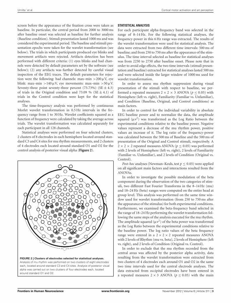
Statistical analyses were performed on four selected clusters,2 clusters of 8 electrodes in each hemisphere located around stan-dard C3 and C4 sites for mu rhythm measurements, and 2 clustersof 4 electrodes each located around standard O1 and O2 for thecontrol analysis of posterior visual alpha (Figure 2).
FIGURE 2 | Clusters of electrodes selected for statistical analyses.
Analysis of mu rhythm was performed on two clusters of eight electrodeseach, located around standard C3 and C4 sites. Analysis of posterior visualalpha was carried out on two clusters of four electrodes each, locatedaround standard O1 and O2.
STATISTICAL ANALYSISFor each participant alpha-frequency band was selected in therange of 8–14 Hz. For the following statistical analyses, thefrequency-power in this 6 Hz range was extracted. The results ofthe wavelet transformation were used for statistical analysis. Thedata were extracted from two different time intervals: 500 ms ofbaseline; and from 250 to 750 ms after the appearance of the stim-ulus. The time interval selected as baseline for statistical analyseswas from 2250 to 2750 after baseline onset. Please note that inorder to avoid edge effects, the two time intervals (stimuli presen-tation and baseline) extracted for statistical analyses lasted 500 msand were selected inside the larger window of 1000 ms used forwavelet transformation.
In order to assess mu rhythm suppression during visualpresentation of the stimuli with respect to baseline, we per-formed a repeated measures 2 × 2 × 3 ANOVA (p ≤ 0.05) withHemisphere (left vs. right), Familiarity (Familiar vs. Unfamiliar),and Condition (Baseline, Original, and Control conditions) asmain factors.
In order to control for the individual variability in absoluteEEG baseline power and to normalize the data, the amplitudesquared (µv2) was transformed as the Log Ratio between theexperimental conditions relative to the baseline power. Negativevalues represent a decrease of the mu rhythm power, positivevalues an increase of it. The log ratio of the frequency-powerwas calculated between the 500 ms of Baseline and the 500 ms ofpresentation of the Original and Control stimuli, respectively. A2 × 2 × 2 repeated measures ANOVA (p ≤ 0.05) was performedwith 2 levels of Hemisphere (left vs. right), 2 levels of Familiarity(Familiar vs. Unfamiliar), and 2 levels of Condition (Original vs.Control).
Post-hoc analyses (Newman–Keuls, test p ≤ 0.05) were appliedon all significant main factors and interactions resulted from theANOVAs.
In order to investigate the possible modulation of the betacomponent during the observation of the two categories of stim-uli, two different Fast Fourier Transforms in the 8–14 Hz (mu)and 18–24 Hz (beta) ranges were computed on the entire head atgroup level. This analysis was performed on the same time win-dow used for wavelet transformation (from 250 to 750 ms afterthe appearance of the stimulus) for both experimental conditions.Furthermore, we examined the beta-frequency band selected inthe range of 18–24 Hz performing the wavelet transformation fol-lowing the same steps of the analysis executed for the mu rhythm.The amplitude squared (µv2) of the beta power was transformedas the Log Ratio between the experimental conditions relative tothe baseline power. The log ratio values of the beta frequencyrange were entered in a 2 × 2 × 2 repeated measures ANOVAwith 2 levels of Rhythm (mu vs. beta), 2 levels of Hemisphere (leftvs. right), and 2 levels of Condition (Original vs. Control).
In order to exclude that the mu rhythm recorded from thecentral areas was affected by the posterior alpha activity, dataresulting from the wavelet transformation were extracted fromtwo clusters of 4 electrodes each around O1 and O2 in the sametwo time intervals used for the central electrode analyses. Thedata extracted from occipital electrodes have been entered ina repeated measures 2 × 3 ANOVA (p ≤ 0.05) with the main
Frontiers in Human Neuroscience www.frontiersin.org November 2012 | Volume 6 | Article 311 | 3
Umilta’ et al. Cortical motor activation and art perception
factors of Hemisphere (left vs. right), and Condition (Baseline,Original, and Control stimuli).
The averages of the score that each participant gave to the“Amount of movement,” the “Aesthetic value,” and the “Artisticnature” of the 3 Original and the 3 Control stimuli wereused to perform three different repeated measures ANOVAs(p ≤ 0.05) with 2 levels of Familiarity (Familiar vs. Unfamiliar)and two levels of Condition (Original vs. Control). Post-hoc anal-ysis (Newman–Keuls, test p ≤ 0.05) was applied on all significantmain factors and interactions.
EMGPARTICIPANTSTwelve healthy volunteers (6 males, 6 females, mean age 28.4years; SD ± 5.9) participated in the experiment. The partici-pants involved in the EMG recordings belonged to a differentgroup from those already involved in the EEG experiments inorder to avoid any training and/or experience effect. Writteninformed consent was obtained from all participants before enter-ing the study. The Ethical Committee of the University of Parmaapproved the study.
EMG RECORDINGSIn order to measure hand and arm EMG activation, Ag/AgClsurface electrodes (diameter 3 mm) were attached bipolarly overthe right First Dorsal Interosseous (FDI) and right Biceps mus-cles. Continuous electromyography (EMG) recordings from bothmuscles were simultaneously acquired with a CED Micro 1401analog-to-digital converting unit (Cambridge Electronic Design,Cambridge, UK). The EMG signal was amplified (1000×) digi-tized (sampling rate: 1 kHz) and stored on a computer for off-lineanalysis.
EMG DATA ANALYSISOff-line, data were submitted to a 50–500 Hz band-pass filter toreduce movement-related artifacts and environmental noise, andhave been full-wave rectified. Data were then visually inspected,and trials with remaining artifacts were excluded from furtherstatistical analyses. For each participant and trial, the averagedEMG responses of the two muscles were subdivided in 4 time-binsof 250 ms each. For each participant and trial four time epochsof 250 ms preceding the appearance of the fixation cross havebeen used as baseline epochs for normalization of the EMG activ-ity. Each epoch was normalized dividing it by the correspondingtime-bin of the baseline.
STATISTICAL ANALYSISEmg data were entered into a 2 (Condition: Original andControl stimuli) × 2 (Muscles: FDI and Biceps) × 4 (Epochs:0–250 ms, 250–500 ms, 500–750 ms, 750–1000 ms) repeated mea-sures ANOVA (p < 0.05.), with Condition, Muscle, and Epochsas the within-subjects factors and Familiarity (Familiar vs.Unfamiliar) as between-subjects factor.
RESULTSRepeated measures ANOVA was performed in order to assessmu rhythm suppression during observation of Original stim-uli with respect to Control stimuli and to the baseline. The
results showed a significant main effect of Condition [F(2, 24) =6.095; MS = 0.602; p < 0.01] regardless of Hemisphere andFamiliarity [Emisphere × Condition × Familiarity: F(2, 24) =0.948; MS = 0.051; p > 0.1]. The comparisons among the threeconditions showed that the squared amplitude of mu rhythm wassignificantly different from the Baseline only during the observa-tion of Original artworks (p < 0.01), and that the observation ofControl stimuli was significantly less effective (p < 0.05) in murhythm suppression than Original stimuli.
The stronger mu rhythm suppression during observation ofOriginal stimuli with respect to Control stimuli and to theBaseline is well exemplified in Figure 3 illustrating the EEG time-frequency spectrum of one participant extracted from C3 and C4sites in the two experimental conditions and in the Baseline.
The repeated measures ANOVA performed on the logratio of the frequency-power confirmed the previous results,showing only a significant effect of Condition [F(1, 12) = 6.621;MS = 0.034; p < 0.05]. The comparison between the two condi-tions demonstrated a stronger mu rhythm suppression (p < 0.05)during observation of the original artworks (Figure 4). Theamount of mu rhythm suppression was higher when observingoriginal artworks than control stimuli, again, regardless ofparticipants’ familiarity with the original artworks [Condition ×Familiarity: F(1, 12) = 0.078; MS = 0.000; p > 0.5; Emisphere ×Condition × Familiarity: F(2, 24) = 0.948; MS = 0.001; p > 0.5].
Figure 5 illustrates the topo maps of the fast Fourier trans-forms performed on the alpha and beta ranges in both exper-imental conditions. Mu suppression was bilateral and mainlylocalized over the sensorimotor areas. Furthermore, mu suppres-sion was much stronger during the observation of the OriginalArtworks compared with the observation of Control stimuli.Differently from mu rhythm, beta range modulation was absentin both experimental conditions. The confirmation of the lackof beta modulation during the observation of both categoriesof stimuli came from the results of the statistical analysis per-formed on the log ratio of the beta range (18–24 Hz). The resultsof the ANOVA showed a significant main effect of Rhythm[F(1, 13) = 26.322; MS = 0.004; p < 0.00] and a significant inter-action Rhythm × Condition [F(1, 13) = 4.39; MS = 0.013; p =0.05]. Newman–Keuls post-hoc comparisons applied to maineffect of Rhythm showed that alpha suppression was significantlystronger than beta (p < 0.00). Moreover, post-hoc applied to thesignificant interaction Rhythm × Condition indicated that whilemu was modulated by conditions, as previously demonstrated,with the strongest suppression during the observation of OriginalArtworks (p < 0.00), beta rhythm remained unchanged acrossconditions (p > 0.8).
The results of the analysis performed on the alpha powerextracted from 2 clusters of 4 electrodes each around O1 andO2 showed only a significant main effect of Condition [F(2, 26) =8.673; MS = 2.696; p < 0.005] (see Figure 6). The further analy-sis with the Newman–Keuls test, however, revealed that the obser-vation of the two different categories of stimuli did not evokeany difference in alpha rhythm squared amplitude (p > 0.1) whileboth of them were different from the baseline (ps < 0.05).
The results of the repeated measures ANOVA performedon participants’ “Aesthetic appraisal” (see Figure 7A) showed
Frontiers in Human Neuroscience www.frontiersin.org November 2012 | Volume 6 | Article 311 | 4

Umilta’ et al. Cortical motor activation and art perception
FIGURE 3 | EEG time-frequency spectrum of one participant extracted
from C3 and C4 sites in the two experimental Conditions and in the
Baseline. A continuous wavelet transformation was performed for thefrequencies from 1 to 30 Hz in a time window of 1 s, which correspondsto the total duration of stimulus presentation. Red color indicates
increased mu rhythm squared amplitude, while blue color indicatesdecreased mu rhythm squared amplitude in a given frequency band(see scale bar). The rectangles added to each panel highlight the 7 Hzwide frequency band (8–14 Hz) and the time window (250–750 ms) usedfor statistical analysis.
a significant main effect of Condition [F(1, 12) = 55.977;MS = 173.33; p < 0.00] accompanied by a significant interac-tion Condition × Familiarity [F(1, 12) = 9.258; MS = 26.67;p < 0.05]. The Newman–Keuls comparisons done on the maineffect of Condition showed that participants’ aesthetic appraisalwas higher for original artworks than for control stimuli(p < 0.001). The investigation of the significant interactionCondition × Familiarity showed that in both groups of partic-ipants stimuli familiarity did not affect aesthetic appraisal ofOriginal artworks and Control stimuli (ps > 0.1). However, bothgroups of participants gave a higher aesthetic appraisal whenobserving Original artworks than Control stimuli (ps < 0.01).
The analysis of the score that participants gave to the “Amountof perceived movement” of the Original and Control stimuli(see Figure 7B) showed a significant main effect of Condition[F(1, 12) = 28.683; MS = 88.099; p < 0.001] and a significant
interaction Condition × Familiarity [F(1, 12) = 4.807; MS =14.765; p < 0.05]. The post-hoc comparisons, performed on thesignificant main effect of Condition, clearly showed that theamount of perceived movement scored by participants was sig-nificantly higher when they saw the original artworks if com-pared to the observation of the Control images (p < 0.005).When the comparisons were made on the significant interac-tion Condition × Familiarity, the amount of movement perceivedin Original stimuli was higher among Familiar participantswhen compared to the Unfamiliar group (p < 0.05). In addi-tion, movement perception for original artworks, compared withControl stimuli, was higher in both groups of participants,regardless of their being familiar (p < 0.005) or unfamiliar withthem (p < 0.05).
Mean scores of the “Artistic nature” questionnaire were:Familiar 100% SE ± 0 for Original stimuli and 9.52% SE ± 4.76
Frontiers in Human Neuroscience www.frontiersin.org November 2012 | Volume 6 | Article 311 | 5

Umilta’ et al. Cortical motor activation and art perception
FIGURE 4 | Amplitude-squared of mu rhythm expressed as Log Ratio
in Control and Original conditions. The amplitude squared (µv2) wastransformed as the Log Ratio between the experimental conditions relativeto the Baseline power. Negative values represent a decrease of the murhythm power, positive values an increase of it. Plots represent theinteraction Condition × Familiarity (p > 0.5) resulted from the ANOVAperformed on mu rhythm expressed as Log Ratio.
FIGURE 5 | Topo maps of the fast Fourier transforms performed on the
alpha and beta ranges in both experimental conditions. Musuppression was bilateral and stronger during the observation of theOriginal Artworks compared with the observation of Control stimuli. Betarange modulation was absent in both experimental conditions.
for Control stimuli; Unfamiliar 66.66% SE ± 4.76 for Originalstimuli and 0% SE ± 0 for Control stimuli (see Figure 7C). Thestatistical analysis showed that the main effects of Condition[F(1, 12) = 99.00; MS = 4.321; p < 0.001] and Familiarity[F(1, 12) = 7.363; MS = 0.321; p < 0.05] were significant,while the interaction between them was not [F(1, 12) = 2.272;MS = 0.099; p > 0.1] (Figure 7C). Post-hoc comparisons wereapplied to the two significant main effects. Original artworks,when compared with Control images, were considered moreoften as real artworks (p < 0.005) by both groups of participants.
FIGURE 6 | Results of the analysis performed on the alpha power
extracted from occipital electrodes. Plots show that that the observationof the two different categories of stimuli did not evoke any difference in theposterior visual alpha modulation in both hemispheres.
Familiar participants gave more affirmative responses than theUnfamiliar group (p < 0.05). The lack of significance of the inter-action between factors indicates that both groups of participantswere more prone to judge the Original stimuli as real art works.
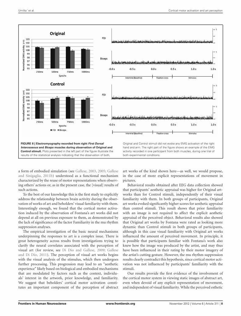
None of the main factors or interactions resulted from theANOVA performed on the EMG data were statistically significant(all ps > 0.05).
These results clearly demonstrate that the observation of bothOriginal and Control stimuli did not evoke any EMG activationof participants’ right hand (FDI) and arm (Biceps) muscles (seeFigure 8).
DISCUSSIONThe present results show that in spite of the similarity betweenthe digitized versions of the Original artworks and the Controlstimuli, only the former evoke cortical motor activation inthe beholder’s brain, as exemplified by mu rhythm suppres-sion recorded from two selected clusters of 8 electrodes ineach hemisphere located around standard C3 and C4 sites.The same clusters were used in previous studies demonstratingmu rhythm suppression during the observation of hand motoracts (Muthukumaraswamy et al., 2004; Muthukumaraswamy andJohnson, 2004a,b; Streltsova et al., 2010).
The motor activation induced by the observation of Originalart works is confirmed by the results of the analysis performedon occipital clusters of electrodes. Such analysis clearly shows thatmu rhythm suppression does not reflect posterior alpha activity,since the observation of the two different categories of stimuli didnot evoke any difference in occipital visual alpha.
The results of the analyses performed on beta rhythm didnot reveal any significant modulation across conditions. Someauthors argued that the beta component would predominantlyoriginate from motor cortex whereas the alpha component wouldmainly arise from postcentral sensory cortex (Hari and Salmelin,1997). However, most recent studies aiming at correlating fMRIBOLD signal with EEG signal during motor tasks demonstrateda negative significant correlation between alpha ERD and BOLDsignal not only on sensorimotor cortex, but also and most
Frontiers in Human Neuroscience www.frontiersin.org November 2012 | Volume 6 | Article 311 | 6

Umilta’ et al. Cortical motor activation and art perception
FIGURE 7 | Results of the ANOVAs performed on the aesthetic appraisal (A), amount of perceived movement (B), and artistic nature (C) in both
groups of participants. Plots of panels (A) and (B) represent the significant Condition × Familiarity interaction. The interaction was not significant in panel (C).
interestingly on premotor cortices (Arnstein et al., 2011; Yuanet al., 2011; Mizuhara, 2012). Furthermore, it should be addedthat even granting that sensorimotor alpha ERD originates moreposteriorly than beta desynchronization, recent data in humansemphasize that the postcentral cortex, and in particular BA 2,must be considered integral part of the mirror neuron system (seeKeysers et al., 2010; Arnstein et al., 2011).
Why should the observation of a static image like a cut in acanvas activate the observer’s motor cortex? It is well known thatthe observation of specific categories of visual stimuli induces theactivation of the observer’s cortical motor system (see Gallese andSinigaglia, 2011a). In particular, it has been shown both in mon-keys and humans that the observation of motor acts and gesturesactivates their motor representations in the observers’ brain (forreview, see Rizzolatti and Sinigaglia, 2010).
More specifically, several EEG studies have shown mu rhythmsuppression during the observation of different types of grasp-ing (Muthukumaraswamy et al., 2004; Muthukumaraswamy andJohnson, 2004a; Perry and Bentin, 2009; Streltsova et al., 2010),meaningless gestures (Babiloni et al., 2002; Streltsova et al., 2010)and sequential finger movements (Calmels et al., 2006). It is inter-esting to note that such motor activation can also be induced bythe observation of static images portraying actions (Johnson-Freyet al., 2003; Mado-Proverbio et al., 2009).
The present results extend the notion of cortical motor activa-tion during the observation of visual stimuli to the observationof abstract static images. Our interpretation is that the observa-tion of the cuts on canvases activates the motor representation ofthe same gesture in the brain of beholders. In contrast, Controlstimuli, because of their lack of implicit dynamicity, as shownby participants’ score on the perceived amount of movement, aremerely processed as images that are not the manifest consequenceof someone’s gesture. Beside the observation of dynamic or staticmotor acts, also the observation of the static consequences ofa motor act is capable of activating its motor representation inthe observer’s brain. Our data demonstrate that viewing cuts andviewing lines differently impact on beholders’ cortical motor sys-tem. Of course, we neither claim that the perceived esthetic statusof the work is a necessary condition for the evocation of suchcortical motor activation, nor that cortical motor activation is anecessary component of esthetic ranking. What we have found,however, is that such cortical motor activation is a componentof what happens in beholders’ brain during the observation ofart works such as those of Lucio Fontana used as stimuli in thepresent study. Furthermore, cortical motor activation occurs inspite of the lack of activation of beholders’ contralateral handand arm muscles, as demonstrated by the EMG recordings. Forthese reasons such cortical motor activation may be regarded as
Frontiers in Human Neuroscience www.frontiersin.org November 2012 | Volume 6 | Article 311 | 7
Umilta’ et al. Cortical motor activation and art perception
FIGURE 8 | Electromyography recorded from right First Dorsal
Interosseous and Biceps muscles during observation of Original and
Control stimuli. Plots presented in the left part of the figure illustrate theresults of the statistical analysis indicating that the observation of both,
Original and Control stimuli did not evoke any EMG activation of the righthand and arm. The right part of the figure shows an example of the EMGactivity recorded in one participant from both muscles, during one trial ofboth experimental conditions.
a form of embodied simulation (see Gallese, 2003, 2005; Galleseand Sinigaglia, 2011b) understood as a functional mechanismcharacterized by the reuse of motor representations when observ-ing others’ actions or, as in the present case, the [visual] results ofsuch actions.
To the best of our knowledge this is the first study to explicitlyaddress the relationship between brain activity during the obser-vation of works of art and beholders’ visual familiarity with them.Interestingly enough, we found that the cortical motor activa-tion induced by the observation of Fontana’s art works did notdepend at all on previous exposure to them, as demonstrated bythe lack of significance of the factor Familiarity in the mu rhythmsuppression analyses.
The empirical investigation of the basic neural mechanismsunderpinning the responses to art is a complex issue. There isgreat heterogeneity across results from investigations trying toclarify the neural correlates associated with the perception ofvisual art (for review, see Di Dio and Gallese, 2009; Galleseand Di Dio, 2011). The perception of visual art works beginswith the visual analysis of the stimulus, which then undergoesfurther processing. This progression may lead to an “aestheticexperience” likely based on biological and embodied mechanismsthat are modulated by factors such as the context, individu-als’ interest in the artwork, prior knowledge, and familiarity.We suggest that beholders’ cortical motor activation consti-tutes an important component of the perception of abstract
art works of the kind shown here—as well, we would propose,in the case of more explicit representations of movement inpictures.
Behavioral results obtained after EEG data collection showedthat participants’ aesthetic appraisal was higher for Original art-works than for Control stimuli, independently of their visualfamiliarity with them. In both groups of participants, Originalart works evoked significantly higher scores for aesthetic appraisalthan control stimuli. This result shows that prior familiaritywith an image is not required to affect the explicit aestheticappraisal of the perceived object. Behavioral results also showedthat Original art works by Fontana were rated as looking moredynamic than Control stimuli in both groups of participants,although in this case visual familiarity with Original art worksinfluenced the amount of perceived movement. In principle, itis possible that participants familiar with Fontana’s work alsoknew how the image was produced by the artist, and may thushave been influenced in their rating by their motor imagery ofthe artist’s cutting gesture. However, the mu rhythm suppressionresults clearly contradict this hypothesis, since cortical motor acti-vation was not influenced by participants’ familiarity with thestimuli.
Our results provide the first evidence of the involvement ofthe cortical motor system in viewing static images of abstract art,even when devoid of any explicit representation of movement,and independent of visual familiarity. While the perceived esthetic
Frontiers in Human Neuroscience www.frontiersin.org November 2012 | Volume 6 | Article 311 | 8

Umilta’ et al. Cortical motor activation and art perception
status of these works is probably not a necessary requisite to evokecortical motor activation, we do take such activation to be animportant component of the perception of such works of art. Thepresent results lend empirical support to the role of motor sim-ulation in the observation of abstract art (Freedberg and Gallese,2007; Di Dio and Gallese, 2009).
ACKNOWLEDGMENTSThis work was supported by the EU grant TESIS.
FUNDINGThe funders had no role in study design, data collection andanalysis, decision to publish, or preparation of the manuscript.
REFERENCESArnheim, R. (1954). Art and Visual
Perception: A Psychology of theCreative Eye. Berkeley, CA:University of California Press.
Arnheim, R. (1988). The Power ofthe Center: A Study of Compositionin the Visual Arts. Berkeley, CA:University of California Press.
Arnstein, D., Cui, F., Keysers, C.,Maurits, N. M., and Gazzola, V.(2011). µ-suppression duringaction observation and executioncorrelates with BOLD in dor-sal premotor, inferior parietal,and SI cortices. J. Neurosci. 31,14243–14249.
Babiloni, C., Babiloni, F., Carducci,F., Cincotti, F., Cocozza, G., DelPercio, C., et al. (2002). Humancortical electroencephalogra-phy (EEG) rhythms during theobservation of simple aimlessmovements: a high resolutionEEG study. Neuroimage 17,559–572.
Calmels, C., Holmes, P., Jarry, G.,Lévèque, J. M., Hars, M., andStam, C. J. (2006). Cortical activ-ity prior to, and during, observa-tion and execution of sequential fin-ger movements. Brain Topogr. 19,77–88.
Di Dio, C., and Gallese, V. (2009).Neuroesthetics: a review. Curr.Opin. Neurobiol. 19, 682–687.
Freedberg, D., and Gallese, V. (2007).Motion, emotion and empathy inesthetic experience. Trends Cogn.Sci. 11, 197–203.
Gallese, V. (2003). The manifold natureof interpersonal relations: the questfor a common mechanism. Philos.Trans. R. Soc. Lond. B Biol. Sci. 358,517–528.
Gallese, V. (2005). Embodied simula-tion: from neurons to phenomenalexperience. Phenomenol. Cogn. Sci.4, 23–48.
Gallese, V., and Di Dio, C. (2012).“Neuroesthetics: the body in esthetic
experience,” in The Encyclopediaof Human Behavior, Vol. 2, edV. S. Ramachandran (Amsterdam:Elsevier Academic Press), 687–693.
Gallese, V., and Sinigaglia, C. (2011a).“Cognition in action. A new lookat the cortical motor system,” inJoint Attention and Agency, eds J.Metcalfe and H. Terrace (Oxford:Oxford University Press).
Gallese, V., and Sinigaglia, C. (2011b).What is so special with embodiedsimulation. Trends Cogn. Sci. 15,512–519.
Hari, R., and Salmelin, R. (1997).Human cortical oscillations: a neu-romagnetic view through the skull.Trends Neurosci. 20, 44–49.
Johnson-Frey, S. H., Maloof, F. R.,Newman-Norlund, R., Farrer, C.,Inati, S., and Grafton, S. T. (2003).Actions or hand-object interac-tions? Human inferior frontal cortexand action observation. Neuron 39,1053–1058.
Keysers, C., and Gazzola, V. (2009).Expanding the mirror: vicariousactivity for actions, emotions, andsensations. Curr. Opin. Neurobiol.19, 666–671.
Keysers, C., Kaas, J. H., and Gazzola,V. (2010). Somatosensation in socialperception. Nat. Rev. Neurosci. 11,417–428.
Locher, P., Krupinski, E. A., Mello-Thoms, C., and Nodine, C. F.(2007). Visual interest in pictorialart during an aesthetic experience.Spat. Vis. 21, 55–77.
Locher, P. J., and Stappers, P. J. (2002).Factors contributing to the implicitdynamic quality of static abstractdesigns. Perception 31, 1093–1107.
Longcamp, M., Tanskanen, T., andHari, R. (2006). The imprint ofaction: motor cortex involvementin visual perception of handwrittenletters. Neuroimage 33, 681–688.
Mado-Proverbio, A. M., Riva, F.,and Zani, A. (2009). Observationof static pictures of dynamic
actions enhances the activity ofmovement-related brain areas.PLoS ONE 4:e5389. doi: 10.1371/journal.pone.0005389
Mizuhara, H. (2012). Cortical dynam-ics of human scalp EEG origins ina visually guided motor execution.Neuroimage 62, 1884–1895.
Muthukumaraswamy, S. D., andJohnson, B. W. (2004a). Changesin rolandic mu rhythm duringobservation of a precision grip.Psychophysiology 41, 152–156.
Muthukumaraswamy, S. D., andJohnson, B. W. (2004b). Primarymotor cortex activation duringaction observation revealed bywavelet analysis of the EEG. Clin.Neurophysiol. 115, 1760–1766.
Muthukumaraswamy, S. D., Johnson,B. W., and McNair, N. A. (2004). Murhythm modulation during obser-vation of an object-directed grasp.Brain Res. Cogn. Brain Res. 19,195–201.
Oldfield, R. C. (1971). The assess-ment and analysis of handedness:the Edinburgh inventory. Neuro-psychologia 9, 77–113.
Perry, A., and Bentin, S. (2009). Mirroractivity in the human brain whileobserving hand movements: a com-parison between EEG desynchro-nization in the mu-range and pre-vious fMRI results. Brain Res. 1282,126–132.
Ramachandran, V. S., and Hirstein,W. (1999). The science of art: aneurological. Theory of aestheticexperience. J. Conscious. Stud. 6,15–51.
Redies, C. (2007). A universal modelof esthetic perception based on thesensory coding of natural stimuli.Spat. Vis. 21, 97–117.
Rizzolatti, G., and Sinigaglia, C.(2010). The functional role ofthe parieto-frontal mirror circuit:interpretations and misinterpre-tations. Nat. Rev. Neurosci. 11,264–274.
Streltsova, A., Berchio, C., Gallese, V.,and Umilta’, M. A. (2010). Timecourse and specificity of sensory-motor alpha modulation duringthe observation of hand motoracts and gestures: a high densityEEG study. Exp. Brain Res. 205,363–373.
Taylor, R. P., Spehar, B., Van Donkelaar,P., and Hagerhall, C. M. (2011).Perceptual and physiologicalresponses to Jackson Pollock’sfractals. Front. Hum. Neurosci. 5:60.doi: 10.3389/fnhum.2011.00060
Yuan, H., Perdoni, C., Yang, L.,and He, B. (2011). Differentialelectrophysiological couplingfor positive and negative BOLDresponses during unilateral handmovements. J. Neurosci. 31,9585–9593.
Zeki, S. (1999). Art and the Brain.J. Conscious. Stud. 6, 76–96.
Conflict of Interest Statement: Theauthors declare that the researchwas conducted in the absence of anycommercial or financial relationshipsthat could be construed as a potentialconflict of interest.
Received: 24 July 2012; accepted: 30October 2012; published online: 16November 2012.Citation: Umilta’ MA, Berchio C, SestitoM, Freedberg D and Gallese V (2012)Abstract art and cortical motor acti-vation: an EEG study. Front. Hum.Neurosci. 6:311. doi: 10.3389/fnhum.2012.00311Copyright © 2012 Umilta’, Berchio,Sestito, Freedberg and Gallese. This isan open-access article distributed underthe terms of the Creative CommonsAttribution License, which permits use,distribution and reproduction in otherforums, provided the original authorsand source are credited and subject to anycopyright notices concerning any third-party graphics etc.
Frontiers in Human Neuroscience www.frontiersin.org November 2012 | Volume 6 | Article 311 | 9


!['Caseof Bilateral Occipitoparietal Brain Injury · 'Caseof Bilateral Occipitoparietal Brain Injury BY A. R. LURIA [Reprinted/rom BRAIN, Vol. 82, Part 111,1959, pp. 437-449] MACMILLAN](https://static.fdocuments.in/doc/165x107/5bd3fa1a09d3f29b578b8349/caseof-bilateral-occipitoparietal-brain-injury-caseof-bilateral-occipitoparietal.jpg)


